Article

Withania somnifera-Functionalized Selenium Nanoparticles Attenuate Glycerol-Induced Rhabdomyolysis-Associated Acute Renal Failure

by
Hala Fouad Elmazar
1,2,
Khaled M. Alam-ElDein
3,*,
Mariam G. Elneel
3,
Habiba A. Abbas
3,
Doaa Y. Ahmed Shalaby
3,
Fatma H. Negm
3,
Mariam S. Gerges Aryan
3,
Basmala H. E. Khalaf
3,
Ahmed Hassan Ibrahim Faraag
3,4,
Khaled Abuelhaded
3,
Ahmed M. Ashour
5,
Ali Khames
6,
Mohamed H. A. Gadelmawla
7,*,
Mariam O. A. Hamed
3 and
Sara Youssif Ibrahim
8
1
Department of Anatomy and Histology, Faculty of Medicine, Mutah University, Alkarak 61710, Jordan
2
Department of Histology and Cell Biology, Faculty of Medicine, Menoufiya University, Shibin El-Kom 32511, Egypt
3
School of Biotechnology, Badr University in Cairo (BUC), Cairo 11829, Egypt
4
Botany and Microbiology Department, Faculty of Science, Helwan University, Cairo 11795, Egypt
5
Department of Pharmacology and Toxicology, College of Pharmacy, Umm Al-Qura University, P.O. Box 13578, Makkah 21955, Saudi Arabia
6
Department of Pharmacology and Toxicology, Faculty of Pharmacy, Sohag University, Sohag 82511, Egypt
7
Life Sciences Department, Faculty of Biotechnology, Sinai University, Kantara Branch, Ismailia 41636, Egypt
8
Department of Clinical Pharmacy and Pharmacy Practice, Faculty of Pharmacy, Ahram Canadian University, Giza 12451, Egypt
*
Authors to whom correspondence should be addressed.
Int. J. Mol. Sci. 2026, 27(15), 6746; https://doi.org/10.3390/ijms27156746
Submission received: 30 June 2026 / Revised: 22 July 2026 / Accepted: 26 July 2026 / Published: 28 July 2026

Abstract

Rhabdomyolysis-associated acute kidney injury is driven by myoglobin-mediated oxidative stress, inflammation, mitochondrial impairment, and tubular cell death. This study evaluated the nephroprotective activity of green-synthesized Withania somnifera-functionalized selenium nanoparticles (Ws-SeNPs) in glycerol-induced renal injury and compared their efficacy with native W. somnifera extract and sodium selenite. The chemical profile of the plant extract was characterized by LC–MS/MS, and Ws-SeNPs were evaluated using dynamic light scattering, zeta potential analysis, transmission electron microscopy, and FTIR spectroscopy. Thirty-five male rats were assigned to Control, ARF, ARF & Ws, ARF & selenium, and ARF & Ws-SeNPs. ARF was induced by intramuscular injection of 50% glycerol. Glycerol administration induced marked skeletal muscle injury, renal dysfunction, tubular damage, oxidative stress, inflammation, mitochondrial dysregulation, pyroptosis, apoptosis, and histopathological alterations. Both Ws and sodium selenite provided partial protection, whereas Ws-SeNPs produced the greatest improvement in renal function and tissue architecture. Their protective effect was associated with restoration of Nrf2-dependent antioxidant defenses, suppression of NF-κB/NLRP3/GSDMD-associated inflammatory and pyroptotic signaling, preservation of mitochondrial regulatory pathways, and attenuation of apoptosis. These findings indicate that Ws-SeNPs provide multi-target protection against glycerol-induced rhabdomyolysis-associated renal injury and may represent a promising phytochemical-based selenium nanoformulation for further preclinical investigation.

1. Introduction

Rhabdomyolysis-associated acute kidney injury is a severe clinical complication resulting from extensive skeletal muscle breakdown and the subsequent release of intracellular constituents, particularly myoglobin, creatine kinase, and lactate dehydrogenase, into the circulation [1,2]. In experimental settings, intramuscular glycerol administration is widely used to reproduce this condition because it induces myofiber damage followed by myoglobin-mediated renal injury [3]. Filtered myoglobin accumulates within the renal tubules, where its heme component promotes vasoconstriction, tubular obstruction, iron-dependent reactive oxygen species generation, lipid peroxidation, mitochondrial injury, and direct tubular epithelial toxicity [4,5]. These events impair renal filtration and are accompanied by elevations in creatinine, urea, cystatin C, kidney injury molecule-1, and neutrophil gelatinase-associated lipocalin, together with tubular degeneration, cast formation, inflammatory infiltration, and acute tubular necrosis [6,7].
At the molecular level, rhabdomyolysis-associated renal injury is sustained by interconnected redox, inflammatory, mitochondrial, and regulated cell death pathways. Excessive reactive oxygen species overwhelm endogenous antioxidant defenses and disturb the Keap1/Nrf2 signaling axis, thereby reducing the activity of protective enzymes such as superoxide dismutase, catalase, glutathione peroxidase, and reduced glutathione [8,9]. Oxidative injury also activates NF-κB-dependent inflammatory signaling and increases pro-inflammatory cytokine production. In parallel, mitochondrial stress and damage-associated signals can activate the NLRP3 inflammasome and gasdermin D-mediated pyroptosis, while disruption of SIRT1/AMPK/PGC-1α-dependent mitochondrial regulation promotes energetic failure and structural instability. Persistent mitochondrial dysfunction further shifts the Bax/Bcl-2 balance toward caspase-dependent apoptosis, thereby amplifying tubular epithelial loss [10].
Because these pathogenic processes are highly interconnected, interventions directed at only one pathway may provide incomplete protection. A therapeutic strategy capable of simultaneously restoring antioxidant defenses, suppressing inflammatory and pyroptotic signaling, preserving mitochondrial homeostasis, and limiting apoptosis may therefore be more effective in attenuating rhabdomyolysis-associated renal injury. This has encouraged growing interest in plant-derived bioactive compounds and redox-active trace elements with complementary cytoprotective properties [11].
Withania somnifera is a medicinal plant rich in withanolides, withanosides, alkaloids, and phenolic constituents that exhibit antioxidant, anti-inflammatory, mitochondrial-protective, and anti-apoptotic activities. Experimental evidence indicates that Ws-derived compounds can enhance Nrf2-dependent cytoprotective responses, reduce NF-κB- and MAPK-mediated inflammatory signaling, and preserve mitochondrial function under oxidative stress [12,13]. These properties make Ws a plausible candidate for attenuating the multiple molecular disturbances involved in rhabdomyolysis-associated kidney injury. However, the therapeutic efficacy of native Ws extract may be limited by variability in phytochemical composition, solubility, stability, and systemic bioavailability [14].
Selenium is an essential trace element incorporated into selenoproteins, including glutathione peroxidases and thioredoxin reductases, which are central to peroxide detoxification, redox regulation, and mitochondrial protection [15]. Selenium supplementation may therefore strengthen antioxidant capacity and reduce oxidative tissue injury. Nevertheless, conventional inorganic selenium salts, particularly sodium selenite, have a narrow therapeutic window and may produce toxicity at relatively high or prolonged exposures. These limitations have prompted the development of selenium nanoparticles as alternative delivery systems with potentially improved biological activity and reduced toxicity [16].
Plant-mediated selenium nanoparticles offer a dual functional platform in which phytochemicals serve as reducing and stabilizing agents while also contributing their own biological activity. Compared with sodium selenite, SeNPs may provide improved cellular delivery, enhanced biological interaction, and lower systemic toxicity [17,18,19]. Functionalization with W. somnifera phytoconstituents may further combine selenium-dependent antioxidant support with the redox-modulating, anti-inflammatory, mitochondrial-protective, and anti-apoptotic actions of Ws. Accordingly, Ws-functionalized SeNPs may produce broader protection than either native Ws extract or inorganic selenium alone [20].
Despite the growing interest in plant-mediated selenium nanoparticles, the protective efficacy of Ws-functionalized SeNPs against rhabdomyolysis-associated renal injury has not been adequately investigated. It also remains unclear whether this nanoformulation provides greater protection than native Ws extract or sodium selenite and whether its effects involve coordinated regulation of Nrf2/Keap1 antioxidant signaling, NF-κB/NLRP3/GSDMD-associated inflammation and pyroptosis, mitochondrial homeostasis, and apoptosis. Therefore, the present study compared the nephroprotective effects of Ws extract, sodium selenite, and Ws-SeNPs in glycerol-induced acute renal injury in rats. We hypothesized that Ws-SeNPs would provide superior protection by simultaneously improving renal function and histoarchitecture, restoring redox and mitochondrial homeostasis, and suppressing inflammatory, pyroptotic, and apoptotic pathways.

2. Results

2.1. LC–MS/MS Chemical Profile of the Extract

The Withania somnifera extract was profiled using LC–MS/MS in both positive and negative ion modes. The total ion chromatograms are shown in Figure 1 (positive mode) and Figure 2 (negative mode). Seven main peaks were identified, all eluting between 9.95 and 12.78 min (Table 1). Six gave [M+H]+ ions in positive mode, while withanoside IV gave the [M–H] ion in negative mode. Six of the seven are withanolides (C28 ergostane-type steroidal lactones); the seventh, withanoside IV, is a glycosylated withanolide.
Three of the withanolides 12-deoxywithastramonolide, withaferin A, and withanone are isobaric: they share the formula C28H38O6 (MW 470.60) and the same [M+H]+ precursor at m/z 471. They were distinguished by retention time (12.26, 11.76, and 12.78 min, respectively) and by their product-ion spectra. Because these three would otherwise be difficult to tell apart, the gradient was set to resolve them chromatographically.
In positive mode, the withanolide aglycones lost water in steps from the hydroxylated steroid nucleus, giving the m/z 317 → 299 → 281 series. Ixocarpalactone ([M+H]+ m/z 506.1) gave m/z 317.06 and 299.07; viscosalactone B ([M+H]+ m/z 489.1) gave m/z 435.10, 317.01, 299.05, and 281.00; and withaferin A ([M+H]+ m/z 471.1) gave m/z 299.05 and 280.95. In negative mode, withanoside IV showed [M–H] at m/z 781.2, with a water-loss fragment at m/z 763.09 and a lower-mass ion at m/z 221.09, consistent with a glycosylated withanolide. In every case, the measured precursor mass matched the value calculated for the proposed formula (Figure 1) (Table 1).

2.2. Physicochemical Characterization of Ws-SeNPs

2.2.1. Particle-Size Distribution and Zeta Potential

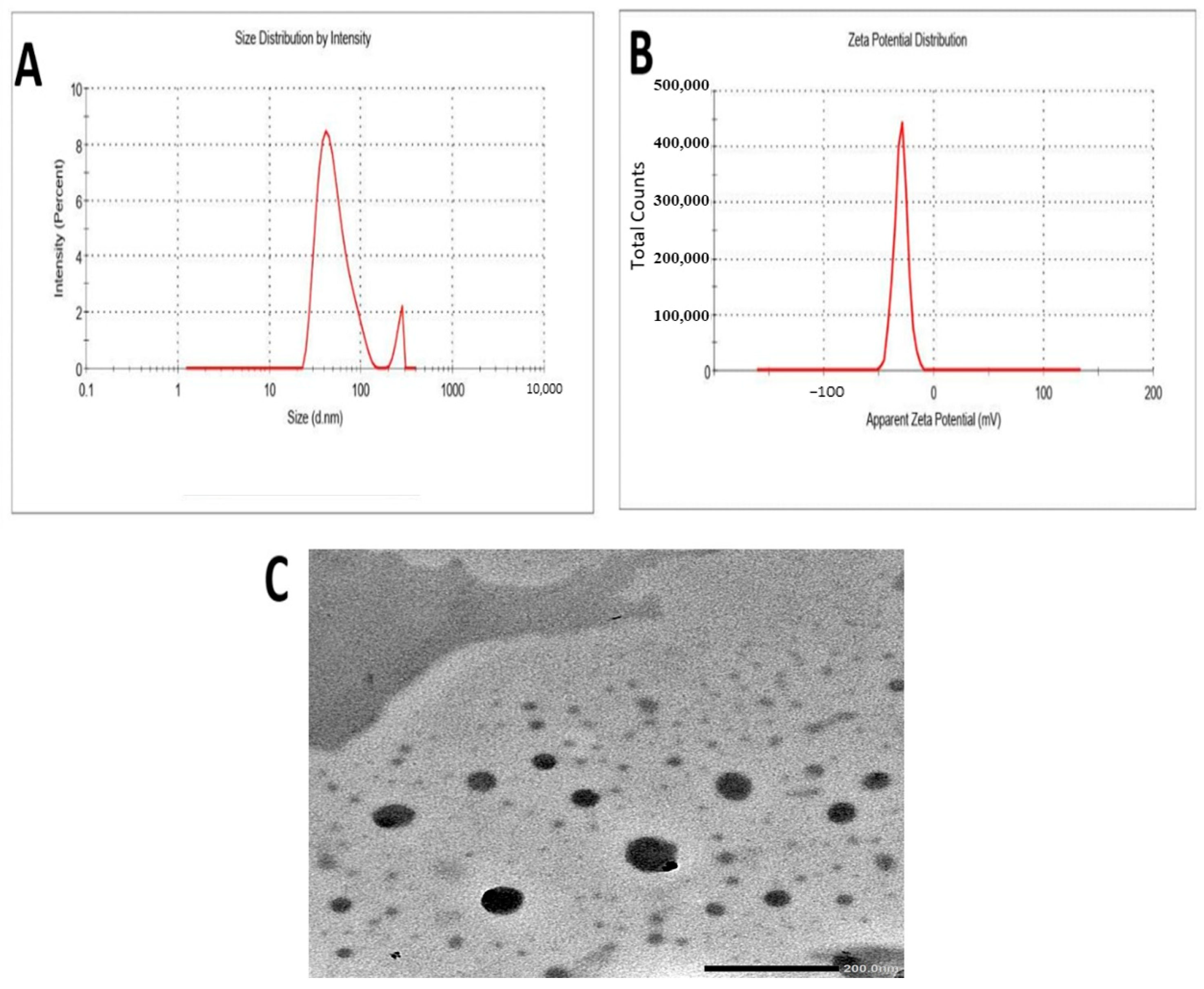
Dynamic light-scattering analysis showed that Ws-SeNPs exhibited a principal hydrodynamic average size of 69.8 nm, together with a minor secondary population at larger diameters, indicating limited aggregation. Zeta potential analysis revealed a negatively charged nanoparticle surface, with the principal distribution located between approximately −15 and −25 mV, indicating moderate colloidal stability (Figure 2).

2.2.2. Transmission Electron Microscopy

TEM showed that the particles produced were in the nanoscale range. Most of the nanoparticles in the image were spherical, had relatively smooth edges, and exhibited a uniform distribution in general. Most particles were well dispersed, with slight aggregation in particular spots (Figure 2).

2.2.3. FTIR Analysis of Ws-SeNPs

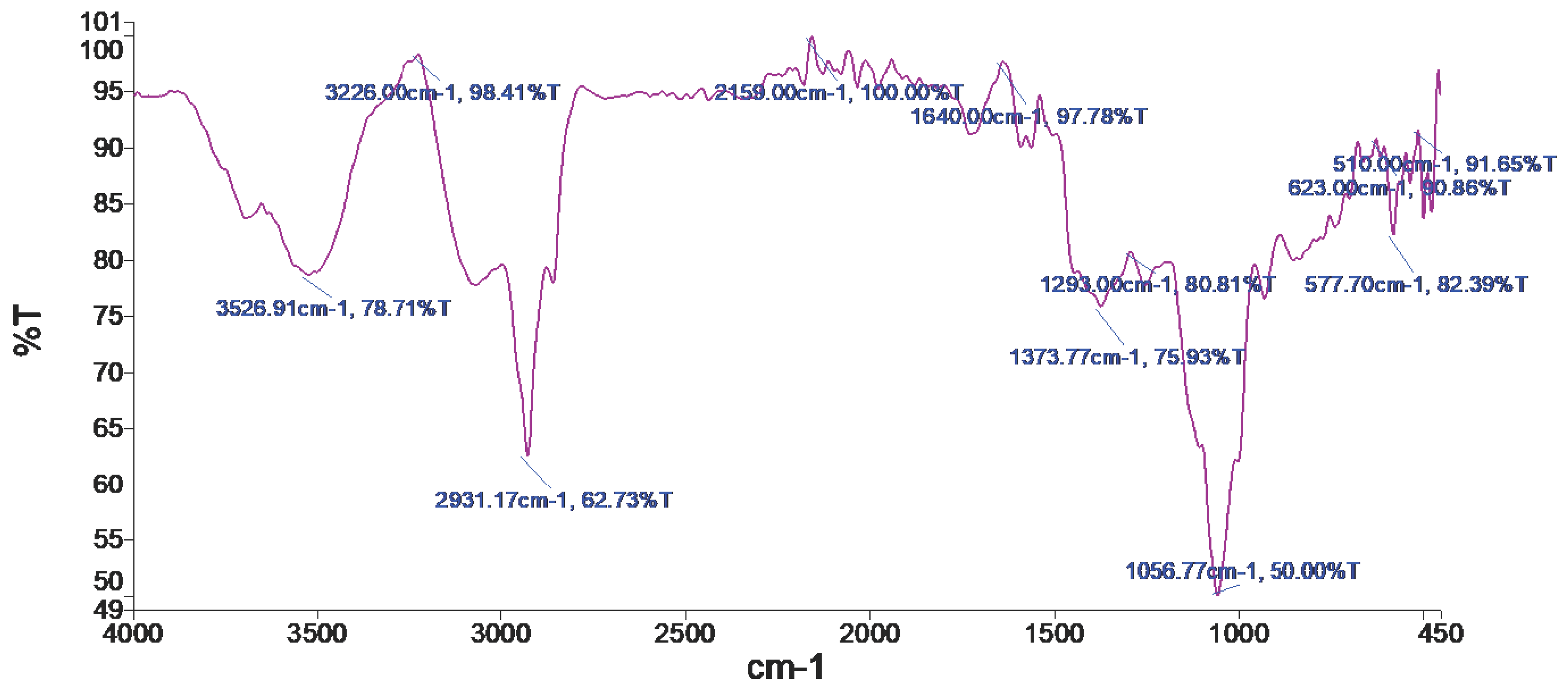
FTIR analysis showed a broad absorption band at 3526–3226 cm−1, corresponding mainly to O–H stretching vibrations, and a band near 2931 cm−1 attributed to aliphatic C–H stretching. The signal at approximately 1640 cm−1 was assigned to C=O and/or aromatic C=C vibrations, whereas bands between 1373 and 1056 cm−1 were associated with C–O stretching. These functional groups are consistent with Ws-derived phytoconstituents involved in selenium-ion reduction, nanoparticle capping, and stabilization (Figure 3).

2.3. Kidney Weight and Relative Kidney Weight

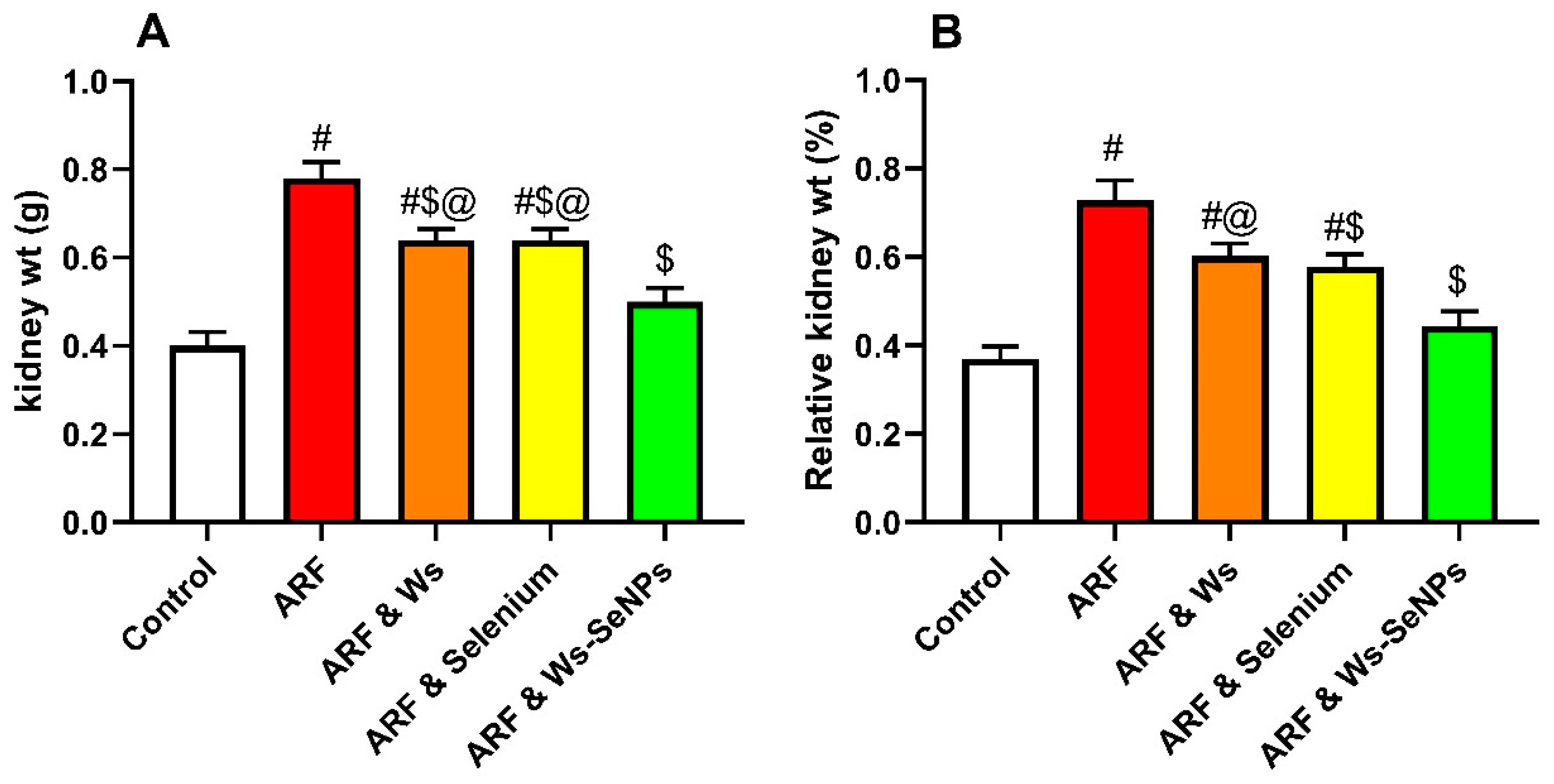
As illustrated in Figure 4, glycerol-induced ARF produced a marked increase in both absolute kidney weight (Figure 5A) and relative kidney weight (Figure 5B) compared with the control group, reflecting renal enlargement, tissue edema, and structural alterations associated with acute renal injury. Post-treatment with Withania somnifera (Ws) and sodium selenite partially attenuated these increases; however, kidney weight parameters remained elevated compared with the control group, indicating incomplete recovery.
Notably, treatment with Ws-SeNPs produced the most pronounced improvement, significantly reducing both kidney weight and relative kidney weight compared with the untreated ARF group (p < 0.05). These findings suggest that Ws-SeNPs exerted superior renoprotective effects by limiting glycerol-induced renal swelling and preserving kidney structural integrity more effectively than Ws extract or sodium selenite alone.

2.4. Rhabdomyolysis-Associated Muscle Injury Markers

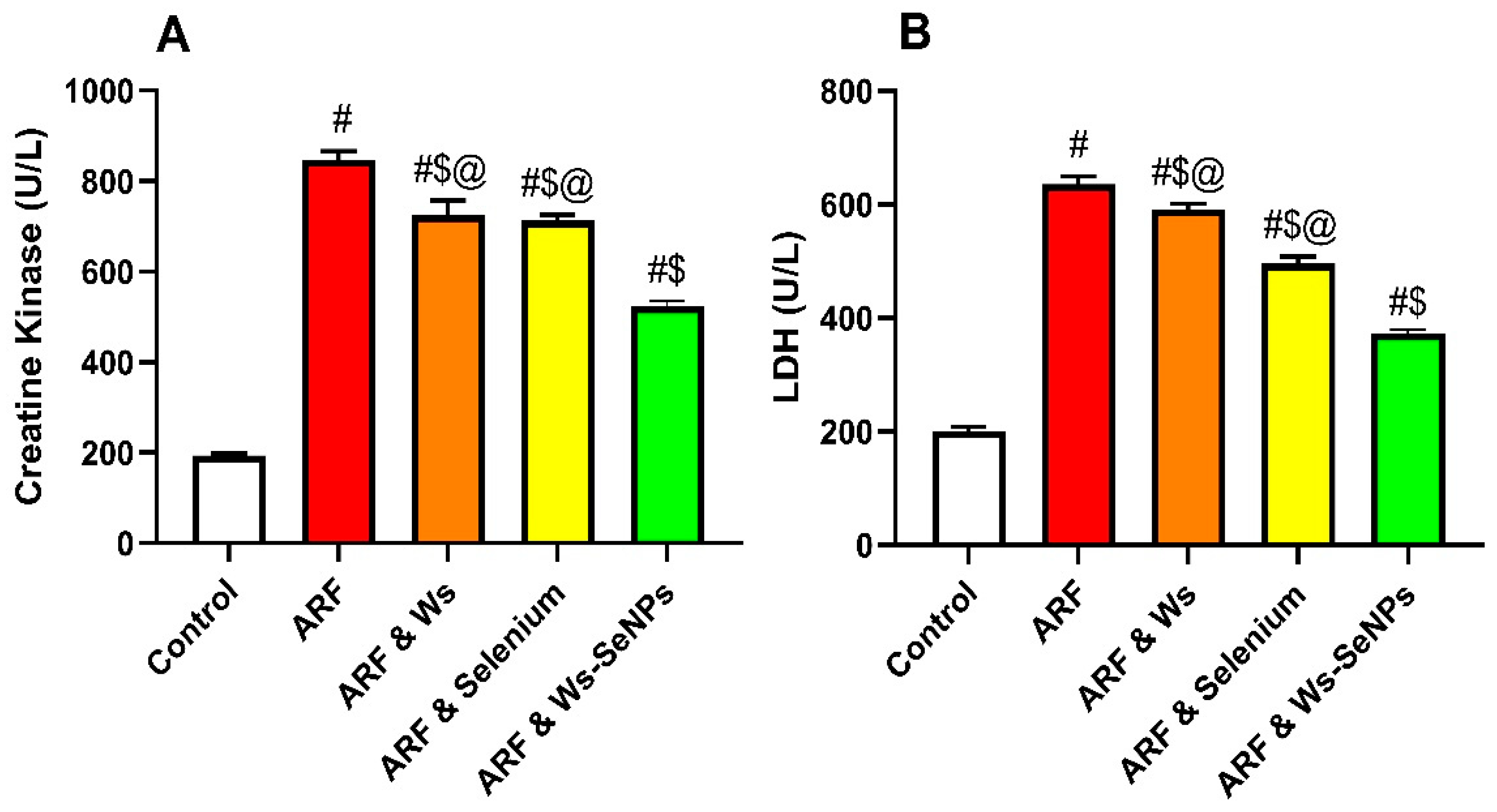
As shown in Figure 5, glycerol-induced ARF was associated with a marked elevation in serum creatine kinase (CK; Figure 6A) and lactate dehydrogenase (LDH; Figure 6B) activities compared with the control group (p < 0.05), confirming the successful induction of rhabdomyolysis-associated muscle and tissue injury.
Treatment with Withania somnifera (Ws) produced a partial reduction in both CK and LDH activities relative to the untreated ARF group, indicating a modest protective effect against glycerol-induced myocellular damage. Sodium selenite treatment also attenuated the rise in these enzymes, with a more evident reduction, particularly in LDH activity. Notably, Ws-SeNPs exerted the most pronounced protective effect, significantly lowering both CK and LDH activities compared with the ARF group (p < 0.05).

2.5. Effect of WS, Selenium, and Ws-SeNPs on Kidney Function and Tubular Injury Biomarkers

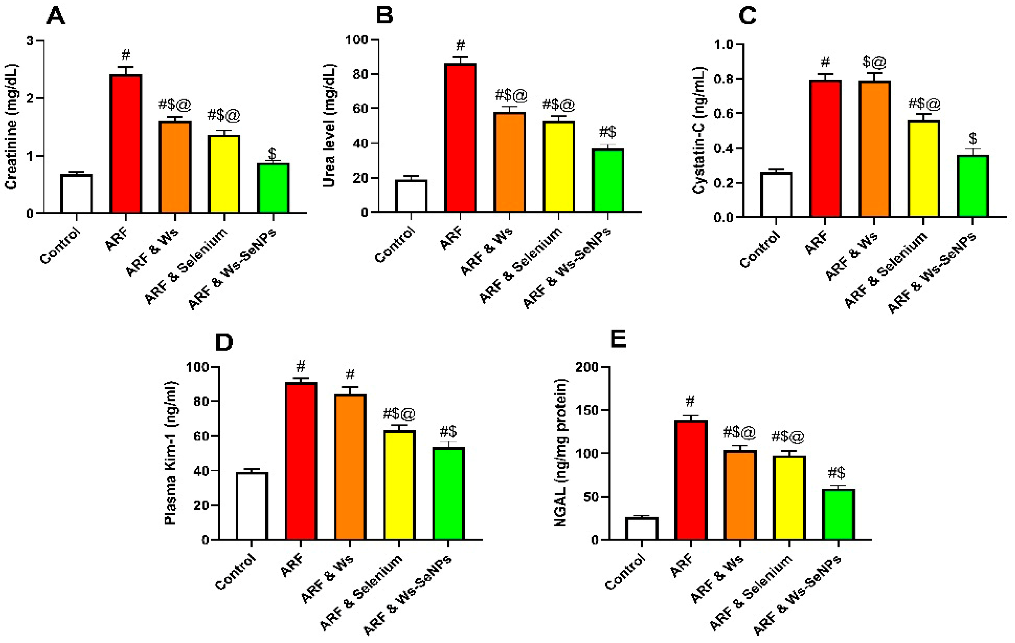
As presented in Figure 6, glycerol-induced ARF caused a marked impairment in renal function, as evidenced by significant elevations in serum creatinine and urea levels compared with the control group (p < 0.05). In parallel, ARF markedly increased cystatin-C, plasma KIM-1, and NGAL, indicating glomerular filtration disturbance, tubular epithelial injury, and loss of renal structural integrity. Treatment with Withania somnifera (Ws) produced partial improvement in most renal function indices; however, some tubular injury markers remained markedly elevated, suggesting incomplete renal recovery.
Sodium selenite also attenuated glycerol-induced renal dysfunction and reduced tubular injury biomarkers, with a more evident effect on KIM-1 and NGAL levels. Notably, Ws-SeNPs exerted the most pronounced renoprotective effect, significantly reducing creatinine, urea, cystatin-C, KIM-1, and NGAL levels compared with the untreated ARF group (p < 0.05). These findings indicate that Ws-SeNPs more effectively preserved renal filtration capacity and mitigated tubular injury than Ws extract or sodium selenite alone.

2.6. Evaluation of Renal Oxidative Stress and Antioxidant Defense Markers

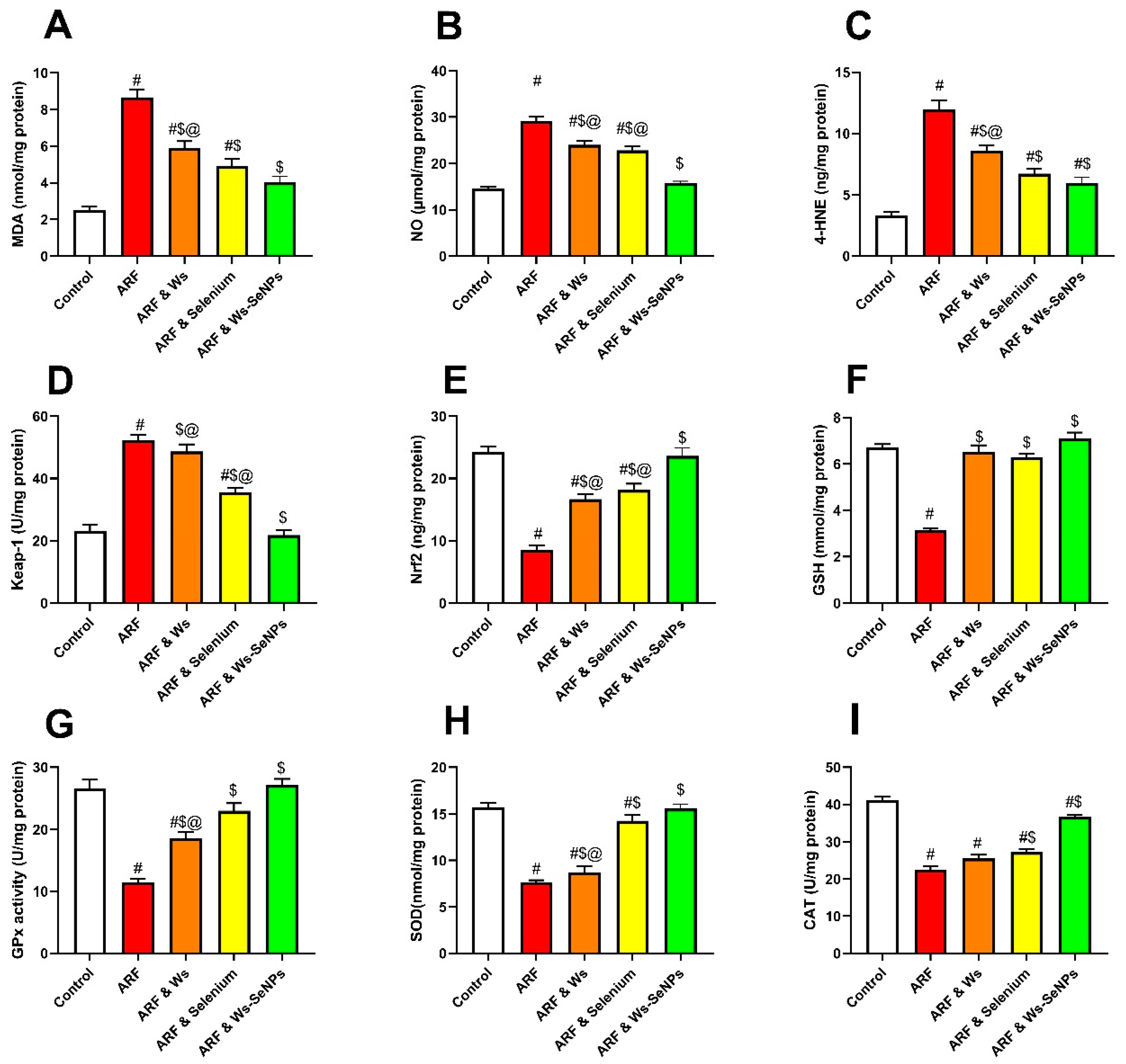
The ARF induced by glycerol caused a significant increase in the levels of malondialdehyde (MDA), nitric oxide (NO), and 4-hydroxynonenal (4-HNE) in comparison with the control group (p < 0.05), as seen in Figure 7. The changes suggest increased lipid peroxidation, nitrosative stress, and oxidative membrane damage after glycerol. Concurrently, ARF significantly elevated the expression of renal Keap-1, but significantly reduced the expression of renal Nrf2, indicating renal antioxidant signaling axis disruption by ARF. Furthermore, the ARF group showed a marked decrease in endogenous antioxidant components, such as reduced glutathione (GSH), glutathione peroxidase (GPx), superoxide dismutase (SOD), and catalase (CAT), indicating a decrease in renal antioxidant capacity under oxidative stress induced by rhabdomyolysis.
Treatment with Ws or sodium selenite partially reversed these redox disturbances, which appeared as a reduction in oxidative stress markers and partial restoration of antioxidant enzyme activities compared with the untreated ARF group. In comparison, a relatively stronger effect of sodium selenite on GPx was observed, which is well correlated to the selenium-dependent activity of GPx. The antioxidant activity was most evident with Ws-SeNPs, which significantly reduced the levels of MDA, NO, 4-HNE, and Keap-1 and significantly increased the levels of Nrf2, GSH, GPx, SOD, and CAT activities (p < 0.05).

2.7. Evaluation of Renal Inflammatory/Anti-Inflammatory and Pyroptosis Mechanism

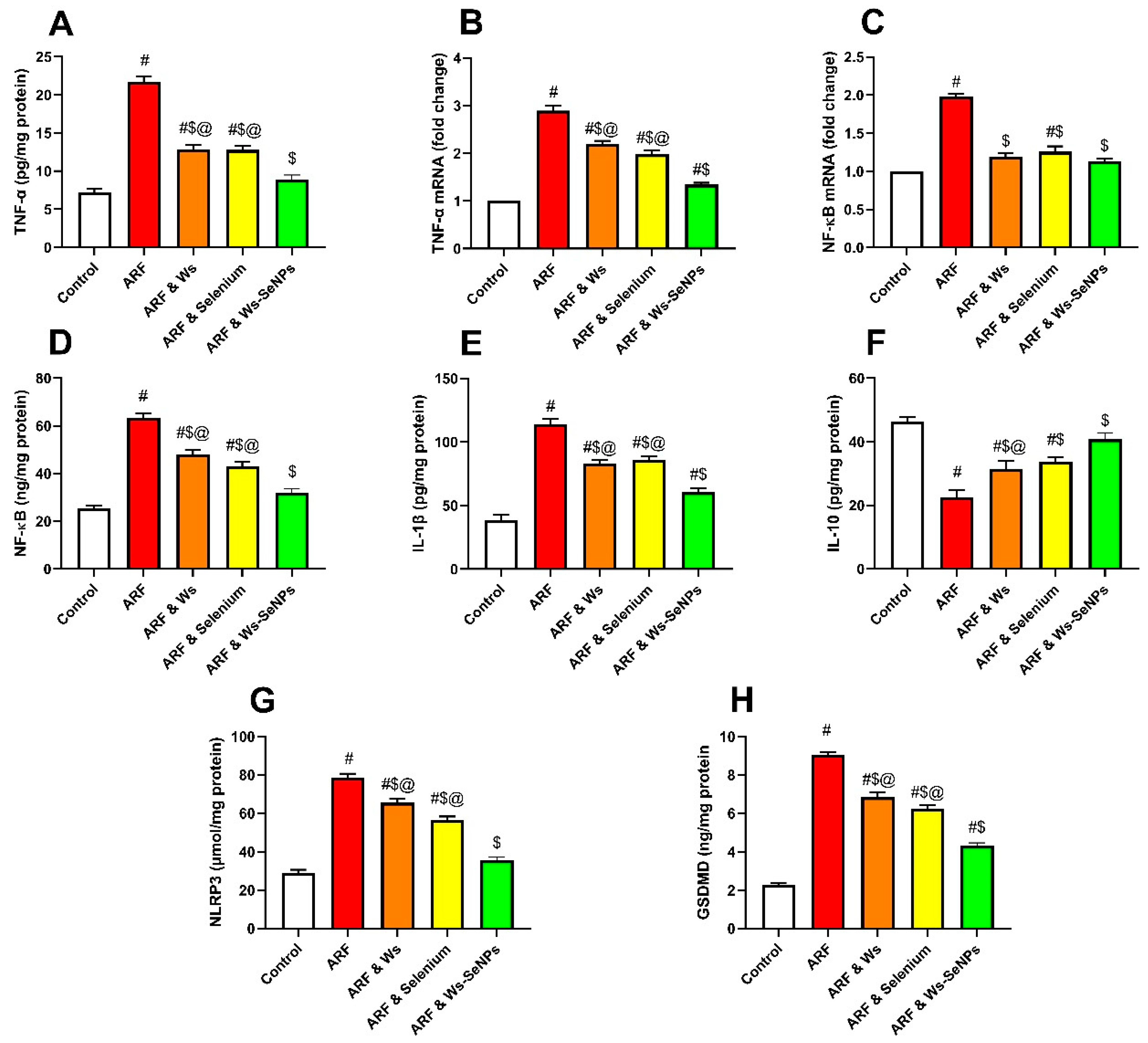
The renal inflammatory response was more severe in ARF (induced by glycerol) than in control groups, with significant increases in the protein level of the inflammatory mediator TNF-α and in the expression of the mRNA for the protein level of TNF-α (p < 0.05) (Figure 8A). Concurrently, ARF dramatically elevated the mRNA expression of NF-κB and also the level of NF-κB protein in the kidney, suggesting activation of the NF-κB-mediated inflammatory signaling pathway. High levels of inflammasome components such as IL-1β, NLRP3, and GSDMD also confirmed this inflammatory activation, suggesting inflammasome-mediated pyroptotic tubular injury. In contrast, the anti-inflammatory cytokine IL-10 was significantly decreased in the ARF group, suggesting the presence of a defective endogenous anti-inflammatory mechanism. The anti-inflammatory effect of Ws partially reduced the inflammatory response, as reflected by the reduced amounts of TNF-α, NF-κB, IL-1β, NLRP3, and GSDMD, with a partial restoration of IL-10. Sodium selenite also positively influenced the inflammatory profile by downregulating NF-κB-related signaling and partially reducing the levels of markers involved in inflammasomes. Specifically, the anti-inflammatory and anti-pyroptotic effects of Ws-SeNPs were most significant, reducing the contents of tumor necrosis factor alpha (TNF-α), nuclear factor kappa B (NF-κB), interleukin-1 beta (IL-1β), Nlrp3, and GSDMD (p < 0.05) and upregulating the content of interleukin-10 (IL-10) (p < 0.05) compared with the untreated ARF group. The results showed that Ws-SeNPs were able to alleviate renal inflammation caused by glycerol, at least in part, by the inhibition of the NF-kB/NLRP3/GSDMD inflammatory–pyroptotic axis (Figure 8).

2.8. Evaluation of Renal Mitochondrial Regulatory Markers

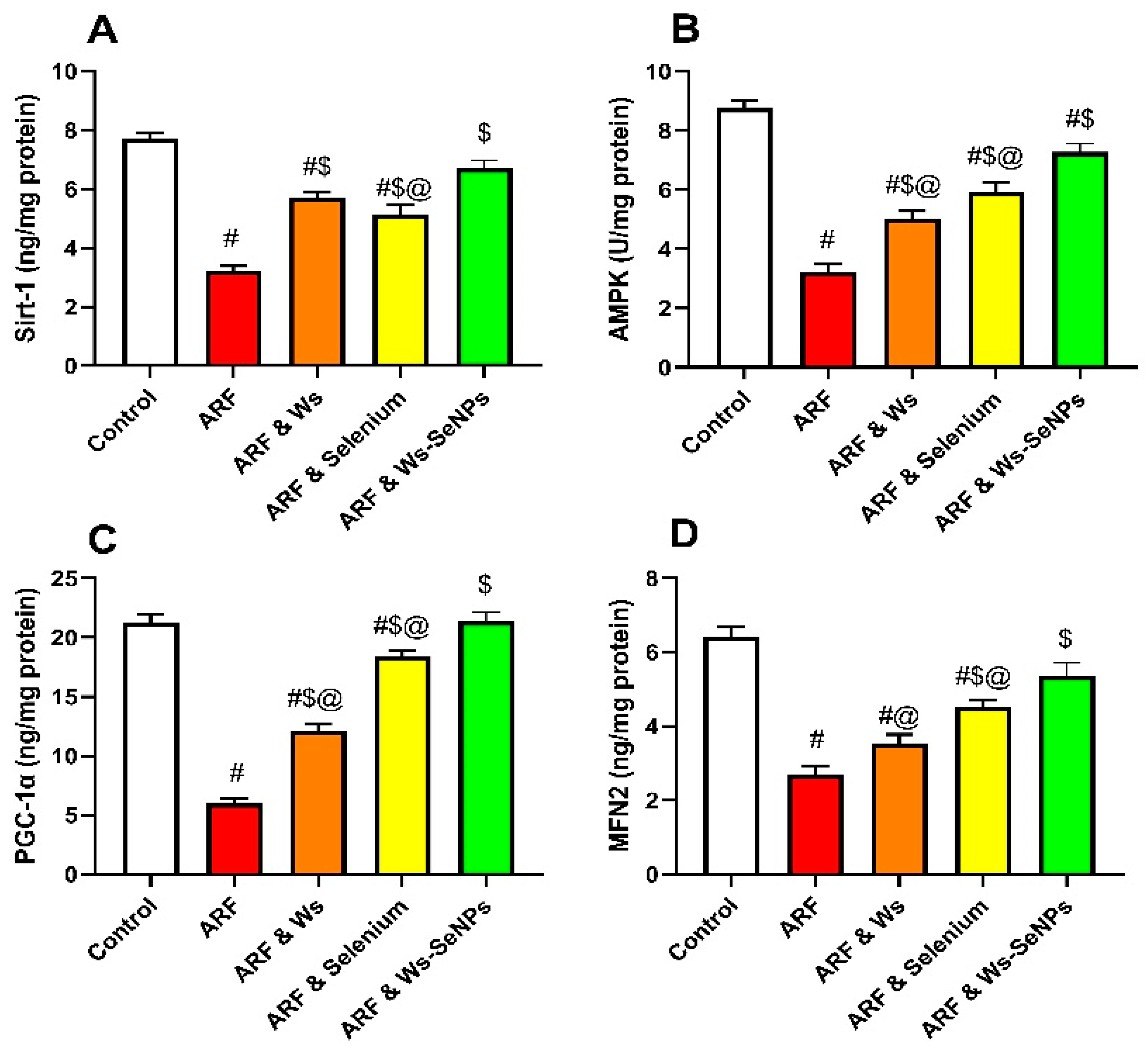
The glycerol-induced ARF strongly impaired renal mitochondrial regulatory signaling, characterized by a significant drop in SIRT1, AMPK, PGC-1α, and MFN2 activity compared to the control group (p < 0.05), as seen in Figure 9. These changes suggest that cellular energy sensing is disturbed, mitochondrial biogenesis is impaired, and mitochondrial fusion is decreased, all of which suggests that there is prominent mitochondrial dysfunction in the renal injury associated with rhabdomyolysis.
Partial restoration was observed with Ws, indicating moderate restoration of mitochondrial homeostasis. Sodium selenite also increased AMPK, PGC-1α, and MFN2 expression, and the increase in some of these markers was more noticeable than the increase for Ws in the mitochondrial regulatory recovery. In particular, the levels of SIRT1, AMPK, PGC-1α, and MFN2 were significantly elevated in the Ws-SeNP group compared to the ARF group (p < 0.05). These results indicated that Ws-SeNPs showed a stronger protective effect on mitochondria, possibly by upregulating the SIRT1/AMPK/PGC-1α pathway and restoring mitochondrial integrity related to MFN2 (Figure 9).

2.9. Evaluation of Renal Apoptotic Markers

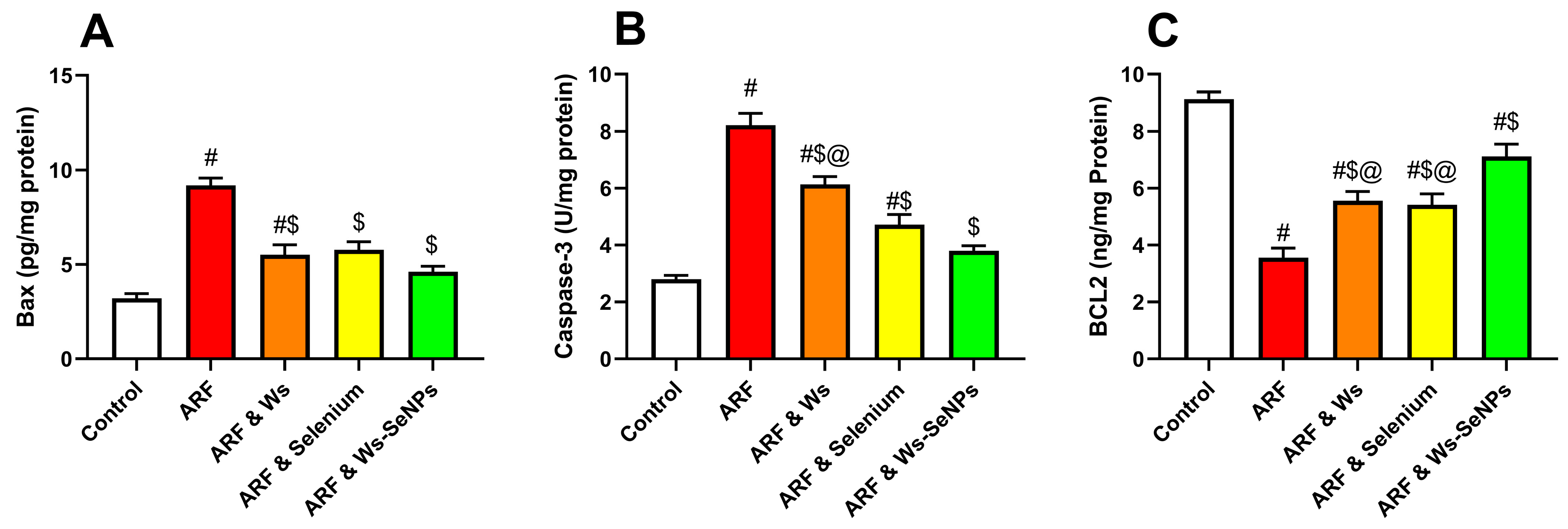
Glycerol-induced ARF showed a significant activation of apoptotic signals in the kidneys, associated with a significant increase in pro-apoptotic markers like Bax and Caspase-3, as shown in Figure 10, compared to the control group (p < 0.05). In contrast, the ARF group showed a significant decrease in the anti-apoptotic marker Bcl-2, suggesting a disbalance of pro- and anti-apoptotic regulatory mechanisms involved in tubular cell damage, thereby confirming the role of apoptotic tubular cell damage in glycerol-induced renal injury. Partially, Withania somnifera (Ws) inhibited these apoptotic changes, as shown by reduced levels of Bax and caspase-3 and partial restoration of Bcl-2. Sodium selenite also had a beneficial effect on the apoptotic profile, indicative of protection against renal cell death. Importantly, Ws-SeNPs were the most powerful anti-apoptotic agent, significantly reducing Bax levels and significantly increasing the level of Bcl-2 compared with the untreated ARF group (p < 0.05).

2.10. Immunohistochemistry

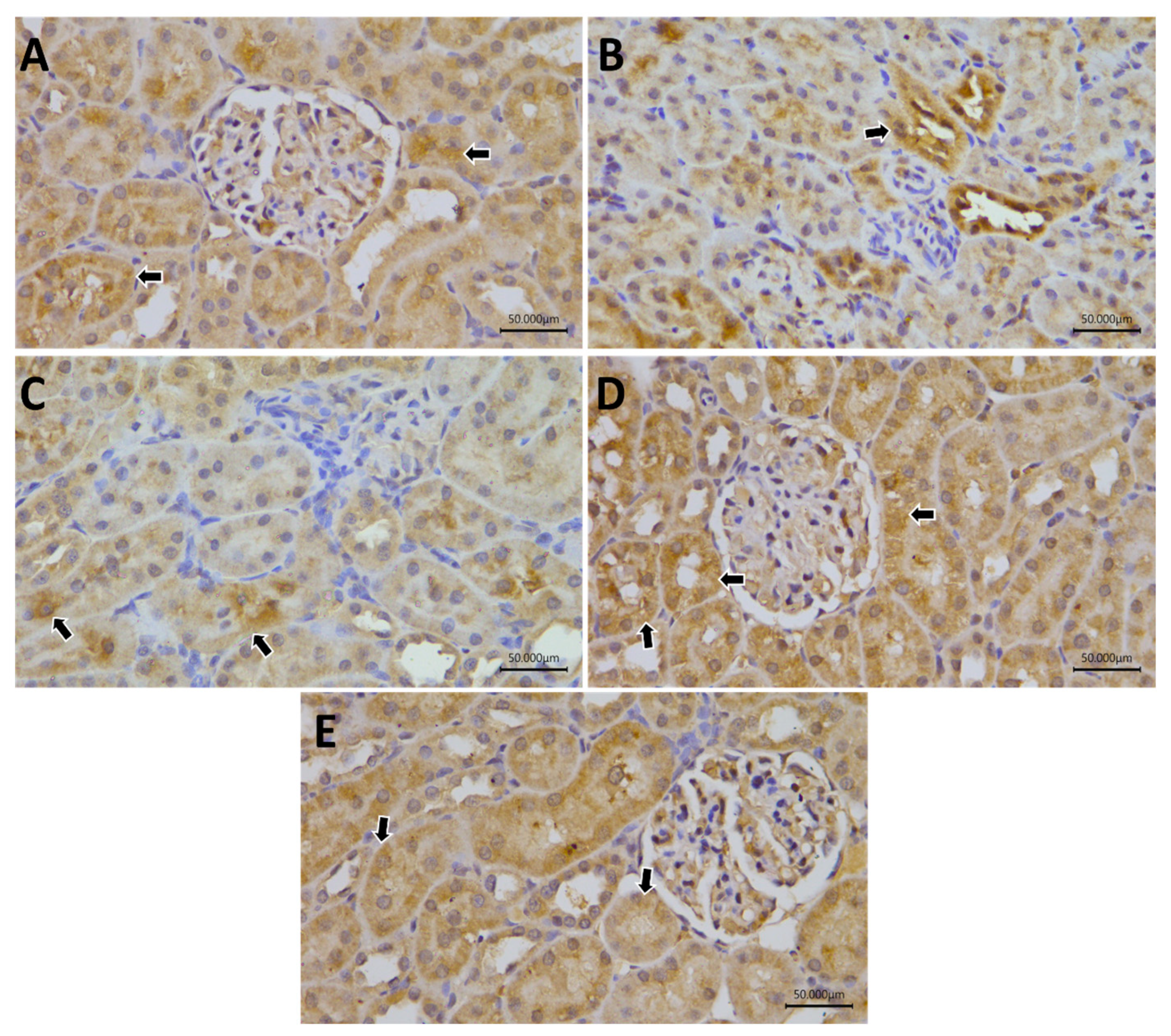
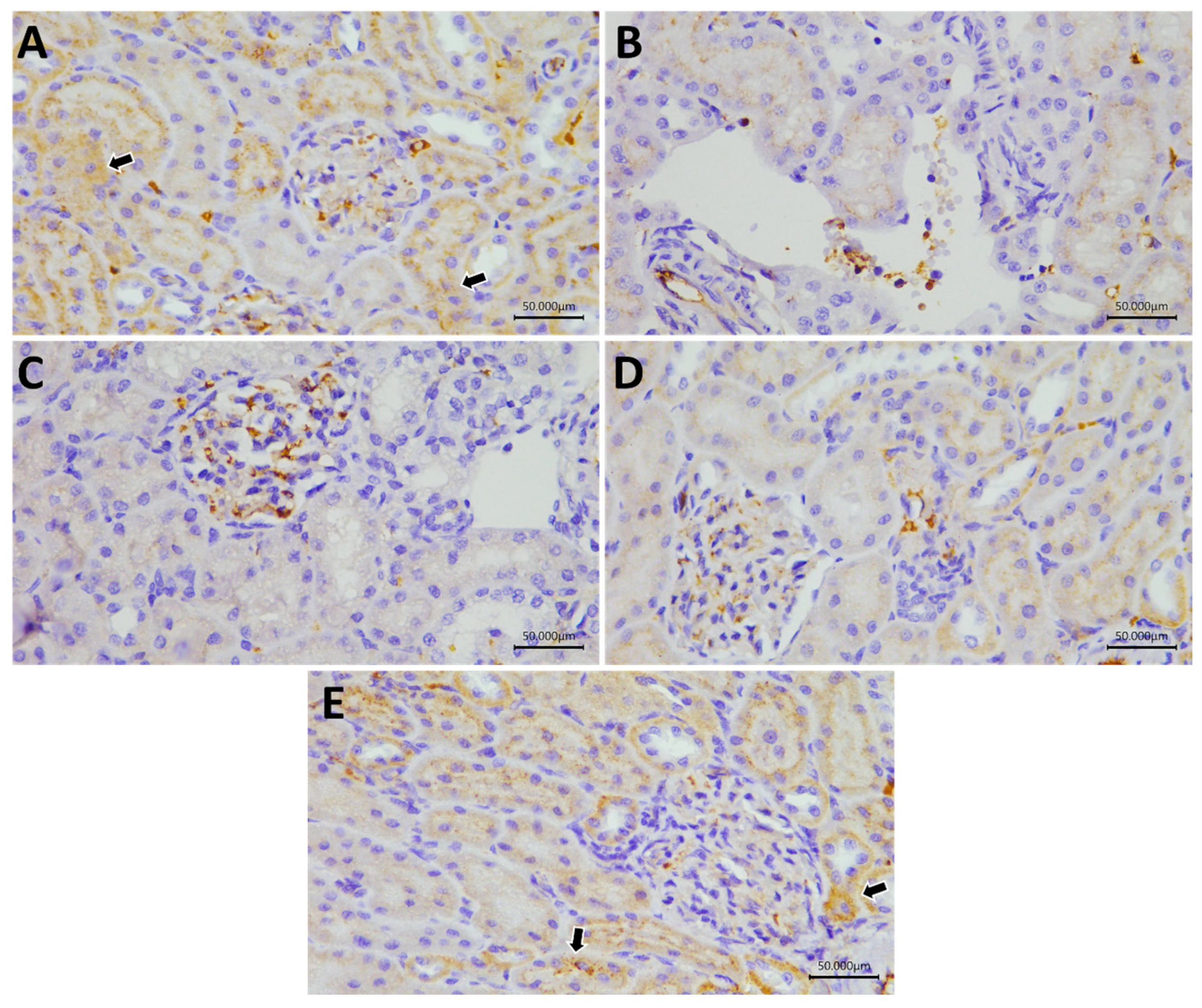
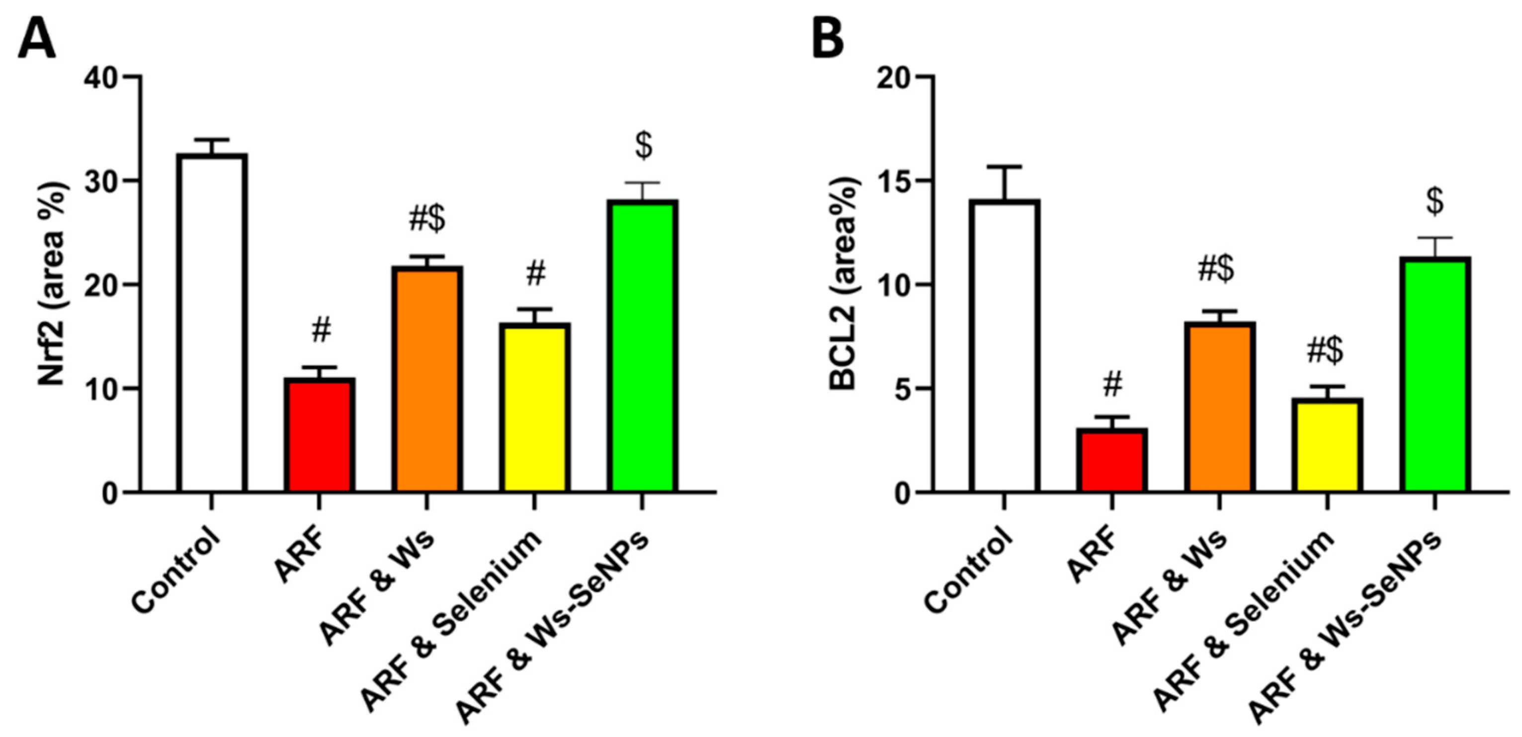
Immunohistochemical assessment of renal Nrf2 and Bcl-2 expression further supported the biochemical findings (Figure 11, Figure 12 and Figure 13). Regarding Nrf2 immunoexpression, the control group showed marked cytoplasmic reactivity, whereas the ARF group exhibited weak Nrf2 staining, indicating suppression of the renal antioxidant response following glycerol-induced injury. Similarly, weak Nrf2 immunoreactivity was observed in the ARF & sodium selenite group, while the ARF & Ws group showed evident improvement. Notably, the ARF & Ws-SeNP group demonstrated strong Nrf2 immunoreactivity, indicating restoration of Nrf2-mediated cytoprotective signaling. In parallel, Bcl-2 immunostaining was weak in the ARF and ARF & sodium selenite groups, reflecting impaired anti-apoptotic defense, whereas moderate cytoplasmic Bcl-2 reactivity was observed in the control and ARF & Ws-SeNP groups. The ARF & Ws group showed mild Bcl-2 expression. Quantitative immunohistochemical analysis revealed a significant reduction in Nrf2 and Bcl-2 immunoreactivity in the ARF and ARF & sodium selenite groups compared with the control group (p < 0.01). Conversely, Ws-SeNP treatment significantly enhanced both Nrf2 and Bcl-2 immunoreactivity compared with the untreated ARF group (p < 0.01), suggesting that Ws-SeNPs restored renal antioxidant and anti-apoptotic defense mechanisms in glycerol-induced ARF.

2.11. Histopathological Analysis

2.11.1. Skeletal Muscle

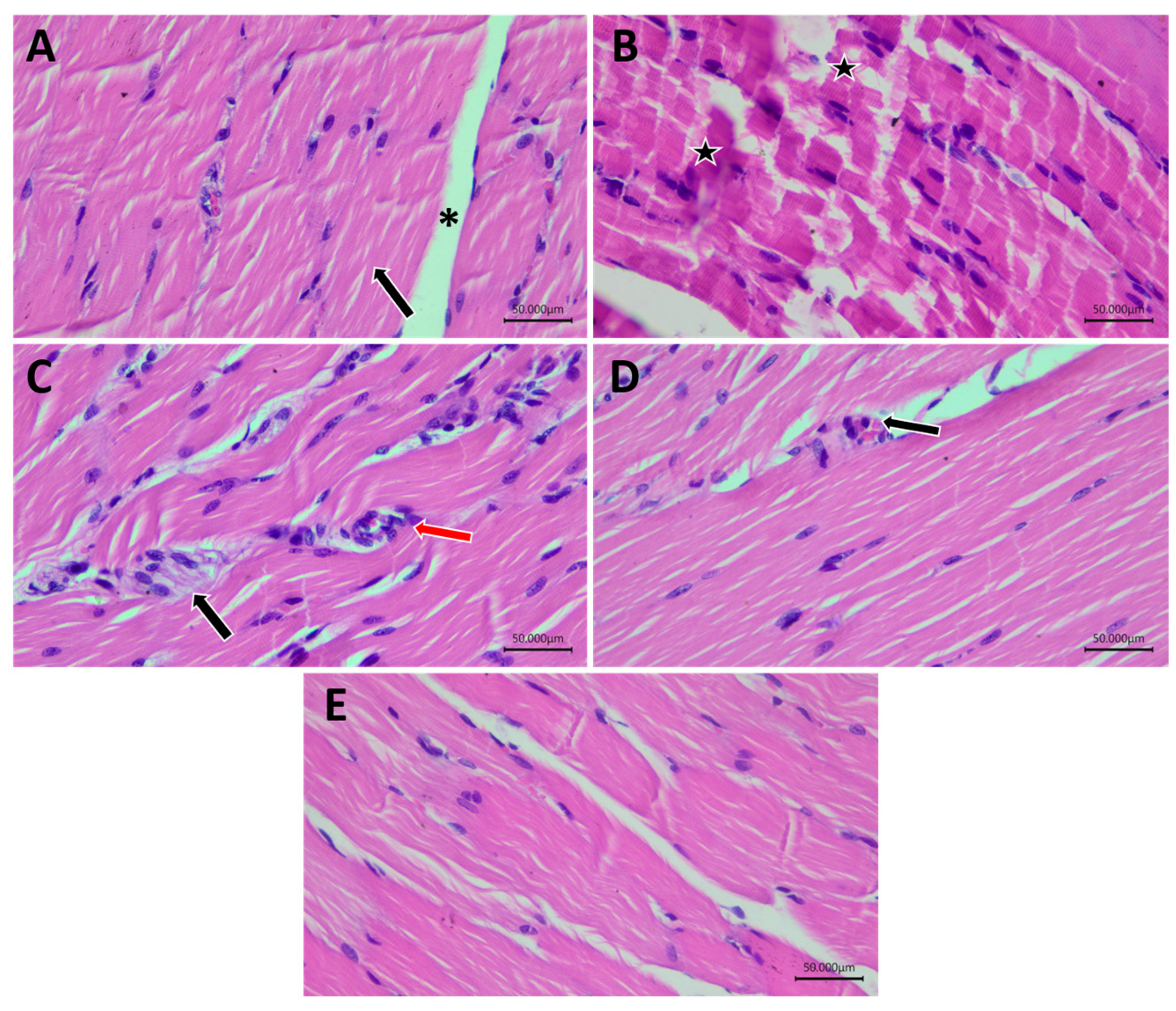
Histopathological examination of skeletal muscle sections from the control group revealed normal skeletal muscle architecture, characterized by regularly arranged myofibers with eosinophilic sarcoplasm, peripheral nuclei, compact myofibrillar organization, and delicate endomysial connective tissue. In contrast, the ARF group exhibited marked myopathic alterations, including endomysial edema, inflammatory cellular infiltration, focal rhabdomyolysis, myofiber hyalinization, and localized necrotic changes. The ARF & sodium selenite group showed persistent skeletal muscle injury, manifested by degenerated myocytes with reduced fiber diameter and inflammatory cellular infiltration within the perimysial connective tissue. Conversely, the ARF & Ws and ARF & Ws-SeNP groups demonstrated notable improvement in skeletal muscle morphology, with nearly preserved myofiber organization, peripheral nuclei, acidophilic sarcoplasm, and relatively normal surrounding connective tissue layers. These findings indicate that Ws and, more prominently, Ws-SeNPs attenuated glycerol-induced skeletal muscle degeneration and rhabdomyolysis-associated histopathological damage (Figure 14).

2.11.2. Renal Tissue

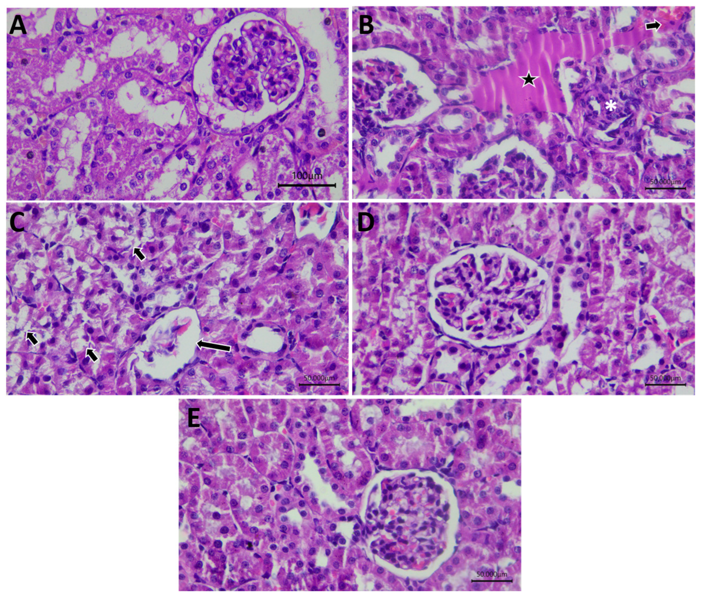
Renal cortical sections from the control group showed normal histological architecture, including intact renal corpuscles, preserved glomerular structure, and regularly arranged renal tubules. In the ARF group, renal sections revealed prominent pathological alterations consistent with acute renal injury, including inflammatory cellular infiltration, tubular vacuolar degeneration, focal necrotic areas, and disruption of renal cortical architecture. The ARF & sodium selenite group showed partial but incomplete improvement, with persistent degeneration of tubular epithelial cells, vacuolated renal tubules, and glomerular alterations. The ARF & Ws group demonstrated an improved renal cortical appearance, although some vascular congestion and mild tubular alterations remained evident. Notably, the ARF & Ws-SeNP group exhibited a nearly normal renal histological pattern, with marked preservation of tubular and glomerular architecture and minimal inflammatory or degenerative changes. These observations support the biochemical findings and suggest that Ws-SeNPs provided superior histological protection against glycerol-induced renal cortical injury (Figure 15).

3. Discussion

Acute renal failure (ARF) following rhabdomyolysis is a clinically significant form of acute kidney injury that results from the breakdown of skeletal muscle and sets in motion a chain of systemic and renal pathological events. The molecular mechanisms underlying the connection between rhabdomyolysis and oxidative stress, tubular damage, mitochondrial injury, inflammatory response, pyroptosis, and apoptosis are still not fully understood, although glycerol-induced ARF is a well-established experimental model that closely resembles myoglobin-mediated renal damage. Furthermore, the antioxidant and anti-inflammatory properties of Withania somnifera (Ws), the redox-regulating effect of selenium, and the comparative efficacy of native Ws extract, inorganic selenium, and Ws-functionalized selenium nanoparticles (Ws-SeNPs) against rhabdomyolysis-associated renal injury have not been well clarified. Thus, the present study aimed to fill this gap by investigating the renoprotective efficacy of green-synthesized Ws-SeNPs in comparison to Ws extract and sodium selenite alone in glycerol-induced ARF.
The present findings showed that administration of glycerol was able to induce ARF associated with rhabdomyolysis, which was confirmed by the increased kidney weight indices, significant increases in rhabdomyolysis markers, worsening of renal function, increased tubular injury markers, oxidative stress, activation of inflammation, mitochondrial dysfunction, and apoptotic cell death. Importantly, Ws-SeNPs had the most prominent protective impact on most of the tested parameters. This superior effect was not exclusive to a single biochemical pathway; instead, it was found to involve coordinated regulation of redox homeostasis, Nrf2/Keap1 signaling, inflammatory and pyroptotic responses, mitochondrial regulatory pathways, and apoptosis-related mechanisms.
The absolute and relative kidney weight of the glycerol group in ARF are attributed to renal swelling, tissue edema, congestion, inflammatory infiltration, and acute tubular alteration of structure. These changes are frequently seen with acute tubular injury and diminished renal hemodynamics after rhabdomyolysis [24,25]. Partial improvement seen after Ws and sodium selenite indicates a partial reduction in renal damage after both treatments. However, the more obvious decrease in kidney weight indices in the Ws-SeNPs-treated group suggests greater preservation of renal tissue architecture and less swelling arising from injury. The anti-inflammatory and antioxidant activity of the phytoconstituents from Ws, plus the selenium-dependent stimulation of cytoprotective mechanisms, may explain this effect [11,26].
The high levels of serum CK and LDH activity in the ARF group corroborate the presence of significant skeletal muscle damage after glycerol administration. CK is more sensitive in detecting the disruption of muscle fibers, while LDH is a more general indicator of damage to the cell membrane and tissue injury [27]. The histopathology of the gastrocnemius muscle also confirmed these biochemical results, showing the presence of focal myofiber necrosis, hyalinization, and necrotic myocytes in the ARF group [28]. Ws and sodium selenite are able to partially reverse these changes, while Ws-SeNPs showed the highest efficacy in reducing the activities of CK and LDH and showing a more marked preservation of the architecture of the skeletal muscle. This indicates that Ws-SeNPs could decrease the load of rhabdomyolysis itself and, thus, the release of myoglobin and other muscle-derived nephrotoxic mediators into the systemic circulation. This effect is important in particular, as the severity of muscle injury affects the amount of myoglobin filtered and, therefore, the degree of renal tubular toxicity [29].
The ARF group showed a significant increase in serum creatinine and urine urea, which indicates a worsening of kidney function. These markers are indicative of dysfunction in glomerular filtration and decreased renal elimination of nitrogenous waste products. Meanwhile, cystatin-C was significantly elevated, corroborating the presence of an early filtration defect [1,30]. The increase in KIM-1 and NGAL is more importantly a direct tubular epithelial injury, specifically in the proximal tubules, which are most sensitive to myoglobin-induced oxidative damage. These injury markers, all of which are functional and tubular-based, indicate that glycerol-induced rhabdomyolysis caused filtration disturbances and tubular structural damage [31,32]. These parameters were further enhanced by Ws and sodium selenite but not completely. In contrast to the others, Ws-SeNPs showed the highest correction of creatinine, urea, cystatin-C, KIM-1, and NGAL, suggesting better renal filtration capacity and preservation of tubular epithelial integrity. This improvement was significantly corroborated by renal histopathology, where the animals treated with Ws-SeNPs had an almost normal pattern of renal cortex and no inflammatory infiltration; focal necrosis, tubular degeneration and glomerular injury were seen in the untreated ARF group.
Oxidative stress is a key mechanism in glycerol-induced ARF. Rhabdomyolysis causes myoglobin to be filtered by the glomeruli and to be deposited in the renal tubules [33]. The heme of myoglobin is a pro-oxidant that catalyzes the generation of free radicals, lipid peroxidation, damage to mitochondria and tubular epithelial toxicity, all of which depend on the presence of iron [34,35]. Renal MDA, 4-HNE, and NO levels were significantly elevated in the ARF group in the present study, reflecting increased lipid peroxidation, oxidative membrane damage, and nitrosative stress. In parallel, there was an obvious decrease in the amounts of GSH, SOD, CAT, and GPx, which verified the depletion of the endogenous antioxidant defense system. This redox imbalance may have been a contributing factor to tubular epithelial injury, inflammatory activation, and mitochondrial dysfunction [36,37].
The Nrf2/Keap1 pathway is an important cytoprotective mechanism against oxidative stress [38]. Under physiological conditions, Keap1 is a negative regulator of Nrf2 activity, but during oxidative challenges, Nrf2 activation leads to the induction of antioxidant and detoxifying enzymes [39]. In the present study, ARF induced by glycerol was associated with an increase in Keap1 and a decrease in Nrf2, which resulted in the suppression of this antioxidant pathway [40]. This was also corroborated by the low immunoexpression of Nrf2 in renal sections from the ARF group. There was partial improvement in the oxidative stress profile during treatment with Ws and sodium selenite. The more positive restoration seen with Ws-SeNPs (decreased MDA, 4-HNE, NO, Keap1, and increased Nrf2, GSH, SOD, CAT, and GPx) indicated that Ws-SeNPs were able to reactivate antioxidant defenses. The selenium component might have a direct role in this effect by supporting selenoprotein-dependent antioxidant enzymes, such as GPx [41,42], and the phytochemicals derived from Ws might have a greater effect in supporting cytoprotection pathways via Nrf2 due to their phenolic and withanolide components [11,43,44]. This makes Ws-SeNPs a potential dual antioxidant system, combining the enzymatic antioxidant activity of selenium and the redox-modulating activity of plants.
Another important factor in the development of renal injury in rhabdomyolysis is inflammation. The increase in oxidative stress and damage to tubular cells results in pro-inflammatory transcriptional signaling, including activation of the nuclear factor-kappaB (NF-κB) pathway, leading to increased production of cytokines, including tumour necrosis factor (TNF)-alpha, interleukin (IL)-1 beta, and IL-6 [45,46,47]. In the present study, glycerol-induced ARF strongly upregulated the protein levels of TNF-α, NF-κB, IL-1β, and IL-6, as well as the mRNA levels of NF-κB and TNF-α, which clearly demonstrated the activation of inflammatory signaling. Concurrently, the endogenous anti-inflammatory counter-regulatory mechanism was impaired by a decrease in IL-10. This inflammatory change may have further exacerbated tubular injury through recruitment of leukocytes, release of cytokines, propagation of oxidative stress, and signaling of cell death [48].
The upregulation of NLRP3 and GSDMD also supports the notion that pyroptotic mechanisms via the inflammasome played a role in renal injury. Mitochondrial ROS, ionic imbalance, and cell danger signals are potent activators of the NLRP3 inflammasome [49,50]. It activates downstream inflammatory cell death pathways, such as GSDMD-dependent membrane pore formation and IL-1β release [51]. Thus, the co-upregulation of NF-κB, NLRP3, IL-1β, and GSDMD in the ARF group indicates the activation of an inflammatory–pyroptotic axis [52]. Ws and sodium selenite partially inhibited these inflammatory markers, but Ws-SeNPs had the greatest anti-inflammatory and anti-pyroptotic effect. This was indicated by lower levels of TNF-α, NF-κB, IL-1 β, IL-6, NLRP3, and GSDMD, and restoration of IL-10. The results indicate that Ws-SeNPs can suppress renal inflammation by inhibiting the NF-κB/NLRP3/GSDMD axis, which might be attributed to the reduction of oxidative stress and enhancement of mitochondrial homeostasis [53].
Oxidative stress, inflammation, apoptosis, and tubular necrosis are important mechanistic links of glycerol-induced ARF and can be explained by mitochondrial dysfunction [54]. Renal tubular cells have an increased reliance on mitochondrial energy production, and dysfunction of the mitochondria will limit ATP production, ion transport, and cell survival [55,56]. In the current study, ARF was highly effective at decreasing SIRT1, AMPK, PGC-1α, and MFN2 expression. AMPK regulates metabolic adaptation during stress, while SIRT1 protects mitochondria, triggers antioxidant responses, and inhibits inflammation. PGC-1α is a master regulator of mitochondrial biogenesis, and MFN2 is involved in mitochondrial fusion and structural integrity [57].
Glycerol-induced renal injury was found to be apoptosis-mediated, as evidenced by increased levels of Bax and caspase-3 and decreased expression of Bcl-2 in the ARF group. Bax favors the permeabilization of the outer mitochondrial membrane and the activation of apoptotic signals, while Bcl-2 acts in the opposite direction by preventing these events and favoring cell survival [58]. Caspase-3 is one of the executioners of apoptosis and is an indicator of the final stage of apoptotic cell death [59,60]. The bax and caspase-3 expression was observed to rise while bcl-2 was found to decrease, thereby suggesting activation of mitochondrial apoptosis in renal tissue. This was also corroborated by immunostaining of Bcl-2 in renal sections of the ARF group, which was not strong [59,61]. The apoptotic changes were partially inhibited by Ws and sodium selenite; however, Ws-Selenppt showed the most anti-apoptotic effect, decreasing the expression of Bax and caspase-3, and increasing the expression of Bcl-2. Furthermore, the immunoreactivity of Bcl-2 in the Ws-SeNP group indicated its cytoprotective activity at the tissue level. These results indicated that Ws-SeNPs could improve the Bax/Bcl-2 ratio and inhibit the apoptotic execution by caspase-3, thereby preventing renal tubular apoptosis.
Histopathological results strongly support the biochemical and molecular results. Glycerol-induced ARF resulted in severe inflammatory infiltration of cells, focal necrosis, tubular degeneration, and glomerular injury in the renal cortex [12,28,49]. These changes are typical of acute tubular necrosis and inflammatory renal damage resulting from myoglobin-mediated toxicity [62]. Some pathological parameters were improved by the use of sodium selenite, but the changes in tubular and glomerular parameters were not reversed. Ws treatment resulted in an improvement of cortical architecture, while Ws-SeNPs created an almost normal histological pattern. In a manner similar to this, severe injury to muscle fibers was observed in the ARF group, as confirmed by skeletal muscle examination, and Ws-SeNPs were seen to significantly reduce the degeneration of muscle fibers.
Several complementary mechanisms can account for the superior effect of Ws-SeNPs compared to Ws extract or sodium selenite alone [63]. First, nanosizing selenium can overcome the toxicity constraint of inorganic selenium, enhance biological distribution, and increase interaction with the cells [17,19]. Second, Ws phytoconstituents can also provide intrinsic antioxidant and anti-inflammatory effects, functioning as capping and stabilizing agents that promote the surface of nanoparticles. Third, the combined formulation may exhibit a greater multi-target effect through various mechanisms such as increasing the activity of antioxidant enzymes dependent on selenium, upregulating nuclear factor E2-related factor 2 signaling, enhancing mitochondrial control mechanisms, decreasing NF-κB/NLRP3-mediated inflammation, and decreasing apoptotic cell death [64].
Although the present study was conducted in a glycerol-induced rat model of rhabdomyolysis-associated acute renal failure, several aspects of the findings are likely to extend to other experimental models and potentially to human acute kidney injury. The pathogenic mechanisms investigated in this study—including oxidative stress, Nrf2/Keap1 dysregulation, NF-κB-mediated inflammation, NLRP3 inflammasome activation, mitochondrial dysfunction, pyroptosis, and apoptosis—are highly conserved across mammalian species and represent common molecular pathways implicated in diverse forms of acute kidney injury. Therefore, the observed ability of Ws-SeNPs to simultaneously modulate these interconnected signaling networks suggests that their renoprotective effects may not be restricted to glycerol-induced injury alone but could also apply to other experimental conditions characterized by excessive oxidative and inflammatory damage, such as ischemia-reperfusion injury, nephrotoxic drug-induced nephropathy, contrast-induced nephropathy, and septic acute kidney injury. Moreover, because rhabdomyolysis-induced acute kidney injury in humans is likewise driven by myoglobin-mediated oxidative injury, tubular dysfunction, and inflammatory activation, the present findings provide mechanistic support for the potential clinical development of Ws-SeNPs as a multi-target therapeutic strategy. Nevertheless, direct translation to human disease should be approached cautiously because important interspecies differences exist in renal physiology, immune responses, selenium metabolism, nanoparticle biodistribution, and pharmacokinetic behavior. Consequently, additional investigations in large-animal models and well-designed preclinical safety studies are required before clinical trials can be considered.
The present study has several limitations. Only male rats were included, limiting the assessment of sex-dependent responses. In addition, N-terminal GSDMD and cleaved caspase-3 were not measured; therefore, pyroptosis and apoptosis were inferred from related signaling markers rather than directly confirmed. The oral bioavailability, pharmacokinetics, renal accumulation, biodistribution, and clearance of Ws-SeNPs were also not evaluated. Future studies should include both sexes and incorporate pathway-specific protein validation together with ICP–MS-based selenium quantification and labeled-nanoparticle biodistribution analyses.

4. Materials and Methods

4.1. Extraction and Preparation of Ws

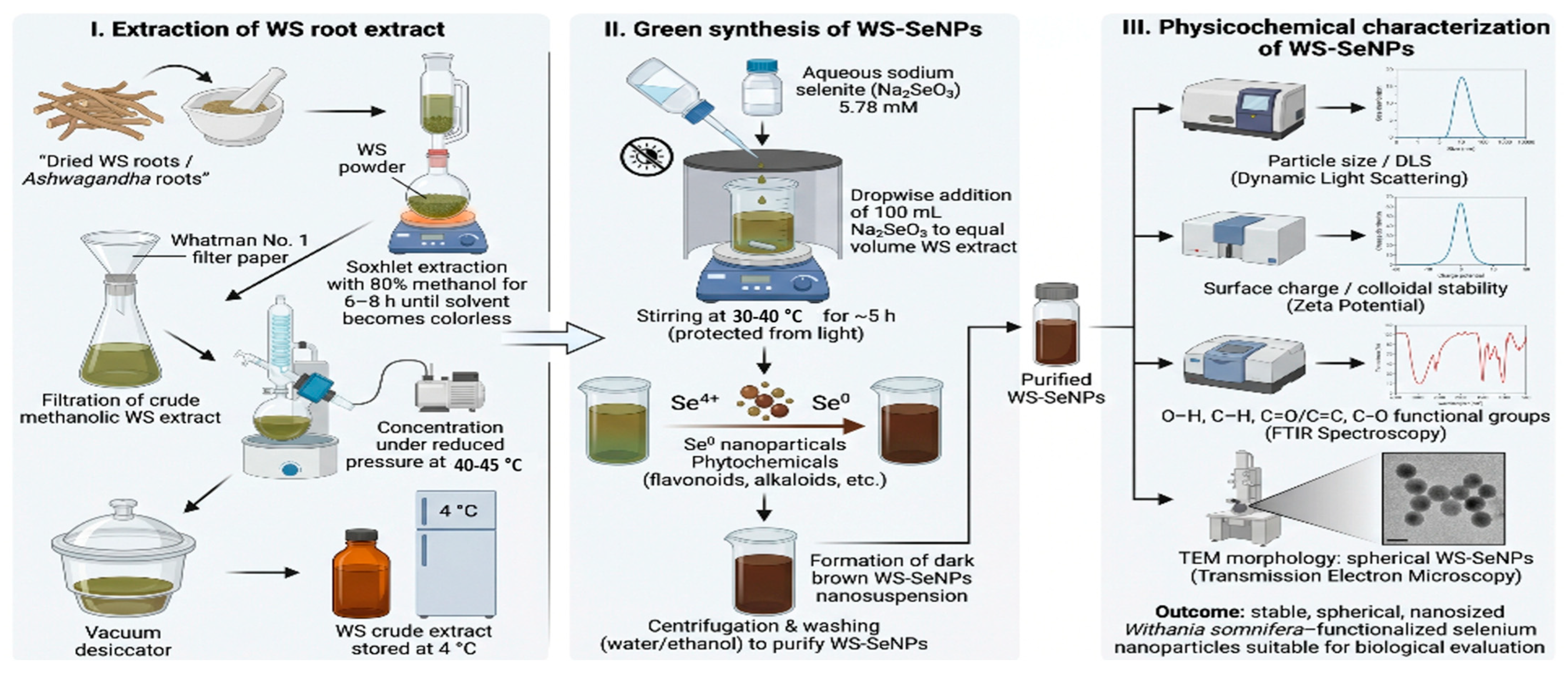
Dried roots of Withania somnifera (Ws) were ground and powdered using a mechanical grinder and extracted with 80% methanol via a Soxhlet apparatus for 6–8 h until the solvent became colorless. The resulting extract was filtered employing Whatman No.1 filter paper and condensed under decreased pressure via a rotary evaporator at 40–45 °C to eliminate the solvent. The concentrated residue was further dried in a vacuum desiccator to yield a semisolid methanolic crude extract of Ws. The dried extract was weighed, and the extraction yield was determined as a percentage of the initial plant material. The extract was stored at 4 °C in airtight amber glass bottles until further use in experimental procedures [65].

4.2. LC–MS/MS

The dried residue was dissolved in LC–MS-grade methanol at 1 mg/mL, vortexed, and centrifuged at 14,000× g for 10 min at 4 °C. The supernatant was passed through a 0.22 µm PTFE filter into amber vials. We injected 5 µL of each sample.
Separation was performed using a reversed-phase C18 column (100 × 2.1 mm, 1.8 µm) at 40 °C. Mobile phase A was water with 0.1% formic acid, and mobile phase B was acetonitrile with 0.1% formic acid. The flow rate was 0.3 mL/min, and the autosampler was kept at 8 °C. A linear gradient brought the withanolides off the column between about 9.9 and 12.8 min, with a total run time of 20 min, including re-equilibration.
An electrospray ionization (ESI) tandem mass spectrometer was run separately in positive and negative ion modes. Typical source settings included an ion-spray voltage of +4.5 kV in positive mode and −3.5 kV in negative mode, a source temperature of 450 °C, and nitrogen as the nebulizer, drying, and collision gas. Survey scans covered m/z 100–1200, and the most intense precursors were selected for data-dependent MS/MS with collision energy ramped from 20 to 40 eV. The withanolide aglycones gave mainly [M+H]+ ions in positive mode, while the glycoside was seen as [M–H] in negative mode.
We annotated each peak using its retention time, measured precursor ion, and MS/MS product ions, then checked the assignment against authentic standards (when available), published Withania somnifera data and spectral databases. The withanolide aglycones lose water in steps (−18 Da each) from the hydroxylated steroid nucleus, which gives the recurring ions at m/z 317, 299 and 281. Withanosides, on the other hand, lose a hexose unit (−162 Da), which helped flag the glycosylated compound. Peak picking and spectral matching were done in the instrument software, together with open-source tools.

4.3. Preparation and Characterization of Selenium Nanoparticles Using Withania somnifera

Ws-SeNPs were synthesized through a green reduction approach adapted from [66]. Briefly, 100 mL of sodium selenite solution (Na2SeO3, 5.78 mM) was mixed with an equal volume of Ws extract solution at a concentration of approximately 2.02 mg/mL under continuous magnetic stirring. This corresponded to a Ws extract-to-sodium selenite mass ratio of approximately 2.02:1 and a Ws extract-to-elemental selenium mass ratio of approximately 4.43:1. This solution was subsequently added dropwise to an equal volume of the prepared methanolic Ws extract solution under continuous magnetic stirring. The reaction mixture was maintained at 30–40 °C and protected from light throughout the synthesis process to minimize photodegradation of the bioactive phytoconstituents. During the reaction, the gradual development of a dark brown coloration over approximately 5 h was considered indicative of the reduction of selenite ions and the formation of Ws-SeNPs. Following synthesis, the resulting nanoparticle suspension was collected and processed for subsequent physicochemical characterization, including particle size analysis, zeta potential measurement, FT-IR, and TEM (Figure 16).
The hydrodynamic diameter and particle size distribution of Ws-SeNPs were evaluated by dynamic light scattering (DLS), while their surface charge and colloidal stability were assessed by zeta-potential analysis using a Zetasizer Nano ZS90 instrument (Malvern Panalytical Ltd., Malvern, Worcestershire, UK). These analyses were performed to determine the dispersion behavior, size homogeneity, and electrostatic stability of the biosynthesized nanoparticles.
The morphology, structural appearance, and approximate particle dimensions of Ws-SeNPs were further investigated using high-resolution transmission electron microscopy (HR-TEM; JEOL Ltd., Tokyo, Japan). For TEM examination, an aliquot of the Ws-SeNPs was mounted on a grid and subsequently visualized and photographed under the electron microscope to assess particle shape, distribution, and aggregation pattern.
FTIR spectroscopy was employed to characterize the surface-associated functional groups involved in the biosynthesis and stabilization of Ws-SeNPs. Spectral analysis was performed to identify the characteristic chemical moieties derived from W. somnifera phytoconstituents that may contribute to selenium-ion reduction, nanoparticle capping, and colloidal stabilization. The detected absorption bands were interpreted to clarify the probable phytochemical interactions underlying the successful formation of the green-synthesized Ws-SeNPs.

4.4. Experimental Animals

The present study was conducted to assess the nephroprotective efficacy of Ws-SeNPs against GLY-induced ARF in rats. A total of thirty-five healthy adult male albino rats (100–120 g) were recruited. The animals were obtained from an accredited facility and maintained under standard laboratory settings, with a relative humidity of 50–60%, a temperature of 22 ± 2 °C, and a 12 h light/12 h dark cycle. The rats were acclimatized for one week before the beginning of the experiment and fed regular laboratory chow and water ad libitum. To induce renal stress, water was withheld for 24 h before glycerol administration. The study protocol was reviewed and approved by the relevant institutional animal ethics committee under ethical approval number 131/A/2026.
Only male rats were included to reduce biological variability associated with estrous-cycle-related hormonal fluctuations and to maintain consistency with commonly used glycerol-induced acute kidney injury models.
The rats were divided into the following experimental groups;
Control Group (Control, n = 7): For 21 consecutive days, the animals received normal saline solution (0.9% NaCl) orally. On day 20, they received a 1 mL i.m. saline injection.
Glycerol Group (ARF, n = 7): For 21 days in a row, the animals in this group were given normal saline orally. On day 20, they received a single i.m. injection of 50% GLY (10 mL/kg/day) [11].
Withania somnifera Group (ARF & Ws, n = 7): The animals received Withania somnifera orally for 21 days (200 mg/kg/day) [61]. On day 20, they received a single dose of i.m. injection of 50% GLY (10 mL/kg/day).
Sodium selenite Group (ARF & Selenium, n = 7): The animals received Na2SeO3 orally for 21 days (0.5 mg/kg/day) [63]. On day 20, the animals received a single dose of GLY (10 mL/kg/day) [17].
SeNPs biosynthesized via Withania somnifera Group (ARF & Ws-SeNPs, n = 7): The animals received Ws-SeNPs orally for 21 days (0.5 mg/kg/day) [11,43]. On day 20, the animals received a single dose of GLY (10 mL/kg/day).
The sample size was determined based on previous experimental studies employing the glycerol-induced acute renal failure model and evaluating the nephroprotective effects of natural products and selenium nanoparticles. A total of 35 adult male albino rats were included and randomly allocated into five experimental groups (n = 7 per group). This sample size was considered sufficient to detect biologically meaningful differences in biochemical, molecular, and histopathological outcomes while adhering to the principles of the 3Rs (Replacement, Reduction, and Refinement) to minimize animal use without compromising statistical validity.

4.5. Sample and Tissue Collection

At the end of the experiment, the rats received deep anesthesia induced by intraperitoneal ketamine/xylazine (100 and 10 mg/kg body weight, respectively), and then euthanized by decapitation. Blood samples were immediately collected and left to clot at room temperature for 15 min. The samples were then centrifuged for 10 min at 3000 rpm at 4 °C, and the separated serum was stored for subsequent analysis of renal function biomarkers. Kidney tissues were removed, rinsed in ice-saline and subjected to biochemical analysis. One milliliter of PBS was used to dilute a 10% (w/v) tissue homogenate with the help of a glass homogenizer under cold conditions. The homogenates were then centrifuged at 10,000 rpm for 10 min at 4 °C, and the supernatants were used to estimate OS indices, inflammatory cytokines, and other biochemical markers using validated commercial ELISA assay kits and colorimetric assay kits, as per the manufacturers’ protocols.
To perform histopathological and immunohistochemical (IHC) analyses, representative samples of the kidney were fixed in formalin, dried, and stored in paraffin blocks. Slices of 450–500 µm thickness were made, placed on glass slides and stained with regular hematoxylin and eosin (H&E). Immunohistochemical analysis was performed to assess structural and cellular changes using a light microscope.

4.6. Relative Kidney Weight (RKW) Estimation

RKW was assessed using the previously described method by dividing the the kidney weight with the total body weight of each animal, thereby expressing the organ weight as a proportion of the animal’s body mass:
RKW   =   L e f t   k i d n e y   w t   ( g ) B o d y   w i e g h t   ( g )   × 100

4.7. Kidney Function Test Level

Serum urea, creatinine, KIM-1, and NGAL levels were assessed according to the manufacturers’ protocols. Urea was determined using the Urea Colorimetric Assay Kit II (BioVision, USA; Cat. No. K376-100) at 505 nm, while creatinine was measured using the Creatinine Colorimetric/Fluorometric Assay Kit (BioVision, Milpitas, CA, USA; Cat. No. K625-100) at 570 nm. Kidney injury molecule-1 (KIM-1) was quantified using the Rat KIM-1 ELISA Assay Kit (Eagle Biosciences, Amherst, NH, USA; Cat. No. RKM29-K01), whereas neutrophil gelatinase-associated lipocalin (NGAL) was measured using a Rat NGAL ELISA Kit (MyBioSource, CA, USA; Cat. No. MBS260195). Absorbance for ELISA-based assays was recorded at 450 nm.

4.8. CK and LDH Activities: Determination of Activities

Serum creatine kinase (CK) and lactate dehydrogenase (LDH) activities were determined using commercially available assay kits according to the manufacturers’ instructions. CK activity was measured using a CK Activity Assay Kit (MyBioSource, CA, USA, Cat. No. MBS1600481), and LDH activity was assessed using an LDH Activity Assay Kit (MyBioSource, USA; Cat. No. MBS2018912). Enzyme activities were calculated according to the standard procedures provided in the kit protocols.

4.9. Oxidant/Antioxidant Biomarkers: Assessment

Oxidative and antioxidant stress markers were assessed in renal tissue homogenates using commercially available assay kits supplied by MyBioSource (San Diego, CA, USA), according to the manufacturer’s instructions. Lipid peroxidation was evaluated by measuring malondialdehyde (MDA; Cat. No. MBS738685), whereas nitrosative stress was assessed by determining nitric oxide (NO; Cat. No. MBS2604161). Endogenous antioxidant defense was evaluated through the measurement of reduced glutathione (GSH; Cat. No. MBS265966), catalase (CAT; Cat. No. MBS2600683), superoxide dismutase (SOD; Cat. No. MBS036924), and glutathione peroxidase (GPx; Cat. No. MBS032696).
In addition, the Nrf2/Keap1 antioxidant signaling axis was investigated by quantifying Kelch-like ECH-associated protein 1 (Keap1; Cat. No. MBS7218529) and nuclear factor erythroid 2-related factor 2 (Nrf2; Cat. No. MBS752046). All assays were performed in accordance with the respective kit protocols, and the obtained values were normalized to total tissue protein content where applicable.

4.10. Determination of Inflammatory/Anti-Inflammatory Markers

Inflammatory and anti-inflammatory biomarkers were quantified using commercially available ELISA kits according to the manufacturers’ protocols. Interleukin-1 (IL-1) levels were measured using an ELISA kit (Elabscience; Wuhan, China, Cat. No. E-EL-R0012). Tumor necrosis factor-alpha (TNF-α) was determined using a Rat TNF-α ELISA Kit (Cloud-Clone Corp., Katy, TX, USA; Cat. No. SEA133Ra), while nuclear factor kappa B (NF-κB) was assessed using a Rat NF-κB ELISA Kit (FineTest, Wuhan, China; Cat. No. ER1186). Interleukin-10 (IL-10) was quantified using a Rat IL-10 ELISA Kit (MyBioSource, CA, USA; Cat. No. MBS9135769). Gasdermin D (GSDMD) was quantified using a Rat ELISA Kit (MyBioSource, CA, USA; Cat. No. MBS2032003).

4.11. qRT-PCR Analysis

RT-qPCR was used to assess the expression values of TNF-α and NF-κB genes in renal tissue samples. Total RNA was extracted following a standard protocol and then converted into complementary DNA (cDNA) via a reverse transcription kit. Gene amplification was performed using specific primers (Table 2), and GAPDH was utilized for normalization. Relative gene expression was measured via the 2−ΔΔCt approach and presented as fold changes compared with the control group.

4.12. Immunohistochemical Analysis of Bcl-2 and Nrf-2

Nrf-2 and Bcl-2 protein immunohistochemical detection of paraffin-embedded renal sections (4–5 µm) was done. The sections were subjected to deparaffinization, followed by rehydration using a series of ethanol, followed by antigen retrieval under optimum conditions. Endogenous peroxidase activity was blocked, and non-specific binding of tissues was followed by the incubation of tissues with the primary antibodies against Nrf2 and Bcl-2 (CAT # NBP3-13682 and MAB8272 respectively; Novus Biologicals, Littleton, CO, USA), and the biotinylated secondary antibodies and streptavidin–peroxidase complex, depending on the species. The immunoreactive sites and nuclei were stained with DAB as the chromogenic substrate, and the nuclei were stained with hematoxylin, respectively. The stained slides were observed using a light microscope to determine the concentration, localization, and distribution of target protein expression in renal tissue [67,68]. Negative control sections were processed in parallel by omitting the primary antibody (or replacing it with species-matched non-immune IgG), while maintaining identical staining conditions. The absence of specific staining confirmed the specificity of the immunohistochemical procedure. Representative negative control images are provided in Supplementary Figures S1 and S2.

4.13. Quantitative Assessment of IHC Staining

This procedure is useful for examining protein localization within the tissue. ImageJ Fiji software version 1.54n is utilized for semi-qualitative IHC. Downstream processing and deconvolution were performed. The percentage of NF-Nrf-2 and Bcl2 immunoreaction area was quantified at ×400 magnification in all groups [69,70].

4.14. Histopathological Examination

To measure the general histoarchitecture, such as renal cortical arrangement and skeletal muscle cellular integrity, renal tissue and skeletal muscle were fixed, processed, and stained with H&E [71].

4.15. Statistical Analysis

Data normality was assessed using the Shapiro–Wilk test. Normally distributed data were expressed as mean ± standard error (SE). Statistical comparisons among groups were performed using one-way analysis of variance (ANOVA), followed by Tukey’s post hoc test for multiple comparisons when a significant overall difference was detected. All statistical analyses and graphical presentations were performed using GraphPad Prism software version 9.0 (GraphPad Software, San Diego, CA, USA). A p-value < 0.05 was considered statistically significant.

5. Conclusions

Finally, the present study highlights the multi-faceted effect of rhabdomyolysis induced by glycerol, which led to severe ARF, involving muscle injury, renal dysfunction, tubular damage, oxidative stress, suppression of the Nrf2-mediated antioxidant defense, inflammatory and pyroptotic activation, mitochondrial dysfunction, and apoptotic cell death. Ws and sodium selenite were found to be partially protective, while Ws-SeNPs provided the most complete renoprotection. The protective action of Ws-SeNPs is thought to be achieved by supporting antioxidant capacity, activating the Nrf2 signaling pathway, inhibiting inflammatory pyroptosis through the NF-κB/NLRP3/GSDMD signaling pathway, improving mitochondrial homeostasis through the SIRT1/AMPK/PGC-1α/MFN2 signaling pathway, and inhibiting Bax/caspase-3-dependent apoptosis.

Supplementary Materials

The following supporting information can be downloaded at: https://www.mdpi.com/article/10.3390/ijms27156746/s1.

Author Contributions

Conceptualization, K.M.A.-E., K.A. and M.H.A.G.; methodology, H.F.E., A.M.A., A.K., S.Y.I., K.M.A.-E., M.G.E., H.A.A., D.Y.A.S., F.H.N., M.S.G.A., B.H.E.K., A.H.I.F. and M.H.A.G.; software, M.S.G.A., B.H.E.K., M.O.A.H., K.M.A.-E. and M.H.A.G.; validation, A.H.I.F., K.A., M.H.A.G. and K.M.A.-E.; formal analysis, H.F.E., A.M.A., A.K., S.Y.I., K.M.A.-E., M.G.E., H.A.A., D.Y.A.S., F.H.N. and M.H.A.G.; investigation, A.M.A., A.K., S.Y.I., M.G.E., H.A.A., D.Y.A.S., F.H.N., M.S.G.A., B.H.E.K., M.O.A.H. and K.M.A.-E.; resources, K.M.A.-E., K.A., A.H.I.F. and M.H.A.G.; data curation, A.M.A., A.K., S.Y.I., M.G.E., H.A.A., M.S.G.A., B.H.E.K., M.O.A.H. and K.M.A.-E.; writing—original draft preparation, H.F.E.,K.M.A.-E., M.G.E., H.A.A., D.Y.A.S., F.H.N., M.S.G.A., B.H.E.K. and M.O.A.H.; writing—review and editing, K.M.A.-E., K.A., A.H.I.F. and M.H.A.G.; Funding acquisition, A.M.A. visualization, H.F.E., M.S.G.A., B.H.E.K., M.O.A.H., K.M.A.-E. and M.H.A.G.; supervision, K.M.A.-E., K.A., A.H.I.F. and M.H.A.G.; project administration, K.M.A.-E., K.A. and M.H.A.G. All authors have read and agreed to the published version of the manuscript.

Funding

This research was funded by Umm Al-Qura University, Saudi Arabia, through grant number 26UQU4310007GSSR05.

Institutional Review Board Statement

The animal study protocol was approved by the Ethics Committee of the School of Biotechnology, Badr University in Cairo, Egypt (protocol code BUC-IACUC/BIOT/131/A/2026, approved on 14 February 2026). Animal pain or suffering was minimized as much as possible during experimentation. All processes of animal experimentation were executed as stated in the ARRIVE guidelines and in accordance with the UK Animals Act, 1986, and approved by the Ethics Committee. We ensure that all aspects of the research comply with the guidelines of the ethical review committee and international ethical research standards.

Informed Consent Statement

Not applicable.

Data Availability Statement

The original contributions presented in this study are included in the article/Supplementary Materials. Further inquiries can be directed to the corresponding authors.

Acknowledgments

The authors extend their appreciation to Umm Al-Qura University, Saudi Arabia, for funding this research work through grant number 26UQU4310007GSSR05.

Conflicts of Interest

The authors declare no conflicts of interest.

Abbreviations

The following abbreviations are used in this manuscript:
AbbreviationDefinition
AMPKAMP-activated protein kinase
ARFAcute renal failure
BaxBcl-2-associated X protein
Bcl-2B-cell lymphoma 2
BUNBlood urea nitrogen
CATCatalase
CKCreatine kinase
DAB3,3′-Diaminobenzidine
DLSDynamic light scattering
FTIRFourier-transform infrared spectroscopy
GPxGlutathione peroxidase
GSDMDGasdermin D
GSHReduced glutathione
H&EHematoxylin and eosin
HO-1Heme oxygenase-1
HR-TEMHigh-resolution transmission electron microscopy
ILInterleukin
IL-1βInterleukin-1 beta
IL-6Interleukin-6
IL-10Interleukin-10
Keap1Kelch-like ECH-associated protein 1
KIM-1Kidney injury molecule-1
LC–MS/MSLiquid chromatography–tandem mass spectrometry
LDHLactate dehydrogenase
MAPKMitogen-activated protein kinase
MDAMalondialdehyde
MFN2Mitofusin-2
mRNAMessenger ribonucleic acid
NF-κBNuclear factor kappa B
NGALNeutrophil gelatinase-associated lipocalin
NLRP3NOD-like receptor family pyrin domain-containing 3
NONitric oxide
Nrf2Nuclear factor erythroid 2-related factor 2
PGC-1αPeroxisome proliferator-activated receptor gamma coactivator-1 alpha
ROSReactive oxygen species
RTRetention time
RT-qPCRReverse transcription quantitative polymerase chain reaction
SeSelenium
SeNPsSelenium nanoparticles
SEMStandard error of the mean
SIRT1Sirtuin 1
SODSuperoxide dismutase
TEMTransmission electron microscopy
TICTotal ion chromatogram
TNF-αTumor necrosis factor-alpha
WsWithania somnifera
Ws-SeNPsWithania somnifera-functionalized selenium nanoparticles
ZPZeta potential

References

  1. Almazmomi, M.A.; Esmat, A.; Naeem, A. Acute Kidney Injury: Definition, Management, and Promising Therapeutic Target. Cureus 2023, 15, e51228. [Google Scholar] [CrossRef] [PubMed]
  2. Crespo-Ortega, L.; Villaseñor-Echavarri, R.; Liberman-Borovoy, V.; Rincon-Ángel, L.; López-Gil, S. Prognostic Significance of Acute Kidney Injury Subtype among Patients with Decompensated Cirrhosis: A 10-Year Retrospective Analysis. Arch. Med. Res. 2026, 57, 103435. [Google Scholar] [CrossRef] [PubMed]
  3. Yuqiang, C.; Lisha, Z.; Jiejun, W.; Qin, X.; Niansong, W. Pifithrin-α Ameliorates Glycerol Induced Rhabdomyolysis and Acute Kidney Injury by Reducing P53 Activation. Ren. Fail. 2022, 44, 473–481. [Google Scholar] [CrossRef] [PubMed]
  4. Wang, Q.; Qi, G.; Zhou, H.; Cheng, F.; Yang, X.; Liu, X.; Wang, R. Protective Effect of Thymol on Glycerol-Induced Acute Kidney Injury. Ren. Fail. 2023, 45, 2227728. [Google Scholar] [CrossRef] [PubMed]
  5. El-Khadragy, M.F.; Abdel-Gaber, R.; Mohamed, N.H.; Salem, F.E.H. Galngin Mitigates Glycerol-Induced Rhabdomyolysis-Associated Acute Kidney Injury by Modulating Nf-Κb-Mediated Cytokine Pathways and Apoptotic Responses. Indian J. Anim. Res. 2025, 59, 1672–1680. [Google Scholar] [CrossRef]
  6. Cheng, H.; Heng, J.; Liao, S.; Zhang, Y. Rosmanol Alleviates Glycerol-Induced Acute Kidney Injury through Its Antioxidative and Anti-Apoptotic Properties via Ameliorating the PGC-1α/SIRT3 Signalling Pathway. J. Mol. Histol. 2026, 57, 188. [Google Scholar] [CrossRef] [PubMed]
  7. Alrashdi, B.; Ashry, M.; Germoush, M.; Fouda, M.; Farid, I.; Massoud, D.; Shaldoum, F.; Moneim, A.; Rab, A.; Mahrous, M.; et al. Anti-Nephrotoxic, Antioxidant and Anti-Inflammatory Efficiency of Nigella Sativa Ethanolic Extract against CCl4-Induced Nephrotoxicity in Rats. Open Vet. J. 2025, 15, 402. [Google Scholar] [CrossRef] [PubMed]
  8. Madiraju, S.R.M.; Possik, E.; Al-Mulla, F.; Nolan, C.J.; Prentki, M. Glycerol and Glycerol-3-Phosphate: Multifaceted Metabolites in Metabolism, Cancer, and Other Diseases. Endocr. Rev. 2026, 47, 93–120. [Google Scholar] [CrossRef] [PubMed]
  9. Madkour, A.H.; Helal, M.G.; Said, E.; Salem, H.A. Dose-Dependent Renoprotective Impact of Lactoferrin against Glycerol-Induced Rhabdomyolysis and Acute Kidney Injury. Life Sci. 2022, 302, 120646, Erratum in Life Sci. 2025, 383, 124080. [Google Scholar] [CrossRef] [PubMed]
  10. Elhemiely, A.A.; El-Fayoumi, S.H.; Gadelmawla, M.H.A.; Mahran, N.A.; Gad, A.M. Hesperidin Reduces Hepatic Injury Induced by Doxorubicin in Rat Model Through Its Antioxidative and Anti-Inflammatory Effects, Focusing on SIRT-1/NRF-2 Pathways. J. Biochem. Mol. Toxicol. 2025, 39, e70465. [Google Scholar] [CrossRef] [PubMed]
  11. Ashour, A.M.; Khames, A.; Alam-ElDein, K.M.; Faraag, A.H.I.; Mahran, N.A.; Aljazzaf, B.; Alghazeer, R.; Akmal, F.; Mahmoud, M.A.; Gadelmawla, M.H. Nephroprotective Effects of Quercetin–Selenium Nanoparticles Against Glycerol-Induced AKI. Int. J. Mol. Sci. 2025, 26, 12187. [Google Scholar] [CrossRef] [PubMed]
  12. Bhargavan, D.; Deepa, B.; Shetty, H.; Krishna, A.P. The Protective Effect of Withania somnifera against Oxidative Damage Caused by Ethanol in the Testes of Adult Male Rats. Int. J. Basic Clin. Pharmacol. 2015, 4, 1104–1108. [Google Scholar] [CrossRef]
  13. Zhou, J.; Zhou, H.; Zhu, J.; Fang, S. Kaempferol Inhibits Cardiomyocyte Pyroptosis via Promoting O-GlcNAcylation of GSDME and Improved Acute Myocardial Infarction. BMC Pharmacol. Toxicol. 2025, 26, 76. [Google Scholar] [CrossRef] [PubMed]
  14. El-Gneady, F.F.; Ashour, A.M.; Ashehri, F.S.; Khames, A.; Elhemiely, A.A.; Ahmed Mahmoud, M.; Mahran, N.A.; Alam-ElDein, K.M.; Gadelmawla, M.H.A. Decoding the Effect of Diceratella elliptica on the Oxidative Stress–Inflammation Axis in Hyperthyroid-Induced Hepatotoxicity. J. Genet. Eng. Biotechnol. 2025, 23, 100625. [Google Scholar] [CrossRef] [PubMed]
  15. Abd El-Fattah, W.; Guesmi, A.; Hamadi, N.B.; Hafez, H.S.; Ali, M.A.; Alam-ElDein, K.M.; Gadelmawla, M.H.A. Amelioration of 5-Fluorouracil–Induced Hepatorenal Toxicity by Epigallocatechin Gallate–Functionalized Selenium Nanoparticles: A Multi-Targeted Protective Approach. Int. J. Mol. Sci. 2026, 27, 3887. [Google Scholar] [CrossRef] [PubMed]
  16. Karthik, K.K.; Cheriyan, B.V.; Rajeshkumar, S.; Gopalakrishnan, M. A Review on Selenium Nanoparticles and Their Biomedical Applications. Biomed. Technol. 2024, 6, 61–74. [Google Scholar] [CrossRef]
  17. Alam-ElDein, K.M.; Faraag, A.H.I.; El-Yamany, N.A.; Abdel Moneim, A.E.; Abdelfattah, M.S.; Elkhadragy, M.; Elmasry, H.M. Studying the Potential Ameliorative Effect of Biosynthesized Selenium Nanoparticles Using Epigallocatechin Gallate against Depression in Rats. Front. Pharmacol. 2026, 16, 1691567. [Google Scholar] [CrossRef] [PubMed]
  18. Ashry, M.; Askar, H.; Obiedallah, M.M.; Elankily, A.H.; Galal El-Sahra, D.; Zayed, G.; Mustafa, M.A.; El-Shamy, S.A.E.-M.; Negm, S.A.; El-Beltagy, M.A.; et al. Hormonal and Inflammatory Modulatory Effects of Hesperidin in Hyperthyroidism-Modeled Rats. Front. Immunol. 2023, 14, 1087397. [Google Scholar] [CrossRef] [PubMed]
  19. Elyan, S.S.; Elsayed, M.; Ashraf, N.; Sayed, N.; Saleh, M.; Osama, M.; Ahmed, S.; Gadelmawla, M.H.A.; Alam-ElDein, K.M. Rutin-Biosynthesized Selenium Nanoparticles Mitigate 5-Fluorouracil Induced Neurotoxicity via Modulation of the ROS/Nrf2/NF-κB Mitochondrial Apoptosis Axis. Nano-Struct. Nano-Objects 2026, 46, 101697. [Google Scholar] [CrossRef]
  20. Jamnekar, P.P.; Dehankar, T.J.; Bedre, R.V.; Dharan, B.G.; Agravat, B.; Agravat, H. Ashwagandha as an Adaptogenic Herb: A Comprehensive Review of Immunological and Neurological Effects. Cureus 2025, 17, e96183. [Google Scholar] [CrossRef] [PubMed]
  21. Girme, A.; Saste, G.; Pawar, S.; Balasubramaniam, A.K.; Musande, K.; Darji, B.; Satti, N.K.; Verma, M.K.; Anand, R.; Singh, R.; et al. Investigating 11 Withanosides and Withanolides by UHPLC–PDA and Mass Fragmentation Studies from Ashwagandha (Withania somnifera). ACS Omega 2020, 5, 27933–27943. [Google Scholar] [CrossRef] [PubMed]
  22. Trivedi, M.K.; Panda, P.; Sethi, K.K.; Jana, S. Metabolite Profiling in Withania somnifera Roots Hydroalcoholic Extract Using LC/MS, GC/MS and NMR Spectroscopy. Chem. Biodivers. 2017, 14, e1600280. [Google Scholar] [CrossRef] [PubMed]
  23. Singh, A.; Ali, M.; Mishra, D.K.; Kanojiya, S. Comparative Profiling of Withanolides in Tissues of Withania somnifera and Datura metel Using UHPLC–MS/MS. Chem. Biodivers. 2025, 22, e01319. [Google Scholar] [CrossRef] [PubMed]
  24. Sharma, M.; Singh, V.; Sharma, R.; Koul, A.; McCarthy, E.T.; Savin, V.J.; Joshi, T.; Srivastava, T. Glomerular Biomechanical Stress and Lipid Mediators during Cellular Changes Leading to Chronic Kidney Disease. Biomedicines 2022, 10, 407. [Google Scholar] [CrossRef] [PubMed]
  25. El-Saadony, M.T.; Saad, A.M.; Alkafaas, S.S.; Dladla, M.; Ghosh, S.; Mohammed, D.M.; Soliman, T.N.; Elkelish, A.; Khalil, F.M.A.; Fahmy, M.A.; et al. Selenium Nanoparticles: Eco-Friendly Synthesis, Biological Activities and Biomedical Applications—A Comprehensive Review. Mater. Today Bio 2026, 37, 102946. [Google Scholar] [CrossRef] [PubMed]
  26. El-Tanbouly, G.; Khalil, R. Novel Therapeutic Approaches of Sildenafil against Rhabdomyolysis-Associated Acute Kidney Injury. J. Adv. Pharm. Res. 2022, 6, 144–154. [Google Scholar] [CrossRef]
  27. Mard, S.A.; Hoseinynejad, K.; Nejaddehbashi, F. Gallic Acid Improves Therapeutic Effects of Mesenchymal Stem Cells Derived from Adipose Tissue in Acute Renal Injury Following Rhabdomyolysis Induced by Glycerol. Inflammation 2022, 45, 2294–2308. [Google Scholar] [CrossRef] [PubMed]
  28. Ahmed, A.; El Agaty, S.; El-Sayed, S. Histological and Morphometric Study on the Effect of Triiodothyronine on the Histological Changes of Gastrocnemius Muscle of Aged Male Albino Rat Associated with Thyroid Dysfunction. Egypt. J. Histol. 2022, 47, 77–94. [Google Scholar] [CrossRef]
  29. Mu, L.; Song, H.; Jin, M.; Li, K.; Guo, Y.; Jiang, N. Role of the Admission Muscle Injury Indicators in Early Coagulopathy, Inflammation and Acute Kidney Injury in Patients with Severe Multiple Injuries. World J. Emerg. Surg. 2025, 20, 19, Correction in World J. Emerg. Surg. 2025, 20, 68. https://doi.org/10.1186/s13017-025-00638-y. [Google Scholar] [CrossRef] [PubMed]
  30. Lin, W. Clinical Significance of Serum Creatinine, Urea Nitrogen and Uric Acid Levels in Patients with Chronic Renal Failure. Int. J. Biol. Life Sci. 2023, 3, 19–26. [Google Scholar] [CrossRef]
  31. Szumilas, D.; Owczarek, A.J.; Brzozowska, A.; Niemir, Z.I.; Olszanecka-Glinianowicz, M.; Chudek, J. The Value of Urinary NGAL, KIM-1, and IL-18 Measurements in the Early Detection of Kidney Injury in Oncologic Patients Treated with Cisplatin-Based Chemotherapy. Int. J. Mol. Sci. 2024, 25, 1074. [Google Scholar] [CrossRef] [PubMed]
  32. Wang, Y.; Gu, Y.; Gu, X.; Cooper, D.B.; Lewis, D.F. Evidence of Kidney Injury in Preeclampsia: Increased Maternal and Urinary Levels of NGAL and KIM-1 and Their Enhanced Expression in Proximal Tubule Epithelial Cells. Front. Med. 2023, 10, 1130112. [Google Scholar] [CrossRef] [PubMed]
  33. Martemucci, G.; Costagliola, C.; Mariano, M.; D’andrea, L.; Napolitano, P.; D’Alessandro, A.G. Free Radical Properties, Source and Targets, Antioxidant Consumption and Health. Oxygen 2022, 2, 48–78. [Google Scholar] [CrossRef]
  34. Wu, H.; Bak, K.H.; Goran, G.V.; Tatiyaborworntham, N. Inhibitory Mechanisms of Polyphenols on Heme Protein-Mediated Lipid Oxidation in Muscle Food: New Insights and Advances. Crit. Rev. Food Sci. Nutr. 2024, 64, 4921–4939. [Google Scholar] [CrossRef] [PubMed]
  35. Mansour, R.M.; Hemdan, M.; Eldein, K.M.A.; Sallam, A.-A.M.; Elimam, H.A.; Elazazy, O.; Mageed, S.S.A.; Mohammed, O.A.; Rizk, N.I.; Abdelaziz, M.M.; et al. Crosstalk between miRNAs and Oxidative Stress: Implications for Cancer Pathogenesis and Therapy. Funct. Integr. Genom. 2025, 25, 259. [Google Scholar] [CrossRef] [PubMed]
  36. Bellanti, F.; Coda, A.R.D.; Trecca, M.I.; Lo Buglio, A.; Serviddio, G.; Vendemiale, G. Redox Imbalance in Inflammation: The Interplay of Oxidative and Reductive Stress. Antioxidants 2025, 14, 656. [Google Scholar] [CrossRef] [PubMed]
  37. Fontecha-Barriuso, M.; Lopez-Diaz, A.M.; Guerrero-Mauvecin, J.; Miguel, V.; Ramos, A.M.; Sanchez-Niño, M.D.; Ruiz-Ortega, M.; Ortiz, A.; Sanz, A.B. Tubular Mitochondrial Dysfunction, Oxidative Stress, and Progression of Chronic Kidney Disease. Antioxidants 2022, 11, 1356. [Google Scholar] [CrossRef] [PubMed]
  38. Verza, F.A.; DA Silva, G.C.; Nishimura, F.G. The Impact of Oxidative Stress and the NRF2-KEAP1-ARE Signaling Pathway on Anticancer Drug Resistance. Oncol. Res. 2025, 33, 1819–1834. [Google Scholar] [CrossRef] [PubMed]
  39. Zhang, M.-J.; Sun, W.-W.; Yang, J.; Shi, D.-D.; Dai, X.-F.; Li, X.-M. The Effect of Preventing Oxidative Stress and Its Mechanisms in the Extract from Sonchus Brachyotus DC. Based on the Nrf2-Keap1-ARE Signaling Pathway. Antioxidants 2023, 12, 1677. [Google Scholar] [CrossRef] [PubMed]
  40. Eryilmaz, I.E.; Colakoglu Bergel, C.; Arioz, B.; Huriyet, N.; Cecener, G.; Egeli, U. Luteolin Induces Oxidative Stress and Apoptosis via Dysregulating the Cytoprotective Nrf2-Keap1-Cul3 Redox Signaling in Metastatic Castration-Resistant Prostate Cancer Cells. Mol. Biol. Rep. 2025, 52, 65. [Google Scholar] [CrossRef] [PubMed]
  41. Alam-ElDein, K.M.; Abdelaziz, M.M.; Elgezery, S.S.; Fouad, Y.T.; Mousa, M.A.; Elmosilhi, A.M.; Mohamed, M.M.; Abdo, S.A.; Ali, M.T.; Mohamed, B.S.; et al. EGCG-Mediated Selenium Nanoparticles Protect against 5-Fluorouracil–Induced Cardiotoxicity via Nrf2/Keap1 Signaling. Naunyn-Schmiedeberg’s Arch. Pharmacol. 2026, 399, 16331–16352. [Google Scholar] [CrossRef] [PubMed]
  42. Żarczyńska, K.; Różańska, K.; Świerczek, O.; Tobolski, D. The Role of Selenium in the Antioxidant System of Cattle, Pigs, and Small Ruminants: Implications for Animal Health and Productivity. Animals 2026, 16, 1019. [Google Scholar] [CrossRef] [PubMed]
  43. Alam-ElDein, K.M.; Shaker, A.A.S.; El-Khadragy, M.F.; Alabbadi, I.K.M.; Abdel Moneim, A.E.; Elhemiely, A.A.; Mahran, N.A.; Ramsis, T.M.; El-Fayoumi, S.H.; Ali, M.A.; et al. Neuroprotective Effects of Rutin, Sodium Selenite, and Rutin-Conjugated Selenium Nanoparticles in a Social Isolation Model. Front. Pharmacol. 2026, 17, 1782734. [Google Scholar] [CrossRef] [PubMed]
  44. Ryabova, Y.V.; Sutunkova, M.P.; Minigalieva, I.A.; Shabardina, L.V.; Filippini, T.; Tsatsakis, A. Toxicological Effects of Selenium Nanoparticles in Laboratory Animals: A Review. J. Appl. Toxicol. 2024, 44, 4–16. [Google Scholar] [CrossRef] [PubMed]
  45. Alruhaimi, R.S.; Hassanein, E.H.M.; Ahmeda, A.F.; Alnasser, S.M.; Atwa, A.M.; Sabry, M.; Alzoghaibi, M.A.; Mahmoud, A.M. Attenuation of Inflammation, Oxidative Stress and TGF-Β1/Smad3 Signaling and Upregulation of Nrf2/HO-1 Signaling Mediate the Protective Effect of Diallyl Disulfide against Cadmium Nephrotoxicity. Tissue Cell 2024, 91, 102576. [Google Scholar] [CrossRef] [PubMed]
  46. Jin, Q.; Liu, T.; Qiao, Y.; Liu, D.; Yang, L.; Mao, H.; Ma, F.; Wang, Y.; Peng, L.; Zhan, Y. Oxidative Stress and Inflammation in Diabetic Nephropathy: Role of Polyphenols. Front. Immunol. 2023, 14, 1185317. [Google Scholar] [CrossRef] [PubMed]
  47. Ng, C.; Kim, M.; Yanti; Kwak, M.-K. Oxidative Stress and NRF2 Signaling in Kidney Injury. Toxicol. Res. 2025, 41, 131–147. [Google Scholar] [CrossRef] [PubMed]
  48. Kwaśny, D.; Borczak, B.; Zagrodzki, P.; Kapusta-Duch, J.; Prochownik, E.; Doskočil, I. Antioxidant Activity, Total Polyphenol Content, and Cytotoxicity of Various Types of Starch with the Addition of Different Polyphenols. Molecules 2025, 30, 2458. [Google Scholar] [CrossRef] [PubMed]
  49. Liu, Y.; Lei, H.; Zhang, W.; Xing, Q.; Liu, R.; Wu, S.; Liu, Z.; Yan, Q.; Li, W.; Liu, X.; et al. Pyroptosis in Renal Inflammation and Fibrosis: Current Knowledge and Clinical Significance. Cell Death Dis. 2023, 14, 472. [Google Scholar] [CrossRef] [PubMed]
  50. Zhou, Z.; Li, Q. The Role of Pyroptosis in the Pathogenesis of Kidney Diseases. Kidney Dis. 2023, 9, 443–458. [Google Scholar] [CrossRef] [PubMed]
  51. Li, Z.; Ji, S.; Jiang, M.-L.; Xu, Y.; Zhang, C.-J. The Regulation and Modification of GSDMD Signaling in Diseases. Front. Immunol. 2022, 13, 893912. [Google Scholar] [CrossRef] [PubMed]
  52. Zhang, J.; Liu, D.; Jia, C.; Wang, S.; Li, Y. Danshensu Sodium Attenuates Pyroptosis in Ischemic Stroke by Targeting the HMGB1/RAGE Axis and Downstream NLRP3/GSDMD/ASC/Caspase-1 Pathway. Cytokine 2026, 199, 157109. [Google Scholar] [CrossRef] [PubMed]
  53. Yan, M.; Chen, Z.; Song, Y.; Yuan, Z. IL-36G Drives Sepsis-Induced Lung Injury via NF-κB-Dependent Activation of the NLRP3 Inflammasome in Macrophage Pyroptosis. Exp. Lung Res. 2026, 52, 27–38. [Google Scholar] [CrossRef] [PubMed]
  54. Bishr, A.; Atwa, A.M.; El-Mokadem, B.M.; El-Din, M.N. Canagliflozin Potentially Promotes Renal Protection against Glycerol-Induced Acute Kidney Injury by Activating the AMPK/SIRT1/FOXO-3a/PGC-1α and Nrf2/HO-1 Pathways. Naunyn-Schmiedeberg’s Arch. Pharmacol. 2025, 398, 13967–13979. [Google Scholar] [CrossRef] [PubMed]
  55. Muñoz-Reyes, D.; Casanova, A.G.; González-Paramás, A.M.; Martín, Á.; Santos-Buelga, C.; Morales, A.I.; López-Hernández, F.J.; Prieto, M. Protective Effect of Quercetin 3-O-Glucuronide against Cisplatin Cytotoxicity in Renal Tubular Cells. Molecules 2022, 27, 1319. [Google Scholar] [CrossRef] [PubMed]
  56. Yao, L.; Liang, X.; Qiao, Y.; Chen, B.; Wang, P.; Liu, Z. Mitochondrial Dysfunction in Diabetic Tubulopathy. Metabolism 2022, 131, 155195. [Google Scholar] [CrossRef] [PubMed]
  57. Shelbayeh, O.A.; Arroum, T.; Morris, S.; Busch, K.B. PGC-1α Is a Master Regulator of Mitochondrial Lifecycle and ROS Stress Response. Antioxidants 2023, 12, 1075. [Google Scholar] [CrossRef] [PubMed]
  58. Chang, S.N.; Haroon, M.; Dey, D.K.; Kang, S.C. Rhabdomyolysis-Induced Acute Kidney Injury and Concomitant Apoptosis Induction via ROS-Mediated ER Stress Is Efficaciously Counteracted by Epigallocatechin Gallate. J. Nutr. Biochem. 2022, 110, 109134. [Google Scholar] [CrossRef] [PubMed]
  59. Mahmood, Y.S.; Kathem, S.H. Protective Effect of Citronellol in Rhabdomyolysis-Induced Acute Kidney Injury in Mice. J. Med. Life 2023, 16, 1057–1061. [Google Scholar] [CrossRef] [PubMed]
  60. Mahran, N.A.; El-Gneady, F.F.; Khames, A.; Elhemiely, A.A.; Alam-ElDein, K.M.; Gadelmawla, M.H.A. Therapeutic Effect of Diceratella Elliptica on Hyperthyroid-Induced Renal and Pancreatic Damage. Tissue Cell 2026, 103, 103697. [Google Scholar] [CrossRef] [PubMed]
  61. Jafari, M.; Akbari, A.; Esmailpour, Z.; Nadi, Z.; Baazm, M. Protective Effects of Withania somnifera against Cyclophosphamide-Induced Testicular Damage in Rats. Clin. Exp. Reprod. Med. 2024, 51, 205. [Google Scholar] [CrossRef] [PubMed]
  62. Luan, Y.; Huang, E.; Huang, J.; Yang, Z.; Zhou, Z.; Liu, Y.; Wang, C.; Wu, M. Serum Myoglobin Modulates Kidney Injury via Inducing Ferroptosis after Exertional Heatstroke. J. Transl. Intern. Med. 2023, 11, 178–188. [Google Scholar] [CrossRef] [PubMed]
  63. Sayed, F.A.-Z.; Maher, M.A.; Elborlosy, M.E.; Safwat, M.A.; Mahmoud, M.S.; Elmahdy, F.Y.; Tarek, R.; Faraag, A.H.I.; Abuelhaded, K.; Ashour, A.M.; et al. EGCG-Functionalized Selenium Nanoparticles Mitigate High-Fat Diet-Induced Hepatic Lipotoxicity Through Keap1/Nrf2 Redox Modulation and Transcriptional Regulation of AMPK/SIRT1/PGC-1α/MFN2-Associated Mitochondrial Homeostasis. Int. J. Mol. Sci. 2026, 27, 5768. [Google Scholar] [CrossRef] [PubMed]
  64. Demiray, S.; Demiray, A.; Akgün, H.; Tokgöz, B. Rhabdomyolysis, Acute Kidney Injury, and Exudative Retinal Detachment Associated with Nonsteroidal Anti-Inflammatory Drug and Herbal Product Use: A Case Report. J. Clin. Pract. Res. 2026, 48, 92–96. [Google Scholar] [CrossRef] [PubMed]
  65. Shinde, S.; Balasubramaniam, A.K.; Mulay, V.; Saste, G.; Girme, A.; Hingorani, L. Recent Advancements in Extraction Techniques of Ashwagandha (Withania somnifera) with Insights on Phytochemicals, Structural Significance, Pharmacology, and Current Trends in Food Applications. ACS Omega 2023, 8, 40982–41003. [Google Scholar] [CrossRef] [PubMed]
  66. Lutfy, R.H.; Ashour, A.M.; Khames, A.; Elhemiely, A.A.; Alam-ElDein, K.M.; Faraag, A.H.I.; Hamed, M.O.; Abdel Daim, Z.J.; Attia, N.I.; Gadelmawla, M.H. Targeting Oxidative Stress and Neuroinflammation: Epigallocatechin-3-Gallate-Selenium Nanoparticles Mitigate Sleep Deprivation-Induced Cortical Impairment. Int. J. Mol. Sci. 2025, 26, 11173. [Google Scholar] [CrossRef] [PubMed]
  67. Alrashdi, B.M.; Askar, H.; Germoush, M.O.; Fouda, M.; Massoud, D.; Alzwain, S.; Abdelsater, N.; Salim, L.M.S.; Gadelmawla, M.H.A.; Ashry, M. Cardioprotective, Anti-Inflammatory, and Antioxidative Outcome of Costus against Bleomycin-Induced Cardiotoxicity in Rat Model. J. Genet. Eng. Biotechnol. 2025, 23, 100466. [Google Scholar] [CrossRef] [PubMed]
  68. Alrashdi, B.; Askar, H.; Germoush, M.; Fouda, M.; Abdel-Farid, I.; Massoud, D.; Alzwain, S.; Gadelmawla, M.H.A.; Ashry, M. Evaluation of the Anti-Diabetic and Anti-Inflammatory Potentials of Curcumin Nanoparticle in Diabetic Rat Induced by Streptozotocin. Open Vet. J. 2024, 14, 3375–3387. [Google Scholar] [CrossRef] [PubMed]
  69. Gad, E.S.; Ashour, A.M.; Gad, A.M.; Khames, A.; Ibrahim, S.G.; Gadelmawla, M.H.A.; Mansour, M. Hepatoprotection by Methylene Blue Against Doxorubicin Toxicity Through Coordinated Modulation of Oxidative Stress, ER Stress, and Apoptotic Pathways. Pharmaceuticals 2025, 18, 1625. [Google Scholar] [CrossRef] [PubMed]
  70. Ibrahim, S.G.; Abu-Dief, A.M.; Gad, A.M.; Gad, E.S.; Alzahrani, A.Y.A.; Alraih, A.M.; Barnawi, I.O.; Mansour, M.; Gadelmawla, M.H.A.; Khames, A. Methylene Blue Mitigates Doxorubicin-Induced Cardiotoxicity via KEAP1/NRF2/GPX-4/Caspase3 Modulation. Int. J. Mol. Sci. 2025, 26, 7680. [Google Scholar] [CrossRef] [PubMed]
  71. Mansour, M.; Ashour, A.M.; Gad, A.M.; Khames, A.; Ibrahim, S.G.; Gadelmawla, M.H.A.; Gad, E.S. Protective Role of Menthol Against Doxorubicin-Induced Cardiac Injury Through Suppression of TLR4/MAPK/NF-κB Signaling and Oxidative Stress. Pharmaceuticals 2026, 19, 59. [Google Scholar] [CrossRef] [PubMed]
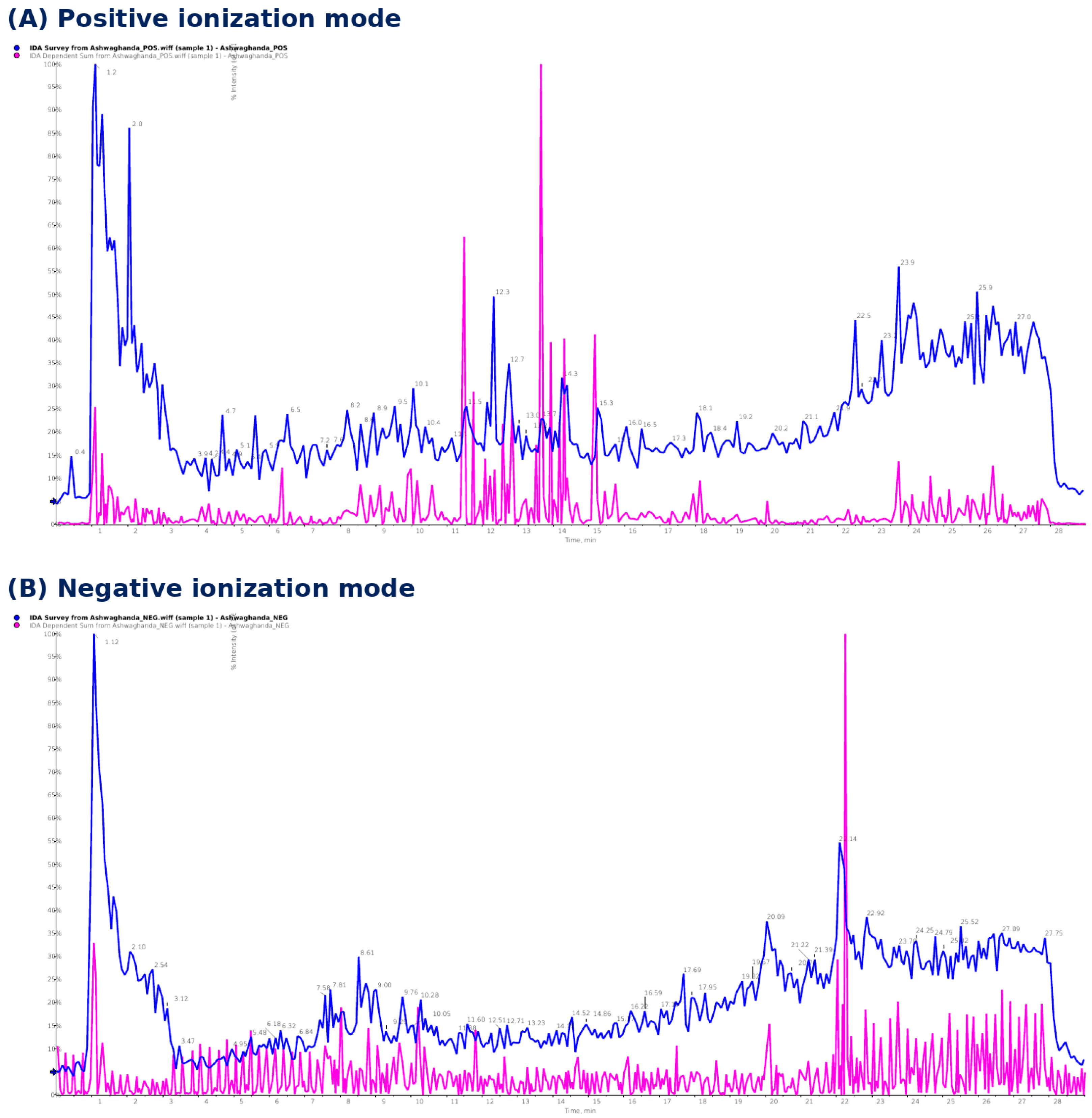
Figure 1. LC–MS/MS total ion chromatograms of the Withania somnifera extract. Representative chromatograms acquired in (A) positive-ion mode and (B) negative-ion mode. The blue trace represents the full-scan survey chromatogram obtained by information-dependent acquisition, whereas the magenta trace represents the summed MS/MS product-ion signal. The x-axis indicates retention time (min), and the y-axis indicates relative intensity as a percentage of the base peak. Seven major constituents were detected between 9.95 and 12.78 min. Six withanolide aglycones were identified as [M+H]+ ions in positive mode, while withanoside IV was detected as a [M−H] ion in negative mode. Chromatographic separation enabled the discrimination of the three isobaric compounds—12-deoxywithastramonolide, withaferin A, and withanone—each with a precursor ion at m/z 471. TIC, total ion chromatogram; IDA, information-dependent acquisition.
Ijms 27 06746 g001
Figure 2. (A) Particle size through DLS, (B) zeta potential, and (C) transmission electron microscopy analyses of Ws-SeNPs (scale 200 nm).
Ijms 27 06746 g002
Figure 3. FTIR spectrum of Ws-SeNPs with characteristic absorption bands of O-H, C-H, C=O/C=C, and C-O functional groups for reducing and stabilizing nanoparticles.
Ijms 27 06746 g003
Figure 4. Impact of Ws, selenium, and Ws-SeNPs on kidney weight indices in glycerol-induced ARF rats. Absolute kidney weight (A) and relative kidney weight (B). Data are expressed as mean ± SEM (n = 7). Statistical analysis was performed using one-way ANOVA followed by Tukey’s post hoc test. # vs. Control; $ vs. ARF; @ vs. ARF & Ws-SeNPs (p < 0.05).
Ijms 27 06746 g004
Figure 5. Impact of Ws, selenium, and Ws-SeNPs on rhabdomyolysis-associated muscle injury markers in glycerol-induced ARF rats. Serum creatine kinase (CK; (A)) and lactate dehydrogenase (LDH; (B)). Data are expressed as mean ± SEM (n = 7). Statistical analysis was performed using one-way ANOVA followed by Tukey’s post hoc test. # vs. Control; $ vs. ARF; @ vs. ARF & Ws-SeNPs (p < 0.05).
Ijms 27 06746 g005
Figure 6. Impact of Ws, selenium, and Ws-SeNPs on kidney function and tubular injury biomarkers in glycerol-induced ARF rats. Serum creatinine (A), urea (B), cystatin-C (C), plasma KIM-1 (D), and renal NGAL (E) levels. Data are expressed as mean ± SEM (n = 7). Statistical analysis was performed using one-way ANOVA followed by Tukey’s post hoc test. # vs. Control; $ vs. ARF; @ vs. ARF & Ws-SeNPs (p < 0.05).
Ijms 27 06746 g006
Figure 7. Impact of Ws, selenium, and Ws-SeNPs on renal oxidative stress and antioxidant defense markers in glycerol-induced ARF rats. Renal levels of MDA (A), NO (B), 4-HNE (C), Keap-1 (D), Nrf2 (E), GSH (F), GPx (G), SOD (H), and CAT (I). Data are expressed as mean ± SEM (n = 7). Statistical analysis was performed using one-way ANOVA followed by Tukey’s post hoc test. # vs. Control; $ vs. ARF; @ vs. ARF & Ws-SeNPs (p < 0.05).
Ijms 27 06746 g007
Figure 8. Impact of Ws, selenium, and Ws-SeNPs on renal inflammatory and pyroptosis-related markers in glycerol-induced ARF rats. Renal TNF-α protein level (A), TNF-α mRNA expression (B), NF-κB mRNA expression (C), NF-κB protein level (D), IL-1β (E), IL-10 (F), NLRP3 (G), and GSDMD (H). Data are expressed as mean ± SEM (n = 7). Statistical analysis was performed using one-way ANOVA followed by Tukey’s post hoc test. # vs. Control; $ vs. ARF; @ vs. ARF & Ws-SeNPs (p < 0.05).
Ijms 27 06746 g008
Figure 9. Impact of Ws, selenium, and Ws-SeNPs on renal mitochondrial regulatory markers in glycerol-induced ARF rats. Renal levels of SIRT1 (A), AMPK (B), PGC-1α (C), and MFN2 (D). Data are expressed as mean ± SEM (n = 7). Statistical analysis was performed using one-way ANOVA followed by Tukey’s post hoc test. # vs. Control; $ vs. ARF; @ vs. ARF & Ws-SeNPs (p < 0.05).
Ijms 27 06746 g009
Figure 10. Impact of Ws, selenium, and Ws-SeNPs on renal apoptotic markers in glycerol-induced ARF rats. Renal levels of Bax (A), Bcl-2 (B), and caspase-3 (C). Data are expressed as mean ± SEM (n = 7). Statistical analysis was performed using one-way ANOVA followed by Tukey’s post hoc test. # vs. Control; $ vs. ARF; @ vs. ARF & Ws-SeNPs (p < 0.05).
Ijms 27 06746 g010
Figure 11. Impact of Ws, selenium, and Ws-SeNPs on renal Nrf2 immunoexpression in glycerol-induced ARF rats. Representative photomicrographs of renal sections immunostained for Nrf2 showing marked cytoplasmic immunoreactivity in the Control group (A), ARF & Ws group (D), and ARF & Ws-SeNP group (E). In contrast, weak Nrf2 immunoreactivity was observed in the ARF group (B) and the ARF & selenium group (C). Positive Nrf2 staining is indicated by brown DAB cytoplasmic reactivity (arrow). DAB, ×400.
Ijms 27 06746 g011
Figure 12. Impact of Ws, selenium, and Ws-SeNPs on renal Bcl-2 immunoexpression in glycerol-induced ARF rats. Representative photomicrographs of renal sections immunostained for Bcl-2 showing moderate cytoplasmic immunoreactivity in the Control group (A) and the ARF & Ws-SeNP group (E), while the ARF & Ws group (D) exhibited mild Bcl-2 expression. In contrast, weak Bcl-2 immunoreactivity was observed in the ARF group (B) and the ARF & selenium group (C). Positive Bcl-2 staining is indicated by brown DAB cytoplasmic reactivity (arrow). DAB, ×400.
Ijms 27 06746 g012
Figure 13. Quantitative analysis of renal Nrf2 and Bcl-2 immunoreactivity in glycerol-induced ARF rats. Semi-quantitative assessment of Nrf2 immunoexpression (A) and Bcl-2 immunoexpression (B) in renal tissue sections from the different experimental groups. Data are expressed as mean ± SEM (n = 7). Statistical analysis was performed using one-way ANOVA followed by Tukey’s post hoc test. # vs. Control; $ vs. ARF (p < 0.05).
Ijms 27 06746 g013
Figure 14. Histopathological examination of gastrocnemius skeletal muscle in glycerol-induced ARF rats. Representative photomicrographs of H&E-stained gastrocnemius muscle sections from the different experimental groups. The Control group (A) showed normal skeletal muscle architecture with intact, regularly arranged muscle fibers (black arrow) surrounded by perimysium (*). The ARF group (B) exhibited marked myopathic alterations, including focal myofiber necrosis, hyalinization (star), and necrotic myocytes. The ARF & selenium group (C) showed persistent myocyte degeneration (black arrow) accompanied by inflammatory cell infiltration (red arrow). In contrast, the ARF & Ws group (D) and ARF & Ws-SeNP group (E) displayed a nearly preserved skeletal muscle pattern with marked attenuation of glycerol-induced myopathic changes. H&E, ×400.
Ijms 27 06746 g014
Figure 15. Histopathological examination of the renal cortex in glycerol-induced ARF rats. Representative photomicrographs of H&E-stained renal cortical sections from the different experimental groups. The Control group (A) showed normal renal cortical architecture with intact glomeruli and renal tubules. The ARF group (B) exhibited marked renal pathological alterations, including inflammatory cellular infiltration (*) and focal necrotic areas (star). The ARF & selenium group (C) showed persistent renal injury characterized by degeneration of tubular epithelial cells (short arrow) and glomerular degeneration (arrowhead). The ARF & Ws group (D) demonstrated improved renal cortical architecture with attenuation of tubular and glomerular damage. The ARF & Ws-SeNP group (E) showed a nearly normal histological pattern, indicating marked preservation of renal cortical structure against glycerol-induced injury. H&E, ×400.
Ijms 27 06746 g015
Figure 16. Schematic workflow for the extraction of Withania somnifera root extract, green synthesis of Withania somnifera–functionalized selenium nanoparticles (Ws-SeNPs), and their physicochemical characterization.
Ijms 27 06746 g016
Table 1. Constituents of Ashwagandha (Withania somnifera) identified by LC–MS/MS.
No.RT (min)Identified CompoundClassMolecular
Formula
MW
(g/mol)
AdductPrecursor
m/z (obs.)
Calc.
m/z
Major MS/MS Fragments (m/z)Ref.
110.02Ixocarpalactone AWithanolideC28H40O8504.61[M+H]+ (pos)506.1505.2796317.06, 299.07[21]
212.2612-DeoxywithastramonolideWithanolideC28H38O6470.60[M+H]+ (pos)471.1471.2741171.10, 280.97, 299.02, 399.07[21]
39.95Viscosalactone BWithanolideC28H40O7488.61[M+H]+ (pos)489.1489.2847281.00, 299.05, 317.01, 435.10[21]
410.97Dihydrowithaferin AWithanolideC28H40O6472.61[M+H]+ (pos)473.1473.2898282.99, 301.05[21]
511.76Withaferin AWithanolideC28H38O6470.60[M+H]+ (pos)471.1471.2741175.10, 280.95, 299.05[21,22]
612.78WithanoneWithanolideC28H38O6470.60[M+H]+ (pos)471.1471.2741262.98, 289.00[21]
710.08Withanoside IVWithanoside (glycoside)C40H62O15782.91[M−H]781.2781.4011763.09, 221.09[21,23]
Molecular formulas and calculated monoisotopic m/z values ([M+H]+ or [M–H]) are listed for the proposed structures; observed precursor and product-ion values are as measured. Identities were confirmed against reference standards where available and were otherwise proposed from accurate mass, fragmentation and the literature. RT, retention time.
Table 2. Primer sequences of genes analyzed by qRT-PCR.
Gene NameForward SequenceReverse Sequence
TNF-αACTGAACTTCGGGGT GATCGGCTTGGTGGTTTGCTACGAC
NF-κBGTCTCAAACCAAACAGCCTCACCAGTGTCTTCCTCGACATGGAT
GAPDHATGGTGAAGGTCGGTGTGAACGTGGTGAAGACGCCAGTAGACTC
Disclaimer/Publisher’s Note: The statements, opinions and data contained in all publications are solely those of the individual author(s) and contributor(s) and not of MDPI and/or the editor(s). MDPI and/or the editor(s) disclaim responsibility for any injury to people or property resulting from any ideas, methods, instructions or products referred to in the content.