Immature Neurons in the Postnatal Brain: Markers, Modulation, and Involvement in Normal and Aberrant Plasticity


Abstract
1. Introduction
2. Identification, Representation and Functional Significance of cINs
2.1. cIN Markers

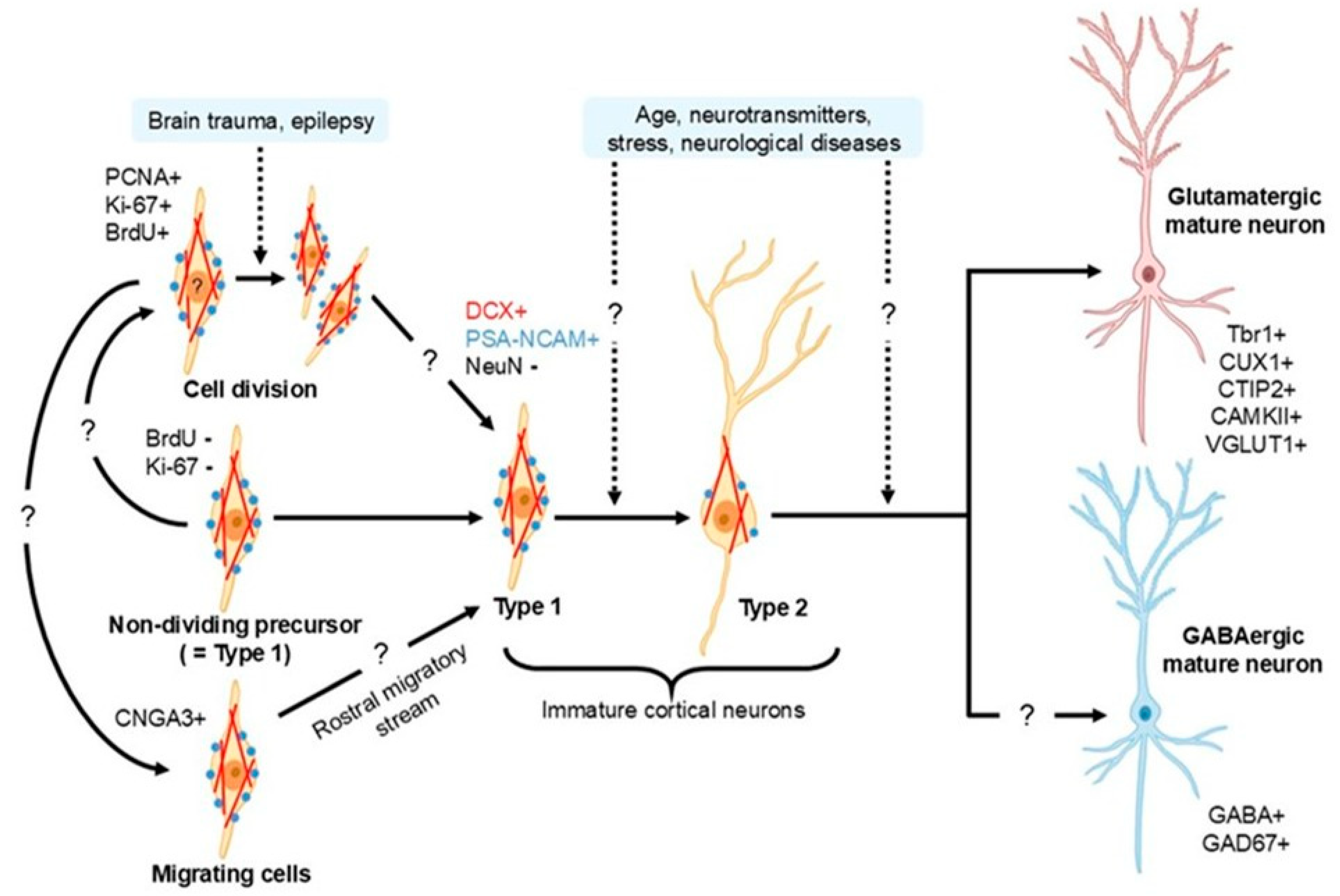
| Marker | Function/Annotation | Expression in cINs | Co-Expression with DCX | Species/Area | References |
|---|---|---|---|---|---|
| DCX | Microtubule-associated migration/plasticity | Positive | 100% | Neocortex (large-brain mammalian); paleocortex (rodents) | [35,45,58,80,81,82] |
| PSA-NCAM | Anti-adhesive molecule; synaptic insulation; delayed maturation | Positive | 94% | Human temporal cortex | [83] |
| Positive | 98% | Human temporal cortex (TLE) | [83] | ||
| Positive | 53% | Human neocortex | [36] | ||
| Positive | 60% | Mice piriform cortex | [32] | ||
| Positive | – | Cat neocortex | [84] | ||
| Positive | 100% | Guinea pig neocortex | [65] | ||
| NeuN | RNA-binding protein regulating neuronal differentiation and splicing/marker of mature neurons | Low | 9% | Human neocortex | [36] |
| Low | – | Human temporal cortex (TLE) | [58] | ||
| Emerges with maturation | – | Mice piriform cortex | [73] | ||
| Low | 19% | Mice piriform cortex | [32] | ||
| Low | 20% of PSA-NCAM cells | Cat cerebral cortex | [84] | ||
| Low | 14% of PSA-NCAM cells | Rat piriform cortex | [31] | ||
| Appears in type 2 cINs | – | Guinea pig neocortex | [65] | ||
| Emerges with maturation | – | Cat neocortex | [66] | ||
| Low | 17–18% | Rodents, cat and marmoset neocortex | [33] | ||
| MAP2 | Cytoskeletal protein playing a crucial role in dendritic development | Negative/low | Rat piriform cortex | [31] | |
| Ankyrin-G | Formation and maintenance of the axon initial segment; Marker of neuronal maturation | Negative | – | Human neocortex | [36] |
| Reelin | Coordination of neuron migration | Negative | – | Rodent piriform cortex; guinea pig neocortex | [65,85] |
| c-Fos | Immediate early gene | Negative | – | Rat piriform cortex | [31] |
| Arc | Immediate early gene | Negative | – | Rat piriform cortex | [31] |
| PV | Calcium-binding proteins | Negative | – | Human/cat/guinea pig neocortex | [65,66,83,84] |
| Positive | – | Cat cerebral cortex layer II | [66] | ||
| Calretinin | Calcium-binding proteins | Negative | – | Human/cat/guinea pig neocortex | [65,66,83,84] |
| Calbindin | Calcium-binding proteins | Negative | – | Human/cat/guinea pig neocortex | [65,66,83,84] |
| Positive | – | Cat neocortex | [66] | ||
| VGLUT1 | Excitatory neuron marker | Emerges with maturation | – | Human neocortex | [36] |
| CUX1 | Excitatory neuron marker | Positive | 73% | Human neocortex | [36] |
| CTIP2 | Excitatory neuron marker | Positive | 21% | Human neocortex layer II | [36] |
| TBR1 | Postmitotic excitatory neuron marker | Positive | 100% | Human neocortex | [36] |
| Positive | 96% of PSA-NCAM cells | Cat neocortex | [84] | ||
| Positive | – | Murine piriform cortex | [37] | ||
| Positive | – | Rodent piriform cortex; guinea pig and rabbit neocortex | [35] | ||
| GluN1 | Subunit of NMDA receptor; marker for glutamatergic signaling | Appears in type 2 of cINs | – | Human neocortex | [36] |
| Appears in type 2 of cINs | 92% of PSA-NCAM cells | Rat piriform cortex | [31] | ||
| Ng | Marker of cerebral principal neurons | Negative | – | Guinea pig neocortex | [65] |
| CAMKII | Marker of mature principal neurons | Negative | – | Cat neocortex; rat piriform cortex | [31,84] |
| Positive | 2–12% | Rat piriform cortex | [37] | ||
| GR | Indicator of stress hormone signaling | Negative | – | Rat piriform cortex | [31] |
| NPY | Marker of the corresponding population of interneurons | Negative | – | Cat neocortex; rat piriform cortex | [31,84] |
| CCK | Marker of the corresponding population of interneurons | Negative | – | Cat cerebral cortex layer II; rat piriform cortex layer II | [31,84] |
| SRIF | Marker of the corresponding population of interneurons | Negative | – | Cat neocortex; rat piriform cortex | [31,84] |
| VIP | Marker of the corresponding population of interneurons | Negative | – | Cat neocortex; rat piriform cortex | [31,84] |
| GAD67 | A key enzyme responsible for synthesizing GABA; marker of inhibitory interneurons | Negative | – | Human neocortex | [36] |
| Negative | – | Cat neocortex | [84] | ||
| Negative | – | Rat piriform cortex | [31] | ||
| Positive | – | Guinea pig neocortex | [86] | ||
| Emerges with maturation | – | Cat neocortex | [66] | ||
| Low | – | Guinea pig neocortex | [65] | ||
| GABA | Marker of GABAergic neurons | Low | – | Guinea pig neocortex | [65] |
| Positive | – | Rhesus monkeys | [46] | ||
| Emerges with maturation | – | Cat neocortex | [66] | ||
| SOX2 | Stem/progenitor marker | Low | – | Human temporal cortex (TLE) | [58] |
| CD68 | Microglia marker | Positive | – | Human temporal cortex (TLE) | [58] |
| CD34 | Marker for hematopoietic stem and progenitor cells | Negative | – | Human temporal cortex (TLE) | [58] |
| GFAP | Marker for astrocytes | Very low | 2.7% | Human neocortex | [36] |
| Negative | – | Guinea pig neocortex | [65] | ||
| Positive | 26% | Non-human primate (Chlorocebus) neocortex | [81] | ||
| Negative | – | Human temporal cortex (TLE) | [58] | ||
| Negative | – | Rat piriform cortex | [31] | ||
| PDGFRβ | A receptor of tyrosine kinase involved in cell proliferation, migration, differentiation, and survival | Positive | – | Human temporal cortex (TLE) | [58] |
| OLIG2 | Marker for oligodendrocyte precursors and motor neuron progenitors | Low | – | Human white matter of temporal cortex (TLE) | [58] |
| IBA1 | Marker for microglia and macrophages | Negative | – | Human neocortex layer II | [36] |
| Positive | – | Human temporal cortex layer II (TLE) | [58] | ||
| Nestin | Marker for neural stem and progenitor cells | Negative | – | Human temporal cortex layer II (TLE) | [58] |
| Positive | – | Rat piriform cortex | [87] | ||
| BrdU | Cell birth dating method; proliferative marker | Negative at adult labeling | – | Cat/guinea pig neocortex; rodent piriform cortex | [31,32,33,84,87,88] |
| Positive | – | Rhesus monkey temporal cortex | [89] | ||
| Positive | – | Guinea pig neocortex | [86] | ||
| Tuj1 | Marker of immature neurons | Positive | 43% | Human temporal cortex | [83] |
| Positive | 82% | Human temporal cortex (TLE) | [83] | ||
| Positive | 70% | Human neocortex (stroke) | [90] | ||
| Positive | – | Guinea pig neocortex | [86] | ||
| CNGA-3 | Marker for cells of the rostral migratory stream | Positive | 98% of PSA-NCAM cells | Rat piriform cortex | [31] |
| p-CREB | Participates in the survival, differentiation, and maturation of newborn neurons | Positive | 95% of PSA-NCAM cells | Rat piriform cortex | [31] |
| ßIV-spectrin | Marker for axon initial segments | Positive | – | Mice piriform cortex | [79] |
| Caspase 3 | Caspase-3 orchestrates execution-phase apoptosis events | Negative | – | Guinea pig neocortex | [91] |
| nNOS | Marker of nitrergic neurons | Positive | – | Cat neocortex | [66] |
| Low | – | Guinea pig neocortex | [65] | ||
| Negative | – | Cat neocortex | [84] | ||
| NADPH-d | Marker of nitrergic neurons | Positive | – | Cat neocortex | [66] |
| Low | – | Guinea pig neocortex | [65] | ||
| MCM2 | Proliferative marker | Negative | – | Human temporal cortex (TLE) | [58] |
| PCNA | Proliferative marker | Positive | <6% | Human temporal cortex | [83] |
| Positive | 64% | Human temporal cortex (TLE) | [83] | ||
| Ki-67 | Proliferative marker | Very low | – | Human neocortex | [92] |
| Positive | – | Human neocortex (TBI) | [92] | ||
| Negative | – | Human neocortex (stroke) | [90] | ||
| Negative | Rabbit and sheep neocortex | [33] | |||
| Positive | – | CD1 mice piriform cortex | [54] | ||
| TUC4 | Marker of newborn and immature neurons | Positive | 69% of PSA-NCAM cells | Rat piriform cortex | [31] |
| NG2 | Marker for oligodendrocyte precursor cells | Negative | – | Human neocortex | [36] |
| Low | – | Rat piriform cortex | [31] |
2.2. cINs Morphology
2.3. cIN Origin
2.4. cIN Differentiation
3. Factors Modulating cINs Phenotype
3.1. Neurotransmitter Modulation and Pharmacological Effects
3.2. Stress-Related Factors
3.3. Sensory Stimulation/Deprivation
3.4. Developmental and Age-Related Factors
4. Neurological Diseases
5. Open Questions and Future Perspectives
6. “Neurogenesis Without Division”: Concluding Remarks
Author Contributions
Funding
Institutional Review Board Statement
Informed Consent Statement
Data Availability Statement
Conflicts of Interest
References
- Altman, J. Are New Neurons Formed in the Brains of Adult Mammals? Science 1962, 135, 1127–1128. [Google Scholar] [CrossRef] [PubMed]
- Altman, J. Autoradiographic investigation of cell proliferation in the brains of rats and cats. Anat. Rec. 1963, 145, 573–591. [Google Scholar] [CrossRef] [PubMed]
- Altman, J.; Das, G.D. Autoradiographic and histological studies of postnatal neurogenesis. I. A longitudinal investigation of the kinetics, migration and transformation of cells incoorporating tritiated thymidine in neonate rats, with special reference to postnatal neurogenesis in some brain regions. J. Comp. Neurol. 1966, 126, 337–389. [Google Scholar] [CrossRef] [PubMed]
- Bengzon, J.; Kokaia, Z.; Elmér, E.; Nanobashvili, A.; Kokaia, M.; Lindvall, O. Apoptosis and proliferation of dentate gyrus neurons after single and intermittent limbic seizures. Proc. Natl. Acad. Sci. USA 1997, 94, 10432–10437. [Google Scholar] [CrossRef] [PubMed]
- Kempermann, G.; Kuhn, H.G.; Gage, F.H. More hippocampal neurons in adult mice living in an enriched environment. Nature 1997, 386, 493–495. [Google Scholar] [CrossRef] [PubMed]
- van Praag, H.; Christie, B.R.; Sejnowski, T.J.; Gage, F.H. Running enhances neurogenesis, learning, and long-term potentiation in mice. Proc. Natl. Acad. Sci. USA 1999, 96, 13427–13431. [Google Scholar] [CrossRef] [PubMed]
- van Praag, H.; Kempermann, G.; Gage, F.H. Running increases cell proliferation and neurogenesis in the adult mouse dentate gyrus. Nat. Neurosci. 1999, 2, 266–270. [Google Scholar] [CrossRef] [PubMed]
- Parent, J.M. The role of seizure-induced neurogenesis in epileptogenesis and brain repair. Epilepsy Res. 2002, 50, 179–189. [Google Scholar] [CrossRef] [PubMed]
- Nygren, J.; Wieloch, T.; Pesic, J.; Brundin, P.; Deierborg, T. Enriched Environment Attenuates Cell Genesis in Subventricular Zone After Focal Ischemia in Mice and Decreases Migration of Newborn Cells to the Striatum. Stroke 2006, 37, 2824–2829. [Google Scholar] [CrossRef] [PubMed]
- Rola, R.; Mizumatsu, S.; Otsuka, S.; Morhardt, D.R.; Noble-Haeusslein, L.J.; Fishman, K.; Potts, M.B.; Fike, J.R. Alterations in hippocampal neurogenesis following traumatic brain injury in mice. Exp. Neurol. 2006, 202, 189–199. [Google Scholar] [CrossRef] [PubMed]
- Scharfman, H.E. Review: Epilepsy as an Example of Neural Plasticity. Neuroscientist 2002, 8, 154–173. [Google Scholar] [CrossRef] [PubMed]
- Michael, R.D.; Rene, H. Adult Hippocampal Neurogenesis as Target for the Treatment of Depression. CNS Neurol. Disord.-Drug Targets 2007, 6, 205–218. [Google Scholar] [CrossRef] [PubMed]
- Gould, E.; McEwen, B.S.; Tanapat, P.; Galea, L.A.M.; Fuchs, E. Neurogenesis in the Dentate Gyrus of the Adult Tree Shrew Is Regulated by Psychosocial Stress and NMDA Receptor Activation. J. Neurosci. 1997, 17, 2492–2498. [Google Scholar] [CrossRef] [PubMed]
- Brummelte, S.; Galea, L.A.M. Chronic high corticosterone reduces neurogenesis in the dentate gyrus of adult male and female rats. Neuroscience 2010, 168, 680–690. [Google Scholar] [CrossRef] [PubMed]
- Malberg, J.E.; Eisch, A.J.; Nestler, E.J.; Duman, R.S. Chronic Antidepressant Treatment Increases Neurogenesis in Adult Rat Hippocampus. J. Neurosci. 2000, 20, 9104–9110. [Google Scholar] [CrossRef] [PubMed]
- Lagace, D.C.; Whitman, M.C.; Noonan, M.A.; Ables, J.L.; DeCarolis, N.A.; Arguello, A.A.; Donovan, M.H.; Fischer, S.J.; Farnbauch, L.A.; Beech, R.D.; et al. Dynamic Contribution of Nestin-Expressing Stem Cells to Adult Neurogenesis. J. Neurosci. 2007, 27, 12623–12629. [Google Scholar] [CrossRef] [PubMed]
- Green, A.D.; Galea, L.A.M. Adult hippocampal cell proliferation is suppressed with estrogen withdrawal after a hormone-simulated pregnancy. Horm. Behav. 2008, 54, 203–211. [Google Scholar] [CrossRef] [PubMed]
- Olson, A.K.; Eadie, B.D.; Ernst, C.; Christie, B.R. Environmental enrichment and voluntary exercise massively increase neurogenesis in the adult hippocampus via dissociable pathways. Hippocampus 2006, 16, 250–260. [Google Scholar] [CrossRef] [PubMed]
- Couillard-Despres, S.; Wuertinger, C.; Kandasamy, M.; Caioni, M.; Stadler, K.; Aigner, R.; Bogdahn, U.; Aigner, L. Ageing abolishes the effects of fluoxetine on neurogenesis. Mol. Psychiatry 2009, 14, 856–864. [Google Scholar] [CrossRef] [PubMed]
- Sorrells, S.F.; Paredes, M.F.; Cebrian-Silla, A.; Sandoval, K.; Qi, D.; Kelley, K.W.; James, D.; Mayer, S.; Chang, J.; Auguste, K.I.; et al. Human hippocampal neurogenesis drops sharply in children to undetectable levels in adults. Nature 2018, 555, 377–381. [Google Scholar] [CrossRef] [PubMed]
- Patzke, N.; Spocter, M.A.; Karlsson, K.Æ.; Bertelsen, M.F.; Haagensen, M.; Chawana, R.; Streicher, S.; Kaswera, C.; Gilissen, E.; Alagaili, A.N.; et al. In contrast to many other mammals, cetaceans have relatively small hippocampi that appear to lack adult neurogenesis. Brain Struct. Funct. 2015, 220, 361–383. [Google Scholar] [CrossRef] [PubMed]
- Bonfanti, L.; La Rosa, C.; Ghibaudi, M.; Sherwood, C.C. Adult neurogenesis and “immature” neurons in mammals: An evolutionary trade-off in plasticity? Brain Struct. Funct. 2023, 229, 1775–1793. [Google Scholar] [CrossRef] [PubMed]
- Bonfanti, L.; Seki, T. The PSA-NCAM-Positive “Immature” Neurons: An Old Discovery Providing New Vistas on Brain Structural Plasticity. Cells 2021, 10, 2542. [Google Scholar] [CrossRef] [PubMed]
- Benedetti, B.; Couillard-Despres, S. Why Would the Brain Need Dormant Neuronal Precursors? Front. Neurosci. 2022, 16, 877167. [Google Scholar] [CrossRef] [PubMed]
- La Rosa, C.; Ghibaudi, M.; Bonfanti, L. Newly Generated and Non-Newly Generated “Immature” Neurons in the Mammalian Brain: A Possible Reservoir of Young Cells to Prevent Brain Aging and Disease? J. Clin. Med. 2019, 8, 685. [Google Scholar] [CrossRef] [PubMed]
- Ghibaudi, M.; Bonfanti, L. How Widespread Are the “Young” Neurons of the Mammalian Brain? Front. Neurosci. 2022, 16, 918616. [Google Scholar] [CrossRef] [PubMed]
- Page, C.E.; Biagiotti, S.W.; Alderman, P.J.; Sorrells, S.F. Immature excitatory neurons in the amygdala come of age during puberty. Dev. Cogn. Neurosci. 2022, 56, 101133. [Google Scholar] [CrossRef] [PubMed]
- Ghibaudi, M.; Marchetti, N.; Vergnano, E.; La Rosa, C.; Benedetti, B.; Couillard-Despres, S.; Farioli-Vecchioli, S.; Bonfanti, L. Age-related changes in layer II immature neurons of the murine piriform cortex. Front. Cell. Neurosci. 2023, 17, 1205173. [Google Scholar] [CrossRef] [PubMed]
- Simard, S.; Rahimian, R.; Davoli, M.A.; Théberge, S.; Matosin, N.; Turecki, G.; Nagy, C.; Mechawar, N. Spatial transcriptomic analysis of adult hippocampal neurogenesis in the human brain. J. Psychiatry Neurosci. 2024, 49, E319–E333. [Google Scholar] [CrossRef] [PubMed]
- Alonso, M.; Petit, A.-C.; Lledo, P.-M. The impact of adult neurogenesis on affective functions: Of mice and men. Mol. Psychiatry 2024, 29, 2527–2542. [Google Scholar] [CrossRef] [PubMed]
- Gómez-Climent, M.Á.; Castillo-Gómez, E.; Varea, E.; Guirado, R.; Blasco-Ibáñez, J.M.; Crespo, C.; Martínez-Guijarro, F.J.; Nácher, J. A Population of Prenatally Generated Cells in the Rat Paleocortex Maintains an Immature Neuronal Phenotype into Adulthood. Cereb. Cortex 2008, 18, 2229–2240. [Google Scholar] [CrossRef] [PubMed]
- Rubio, A.; Belles, M.; Belenguer, G.; Vidueira, S.; Fariñas, I.; Nacher, J. Characterization and isolation of immature neurons of the adult mouse piriform cortex. Dev. Neurobiol. 2016, 76, 748–763. [Google Scholar] [CrossRef] [PubMed]
- La Rosa, C.; Cavallo, F.; Pecora, A.; Chincarini, M.; Ala, U.; Faulkes, C.G.; Nacher, J.; Cozzi, B.; Sherwood, C.C.; Amrein, I.; et al. Phylogenetic variation in cortical layer II immature neuron reservoir of mammals. eLife 2020, 9, e55456. [Google Scholar] [CrossRef] [PubMed]
- Márquez-Valadez, B.; Gallardo-Caballero, M.; Llorens-Martín, M. Human adult hippocampal neurogenesis is shaped by neuropsychiatric disorders, demographics, and lifestyle-related factors. Cell Stem Cell 2025, 32, 1577–1594.e5. [Google Scholar] [CrossRef] [PubMed]
- Luzzati, F.; Bonfanti, L.; Fasolo, A.; Peretto, P. DCX and PSA-NCAM Expression Identifies a Population of Neurons Preferentially Distributed in Associative Areas of Different Pallial Derivatives and Vertebrate Species. Cereb. Cortex 2008, 19, 1028–1041. [Google Scholar] [CrossRef] [PubMed]
- Coviello, S.; Gramuntell, Y.; Klimczak, P.; Varea, E.; Blasco-Ibañez, J.M.; Crespo, C.; Gutierrez, A.; Nacher, J. Phenotype and Distribution of Immature Neurons in the Human Cerebral Cortex Layer II. Front. Neuroanat. 2022, 16, 851432. [Google Scholar] [CrossRef] [PubMed]
- Rotheneichner, P.; Belles, M.; Benedetti, B.; König, R.; Dannehl, D.; Kreutzer, C.; Zaunmair, P.; Engelhardt, M.; Aigner, L.; Nacher, J.; et al. Cellular Plasticity in the Adult Murine Piriform Cortex: Continuous Maturation of Dormant Precursors Into Excitatory Neurons. Cereb. Cortex 2018, 28, 2610–2621. [Google Scholar] [CrossRef] [PubMed]
- Zhao, C.; Teng, E.M.; Summers, R.G.; Ming, G.-l.; Gage, F.H. Distinct Morphological Stages of Dentate Granule Neuron Maturation in the Adult Mouse Hippocampus. J. Neurosci. 2006, 26, 3–11. [Google Scholar] [CrossRef] [PubMed]
- Nagayama, S.; Homma, R.; Imamura, F. Neuronal organization of olfactory bulb circuits. Front. Neural Circuits 2014, 8, 98. [Google Scholar] [CrossRef] [PubMed]
- König, R.; Benedetti, B.; Rotheneichner, P.; O’ Sullivan, A.; Kreutzer, C.; Belles, M.; Nacher, J.; Weiger, T.M.; Aigner, L.; Couillard-Després, S. Distribution and fate of DCX/PSA-NCAM expressing cells in the adult mammalian cortex: A local reservoir for adult cortical neuroplasticity? Front. Biol. 2016, 11, 193–213. [Google Scholar] [CrossRef]
- Liang, Z.; Jin, N.; Guo, W. Neural stem cell heterogeneity in adult hippocampus. Cell Regen. 2025, 14, 6. [Google Scholar] [CrossRef] [PubMed]
- Ai, J.-Q.; Luo, R.; Tu, T.; Yang, C.; Jiang, J.; Zhang, B.; Bi, R.; Tu, E.; Yao, Y.-G.; Yan, X.-X. Doublecortin-Expressing Neurons in Chinese Tree Shrew Forebrain Exhibit Mixed Rodent and Primate-Like Topographic Characteristics. Front. Neuroanat. 2021, 15, 727883. [Google Scholar] [CrossRef] [PubMed]
- Torrijos-Saiz, L.I.; Freixes, J.; Desfilis, E.; Medina, L.; Sawamoto, K.; García-Verdugo, J.M.; Herranz-Pérez, V. Cellular Organization and Migration Pathways of the Ventricular–Subventricular Zone in the Juvenile Swine Brain (Sus scrofa domesticus). J. Comp. Neurol. 2025, 533, e70070. [Google Scholar] [CrossRef] [PubMed]
- Ghibaudi, M.; Zanone, A.; Bonfanti, L. Brain structural plasticity in large-brained mammals: Not only narrowing roads. Neural Regen. Res. 2026, 21, 1669–1680. [Google Scholar] [CrossRef] [PubMed]
- Pattaro, A.; Ghibaudi, M.; Corrente, C.; Telitsyn, N.; Graic, J.-M.; Aresu, L.; Sherwood, C.C.; Bonfanti, L. Phylogenetic variation of layer II cortical immature neurons in dog and horse confirms covariance with brain size and neocortical surface. Brain Struct. Funct. 2025, 230, 115. [Google Scholar] [CrossRef] [PubMed]
- Zhang, X.-m.; Cai, Y.; Chu, Y.; Chen, E.-Y.; Feng, J.-C.; Luo, X.-G.; Xiong, K.; Struble, R.G.; Clough, R.W.; Patrylo, P.R.; et al. Doublecortin-expressing cells persist in the associative cerebral cortex and amygdala in aged nonhuman primates. Front. Neuroanat. 2009, 3, 2009. [Google Scholar] [CrossRef] [PubMed]
- Li, Y.-N.; Hu, D.-D.; Cai, X.-L.; Wang, Y.; Yang, C.; Jiang, J.; Zhang, Q.-L.; Tu, T.; Wang, X.-S.; Wang, H.; et al. Doublecortin-Expressing Neurons in Human Cerebral Cortex Layer II and Amygdala from Infancy to 100 Years Old. Mol. Neurobiol. 2023, 60, 3464–3485. [Google Scholar] [CrossRef] [PubMed]
- Chen, P.; Guo, Z.; Zhou, B. Insight into the role of adult hippocampal neurogenesis in aging and Alzheimer’s disease. Ageing Res. Rev. 2023, 84, 101828. [Google Scholar] [CrossRef] [PubMed]
- Lupo, G. Adult neurogenesis and aging mechanisms: A collection of insights. Sci. Rep. 2023, 13, 18104. [Google Scholar] [CrossRef] [PubMed]
- Gage, F.H. Adult Neurogenesis in the Human Dentate Gyrus. Hippocampus 2025, 35, e23655. [Google Scholar] [CrossRef] [PubMed]
- Sakelaris, B.; Riecke, H. Adult neurogenesis reconciles flexibility and stability of olfactory perceptual memory. eLife 2025, 14, RP104443. [Google Scholar] [CrossRef] [PubMed]
- Coviello, S.; Gramuntell, Y.; Castillo-Gomez, E.; Nacher, J. Effects of Dopamine on the Immature Neurons of the Adult Rat Piriform Cortex. Front. Neurosci. 2020, 14, 574234. [Google Scholar] [CrossRef] [PubMed]
- Nacher, J.; Pham, K.; Gil-Fernandez, V.; McEwen, B.S. Chronic restraint stress and chronic corticosterone treatment modulate differentially the expression of molecules related to structural plasticity in the adult rat piriform cortex. Neuroscience 2004, 126, 503–509. [Google Scholar] [CrossRef] [PubMed]
- Lopatina, O.; Panina, Y.A.; Komleva, Y.K.; Salmina, A. Olfactory Stimuli Affect Establishment of Behavioral Phenotype and Fate of Immature Neurons in the Developing Piriform Cortex of CD1 Mice. Opera Medica Physiol. 2022, 9, 110–125. [Google Scholar] [CrossRef]
- Naffaa, M.M. Neurogenesis dynamics in the olfactory bulb: Deciphering circuitry organization, function, and adaptive plasticity. Neural Regen. Res. 2025, 20, 1565–1581. [Google Scholar] [CrossRef] [PubMed]
- Wang, K.; Liu, X.-Y.; Liu, S.-F.; Wang, X.-X.; Wei, Y.-H.; Zhu, J.-R.; Liu, J.; Xu, X.Q.; Wen, L. Rbm24/Notch1 signaling regulates adult neurogenesis in the subventricular zone and mediates Parkinson-associated olfactory dysfunction. Theranostics 2024, 14, 4499–4518. [Google Scholar] [CrossRef] [PubMed]
- Khalil, M.H. The BDNF-Interactive Model for Sustainable Hippocampal Neurogenesis in Humans: Synergistic Effects of Environmentally-Mediated Physical Activity, Cognitive Stimulation, and Mindfulness. Int. J. Mol. Sci. 2024, 25, 12924. [Google Scholar] [CrossRef] [PubMed]
- Liu, J.Y.W.; Matarin, M.; Reeves, C.; McEvoy, A.W.; Miserocchi, A.; Thompson, P.; Sisodiya, S.M.; Thom, M. Doublecortin-expressing cell types in temporal lobe epilepsy. Acta Neuropathol. Commun. 2018, 6, 60. [Google Scholar] [CrossRef] [PubMed]
- Srikandarajah, N.; Martinian, L.; Sisodiya, S.M.; Squier, W.; Blumcke, I.; Aronica, E.; Thom, M. Doublecortin expression in focal cortical dysplasia in epilepsy. Epilepsia 2009, 50, 2619–2628. [Google Scholar] [CrossRef] [PubMed]
- Wu, A.; Zhang, J. Neuroinflammation, memory, and depression: New approaches to hippocampal neurogenesis. J. Neuroinflamm. 2023, 20, 283. [Google Scholar] [CrossRef] [PubMed]
- Zhang, K.; Wang, F.; Zhai, M.; He, M.; Hu, Y.; Feng, L.; Li, Y.; Yang, J.; Wu, C. Hyperactive neuronal autophagy depletes BDNF and impairs adult hippocampal neurogenesis in a corticosterone-induced mouse model of depression. Theranostics 2023, 13, 1059–1075. [Google Scholar] [CrossRef] [PubMed]
- Rafiei, A.; Cruz Tetlalmatzi, S.; Edrington, C.H.; Lee, L.; Crowder, D.A.; Saltzberg, D.J.; Sali, A.; Brouhard, G.; Schriemer, D.C. Doublecortin engages the microtubule lattice through a cooperative binding mode involving its C-terminal domain. elife 2022, 11, e66975. [Google Scholar] [CrossRef] [PubMed]
- Dema, A.; Charafeddine, R.A.; van Haren, J.; Rahgozar, S.; Viola, G.; Jacobs, K.A.; Kutys, M.L.; Wittmann, T. Doublecortin reinforces microtubules to promote growth cone advance in soft environments. Curr. Biol. 2024, 34, 5822–5832.e5. [Google Scholar] [CrossRef] [PubMed]
- Abellán-Álvaro, M.; Teruel-Sanchis, A.; Madeira, M.F.; Lanuza, E.; Santos, M.; Agustín-Pavón, C. Doublecortin-immunoreactive neurons in the piriform cortex are sensitive to the long lasting effects of early life stress. Front. Neurosci. 2024, 18, 1446912. [Google Scholar] [CrossRef] [PubMed]
- Xiong, K.; Luo, D.-W.; Patrylo, P.R.; Luo, X.-G.; Struble, R.G.; Clough, R.W.; Yan, X.-X. Doublecortin-expressing cells are present in layer II across the adult guinea pig cerebral cortex: Partial colocalization with mature interneuron markers. Exp. Neurol. 2008, 211, 271–282. [Google Scholar] [CrossRef] [PubMed]
- Cai, Y.; Xiong, K.; Chu, Y.; Luo, D.-W.; Luo, X.-G.; Yuan, X.-Y.; Struble, R.G.; Clough, R.W.; Spencer, D.D.; Williamson, A.; et al. Doublecortin expression in adult cat and primate cerebral cortex relates to immature neurons that develop into GABAergic subgroups. Exp. Neurol. 2009, 216, 342–356. [Google Scholar] [CrossRef] [PubMed]
- Micheli, L.; Caruso, M.; D’Andrea, G.; Volpe, D.; Ceccarelli, M.; Tirone, F. Survey of transcriptome analyses of hippocampal neurogenesis with focus on adult dentate gyrus stem cells. Front. Cell Dev. Biol. 2025, 13, 1605116. [Google Scholar] [CrossRef] [PubMed]
- Ghibaudi, M.; Amenta, A.; Agosti, M.; Riva, M.; Graïc, J.-M.; Bifari, F.; Bonfanti, L. Consistency and Variation in Doublecortin and Ki67 Antigen Detection in the Brain Tissue of Different Mammals, including Humans. Int. J. Mol. Sci. 2023, 24, 2514. [Google Scholar] [CrossRef] [PubMed]
- Duncan, B.W.; Murphy, K.E.; Maness, P.F. Molecular Mechanisms of L1 and NCAM Adhesion Molecules in Synaptic Pruning, Plasticity, and Stabilization. Front. Cell Dev. Biol. 2021, 9, 625340. [Google Scholar] [CrossRef] [PubMed]
- Sato, C.; Kitajima, K. Polysialylation and disease. Mol. Asp. Med. 2021, 79, 100892. [Google Scholar] [CrossRef] [PubMed]
- Conchonaud, F.; Nicolas, S.; Amoureux, M.-C.; Ménager, C.; Marguet, D.; Lenne, P.-F.; Rougon, G.; Matarazzo, V. Polysialylation Increases Lateral Diffusion of Neural Cell Adhesion Molecule in the Cell Membrane. J. Biol. Chem. 2007, 282, 26266–26274. [Google Scholar] [CrossRef] [PubMed]
- Nacher, J.; Alonso-Llosa, G.; Rosell, D.; McEwen, B. PSA-NCAM expression in the piriform cortex of the adult rat. Modulation by NMDA receptor antagonist administration. Brain Res. 2002, 927, 111–121. [Google Scholar] [CrossRef] [PubMed]
- Coviello, S.; Benedetti, B.; Jakubecova, D.; Belles, M.; Klimczak, P.; Gramuntell, Y.; Couillard-Despres, S.; Nacher, J. PSA Depletion Induces the Differentiation of Immature Neurons in the Piriform Cortex of Adult Mice. Int. J. Mol. Sci. 2021, 22, 5733. [Google Scholar] [CrossRef] [PubMed]
- Rodríguez, J.J.; Gardenal, E.; Zallo, F.; Cabot, J.; Busquets, X. Early PSA-NCAM reduction in the dentate gyrus and impaired plasticity in the Alzheimer’s disease 3xTg-mice model. Acta Histochem. 2024, 126, 152194. [Google Scholar] [CrossRef] [PubMed]
- Gascon, E.; Vutskits, L.; Jenny, B.; Durbec, P.; Kiss, J.Z. PSA-NCAM in postnatally generated immature neurons of the olfactory bulb:a crucial role in regulating p75 expression and cell survival. Development 2007, 134, 1181–1190. [Google Scholar] [CrossRef] [PubMed][Green Version]
- Massaro, A.R.; Sbriccoli, A.; Tonali, P. Reactive astrocytes within the acute plaques of multiple sclerosis are PSA-NCAM positive. Neurol. Sci. 2002, 23, 255–256. [Google Scholar] [CrossRef] [PubMed]
- Chareyron, L.J.; Amaral, D.G.; Lavenex, P. Selective lesion of the hippocampus increases the differentiation of immature neurons in the monkey amygdala. Proc. Natl. Acad. Sci. USA 2016, 113, 14420–14425. [Google Scholar] [CrossRef] [PubMed]
- Sorrells, S.F.; Paredes, M.F.; Velmeshev, D.; Herranz-Pérez, V.; Sandoval, K.; Mayer, S.; Chang, E.F.; Insausti, R.; Kriegstein, A.R.; Rubenstein, J.L.; et al. Immature excitatory neurons develop during adolescence in the human amygdala. Nat. Commun. 2019, 10, 2748. [Google Scholar] [CrossRef] [PubMed]
- Benedetti, B.; Dannehl, D.; König, R.; Coviello, S.; Kreutzer, C.; Zaunmair, P.; Jakubecova, D.; Weiger, T.M.; Aigner, L.; Nacher, J.; et al. Functional Integration of Neuronal Precursors in the Adult Murine Piriform Cortex. Cereb. Cortex 2019, 30, 1499–1515. [Google Scholar] [CrossRef] [PubMed]
- Bai, J.; Ramos, R.L.; Paramasivam, M.; Siddiqi, F.; Ackman, J.B.; LoTurco, J.J. The Role of DCX and LIS1 in Migration through the Lateral Cortical Stream of Developing Forebrain. Dev. Neurosci. 2007, 30, 144–156. [Google Scholar] [CrossRef] [PubMed]
- Bloch, J.; Kaeser, M.; Sadeghi, Y.; Rouiller, E.M.; Redmond, D.E., Jr.; Brunet, J.-F. Doublecortin-positive cells in the adult primate cerebral cortex and possible role in brain plasticity and development. J. Comp. Neurol. 2011, 519, 775–789. [Google Scholar] [CrossRef] [PubMed]
- De Nevi, E.; Marco-Salazar, P.; Fondevila, D.; Blasco, E.; Pérez, L.; Pumarola, M. Immunohistochemical study of doublecortin and nucleostemin in canine brain. Eur. J. Histochem. 2013, 57, e9. [Google Scholar] [CrossRef] [PubMed][Green Version]
- Liu, Y.W.J.; Curtis, M.A.; Gibbons, H.M.; Mee, E.W.; Bergin, P.S.; Teoh, H.H.; Connor, B.; Dragunow, M.; Faull, R.L.M. Doublecortin expression in the normal and epileptic adult human brain. Eur. J. Neurosci. 2008, 28, 2254–2265. [Google Scholar] [CrossRef] [PubMed]
- Varea, E.; Belles, M.; Vidueira, S.; Blasco-Ibañez, J.M.; Crespo, C.; Pastor, A.M.; Nacher, J. PSA-NCAM is Expressed in Immature, but not Recently Generated, Neurons in the Adult Cat Cerebral Cortex Layer II. Front. Neurosci. 2011, 5, 9449. [Google Scholar] [CrossRef] [PubMed]
- Carceller, H.; Rovira-Esteban, L.; Nacher, J.; Castrén, E.; Guirado, R. Neurochemical Phenotype of Reelin Immunoreactive Cells in the Piriform Cortex Layer II. Front. Cell. Neurosci. 2016, 10, 65. [Google Scholar] [CrossRef] [PubMed]
- Clough, R.W.; Xiong, K.; Luo, X.-G.; Patrylo, P.R.; Struble, R.G.; Yan, X.-X. Intrinsic neurogenesis in the cerebral cortex continues in adult guinea pig and gives rise to new GABAergic neurons. FASEB J. 2008, 22, 974.4. [Google Scholar] [CrossRef]
- Vadodaria, K.C.; Yanpallewar, S.U.; Vadhvani, M.; Toshniwal, D.; Liles, L.C.; Rommelfanger, K.S.; Weinshenker, D.; Vaidya, V.A. Noradrenergic regulation of plasticity marker expression in the adult rodent piriform cortex. Neurosci. Lett. 2017, 644, 76–82. [Google Scholar] [CrossRef] [PubMed]
- Yang, Y.; Xie, M.-X.; Li, J.-M.; Hu, X.; Patrylo, P.; Luo, X.-G.; Cai, Y.; Li, Z.; Yan, X.-X. Prenatal genesis of layer II doublecortin expressing neurons in neonatal and young adult guinea pig cerebral cortex. Front. Neuroanat. 2015, 9, 109. [Google Scholar] [CrossRef] [PubMed]
- Heyworth, N.; Rosene, D.L. Immature Neurons in the Temporal Cortex of the Aging Rhesus Monkey. FASEB J. 2013, 27, 533.13. [Google Scholar] [CrossRef]
- Hua, R.; Doucette, R.; Walz, W. Doublecortin-expressing cells in the ischemic penumbra of a small-vessel stroke. J. Neurosci. Res. 2008, 86, 883–893. [Google Scholar] [CrossRef] [PubMed]
- Huang, J.; Huang, K.; Shang, L.; Wang, H.; Zhang, M.; Fan, C.-L.; Chen, D.; Yan, X.; Xiong, K. Chronic lead exposure reduces doublecortin-expressing immature neurons in young adult guinea pig cerebral cortex. BMC Neurosci. 2012, 13, 82. [Google Scholar] [CrossRef] [PubMed]
- Zheng, W.; ZhuGe, Q.; Zhong, M.; Chen, G.; Shao, B.; Wang, H.; Mao, X.; Xie, L.; Jin, K. Neurogenesis in adult human brain after traumatic brain injury. J. Neurotrauma 2013, 30, 1872–1880. [Google Scholar] [CrossRef] [PubMed]
- Gomez-Climent, M.A.; Guirado, R.; Varea, E.; Nàcher, J. “Arrested development”. Immature, but not recently generated, neurons in the adult brain. Arch. Ital. Biol. 2010, 148, 159–172. [Google Scholar] [CrossRef] [PubMed]
- Yeroshenko, D.; de Silva, C.; Burli, A.; Bellizzi, S.; Singh, V.; Conover, J. Developmental regulation of long-range neuroblast migration by Eph/ephrin signaling. Front. Neurosci. 2025, 19, 1670635. [Google Scholar] [CrossRef] [PubMed]
- Miyamoto, T.; Kuboyama, K.; Honda, M.; Ohkawa, Y.; Oki, S.; Sawamoto, K. High spatial resolution gene expression profiling and characterization of neuroblasts migrating in the peri-injured cortex using photo-isolation chemistry. Front. Neurosci. 2025, 18, 1504047. [Google Scholar] [CrossRef] [PubMed]
- Wu, N.; Sun, X.; Zhou, C.; Yan, J.; Cheng, C. Neuroblasts migration under control of reactive astrocyte-derived BDNF: A promising therapy in late neurogenesis after traumatic brain injury. Stem Cell Res. Ther. 2023, 14, 2. [Google Scholar] [CrossRef] [PubMed]
- Pardillo-Díaz, R.; Pérez-García, P.; Ortego-Domínguez, M.; Gómez-Oliva, R.; Martínez-Gómez, N.; Domínguez-García, S.; García-Cózar, F.; Muñoz-Miranda, J.P.; Hernández-Galán, R.; Carrascal, L.; et al. The subventricular zone neurogenic niche provides adult born functional neurons to repair cortical brain injuries in response to diterpenoid therapy. Stem Cell Res. Ther. 2025, 16, 1. [Google Scholar] [CrossRef] [PubMed]
- Bressan, C.; Saghatelyan, A. Intrinsic Mechanisms Regulating Neuronal Migration in the Postnatal Brain. Front. Cell. Neurosci. 2021, 14, 620379. [Google Scholar] [CrossRef] [PubMed]
- Bernier, P.J.; Bédard, A.; Vinet, J.; Lévesque, M.; Parent, A. Newly generated neurons in the amygdala and adjoining cortex of adult primates. Proc. Natl. Acad. Sci. USA 2002, 99, 11464–11469. [Google Scholar] [CrossRef] [PubMed]
- Zhang, L.; Zetter, M.A.; Hernández, V.S.; Hernández-Pérez, O.R.; Jáuregui-Huerta, F.; Krabichler, Q.; Grinevich, V. Morphological Signatures of Neurogenesis and Neuronal Migration in Hypothalamic Vasopressinergic Magnocellular Nuclei of the Adult Rat. Int. J. Mol. Sci. 2024, 25, 6988. [Google Scholar] [CrossRef] [PubMed]
- Budinich, C.S.; Chen, H.; Lowe, D.; Rosenberger, J.G.; Bernstock, J.D.; McCabe, J.T. Mouse brain PSA-NCAM levels are altered by graded-controlled cortical impact injury. Neural Plast. 2012, 2012, 378307. [Google Scholar] [CrossRef] [PubMed]
- Kannangara, T.S.; Carter, A.; Xue, Y.; Dhaliwal, J.S.; Béïque, J.C.; Lagace, D.C. Excitable Adult-Generated GABAergic Neurons Acquire Functional Innervation in the Cortex after Stroke. Stem Cell Rep. 2018, 11, 1327–1336. [Google Scholar] [CrossRef] [PubMed]
- Inta, D.; Gass, P. Is forebrain neurogenesis a potential repair mechanism after stroke? J. Cereb. Blood Flow Metab. 2015, 35, 1220–1221. [Google Scholar] [CrossRef] [PubMed]
- Zhang, J.; Giesert, F.; Kloos, K.; Vogt Weisenhorn, D.M.; Aigner, L.; Wurst, W.; Couillard-Despres, S. A powerful transgenic tool for fate mapping and functional analysis of newly generated neurons. BMC Neurosci. 2010, 11, 158. [Google Scholar] [CrossRef] [PubMed]
- Benedetti, B.; Reisinger, M.; Hochwartner, M.; Gabriele, G.; Jakubecova, D.; Benedetti, A.; Bonfanti, L.; Couillard-Despres, S. The awakening of dormant neuronal precursors in the adult and aged brain. Aging Cell 2023, 22, e13974. [Google Scholar] [CrossRef] [PubMed]
- Buenrostro-Jáuregui, M.H.; Muñóz-Sánchez, S.; Rojas-Hernández, J.; Alonso-Orozco, A.I.; Vega-Flores, G.; Tapia-de-Jesús, A.; Leal-Galicia, P. A Comprehensive Overview of Stress, Resilience, and Neuroplasticity Mechanisms. Int. J. Mol. Sci. 2025, 26, 3028. [Google Scholar] [CrossRef] [PubMed]
- Salmina, A.B.; Uspenskaya, Y.A.; Panina, Y.A.; Gorina, Y.V.; Lopatina, O.L. Changes in the Population of Immature Neurons in the Piriform Cortex of Experimental Animals Studies in the Long-Term Period after Early Life Stress. Cell Tissue Biol. 2023, 17, 420–427. [Google Scholar] [CrossRef]
- Pesarico, A.P.; Bueno-Fernandez, C.; Guirado, R.; Gómez-Climent, M.Á.; Curto, Y.; Carceller, H.; Nacher, J. Chronic Stress Modulates Interneuronal Plasticity: Effects on PSA-NCAM and Perineuronal Nets in Cortical and Extracortical Regions. Front. Cell. Neurosci. 2019, 13, 197. [Google Scholar] [CrossRef] [PubMed]
- Gómez-Climent, M.Á.; Hernández-González, S.; Shionoya, K.; Belles, M.; Alonso-Llosa, G.; Datiche, F.; Nacher, J. Olfactory bulbectomy, but not odor conditioned aversion, induces the differentiation of immature neurons in the adult rat piriform cortex. Neuroscience 2011, 181, 18–27. [Google Scholar] [CrossRef] [PubMed]
- Varea, E.; Castillo-Gómez, E.; Gómez-Climent, M.Á.; Guirado, R.; Blasco-Ibáñez, J.M.; Crespo, C.; Martínez-Guijarro, F.J.; Nácher, J. Differential evolution of PSA-NCAM expression during aging of the rat telencephalon. Neurobiol. Aging 2009, 30, 808–818. [Google Scholar] [CrossRef] [PubMed]
- Piumatti, M.; Palazzo, O.; La Rosa, C.; Crociara, P.; Parolisi, R.; Luzzati, F.; Lévy, F.; Bonfanti, L. Non-Newly Generated, “Immature” Neurons in the Sheep Brain Are Not Restricted to Cerebral Cortex. J. Neurosci. 2018, 38, 826–842. [Google Scholar] [CrossRef] [PubMed]
- Henning, O.; Heuser, K.; Larsen, V.S.; Kyte, E.; Kostov, H.; Marthinsen, P.; Egge, A.; Alfstad, K.; Nakken, K. Temporal lobe epilepsy. Tidsskr. Nor. Laegeforen. 2023, 143, 2. [Google Scholar] [CrossRef] [PubMed]
- Vinti, V.; Dell’Isola, G.; Tascini, G.; Mencaroni, E.; Cara, G.; Striano, P.; Verrotti, A. Temporal Lobe Epilepsy and Psychiatric Comorbidity. Front. Neurol. 2021, 12, 775781. [Google Scholar] [CrossRef] [PubMed]
- Neumann, A.; Britsch, S. Molecular Genetics of Acquired Temporal Lobe Epilepsy. Biomolecules 2024, 14, 669. [Google Scholar] [CrossRef] [PubMed]
- Al-Dhahi, A.s.; Al-kuraishy, H.M.; Albuhadily, A.K.; Al-Gareeb, A.I.; Abdelaziz, A.M.; Alexiou, A.; Papadakis, M.; Alruwaili, M.; El-Saber Batiha, G. The possible role of neurogenesis activators in temporal lobe epilepsy: State of art and future perspective. Eur. J. Pharmacol. 2025, 998, 177646. [Google Scholar] [CrossRef] [PubMed]
- Segasby, T.; Sanaei, R.; Aleksejenko, N.; Mamad, O.; Henshall, D.C.; Floudas, A.; Heller, J.P. Aberrant hippocampal neurogenesis produces glial cells in epilepsy: New targets for regenerative medicine. Regen. Med. Rep. 2025, 2, 7–14. [Google Scholar] [CrossRef]
- Mauriello, S.; Treglia, M.; Pallocci, M.; Bonfiglio, R.; Giacobbi, E.; Passalacqua, P.; Cammarano, A.; D’Ovidio, C.; Marsella, L.T.; Scimeca, M. Antigenicity Preservation Is Related to Tissue Characteristics and the Post-Mortem Interval: Immunohistochemical Study and Literature Review. Healthcare 2022, 10, 1495. [Google Scholar] [CrossRef] [PubMed]
- Krassner, M.M.; Kauffman, J.; Sowa, A.; Cialowicz, K.; Walsh, S.; Farrell, K.; Crary, J.F.; McKenzie, A.T. Postmortem changes in brain cell structure: A review. Free Neuropathol. 2023, 4, 10. [Google Scholar] [CrossRef] [PubMed]
- Chee, K.; Razmara, A.; Geller, A.S.; Harris, W.B.; Restrepo, D.; Thompson, J.A.; Kramer, D.R. The role of the piriform cortex in temporal lobe epilepsy: A current literature review. Front. Neurol. 2022, 13, 1042887. [Google Scholar] [CrossRef] [PubMed]
- Ochoa-Lantigua, P.; Moreira-Mendoza, J.; García Ríos, C.A.; Rodas, J.A.; Leon-Rojas, J.E. The Little-Known Ribbon-Shaped Piriform Cortex: A Key Node in Temporal Lobe Epilepsy—Anatomical Insights and Its Potential for Surgical Treatment. Diagnostics 2024, 14, 2838. [Google Scholar] [CrossRef] [PubMed]
- Wang, S.; He, C.; Chen, X.; Ye, H.; Hu, L.; Yang, Y.; Xu, S.; Chen, C.; Ding, Y.; Jiang, H.; et al. Resection of the temporal piriform cortex in mesial temporal lobe epilepsy: Association with seizure freedom in a retrospective cohort study. Epilepsia 2025, 66, 4211–4222. [Google Scholar] [CrossRef] [PubMed]
- Piper, R.J.; Dasgupta, D.; Eriksson, M.H.; Ripart, M.; Moosa, A.; Chari, A.; Seunarine, K.K.; Clark, C.A.; Duncan, J.S.; Carmichael, D.W.; et al. Extent of piriform cortex resection in children with temporal lobe epilepsy. Ann. Clin. Transl. Neurol. 2023, 10, 1613–1622. [Google Scholar] [CrossRef] [PubMed]
- Ahmed, Z.; Chaudhary, F.; Agrawal, D.K. Epidemiology, Pathophysiology, and Current Treatment Strategies in Stroke. Cardiol. Cardiovasc. Med. 2024, 8, 389–404. [Google Scholar] [CrossRef] [PubMed]
- Purroy, F.; Montalà, N. Epidemiology of stroke in the last decade: A systematic review. Rev. Neurol. 2021, 73, 321–336. [Google Scholar] [CrossRef] [PubMed]
- Yoshida, K.; Chambers, J.K.; Uchida, K. Immunohistochemical study of neural stem cell lineage markers in canine brains, gliomas, and a glioma cell line. Vet. Pathol. 2023, 60, 35–46. [Google Scholar] [CrossRef] [PubMed]
- Rapp, A.; Kobeissi, H.; Fahim, D.K. Updated Review of the Management of and Guidelines for Traumatic Brain Injury. J. Clin. Med. 2025, 14, 6796. [Google Scholar] [CrossRef] [PubMed]
- Freire, M.A.M.; Rocha, G.S.; Bittencourt, L.O.; Falcao, D.; Lima, R.R.; Cavalcanti, J.R.L.P. Cellular and Molecular Pathophysiology of Traumatic Brain Injury: What Have We Learned So Far? Biology 2023, 12, 1139. [Google Scholar] [CrossRef] [PubMed]
- Joannides, A.; Korhonen, T.K.; Clark, D.; Gnanakumar, S.; Venturini, S.; Mohan, M.; Bashford, T.; Baticulon, R.; Bhagavatula, I.D.; Esene, I.; et al. An international, prospective observational study on traumatic brain injury epidemiology study protocol: GEO-TBI: Incidence. NIHR Open Res. 2023, 3, 34. [Google Scholar] [CrossRef] [PubMed]
- Pagin, M.; Pernebrink, M.; Giubbolini, S.; Barone, C.; Sambruni, G.; Zhu, Y.; Chiara, M.; Ottolenghi, S.; Pavesi, G.; Wei, C.-L.; et al. Sox2 Controls Neural Stem Cell Self-Renewal Through a Fos-Centered Gene Regulatory Network. Stem Cells 2021, 39, 1107–1119. [Google Scholar] [CrossRef] [PubMed]
| Characteristic | Cortical Immature Neurons | Adult-Born Neurons |
|---|---|---|
| Developmental Timing | Prenatally generated (embryonic); maintain immature phenotype into adulthood [22,28] | Continuously generated throughout adult life from SVZ/SGZ stem cells [29,30] |
| Proliferation Status | Mostly post-mitotic [31,32,33] | Initially proliferative; become post-mitotic upon differentiation [29,34] |
| Morphology—Immature | Two types: (1) Small tangled cells with short processes; (2) Complex cells with apical dendrites [35,36,37] | Stereotypical developmental progression from round cells to mature granule cell/interneuron morphology [38,39] |
| Morphology—Mature | Mostly develop into pyramidal-like neurons with typical dendritic arbor and spines [37,40] | Hippocampus: granule cells; OB: interneurons (granule cells; periglomerular cells) [38,41] |
| Location—Rodents | Restricted mainly to paleocortical layer II (piriform) [22,33,42] | SVZ → olfactory bulb; SGZ → dentate gyrus granule cell layer [22,43,44] |
| Location—Large Mammals | Widespread in neocortical layer II; amygdala; associative cortical areas [22,45,46] | Limited adult neurogenesis in SVZ/SGZ; controversial in primates/humans [22,44] |
| Species Distribution | Inverse correlation with adult neurogenesis—more abundant in large-brained mammals [22,44] | More prominent in small mammals; reduced in large-brained species [22,44] |
| Age-related Changes | Numbers decrease progressively with age as cells mature and differentiate [28,46,47] | Adult neurogenesis declines dramatically with age in most species [48,49] |
| Functional Role | Proposed structural plasticity reservoir [44]; | Pattern separation (hippocampus); olfactory discrimination (OB); memory formation [50,51] |
| Regulation | Modulated by neurotransmitters; stress; sensory stimulation [52,53,54] | Regulated by neurogenesis factors; activity; stress; environmental enrichment [50,55,56,57] |
| Clinical Relevance | Altered in epilepsy; stress disorders; potential role in psychiatric conditions [36,58,59] | Implicated in depression; cognitive decline; neurodegenerative diseases [50,55,60,61] |
| Factor | Effect | Region | Species | Reference |
|---|---|---|---|---|
| Aging | Progressive ↓ PSA-NCAM+ and DCX+ cells | Neocortex/piriform cortex layer II; amygdala | Rat, cat, primate, human | [28,46,47] |
| Neurotransmitter modulation and pharmacological effects | ||||
| Dopamine D2 receptor antagonist (haloperidol) | ↑ PSA-NCAM+ cells | Piriform cortex layer II | Rat | [52] |
| Dopamine D2 receptor agonist (PPHT) | ↓ PSA-NCAM+ cells | Piriform cortex layer II | Rat | [52] |
| Norepinephrine depletion (DSP-4) | ↑ PSA-NCAM+, DCX+ and nestin cells | Piriform cortex layer II | Rat | [87] |
| α2-adrenergic receptor agonist (guanabenz) | ↑ PSA-NCAM+, DCX+ and nestin cells | Piriform cortex layer II | Rat | [87] |
| α2-adrenergic receptor antagonist (yohimbine) | ↓ PSA-NCAM+, DCX+ and nestin cells | Piriform cortex layer II | Rat | [87] |
| PSA enzymatic depletion (Endo-N) | Accelerated maturation (loss of PSA-NCAM) | Piriform cortex layer II | Mouse | [73] |
| NMDA receptor antagonist (CGP43487) | ↑ PSA-NCAM+ cells (7 days) and DCX+ cells (21 days) | Piriform cortex | Rat | [72] |
| Stress-related factors | ||||
| Chronic stress | ↑ PSA-NCAM+ cells | Piriform cortex | Rat | [53,108] |
| Chronic corticosterone | ↓ PSA-NCAM+ cells | Piriform cortex | Rat | [53] |
| Early life stress | ↓↑ DCX+/PSA-NCAM+ cells | Piriform cortex; amygdala | Mouse | [64,78,107] |
| Sensory stimulation/deprivation | ||||
| Olfactory bulbectomy | ↓ DCX+/PSA-NCAM+, ↓ GAD67 ↑ NeuN cells | Piriform cortex | Rat | [109] |
| Olfactory stimulation | ↑ DCX + PSA-NCAM (21 day after birth (dab)) ↑ DCX/PSA-NCAM/Ki67+ (60 dab) | Piriform cortex | Mouse | [54] |
| Category | Question |
|---|---|
| Terminology | What standardized terminology should be adopted for cINs to avoid confusion with other DCX+/PSA-NCAM+ cell populations, and how can the field achieve consensus on definitional criteria? |
| Molecular mechanisms | What are the precise molecular programs—transcriptional, epigenetic, and post-translational—that maintain cINs in an immature developmental state for extended periods? |
| Molecular mechanisms | What are the specific signals that trigger the transition from quiescent immature state to active maturation in cINs, and do different triggers activate distinct differentiation programs? |
| Molecular mechanisms | What role do specific microRNAs play in maintaining the immature phenotype of cINs, and can their manipulation accelerate or prevent maturation? |
| Molecular mechanisms | Do cINs possess unique epigenetic signatures (DNA methylation or histone modifications) that distinguish them from both immature adult-born neurons and mature cortical neurons? |
| Molecular mechanisms | What prevents cINs from undergoing apoptosis during their extended period of incomplete differentiation, and do they express unique anti-apoptotic programs? |
| Heterogeneity | What is the extent of heterogeneity within cIN populations regarding origin, molecular profile, functional capacity, and regulatory mechanisms across brain regions? |
| Distribution | Are there undiscovered populations of non-newly generated DCX+/PSA-NCAM+ immature neurons in brain regions beyond the studied regions? |
| Human translation | How can findings from rodent cIN studies, primarily in paleocortical regions, be translated in order to understand neocortical cINs in humans, where they are more prevalent? |
| Development | Are cINs specified as a distinct population during embryonic development or are they formed from a common pool of precursors? |
| Development | Which embryonic signaling gradients or transcription factor combinations determine whether a neuron generated during cortical neurogenesis becomes a cIN versus a conventionally maturing neuron? |
| Origin | What is the definitive contribution of SVZ-derived migration, as opposed to local persistence, to adult cIN populations under normal physiological conditions? |
| Origin | Are the proliferating DCX+ cells observed under pathological conditions (TBI and TLE) derived from resident cINs, local progenitors, or migrating SVZ neuroblasts? |
| Electrophysiology | Do cINs contribute to network activity through subthreshold membrane oscillations or gap junction coupling before acquiring action potential generation capacity? |
| Electrophysiology | What is the critical period during which newly matured cINs exhibit enhanced synaptic plasticity compared to existing circuit elements? |
| Circuit integration | How do newly matured cINs establish appropriate synaptic partnerships—which molecular cues guide their axonal targeting and dendritic positioning? |
| Circuit integration | What proportion of cINs that initiate maturation successfully integrate into functional circuits versus undergoing elimination through activity-dependent pruning? |
| Function | Which specific cognitive, sensory, or behavioral functions depend on the presence and maturation of cINs? |
| Function | Do cINs serve as a cellular “reserve” that can be recruited during learning, environmental enrichment, injury, or disease, and what determines their mobilization? |
| Function | Do cINs in the piriform cortex contribute to olfactory learning and memory, and if so, at which stage of processing—pattern separation, pattern completion, or memory consolidation? |
| Function | Does the rate of cIN maturation correlate with individual differences in learning capacity, cognitive flexibility, or stress resilience? |
| Function | Can sensory experience or cognitive training selectively recruit cIN maturation in relevant cortical regions? |
| Disease | How does neuroinflammation affect cIN maintenance, maturation, and survival—do microglia regulate cIN phenotype? |
| Disease | Can aberrant cIN maturation contribute to the development of cortical malformations or heterotopias when developmental regulation fails? |
| Disease | Can cINs contribute to epileptogenesis through integration into hyperexcitable networks, or do they provide compensatory inhibition that limits seizure propagation? |
| Disease | Do cINs in the human piriform cortex play a specific role in temporal lobe epilepsy given this region’s proposed function as a seizure amplification zone? |
| Disease | Do cIN alterations precede symptom onset in neurodevelopmental disorders, potentially serving as early biomarkers or therapeutic targets? |
| Disease | What is the fate of cINs in neurodegenerative diseases—are they selectively vulnerable, relatively resistant, or capable of compensatory responses? |
| Sex Differences | Do cIN populations differ between males and females, and are they regulated by sex hormones during puberty, reproductive cycling, or menopause? |
| Hormonal | How do thyroid hormones, which profoundly influence brain development and maturation, affect cIN maintenance and differentiation? |
| Aging | Does the rate of cIN decline correlate with age-related cognitive decline, and can interventions that preserve cIN populations maintain cognitive function? |
| Aging | Are there “long-lived” cINs that persist throughout the lifespan versus cINs that mature during specific developmental windows? |
| Aging | How does aging affect the capacity of remaining cINs to undergo maturation in response to appropriate stimuli? |
| Comparative | Do marine mammals (cetaceans and pinnipeds) possess cIN populations, and if so, how do they compare to terrestrial mammals with similar brain sizes? |
| Comparative | Are there systematic differences in cIN properties between cortical regions with different evolutionary ages (paleocortex versus neocortex) within the same species? |
| Methods | Can single-cell multi-omics approaches (transcriptomics, epigenomics, and proteomics) identify molecular signatures unique to cINs that enable development of specific genetic tools? |
| Methods | What non-invasive imaging approaches might enable detection or monitoring of cIN populations in living humans? |
| Methods | How can the field establish standardized protocols for cIN quantification that account for tissue processing variables and enable cross-study comparisons? |
| Methods | Can organoid or assembloid models recapitulate cIN biology sufficiently to enable mechanistic studies not feasible in vivo? |
| Therapeutics | Can pharmacological modulation of PSA-NCAM (through polysialyltransferase inhibitors or endoneuraminidase-N delivery) be developed as a therapeutic strategy to promote cIN maturation? |
| Therapeutics | What is the therapeutic window for promoting cIN maturation after brain injury and does early versus delayed intervention produce different outcomes? |
| Therapeutics | Could cINs serve as targets for cell replacement strategies, either through transplantation of exogenous immature neurons or in situ reprogramming? |
| Therapeutics | Which biomarkers could serve as endpoints for cIN-targeted therapies in clinical trials? |
| Microenvironment | How does the composition of the extracellular matrix surrounding cINs differ from that surrounding mature neurons, and does it contribute to phenotype maintenance? |
| Microenvironment | How does vascular proximity influence cIN distribution, maintenance, and maturation, given known relationships between angiogenesis and neurogenesis? |
| Systems | Do cIN populations in different cortical regions coordinate their maturation timing, potentially through long-range circuit activity or hormonal signals? |
| Factor | Effect | Region | Species | Reference |
|---|---|---|---|---|
| Chronic lead exposure | ↓ DCX+ cells | Neocortex layer II | Guinea pig | [91] |
| Small-vessel stroke | ↑ DCX+ cells No DCX+/ki67 cells detected | Neocortex and corpus callosum | Rat | [90] |
| Traumatic brain injury | ↑↓ DCX+/PSA-NCAM+, ↑ DCX+/ki67 cells | Neocortex | Mouse, human | [92,101] |
| Bipolar disorder | No changes | Neocortex | Human | [36] |
| Schizophrenia | No changes | Neocortex | Human | [36] |
| Major depression | No changes | Neocortex | Human | [36] |
| Epilepsy | No changes [36] ↑ DCX+ cells [58] ↑ DCX+/PCNA cells [58] | Neocortex (significantly in temporal lobe); piriform cortex | Human, rat | [36,58] |
| Focal cortical dysplasia type Ia | ↑ DCX+ cells | Neocortex layer II | Human | [59] |
| Focal cortical dysplasia type IIb | No changes | Neocortex | Human | [59] |
| Grey matter heterotopia | No changes | Neocortex | Human | [59] |
| Pediatric hippocampal sclerosis | No changes | Neocortex | Human | [59] |
| Temporal lobe sclerosis | No changes | Neocortex | Human | [59] |
| Glioneuronal tumors | No changes | Neocortex | Human | [59] |
Disclaimer/Publisher’s Note: The statements, opinions and data contained in all publications are solely those of the individual author(s) and contributor(s) and not of MDPI and/or the editor(s). MDPI and/or the editor(s) disclaim responsibility for any injury to people or property resulting from any ideas, methods, instructions or products referred to in the content. |
© 2026 by the authors. Licensee MDPI, Basel, Switzerland. This article is an open access article distributed under the terms and conditions of the Creative Commons Attribution (CC BY) license.
Share and Cite
Riga, V.; Aniol, V.; Gulyaeva, N. Immature Neurons in the Postnatal Brain: Markers, Modulation, and Involvement in Normal and Aberrant Plasticity. Int. J. Mol. Sci. 2026, 27, 6696. https://doi.org/10.3390/ijms27156696
Riga V, Aniol V, Gulyaeva N. Immature Neurons in the Postnatal Brain: Markers, Modulation, and Involvement in Normal and Aberrant Plasticity. International Journal of Molecular Sciences. 2026; 27(15):6696. https://doi.org/10.3390/ijms27156696
Chicago/Turabian StyleRiga, Viacheslav, Victor Aniol, and Natalia Gulyaeva. 2026. "Immature Neurons in the Postnatal Brain: Markers, Modulation, and Involvement in Normal and Aberrant Plasticity" International Journal of Molecular Sciences 27, no. 15: 6696. https://doi.org/10.3390/ijms27156696
APA StyleRiga, V., Aniol, V., & Gulyaeva, N. (2026). Immature Neurons in the Postnatal Brain: Markers, Modulation, and Involvement in Normal and Aberrant Plasticity. International Journal of Molecular Sciences, 27(15), 6696. https://doi.org/10.3390/ijms27156696