Review

Adipose Tissue Heterogeneity: Depot-Specific Location and Functional Specialization in Obesity-Related Disease


by
Mara Patricia Chávez-Ortega
1,2,
Mario Peña-Peña
3,
Roxana Carbó
4,
Aida Medina-Urrutia
5,
Horacio Osorio-Alonso
6,
Baohong Jiang
7,
Julio C. Almanza-Pérez
8,
Gerardo Blancas-Flores
8,
Santiago Villafaña
9,
Rodrigo Romero-Nava
10,
Fausto Sánchez-Muñoz
3,* and
Fengyang Huang
2,*
1
Posgrado en Biología Experimental, División de Ciencias Biológicas y de la Salud, Universidad Autónoma Metropolitana, Ave. San Rafael Atlixco 186, Mexico City 09340, Mexico
2
Laboratorio de Investigación en Obesidad y Asma, Hospital Infantil de México Federico Gómez, St. Dr. Márquez 162, Mexico City 06720, Mexico
3
Departamento de Fisiología, Instituto Nacional de Cardiología Ignacio Chávez, Juan Badiano 1, Mexico City 14080, Mexico
4
Departamento de Biomedicina Cardiovascular, Instituto Nacional de Cardiología Ignacio Chávez, Juan Badiano 1, Mexico City 14080, Mexico
5
Departamento de Endocrinología, Instituto Nacional de Cardiología Ignacio Chávez, Juan Badiano 1, Mexico City 14080, Mexico
6
Departamento de Fisiopatología Cardio-Renal, Instituto Nacional de Cardiología Ignacio Chávez, Juan Badiano 1, Mexico City 14080, Mexico
7
Shanghai Institute of Materia Medica, Chinese Academy of Sciences, 501# Haike Road, Shanghai 201203, China
8
Laboratorio de Farmacología, Departamento de Ciencias de la Salud, División de Ciencias Biológicas y de la Salud, Universidad Autónoma Metropolitana, Ave. San Rafael Atlixco 186, Mexico City 09340, Mexico
9
Laboratorio de Terapia Génica Experimental, Escuela Superior de Medicina, Instituto Politécnico Nacional, Sección de Estudios de Posgrado e Investigación, Mexico City 11340, Mexico
10
Laboratorio de Investigación en Genética de Enfermedades Metabólicas, Escuela Superior de Medicina, Instituto Politécnico Nacional, Sección de Estudios de Posgrado e Investigación, Mexico City 11340, Mexico
*
Authors to whom correspondence should be addressed.
Int. J. Mol. Sci. 2026, 27(15), 6693; https://doi.org/10.3390/ijms27156693
Submission received: 10 June 2026 / Revised: 21 July 2026 / Accepted: 24 July 2026 / Published: 27 July 2026
(This article belongs to the Special Issue Molecular Mechanisms of Adipose Tissue Dysfunction)

Abstract

Adipose tissue is now recognized as a heterogeneous endocrine and metabolic organ rather than a passive lipid reservoir. Beyond the classical white, brown, and beige adipocytes, several depot- and organ-specific lipid-storing cell populations, including pink adipocytes, bone marrow (yellow) adipocytes, and hepatic stellate cells, contribute to energy homeostasis, thermogenesis, immune regulation, bone marrow function, and hepatic retinoid storage. Most existing reviews address these populations separately or focus narrowly on classical depots. Here, we integrate classical and non-classical adipocyte populations within a single depot-specific framework, examining how anatomical location and functional specialization jointly determine their contribution to obesity-related non-communicable diseases, including type 2 diabetes mellitus, metabolic-associated fatty liver disease, hypertension, and atherosclerotic cardiovascular disease. We further highlight unresolved mechanistic questions, including macrophage phenotypic heterogeneity beyond the classical M1/M2 model, the translational limits of brown adipose tissue activation, and the paracrine role of perivascular adipose tissue, that represent priority areas for future investigation.

1. Introduction

Obesity is defined as an excessive accumulation of adipose tissue that can impair biological functions [1] and is a complex health issue influenced by genetic, psychological, sociocultural, and economic factors [2]. In 2024, the World Atlas of Obesity indicated that the global prevalence of obesity has increased significantly, especially in low-income countries [3]. Consequently, the World Health Organization (WHO) is unlikely to achieve its 2025 targets for reducing obesity rates to 2010 levels [1,2,3]. In contrast, projections indicate that by 2035, 24% of the world’s population (approximately 1.77 billion people) will be classified as obese [3].
These epidemiological trends support the relevance of obesity as a major risk factor for several non-communicable diseases (NCDs) [4,5]. NCDs affect approximately 80% of individuals with obesity and are the leading cause of mortality worldwide [4]. Among the most critical NCDs associated with obesity are cardiovascular disease, metabolic-associated fatty liver disease (MAFLD), and type 2 diabetes mellitus (T2DM) [6,7].
A deeper understanding of the mechanisms linking adipose tissue expansion to obesity-related diseases is therefore essential for clarifying the biological basis of these complications [8,9,10]. Current evidence indicates that excessive adipose tissue accumulation in specific anatomical regions is particularly relevant, as adipocyte dysfunction within these depots may contribute to local and systemic metabolic alterations [11]. Different types of adipocytes as well as other lipid-storing specialized cells such as hepatic stellate cells are affected by the development of obesity. Thus, adipose tissue should not be interpreted solely as a pathological structure, but as a tissue whose adaptive or maladaptive responses depend on biological context [12].
The inclusion of non-classical, adipocyte-like populations alongside classical white, brown, and beige adipose tissue in this review is not intended to imply a shared developmental origin. Rather, these populations are grouped because they share three features directly relevant to obesity pathophysiology: intracellular lipid storage as a core structural and functional property, regulation that is specific to their anatomical or organ microenvironment (bone marrow, liver, breast, vasculature), and demonstrated plasticity or dysfunction in response to the obese state. Framing these diverse cell types within a single depot-specific model allows their shared and divergent contributions to metabolic disease to be compared directly, which is the central aim of this review.
For this reason, this review provides an overview of the role of adipose tissue in the progression of obesity-associated diseases, with particular attention to conditions affecting cardiovascular and metabolic health. This analysis also emphasizes how adipocyte anatomical location and morphology influence biological processes that are essential for tissue function and systemic homeostasis.

2. Adipose Tissue: A Journey of Discovery

Adipose tissue was long regarded as an inert structure providing mechanical support and thermal insulation, following early descriptions of lipid-laden polygonal cells in 1846 [13]. This view was progressively overturned across the 20th century: studies in the following decades showed that lipid droplet size responded to feeding state [14], that adipose tissue participated in energy-homeostatic signaling from nerves and hormones [15,16,17,18], and that morphologically distinct adipocyte populations existed within the tissue, later identified as brown adipocytes [19,20,21].
A pivotal conceptual shift occurred with the discovery that adipocytes secrete signaling molecules, adipokines, beginning with adipsin and followed by leptin and adiponectin [22,23,24,25], establishing adipose tissue as a bona fide endocrine organ now known to produce more than 60 such factors with systemic effects on the brain, liver, pancreas, cardiovascular system, and immune system [26,27]. In parallel, epidemiological studies in the 1980s linked fat accumulation in specific anatomical regions, rather than total adiposity, to the risk of type 2 diabetes, hypertension, and cardiovascular disease [28,29,30,31,32], establishing depot location as a determinant of metabolic risk.
Collectively, these discoveries transformed adipose tissue from a presumed passive depot into a heterogeneous, secretory, and anatomically diverse organ whose dysfunction actively drives systemic disease. This reconceptualization underlies the depot-specific framework adopted throughout the present review: rather than treating adipose tissue as a single functional entity, we examine how location-specific and lineage-specific differences between adipocyte populations translate into distinct contributions to obesity-related pathology.

3. Physiology of Adipose Tissue, Adipocytes and Its Role in Obesity-Related Disease Progression

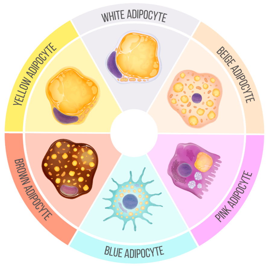
In healthy individuals, adipose tissue accounts for approximately 20–30% of total body weight and is mainly composed of triglycerides stored within adipocytes [33]. As previously mentioned, adipocytes are the main cellular component of adipose tissue. They are characterized by an oval shape, variable diameter, one or more lipid droplets, thin cytoplasm, and a nucleus that is displaced toward the periphery [34,35]. Adipocytes constitute approximately 60–70% of adipose tissue cells; however, this tissue also contains a stromal vascular fraction (SVF) embedded within an extracellular matrix [35,36]. SVF includes capillaries, endothelial cells, nerves, adipocyte precursor cells, and immune cells, including macrophages, T lymphocytes, and mast cells [36]. In humans, adipose tissue begins to form during intrauterine development and, in most individuals, continues to remodel throughout life [37,38]. This morphological and functional diversity supports classifying adipose tissue into several types, as summarized in Figure 1.

3.1. Developmental and Embryological Origins of Adipocyte Populations

Adipocyte populations arise from distinct embryonic precursor lineages, which partly explain their functional divergence. Classical brown adipocytes derive from Myf5-expressing precursors in the dermomyotome, a lineage shared with skeletal myocytes, consistent with the shared mitochondrial and oxidative machinery between brown fat and muscle [39]. White adipocytes were originally thought to arise exclusively from Myf5-negative precursors; however, lineage-tracing studies show that a subset of white adipocytes in some subcutaneous depots also derives from Myf5-lineage cells, and that visceral adipocytes may arise from a distinct Wt1+ lateral-plate-mesoderm precursor population separate from subcutaneous precursors [40]. More recent single-cell RNA sequencing of human subcutaneous adipose tissue confirms that precursor populations differ by depot even within subcutaneous fat, since abdominal versus gluteofemoral precursors show distinct commitment and metabolic gene signatures, indicating that depot-specific developmental programming persists into adult human tissue and is not simply a rodent phenomenon [41]. Beige adipocytes, interspersed within white adipose tissue and induced by cold or β3-adrenergic stimulation, do not arise from the Myf5 lineage; their precursor identity remains incompletely defined. Furthermore, the precursor identity of beige adipocytes and the extent to which rodent lineage-tracing models generalize to humans remain open questions requiring further human-tissue lineage studies.

3.2. White Adipose Tissue (WAT) and White Adipocytes

White adipose tissue (WAT) is the most extensively studied adipose depot, partly because it is present throughout life and represents the main site of lipid storage [42]. It is also the most abundant, accounting for approximately 10–20% of body weight in an adult individual [43]. Its pale appearance is related to its moderate vascularization and the predominance of lipid-loaded adipocytes [42]. These adipocytes are characterized by a large unilocular lipid droplet, which supports efficient energy storage [42,43]. The formation of adipocytes during intrauterine growth begins with undifferentiated mesenchymal cells forming lipoblasts at the periphery of blood vessels through the activation/repression of transcription factors [38]. With the appearance of slight lipid inclusions near the nucleus, lipoblasts acquire an oval shape and are called intermediate lipoblasts [35]. When lipid droplets coalesce into a large droplet, the cells are termed mature adipocytes [38,42].
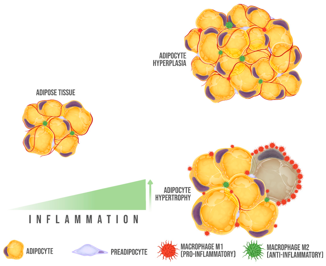
Mature white adipocytes, which vary in size from 20 to 200 micrometers in diameter, demonstrate remarkable structural plasticity [38]. This variability enables adipocytes to expand or contract in response to an individual’s physiological state, sex, and tissue location [38,42]. WAT can expand through two mechanisms: hyperplasia, an increase in the number of adipocytes, and hypertrophy, an increase in adipocyte size [38,42,43]. Both mechanisms are influenced by age. During early life, WAT expansion occurs through both hyperplasia and hypertrophy [44]. However, tissue hyperplasia gradually decreases during adulthood, and hypertrophy predominates [44,45].
WAT hyperplasia is a process modulated by insulin, insulin-like growth factor 1 (IGF-1), and fibroblast growth factor 1 (FGF-1) [45]. In the case of hypertrophy, the peroxisome proliferator-activated receptor gamma (PPAR-γ) plays a crucial role by regulating fatty acid storage and suppressing lipolysis [44,45]. The presence of modulators of these WAT expansion mechanisms is crucial because the number and size of adipocytes are key factors in metabolic health [43,45]. A reduction in adipogenic capacity, together with an increase in white adipocyte size, is associated with WAT dysfunction [43].
Before considering its role in disease, it is important to note that healthy WAT performs several essential physiological functions beyond lipid storage. As an endocrine organ, WAT secretes adiponectin, leptin, and other adipokines under basal, non-inflammatory conditions to regulate systemic insulin sensitivity, food intake, and energy expenditure [26,27]. Its lipid buffering capacity, the ability to expand adipocyte number and size in response to caloric surplus, protects non-adipose tissues (liver, muscle, pancreas) from ectopic lipid accumulation, provided this expansion occurs primarily through hyperplasia rather than hypertrophy [43,44,45]. Healthy WAT also demonstrates metabolic flexibility, shifting between lipogenesis and lipolysis according to feeding state and energy demand, coordinated by insulin and catecholamine signaling. Loss of this flexibility, rather than adiposity per se, is increasingly recognized as a determinant of metabolic dysfunction, underscoring that WAT pathology reflects a failure of adaptive capacity rather than an intrinsic property of the tissue itself.
When white adipocytes become hypertrophic, they may initiate coordinated changes in the tissue microenvironment that lead to structural remodeling (Figure 2) [38,43]. Recently, spatially resolved single-nucleus analyses of human abdominal subcutaneous adipose tissue showed that obesity is associated with senescence-prone metabolic, precursor, and vascular cell states. In contrast, weight loss reverses much of this remodeling but does not completely normalize macrophage activation. Hypertrophic WAT alters its secretory profile, increasing the release of inflammatory mediators (visfatin, interleukin-6, interleukin-8, tumor necrosis factor alpha, and resistin) and decreasing the secretion of adipocytokines with anti-inflammatory actions (interleukin-10, interleukin-4, and interleukin-13) [46]. It also increases leptin release, which regulates food intake in the hypothalamus, and conversely decreases adiponectin secretion, thereby reducing insulin sensitivity, lipid oxidation, and modulation of inflammation [43,46,47]. Furthermore, hypertrophic WAT promotes the infiltration and activation of macrophages, which are classified into two main subtypes: M1, with pro-inflammatory properties, and M2, with an anti-inflammatory profile [42]. Although this classification has been well described in experimental models, its relevance and delineation in humans are less specific [48,49]. Hypertrophic WAT exhibits an activated inflammatory M1 phenotype, comprising up to 40% of WAT cells.
However, single-cell genomic studies have identified additional adipose tissue macrophage (ATM) subpopulations that do not fit this binary, including metabolically activated macrophages (MMe), lipid-associated macrophages (LAMs) and their precursor state (pre-LAM), Mox macrophages, and perivascular macrophages (PVMs) that decline in number with obesity [50]. A 2024 single-cell genomics review notes that although several ATM clusters expand with obesity, none of them truly resembles the classical M1 phenotype described in early rodent work, underscoring that the field has moved well past the binary model [51]. But research is lacking on whether specific intermediate ATM phenotypes are causally linked to insulin resistance in humans, or are secondary markers of tissue remodeling.
Other consequences of sustained hypertrophy include extracellular matrix accumulation and insufficient angiogenic adaptation, which may contribute to fibrosis and WAT hypoxia [43]. Hypoxia, in turn, inhibits white adipocyte hyperplasia, generates local and systemic inflammation, promotes apoptosis in WAT, and leads to insulin resistance (IR) [38,42,43,44,46].
The implications of IR are significant, given that insulin plays a crucial role in regulating glucose and lipid metabolism within white adipocytes [49]. In healthy individuals, insulin exerts its effect by binding to its receptor, which autophosphorylates on tyrosine residues and phosphorylates the insulin receptor substrate 1 (IRS-1) [49,52]. The activation of IRS-1 facilitates the formation of molecular complexes within the phosphatidylinositol-3-kinase (PI3K) pathway. Consequently, glucose can enter the adipocyte through the translocation of glucose transporter 4 (GLUT-4) [49]. In contrast, in individuals with IR, the hormone’s ability to perform its biological functions is diminished [53]. This alteration is mainly due to reduced insulin interaction with the receptor, its expression, and kinase activity [49,53]. Likewise, ISR-1 increases its phosphorylation at serine/threonine residues, altering its ability to associate and causing attenuation of GLUT-4 translocation [53].
Beyond IRS-1 serine/threonine phosphorylation, several converging pathways link adipocyte hypertrophy and inflammation to insulin resistance. Pro-inflammatory cytokines (TNF-α, IL-1β) activate c-Jun N-terminal kinase (JNK), which directly phosphorylates IRS-1 at inhibitory serine residues, and the IKKβ/NF-κB pathway, which impairs insulin signaling primarily through transcriptional upregulation of inflammatory mediators rather than direct IRS-1 modification [54,55]. Chronic nutrient overload also induces endoplasmic reticulum (ER) stress; the PERK-eIF2α and IRE1 branches of the unfolded protein response converge on both NF-κB activation and induction of the TXNIP-NLRP3 inflammasome, driving IL-1β secretion and further impairing insulin signaling [56]. Adipocyte mitochondrial dysfunction, characterized by increased reactive oxygen species production and impaired oxidative capacity, contributes to and is exacerbated by these inflammatory pathways, creating a feed-forward cycle. Together, JNK, IKKβ/NF-κB, ER stress, mitochondrial dysfunction, and inflammasome activation represent overlapping rather than independent mechanisms, converging on impaired IRS-1 function and reduced GLUT-4 translocation. The relative contribution of each pathway in human versus rodent adipose tissue, and whether they can be therapeutically targeted selectively, remains incompletely defined.
IR is a key factor in the development of T2DM and metabolic syndrome; however, when combined with WAT dysfunction, its impact extends beyond these conditions. IR reduces adipocytes’ ability to store lipids, increasing free fatty acid (FFA) concentrations [45,47]. Elevated circulating FFAs are associated with ectopic lipid accumulation, a process involved in the progression of several obesity-associated diseases [43,46,47].
For instance, the deposition of WAT in the liver and IR leads to ectopic fat accumulation in organs such as the liver, muscle, kidney, and pancreas. This promotes oxidative stress and mitochondrial dysfunction. Consequently, liver injury, inflammation, and fibrosis, which are hallmarks of MAFLD, are promoted [52]. Additionally, for the development of cardiovascular diseases, atherosclerosis, ventricular hypertrophy, and diastolic dysfunction, the accumulation of pro-inflammatory macrophages, hypertriglyceridemia, epicardial WAT deposition, and inflammation play a crucial role [57]. In hypertension, it is essential to consider that insulin plays a role in blood pressure regulation [52,58,59], since it promotes vasorelaxation by stimulating nitric oxide production in the endothelium and modulates sodium reabsorption in the kidney [59]. Consequently, 80% of patients with T2DM also have hypertension [59,60]. Moreover, certain types of cancer are frequently associated with WAT inflammation, as the accumulation of macrophages, hypoxia, and increased cytokine secretion promote the activation of pathways involved in carcinogenesis [61], for example, tumor necrosis factor-alpha (TNF-α), which in certain circumstances can be a tumor promoter by activating cell life/death signaling pathways such as the nuclear factor kappa B (NF-κB) pathway and specific mitogen-activated protein kinase (MAPK) cascades involved in apoptotic or survival processes [52,57,61]. Finally, cognitive decline is also a consequence of IR. Therefore, in obese middle-aged patients with glucose intolerance, there is a risk of developing dementia, memory loss, or even neurodegenerative diseases such as Alzheimer’s increases [62,63]. Likewise, recent studies indicate the association between increased secretion of adipokines such as visfatin and cystatin C with the development of psychiatric disorders, multiple sclerosis, and Alzheimer’s [63].
Considering the association between WAT and the obesity comorbidities mentioned above, WAT has been extensively studied for many decades. On the one hand, in vivo, models have been implemented in animals induced to obesity through mutations in leptin (ob/ob mouse, Zucker rat, etc.), by changes in diet (high-fat diet, high-carbohydrate diet, etc.), or by surgical and chemical methods (ovariectomy, lesion of the ventromedial hypothalamus or the hypothalamic paraventricular nucleus) [64,65,66,67].
WAT is identified by the expression of genes such as Hoxc8 and Hoxc9, which exhibit region-specific patterns [68]. Hoxc8 is predominantly expressed in intra-abdominal adipose depots, such as epididymal and perirenal fat, whereas Hoxc9 is more associated with subcutaneous fat depots [69]. However, neither gene is an exclusive marker of adipocytes, as their expression extends to other cellular components within the stromal vascular fraction of adipose tissue [68,69]. However, the amino acid transporter ASC-1 is a white adipocyte-specific cell surface protein with low expression in other adipocytes [68].
These genes are part of broader transcriptomic signatures that vary by adipose depot and developmental stage, reflecting the complex biology of adipose tissue rather than serving as definitive markers for WAT [69,70]. In the case of in vitro and ex vivo models, mouse cell lines (3T3-L1, 3T3-F442A, C3H/10T1/2) or human cell lines (immortalized cell lines, human adipose tissue-derived stem cells) are implemented [70]. To corroborate the presence of white adipocytes, some commonly used surface markers are Serpina3K, Wdnm1-like, and Asc1 [68].
As a result of extensive research, various strategies have been developed to control excessive WAT growth and manage obesity. Traditional approaches, such as dietary therapy, focus on reducing lipid intake, portion sizes, and energy content while incorporating fruits and vegetables [71,72,73]. Physical activity and regular monitoring by health professionals remain integral components of long-term weight management [71,74]. Pharmacological treatments have undergone significant evolution over the years. Incretin mimetics, such as GLP-1 receptor agonists (e.g., semaglutide and liraglutide), have emerged as effective therapies for obesity and related metabolic disorders [75]. These agents promote weight loss by enhancing satiety, delaying gastric emptying, and improving insulin sensitivity. Similarly, melanocortin-4 receptor (MC4R) agonists, such as set-melanotide, represent a breakthrough for rare genetic forms of obesity by regulating appetite and energy homeostasis [76]. Older pharmacological options like phentermine, benzphetamine, and orlistat, while still in use, have been complemented by these novel agents [67,74]. Bariatric surgery remains a highly effective option for individuals with a BMI ≥ 40 kg/m2, resulting in significant and sustainable weight loss, as well as reduced prevalence of DM, dyslipidemia, and hypertension [77,78].
Dietary supplements have also been explored as complementary strategies for weight management and metabolic modulation [79,80]. Some commonly used products include spirulina, chitosan, green tea extract, probiotics, and omega-3 polyunsaturated fatty acids (ω-3 PUFAs) [79]. The latter is mainly composed of eicosapentaenoic acid (EPA) and docosahexaenoic acid (DHA) [79,81]. So far, ω-3 PUFAs appear to have therapeutic potential for heart disease, T2DM, and MAFLD due to their anti-inflammatory, hypolipidemic, and insulin-sensitizing effects [81,82,83]. It is essential to note that, despite the variety of supplements and evidence of their health benefits, the long-term effectiveness of these products remains to be determined [79,81].

3.3. Brown Adipose Tissue (BAT)

Brown adipose tissue (BAT) also stores lipids, as in WAT; however, in humans, BAT is much less abundant and rarely exceeds 1% of body weight [84]. Moreover, the morphology, origin, and function of its adipocytes differ. BAT adipocytes are characterized by their small size (10 to 25 micrometers in diameter), storing triglycerides in multiple lipid droplets (multilocular), having a rounded nucleus, and a reduced endoplasmic reticulum [34,35]. The distinctive color of BAT reflects its dense vascularization, rich sympathetic innervation, and high mitochondrial content [36,42,85,86,87]. Regarding their origin, brown adipocytes form during embryonic development, as do white adipocytes. However, BAT adipocytes share the same lineage as skeletal muscle myocytes [42,85,86].
Unlike WAT, BAT’s primary function is not energy storage. On the contrary, it specializes in lipid catabolism and adaptive thermogenesis [42]. This function is conferred by the uncoupling protein 1 (UCP-1) located in the inner membrane of its mitochondria [83,84,88,89]. UCP-1 uncouples oxidative phosphorylation, dissipating the proton electrochemical gradient across the mitochondrial membrane that is typically used to generate ATP [86,87,88]. As a result of this mechanism, energy is dissipated as heat, and fatty acid oxidation is stimulated [88,89]. UCP-1 activation is triggered by exposure of the organism to cold, fatty acids, the release of insulin, and norepinephrine by the sympathetic nervous system [90,91].
Due to its primary function of thermogenesis, BAT was initially thought to be limited to hibernating mammals and was therefore referred to as the hibernation gland [82]. However, it was later discovered that this tissue is also present in other non-hibernating mammals, such as rats, mice, cats, and humans [86,88]. BAT is now recognized as essential for thermoregulation and survival in newborn small mammals. Since they are predisposed to lose heat due to their body volume-to-surface area ratio [86,88], they lack sufficient muscle mass to regulate their temperature through shivering [86].
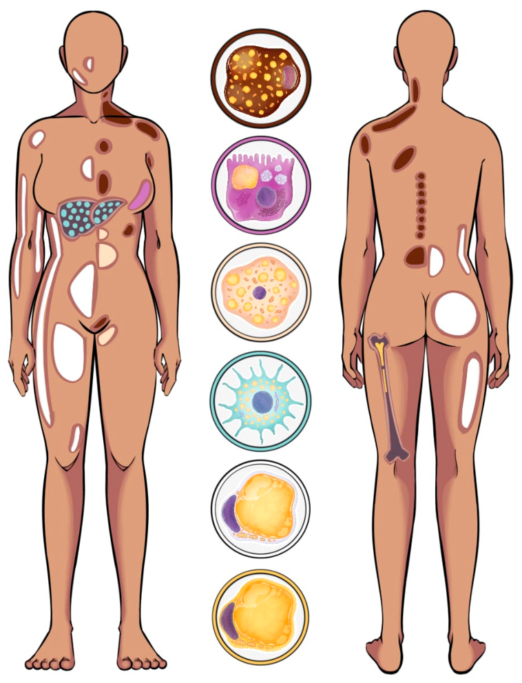
Regarding its location, BAT in rodents accumulates in the interscapular, subscapular, perirenal, and periaortic regions [85]. In human neonates, BAT is primarily distributed in the interscapular, supraclavicular, and paravertebral regions [86,87]. Finally, studies on adult humans have been limited until recently, as it was previously thought to be present only in newborns. In 2007, evidence indicated that BAT does not completely atrophy during childhood [92]. On the contrary, a portion of BAT is preserved in the supraclavicular and paravertebral areas (Figure 3) [92,93]. However, BAT in adults differs from that previously described in small mammals, as adipocytes contain less UCP-1 and are surrounded by white adipocytes [94,95]. Evidence supports the existence of metabolically active BAT in adults, which regulates total body energy expenditure and body fat. Additionally, BAT presence is influenced by diabetic status or blood glucose levels [96].
In contrast to WAT, BAT does not appear to increase with obesity, and studies even indicate a negative correlation between BAT activation and body mass index (BMI) [83,86]. Furthermore, BAT is notably reduced in individuals with T2DM [91,94]. The mechanism by which BAT decreases is not well defined; however, it has been suggested that it may result from insulin resistance [94,97]. For this reason, BAT activation is generally not considered a driver of obesity-related comorbidities. Accordingly, BAT activation has been explored as a potential therapeutic strategy for conditions associated with excessive WAT expansion [84,93,94]. BAT is a tissue capable of increasing energy expenditure through thermogenesis using substrates such as triglycerides (primarily), glucose, and some amino acids [42].
Cold exposure is considered the classical stimulus for BAT activation [90,91]. This mechanism occurs in brown adipocytes via a signal transduction pathway initiated by norepinephrine binding to the β3-adrenergic receptor (β3-AR) [90,98,99]. This signaling pathway promotes UCP-1 expression and lipid β-oxidation to produce heat [98]. Currently, studies in rodents exposed to intense cold (between 4 °C and 10 °C) for extended periods show that brown adipocyte activity increases by approximately 1.5-fold [100,101]. In related human studies, daily exposure to cold at 17 °C for 2 h over 6 weeks has been shown to significantly increase BAT activity and reduce body fat mass [101]. However, a limitation is that this cold exposure is not readily applicable to humans [100]. Similarly, systemic increases in norepinephrine are not selective and may adversely affect cardiovascular function and cognition [84,100]. For this reason, researchers are currently seeking to develop selective methods of BAT activation [80], for example, supplements (capsaicin, ephedrine, catechin, and retinoic acid) or BAT transplantation [84,91,99].
So far, some positive effects of BAT activation include reductions in hyperglycemia and dyslipidemia typical of T2DM and metabolic syndrome [97,102]. It is suggested that 40 g of active BAT could promote approximately 20% of the total energy expenditure of an adult [102]. In cardiovascular diseases, experiments in rodents have indicated that BAT has a protective effect [98,102]. In addition to its thermoregulatory function, this tissue is also a secretory organ. Some adipokines secreted by BAT, such as fibroblast growth factor 21 (FGF21), delay the development of hypertension and protect the heart from inflammation, oxidative stress, and decreased HDL [102,103]. Likewise, in mice with hyperlipidemia, the administration of β-adrenalin (to promote BAT activation) delayed the development of atherosclerosis [102]. This effect is associated with the increased expression of heme oxygenase, mediated by adipocyte-specific overexpression of the peroxisome proliferator-activated receptor gamma coactivator 1-alpha (PGC-1α), which contributes to vasodilation and improved vascular function [102,103]. Regarding cancer, recent studies in mice suggest that BAT activation could reduce the size of fibrosarcoma, melanoma, and pancreatic cancer tumors [104]. To date, it is suggested that the decrease in glucose supply to the tumor microenvironment could decrease hypoxia, angiogenesis, and cell proliferation [104,105].
Despite the growing interest in BAT for its therapeutic potential, its study, even to date, remains a challenge. Some techniques used to identify brown adipocytes based on their morphology are light microscopy, transmission, and scanning electron microscopy [106,107]. These approaches allow the identification of multilocular adipocytes with high mitochondrial density and abundant cristae [106]. Primary cell cultures and BAT explants are also implemented [108]. These are proliferated and differentiated in vitro, with the SV40 T antigen used for immortalization. Identification is achieved by analyzing BAT-specific markers such as UCP-1, Cidea, and Prdm16, although these are not surface markers but intracellular or transcriptional markers [68,108,109]. Finally, imaging techniques such as magnetic resonance imaging are applied for the non-invasive study of the structure and function of BAT in vivo (animals and humans) [110]. However, despite being a cost-effective method, it is not entirely accurate because it is based on the water and fat content of BAT [110,111]. For this reason, for some years now, the preferred method for characterizing BAT has been positron emission tomography with fluorine-18-fluorodeoxyglucose (18 FDG-PET/CT) [111,112]. However, it is warned that this technique also has limitations. For example, 18 FDG-PET/CT determines the absorption of glucose in BAT, which does not accurately reflect its thermogenic activity since triglycerides are its primary energy source [111].
Despite these promising findings, several translational barriers temper the therapeutic outlook for BAT activation. Metabolically active BAT mass in adult humans is limited, constraining its maximal contribution to whole-body energy expenditure regardless of activation strategy [113]. BAT presence and activity also show marked interindividual variability, influenced by age, sex, adiposity, and cold-exposure history [114]. The durability of pharmacologically or cold-induced BAT activation is also uncertain. Together, these findings suggest that human BAT, even under optimized activation, may be insufficient on its own to meaningfully reverse obesity-associated cardiometabolic disease, and is more likely to require combination with other interventions rather than deployment as a standalone therapy [114]. It is worth noting that selective pharmacological BAT activators without systemic adrenergic side effects, and reliable methods to quantify thermogenic (not just glucose-avid) BAT activity in vivo, remain unmet needs. Therefore, several opportunities remain to further investigate BAT function. More specific methods for studying BAT are needed to clarify the therapeutic relevance of BAT activation in individuals with obesity and metabolic dysfunction.

3.4. Beige Adipose Tissue (BeAT)

In addition to BAT, another type of thermogenic adipose tissue called beige or brite adipose tissue (BeAT) was initially described in 2008 by Seale and colleagues [115]. Because BeAT is present in limited and heterogeneous depots in humans, its total proportion has not been precisely established, although it appears to be comparable to that of BAT [116,117]. BeAT shares a mesenchymal origin with WAT and is found within subcutaneous cervical, supraclavicular, axillary, and inguinal depots, where beige adipocytes are interspersed among white adipocytes (Figure 3) [110,116,117]. Although its localization is heterogeneous, beige adipocyte morphology resembles that of brown adipocytes, with centrally located nuclei, multilocular lipid droplets, and abundant mitochondria [117].
The primary function of BeAT is thermoregulation, and beige adipocytes are often considered inducible or transient thermogenic cells. However, BeAT can also store energy because it exhibits an intermediate phenotype: it may express UCP-1, as observed in BAT, or store lipids in droplets, as occurs in WAT [117,118,119]. The mechanism through which this occurs is called browning, and even today, it is not fully understood. To date, two models have been proposed: the first suggests that WAT has the property of converting to BeAT through a reversible transdifferentiation process; the second indicates that BeAT adipocytes come from multiple precursor lineages that, when active, share characteristics with BAT and, when inactive, resemble WAT [116,120]. Despite this uncertainty, we currently know that, upon the activation of BAT, browning occurs in response to prolonged exposure to cold and β-adrenergic stimulation [37,86,117,120,121].
Recently, Wang et al. described that two distinct subpopulations of beige adipocytes coexist to dissipate heat. Heat dissipation may occur through UCP-1-dependent mechanisms or ATP-dependent futile cycles (FCs), defining UCP-1 beige adipocytes and FC adipocytes, respectively. The FC adipocyte subpopulation is highly metabolically active and uses substrate cycles involving creatine, calcium, and lipids, resulting in net ATP consumption and UCP-1 independent energy dissipation [122]. These cells are essential to direct systemic energy homeostasis and are linked to glucose metabolism and obesity resistance in humans and regulators of energy homeostasis in mammals.
Studies indicate that, since active BeAT increases insulin sensitivity and can catabolize lipids (like BAT), it does not increase in obesity (like WAT) [110,120,121]. However, recent studies indicate that, in the presence of inflammation in BeAT adipocytes, the expression of the UCP-1 protein decreases, and its predominant function is lipid storage [121,123,124]. For this reason, BeAT activation and WAT browning have attracted interest as potential strategies for metabolic disorders associated with obesity [121].
As previously mentioned, the primary stimulus for BeAT browning is cold; however, in recent decades, other alternatives have been explored to promote the activity of this tissue [117,120,121]. For example, exercise increases irisin secretion in skeletal muscle, promoting white adipocyte browning [120,121,125,126]. Therefore, in animal models, it increases energy consumption, reduces body weight, decreases WAT, and improves glucose tolerance [125,126]. Studies have indicated that these effects were potentiated by implementing a balanced diet [125]. Likewise, the intervention of dietary components such as resveratrol, curcumin, and omega-3 polyunsaturated fatty acids (EPA and DHA) also promotes the browning of WAT and thermogenesis in obese rodents [127,128,129,130]. To date, it is suggested that this effect results from activating the AMPK/SIRT1/PGC-1α pathway and the transcription factors PPAR-α/γ [117,119,120,127].
However, it is essential to note that research on the BeAT presents a challenge due to its physiological and morphological similarities to BAT [37,121]. Primary cell cultures (3T3-L1 preadipocytes subsequently differentiated) or BeAT explants are identified with surface markers such as Tmem26, Tbx1, and CD137 [37,131]. In addition, the amino acid transporter PAT2 and the purinergic receptor P2RX5 are cell surface markers expressed in brown and beige adipocytes in mice. These markers also selectively mark brown/beige and white adipocytes in human tissue [68].
Regarding in vivo studies, techniques such as the Bruker In Vivo Xtreme imaging system or multispectral optoacoustic tomography are commonly used to detect BeAT in rodents due to being minimally invasive [131]. Finally, radiopharmaceuticals such as 18 FDG-PET/CT and TPSO-18 kDa are the most optimal imaging modalities [131,132,133]. It is important to note that to understand further what we know about BeAT and the extent of the activation of these adipocytes on the metabolic state, it is essential to find better BeAT markers and more optimal techniques capable of distinguishing BeAT from BAT [37,126].

3.5. Pink Adipose Tissue (PAT)

Pink adipose tissue (PAT) was characterized in murine models over the past two decades [134]. Since its initial mention, its essential participation in the alveolar epithelium of the mammary glands during milk production and secretion has been established [134,135,136]. Additionally, it was indicated that pink adipocytes, like white, brown, and beige adipocytes, can store lipids [136]. However, their morphology differs, comprising brick-like epithelial cells [134,137]. PAT adipocytes are equipped with multiple lipid droplets that contain milk protein inside and are bigger than those observed in brown adipocytes [134,138,139]. Additionally, pink adipocytes have a thin cytoplasm, a centrally located nucleus, a developed Golgi apparatus, abundant rough endoplasmic reticulum (RER) cisternae, and larger mitochondria than those present in white adipocytes [134,139]. Regarding its origin, PAT comes from the subcutaneous adipose tissue of the mammary gland [134,138], which, through physiological stimuli during pregnancy, undergoes a transdifferentiation process commonly called “pinking” [134,139].
Before pregnancy, the mammary glands of females comprise a large amount of white adipose tissue infiltrated in branched ducts that end in the nipple [134,136]. However, during pregnancy and lactation, a milk-producing structure is formed, integrated by adipose epithelial cells (pink adipocytes) that supply energy to newborns through feeding (Figure 3) [134,136]. This event is called alveologenesis and occurs in females from the first 18 days of pregnancy [134,138]. The process begins when the alveoli of the mammary glands expand progressively until they occupy 90% of the mammary volume [135]. These milk-producing and secreting structures persist until the end of lactation and present a pink color at the macroscopic level, giving their characteristic name to PAT [134,136,140]. After lactation, the mammary gland begins an involution process during the first days [134]. These initiate the progressive decrease of the alveolar structures until they are replaced by white adipose tissue, reverting to the anatomy of the breast before pregnancy [136,140]. In the case of PAT adipocytes, the number of adipo-epithelial cells decreases until they disappear, and in parallel, the presence of cells with the typical morphology of WAT is reincorporated [134,136].
It should be noted that the molecular mechanisms that allow the formation of pink adipocytes are not fully understood. However, pregnancy hormones and the paracrine activity of the mammary glands are involved [131]. Additionally, studies published in 2014 indicated that secreted phosphoprotein 1 (SPP1) plays a vital role in WAT transdifferentiation to PAT [140,141]. Likewise, for the transdifferentiation of PAT to WAT, the transcription factor PPAR-γ is key in the involution of the mammary gland [138,141].
Studies on PAT have contributed to the understanding of adipose tissue plasticity and suggest that transdifferentiation may represent a physiological property of the adipose organ [136,138]. This concept is supported by evidence that adipocyte conversion is not limited to the white to pink transition and its reversal [136]. Some studies with electron microscopy and lineage tracing indicate that PAT can also convert to BAT, although there is currently no evidence of BAT conversion to PAT [136,138].
It is important to note that, to date, a direct association between the formation of PAT and the development of metabolic diseases associated with obesity has not been established. Conversely, breastfeeding has been associated with reduced offspring susceptibility to obesity, cardiovascular disease, and T2DM [142,143]. Studies suggest that this is because pink adipocytes, in addition to milk, secrete hormones such as leptin and adiponectin [134,137,138,139].
In addition, interesting results in obesity-induced rodents concluded that the transdifferentiation of pink adipocytes decreased considerably during pregnancy [134,136]. However, the mechanisms by which increased WAT may impair adequate PAT formation remain unclear [144,145]. As for other pathologies associated with obesity, studies indicate that the partial involution of the mammary gland at the end of lactation results in developing a microenvironment rich in pro-inflammatory mediators that can lead to tumorigenesis [141,142,143,144,145]. Likewise, mammary adipose tissue secretes growth factors and an energy source that cancer cells can use for survival [141]. This explains why the risk of developing breast cancer increases transiently with pregnancy, and the incidence of death associated with this type of cancer increases within the first five years after childbirth [145,146].
To date, the study of this type of adipose tissue still involves multiple difficulties due to its technical limitations [134]. Histological studies, lineage tracing experiments, and PAT explants in rodents use the whey acidic protein (WAP) as a marker for identifying PAT adipo-epithelial cells [134,136,138,139]. Because PAT has been described relatively recently, it remains unclear whether WAT transdifferentiation into milk-producing structures occurs similarly in humans during pregnancy [137]. Likewise, much remains to be explored about the participation of these adipocytes in energy homeostasis in mothers and offspring [143]. Finally, the effect of obesity-related inflammation on PAT function remains to be elucidated [147].

3.6. Yellow Adipose Tissue (YAT)

Yellow adipose tissue (YAT) was initially considered inert and was thought to primarily fill bony spaces; however, it is now recognized as an active component of the bone marrow microenvironment (Figure 3). In terms of its morphology, the yellow adipocyte is comparable to the white adipocyte, as it has a lipid droplet (90% of its cell volume), a thin cytoplasm, a nucleus located at the periphery, and a moderate number of mitochondria [148,149,150]. Regarding size, yellow adipocytes are larger than brown and beige adipocytes, as it has a diameter between 40 and 90 micrometers [148,149].
Despite its morphological similarities with WAT, YAT has distinct function, including regulation of the bone marrow microenvironment and participation in hematopoiesis [149,151,152,153]. Additionally, evidence indicates that the triglycerides stored in YAT do not provide energy during periods of caloric restriction; however, this energy can contribute to bone regeneration [149,150]. Recent evidence indicates that YAT functions as an active endocrine compartment that participates in the regulation of bone metabolism (and probably systemic metabolism), as it is a secretor of adipokines such as adiponectin, leptin, and pro-inflammatory cytokines [149,151,154,155].
In the case of its origin, yellow adipocyte (like WAT) formation occurs from mesenchymal progenitor cells and with the regulation of PPAR-γ [143,156,157]. This age-dependent process occurs from the distal to the proximal area [149,155]. For this reason, yellow adipocytes are formed during childhood in the phalanges and, later in puberty, accumulate in the appendicular and axial skeleton (mainly in the trabecular cavity) [149,151,156]. In adults between 25 and 50 years old, bone marrow is composed of 70% YAT, making it integrate approximately 10% of the total fatty tissue in healthy adults [148,155,156]. However, men generally have a greater number of yellow adipocytes than women of the same age [151,157].
On the other hand, evidence obtained recently from studies with rodents indicates that YAT is composed of two subtypes of adipocytes: constitutive yellow adipocytes (YAc) and regulatory yellow adipocytes (YAr) [148]. YAc are formed during the early stages of life, are maintained throughout the years in the appendicular skeleton (even in the presence of pathologies or environmental changes), and store mainly saturated lipids [148,149,150,157]. Likewise, YAc cells integrate a portion of bone marrow commonly called yellow due to its abundant large adipocytes [143,150]. As for YAr, their formation occurs in the axial skeleton within the red bone marrow, which gets its name from being composed primarily of hematopoietic tissue [148,156]. Unlike YAc, YAr accumulates mainly unsaturated lipids and tends to be smaller [148,149,156]. Likewise, YAr are more labile and respond to various physiological conditions [150,155,156]. For example, YAr and red bone marrow decrease with aging, obesity, hypertension, and T2DM, causing YAT to be composed predominantly of YAc [141,150,158]. This phenomenon is associated with reduced regenerative capacity and lower bone density, particularly in trabecular bone [149,151,155,157,158]. Additionally, recently published studies have suggested that the increase in adiposity percentage and waist circumference in humans is correlated with the risk of osteoporotic fractures, particularly in men and postmenopausal women [149,157,159]. Finally, these negative consequences on bone marrow health were also identified in people who abuse alcohol or in patients medicated with glucocorticoids, thiazolidinediones, or chemotherapy [148,149,155,157].
Knowledge about YAT in humans remains limited, as some limitations to studying these adipocytes are the difficulties in collecting and isolating these cells infiltrated in hematopoietic tissue [148,160]. For this reason, most studies are conducted in in vivo murine or in vitro human models [160]. Further studies are required to clarify the differences between YAT adipocyte subtypes (YAc/YAr), delve deeper into lipid accumulation or adipokine secretion mechanisms, and establish how YAT contributes to energy homeostasis [160]. Since its size can vary in response to increased fat intake, age, and visceral fat, it is also strongly influenced by estrogen, insulin, or glucocorticoid signals [148,160]. Finally, diseases such as anorexia nervosa and lipodystrophy are associated with decreased YAT [161,162].

3.7. Hepatic Stellate Cells (HSCs)—Blue Adipocytes

Hepatic stellate cells (HSCs) were first described in 1876 by Karl Kupffer and have been widely studied due to their critical roles in liver physiology and pathology [163,164]. In their quiescent state, these cells contain multiple lipid droplets measuring two to eight micrometers in diameter, an oval nucleus located centrally, and a developed Golgi apparatus and rough endoplasmic reticulum [163,164,165]. Furthermore, these cells have elongated processes in contact with endothelial cells [163].
HSCs, also referred to as blue adipocyte-like cells in some contexts, are located in the liver and positioned in the space of Disse, between sinusoidal endothelial cells and hepatocytes (Figure 3) [163,166,167]. They are also of mesenchymal origin and in humans are formed from the third month of gestation [163,164,166,168]. The importance of these adipocyte-like cells lies in the fact that they constitute approximately 10% to 15% of cells in the healthy liver and, due to their sensitivity to extracellular stimuli, they have very varied functions [167,168]. For example, intercellular communication, regulation of the remodeling of extracellular matrix components, regulation of sinusoidal blood flow, and communication with immune cells [166,169,170]. However, one of their best characterized functions in the quiescent state is the storage of lipid droplets containing triglycerides, cholesterol, and vitamin A in the form of retinol [165,166,169]. Retinol is the main component of these droplets, and 90% of hepatic vitamin A and 80% of the total is accumulated in them [163,166]. This characteristic is important because retinol regulates cell proliferation, differentiation, and morphogenesis [166,169]. In cell cultures, lipid droplets with vitamin A exhibit blue/green fluorescence at a wavelength of approximately 330 nm, giving them their characteristic name [165].
On the other hand, another vital function of HSCs is their ability to act as defense cells, responding to liver abnormalities such as infections, inflammation, tissue injury, or excess lipopolysaccharides [167,168,170]. During this process, HSCs become activated and interact with immune cells while promoting changes in the extracellular matrix [165,167]. During this process, these cells lose their lipid droplets, transdifferentiate into cells like myofibroblasts, and promote collagen production [165,166,167,168]. Likewise, these cells secrete pro-inflammatory cytokines, growth factors, leptin, and adiponectin [168]. These responses contribute to the removal of damaged hepatocytes and scar formation at the injury site, limiting further tissue damage; however, prolonged and repeated activation of this mechanism causes fibrosis, cirrhosis, and hepatocarcinogenesis [169,171,172].
In their quiescent state, HSCs, which share some adipocyte-like features, are not directly associated with liver or metabolic dysfunction [169]. Likewise, it does not appear to increase proportionately with pathologies such as obesity [166,169]. However, studies indicate that some triggers of stellate cell activation are obesity, dyslipidemia, hypertension, and T2DM [170]. These conditions may promote hepatic fibrosis through mechanisms in which inflammation plays a central role [171,172].
For this reason, further studies are required to clarify less explored roles of HSCs and their functional parallels with adipocytes. For example, their abundant expressions of leptin, adiponectin, PPAR-γ, and sterol regulatory element binding protein 1c (SREBP-1c) [164,173]. The latter two are characterized by being two key regulators of adipogenesis [174]. Once again, the main limitation to expanding our knowledge is the challenge of studying cells that are difficult to isolate in vivo [169]. However, the association between HSCs and adipocytes has been accentuated thanks to the studies by Tsukamoto and collaborators, which indicate that the transcriptional program involved in adipocyte differentiation overlaps with the program required to maintain HSCs in their quiescent vitamin A storing phenotype [164,173].

4. Anatomical Location of Adipose Tissue and Its Role in Obesity Progression

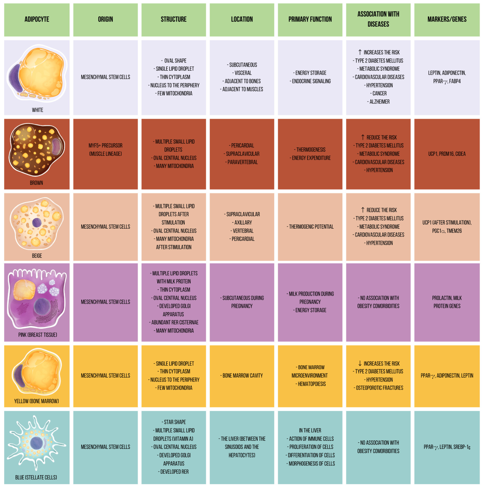
As previously mentioned, adipose tissue can be classified based on its physiology. The unique characteristics, molecular markers, origins, and physiological roles of the different types of adipose tissues, including WAT, BAT, BeAT, PAT, YAT, and BluAT (HSC), are summarized in Figure 4. However, this physiological classification should be complemented by anatomical location (Figure 3). Over the past 80 years, several studies have shown that adipocyte depot location influences lipid storage capacity, endocrine activity, lipolytic response, and sensitivity to physiological stimuli [175]. Consequently, adipose tissue distribution pattern may be more relevant than total adiposity percentage for the development of metabolic diseases [176,177].
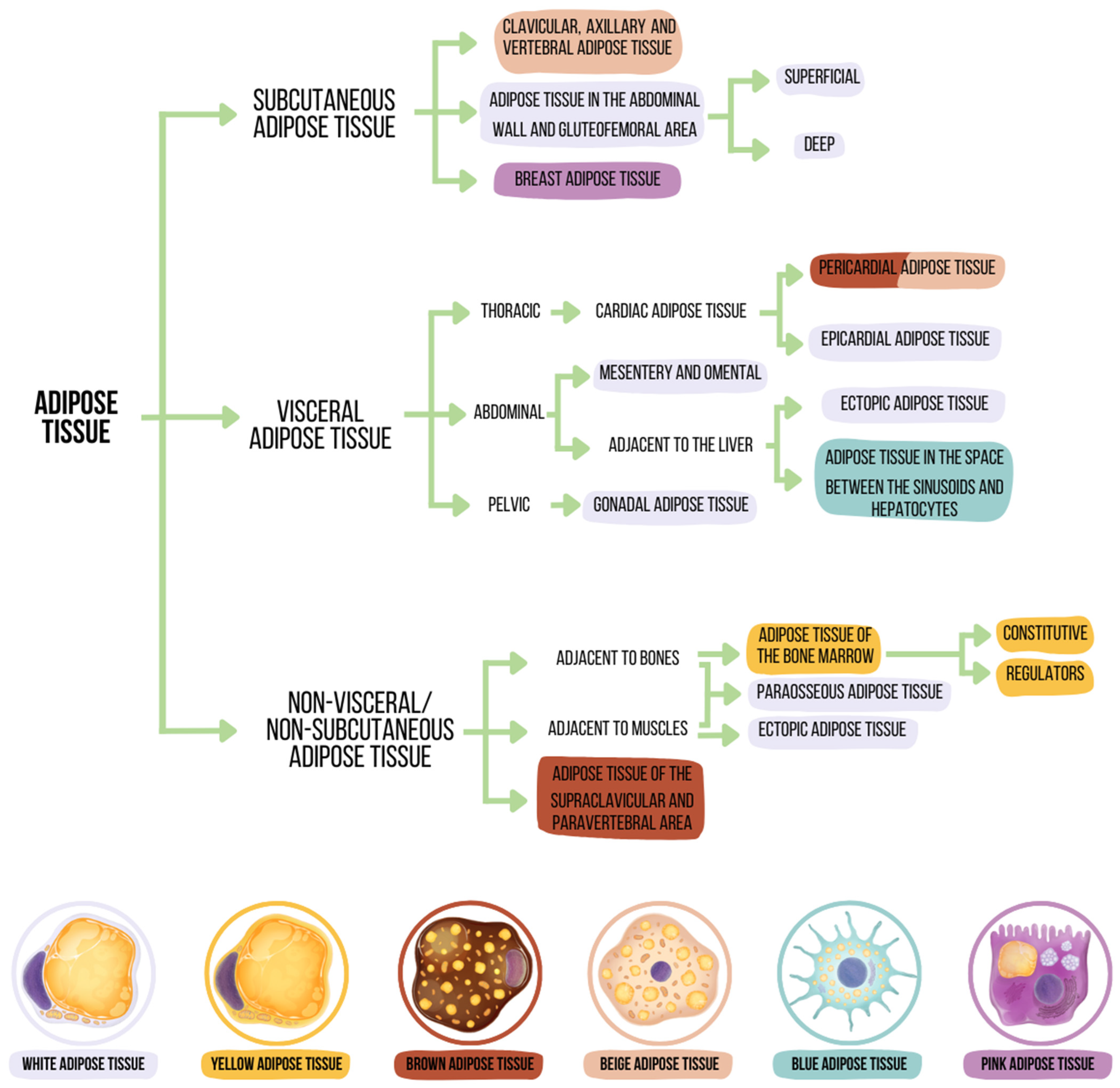
This review suggests an integrative classification of adipose tissue that considers its physiology and anatomical location as factors in the analysis. It is important to emphasize that the anatomical distribution of adipose tissue is influenced by sex, age, diet, physical activity, and medication use. Based on studies conducted in healthy adults, adipose tissue can be anatomically classified into three main compartments: subcutaneous adipose tissue, visceral adipose tissue, and non-visceral/non-subcutaneous adipose tissue (Figure 5) [175,176].

4.1. Subcutaneous Adipose Tissue (SAT)

Subcutaneous adipose tissue (SAT) is located between the dermis and the connective membrane that covers the muscles (aponeurosis) [175]. SAT is generally composed of relatively small adipocytes that secrete abundant leptin, show high insulin sensitivity, and have a lower lipolytic rate than visceral adipose tissue (50% lower than visceral adipose tissue) [176,177] SAT is also highly dynamic, as specific stimuli can induce transdifferentiation toward pink adipocytes during pregnancy and lactation or beige adipocytes in response to cold exposure and exercise [117,121,136,139]. A recent single cell methylome and 3D genome atlas of human subcutaneous adipose tissue identified cell type-specific epigenomic programs and showed that abdominal obesity risk variants are preferentially enriched in adipocyte regulatory compartments, whereas inflammatory enrichment is concentrated in myeloid compartments. However, the most explored adipocytes of the SAT are the white ones, since they are more abundant (80% of the total fatty tissue) (Figure 5) [176]. However, excessive expansion of white adipocytes within SAT may contribute to obesity-related diseases [177].
Regarding its location, SAT is found in the clavicular, axillary, and vertebral regions (beige subcutaneous adipose tissue), abdominal wall, breast tissue (pink subcutaneous adipose tissue), and gluteofemoral area (Figure 5) [169,170,171]. This last depot is generally the largest because it constitutes 50% of the total SAT and, due to its abundance of estrogen receptors, accumulates more in women than in men [176,177,178].
It should be noted that white SAT expansion is significantly influenced by hormonal changes, particularly those related to sex hormones [179]. For example, estrogen increases the size and number of subcutaneous adipocytes and attenuates lipolysis [179,180]. Consequently, preferential accumulation of SAT (particularly in the gluteofemoral region) is promoted [180]. At the same time, androgen levels are inversely correlated with white SAT deposition, contributing to its lower abundance in men [179].
On the other hand, the white SAT stores large amounts of lipids, expanding primarily through hyperplasia (an increase in the number of adipocytes) [176]. For this reason, white SAT is considered a metabolic buffer that can temporarily limit the adverse effects of energy surplus and is associated with a lower risk of metabolic diseases [176,177,180]. However, despite the protective effect of white SAT, when the tissue’s storage capacity is exceeded, the mechanism for generating new adipocytes deteriorates [177]. Consequently, inflammation, insulin resistance, and ectopic lipid accumulation (skeletal muscle, heart, and liver) are promoted [176,177,178]. For this reason, excess white SAT over the long term is associated with elevated blood levels of triglycerides, cholesterol, and glucose [178]. Likewise, studies reveal a relationship between high percentages of white SAT and hypertension, heart disease, and T2DM [176,178]. In this context, lipid storage in white SAT may delay metabolic deterioration, although it does not fully prevent it [176,177,178,180].
Hormonal stimulation during puberty, pregnancy, and menopause may promote pathological growth of subcutaneous adipose tissue [179]. For example, loss of ovarian function increases the risk of diseases such as metabolic syndrome and T2DM, which increases cardiovascular risk [177,179]. Furthermore, menopause is associated with redistribution and morphological changes in the white SAT [179]. This remodeling is characterized by adipocyte hypertrophy, fibrosis, and chronic inflammation, which may reinforce insulin resistance and perpetuate tissue dysfunction [171,179].
On the other hand, the participation of various hormones, such as cortisol, can influence the expansion of this tissue, since cortisol is generated in the white SAT by the enzyme 11β-hydroxysteroid dehydrogenase type 1 (11βHSD1), which converts inactive cortisone into active cortisol [181]. In consequence, the activity of this enzyme in this tissue increases body weight [179,181]. Likewise, growth hormone (GH) can modify the white SAT by affecting its mass, distribution, composition, endocrine function, and inflammatory and fibrotic status. Previous studies indicate that humans and rodents with elevated GH levels experience a decrease in longevity and are more susceptible to cardiovascular diseases and cancer [182]. Finally, insulin plays a central role in the expansion of white SAT by stimulating lipogenesis and inhibiting lipolysis, favoring fat storage in conditions of energy surplus [178].
The deep fascia divides white SAT into two compartments: superficial SAT, which protects organs from external compression, and deep SAT, which contributes to the mechanical adaptation of the skin in response to external forces [176,178,183]. Superficial white SAT is composed of small, organized adipocytes that form a thin layer of compact tissue close to the skin of the abdomen [178,183,184]. It also predominantly accumulates monounsaturated fatty acids and secretes abundant leptin and adiponectin [185,186]. In contrast, deep SAT has irregularly distributed adipocytes that form a thick layer of loose, highly vascularized tissue adjacent to muscle fascia [184]. For this reason, deep SAT is a major site of adiposity accumulation [184,185,186].
Previous studies indicate that superficial and deep SAT differ in characteristics that may influence their associations with obesity-related diseases [183,186]. For example, deep SAT exhibits greater lipolytic activity, greater infiltration of pro-inflammatory cells, and lower adiponectin secretion compared to superficial SAT [187,188]. Additionally, deep SAT shows elevated gene expression levels of leptin, lipoprotein lipase, fatty acid synthase (FASN), IL-6, monocyte chemoattractant protein 1 (MCP-1), and TNF-α. As a result, deep SAT has a diminished capacity for lipid storage and a tendency toward chronic inflammation [187,188,189].
For this reason, deep SAT has been suggested to expand excessively in individuals with obesity, with features that partially resemble visceral adipose tissue [185,186]. Likewise, it is proposed that its increase is associated with altered systemic metabolism, especially in men [190,191,192]. This could be partially explained by the fact that in men, the increase in subcutaneous adiposity occurs mainly in the deep SAT. On the contrary, in women, both the deep and superficial SAT expand proportionally [190].
Deep SAT appears to be associated with several obesity-related pathologies in both sexes. For example, T2DM, metabolic syndrome, non-alcoholic fatty liver disease, hypertension, and heart disease [184,190]. As measures to reduce SAT (deep and superficial SAT), physical activity and dietary changes are recommended [192,193]. Likewise, surgical procedures such as liposuction and lipectomy are also very effective (mainly for decreasing deep SAT). However, some questions have been raised about the safety of these procedures [193,194,195]. For example, considering the initial protective effect of superficial SAT, is it possible that the reduction of this tissue generates adverse metabolic effects? [194]. Likewise, because of the adipose tissue regulation system, could the sudden elimination of the white SAT favor activating recovery mechanisms for the removed tissue? [195]. Studies in rodents fed a high-fat diet and subsequently undergoing lipectomy observed insulin resistance and increased triglyceride concentrations [194]. Furthermore, weeks after the surgical intervention, they quantified an increase in adipose tissue in areas of white SAT not subjected to lipectomy [195]. However, it is essential to note that studies conducted with humans do not generate sufficient evidence to establish benefits or contraindications [177,185,194].
Another adipose compartment adjacent to SAT, known as dermal white adipose tissue (DWAT), has recently received increasing attention. DWAT, as its name suggests, is a layer of white adipocytes located within the reticular dermis of the skin [188,189,196]. Specifically, in rodents, this layer is 2 to 15 cells thick and is found in the panniculus carnosus between the skin and the deep fascia of the DWAT [187,188]. In humans, these adipocytes are concentrated in a conical arrangement around the shaft units, hair follicles, and the sebaceous glands of the cheek, neck, chest, abdomen, and buttocks [187,196].
Another notable difference from the SAT is that, due to its transdifferentiation capacity, DWAT has functions not directly associated with metabolic homeostasis, as it participates in the hair cycle, wound healing, thermogenesis, and immune defense against infections [187,188]. After skin injury, mature adipocytes in DWAT can undergo lipolysis and dedifferentiate into fibroblast-like cells [196]. Furthermore, these dermal adipocytes are rich in adipokines, and cytokines are released in response to injury [188]. For this reason, DWAT is a relevant area of research due to its plasticity and role in immune defense [187,196].

4.2. Non-Visceral/Non-Subcutaneous Adipose Tissue

Non-visceral or non-subcutaneous adipose tissue is found in several anatomical regions, including the supraclavicular and paravertebral areas (composed of brown adipocytes), within bone marrow, where yellow adipocytes predominate; within skeletal muscle, where intramuscular white adipocytes are present; and at the muscle–bone interface, where paraosseous white adipocytes can be found (Figure 5) [185].
The accumulation of non-visceral or non-subcutaneous adipose tissue composed of white adipocytes is favored by aging, obesity, and physical inactivity [176,177]. In addition, it decreases with physical activity [176]. These compartments are challenging to study in humans because they are difficult to dissect and generally require high-resolution imaging methods for visualization [169,170]. However, rodent studies suggest that internal non-visceral white adipose tissue accumulation may adversely affect metabolic health, as this tissue is associated with inflammatory processes and insulin resistance [175,176,177].

4.3. Visceral Adipose Tissue (VAT)

Visceral adipose tissue (VAT) covers the internal organs (white adipose tissue and, to a lesser extent, brown and beige adipose tissue). In addition, it is also found within them (blue adipose tissue) as seen in Figure 5. The VAT constitutes five to eight percent of total adipose tissue in women and 10 to 20% in men [189].
In white VAT, adipocytes are generally larger, and their secretory profile is predominantly proinflammatory, with increased secretion of IL-6, TNF-α, and resistin [176,178]. Additionally, unlike white SAT, white VAT secretes less adiponectin, is highly vascularized, shows abundant infiltration of proinflammatory cells, has higher lipolytic activity, and expands predominantly through hypertrophy (an increase in adipocyte size) [11,177,178].
These characteristics make white VAT a less suitable depot for long-term lipid storage [197]. Moreover, this tissue significantly promotes inflammation, hyperglycemia, dyslipidemia, and insulin resistance [176,178]. Consequently, epidemiological studies indicate a higher risk of developing T2DM, MAFLD, metabolic syndrome, cardiovascular diseases, hypertension, certain types of cancer (colon, breast, and prostate), and stroke [11,176,177,178,182]. These risks associated with white VAT accumulation are clinically relevant, and, due to the large number of androgen receptors in this tissue, accumulation is greater in men than in women [176,177]. However, it should be noted that the percentage of visceral adiposity increases in women with menopause [176].
It should be emphasized that some studies using animal models have indicated that not all white VAT is the same. Therefore, it has been divided into three main anatomical compartments: thoracic visceral adipose tissue (adjacent to the heart), abdominal visceral adipose tissue (retroperitoneal and intraperitoneal), and pelvic visceral adipose tissue (gonadal) [175,177]. However, the applicability of this classification to humans remains under investigation [177].
Thoracic visceral adipose tissue, one of the most studied visceral depots because of its physiological relevance, includes epicardial adipose tissue located between the visceral pericardium and the outer surface of the myocardium [175,198]. Under physiological conditions, epicardial adipose tissue represents approximately 20% of the heart mass and is commonly found in the atrioventricular and interventricular grooves [198]. This tissue serves as a local energy source and is composed of white adipocytes [175]. However, excessive accumulation of this tissue may impair cardiac function by increasing mechanical load during cardiac contraction [198]. In addition, the adipose tissue pericardial is a source of pro-inflammatory cytokines associated with adverse cardiovascular conditions and coronary artery calcifications [199]. Likewise, thoracic adipose tissue has been associated with pulmonary hypertension and may exert local and systemic effects [199,200,201]. Also, adipose tissue adjacent to the lungs, together with its secreted proinflammatory factors, may indirectly contribute to the severity of influenza virus infection [200,201].
In the case of abdominal visceral adipose tissue, it drains into the portal vein and has its main sites of accumulation in the mesentery and omentum [175,198]. Likewise, as in thoracic adipose tissue, the abdominal visceral adipose tissue is composed of white adipocytes [177].
Its expansion is a hallmark of central obesity and is strongly associated with insulin resistance, thrombotic vascular disease, kidney damage, and local intestinal inflammation, which may contribute to conditions such as Crohn’s disease (CD) and mucosal ulceration [177,198,202,203,204]. It should be noted that the mechanisms underlying pathologies associated with mesenteric adipose tissue are only partially understood [202]. However, it is well known that visceral obesity promotes infiltration of macrophages, the translocation of bacteria from the intestinal lumen (resulting in creeping fat formation surrounding the inflamed bowel), and the secretion of pro-inflammatory adipokines and cytokines, including TNF-α, IL-1, IL-6, and IL-8 [202,203].
Another consequence of the excessive increase of abdominal visceral adipose tissue is hypertrophied adipocytes [177,200]. This may contribute to increased intra-abdominal pressure in individuals with obesity, while hypertrophic adipocytes may become more susceptible to rupture under ordinary physical forces (cough and/or exercise) [177]. In addition, adipose cell apoptosis starts a cascade, leading to a chronic inflammatory state. For example, cytokine release may stimulate hepatic C-reactive protein (CRP) production, while immune cells accumulate around ruptured adipocytes and residual lipid droplets, and cells sequester residual lipid droplets [177,200].
Finally, pelvic visceral adipose tissue is in the periphery of the gonads (bladder, uterus/testes, rectum) and is formed of white adipocytes [175]. Studies in rodents indicate that this type of tissue grows primarily in the initial phase of weight gain and is the first region of visceral adipose tissue to increase [196,197]. Moreover, they concluded that upon reaching its maximum storage capacity, pelvic visceral adipose tissue promotes the accumulation of abdominal visceral adipose tissue [205,206].
Some consequences of the exacerbated presence of pelvic visceral adipose tissue are increased infiltration of pro-inflammatory macrophages and insulin resistance [206]. In addition, this adipose tissue has been proposed as a marker of oncologic spread and has been associated with endometriosis, in which fibrotic adipose tissue may be found adjacent to the peritoneal region [207,208]. Finally, excessive deposition of mature adipose tissue in the pelvic cavity is characteristic of pelvic lipomatosis, a disease characterized by the deposition of mature fat tissue in the pelvic cavity without delimitation by a capsule [206,209]. This results in inflammation and compression of the bladder, rectum, and blood vessels. Its incidence remains undetermined but is most prevalent in men and dark-skinned individuals [209].
Unlike the white SAT, liposuction is not a viable or applicable strategy to decrease the percentage of white VAT. White VAT is mainly reduced through lifestyle modification and, when indicated, pharmacotherapy [175,189]. Further studies on this adipose compartment are needed to better understand its contribution to the progression of obesity-related diseases.

4.4. Perivascular Adipose Tissue (PVAT)

Perivascular adipose tissue (PVAT) surrounds most blood vessels, with the notable exception of pulmonary and cerebral vasculature, and was historically regarded as inert structural support. PVAT is now recognized as an active paracrine organ with direct outside-in signaling to the vessel wall. Depending on vascular bed, PVAT can resemble either brown or white adipose tissue; thoracic (periaortic) PVAT shares thermogenic, UCP-1-expressing characteristics with BAT, while PVAT surrounding other vessels is more white-adipocyte-like, and recent evidence suggests PVAT arises from its own dedicated precursor population distinct from adjacent subcutaneous or visceral depots.
In the healthy state, PVAT exerts anticontractile and anti-inflammatory effects on the vessel wall through secretion of adiponectin, nitric oxide, and omentin, helping maintain vascular tone and endothelial function [210]. In obesity, hypertension, and atherosclerosis, PVAT undergoes phenotypic remodeling, including reduced adiponectin secretion, macrophage infiltration, and a shift toward a pro-inflammatory, pro-oxidative secretory profile, that promotes vascular inflammation and contributes to disease progression. Clinically, non-invasive CT-derived indices of PVAT attenuation are being explored as imaging biomarkers of vascular inflammation, and reductions in these indices have been observed following statin therapy and anti-inflammatory treatment, highlighting PVAT’s potential as both a biomarker and therapeutic target in cardiovascular disease [211].
However, whether PVAT dysfunction is a cause or consequence of vascular disease in humans, and whether PVAT-targeted therapies (as opposed to biomarker use) are feasible, remain open questions.

5. Conclusions

The study of adipose tissue has provided valuable insights, revealing that different adipose tissue depots possess distinct and previously underestimated functions. These findings underscore that the predisposition to dysfunction varies significantly across different fat distribution areas, as they exhibit varying levels of hypoxia, adipokine secretion alterations, and immune cell infiltration. Consequently, the risk of developing conditions such as T2DM, hypertension, and cardiovascular disease increases, with the loss of adipose tissue diversity (decrease in brown, beige, pink, yellow, and blue adipose tissue) and with the increase in white adipose tissue (especially in the visceral area).
Analyzing adipose tissue from physiological and anatomical perspectives is essential to comprehending the mechanisms underlying the progression of obesity-related pathologies and identifying potential therapeutic approaches. Moreover, recent advances highlight the possible roles of lesser-studied adipose tissue types (beige and brown adipose tissue) in metabolic regulation and disease prevention, offering promising areas for targeted intervention.
Despite this progress, significant gaps remain in our understanding. Across the adipocyte populations discussed in this review, several open questions recur: the human relevance of rodent-derived lineage and macrophage-polarization models, the relative contribution of distinct insulin-resistance pathways in human tissue, the translational ceiling of BAT-directed therapy given limited adult BAT mass and interindividual variability, and the causal versus consequential role of depot-specific dysfunction (e.g., in perivascular adipose tissue) in disease progression. Addressing these gaps will likely require greater reliance on human single-cell and spatial transcriptomic data, standardized in vitro models of human adipocyte subtypes, and longitudinal human studies linking depot-specific remodeling to clinical outcomes. Expanding research in these areas may contribute to the development of more precise approaches to address the complexity of obesity and its associated disorders.

Author Contributions

Conceptualization: M.P.C.-O., F.S.-M. and G.B.-F.; Methodology, formal analysis, and investigation: M.P.C.-O., M.P.-P., A.M.-U., R.C., S.V. and R.R.-N.; Writing—original draft preparation: M.P.C.-O., F.H., R.C., A.M.-U., J.C.A.-P. and F.S.-M.; Funding acquisition: F.S.-M. Supervision: F.S.-M. and G.B.-F.; Review and Editing: F.H., R.C., A.M.-U., M.P.-P., F.S.-M., J.C.A.-P., H.O.-A., B.J., G.B.-F., S.V. and R.R.-N. All authors have read and agreed to the published version of the manuscript.

Funding

This work was supported by the Mexican government for the Hospital Infantil de México Federico Gómez under grant numbers HIM/2018/036 SSa. 1499 and HIM/2020/030, SSa. 1667.

Institutional Review Board Statement

Not applicable.

Informed Consent Statement

Not applicable.

Data Availability Statement

No new data were created or analyzed in this study. Data sharing is not applicable to this article.

Conflicts of Interest

The authors declare no conflicts of interest.

Abbreviations

The following abbreviations are used in this manuscript:
11βHSD111β-hydroxysteroid dehydrogenase type 1
18 FDG-PET/CTPositron emission tomography with fluorine-18-fluorodeoxyglucose
BATBrown adipose tissue
BeATBrite adipose tissue
BMIBody mass index
CDCrohn’s disease
CRPC-reactive protein
DHADocosahexaenoic acid
DWATDermal white adipose tissue
EPAEicosapentaenoic acid
FASNFatty acid synthase
FCsFutile cycles
FFAsFree fatty acids
FGF-1Fibroblast growth factor 1
GHGrowth hormone
GLUT-4Glucose transporter 4
HSCsHepatic stellate cells
IGF-1Insulin-like growth factor 1
IRInsulin resistance
IRS-1Insulin receptor substrate 1
MAFLDMetabolic-associated fatty liver disease
MAPKMitogen-activated protein kinase
MC4RMelanocortin-4 receptor
MCP-1Monocyte chemoattractant protein 1
NCDsNon-communicable diseases
NF-κBNuclear factor kappa B
PATPink adipose tissue
PI3KPhosphatidylinositol-3-kinase
PPAR-γPeroxisome proliferator-activated receptor gamma
SATSubcutaneous adipose tissue
SPP1Secreted phosphoprotein 1
SREBP-1cSterol regulatory element binding protein 1c
SVFStromal vascular fraction
T2DMType 2 diabetes mellitus
TNF-αTumor necrosis factor-alpha
UCP-1Uncoupling protein 1
VATVisceral adipose tissue
WAPWhey acidic protein
WATWhite adipose tissue
WHOWorld Health Organization
YAcConstitutive yellow adipocytes
YarRegulatory yellow adipocytes
β3-ARβ3-adrenergic receptor
ω-3 PUFAOmega-3 polyunsaturated fatty acids

References

  1. Obesity and Overweight. Available online: https://www.who.int/news-room/fact-sheets/detail/obesity-and-overweight (accessed on 16 October 2025).
  2. Williams, E.P.; Mesidor, M.; Winters, K.; Dubbert, P.M.; Wyatt, S.B. Overweight and Obesity: Prevalence, Consequences, and Causes of a Growing Public Health Problem. Curr. Obes. Rep. 2015, 4, 363–370. [Google Scholar] [CrossRef] [PubMed]
  3. World Obesity Atlas 2024: No Area of the World Is Unaffected by the Consequences of Obesity. World Obesity Federation. Available online: https://www.worldobesity.org/news/world-obesity-atlas-2024 (accessed on 26 February 2025).
  4. Wagner, K.H.; Brath, H. A global view on the development of non communicable diseases. Prev. Med. 2012, 54, 38–41. [Google Scholar] [CrossRef] [PubMed]
  5. Singh, J.; Nangia, R.; Singh, S. Progress and challenges in achieving noncommunicable diseases targets for the sustainable development goals. FASEB Bioadv. 2021, 3, 563–568. [Google Scholar] [CrossRef] [PubMed]
  6. Finer, N. Medical consequences of obesity. Medicine 2015, 43, 88–93. [Google Scholar] [CrossRef]
  7. Tong, Y.; Xu, S.; Huang, L.; Chen, C. Obesity and insulin resistance: Pathophysiology and treatment. Drug Discov. Today 2022, 27, 822–830. [Google Scholar] [CrossRef] [PubMed]
  8. Kimokoti, R.W.; Newby, P.K.; Gona, P.; Zhu, L.; McKeon-O’Malley, C.; Pablo Guzman, J.; D’Agostino, R.B.; Millen, B.E. Patterns of weight change and progression to overweight and obesity differ in men and women: Implications for research and interventions. Public Health Nutr. 2013, 16, 1463–1475. [Google Scholar] [CrossRef] [PubMed]
  9. Iacobini, C.; Pugliese, G.; Blasetti Fantauzzi, C.; Federici, M.; Menini, S. Metabolically healthy versus metabolically unhealthy obesity. Metabolism 2019, 92, 51–60. [Google Scholar] [CrossRef] [PubMed]
  10. Tsatsoulis, A.; Paschou, S.A. Metabolically Healthy Obesity: Criteria, Epidemiology, Controversies, and Consequences. Curr. Obes. Rep. 2020, 9, 109–120. [Google Scholar] [CrossRef] [PubMed]
  11. Wajchenberg, B.L.; Giannella-Neto, D.; Da Silva, M.E.; Santos, R.F. Depot-specific hormonal characteristics of subcutaneous and visceral adipose tissue and their relation to the metabolic syndrome. Horm. Metab. Res. 2002, 34, 616–621. [Google Scholar] [CrossRef] [PubMed]
  12. Harvey, I.; Boudreau, A.; Stephens, J.M. Adipose tissue in health and disease. Open Biol. 2020, 10, 200291. [Google Scholar] [CrossRef] [PubMed]
  13. Hassall, A. Observations on the development of the fat vesicle. Obes. Res. 1993, 1, 419–420. [Google Scholar] [CrossRef] [PubMed]
  14. Shaw, H.B. A Contribution to the Study of the Morphology of Adipose Tissue. J. Anat. Physiol. 1901, 36, 1–13. [Google Scholar] [PubMed]
  15. Wells, H.G. Adipose tissue, a neglected subject. J. Am. Med. Assoc. 1940, 114, 2177–2183. [Google Scholar] [CrossRef]
  16. Schoenheimer, R.; Rittenberg, D. Deuterium as an indicator in the study of intermediary metabolism. Science 1935, 82, 156–157. [Google Scholar] [CrossRef] [PubMed]
  17. Bernhard, K.; Schoenheimer, R. The rate of formation of stearic and palmitic acids in normal mice. J. Biol. Chem. 1940, 133, 713–720. [Google Scholar] [CrossRef]
  18. Barrett, H.M.; Best, C.H.; Ridout, J.H. A study of the source of liver fat using deuterium as an indicator. J. Physiol. 1938, 93, 367–381. [Google Scholar] [CrossRef] [PubMed]
  19. Sidman, R.L. Histogenesis of brown adipose tissue in vivo and in organ culture. Anat. Rec. 1956, 124, 581–601. [Google Scholar] [CrossRef] [PubMed]
  20. Lever, J.D. The fine structure of brown adipose tissue in the rat with observations on the cytological changes following starvation and adrenalectomy. Anat. Rec. 1957, 128, 361–377. [Google Scholar] [CrossRef] [PubMed]
  21. Hartman, A.D.; Christ, D.W. Effect of cell size, age and anatomical location on the lipolytic response of adipocytes. Life Sci. 1978, 22, 1087–1096. [Google Scholar] [CrossRef] [PubMed]
  22. Cook, K.S.; Groves, D.L.; Min, H.Y.; Spiegelman, B.M. A developmentally regulated mRNA from 3T3 adipocytes encodes a novel serine protease homologue. Proc. Natl. Acad. Sci. USA 1985, 82, 6480–6484. [Google Scholar] [CrossRef] [PubMed]
  23. Lo, J.C.; Ljubicic, S.; Leibiger, B.; Kern, M.; Leibiger, I.B.; Moede, T.; Kelly, M.E.; Chatterjee Bhowmick, D.; Murano, I.; Cohen, P.; et al. Adipsin is an adipokine that improves β cell function in diabetes. Cell 2014, 158, 41–53. [Google Scholar] [CrossRef] [PubMed]
  24. Zhang, Y.; Proenca, R.; Maffei, M.; Barone, M.; Leopold, L.; Friedman, J.M. Positional cloning of the mouse obese gene and its human homologue. Nature 1994, 372, 425–432. [Google Scholar] [CrossRef] [PubMed]
  25. Scherer, P.E.; Williams, S.; Fogliano, M.; Baldini, G.; Lodish, H.F. A novel serum protein similar to C1q, produced exclusively in adipocytes. J. Biol. Chem. 1995, 270, 26746–26749. [Google Scholar] [CrossRef] [PubMed]
  26. Lehr, S.; Hartwig, S.; Sell, H. Adipokines: A treasure trove for the discovery of biomarkers for metabolic disorders. Proteom. Clin. Appl. 2012, 6, 91–101. [Google Scholar] [CrossRef] [PubMed]
  27. Fasshauer, M.; Blüher, M. Adipokines in health and disease. Trends Pharmacol. Sci. 2015, 36, 461–470. [Google Scholar] [CrossRef] [PubMed]
  28. Després, J.P.; Allard, C.; Tremblay, A.; Talbot, J.; Bouchard, C. Evidence for a regional component of body fatness in the association with serum lipids in men and women. Metabolism 1985, 34, 967–973. [Google Scholar] [CrossRef] [PubMed]
  29. Kissebah, A.H.; Peiris, A.N. Biology of regional body fat distribution: Relationship to non-insulin-dependent diabetes mellitus. Diabetes Metab. Rev. 1989, 5, 83–109. [Google Scholar] [CrossRef] [PubMed]
  30. Seidell, J.C.; Björntorp, P.; Sjöström, L.; Kvist, H.; Sannerstedt, R. Visceral fat accumulation in men is positively associated with insulin, glucose, and C-peptide levels, but negatively with testosterone levels. Metabolism 1990, 39, 897–901. [Google Scholar] [CrossRef] [PubMed]
  31. Cassano, P.A.; Segal, M.R.; Vokonas, P.S.; Weiss, S.T. Body fat distribution, blood pressure, and hypertension. A prospective cohort study of men in the normative aging study. Ann. Epidemiol. 1990, 1, 33–48. [Google Scholar] [CrossRef] [PubMed]
  32. Seidell, J.C.; Cigolini, M.; Deslypere, J.P.; Charzewska, J.; Ellsinger, B.M.; Cruz, A. Body fat distribution in relation to serum lipids and blood pressure in 38-year-old European men: The European fat distribution study. Atherosclerosis 1991, 86, 251–260. [Google Scholar] [CrossRef] [PubMed]
  33. Langin, D.; Frühbeck, G.; Frayn, K.N.; Lafontan, M. Adipose Tissue: Development, Anatomy and Functions. In Obesity: Science to Practice; John Wiley & Sons: Hoboken, NJ, USA, 2009; pp. 79–108. [Google Scholar] [CrossRef]
  34. Ross, M.; Pawlina, W. Tejido adiposo. In Histología. Texto Y Atlas, 5th ed.; Editorial Médica Panamericana: Buenos Aires, Argentina, 2007; pp. 258–267. [Google Scholar]
  35. Mescher, A.L. Adipose Tissue. In Junqueira’s Basic Histology Text and Atlas, 17th ed.; McGraw-Hill Medical: New York, NY, USA, 2021; pp. 122–128. [Google Scholar]
  36. Lenz, M.; Arts, I.C.W.; Peeters, R.L.M.; de Kok, T.M.; Ertaylan, G. Adipose tissue in health and disease through the lens of its building blocks. Sci. Rep. 2020, 10, 10433. [Google Scholar] [CrossRef] [PubMed]
  37. Pilkington, A.C.; Paz, H.A.; Wankhade, U.D. Beige Adipose Tissue Identification and Marker Specificity-Overview. Front. Endocrinol. 2021, 12, 599134. [Google Scholar] [CrossRef] [PubMed]
  38. Spiegelman, B.M.; Enerbäck, S. The adipocyte: A multifunctional cell. Cell Metab. 2006, 4, 425–427. [Google Scholar] [CrossRef] [PubMed][Green Version]
  39. Sarjeant, K.; Stephens, J.M. Adipogenesis. Cold Spring Harb. Perspect. Biol. 2012, 4, a008417. [Google Scholar] [CrossRef] [PubMed]
  40. Sanchez-Gurmaches, J.; Guertin, D.A. Adipocyte lineages: Tracing back the origins of fat. Biochim. Biophys. Acta 2014, 1842, 340–351. [Google Scholar] [CrossRef] [PubMed]
  41. Divoux, A.; Whytock, K.L.; Halasz, L.; Hopf, M.E.; Sparks, L.M.; Osborne, T.F.; Smith, S.R. Distinct subpopulations of human subcutaneous adipose tissue precursor cells revealed by single-cell RNA sequencing. Am. J. Physiol. Cell Physiol. 2024, 326, C1248–C1261. [Google Scholar] [CrossRef] [PubMed]
  42. El, H.M.; Buelna-Chontal, M.; Sánchez-Muñoz, F.; Carbó, R. Adipogenesis: A Necessary but Harmful Strategy. Int. J. Mol. Sci. 2019, 20, 3657. [Google Scholar] [CrossRef] [PubMed]
  43. Muir, L.A.; Neeley, C.K.; Meyer, K.A.; Baker, N.A.; Brosius, A.M.; Washabaugh, A.R.; Varban, O.A.; Finks, J.F.; Zamarron, B.F.; Flesher, C.G.; et al. Adipose tissue fibrosis, hypertrophy, and hyperplasia: Correlations with diabetes in human obesity. Obesity 2016, 24, 597–605. [Google Scholar] [CrossRef] [PubMed]
  44. Kim, S.M.; Lun, M.; Wang, M.; Senyo, S.E.; Guillermier, C.; Patwari, P.; Steinhauser, M.L. Loss of white adipose hyperplastic potential is associated with enhanced susceptibility to insulin resistance. Cell Metab. 2014, 20, 1049–1058. [Google Scholar] [CrossRef] [PubMed]
  45. Horwitz, A.; Birk, R. Adipose Tissue Hyperplasia and Hypertrophy in Common and Syndromic Obesity-The Case of BBS Obesity. Nutrients 2023, 15, 3445. [Google Scholar] [CrossRef] [PubMed]
  46. Longo, M.; Zatterale, F.; Naderi, J.; Parrillo, L.; Formisano, P.; Raciti, G.A.; Beguinot, F.; Miele, C. Adipose Tissue dysfunction as determinant of obesity-associated metabolic complications. Int. J. Mol. Sci. 2019, 20, 2358. [Google Scholar] [CrossRef] [PubMed]
  47. Wellen, K.E.; Hotamisligil, G.S. Obesity-induced inflammatory changes in adipose tissue. J. Clin. Investig. 2003, 112, 1785–1788. [Google Scholar] [CrossRef] [PubMed]
  48. Espinosa, A.E.; Søndergaard, E.; Morgan, M.; Lytle, K.; Delivanis, D.A.; Ramos, P.; Carranza, B.G.; Jensen, M.D. Adipose Tissue Inflammation Is Not Related to Adipose Insulin Resistance in Humans. Diabetes 2022, 71, 381–393. [Google Scholar] [CrossRef] [PubMed]
  49. Guo, S. Insulin signaling, resistance, and the metabolic syndrome: Insights from mouse models into disease mechanisms. J. Endocrinol. 2014, 220, 1–23. [Google Scholar] [CrossRef] [PubMed]
  50. Nawaz, A.; Fujisaka, S.; Kado, T.; Jeelani, I.; Tobe, K. Heterogeneity of adipose tissue-resident macrophages-beyond M1/M2 paradigm. Diabetol. Int. 2023, 14, 125–133. [Google Scholar] [CrossRef] [PubMed]
  51. Kang, H.; Lee, J. Adipose tissue macrophage heterogeneity in the single-cell genomics era. Mol. Cells 2024, 47, 100031. [Google Scholar] [CrossRef] [PubMed]
  52. Bray, G.A. Medical consequences of obesity. J. Clin. Endocrinol. Metab. 2004, 89, 2583–2589. [Google Scholar] [CrossRef] [PubMed]
  53. Kahn, B.; Flier, J. Obesity and insulin resistance. J. Clin. Investig. 2000, 106, 473–481. [Google Scholar] [CrossRef] [PubMed]
  54. Shoelson, S.E.; Lee, J.; Goldfine, A.B. Inflammation and insulin resistance. J. Clin. Investig. 2006, 116, 1793–1801. [Google Scholar] [CrossRef] [PubMed]
  55. Solinas, G.; Karin, M. JNK1 and IKKβ: Molecular links between obesity and metabolic dysfunction. FASEB J. 2010, 24, 2596–2611. [Google Scholar] [CrossRef] [PubMed]
  56. Ahmed, B.; Sultana, R.; Greene, M.W. Adipose tissue and insulin resistance in obese. BioMed. Pharmacother. 2021, 137, 111315. [Google Scholar] [CrossRef] [PubMed]
  57. Kachur, S.; Lavie, C.J.; De Schutter, A.; Milani, R.V.; Ventura, H.O. Obesity and cardiovascular diseases. Minerva Medica 2017, 108, 212–228. [Google Scholar] [CrossRef] [PubMed]
  58. Ormazabal, V.; Nair, S.; Elfeky, O.; Aguayo, C.; Salomón, C.; Zúñiga, F.A. Association between insulin resistance and the development of cardiovascular disease. Cardiovasc. Diabetol. 2018, 17, 122. [Google Scholar] [CrossRef] [PubMed]
  59. Zhou, M.S.; Wang, A.; Yu, H. Link between insulin resistance and hypertension: What is the evidence from evolutionary biology? Diabetol. Metab. Syndr. 2014, 6, 12. [Google Scholar] [CrossRef] [PubMed]
  60. Lastra, G.; Dhuper, S.; Johnson, M.S.; Sowers, J.R. Salt, aldosterone, and insulin resistance: Impact on the cardiovascular system. Nat. Rev. Cardiol. 2010, 7, 577–584. [Google Scholar] [CrossRef] [PubMed]
  61. Arcidiacono, B.; Iiritano, S.; Nocera, A.; Possidente, K.; Nevolo, M.T.; Ventura, V.; Foti, D.; Chiefari, E.; Brunetti, A. Insulin resistance and cancer risk: An overview of the pathogenetic mechanisms. Exp. Diabetes Res. 2012, 2012, 789174. [Google Scholar] [CrossRef] [PubMed]
  62. Pugazhenthi, S.; Qin, L.; Reddy, P.H. Common neurodegenerative pathways in obesity, diabetes, and Alzheimer’s disease. Biochim Biophys. Acta Mol. Basis Dis. 2017, 1863, 1037–1045. [Google Scholar] [CrossRef] [PubMed]
  63. Huber, K.; Szerenos, E.; Lewandowski, D.; Toczylowski, K.; Sulik, A. The Role of Adipokines in the Pathologies of the Central Nervous System. Int. J. Mol. Sci. 2023, 24, 14684. [Google Scholar] [CrossRef] [PubMed]
  64. Lutz, T.A.; Woods, S.C. Overview of animal models of obesity. Curr. Protoc. Pharmacol. 2012, 58, 5–61. [Google Scholar] [CrossRef] [PubMed]
  65. Coleman, D.L. Obese and diabetes: Two mutant genes causing diabetes-obesity syndromes in mice. Diabetologia 1978, 14, 141–148. [Google Scholar] [CrossRef] [PubMed]
  66. Gloy, V.; Langhans, W.; Hillebrand, J.J.; Geary, N.; Asarian, L. Ovariectomy and overeating palatable, energy-dense food increase subcutaneous adipose tissue more than intra-abdominal adipose tissue in rats. Biol. Sex. Differ. 2011, 2, 6. [Google Scholar] [CrossRef] [PubMed]
  67. Guan, X.M.; Yu, H.; Van der Ploeg, L.H. Evidence of altered hypothalamic pro-opiomelanocortin/ neuropeptide Y mRNA expression in tubby mice. Brain Res. Mol. Brain Res. 1998, 59, 273–279. [Google Scholar] [CrossRef] [PubMed]
  68. Ussar, S.; Lee, K.; Dankel, S.; Boucher, J.; Haering, M.; Kleinridders, A.; Thomou, T.; Xue, R.; Macotela, Y.; Cypess, A.; et al. ASC-1, PAT2, and P2RX5 are cell surface markers for white, beige, and brown adipocytes. Sci. Transl. Med. 2014, 6, 247ra103. [Google Scholar] [CrossRef] [PubMed]
  69. Yamamoto, Y.; Gesta, S.; Lee, K.Y.; Tran, T.T.; Saadatirad, P.; Kahn, C.R. Adipose depots possess unique developmental gene signatures. Obesity 2010, 18, 872–878, Erratum in Obesity 2010, 18, 1064. [Google Scholar] [CrossRef] [PubMed]
  70. Dufau, J.; Shen, J.X.; Couchet, M.; De Castro, T.; Mejhert, N.; Massier, L.; Griseti, E.; Mouisel, E.; Amri, E.; Lauschke, V.M.; et al. In vitro and ex vivo models of adipocytes. Am. J. Physiol. Cell Physiol. 2021, 320, 822–841. [Google Scholar] [CrossRef] [PubMed]
  71. Thompson, W.; Cook, D.; Clark, M.; Bardia, A.; Levine, J. Treatment of Obesity. Mayo Clin. Proc. 2007, 82, 93–102. [Google Scholar] [CrossRef] [PubMed]
  72. Stern, L.; Iqbal, N.; Seshadri, P.; Chicano, K.; Daily, D.; McGrory, J.; Williams, M.; Gracely, E.; Samaha, F.F. The effects of low-carbohydrate versus conventional weight loss diets in severely obese adults: One-year follow-up of a randomized trial. Ann. Intern. Med. 2004, 140, 778–785. [Google Scholar] [CrossRef] [PubMed]
  73. Due, A.; Toubro, S.; Skov, A.R.; Astrup, A. Effect of normal-fat diets, either medium or high in protein, on body weight in overweight subjects: A randomised 1-year trial. Int. J. Obes. Relat. Metab. Disord. 2004, 28, 1283–1290. [Google Scholar] [CrossRef] [PubMed]
  74. Davidson, M.H.; Hauptman, J.; DiGirolamo, M.; Foreyt, J.P.; Halsted, C.H.; Heber, D.; Heimburger, D.C.; Lucas, C.P.; Robbins, D.C.; Chung, J.; et al. Weight control and risk factor reduction in obese subjects treated for 2 years with orlistat: A randomized controlled trial. JAMA 1999, 281, 235–242. [Google Scholar] [CrossRef] [PubMed]
  75. Kokkorakis, M.; Chakhtoura, M.; Rhayem, C.; Al Rifai, J.; Ghezzawi, M.; Valenzuela-Vallejo, L.; Mantzoros, C.S. Emerging pharmacotherapies for obesity: A systematic review. Pharmacol. Rev. 2025, 77, 100002. [Google Scholar] [CrossRef] [PubMed]
  76. Ferraz, B.; Aquino, F.C.; Bordignon, C.; Palavicini, P.T.; Pereira, I.; Araujo, B.; Cristine, J.; Rodríguez, R.M.; Pereira, N.; Rodrigues, M. Efficacy and Safety of Setmelanotide, a Melanocortin-4 Receptor Agonist, for Obese Patients: A Systematic Review and Meta-Analysis. J. Pers. Med. 2023, 13, 1460. [Google Scholar] [CrossRef] [PubMed]
  77. Santry, H.P.; Gillen, D.L.; Lauderdale, D.S. Trends in bariatric surgical procedures. JAMA 2005, 294, 1909–1917. [Google Scholar] [CrossRef] [PubMed]
  78. Craig, B.M.; Tseng, D.S. Cost-effectiveness of gastric bypass for severe obesity. Am. J. Med. 2002, 113, 491–498. [Google Scholar] [CrossRef] [PubMed]
  79. Lua, P.L.; Roslim, N.A.; Ahmad, A.; Mansor, M.; Aung, M.M.; Hamzah, F. Complementary and Alternative Therapies for Weight Loss: A Narrative Review. J. Evid. Based Integr. Med. 2021, 26, 2515690X211043738. [Google Scholar] [CrossRef] [PubMed]
  80. Esteghamati, A.; Mazaheri, T.; Vahidi, M.; Noshad, S. Complementary and alternative medicine for the treatment of obesity: A critical review. Int. J. Endocrinol. Metab. 2015, 13, e19678. [Google Scholar] [CrossRef] [PubMed]
  81. Ito, M.K. Long-chain omega-3 fatty acids, fibrates and niacin as therapeutic options in the treatment of hypertriglyceridemia: A review of the literature. Atherosclerosis 2015, 242, 647–656. [Google Scholar] [CrossRef] [PubMed]
  82. Vasickova, L.; Stavek, P.; Suchanek, P. Possible effect of DHA intake on body weight reduction and lipid metabolism in obese children. Neuro Endocrinol. Lett. 2011, 32, 64–67. [Google Scholar] [PubMed]
  83. Chávez-Ortega, M.P.; Almanza-Pérez, J.C.; Sánchez-Muñoz, F.; Hong, E.; Velázquez-Reyes, E.; Romero-Nava, R.; Villafaña-Rauda, S.; Pérez-Ontiveros, A.; Blancas-Flores, G.; Huang, F. Effect of Supplementation with Omega-3 Polyunsaturated Fatty Acids on Metabolic Modulators in Skeletal Muscle of Rats with an Obesogenic High-Fat Diet. Pharmaceuticals 2024, 17, 222. [Google Scholar] [CrossRef] [PubMed]
  84. Hachemi, I.; U-Din, M. Brown Adipose Tissue: Activation and Metabolism in Humans. Endocrinol. Metab. 2023, 38, 214–222. [Google Scholar] [CrossRef] [PubMed]
  85. Jung, S.M.; Sanchez-Gurmaches; Guertin, D.A. Brown Adipose Tissue Development and Metabolism. Handb. Exp. Pharmacol. 2019, 251, 3–36. [Google Scholar] [CrossRef] [PubMed]
  86. Lidell, M.E. Brown Adipose Tissue in Human Infants. Handb. Exp. Pharmacol. 2019, 251, 107–123. [Google Scholar] [CrossRef] [PubMed]
  87. Carpentier, A.C.; Blondin, D.P.; Virtanen, K.A.; Richard, D.; Haman, F.; Turcotte, É.E. Brown Adipose Tissue Energy Metabolism in Humans. Front. Endocrinol. 2018, 9, 447. [Google Scholar] [CrossRef] [PubMed]
  88. Cannon, B.; Jong, J.M.A.; Fischer, A.W.; Nedergaard, J.; Petrovic, N. Human brown adipose tissue: Classical brown rather than brite/beige? Exp. Physiol. 2020, 105, 1191–1200. [Google Scholar] [CrossRef] [PubMed]
  89. Levy, S. Brown adipose tissue and type 2 diabetes. Evol. Med. Public Health 2020, 2020, 70–71. [Google Scholar] [CrossRef] [PubMed]
  90. Saito, M.; Okamatsu-Ogura, Y.; Matsushita, M.; Watanabe, K.; Yoneshiro, T.; Nio-Kobayashi, J.; Iwanaga, T.; Miyagawa, M.; Kameya, T.; Nakada, K.; et al. High incidence of metabolically active brown adipose tissue in healthy adult humans: Effects of cold exposure and adiposity. Diabetes 2009, 58, 1526–1531. [Google Scholar] [CrossRef] [PubMed]
  91. Van Marken, W.D.; Vanhommerig, J.W.; Smulders, N.M.; Drossaerts, J.M.; Kemerink, G.J.; Bouvy, N.D.; Schrauwen, P.; Teule, G.J. Cold-activated brown adipose tissue in healthy men. N. Engl. J. Med. 2009, 360, 1500–1508, Erratum in N. Engl. J. Med. 2009, 360, 1917. [Google Scholar] [CrossRef] [PubMed]
  92. Nedergaard, J.; Bengtsson, T.; Cannon, B. Unexpected evidence for active brown adipose tissue in adult humans. Am. J. Physiol. Endocrinol. Metab. 2007, 293, 444–452. [Google Scholar] [CrossRef] [PubMed]
  93. Cypess, A.; Lehman, S.; Williams, G.; Tal, I.; Rodman, D.; Goldfine, A.; Kuo, F.; Palmer, E.; Tseng, Y.; Doria, A.; et al. Identification and importance of brown adipose tissue in adult humans. N. Engl. J. Med. 2009, 360, 1509–1517. [Google Scholar] [CrossRef] [PubMed]
  94. Zingaretti, M.C.; Crosta, F.; Vitali, A.; Guerrieri, M.; Frontini, A.; Cannon, B.; Nedergaard, J.; Cinti, S. The presence of UCP1 demonstrates that metabolically active adipose tissue in the neck of adult humans truly represents brown adipose tissue. FASEB J. 2009, 23, 3113–3120. [Google Scholar] [CrossRef] [PubMed]
  95. Perkins, A.; Mshelia, D.; Symonds, M.; Sathekge, M. Prevalence and pattern of brown adipose tissue distribution of 18F-FDG in patients undergoing PET-CT in a subtropical climatic zone. Nucl. Med. Commun. 2013, 34, 168–174. [Google Scholar] [CrossRef] [PubMed]
  96. Matsushita, M.; Yoneshiro, T.; Aita, S.; Kameya, T.; Sugie, H.; Saito, M. Impact of brown adipose tissue on body fatness and glucose metabolism in healthy humans. Int. J. Obes. 2014, 38, 812–817. [Google Scholar] [CrossRef] [PubMed]
  97. Maliszewska, K.; Kretowski, A. Brown Adipose Tissue and Its Role in Insulin and Glucose Homeostasis. Int. J. Mol. Sci. 2021, 22, 1530. [Google Scholar] [CrossRef] [PubMed]
  98. Hropot, T.; Herman, R.; Janez, A.; Lezaic, L.; Jensterle, M. Brown Adipose Tissue: A New Potential Target for Glucagon-like Peptide 1 Receptor Agonists in the Treatment of Obesity. Int. J. Mol. Sci. 2023, 24, 8592. [Google Scholar] [CrossRef] [PubMed]
  99. Saito, M.; Yoneshiro, T.; Matsushita, M. Activation and recruitment of brown adipose tissue by cold exposure and food ingredients in humans. Best Pract. Res. Clin. Endocrinol. Metab. 2016, 30, 537–547. [Google Scholar] [CrossRef] [PubMed]
  100. Liu, X.; Zhang, Z.; Song, Y.; Xie, H.; Dong, M. An update on brown adipose tissue and obesity intervention: Function, regulation, and therapeutic implications. Front. Endocrinol. 2023, 13, 1065263. [Google Scholar] [CrossRef] [PubMed]
  101. Yoneshiro, T.; Aita, S.; Matsushita, M.; Kayahara, T.; Kameya, T.; Kawai, Y.; Iwanaga, T.; Saito, M. Recruited brown adipose tissue as an antiobesity agent in humans. J. Clin. Investig. 2013, 123, 3404–3408. [Google Scholar] [CrossRef] [PubMed]
  102. Chen, H.J.; Meng, T.; Gao, P.J.; Ruan, C.C. The Role of Brown Adipose Tissue Dysfunction in the Development of Cardiovascular Disease. Front. Endocrinol. 2021, 12, 652246. [Google Scholar] [CrossRef] [PubMed]
  103. Shen, S.H.; Singh, S.P.; Raffaele, M.; Waldman, M.; Hochhauser, E.; Ospino, J.; Arad, M.; Peterson, S.J. Adipocyte-Specific Expression of PGC1α Promotes Adipocyte Browning and Alleviates Obesity-Induced Metabolic Dysfunction in an HO-1-Dependent Fashion. Antioxidants 2022, 11, 1147. [Google Scholar] [CrossRef] [PubMed]
  104. Seki, T.; Yang, Y.; Sun, X.; Lim, S.; Xie, S.; Guo, Z.; Xiong, W.; Kuroda, M.; Sakaue, H.; Hosaka, K.; et al. Brown-fat-mediated tumour suppression by cold-altered global metabolism. Nature 2022, 608, 421–428. [Google Scholar] [CrossRef] [PubMed]
  105. Seeley, R.J.; MacDougald, O.A. Mice as experimental models for human physiology: When several degrees in housing temperature matter. Nat. Metab. 2021, 3, 443–445. [Google Scholar] [CrossRef] [PubMed]
  106. Cinti, S.; Zingaretti, M.; Cancello, R.; Ceresi, E.; Ferrara, P. Morphologic Techniques for the Study of Brown Adipose. Methods Mol. Biol. 2001, 155, 21–51. [Google Scholar] [CrossRef] [PubMed]
  107. Sbarbati, A.; Morroni, M.; Zancanaro, C.; Cinti, S. Rat interscapular brown adipose tissue at different ages: A morphometric study. Int. J. Obes. 1991, 15, 581–588. [Google Scholar] [PubMed]
  108. Samuelson, I.; Vidal-Puig, A. Studying Brown Adipose Tissue in a Human in vitro Context. Front. Endocrinol. 2020, 11, 629. [Google Scholar] [CrossRef] [PubMed]
  109. Christian, M. In Vitro Models for Study of Brown Adipocyte Biology. Handb. Exp. Pharmacol. 2018, 251, 85–96. [Google Scholar] [CrossRef] [PubMed]
  110. Wu, M.; Junker, D.; Branca, R.T.; Karampinos, D.C. Magnetic Resonance Imaging Techniques for Brown Adipose Tissue Detection. Front. Endocrinol. 2020, 11, 421. [Google Scholar] [CrossRef] [PubMed]
  111. McNeill, B.T.; Morton, N.M.; Stimson, R.T. Substrate Utilization by Brown Adipose Tissue: What’s Hot and What’s Not? Front. Endocrinol. 2020, 11, 571659. [Google Scholar] [CrossRef] [PubMed]
  112. Ricci, M.; De Feo, M.S.; Granese, G.M.; Frantellizzi, V.; Carabellese, B.; Lubrano, E.; Cimini, A. 18F-FDG PET/CT technology for the assessment of brown adipose tissue: An updated review. Expert Rev. Med. Devices 2023, 20, 1143–1156. [Google Scholar] [CrossRef] [PubMed]
  113. Carpentier, A.C.; Blondin, D.P. Human brown adipose tissue is not enough to combat cardiometabolic diseases. J. Clin. Investig. 2023, 133, e175288. [Google Scholar] [CrossRef] [PubMed]
  114. Carpentier, A.C.; Blondin, D.P.; Haman, F.; Richard, D. Brown Adipose Tissue-A Translational Perspective. Endocr. Rev. 2023, 44, 143–192, Erratum in Endocr. Rev. 2023, 44, 354. https://doi.org/10.1210/endrev/bnac027. [Google Scholar] [CrossRef] [PubMed]
  115. Seale, P.; Bjork, B.; Yang, W.; Kajimura, S.; Chin, S.; Kuang, S.; Scimè, A.; Devarakonda, S.; Conroe, H.M.; Erdjument-Bromage, H.; et al. PRDM16 controls a brown fat/skeletal muscle switch. Nature 2008, 454, 961–967. [Google Scholar] [CrossRef] [PubMed]
  116. Rui, L. Brown and Beige Adipose Tissues in Health and Disease. Compr. Physiol. 2017, 7, 1281–1306. [Google Scholar] [CrossRef] [PubMed]
  117. Zoico, E.; Rubele, S.; De Caro, A.; Nori, N.; Mazzali, G.; Fantin, F.; Rossi, A.; Zamboni, M. Brown and Beige Adipose Tissue and Aging. Front. Endocrinol. 2019, 10, 368. [Google Scholar] [CrossRef] [PubMed]
  118. Okla, M.; Kim, J.; Koehler, K.; Chung, S. Dietary Factors Promoting Brown and Beige Fat Development and Thermogenesis. Adv. Nutr. 2017, 8, 473–483. [Google Scholar] [CrossRef] [PubMed]
  119. Sidossis, L.; Kajimura, S. Brown and beige fat in humans: Thermogenic adipocytes that control energy and glucose homeostasis. J. Clin. Investig. 2015, 125, 478–486. [Google Scholar] [CrossRef] [PubMed]
  120. Cheng, L.; Wang, J.; Dai, H.; Duan, Y.; An, Y.; Shi, L.; Lv, Y.; Li, H.; Wang, C.; Ma, Q.; et al. Brown and beige adipose tissue: A novel therapeutic strategy for obesity and type 2 diabetes mellitus. Adipocyte 2021, 10, 48–65. [Google Scholar] [CrossRef] [PubMed]
  121. Altınova, A. Beige Adipocyte as the Flame of White Adipose Tissue: Regulation of Browning and Impact of Obesity. J. Clin. Endocrinol. Metab. 2022, 107, 1778–1788. [Google Scholar] [CrossRef] [PubMed]
  122. Wang, T.; Sharma, A.K.; Wu, C.; Maushart, C.I.; Ghosh, A.; Yang, W.; Stefanicka, P.; Kovanicova, Z.; Ukropec, J.; Zhang, J.; et al. Single-nucleus transcriptomics identifies separate classes of UCP1 and futile cycle adipocytes. Cell Metab. 2024, 36, 2130–2145. [Google Scholar] [CrossRef] [PubMed]
  123. Chung, K.J.; Chatzigeorgiou, A.; Economopoulou, M.; Garcia-Martin, R.; Alexaki, V.I.; Mitroulis, I.; Nati, M.; Gebler, J.; Ziemssen, T.; Goelz, S.E.; et al. A self-sustained loop of inflammation-driven inhibition of beige adipogenesis in obesity. Nat. Immunol. 2017, 18, 654–664. [Google Scholar] [CrossRef] [PubMed]
  124. Qiu, Y.; Sun, L.; Hu, X.; Zhao, X.; Shi, H.; Liu, Z.; Yin, X. Compromised browning plasticity of primary subcutaneous adipocytes derived from overweight Chinese adults. Diabetol. Metab. Syndr. 2020, 12, 91. [Google Scholar] [CrossRef] [PubMed]
  125. Li, J.; Yi, X.; Li, T.; Yao, T.; Li, D.; Hu, G.; Ma, Y.; Chang, B.; Cao, S. Effects of exercise and dietary intervention on muscle, adipose tissue, and blood IRISIN levels in obese male mice and their relationship with the beigeization of white adipose tissue. Endocr. Connect. 2022, 11, e210625. [Google Scholar] [CrossRef] [PubMed]
  126. Wu, J.; Boström, P.; Sparks, L.M.; Ye, L.; Choi, J.H.; Giang, A.H.; Khandekar, M.; Virtanen, K.A.; Nuutila, P.; Schaart, G.; et al. Beige adipocytes are a distinct type of thermogenic fat cell in mouse and human. Cell 2012, 150, 366–376. [Google Scholar] [CrossRef] [PubMed]
  127. El Hadi, H.; Di Vincenzo, A.; Vettor, R.; Rossato, M. Food Ingredients Involved in White-to-Brown Adipose Tissue Conversion and in Calorie Burning. Front. Physiol. 2019, 9, 1954. [Google Scholar] [CrossRef] [PubMed]
  128. Takahashi, Y.; Ide, T. Dietary n-3 fatty acids affect mRNA level of brown adipose tissue uncoupling protein 1, and white adipose tissue leptin and glucose transporter 4 in the rat. Br. J. Nutr. 2000, 84, 175–184. [Google Scholar] [CrossRef] [PubMed]
  129. Kim, S.; Jin, Y.; Choi, Y.; Park, T. Resveratrol exerts anti-obesity effects via mechanisms involving down-regulation of adipogenic and inflammatory processes in mice. Biochem. Pharmacol. 2011, 81, 1343–1351. [Google Scholar] [CrossRef] [PubMed]
  130. Lone, J.; Choi, J.H.; Kim, S.W.; Yun, J.W. Curcumin induces brown fat-like phenotype in 3T3-L1 and primary white adipocytes. J. Nutr. Biochem. 2016, 27, 193–202. [Google Scholar] [CrossRef] [PubMed]
  131. Chan, X.H.D.; Balasundaram, G.; Attia, A.B.E.; Goggi, J.L.; Ramasamy, B.; Han, W.; Olivo, M.; Sugii, S. Multimodal imaging approach to monitor browning of adipose tissue in vivo. J. Lipid Res. 2018, 59, 1071–1078. [Google Scholar] [CrossRef] [PubMed]
  132. Holmes, L.R.; Garside, J.C.; Frank, J.; Livingston, E.; Snyder, J.; Abu Khalaf, N.; Yuan, H.; Branca, R.T. In-vivo detection of white adipose tissue browning: A multimodality imaging approach. Sci. Rep. 2023, 13, 15485. [Google Scholar] [CrossRef] [PubMed]
  133. Hartimath, S.; Khanapur, S.; Boominathan, R.; Jiang, L.; Cheng, P.; Yong, F.; Tan, P.; Robins, E.; Goggi, J. Imaging adipose tissue browning using the TSPO-18kDa tracer [18F] FEPPA. Mol. Metab. 2019, 25, 154–158. [Google Scholar] [CrossRef] [PubMed]
  134. Cinti, S. Pink Adipocytes. Trends Endocrinol. Metab. 2018, 29, 651–666. [Google Scholar] [CrossRef] [PubMed]
  135. Cinti, S. White, brown, beige and pink: A rainbow in the adipose organ. Curr. Opinión Endocr. Metab. Res. 2018, 4, 29–36. [Google Scholar] [CrossRef]
  136. Cinti, S. Anatomy and physiology of the nutritional system. Mol. Asp. Med. 2019, 68, 101–107. [Google Scholar] [CrossRef] [PubMed]
  137. Morroni, M.; Giordano, A.; Zingaretti, M.C.; Boiani, R.; De Matteis, R.; Kahn, B.B.; Nisoli, E.; Tonello, C.; Pisoschi, C.; Luchetti, M.M.; et al. Reversible transdifferentiation of secretory epithelial cells into adipocytes in the mammary gland. Proc. Natl. Acad. Sci. USA 2004, 101, 16801–16806. [Google Scholar] [CrossRef] [PubMed]
  138. Giordano, A.; Smorlesi, A.; Frontini, A.; Barbatelli, G.; Cinti, S. White, brown and pink adipocytes: The extraordinary plasticity of the adipose organ. Eur. J. Endocrinol. 2014, 170, 159–171. [Google Scholar] [CrossRef] [PubMed]
  139. Corrêa, L.; Heyn, G.; Magalhaes, K. The Impact of the Adipose Organ Plasticity on Inflammation and Cancer Progression. Cells 2019, 8, 662. [Google Scholar] [CrossRef] [PubMed]
  140. Prokesch, A.; Smorlesi, A.; Perugini, J.; Manieri, M.; Ciarmela, P.; Mondini, E.; Trajanoski, Z.; Kristiansen, K.; Giordano, A.; Bogner-Strauss, J.G.; et al. Molecular Aspects of Adipoepithelial Transdifferentiation in Mouse Mammary Gland. Stem Cells 2014, 32, 2756–2766. [Google Scholar] [CrossRef] [PubMed]
  141. Kothari, C.; Diorio, C.; Durocher, F. The Importance of Breast Adipose Tissue in Breast Cancer. Int. J. Mol. Sci. 2020, 21, 5760. [Google Scholar] [CrossRef] [PubMed]
  142. Smith-Kirwin, S.M.; O’Connor, D.M.; De Johnston, J.; Lancey, E.D.; Hassink, S.G.; Funanage, V.L. Leptin Expression in Human Mammary Epithelial Cells and Breast Milk. J. Clin. Endocrinol. Metab. 1998, 83, 1810–1816. [Google Scholar] [CrossRef] [PubMed]
  143. Palou, A.; Sánchez, J.; Picó, C. Nutrient–Gene Interactions in Early Life Programming: Leptin in breast milk prevents obesity later on in life. Adv. Exp. Med. Biol. 2009, 646, 95–104. [Google Scholar] [CrossRef] [PubMed]
  144. Ramji, N.; Challa, S.; Murphy, P.A.; Quinlan, J.; Crane, J.M.G. A comparison of breastfeeding rates by obesity class. J. Matern. Fetal Neonatal Med. 2018, 31, 3021–3026. [Google Scholar] [CrossRef] [PubMed]
  145. Colleluori, G.; Perugini, J.; Barbatelli, G.; Cinti, S. Mammary gland adipocytes in lactation cycle, obesity and breast cancer. Rev. Endocr. Metab. Disord. 2021, 22, 241–255. [Google Scholar] [CrossRef] [PubMed]
  146. Wang, Q.A.; Scherer, P.E. Remodeling of Murine Mammary Adipose Tissue during Pregnancy, Lactation, and Involution. J. Mammary Gland. Biol. Neoplasia 2019, 24, 207–212. [Google Scholar] [CrossRef] [PubMed]
  147. Chan, C.; Damen, M.S.M.A.; Alarcon, P.C.; Sanchez-Gurmaches, J.; Divanovic, S. Inflammation and Immunity: From an Adipocyte’s Perspective. J. Interf. Cytokine Res. 2019, 39, 459–471. [Google Scholar] [CrossRef] [PubMed]
  148. Attané, C.; Estève, D.; Chaoui, K.; Iacovoni, J.; Corre, J.; Moutahir, M.; Valet, P.; Schiltz, O.; Reina, N.; Muller, C. Human Bone Marrow Is Comprised of Adipocytes with Specific Lipid Metabolism. Cell Rep. 2020, 30, 949–958. [Google Scholar] [CrossRef] [PubMed]
  149. Li, Y.; Meng, Y.; Yu, X. The Unique Metabolic Characteristics of Bone Marrow Adipose Tissue. Front. Endocrinol. 2019, 10, 69. [Google Scholar] [CrossRef] [PubMed]
  150. Mattiucci, D.; Naveiras, O.; Poloni, A. Bone Marrow “Yellow” and “Red” Adipocytes”: Good or Bad Cells? Curr. Mol. Bio. Rep. 2018, 4, 117–122. [Google Scholar] [CrossRef]
  151. Hardaway, A.L.; Herroon, M.K.; Rajagurubandara, E.; Podgorski, I. Bone marrow fat: Linking adipocyte-induced inflammation with skeletal metastases. Cancer Metastasis Rev. 2014, 33, 527–543. [Google Scholar] [CrossRef] [PubMed]
  152. Cawthorn, W.P.; Scheller, E.L.; Learman, B.S.; Parlee, S.D.; Simon, B.R.; Mori, H.; Ning, X.; Bree, A.J.; Schell, B.; Broome, D.T.; et al. Bone marrow adipose tissue is an endocrine organ that contributes to increased circulating adiponectin during caloric restriction. Cell Metab. 2014, 20, 368–375. [Google Scholar] [CrossRef] [PubMed]
  153. Krings, A.; Rahman, S.; Huang, S.; Lu, Y.; Czernik, P.J.; Lecka-Czernik, B. Bone marrow fat has brown adipose tissue characteristics, which are attenuated with aging and diabetes. Bone 2012, 50, 546–552. [Google Scholar] [CrossRef] [PubMed]
  154. Lecka-Czernik, B.; Rosen, C.J.; Kawai, M. Skeletal aging and the adipocyte program: New insights from an “old” molecule. Cell Cycle 2010, 9, 3648–3654. [Google Scholar] [CrossRef] [PubMed]
  155. Zinngrebe, J.; Debatin, K.M.; Fischer-Posovszky, P. Adipocytes in hematopoiesis and acute leukemia: Friends, enemies, or innocent bystanders? Leukemia 2020, 34, 2305–2316. [Google Scholar] [CrossRef] [PubMed]
  156. Rosen, C.J.; Ackert-Bicknell, C.; Rodriguez, J.P.; Pino, A.M. Marrow fat and the bone microenvironment: Developmental, functional, and pathological implications. Crit. Rev. Eukaryot. Gene Expr. 2009, 19, 109–124. [Google Scholar] [CrossRef] [PubMed]
  157. Diedrich, J.; Gusky, H.; Podgorski, I. Adipose tissue dysfunction and its effects on tumor metabolism. Horm. Mol. Biol. Clin. Investig. 2015, 21, 17–41. [Google Scholar] [CrossRef] [PubMed]
  158. Ambrosi, T.H.; Scialdone, A.; Graja, A.; Gohlke, S.; Jank, A.M.; Bocian, C.; Woelk, L.; Fan, H.; Logan, D.W.; Schürmann, A.; et al. Adipocyte Accumulation in the Bone Marrow during Obesity and Aging Impairs Stem Cell-Based Hematopoietic and Bone Regeneration. Cell Stem Cell 2017, 20, 771–784. [Google Scholar] [CrossRef] [PubMed]
  159. Li, Y.; Xu, Z. The relationship of body composition with bone mineral density and 10-year probability of hip fracture in postmenopausal women. Geriatr. Nurs. 2024, 59, 250–255. [Google Scholar] [CrossRef] [PubMed]
  160. Lanske, B.; Rosen, C. Bone Marrow Adipose Tissue: The First 40 Years. J. Bone Min. Res. 2017, 32, 1153–1156. [Google Scholar] [CrossRef] [PubMed]
  161. Sebo, Z.L.; Rendina-Ruedy, E.; Ables, G.P.; Lindskog, D.M.; Rodeheffer, M.S.; Fazeli, P.K.; Horowitz, M.C. Bone Marrow Adiposity: Basic and Clinical Implications. Endocr. Rev. 2019, 40, 1187–1206. [Google Scholar] [CrossRef] [PubMed]
  162. Pham, T.T.; Ivaska, K.K.; Hannukainen, J.C.; Virtanen, K.A.; Lidell, M.E.; Enerbäck, S.; Mäkelä, K.; Parkkola, R.; Piirola, S.; Oikonen, V.; et al. Human Bone Marrow Adipose Tissue is a Metabolically Active and Insulin-Sensitive Distinct Fat Depot. J. Clin. Endocrinol. Metab. 2020, 105, 2300–2310. [Google Scholar] [CrossRef] [PubMed]
  163. Sarema, M.; Znaidaka, R.; Macías, M.; Rey, R. Las células estrelladas del hígado: Su importancia en condiciones normales y patológicas. Gastroenterol. Hepatol. 2006, 29, 93–101. [Google Scholar] [CrossRef]
  164. Friedman, S.L. Hepatic stellate cells: Protean, multifunctional, and enigmatic cells of the liver. Physiol. Rev. 2008, 88, 125–172. [Google Scholar] [CrossRef] [PubMed]
  165. Blaner, W.S.; O’Byrne, S.M.; Wongsiriroj, N.; Kluwe, J.; D’Ambrosio, D.M.; Jiang, H.; Schwabe, R.F.; Hillman, E.M.; Piantedosi, R.; Libien, J. Hepatic stellate cell lipid droplets: A specialized lipid droplet for retinoid storage. Biochim. Biophys. Acta 2009, 1791, 467–473. [Google Scholar] [CrossRef] [PubMed]
  166. Senoo, H. Structure and function of hepatic stellate cells. Med. Electron. Microsc. 2004, 37, 3–15. [Google Scholar] [CrossRef] [PubMed]
  167. Barry, A.E.; Baldeosingh, R.; Lamm, R.; Patel, K.; Zhang, K.; Dominguez, D.A.; Kirton, K.J.; Shah, A.P.; Dang, H. Hepatic Stellate Cells and Hepatocarcinogenesis. Front. Cell Dev. Biol. 2020, 8, 709. [Google Scholar] [CrossRef] [PubMed]
  168. Yin, C.; Evason, K.J.; Asahina, K.; Stainier, D.Y. Hepatic stellate cells in liver development, regeneration, and cancer. J. Clin. Investig. 2013, 123, 1902–1910. [Google Scholar] [CrossRef] [PubMed]
  169. Sufleţel, R.T.; Melincovici, C.S.; Gheban, B.A.; Toader, Z.; Mihu, C.M. Hepatic stellate cells—From past till present: Morphology, human markers, human cell lines, behavior in normal and liver pathology. Rom. J. Morphol. Embryol. 2020, 61, 615–642. [Google Scholar] [CrossRef] [PubMed]
  170. Weiskirchen, R.; Tacke, F. Cellular and molecular functions of hepatic stellate cells in inflammatory responses and liver immunology. Hepatobiliary Surg. Nutr. 2014, 3, 344–363. [Google Scholar] [CrossRef] [PubMed]
  171. Fujita, T.; Narumiya, S. Roles of hepatic stellate cells in liver inflammation: A new perspective. Inflamm. Regen. 2016, 36, 1. [Google Scholar] [CrossRef] [PubMed]
  172. Zhao, Y.Q.; Deng, X.W.; Xu, G.Q.; Lin, J.; Lu, H.Z.; Chen, J. Mechanical homeostasis imbalance in hepatic stellate cells activation and hepatic fibrosis. Front. Mol. Biosci. 2023, 10, 1183808. [Google Scholar] [CrossRef] [PubMed]
  173. Tsukamoto, H. Adipogenic Phenotype of Hepatic Stellate Cells. Alcohol. Clin. Exp. Res. 2005, 29, 132–133. [Google Scholar] [CrossRef] [PubMed]
  174. Kobayashi, M.; Uta, S.; Otsubo, M.; Deguchi, Y.; Tagawa, R.; Mizunoe, Y.; Nakagawa, Y.; Shimano, H.; Higami, Y. Srebp-1c/Fgf21/Pgc-1α Axis Regulated by Leptin Signaling in Adipocytes-Possible Mechanism of Caloric Restriction-Associated Metabolic Remodeling of White Adipose Tissue. Nutrients 2020, 12, 2054. [Google Scholar] [CrossRef] [PubMed]
  175. Shen, W.; Wang, Z.; Punyanita, M.; Lei, J.; Sinav, A.; Kral, J.G.; Imielinska, C.; Ross, R.; Heymsfield, S.B. Adipose Tissue Quantification by Imaging Methods: A Proposed Classification. Obes. Res. 2003, 11, 5–16. [Google Scholar] [CrossRef] [PubMed]
  176. Ibrahim, M.M. Subcutaneous and visceral adipose tissue: Structural and functional differences. Obes. Rev. 2010, 11, 11–18. [Google Scholar] [CrossRef] [PubMed]
  177. Marcadenti, A.; de Abreu-Silva, E.O. Different adipose tissue depots: Metabolic implications and effects of surgical removal. Endocrinol. Nutr. 2015, 62, 458–464. [Google Scholar] [CrossRef] [PubMed]
  178. Fox, C.; Massaro, J.M.; Hoffmann, U.; Pou, K.M.; Maurovich-Horvat, P.; Liu, C.Y.; Vasan, R.S.; Murabito, J.M.; Meigs, J.B.; Cupples, L.A.; et al. Abdominal Visceral and Subcutaneous Adipose Tissue Compartments: Association with Metabolic Risk Factors in the Framingham Heart Study. Circulation 2007, 116, 39–48. [Google Scholar] [CrossRef] [PubMed]
  179. Abildgaard, J.; Ploug, T.; Al-Saoudi, E.; Wagner, T.; Thomsen, C.; Ewertsen, C.; Bzorek, M.; Pedersen, B.K.; Pedersen, A.T.; Lindegaard, B. Changes in abdominal subcutaneous adipose tissue phenotype following menopause is associated with increased visceral fat mass. Sci. Rep. 2021, 11, 14750. [Google Scholar] [CrossRef] [PubMed]
  180. Mathur, N.; Mck, S.; Jensen, M.E.; Naver, L.; Schrölkamp, M.; Laye, M.J.; Watt, M.J.; Nielsen, S.; Krogh-Madsen, R.; Pedersen, B.K.; et al. Human visceral and subcutaneous adipose stem and progenitor cells retain depot-specific adipogenic properties during obesity. Front. Cell Dev. Biol. 2022, 10, 983899. [Google Scholar] [CrossRef] [PubMed]
  181. Mattsson, C.; Olsson, T. Estrogens and glucocorticoid hormones in adipose tissue metabolism. Rev. Curr. Med. Chem. 2007, 14, 2918–2924. [Google Scholar] [CrossRef] [PubMed]
  182. Duran-Ortiz, S.; Berryman, D.E.; Kopchick, J.J. Growth hormone impact on adipose tissue and aging. Curr. Opin. Endocrinol. Metab. Res. 2019, 5, 45–57. [Google Scholar] [CrossRef]
  183. Kosaka, K.; Kubota, Y.; Adachi, N.; Akita, S.; Sasahara, Y.; Kira, T.; Kuroda, M.; Mitsukawa, N.; Bujo, H.; Satoh, K. Human adipocytes from the subcutaneous superficial layer have greater adipogenic potential and lower PPAR-γ DNA methylation levels than deep layer adipocytes. Am. J. Physiol. Cell Physiol. 2016, 311, 322–329. [Google Scholar] [CrossRef] [PubMed]
  184. Cancello, R.; Zulian, A.; Gentilini, D.; Maestrini, S.; Della, A.; Invitti, C.; Corà, D.; Caselle, M.; Liuzzi, A.; Di Blasio, A. Molecular and morphologic characterization of superficial- and deep-subcutaneous adipose tissue subdivisions in human obesity. Obesity 2013, 21, 2562–2570. [Google Scholar] [CrossRef] [PubMed]
  185. Lundbom, J.; Hakkarainen, A.; Lundbom, N.; Taskinen, M. Deep subcutaneous adipose tissue is more saturated than superficial subcutaneous adipose tissue. Int. J. Obes. 2013, 37, 620–622. [Google Scholar] [CrossRef] [PubMed][Green Version]
  186. Walker, G.; Verti, B.; Marzullo, P.; Savia, G.; Mencarelli, M.; Zurleni, F.; Liuzzi, A.; Di Blasio, A. Deep Subcutaneous Adipose Tissue: A Distinct Abdominal Adipose Depot. Obesity 2007, 15, 1933–1943. [Google Scholar] [CrossRef] [PubMed]
  187. Chen, S.X.; Zhang, L.J.; Gallo, R.L. Dermal White Adipose Tissue: A Newly Recognized Layer of Skin Innate Defense. J. Investig. Dermatol. 2019, 139, 1002–1009. [Google Scholar] [CrossRef] [PubMed]
  188. Alexander, C.M.; Kasza, I.; Yen, C.L.; Reeder, S.B.; Hernando, D.; Gallo, R.L.; Jahoda, C.A.; Horsley, V.; MacDougald, O.A. Dermal white adipose tissue: A new component of the thermogenic response. J. Lipid Res. 2015, 56, 2061–2069. [Google Scholar] [CrossRef] [PubMed]
  189. Mittal, B. Subcutaneous adipose tissue & visceral adipose tissue. Indian J. Med. Res. 2019, 149, 571–573. [Google Scholar] [CrossRef] [PubMed]
  190. Brand, T.; Van den Munckhof, I.C.L.; Van der Graaf, M.; Schraa, K.; Dekker, H.M.; Joosten, L.A.B.; Netea, M.G.; Riksen, N.P.; de Graaf, J.; Rutten, J.H.W. Superficial vs Deep Subcutaneous Adipose Tissue: Sex-Specific Associations With Hepatic Steatosis and Metabolic Traits. J. Clin. Endocrinol. Metab. 2021, 106, 3881–3889. [Google Scholar] [CrossRef] [PubMed]
  191. Marinou, K.; Hodson, L.; Vasan, S.K.; Fielding, B.A.; Banerjee, R.; Brismar, K.; Koutsilieris, M.; Clark, A.; Neville, M.J.; Karpe, F. Structural and functional properties of deep abdominal subcutaneous adipose tissue explain its association with insulin resistance and cardiovascular risk in men. Diabetes Care 2014, 37, 821–829. [Google Scholar] [CrossRef] [PubMed]
  192. Kim, S.H.; Chung, J.H.; Song, S.W.; Jung, W.S.; Lee, Y.A.; Kim, H.N. Relationship between deep subcutaneous abdominal adipose tissue and metabolic syndrome: A case control study. Diabetol. Metab. Syndr. 2016, 8, 10. [Google Scholar] [CrossRef] [PubMed]
  193. Benatti, F.B.; Lira, F.S.; Oyama, L.M.; do Nascimento, C.M.; Lancha, A.H., Jr. Strategies for reducing body fat mass: Effects of liposuction and exercise on cardiovascular risk factors and adiposity. Diabetes Metab. Syndr. Obes. 2011, 4, 141–154. [Google Scholar] [CrossRef] [PubMed]
  194. Danilla, S.; Longton, C.; Valenzuela, K.; Cavada, G.; Norambuena, H.; Tabilo, C.; Erazo, C.; Benitez, S.; Sepulveda, S.; Schulz, R.; et al. Suction-assisted lipectomy fails to improve cardiovascular metabolic markers of disease: A meta-analysis. J. Plast. Reconstr. Aesthet. Surg. 2013, 66, 1557–1563. [Google Scholar] [CrossRef] [PubMed]
  195. Hernandez, T.L.; Kittelson, J.M.; Law, C.K.; Ketch, L.L.; Stob, N.R.; Lindstrom, R.C.; Scherzinger, A.; Stamm, E.R.; Eckel, R.H. Fat redistribution following suction lipectomy: Defense of body fat and patterns of restoration. Obesity 2011, 19, 1388–1395. [Google Scholar] [CrossRef] [PubMed]
  196. Li, Y.; Long, J.; Zhang, Z.; Yin, W. Insights into the unique roles of dermal white adipose tissue (dWAT) in wound healing. Front. Physiol. 2004, 15, 1346612. [Google Scholar] [CrossRef] [PubMed]
  197. Chait, A.; den Hartigh, L. Adipose Tissue Distribution, Inflammation and Its Metabolic Consequences, Including Diabetes and Cardiovascular Disease. Front. Cardiovasc. Med. 2020, 7, 22. [Google Scholar] [CrossRef] [PubMed]
  198. Zwick, R.; Guerrero-Juarez, C.; Horsley, V.; Plikus, M. Anatomical, Physiological, and Functional Diversity of Adipose Tissue. Cell Metab. 2018, 27, 68–83. [Google Scholar] [CrossRef] [PubMed]
  199. Al-Naamani, N.; Pan, H.M.; Anderson, M.R.; Torigian, D.A.; Tong, Y.; Oyster, M.; Porteous, M.K.; Palmer, S.; Arcasoy, S.M.; Diamond, J.M.; et al. Thoracic Visceral Adipose Tissue Area and Pulmonary Hypertension in Lung Transplant Candidates. The Lung Transplant Body Composition Study. Ann. Am. Thorac. Soc. 2020, 17, 1393–1400. [Google Scholar] [CrossRef] [PubMed]
  200. Hornung, F.; Schulz, L.; Köse-Vogel, N.; Häder, A.; Grießhammer, J.; Wittschieber, D.; Autsch, A.; Ehrhardt, C.; Mall, G.; Löffler, B.; et al. Thoracic adipose tissue contributes to severe virus infection of the lung. Int. J. Obes. 2023, 47, 1088–1099. [Google Scholar] [CrossRef] [PubMed]
  201. Iacobellis, G.; Bianco, A.C. Epicardial adipose tissue: Emerging physiological, pathophysiological and clinical features. Trends Endocrinol. Metab. 2011, 22, 450–457. [Google Scholar] [CrossRef] [PubMed]
  202. Bilski, J.; Mazur-Bialy, A.; Wojcik, D.; Surmiak, M.; Magierowski, M.; Sliwowski, Z.; Pajdo, R.; Kwiecien, S.; Danielak, A.; Ptak-Belowska, A.; et al. Role of Obesity, Mesenteric Adipose Tissue, and Adipokines in Inflammatory Bowel Diseases. Biomolecules 2019, 9, 780. [Google Scholar] [CrossRef] [PubMed]
  203. Desreumaux, P.; Ernst, O.; Geboes, K.; Gambiez, L.; Berrebi, D.; Müller-Alouf, H.; Hafraoui, S.; Emilie, D.; Ectors, N.; Peuchmaur, M.; et al. Inflammatory alterations in mesenteric adipose tissue in Crohn’s disease. Gastroenterology 1999, 117, 73–81. [Google Scholar] [CrossRef] [PubMed]
  204. Lu, F.; Fan, J.; Li, F.; Liu, L.; Chen, Z.; Tian, Z.; Zuo, L.; Yu, D. Abdominal adipose tissue and type 2 diabetic kidney disease: Adipose radiology assessment, impact, and mechanisms. Abdom. Radiol. 2024, 49, 560–574. [Google Scholar] [CrossRef] [PubMed]
  205. Van, L.; Van, J.; Pronk, A.; Van, A.; Dirven, E.; Rensen, P.; Koning, F.; Willems, K.; Van, V. The limited storage capacity of gonadal adipose tissue directs the development of metabolic disorders in male C57Bl/6J mice. Diabetologia 2015, 58, 1601–1609. [Google Scholar] [CrossRef] [PubMed]
  206. Strissel, K.; Stancheva, Z.; Miyoshi, H.; Perfield, J.; DeFuria, J.; Jick, Z.; Greenberg, A.; Obin, M. Adipocyte death, adipose tissue remodeling, and obesity complications. Diabetes 2007, 56, 2910–2918. [Google Scholar] [CrossRef] [PubMed]
  207. Le Vot, J.; Solacroup, J.C.; Muyard, B.; Dubayle, P.; Nun, P.; Gbehi-Beugre, G.; Gueguen, E.; Moreau, X.; Gisserot, D. The adipose tissue of the pelvis in imaging technics. Ann. Radiol. 1990, 33, 23–30. [Google Scholar] [PubMed]
  208. Kubo, K.; Kamada, Y.; Hasegawa, T.; Sakamoto, A.; Nakatsuka, M.; Matsumoto, T.; Masuyama, H. Inflammation of the adipose tissue in the retroperitoneal cavity adjacent to pelvic endometriosis. J. Obstet. Gynaecol. Res. 2021, 47, 3598–3606. [Google Scholar] [CrossRef] [PubMed]
  209. Cruz, J.M.N.; Vieira Filho, M.A.A.; Mendes, L.V.; Bezerra, A.F., Jr.; Aragão, B.L.O.; Cruz Filho, J.M.N. Pelvic lipomatosis: A case report and literature review. Radiol. Bras. 2012, 45, 175–177. [Google Scholar] [CrossRef]
  210. Abd Rami, A.Z.; Hamid, A.A.; Mohamad Anuar, N.N.; Aminuddin, A.; Ugusman, A. Exploring the Relationship of Perivascular Adipose Tissue Inflammation and the Development of Vascular Pathologies. Mediat. Inflamm. 2022, 2022, 2734321. [Google Scholar] [CrossRef] [PubMed]
  211. Sowa, H.; Karasaki, K.; Ishiwata, M.; Cheng, X.; Hashimoto, M.; Ueda, K. Perivascular adipose tissue and vascular inflammation: From biological insights to clinical implications. Front. Physiol. 2026, 17, 1766197. [Google Scholar] [CrossRef] [PubMed]
Figure 1. Morphology of adipocytes: white, brown, beige, pink (breast tissue), yellow (bone marrow), and blue adipocyte (hepatic stellate cells).
Ijms 27 06693 g001
Figure 2. Expansion of adipose tissue through hyperplasia (an increase in the number of adipocytes) and hypertrophy (an increase in the size of adipocytes), and its association with the progression of obesity. The green gradient and upward arrow indicate increasing inflammation, whereas the gray-brown adipocyte represents a hypertrophic adipocyte.
Ijms 27 06693 g002
Figure 3. Anatomical location of adipose tissue: white, brown, beige, pink (mammary tissue), yellow (bone marrow), and blue (hepatic stellate cells).
Ijms 27 06693 g003
Figure 4. Morphology of Adipocytes. This figure summarizes the key attributes of different types of adipocytes, including their origin, structure, location, primary functions, and associations with metabolic or other health-related conditions. Adipocyte types included are white, brown, beige, pink (mammary tissue), yellow (bone marrow), and blue (stellate cells), highlighting their distinct roles and contributions to human physiology and disease. Upward and downward arrows indicate an increase or decrease, respectively, in the abundance or activity of the corresponding adipocyte population.
Ijms 27 06693 g004
Figure 5. Classification of adipose tissue based on its physiology (white, brown, beige, pink, yellow, and blue) and anatomical location (subcutaneous, visceral, and non-visceral/subcutaneous adipose tissue).
Ijms 27 06693 g005
Disclaimer/Publisher’s Note: The statements, opinions and data contained in all publications are solely those of the individual author(s) and contributor(s) and not of MDPI and/or the editor(s). MDPI and/or the editor(s) disclaim responsibility for any injury to people or property resulting from any ideas, methods, instructions or products referred to in the content.