MicroRNAs in Salivary Gland Cancers Associated with Poor Prognosis: A Systematic Review

Abstract
1. Introduction
2. Methods
2.1. Data Source and Search Strategy
2.1.1. Protocol and Registration
2.1.2. Selection Criteria
2.1.3. Outcomes
- Metastasis (metastasis vs. no metastasis), (lymph node or distant)
- Recurrence (recurrence vs. no recurrence)
- Growth pattern in AdCC (solid growth pattern vs. cribriform/tubular pattern)
- Tumour grade in MEC (high-grade vs. intermediate-grade or low-grade)
- Perineural invasion (perineural invasion vs. no perineural invasion)
- Tumour size/stage (measured in centimetres/T1–T2 vs. T3–T4)
- Localization (major/minor salivary glands)
- Survival outcomes (overall survival, disease-free survival, recurrence-free survival, and cumulative survival rate, cumulative proportion survival), measured in months
2.1.4. Data Extraction
3. Results
3.1. Study Selection
3.2. Study Characteristics
3.3. Data Characteristics
4. Discussion

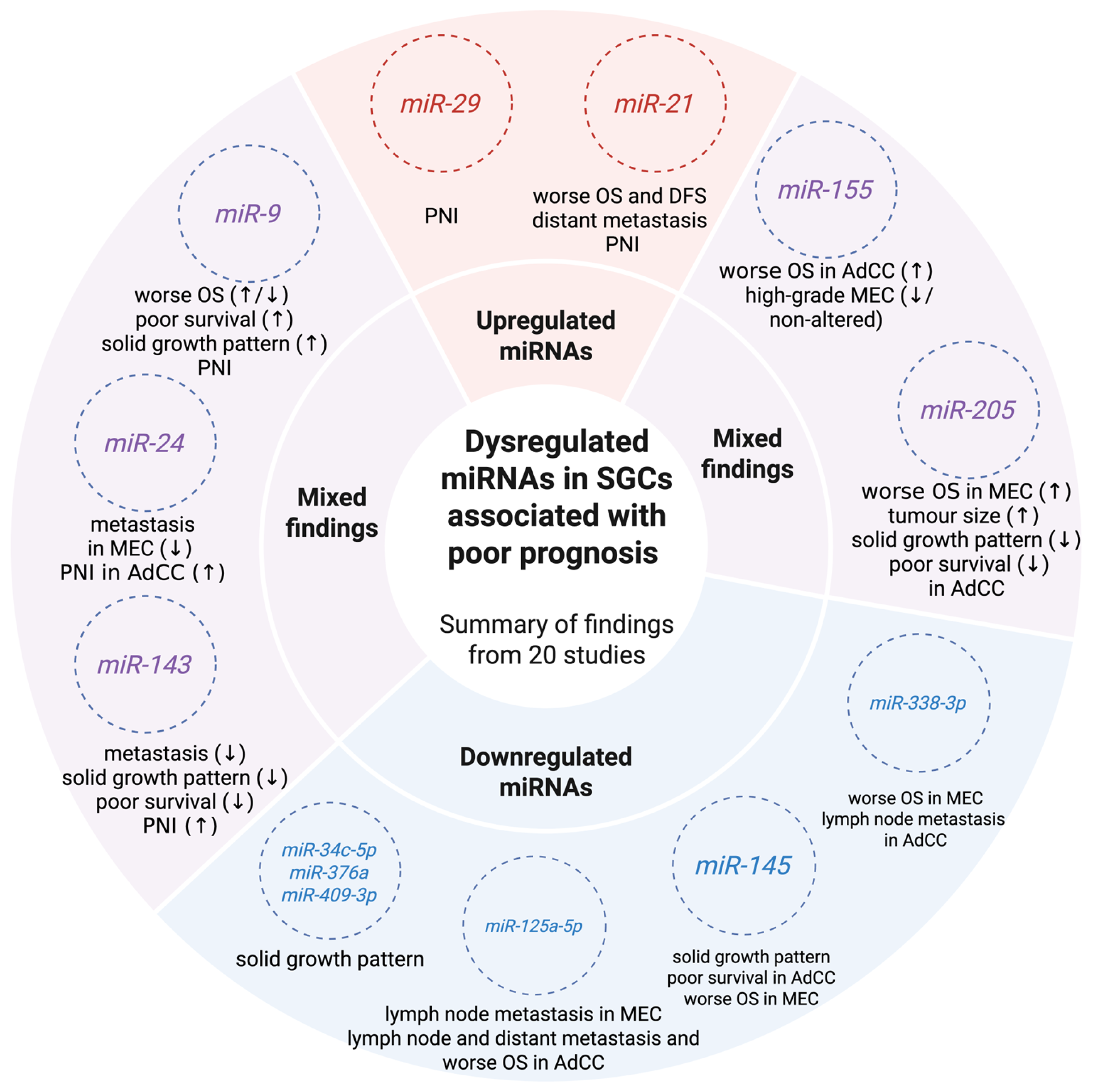
| MiRNA | Role | Propose as a Prognostic Biomarker in Other Cancers | References |
|---|---|---|---|
| miRNA-9 | oncomiR/tumour suppressor | ↑ miR-9-3p vascular invasion, perineural invasion (sinonasal SCC) ↑ miR-9-5p lymph node metastasis, tumour growth, angiogenesis, invasion and influence on radiosensitivity (cervical cancer) ↓ OS (clear cell renal cell carcinoma) ↓ miR-9-5p regional recurrence (sinonasal SCC) | Kovaříková et al. [65], Wei et al. [83], Xie et al. [84] |
| ↑ miRNA-21 | oncomiR | HNC (including oral SCC) ↓ OS, ↓ DFS (HNC (including oral SCC); breast cancer, cholangiocarcinoma) PNI (HNC (including SCC); cholangiocarcinoma) advanced stage (HNC (including SCC) lymph node metastasis (cholangiocarcinoma) | Mariani et al. [85], Yu et al. [77], Šimić et al. [86], Hedbäck et al. [87] Huang et al. [88], Lü et al. [89] |
| miRNA-29 | oncomiR/ tumour suppressor | ↓ miR-29 cisplatin resistance in ovarian cancer; multiple clinicopathological associations reported in various cancers, sometimes with opposing effects | Yu et al. [90], Menon et al. [30], Kwon et al. [66] |
| miRNA-155 | oncomiR/ tumour suppressor | ↑ miR-155 lymph node/distant metastasis, advanced stage, ↓ overall survival, chemoresistance to cisplatin and 5-fluorouracil, affects immune response, thus immunotherapy (colorectal cancer) ↓ miR-155 progression and metastasis (colorectal cancer) ↓ OS (breast cancer) ↑ miR-155-5p regional recurrence, advanced stage (sinonasal SCC). ↓ survival, tumour grade, lymph node metastasis, paclitaxel resistance, invasive breast cancer vs. ductal carcinoma in situ (breast cancer) ↑ miR-155-3p reverses paclitaxel resistance (breast cancer) | Hussen et al. [62], Lü et al. [89], Kovaříková et al. [65], Li et al. [63], Zhang et al. [64] |
| miRNA-205 | oncomiR/tumour suppressor | ↑ miR-205 local recurrence, infiltrative growth pattern, PNI (cutaneous SCC). advanced stage, metastasis (ovarian cancer) ↓ miR-205-5p ↓ OS, distant metastasis, advanced stage (gastric cancer) | Cañueto et al. [91] He et al. [92], Zhang et al. [93], Qin et al. [61] |
5. Future Perspectives
6. Strengths and Limitations
7. Conclusions
Author Contributions
Funding
Data Availability Statement
Conflicts of Interest
Abbreviations
| AdCC | adenoid cystic carcinoma |
| CPS | cumulative proportion survival |
| CSR | cumulative survival rate |
| DFS | disease-free survival |
| DMFS | distant metastasis-free survival |
| EMT | epithelial–mesenchymal transition |
| FFPE | formalin-fixed, paraffin-embedded |
| FISH | fluorescent in situ hybridization |
| HNCs | head and neck cancers |
| ISH | in situ hybridization |
| MEC | mucoepidermoid carcinoma |
| NP | not precise |
| NS | not significant |
| R/M | recurrent or metastatic |
| qRT-PCR | quantitative real-time PCR |
| QUIPS | Quality in Prognosis Studies |
| OS | overall survival |
| SCC | squamous cell carcinoma |
| SGCs | salivary gland cancers |
| WHO | World Health Organization |
References
- Gatta, G.; Guzzo, M.; Locati, L.D.; McGurk, M.; Prott, F.J. Major and minor salivary gland tumours. Crit. Rev. Oncol. Hematol. 2020, 152, 102959. [Google Scholar] [CrossRef] [PubMed]
- Skálová, A.; Hyrcza, M.D.; Leivo, I. Update from the 5th Edition of the World Health Organization Classification of Head and Neck Tumors: Salivary Glands. Head Neck Pathol. 2022, 16, 40–53. [Google Scholar] [CrossRef] [PubMed]
- WHO Classiffcation of Tumours Editorial Board. Head and Neck tumours. In WHO Classifcation of Tumours Series, 5th ed.; IARC: Lyon, France, 2022. [Google Scholar]
- Xu, B.; Mneimneh, W.; Torrence, D.E.; Higgins, K.; Klimstra, D.; Ghossein, R.; Katabi, N. Misinterpreted Myoepithelial Carcinoma of Salivary Gland: A Challenging and Potentially Significant Pitfall. Am. J. Surg. Pathol. 2019, 43, 601–609. [Google Scholar] [CrossRef] [PubMed]
- Cormier, C.; Agarwal, S. Myoepithelial Carcinoma Ex-Pleomorphic Adenoma: A Rare Pathology Misdiagnosed as Pleomorphic Adenoma; With a Novel TERT Promoter Mutation and High PD-L1 Expression. Head Neck Pathol. 2022, 16, 322–330. [Google Scholar] [CrossRef] [PubMed]
- Hernandez-Prera, J.C.; Skálová, A.; Franchi, A.; Rinaldo, A.; Poorten, V.V.; Zbären, P.; Ferlito, A.; Wenig, B.M. Pleomorphic adenoma: The great mimicker of malignancy. Histopathology 2021, 79, 279–290. [Google Scholar] [CrossRef] [PubMed]
- Jia, M.Q.; Gao, M.; Ye, P.; Gao, Y.; Yu, G.Y.; Wang, H.H.; Peng, X. Survival Outcome of Salivary Gland Carcinoma: A 50-Year Retrospective Study With Long-Term Follow-up. J. Oral Maxillofac. Surg. 2022, 80, 2003–2014. [Google Scholar] [CrossRef] [PubMed]
- Liu, F.; Chu, Y.; Zheng, Q.; Hu, Y.; Wang, Y.; Qin, L.; Fu, S.; Wang, S. Major and minor perineural invasion in salivary gland cancer. Front. Oncol. 2024, 14, 1466196. [Google Scholar] [CrossRef] [PubMed]
- Terhaard, C.H.; Lubsen, H.; Van der Tweel, I.; Hilgers, F.J.; Eijkenboom, W.M.; Marres, H.A.; Tjho-Heslinga, R.E.; de Jong, J.M.; Roodenburg, J.L. Salivary gland carcinoma: Independent prognostic factors for locoregional control, distant metastases, and overall survival: Results of the Dutch head and neck oncology cooperative group. Head Neck 2004, 26, 681–692; discussion 692–693. [Google Scholar] [CrossRef]
- Kucharska, E.; Rzepakowska, A.; Żurek, M.; Pikul, J.; Daniel, P.; Oleszczak, A.; Niemczyk, K. Oncologic outcomes of the most prevalent major salivary gland cancers: Retrospective cohort study from single center. Eur. Arch. Otorhinolaryngol. 2024, 281, 4305–4313. [Google Scholar] [CrossRef] [PubMed]
- Hirvonen, K.; Bäck, L.; Saarilahti, K.; Leivo, I.; Hagström, J.; Mäkitie, A.A. Pattern of recurrent disease in major salivary gland adenocystic carcinoma. Virchows Arch. 2015, 467, 19–25. [Google Scholar] [CrossRef] [PubMed]
- Young, A.; Okuyemi, O.T. Malignant Salivary Gland Tumors. In StatPearls; StatPearls Publishing LLC.: Treasure Island, FL, USA, 2022. [Google Scholar]
- Lombardi, D.; Tomasoni, M.; Lorini, L.; Gurizzan, C.; Tomasini, D.; Ardighieri, L.; Battocchio, S.; Bozzola, A.; Mattavelli, D.; Paderno, A.; et al. Baseline prognostic factors affecting survival in recurrent and/or metastatic salivary gland adenoid cystic carcinoma. Oral. Oncol. 2022, 126, 105764. [Google Scholar] [CrossRef] [PubMed]
- van Weert, S.; van der Waal, I.; Witte, B.I.; Leemans, C.R.; Bloemena, E. Histopathological grading of adenoid cystic carcinoma of the head and neck: Analysis of currently used grading systems and proposal for a simplified grading scheme. Oral. Oncol. 2015, 51, 71–76. [Google Scholar] [CrossRef] [PubMed]
- Hamper, K.; Lazar, F.; Dietel, M.; Caselitz, J.; Berger, J.; Arps, H.; Falkmer, U.; Auer, G.; Seifert, G. Prognostic factors for adenoid cystic carcinoma of the head and neck: A retrospective evaluation of 96 cases. J. Oral. Pathol. Med. 1990, 19, 101–107. [Google Scholar] [CrossRef] [PubMed]
- Skalova, A.; Leivo, I.; Hellquist, H.; Agaimy, A.; Simpson, R.H.W.; Stenman, G.; Vander Poorten, V.; Bishop, J.A.; Franchi, A.; Hernandez-Prera, J.C.; et al. High-grade Transformation/Dedifferentiation in Salivary Gland Carcinomas: Occurrence Across Subtypes and Clinical Significance. Adv. Anat. Pathol. 2021, 28, 107–118. [Google Scholar] [CrossRef] [PubMed]
- Bjørndal, K.; Krogdahl, A.; Therkildsen, M.H.; Charabi, B.; Kristensen, C.A.; Andersen, E.; Schytte, S.; Primdahl, H.; Johansen, J.; Pedersen, H.B.; et al. Salivary adenoid cystic carcinoma in Denmark 1990–2005: Outcome and independent prognostic factors including the benefit of radiotherapy. Results of the Danish Head and Neck Cancer Group (DAHANCA). Oral Oncol. 2015, 51, 1138–1142. [Google Scholar] [CrossRef] [PubMed]
- Haderlein, M.; Scherl, C.; Semrau, S.; Lettmaier, S.; Uter, W.; Neukam, F.W.; Iro, H.; Agaimy, A.; Fietkau, R. High-grade histology as predictor of early distant metastases and decreased disease-free survival in salivary gland cancer irrespective of tumor subtype. Head Neck 2016, 38, E2041–E2048. [Google Scholar] [CrossRef] [PubMed]
- Barrett, A.W.; Speight, P.M. Perineural invasion in adenoid cystic carcinoma of the salivary glands: A valid prognostic indicator? Oral Oncol. 2009, 45, 936–940. [Google Scholar] [CrossRef] [PubMed]
- Kazemian, E.; Solinski, M.; Adams, W.; Moore, M.; Thorpe, E.J. The role of perineural invasion in parotid malignancy outcomes: A systematic review and meta-analysis. Oral Oncol. 2022, 130, 105937. [Google Scholar] [CrossRef] [PubMed]
- Nam, S.J.; Roh, J.L.; Cho, K.J.; Choi, S.H.; Nam, S.Y.; Kim, S.Y. Risk Factors and Survival Associated with Distant Metastasis in Patients with Carcinoma of the Salivary Gland. Ann. Surg. Oncol. 2016, 23, 4376–4383. [Google Scholar] [CrossRef] [PubMed]
- Pikul, J.; Machnicki, M.M.; Rzepakowska, A.; Winiarska, N.; Chudy, A.; Moskowicz, A.; Król, K.; Fus, Ł.; Kostrzewa, G.; Stokłosa, T. Potentially actionable molecular alterations in particular related to poor oncologic outcomes in salivary gland carcinomas. BMC Cancer 2025, 25, 42. [Google Scholar] [CrossRef] [PubMed]
- van Herpen, C.; Vander Poorten, V.; Skalova, A.; Terhaard, C.; Maroldi, R.; van Engen, A.; Baujat, B.; Locati, L.D.; Jensen, A.D.; Smeele, L.; et al. Salivary gland cancer: ESMO-European Reference Network on Rare Adult Solid Cancers (EURACAN) Clinical Practice Guideline for diagnosis, treatment and follow-up. ESMO Open 2022, 7, 100602, Correction in ESMO Open 2023, 8, 101630. https://doi.org/10.1016/j.esmoop.2023.101630. [Google Scholar] [CrossRef] [PubMed]
- Geiger, J.L.; Ismaila, N.; Beadle, B.; Caudell, J.J.; Chau, N.; Deschler, D.; Glastonbury, C.; Kaufman, M.; Lamarre, E.; Lau, H.Y.; et al. Management of Salivary Gland Malignancy: ASCO Guideline. J. Clin. Oncol. 2021, 39, 1909–1941. [Google Scholar] [CrossRef] [PubMed]
- Pikul, J.; Rzepakowska, A. Molecular landscape of salivary gland malignancies. What is already known? Contemp. Oncol./Współczesna Onkol. 2024, 28, 201–216, Erratum in Contemp. Oncol./Współczesna Onkol. 2025, 29, 215–216. https://doi.org/10.5114/wo.2025.151841. [Google Scholar] [CrossRef] [PubMed]
- Peng, Y.; Croce, C.M. The role of MicroRNAs in human cancer. Signal Transduct. Target. Ther. 2016, 1, 15004. [Google Scholar] [CrossRef] [PubMed]
- Saliminejad, K.; Khorshid, H.R.K.; Fard, S.S.; Ghaffari, S.H. An overview of microRNAs: Biology, functions, therapeutics, and analysis methods. J. Cell. Physiol. 2019, 234, 5451–5465. [Google Scholar] [CrossRef] [PubMed]
- Smolarz, B.; Durczyński, A.; Romanowicz, H.; Szyłło, K.; Hogendorf, P. miRNAs in Cancer (Review of Literature). Int. J. Mol. Sci. 2022, 23, 2805. [Google Scholar] [CrossRef] [PubMed]
- Kimura, T.C.; de Lima-Souza, R.A.; Maciel, T.F.; Kowalski, L.P.; Coutinho-Camillo, C.M.; Egal, E.S.A.; Altemani, A.; Mariano, F.V. Dynamic Role of miRNAs in Salivary Gland Carcinomas: From Biomarkers to Therapeutic Targets. Head Neck Pathol. 2024, 18, 12. [Google Scholar] [CrossRef] [PubMed]
- Menon, A.; Abd-Aziz, N.; Khalid, K.; Poh, C.L.; Naidu, R. miRNA: A Promising Therapeutic Target in Cancer. Int. J. Mol. Sci. 2022, 23, 11502. [Google Scholar] [CrossRef] [PubMed]
- Bandres, E.; Agirre, X.; Bitarte, N.; Ramirez, N.; Zarate, R.; Roman-Gomez, J.; Prosper, F.; Garcia-Foncillas, J. Epigenetic regulation of microRNA expression in colorectal cancer. Int. J. Cancer 2009, 125, 2737–2743. [Google Scholar] [CrossRef] [PubMed]
- Tsai, K.W.; Liao, Y.L.; Wu, C.W.; Hu, L.Y.; Li, S.C.; Chan, W.C.; Ho, M.R.; Lai, C.H.; Kao, H.W.; Fang, W.L.; et al. Aberrant hypermethylation of miR-9 genes in gastric cancer. Epigenetics 2011, 6, 1189–1197. [Google Scholar] [CrossRef] [PubMed]
- Naakka, E.; Barros-Filho, M.C.; Adnan-Awad, S.; Al-Samadi, A.; Marchi, F.A.; Kuasne, H.; Korelin, K.; Suleymanova, I.; Brown, A.L.; Scapulatempo-Neto, C.; et al. miR-22 and miR-205 Drive Tumor Aggressiveness of Mucoepidermoid Carcinomas of Salivary Glands. Front. Oncol. 2021, 11, 786150. [Google Scholar] [CrossRef] [PubMed]
- Cinpolat, O.; Unal, Z.N.; Ismi, O.; Gorur, A.; Unal, M. Comparison of microRNA profiles between benign and malignant salivary gland tumors in tissue, blood and saliva samples: A prospective, case-control study. Braz. J. Otorhinolaryngol. 2017, 83, 276–284. [Google Scholar] [CrossRef] [PubMed]
- Page, M.J.; McKenzie, J.E.; Bossuyt, P.M.; Boutron, I.; Hoffmann, T.C.; Mulrow, C.D.; Shamseer, L.; Tetzlaff, J.M.; Akl, E.A.; Brennan, S.E.; et al. The PRISMA 2020 statement: An updated guideline for reporting systematic reviews. BMJ 2021, 372, n71. [Google Scholar] [CrossRef] [PubMed]
- Ouzzani, M.; Hammady, H.; Fedorowicz, Z.; Elmagarmid, A. Rayyan-a web and mobile app for systematic reviews. Syst. Rev. 2016, 5, 210. [Google Scholar] [CrossRef] [PubMed]
- Haddaway, N.R.; Page, M.J.; Pritchard, C.C.; McGuinness, L.A. PRISMA2020: An R package and Shiny app for producing PRISMA 2020-compliant flow diagrams, with interactivity for optimised digital transparency and Open Synthesis. Campbell Syst. Rev. 2022, 18, e1230. [Google Scholar] [CrossRef] [PubMed]
- Hayden, J.A.; van der Windt, D.A.; Cartwright, J.L.; Côté, P.; Bombardier, C. Assessing bias in studies of prognostic factors. Ann. Intern. Med. 2013, 158, 280–286. [Google Scholar] [CrossRef] [PubMed]
- McGuinness, L.A.; Higgins, J.P.T. Risk-of-bias VISualization (robvis): An R package and Shiny web app for visualizing risk-of-bias assessments. Res. Synth. Methods 2021, 12, 55–61. [Google Scholar] [CrossRef] [PubMed]
- Andreasen, S.; Tan, Q.; Agander, T.K.; Hansen, T.V.O.; Steiner, P.; Bjørndal, K.; Høgdall, E.; Larsen, S.R.; Erentaite, D.; Olsen, C.H.; et al. MicroRNA dysregulation in adenoid cystic carcinoma of the salivary gland in relation to prognosis and gene fusion status: A cohort study. Virchows Arch. 2018, 473, 329–340. [Google Scholar] [CrossRef] [PubMed]
- Bayat, P.; Mahdavi, N.; Younespour, S.; Kardouni Khoozestani, N. Interactive role of miR-29, miR-93, miR-205, and VEGF in salivary adenoid cystic carcinoma. Clin. Exp. Dent. Res. 2023, 9, 112–121. [Google Scholar] [CrossRef] [PubMed]
- Chen, B.J.; Jiang, J.; Li, T.; Jiang, H.J.; Liang, X.H.; Tang, Y.L. miR-183-5p overexpression orchestrates collective invasion in salivary adenoid cystic carcinoma through the FAT1/YAP1 signaling pathway. Biochem. Biophys. Res. Commun. 2023, 655, 127–137. [Google Scholar] [CrossRef] [PubMed]
- Dai, L.; Xian, H.; Wang, H.; Li, M.; Zhang, M.; Liang, X.H.; Tang, Y.L. Hypoxia induced cell dormancy of salivary adenoid cystic carcinoma through miR-922/DEC2 axis. Transl. Oncol. 2024, 40, 101868. [Google Scholar] [CrossRef] [PubMed]
- Fu, M.; Chen, C.W.; Yang, L.Q.; Yang, W.W.; Du, Z.H.; Li, Y.R.; Li, S.L.; Ge, X.Y. MicroRNA-103a-3p promotes metastasis by targeting TPD52 in salivary adenoid cystic carcinoma. Int. J. Oncol. 2020, 57, 574–586. [Google Scholar] [CrossRef] [PubMed]
- Jiang, L.H.; Ge, M.H.; Hou, X.X.; Cao, J.; Hu, S.S.; Lu, X.X.; Han, J.; Wu, Y.C.; Liu, X.; Zhu, X.; et al. miR-21 regulates tumor progression through the miR-21-PDCD4-Stat3 pathway in human salivary adenoid cystic carcinoma. Lab. Investig. 2015, 95, 1398–1408. [Google Scholar] [CrossRef] [PubMed]
- Kerche, L.E.; de Sousa, E.A.; Squarize, C.H.; Oliveira, K.K.; Marchi, F.A.; Bettim, B.B.; Kowalski, L.P.; Soares, F.A.; Lourenço, S.V.; Coutinho-Camillo, C.M. EMT in salivary gland tumors: The expression of microRNAs miR-155 and miR-200c is associated with clinical-pathological parameters. Mol. Biol. Rep. 2022, 49, 2157–2167. [Google Scholar] [CrossRef] [PubMed]
- Li, J.; Zhao, H. miR-429 suppresses progression and metastasis of salivary adenoid cystic carcinoma by targeting ZEB1. J. Mol. Histol. 2025, 56, 224. [Google Scholar] [CrossRef] [PubMed]
- Li, M.; Zhang, X.; Zhang, L.H.; Wang, J.R.; Fan, H.Y.; Dou, Z.C.; Liang, X.H.; Tang, Y.L. Injectable thermosensitive hydrogel-encapsulated paclitaxel liposomes enriched with miR-455-3p antagomir for dormancy therapy of salivary adenoid cystic carcinoma. J. Mater. Chem. B 2025, 13, 8749–8766. [Google Scholar] [CrossRef] [PubMed]
- Li, Z.; Zhang, Q.; Su, H.; Li, H.Y.; Cao, G.; Xu, J.K.; Wang, J.L.; Niu, C.Z.; Zhang, F.; Yang, J.; et al. miR-5191 acts as a tumor suppressor in salivary adenoid cystic carcinoma by targeting Notch-2. Oral. Dis. 2022, 28, 1871–1881. [Google Scholar] [CrossRef] [PubMed]
- Liang, Y.; Ye, J.; Jiao, J.; Zhang, J.; Lu, Y.; Zhang, L.; Wan, D.; Duan, L.; Wu, Y.; Zhang, B. Down-regulation of miR-125a-5p is associated with salivary adenoid cystic carcinoma progression via targeting p38/JNK/ERK signal pathway. Am. J. Transl. Res. 2017, 9, 1101–1113. [Google Scholar] [PubMed] [PubMed Central]
- Mitani, Y.; Roberts, D.B.; Fatani, H.; Weber, R.S.; Kies, M.S.; Lippman, S.M.; El-Naggar, A.K. MicroRNA profiling of salivary adenoid cystic carcinoma: Association of miR-17-92 upregulation with poor outcome. PLoS ONE 2013, 8, e66778. [Google Scholar] [CrossRef] [PubMed]
- Santos, P.R.B.; Coutinho-Camillo, C.M.; Soares, F.A.; Freitas, V.S.; Vilas-Bôas, D.S.; Xavier, F.C.A.; Rocha, C.A.G.; de Araújo, I.B.; Santos, J.N.D. MicroRNAs expression pattern related to mast cell activation and angiogenesis in paraffin-embedded salivary gland tumors. Pathol. Res. Pract. 2017, 213, 1470–1476. [Google Scholar] [CrossRef] [PubMed]
- Sun, L.; Liu, B.; Lin, Z.; Yao, Y.; Chen, Y.; Li, Y.; Chen, J.; Yu, D.; Tang, Z.; Wang, B.; et al. MiR-320a acts as a prognostic factor and Inhibits metastasis of salivary adenoid cystic carcinoma by targeting ITGB3. Mol. Cancer 2015, 14, 96. [Google Scholar] [CrossRef] [PubMed]
- Trevizani, M.E.S.; Marchi, F.A.; Bizinelli, D.; Oliveira, K.K.; Mariano, F.V.; Nagano, C.P.; Costa, F.D.; Pinto, C.A.L.; Kowalski, L.P.; Lourenço, S.V.; et al. MicroRNA Profiles Distinguishing Metastatic from Non-Metastatic Salivary Mucoepidermoid Carcinoma. J. Clin. Med. 2025, 14, 4957. [Google Scholar] [CrossRef] [PubMed]
- Wang, S.; Zhang, L.; Shi, P.; Zhang, Y.; Zhou, H.; Cao, X. Genome-wide profiles of metastasis-associated mRNAs and microRNAs in salivary adenoid cystic carcinoma. Biochem. Biophys. Res. Commun. 2018, 500, 632–638. [Google Scholar] [CrossRef] [PubMed]
- Wang, W.W.; Chen, B.; Lei, C.B.; Liu, G.X.; Wang, Y.G.; Yi, C.; Wang, Y.Y.; Zhang, S.Y. miR-582-5p inhibits invasion and migration of salivary adenoid cystic carcinoma cells by targeting FOXC1. Jpn. J. Clin. Oncol. 2017, 47, 690–698. [Google Scholar] [CrossRef] [PubMed]
- Xie, S.; Yu, X.; Li, Y.; Ma, H.; Fan, S.; Chen, W.; Pan, G.; Wang, W.; Zhang, H.; Li, J.; et al. Upregulation of lncRNA ADAMTS9-AS2 Promotes Salivary Adenoid Cystic Carcinoma Metastasis via PI3K/Akt and MEK/Erk Signaling. Mol. Ther. 2018, 26, 2766–2778. [Google Scholar] [CrossRef] [PubMed]
- Zanon, M.F.; Scapulatempo-Neto, C.; Gama, R.R.; Marques, M.M.C.; Reis, R.M.; Evangelista, A.F. Identification of MicroRNA Expression Profiles Related to the Aggressiveness of Salivary Gland Adenoid Cystic Carcinomas. Genes 2023, 14, 1220. [Google Scholar] [CrossRef] [PubMed]
- Wang, C.; Li, T.; Yan, F.; Cai, W.; Zheng, J.; Jiang, X.; Sun, J. Effect of simvastatin and microRNA-21 inhibitor on metastasis and progression of human salivary adenoid cystic carcinoma. Biomed. Pharmacother. 2018, 105, 1054–1061. [Google Scholar] [CrossRef] [PubMed]
- Yan, F.; Wang, C.; Li, T.; Cai, W.; Sun, J. Role of miR-21 in the growth and metastasis of human salivary adenoid cystic carcinoma. Mol. Med. Rep. 2018, 17, 4237–4244. [Google Scholar] [CrossRef] [PubMed]
- Qin, A.Y.; Zhang, X.W.; Liu, L.; Yu, J.P.; Li, H.; Wang, S.Z.; Ren, X.B.; Cao, S. MiR-205 in cancer: An angel or a devil? Eur. J. Cell Biol. 2013, 92, 54–60. [Google Scholar] [CrossRef] [PubMed]
- Hussen, B.M.; Sulaiman, S.H.A.; Abdullah, S.R.; Hidayat, H.J.; Khudhur, Z.O.; Eslami, S.; Samsami, M.; Taheri, M. MiRNA-155: A double-edged sword in colorectal cancer progression and drug resistance mechanisms. Int. J. Biol. Macromol. 2025, 299, 140134. [Google Scholar] [CrossRef] [PubMed]
- Li, Y.; Zhang, L.; Dong, Z.; Xu, H.; Yan, L.; Wang, W.; Yang, Q.; Chen, C. MicroRNA-155-5p promotes tumor progression and contributes to paclitaxel resistance via TP53INP1 in human breast cancer. Pathol. Res. Pract. 2021, 220, 153405. [Google Scholar] [CrossRef] [PubMed]
- Zhang, L.; Chen, T.; Yan, L.; Xu, H.; Wang, Y.; Li, Y.; Wang, H.; Chen, S.; Wang, W.; Chen, C.; et al. MiR-155-3p acts as a tumor suppressor and reverses paclitaxel resistance via negative regulation of MYD88 in human breast cancer. Gene 2019, 700, 85–95. [Google Scholar] [CrossRef] [PubMed]
- Kovaříková, J.; Baranová, I.; Laco, J.; Rozkošová, K.; Vošmíková, H.; Vošmík, M.; Dundr, P.; Němejcová, K.; Michálek, J.; Palička, V.; et al. Deregulation of Selected MicroRNAs in Sinonasal Squamous Cell Carcinoma: Searching for Potential Prognostic Biomarkers. Folia Biol. 2019, 65, 142–151. [Google Scholar] [CrossRef]
- Kwon, J.J.; Factora, T.D.; Dey, S.; Kota, J. A Systematic Review of miR-29 in Cancer. Mol. Ther. Oncolytics 2019, 12, 173–194. [Google Scholar] [CrossRef] [PubMed]
- Ragab, M.A.; Fathy, N.; Attia, Y.M.; Amer, E.A.; Fahim, S.A. The paradox of miR-107 in oncology: Dual roles as a tumor suppressor and oncomiR. Cancer Gene Ther. 2026. [Google Scholar] [CrossRef] [PubMed]
- Annese, T.; Tamma, R.; De Giorgis, M.; Ribatti, D. microRNAs Biogenesis, Functions and Role in Tumor Angiogenesis. Front. Oncol. 2020, 10, 581007. [Google Scholar] [CrossRef] [PubMed]
- Svoronos, A.A.; Engelman, D.M.; Slack, F.J. OncomiR or Tumor Suppressor? The Duplicity of MicroRNAs in Cancer. Cancer Res. 2016, 76, 3666–3670. [Google Scholar] [CrossRef] [PubMed]
- Re, M.; Magliulo, G.; Gioacchini, F.M.; Bajraktari, A.; Bertini, A.; Çeka, A.; Rubini, C.; Ferrante, L.; Procopio, A.D.; Olivieri, F. Expression Levels and Clinical Significance of miR-21-5p, miR-let-7a, and miR-34c-5p in Laryngeal Squamous Cell Carcinoma. Biomed. Res. Int. 2017, 2017, 3921258. [Google Scholar] [CrossRef] [PubMed]
- Wu, H.; Huang, M.; Lu, M.; Zhu, W.; Shu, Y.; Cao, P.; Liu, P. Regulation of microtubule-associated protein tau (MAPT) by miR-34c-5p determines the chemosensitivity of gastric cancer to paclitaxel. Cancer Chemother. Pharmacol. 2013, 71, 1159–1171. [Google Scholar] [CrossRef] [PubMed]
- Liu, D.; Song, L.; Dai, Z.; Guan, H.; Kang, H.; Zhang, Y.; Yan, W.; Zhao, X.; Zhang, S. MiR-429 suppresses neurotrophin-3 to alleviate perineural invasion of pancreatic cancer. Biochem. Biophys. Res. Commun. 2018, 505, 1077–1083. [Google Scholar] [CrossRef] [PubMed]
- Yang, L.; Li, H.; Huang, A. MiR-429 and MiR-143-3p Function as Diagnostic and Prognostic Markers for Osteosarcoma. Clin. Lab. 2020, 66. [Google Scholar] [CrossRef] [PubMed]
- Wu, J.; Zhu, Y.; Liu, D.; Cong, Q.; Bai, C. Biological functions and potential mechanisms of miR-143-3p in cancers (Review). Oncol. Rep. 2024, 52, 113. [Google Scholar] [CrossRef] [PubMed]
- Xu, W.X.; Liu, Z.; Deng, F.; Wang, D.D.; Li, X.W.; Tian, T.; Zhang, J.; Tang, J.H. MiR-145: A potential biomarker of cancer migration and invasion. Am. J. Transl. Res. 2019, 11, 6739–6753. [Google Scholar] [PubMed] [PubMed Central]
- Shen, H.; Shen, J.; Wang, L.; Shi, Z.; Wang, M.; Jiang, B.H.; Shu, Y. Low miR-145 expression level is associated with poor pathological differentiation and poor prognosis in non-small cell lung cancer. Biomed. Pharmacother. 2015, 69, 301–305. [Google Scholar] [CrossRef] [PubMed]
- Yu, E.H.; Tu, H.F.; Wu, C.H.; Yang, C.C.; Chang, K.W. MicroRNA-21 promotes perineural invasion and impacts survival in patients with oral carcinoma. J. Chin. Med. Assoc. 2017, 80, 383–388. [Google Scholar] [CrossRef] [PubMed]
- Hurník, P.; Chyra, Z.; Ševčíková, T.; Štembírek, J.; Trtková, K.S.; Gaykalova, D.A.; Buchtová, M.; Hrubá, E. Epigenetic Regulations of Perineural Invasion in Head and Neck Squamous Cell Carcinoma. Front. Genet. 2022, 13, 848557. [Google Scholar] [CrossRef] [PubMed]
- Zhang, M.; Wu, J.S.; Xian, H.C.; Chen, B.J.; Wang, H.F.; Yu, X.H.; Pang, X.; Dai, L.; Jiang, J.; Liang, X.H.; et al. CXCR5 induces perineural invasion of salivary adenoid cystic carcinoma by inhibiting microRNA-187. Aging 2021, 13, 15384–15399. [Google Scholar] [CrossRef] [PubMed]
- Su, R.; Zhong, S.; Wang, P.; Lin, Z. Induction of perineural invasion in salivary adenoid cystic carcinoma by circular RNA RNF111. Clin. Transl. Oncol. 2023, 25, 3152–3164. [Google Scholar] [CrossRef] [PubMed]
- Ye, H.; Zhu, W.; Mei, L.; Lu, Z. Prognostic and clinicopathologic significance of MicroRNA-125a-5p in cancers: A meta-analysis. Medicine 2019, 98, e16685. [Google Scholar] [CrossRef] [PubMed]
- Zarlashat, Y.; Halász, J.; Dósa, E. Dysregulation of MicroRNAs in Hepatocellular Carcinoma: Targeting Oncogenic Signaling Pathways for Innovative Therapies. Int. J. Mol. Sci. 2025, 26, 8365. [Google Scholar] [CrossRef] [PubMed]
- Wei, Y.Q.; Jiao, X.L.; Zhang, S.Y.; Xu, Y.; Li, S.; Kong, B.H. MiR-9-5p could promote angiogenesis and radiosensitivity in cervical cancer by targeting SOCS5. Eur. Rev. Med. Pharmacol. Sci. 2019, 23, 7314–7326. [Google Scholar] [CrossRef] [PubMed]
- Xie, M.; Lv, Y.; Liu, Z.; Zhang, J.; Liang, C.; Liao, X.; Liang, R.; Lin, Y.; Li, Y. Identification and validation of a four-miRNA (miRNA-21-5p, miRNA-9-5p, miR-149-5p, and miRNA-30b-5p) prognosis signature in clear cell renal cell carcinoma. Cancer Manag. Res. 2018, 10, 5759–5766. [Google Scholar] [CrossRef] [PubMed]
- Mariani, M.; Castagna, C.; Boccia, S.; Pastorino, R. The Prognostic Role of Micro-RNAs in Head and Neck Cancers: An Umbrella Review. J. Pers. Med. 2021, 11, 821. [Google Scholar] [CrossRef] [PubMed]
- Šimić, I.; Božinović, K.; Gašperov, N.M.; Kordić, M.; Pešut, E.; Manojlović, L.; Grce, M.; Dediol, E.; Sabo, I.l. Head and Neck Cancer Patients’ Survival According to HPV Status, miRNA Profiling, and Tumour Features-A Cohort Study. Int. J. Mol. Sci. 2023, 24, 3344. [Google Scholar] [CrossRef] [PubMed]
- Hedbäck, N.; Jensen, D.H.; Specht, L.; Fiehn, A.M.; Therkildsen, M.H.; Friis-Hansen, L.; Dabelsteen, E.; von Buchwald, C. MiR-21 expression in the tumor stroma of oral squamous cell carcinoma: An independent biomarker of disease free survival. PLoS ONE 2014, 9, e95193. [Google Scholar] [CrossRef] [PubMed]
- Huang, Q.; Liu, L.; Liu, C.H.; You, H.; Shao, F.; Xie, F.; Lin, X.S.; Hu, S.Y.; Zhang, C.H. MicroRNA-21 regulates the invasion and metastasis in cholangiocarcinoma and may be a potential biomarker for cancer prognosis. Asian Pac. J. Cancer Prev. 2013, 14, 829–834. [Google Scholar] [CrossRef] [PubMed]
- Lü, L.; Mao, X.; Shi, P.; He, B.; Xu, K.; Zhang, S.; Wang, J. MicroRNAs in the prognosis of triple-negative breast cancer: A systematic review and meta-analysis. Medicine 2017, 96, e7085. [Google Scholar] [CrossRef] [PubMed]
- Yu, P.N.; Yan, M.D.; Lai, H.C.; Huang, R.L.; Chou, Y.C.; Lin, W.C.; Yeh, L.T.; Lin, Y.W. Downregulation of miR-29 contributes to cisplatin resistance of ovarian cancer cells. Int. J. Cancer 2014, 134, 542–551. [Google Scholar] [CrossRef] [PubMed]
- Cañueto, J.; Cardeñoso-Álvarez, E.; García-Hernández, J.L.; Galindo-Villardón, P.; Vicente-Galindo, P.; Vicente-Villardón, J.L.; Alonso-López, D.; De Las Rivas, J.; Valero, J.; Moyano-Sanz, E.; et al. MicroRNA (miR)-203 and miR-205 expression patterns identify subgroups of prognosis in cutaneous squamous cell carcinoma. Br. J. Dermatol. 2017, 177, 168–178. [Google Scholar] [CrossRef] [PubMed]
- He, L.; Zhu, W.; Chen, Q.; Yuan, Y.; Wang, Y.; Wang, J.; Wu, X. Ovarian cancer cell-secreted exosomal miR-205 promotes metastasis by inducing angiogenesis. Theranostics 2019, 9, 8206–8220. [Google Scholar] [CrossRef] [PubMed]
- Zhang, J.; Zhang, J.; Pang, X.; Chen, Z.; Zhang, Z.; Lei, L.; Xu, H.; Wen, L.; Zhu, J.; Jiang, Y.; et al. MiR-205-5p suppresses angiogenesis in gastric cancer by downregulating the expression of VEGFA and FGF1. Exp. Cell Res. 2021, 404, 112579. [Google Scholar] [CrossRef] [PubMed]
- Fukada, M.; Matsuhashi, N.; Takahashi, T.; Sugito, N.; Heishima, K.; Akao, Y.; Yoshida, K. Tumor Tissue MIR92a and Plasma MIRs21 and 29a as Predictive Biomarkers Associated with Clinicopathological Features and Surgical Resection in a Prospective Study on Colorectal Cancer Patients. J. Clin. Med. 2020, 9, 2509. [Google Scholar] [CrossRef] [PubMed]
- Bouz Mkabaah, L.; Davey, M.G.; Lennon, J.C.; Bouz, G.; Miller, N.; Kerin, M.J. Assessing the Role of MicroRNAs in Predicting Breast Cancer Recurrence-A Systematic Review. Int. J. Mol. Sci. 2023, 24, 7115. [Google Scholar] [CrossRef] [PubMed]
- Torres-Bustamante, M.I.; Vazquez-Urrutia, J.R.; Solorzano-Ibarra, F.; Ortiz-Lazareno, P.C. The Role of miRNAs to Detect Progression, Stratify, and Predict Relevant Clinical Outcomes in Bladder Cancer. Int. J. Mol. Sci. 2024, 25, 2178. [Google Scholar] [CrossRef] [PubMed]
- Xie, Y.; Ma, X.; Chen, L.; Li, H.; Gu, L.; Gao, Y.; Zhang, Y.; Li, X.; Fan, Y.; Chen, J.; et al. MicroRNAs with prognostic significance in bladder cancer: A systematic review and meta-analysis. Sci. Rep. 2017, 7, 5619. [Google Scholar] [CrossRef] [PubMed]
- Ganci, F.; Sacconi, A.; Manciocco, V.; Covello, R.; Benevolo, M.; Rollo, F.; Strano, S.; Valsoni, S.; Bicciato, S.; Spriano, G.; et al. Altered peritumoral microRNA expression predicts head and neck cancer patients with a high risk of recurrence. Mod. Pathol. 2017, 30, 1387–1401. [Google Scholar] [CrossRef] [PubMed]
- Jayaraj, R.; Polpaya, K.; Kunale, M.; Muthukaliannan, G.K.; Shetty, S.; Baxi, S.; Mani, R.R.; Paranjothy, C.; Purushothaman, V.; Kayarohanam, S.; et al. Clinical Investigation of Chemotherapeutic Resistance and miRNA Expressions in Head and Neck Cancers: A Thorough PRISMA Compliant Systematic Review and Comprehensive Meta-Analysis. Genes 2022, 13, 2325. [Google Scholar] [CrossRef] [PubMed]
- Broseghini, E.; Carosi, F.; Berti, M.; Compagno, S.; Ghelardini, A.; Fermi, M.; Querzoli, G.; Filippini, D.M. Salivary Gland Cancers in the Era of Molecular Analysis: The Role of Tissue and Liquid Biomarkers. Cancers 2025, 17, 660. [Google Scholar] [CrossRef] [PubMed]
- Tiberio, P.; Callari, M.; Angeloni, V.; Daidone, M.G.; Appierto, V. Challenges in using circulating miRNAs as cancer biomarkers. Biomed. Res. Int. 2015, 2015, 731479. [Google Scholar] [CrossRef] [PubMed]
- Roi, A.; Boia, S.; Rusu, L.-C.; Roi, C.I.; Boia, E.R.; Riviș, M. Circulating miRNA as a Biomarker in Oral Cancer Liquid Biopsy. Biomedicines 2023, 11, 965. [Google Scholar] [CrossRef] [PubMed]
- de Gonzalo-Calvo, D.; Pérez-Boza, J.; Curado, J.; Devaux, Y. Challenges of microRNA-based biomarkers in clinical application for cardiovascular diseases. Clin. Transl. Med. 2022, 12, e585. [Google Scholar] [CrossRef] [PubMed]
- Zhao, D.; Wang, Y. MicroRNAs and Cancer Racial Disparities. Wiley Interdiscip. Rev. RNA 2025, 16, e70028. [Google Scholar] [CrossRef] [PubMed]
- Gong, Z.; Wang, J.; Wang, D.; Buas, M.F.; Ren, X.; Freudenheim, J.L.; Belinsky, S.A.; Liu, S.; Ambrosone, C.B.; Higgins, M.J. Differences in microRNA expression in breast cancer between women of African and European ancestry. Carcinogenesis 2019, 40, 61–69. [Google Scholar] [CrossRef] [PubMed]


| Author | Year | Country | Study Design | Type of Malignant SGC | Localization | Malignant Samples | Material | Main Technology Methods | Number of Analyzed MiRNA with Prognosis Assessment | Key Findings | Target Genes or Signalling Pathways |
|---|---|---|---|---|---|---|---|---|---|---|---|
| Andreasen et al. [40] | 2018 | Denmark | human observational study | AdCC | major and minor salivary glands | 64—training cohort 120—validation cohort | FFPE | microarray qPCR (TaqMan) | microarray-numerous qPCR (32 highest-ranking miRNAs in survival analysis) | ↑ miR-6835-3p * ↓ RFS ↓ miR-1180 ** ↓ miR-374c *** ↑ mir-4676 ** ↓OS ↑ mir-21 *** ↑ mir-181a-2 *** ↑ mir-152 *** * results obtained by microarray in the training cohort ** results obtained by qPCR in the training cohort *** results obtained by qPCR in the validation cohort | NP |
| Bayat et al. [41] | 2022 | Iran | human tissue study | AdCC | major and minor salivary glands | 15 | FFPE | qRT-PCR (SYBR Green) | 3 | ↑ miR-29 PNI ↑ mir-93-5p histopathological grade ↑ miR-205 tumour size | VEGF PI3K/AKT and JAK/STAT pathways |
| Chen et al. [42] | 2023 | China | in vitro (cell line) study + human tissue study + animal study | AdCC | major and minor salivary glands | 25 | human tumour tissue NP | FISH | 1 | ↑ miR-183-5p (at the leading edge) minor salivary glands, PNI, distant metastasis | FAT-1/YAP1 |
| Dai et al. [43] | 2023 | China | human tissue study + in vitro (cell line) study + animal study | AdCC | major and minor salivary glands | 23-FISH 11—qRT-PCR | 23 FFPE/ 11 fresh frozen tissue (only cribriform/tubular type) | FISH qRT-PCR | 1 | ↑ miR-922 solid growth pattern, local invasion *, distant metastasis * results obtained by FISH and qRT-PCR | DEC2 HIF-1 |
| Fu et al. [44] | 2020 | China | human tissue study+ in vitro (cell line) study + animal study | AdCC | major and minor salivary glands | 52 | human tumour tissue NP | qRT-PCR (SYBR Green) | 1 | ↑ miR-103a-3p local regional recurrence, distant metastasis | TPD52 |
| Jiang et al. [45] | 2015 | China | human tissue study+ in vitro (cell line) study | AdCC | major and minor salivary glands | 35 | fresh frozen tissue | qRT-PCR (TaqMan) | 1 | ↑ miR-21 distant metastasis, cumulative DFS independent predictor of survival | PDCD4 STAT3 and several other |
| Kerche et al. [46] | 2022 | Brazil | human tissue study | AdCC MEC | major and minor salivary glands | 18 14 | FFPE | qRT-PCR (TaqMan) | 6 | ↑/↓ miR-9 ↓ OS AdCC ↑ miR-155 ↓/non-altered miR-34a ↓ OS MEC ↓/non-altered miR-138 ↓/non-altered miR-155 high grade MEC ↑ miR-200c lymph node metastasis MEC | related to EMT |
| Li, J et al. [47] | 2025 | China | human tissue study+ in vitro (cell line) study + animal study | AdCC | major and minor salivary glands | 26 | fresh frozen tissue | qRT-PCR (SYBR Green) | 1 | ↓ miR-429 T3–T4 vs. T1–T2 PNI and metastasis in cell lines | ZEB1 |
| Li, M et al. [48] | 2025 | China | human tissue study + in vitro (cell line) study + animal study | AdCC | major and minor salivary glands | 22 | FFPE | FISH | 1 | ↓ miR455-3p recurrence, ↓ OS, ↓ RFS, ↓ DMFS metastasis in vitro | GNPNAT1 |
| Li, Z et al. [49] | 2022 | China | human tissue study+ in vitro (cell line) study+ animal study | AdCC | NP | 30 | fresh frozen tissue | qRT-PCR (SYBR Green) | 1 | ↓ miR-5191 solid growth pattern, lymph node and distant metastasis, advanced stage, ↓OS/CSR | NOTCH2 |
| Liang et al. [50] | 2017 | China | human tissue study + in vitro (cell line) study + animal study | AdCC | parotid glands | 106 | human tumour tissue NP | In situ hybridization qRT-PCR * (SYBR Green) | 1 | ↓ miR-125a-5p lymph node metastasis, distant metastasis, ↓ OS independent prognostic factor for poor survival * 40 samples with metastasis or without metastasis | p38/JNK/ERK |
| Mitani et al. [51] | 2013 | USA | human observational study | AdCC | major and minor salivary glands | 30—screening 30—validation | fresh frozen tissue | MiRNA array profiling qRT-PCR (SYBR Green) for validation (miR-455-3p, miR-455-5p, miR-375, miR-142-3p, miR-17, and miR-20a) | NP | ↑ miR-17 ↓CPS ↑ miR-20a poor survival ↓: miR-143/**, miR-145/**, miR-205, miR-299-5p, miR-433, miR-452, miR-483-5p ↑: miR-9/**, miR-17, miR-20a, miR-92a, miR-501-5p, miR-545, miR-1909** solid growth pattern ↓: miR-22, miR-34c-5p, miR-127-3p/5p, miR-136/**, miR-143/**, miR-145, miR-154, miR-205, miR-329, miR-376a **/b/c, miR-377, miR-379, miR-381, miR-382, miR-409-3p, miR-410, miR-411, miR-432, miR-495, miR-654-3p ↑: miR-9/**, miR-17, miR-20a ↑ miR-let-7a tumour size, tumour stage, recurrence ↑ miR-150 lymph node, tumour stage numerous other miRNAs corelated with clinicopathological features not listed ** miRNA star strand | several |
| Naakka et al. [33] | 2022 | Brazil Finland | Human observational study + in vitro (cell line) study | MEC | major and minor salivary glands | 25 | fresh frozen tissue | Agilent SurePrint 8x60K platform | NP | ↑ miR-205-5p ↓ OS ↑ miR-224-5p ↓ miR-139-3p ↓ miR-145-3p ↓ miR-148a-3p ↓ miR-186-5p ↓ miR-338-3p ↓ miR-363-3p ↓ miR-4324 ↓ miR-582-5p high-grade MEC ↓ miR-3125 ↓ miR- 4324 (miR-22 and miR-205- migration and invasion in vitro) | Several As follow: ERK/MAPK signalling, EIF2 signalling, PI3K/AKT |
| Santos et al. [52] | 2017 | Brazil | Human tissue study | AdCC MEC | major and minor salivary glands | 11 9 | FFPE | qRT-PCR (TaqMan) | 6 (miR-9, miR-16, miR-17, miR-132, miR-195 and miR-221) | NS (histological pattern in AdCC, histological grade in MEC, tumour size, localization, PNI) | |
| Sun et al. [53] | 2015 | China | in vitro (cell line) study + animal study + human observational study | AdCC | NP | 450 (from two Centres) | human tumour tissue NP | In situ hybridization | 1 | ↓ miR-320a lymph node metastasis, distant metastasis ↓ CSR ↓ CMR independent indicator for lung metastasis | ITGB3 |
| Trevizani et al. [54] | 2025 | Brazil | human tissue study | MEC | parotid glands | 10 | FFPE | qRT-PCR (TaqMan Array Human MicroRNA A Card) | 377 | lymph node metastasis ↓: miR-16, miR-24, miR-27a, miR-106a/b, miR-125a-5p, miR-191, miR-210, miR-324-3p, miR-339-3p, miR- 374b-5p, miR-484, miR-590-5p, miR-671-3p, miR-744, miR-886-3p/5p ↓ miR-24, ↓ miR-27a distant metastasis | |
| Wang et al. [55] | 2018 | China | in vitro (cell line) study + human tissue study | AdCC | NP | 37 | fresh frozen tissue | qRT-PCR (SYBR Green) | 2 | ↓ miR-338-3p/5p lymph node metastasis | LAMC2 |
| Wang et al. [56] | 2017 | China | human tissue study+ in vitro (cell line) study + animal study | AdCC | NP | 110 | human tumour tissue NP | In situ hybridization | 1 | ↓ miR-582-5p distant metastasis, ↓CSR | FOXC1 |
| Xie et al. [57] | 2018 | China | in vitro (cell line) studv + human tissue study + in vivo | AdCC | NP | 102 | human tumour tissue NP | qRT-PCR | 1 | ↓ miR-143-3p metastasis | ITGA6 |
| Zanon et al. [58] | 2023 | Brazil | human observational study | AdCC | major and minor salivary glands | 19 | FFPE | NanoString nCounter Technology | ~800 | solid growth pattern ↑: miR-146b-3p, miR-326, miR-370-5p, miR-491-3p, miR-516b-5p, miR-631, miR-770-5p, miR-1249-3p, miR-1297, miR-4461, miR-4755-5p ↓:miR-23b-3p, miR-34c-5p, miR-154-5p, miR-181d-3p, miR-320b, miR-323a-3p, miR-330-5p, miR-376a-3p, miR-382-5p, miR-409-3p, miR-433-3p, miR-455-5p, miR-487b-3p, miR-543, miR-766-5p, miR-758-3p + miR-411-3p, miR-3127-5p, miR-3190-3p, miR-5196-3p + miR-6732-3p perineural invasion (49 dysregulated miRNAs): miR-1-3p, miR-9-5p, miR-19b-3p, miR-21-5p, miR-23b-3p, miR-24-3p, miR-27b-3p, miR-29a/c-3p, miR-30a/e-5p, miR-92a-1-5p, miR-122-5p, miR-133a-3p, miR-140-3p, miR-141-3p, miR-142-3p, miR-143-3p, miR-146b-5p, miR-151a-3p, miR-195-5p, miR-203a-3p, miR-206, miR-208b-5p, miR-302d-3p, miR-424-5p, miR-494-3p, miR-497-5p, miR-510-5p, miR-517a-3p, miR-548aa + miR-548t-3p, miR-548z + miR-548h-3p, miR-613, miR-628-3p, miR-664b-3p, miR-1224-5p, miR-1253, miR-1257, miR-1275, miR-1276, miR-1322, miR-1323, miR-1972, miR-1976, miR-3065-5p, miR-3161, miR-3180-5p, miR-4531, miR-5001-5p | several; 13 molecular pathways, such as Notch, Wnt, Hedgehog, TGFB, Hippo, MAPK, STAT, P13K, RAS, chromatin modification, transcriptional regulation, DNA damage control, cell cycle, and apoptosis. |
Disclaimer/Publisher’s Note: The statements, opinions and data contained in all publications are solely those of the individual author(s) and contributor(s) and not of MDPI and/or the editor(s). MDPI and/or the editor(s) disclaim responsibility for any injury to people or property resulting from any ideas, methods, instructions or products referred to in the content. |
© 2026 by the authors. Licensee MDPI, Basel, Switzerland. This article is an open access article distributed under the terms and conditions of the Creative Commons Attribution (CC BY) license.
Share and Cite
Pikul, J.; Cieślik, M.; Niemczyk, K.; Rzepakowska, A. MicroRNAs in Salivary Gland Cancers Associated with Poor Prognosis: A Systematic Review. Int. J. Mol. Sci. 2026, 27, 6101. https://doi.org/10.3390/ijms27146101
Pikul J, Cieślik M, Niemczyk K, Rzepakowska A. MicroRNAs in Salivary Gland Cancers Associated with Poor Prognosis: A Systematic Review. International Journal of Molecular Sciences. 2026; 27(14):6101. https://doi.org/10.3390/ijms27146101
Chicago/Turabian StylePikul, Julia, Maja Cieślik, Kazimierz Niemczyk, and Anna Rzepakowska. 2026. "MicroRNAs in Salivary Gland Cancers Associated with Poor Prognosis: A Systematic Review" International Journal of Molecular Sciences 27, no. 14: 6101. https://doi.org/10.3390/ijms27146101
APA StylePikul, J., Cieślik, M., Niemczyk, K., & Rzepakowska, A. (2026). MicroRNAs in Salivary Gland Cancers Associated with Poor Prognosis: A Systematic Review. International Journal of Molecular Sciences, 27(14), 6101. https://doi.org/10.3390/ijms27146101