Review

Research Advances in the Pathogenesis of Sepsis-Associated Encephalopathy

1
Department of Emergency Medicine, West China Hospital, Sichuan University, Chengdu 610041, China
2
West China School of Clinical Medicine, Sichuan University, Chengdu 610041, China
3
Department of Science and Technology, West China Hospital, Sichuan University, Chengdu 610041, China
*
Author to whom correspondence should be addressed.
These authors contributed equally to this work.
Int. J. Mol. Sci. 2026, 27(12), 5390; https://doi.org/10.3390/ijms27125390
Submission received: 16 May 2026 / Revised: 6 June 2026 / Accepted: 11 June 2026 / Published: 15 June 2026
(This article belongs to the Section Molecular Pathology, Diagnostics, and Therapeutics)

Abstract

Sepsis-associated encephalopathy (SAE) is a frequent neurological complication of sepsis, driven by six interconnected pathophysiological components: (1) systemic inflammation-triggered neuroinflammatory cascades, initiated by systemic recognition of pathogen-associated molecular patterns (PAMPs) and damage-associated molecular patterns (DAMPs) and propagated by pro-inflammatory mediators; (2) central nervous system (CNS) immune cell-mediated neuroinflammation, wherein microglia, regulatory T cells, and neutrophils dynamically regulate inflammatory progression; (3) blood–brain barrier (BBB) disruption, progressing from functional disturbance to structural damage via tight junction degradation and immune infiltration; (4) multimodal programmed cell death, encompassing autophagy, apoptosis, pyroptosis, and ferroptosis driven by mitochondrial dysfunction; (5) neurotransmitter network imbalance, manifesting as cholinergic deficiency and glutamate excitotoxicity; and (6) gut–brain axis dysregulation, characterized by reduced microbiota-derived metabolites such as butyrate and indolepropionic acid. These components are organized along a core pathological axis comprising four sequential stages: neuroinflammatory storm (encompassing components 1 and 2) → BBB disruption and microcirculatory disturbances (component 3) → multimodal programmed cell death (component 4) → neurotransmitter imbalance (component 5), with the gut–brain axis (component 6) functioning as a bidirectional regulatory node that intersects and modulates all four stages. Mitochondrial dysfunction serves as the central converging node linking these pathological axes. Targeted interventions against neuroinflammation, immune cell modulation, BBB restoration, inhibition of aberrant cell death, neurotransmitter homeostasis, and gut microbiota remodeling hold therapeutic promise. Elucidating the crosstalk among these pathways will accelerate the clinical translation of precision therapies for SAE.

1. Introduction

Sepsis-associated encephalopathy (SAE) occurs in more than 50% of hospitalized sepsis patients and is associated with high mortality [1]. The core clinical features of SAE include fluctuating consciousness disorders, executive dysfunction, and neurological deficits [1]. Patients with characteristic consciousness fluctuations face a three-to fivefold higher risk of death, with in-hospital mortality reaching 45–60% in severe cases [2]. Although SAE has been directly linked to worse sepsis outcomes, the complex interactions within the neuro-immune-vascular unit remain poorly understood. The crosstalk among different pathological components has not been fully elucidated. Moreover, most current research focuses on basic mechanisms, with few translational targets available for clinical practice, hindering the development of precise treatment strategies for SAE.
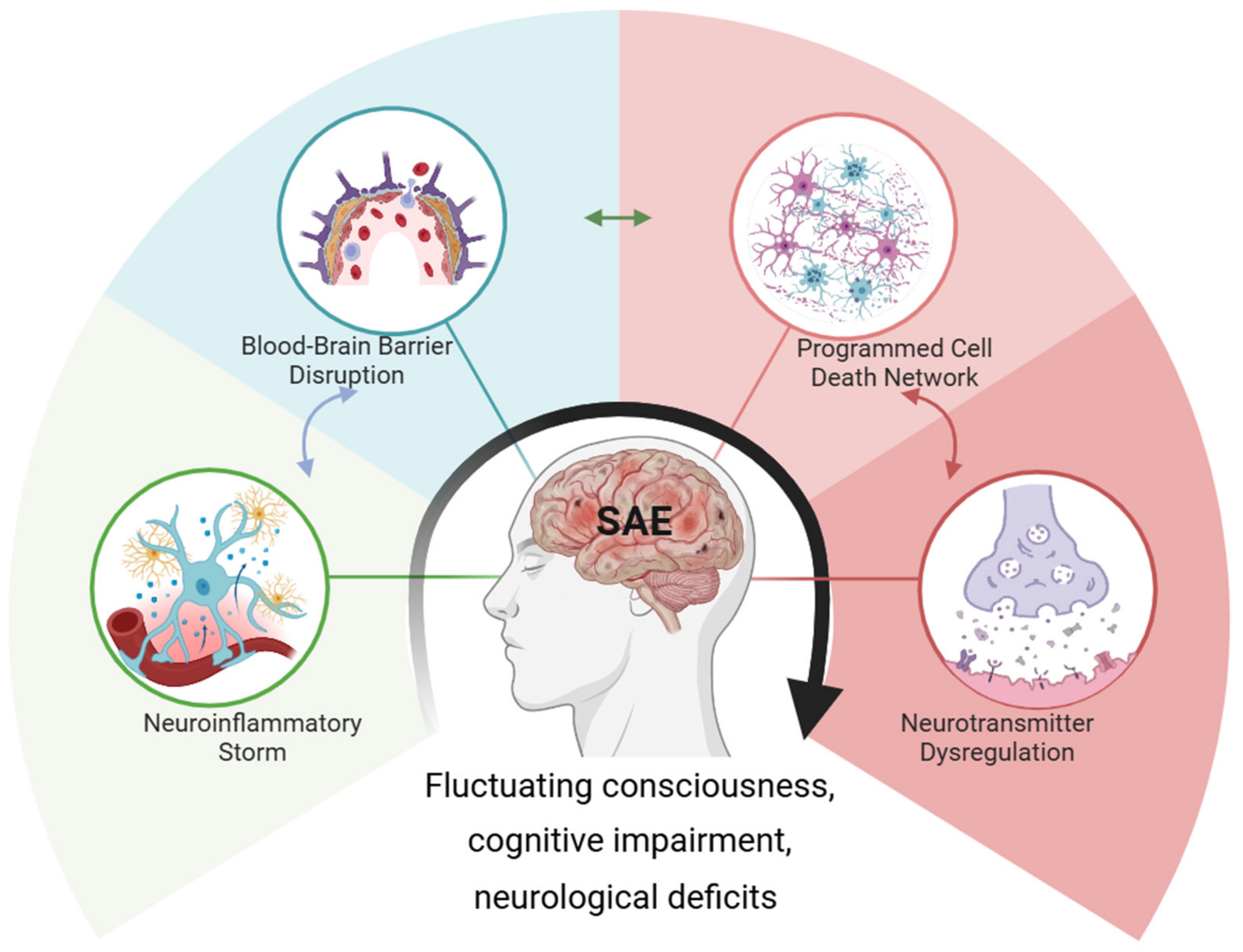
Recent advances in interdisciplinary research paradigms and multi-omics technologies have accelerated mechanistic understanding. Breakthrough discoveries—such as glial cells revealed by single-cell sequencing, dynamic BBB leakage captured by live imaging, and cerebrospinal fluid neurotransmitter imbalances identified via metabolomics—have provided new dimensions for constructing a molecular map of SAE. This review systematically examines and synthesizes current evidence on six core pathological components organized along four sequential axes (Figure 1): (1) neuroinflammatory cascades (Section 3.1) and immune cell-mediated regulation (Section 3.2), collectively constituting the neuroinflammatory storm; (2) blood–brain barrier remodeling and microcirculatory disturbances (Section 3.3); (3) multimodal programmed cell death (Section 3.4); (4) neurotransmitter network imbalance (Section 3.5). The gut–brain axis (Section 3.6) serves as a key regulatory node modulating all four axes.

2. Materials and Methods

A comprehensive literature search was conducted in PubMed, Web of Science, and Embase to identify studies on sepsis-associated encephalopathy (SAE) pathogenesis and therapeutics published between January 2015 and March 2026. The search strategy combined MeSH terms and free-text keywords including “sepsis-associated encephalopathy,” “neuroinflammation,” “blood–brain barrier,” “programmed cell death” (autophagy, apoptosis, pyroptosis, ferroptosis), “gut–brain axis,” and related molecular targets. Boolean operators were used to connect disease terms with mechanism-specific keywords. Given the breadth of mechanisms covered, separate searches were conducted for each pathological component (neuroinflammation, BBB disruption, cell death, neurotransmitter imbalance, gut–brain axis). A representative PubMed search string for the cell death component was: (“sepsis-associated encephalopathy”[Title/Abstract] OR “SAE”[Title/Abstract]) AND (“programmed cell death”[Title/Abstract] OR “autophagy”[Title/Abstract] OR “apoptosis”[Title/Abstract] OR “pyroptosis”[Title/Abstract] OR “ferroptosis”[Title/Abstract]). Similar structures were applied to other components with appropriate mechanism-specific keywords substituted.
Inclusion criteria were: (1) original research articles, reviews, or clinical studies published in English; (2) studies investigating SAE pathogenesis involving neuroinflammatory cascades, BBB disruption, cell death pathways, or gut–brain axis dysregulation; and (3) for interventional studies, reporting clear mechanisms of action; review articles were included if they systematically summarized relevant evidence.
Exclusion criteria included case reports with fewer than 3 subjects, conference abstracts without full text, and non-peer-reviewed publications.
The literature selection followed a three-stage process. Initial screening of titles and abstracts identified 2500 records, of which 380 underwent full-text review. Ultimately, 116 references were included in the primary analysis, comprising preclinical original articles, clinical studies, and reviews; additional recent studies were incorporated during revision to ensure currency.
Data were extracted and synthesized according to four core pathological components consistent with the review’s framework: neuroinflammatory storms, BBB remodeling, neurotransmitter network imbalances, and gut–brain axis dysregulation. Intervention strategies were critically classified by clinical translation stage: clinically available drugs, preclinical candidates, and potential molecular targets. This narrative review employed a critical appraisal approach, prioritizing studies with robust experimental designs and validated methods while acknowledging limitations and interpreting conflicting findings with caution.
Notably, the proposed integrated pathological axis is established by synthesizing evidence from diverse study designs. Most of the included studies are observational or associative in nature, rather than being purposely designed to verify the causal relationships of this integrated model. The causal interactions among these pathological components remain to be validated by dedicated future studies.

3. Pathogenesis and Therapeutic Strategies

3.1. Neuroinflammatory Cascades and Targeted Therapies

The proposed core pathological mechanism of SAE is initiated by a systemic inflammatory storm. Recognition of PAMPs/DAMPs triggers innate immunity, leading to a cascade release of pro-inflammatory mediators such as IL-1β, IL-6, TNF-α. These circulating factors cross the BBB or enter the CNS via pathways like the vagus nerve, activating microglia and astrocytes and forming a self-amplifying neuroinflammation cycle [3].
Neuroinflammation appears to damage neuronal function in part by directly attacking mitochondria. TNF-α and IL-1β released by activated glial cells bind to death receptors on neuronal membranes, inducing persistent opening of the mitochondrial permeability transition pore (mPTP). This leads membrane potential collapse, ATP synthesis interruption, and cytochrome c leakage, thereby initiating apoptosis [4]. Simultaneously, NADPH oxidase (NOX2) and inducible nitric oxide synthase (iNOS) are strongly upregulated in M1-polarized microglia, resulting in burst production of superoxide anion (O2) and nitric oxide (NO). These combine to form peroxynitrite (ONOO), a potent oxidant that directly damages mitochondrial electron transport chain complexes and induces mitochondrial DNA damage [5]. Inflammatory signaling also inhibits PINK1/Parkin-mediated mitophagy and disrupts PGC-1α-regulated mitochondrial biogenesis, leading to the accumulation of damaged mitochondria and widespread energy metabolism imbalance [6].
Parallel to and exacerbating neuroinflammation is severe cholinergic system dysfunction. In the septic environment, choline acetyltransferase (ChAT)-positive neurons in key brain regions such as the hippocampus are reduced in number and activity, leading to insufficient synthesis of the anti-inflammatory mediator acetylcholine (ACh) [7]. At the same time, acetylcholinesterase (AChE) activity is upregulated by inflammatory signals, accelerating ACh breakdown in the synaptic cleft. Consequently, the cholinergic anti-inflammatory pathway (CAP)—a core bridge between neural and immune systems—is impaired. Expression or downstream signaling (e.g., JAK2/STAT3) of its key receptor, the α7 nicotinic acetylcholine receptor (α7nAChR), is impaired in the inflammatory milieu, preventing ACh from effectively inhibiting nuclear factor kappa-light-chain-enhancer of activated B cells (NF-κB) nuclear translocation and pro-inflammatory factor transcription [8]. More fundamentally, sepsis-induced systemic disruption can reduce vagal efferent tone, decreasing ACh release at its source and causing widespread suppression of this physiological anti-inflammatory system [9].
Given these mechanisms, multidimensional intervention strategies are needed. Dexmedetomidine, beyond its sedative effects via α2 receptors, activates the neuronal mitochondrial deacetylase Sirt3, improving oxidative phosphorylation and reducing reactive oxygen species (ROS) production [10,11]. Enhancing mitophagy via the PINK1/Parkin pathway has also emerged as a potential strategy [3]. For the cholinergic system, selective α7nAChR agonists (PNU-282987) can more precisely activate the CAP and inhibit microglial activation [12]. Additionally, targeting the TLR4/NF-κB axis with guggulsterone (preclinical research) and selective mitochondrial antioxidant protection via high-concentration hydrogen inhalation are promising approaches [13,14]. Implementing these multi-dimensional strategies requires a deeper understanding of how different immune cells—from resident microglia to infiltrating T cells and neutrophils—dynamically regulate neuroinflammation. Notably, the central role of mitochondrial dysfunction as a converging node (Figure 1) becomes particularly evident when examining immune cell activation: microglial M1 polarization, T cell metabolic reprogramming, and neutrophil NETosis are all energy-intensive processes dependent on mitochondrial integrity. The following section details this immune cell-mediated regulation and its therapeutic targeting.

3.2. Immune Cell-Mediated Regulation of Neuroinflammation

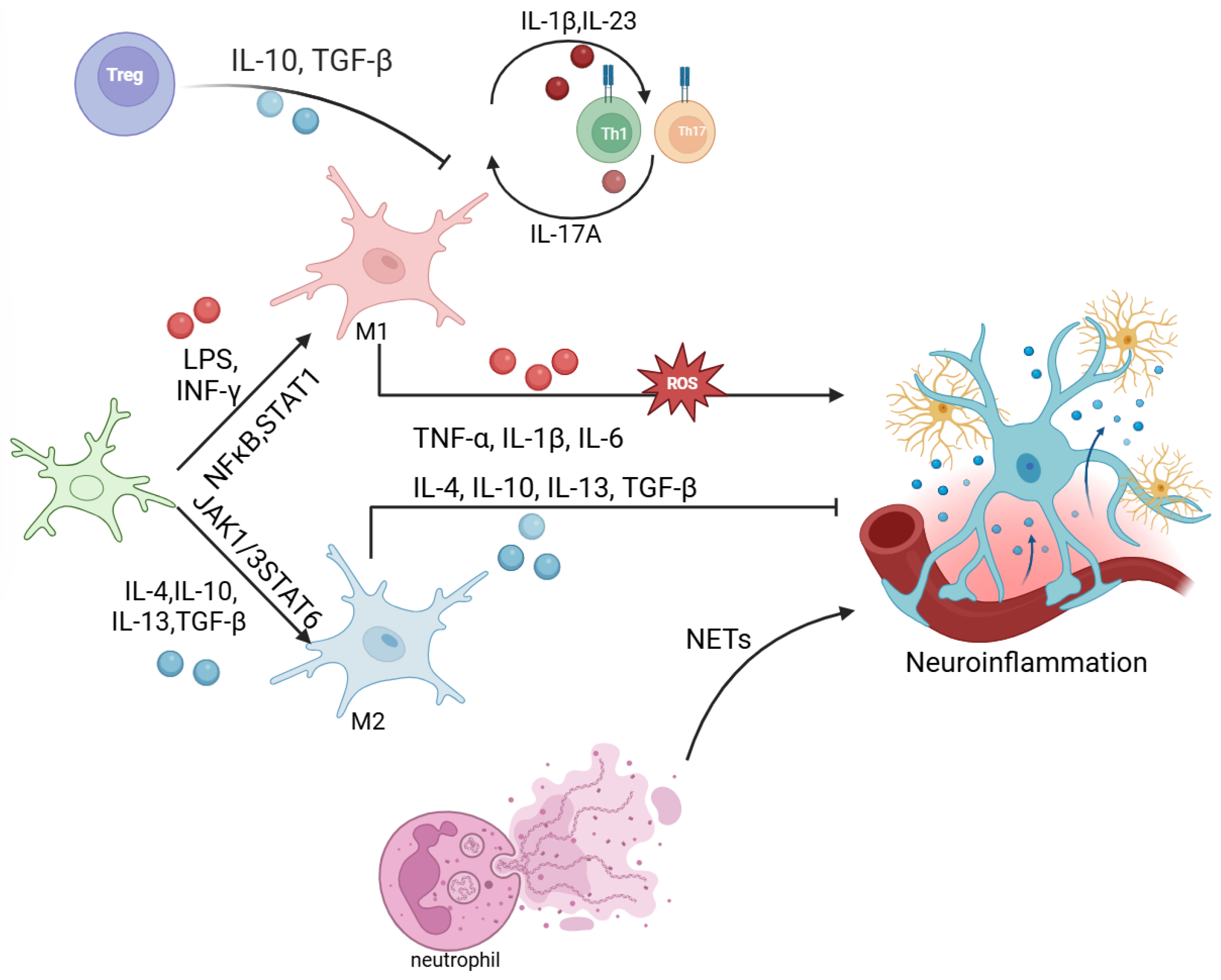
While Section 3.1 focused on how peripheral inflammation triggers neuroinflammation and directly damages neurons via mitochondrial dysfunction, this section delves into the complex regulatory network of immune cells—both CNS-resident (microglia) and infiltrating (T cells, neutrophils, mast cells)—that determines the intensity and outcome of neuroinflammation. These cells drive the initiation and amplification of neuroinflammatory cascades, thereby promoting subsequent cell death and neurotransmitter imbalance (Figure 2).

3.2.1. The Dynamic Regulatory Network of Microglia

Under physiological conditions, microglia surrounding brain microvessels help maintain BBB integrity by enhancing tight junction proteins [15]. During sepsis, these cells undergo dynamic functional transformation: early activation exerts protective effects via Foxc1-mediated IκBα/NF-κB signaling regulation [16]; however, sustained activation triggered by the RAGE ligand S100B exacerbates abnormal BBB permeability [17,18]. Infiltration of peripheral TH1/TH17 cells into the CNS leads to aberrant IL-17A secretion, which further promotes persistent microglial activation via a positive feedback loop [19]. Activated microglia secrete pro-inflammatory factors such as TNF-α, IL-6, and HMGB1. In addition to the mitochondrial damage induced by TNF-α, TNF-α also induces excitotoxic neuronal injury by causing abnormal glutamate secretion from glial cells and inhibiting astrocytic reuptake [20].
Intervention strategies targeting microglia are classified by clinical transformation stage: ① Preclinical research: CD137L neutralizing antibody TKS-1 inhibits M1 polarization [21]; α7nAChR agonist remimazolam activates the Nrf2/HO-1 pathway [22]; miR-25-3p inhibits IL-1β signaling via the TLR4/NLRP3 axis [23]; miR-494 maintains mitochondrial metabolic homeostasis [24]. HMGB1 inhibitors restore synaptic pruning balance [25,26]; MFGE8 enhances αVβ integrin-mediated efferocytosis [27,28]; the microglial depletion agent PLX5622 prevents synaptic engulfment [29]. ② Natural Compounds: β-patchoulene, β-elemene, and resveratrol glycoside exert anti-inflammatory effects by modulating pathways such as Sirt1/Nrf2 and RAC1/MLK3/p38 [30,31,32].

3.2.2. Regulation of Neuroinflammation by Regulatory T Cells

Regulatory T cells (Tregs) maintain tissue microenvironmental balance by suppressing excessive immune responses and can migrate across the BBB into CNS. Activation of nicotinic acetylcholine receptors on their surface promotes clonal expansion within the hippocampus and modulates neuroinflammation via acetylcholine signaling [33,34]. In SAE, brain-infiltrating Tregs antagonize microglial inflammatory cascades by secreting factors like IL-10 and TGF-β, and suppress astrocytic IL-6 synthesis through the amphiregulin (AREG)-EGFR signaling axis [35,36]. Caspase-1-mediated GSDMD cleavage, indicating activation of the pyroptosis pathway, has been observed in peripheral blood immune cells of sepsis patients, suggesting the involvement of programmed cell death mechanisms in disease progression [37].

3.2.3. Synergistic Effects of Neutrophils and T Cells

In sepsis models, elevated levels of IL-1β and IL-6 are accompanied by neutrophils and T cells infiltration [38]. Among these, Treg/Th2 cells can improve SAE-associated psychiatric disorders by alleviating chronic-phase inflammation. The γδ T cell subset exacerbates microglial activation via IL-17A signaling, and intervention with anti-γδTCR/anti-IL-17A significantly ameliorates anxiety-like behavior [39]. Neutrophils and their Neutrophil Extracellular Traps (NETs) mediate BBB disruption, neuronal death, and microglial activation in the hippocampus. Targeted interventions (anti-Gr-1 antibody/DNase I) or gene knockout (GSDMD/PD-L1) can significantly improve the pathological progression. Mechanistically, phosphorylated STAT3 (p-Y705) in neutrophils forms a complex with PD-L1, driving nuclear translocation and upregulating GSDMD expression to promote NETosis [40]. Kynurenic acid (KYNA) exerts neuroprotective effects by inhibiting NET formation, maintaining BBB integrity, and improving mitochondrial function [41]. Clinical studies confirm that cerebrospinal fluid NGAL can serve as a diagnostic biomarker for SAE [42].
Mast cells exacerbate BBB injury and cognitive impairment through the histamine/H1R-TLR2/4-MAPK axis, enhancing the inflammatory response in BMECs. The cascade reaction between myeloid cells and vascular endothelium induces microthrombus formation and perfusion disturbances, forming a central regulatory network of the vascular-immune-neural axis [43].

3.3. Dynamic Remodeling of the Blood–Brain Barrier and Intervention Strategies

The BBB is a sophisticated structure composed of brain microvascular endothelial cells (BMECs) with their tight junctions (TJ), basement membrane, astrocytic end-feet, and pericytes, serving as the critical defensive line for CNS homeostasis [44]. BBB remodeling in SAE progresses through distinct stages with differential molecular signatures and therapeutic windows (Table 1). Understanding this temporal sequence is critical for timing interventions.
In SAE, the BBB undergoes a dynamic remodeling from functional disturbance to structural disruption. The systemic inflammatory storm initiates this process. Circulating TNF-α and IL-1β activate the NF-κB and ROCK signaling pathways within endothelial cells, downregulating tight junction proteins (Occludin, Claudin-5, and ZO-1) and enhancing matrix metalloproteinases (MMP-2/9) activity, which cleaves TJ proteins and promotes their endocytosis, leading to early, reversible increases in permeability [44]. Concurrently, the endothelial glycocalyx is sheds under inflammation and abnormal shear stress, shed components (e.g., syndecan-1) act as DAMPs to amplify inflammation, directly weakening the first physical and charge barrier against macromolecule extravasation [45,46].
BBB integrity highly depends on normal endothelial mitochondrial function. In the inflammatory environment, BMEC mitochondria are also attacked. Uncoupling of oxidative phosphorylation reduces ATP generation, while maintaining TJ structure and supporting active transport are highly energy-consuming processes; this energy crisis directly impairs barrier function [47]. Simultaneously, ROS produced within endothelial cells can oxidatively damage TJ proteins and activate the NLRP3 inflammasome, leading to caspase-1 activation and IL-1β maturation, exacerbating local disruption in an autocrine manner [48]. Mitochondrial dysfunction also causes loss of calcium ion buffering capacity, leading to intracellular calcium overload. This in turn activates calcium-dependent proteases to cleave TJ proteins and induces endothelial cell apoptosis, driving the progression of the BBB from functional leakage to structural damage [49].
BBB injury is associated with opening the gateway for peripheral immune cell infiltration. Activated endothelial cells highly express adhesion molecules such as E-selectin, P-selectin, ICAM-1, and VCAM-1, which interact with neutrophils and monocytes, mediating their rolling, firm adhesion, and transendothelial migration. Infiltrating immune cells release toxic substances and form local inflammatory foci, establishing a vicious cycle [50].
Intervention strategies targeting BBB remodeling include: strengthening tight junctions—dihydroartemisinin (DHA) stabilizes TJ protein expression by inhibiting SNAI1 [51]; mirtazapine and cabergoline also show barrier-fortifying effects [52,53]. For inhibiting immune cell adhesion, citrate-coated gold nanoparticles (cit-AuNP) effectively inhibit ICAM-1-mediated neutrophil adhesion. Multifunctional drugs such as rosuvastatin, memantine, and metformin protect endothelial cells and maintain BBB integrity through anti-inflammatory, antioxidant, and anti-excitotoxic pathways [54,55,56]. Furthermore, treatments targeting impaired meningeal lymphatic drainage (e.g., promoting VEGF-C expression) help accelerate the clearance of harmful substances from the brain, creating a favorable microenvironment for BBB repair [57].

3.4. MultiModal Programmed Cell Death Interaction Mechanisms

The neuroinflammatory environment likely serves as a key driver of multimodal programmed cell death (autophagy, apoptosis, pyroptosis, ferroptosis). These death pathways are interconnected and mutually regulated, collectively constituting the characteristic pathological alterations in SAE, with mitochondrial dysfunction as the common regulatory axis. For instance, excessive pyroptosis may consume cellular ATP and trigger secondary apoptosis, while defective autophagy can concurrently aggravate neuronal apoptosis and ferroptosis. Consistent with these interactive effects, GSDMD deficiency simultaneously suppresses pyroptosis and nuclear autophagy [58], while GSDMD/Drp1-mediated mitochondrial fission contributes to synaptic damage [59]. Dysregulated mitochondrial quality control, including VDAC1-related mechanisms and impaired PINK1/Parkin-mediated mitophagy, further links autophagy dysfunction to neuronal injury [60,61].

3.4.1. Dynamic Regulation and Therapeutic Interventions of Autophagy and Apoptosis

In SAE progression, the interplay between autophagy and apoptosis is a key pathological mechanism. CXCR5 reduces hippocampal autophagy activity by inhibiting the p38 MAPK/NF-κB/STAT3 pathway; targeted intervention can restore autophagic flux and ameliorate cognitive impairment [58]. Conversely, silencing VDAC1 enhances mitochondrial quality control by activating the PINK1/Parkin pathway, synergistically suppressing aberrant autophagy and promoting mitophagy [62]. IRGM1 inhibits neuronal apoptosis and activates protective autophagy by regulating p38 MAPK phosphorylation [60], while sestrin 2 exerts therapeutic effects by enhancing autophagy via the ULK1 pathway [63]. Pharmacological interventions show that dexamethasone has a dual dose-dependent effect: low doses regulate autophagy via mTOR inhibition, whereas high doses activate apoptotic pathways [61].
Mitochondrial regulatory networks indicate that Nogo-A induces mitophagy and apoptosis via the ROS-p-SHP2 axis [64], while Omi/HtrA2 promotes apoptosis through dual mechanisms: inhibiting XIAP and inducing mitochondrial membrane potential depolarization [65]. Blocking the MD-2-mediated apoptosis-necroptosis signaling interaction significantly improves neuronal survival [66]. Intervention strategies classified by clinical transformation stage: ① Clinically available drugs: Melatonin induces M2 microglial polarization through AMPKα2 activation [67]. ② Preclinical research: Emodin simultaneously inhibits apoptosis and activates autophagy via the BDNF/TrkB signaling pathway [68]; umbilical cord mesenchymal stem cells mitigate inflammation and apoptosis via the PI3K/AKT pathway [69]; fisetin inhibits IL-1β secretion via the mitophagy-NLRP3 axis [70]; NMN alleviates oxidative damage through the NAD+/SIRT1 pathway [71]; Lrg1 silencing reduces neuronal apoptosis by suppressing the TGFβ1/SMAD signaling pathway [72]; ③ Non-pharmacological interventions and potential targets: Environmental enrichment (EE) exerts protective effects by enhancing antioxidant, anti-inflammatory, and anti-apoptotic responses [73]; FGR gene silencing regulates mitochondrial homeostasis via the Sirt1/PGC-1α pathway [74].

3.4.2. Molecular Regulatory Networks and Targeted Interventions of Pyroptosis

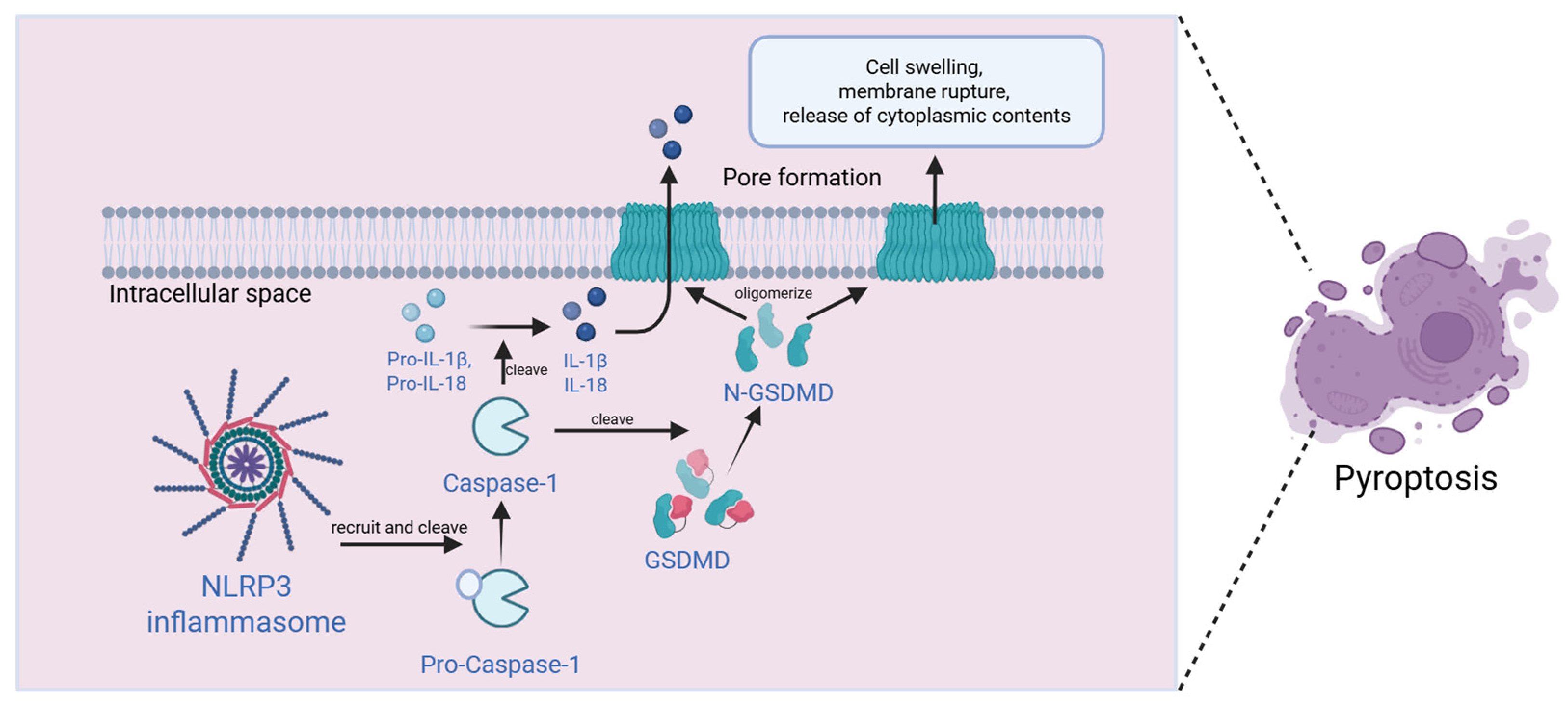
In SAE, NLRP3 inflammasome-mediated pyroptosis is a central pathological mechanism. The NLRP3/caspase-1/GSDMD signaling axis is abnormally activated in the hippocampus of cecal ligation and puncture (CLP) models, accompanied by elevated pyroptosis markers and pro-inflammatory factors, leading to cognitive impairment [75]. Genetic knockout experiments demonstrate that GSDMD deficiency simultaneously inhibits pyroptosis and autophagy [58], while dynamin-related protein 1 (DRP1) inhibitors significantly improve pathological phenotypes by reversing mitochondrial dysfunction [59] (Figure 3).
Molecular mechanisms include:
(1) Ion channel regulation: The P2X7 receptor activates cortical pyroptosis pathways via ERK1/2 phosphorylation [76]; pannexin-1 regulates the autophagy-pyroptosis balance through AMPK signaling [77].
(2) Epigenetic regulation: TRIM45 aggravates microglial pyroptosis by promoting K63-linked polyubiquitination ubiquitination and stabilization of Atg5, thereby disrupting autophagic flux and activating the NLRP3 inflammasome [78]; Maf1 inhibits NF-κB/p65-driven inflammasome assembly [79].
Intervention strategies classified by clinical transformation stage: ① Preclinical research: Ethyl pyruvate inhibits NLRP3 [80]; TXNIP knockdown protects against sepsis-induced brain injury and cognitive decline by suppressing oxidative stress and neuroinflammation [81]; mesenchymal stem cell-derived exosomes deliver miR-140-3p to inhibit HMGB1-mediated pyroptosis [82]; hydrogen-rich saline blocks inflammasome activation via the Nrf2/autophagy pathway [83,84]; echinomycin improves mitochondrial structure and reduces inflammatory factors via the HIF-1α/BNIP3L axis [85]; ② Natural compounds (preclinical): Puerarin enhances BBB stability by blocking GSDMD cleavage; myricitrin alleviates neuroinflammation by modulating the Bax/Bcl-2 balance [86]; malvidin dually inhibits pyroptosis and apoptosis via the AMPKα/UCP2 axis [87].

3.4.3. Molecular Regulatory Axes and Synergistic Interventions of Ferroptosis

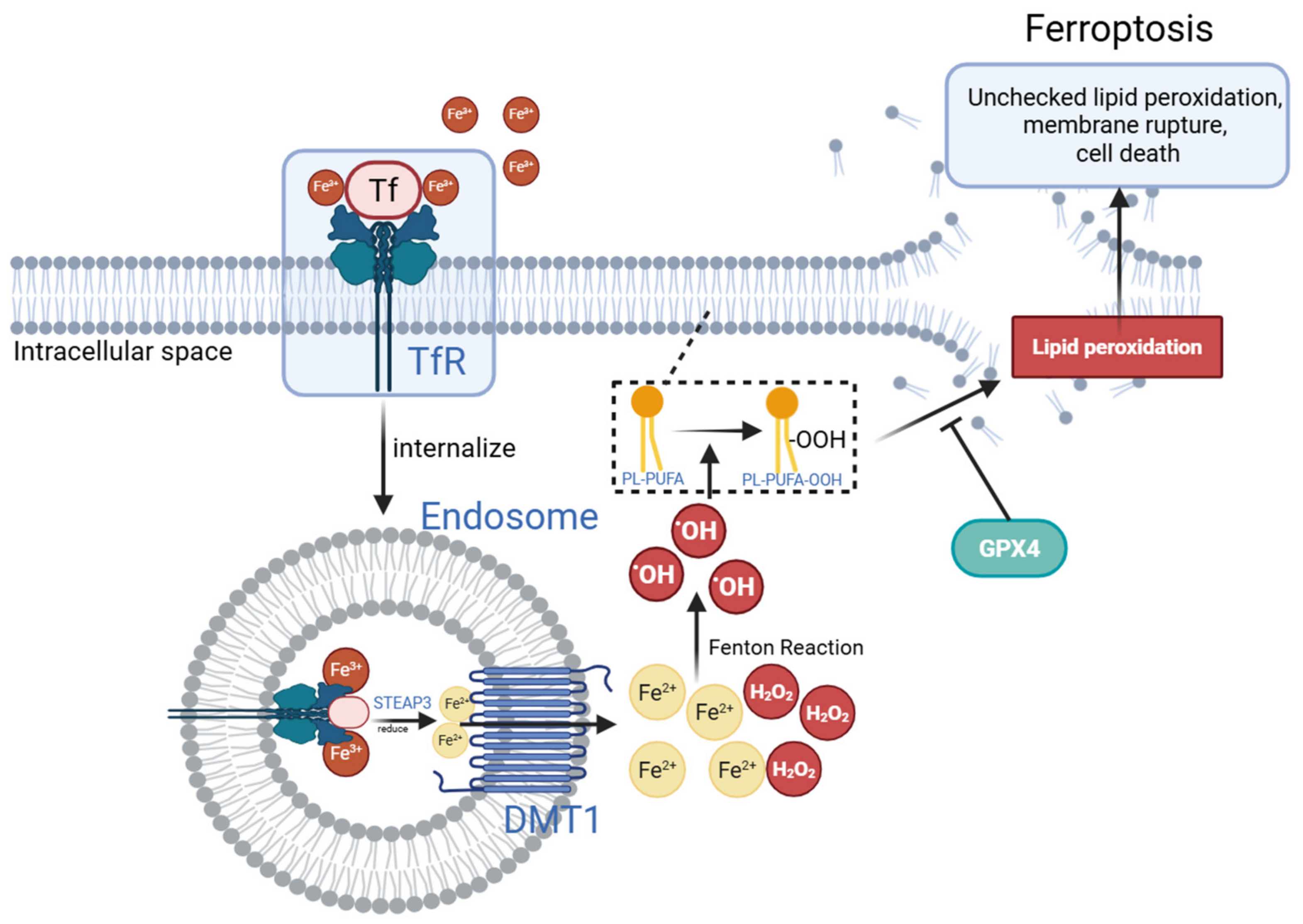
Ferroptosis involves glutathione antioxidant axis dysfunction, iron metabolism disorders, and uncontrolled lipid peroxidation due to GPX4 activity inhibition. Clinical data show that SAE patients exhibit a 2.3-fold and 1.8-fold increase in peripheral blood S100β and GFAP levels, respectively, positively correlating with MDA accumulation (r = 0.72), alongside a 40% decrease in SOD activity, indicating oxidative/antioxidant system imbalance and mitochondrial cristae disruption [88] (Figure 4).
Multi-omics analyses reveal key ferroptosis regulatory networks in SAE: the hippocampal PEBP-1/15-LOX/ACSL4 axis is significantly activated, while GPX4 and xCT transporter (SLC7A11) expression is suppressed, forming a lipid peroxidation feedback loop [88]. Interventions include: (1) acetaminophen (APAP) restores the GPX4 pathway to inhibit ferroptosis [89]; (2) MAR1 reshapes the SLC7A11/GPX4 signaling pathway to suppress microglial activation [90]; (3) Exosomal NEAT1 exacerbates ferroptosis via the miR-9-5p/TFRC-GOT1 axis [91].
Pharmacological synergy studies demonstrate that propofol combined with dexmedetomidine achieves multi-target intervention by regulating the Fpn1 node, reducing FTH1 expression by 58% and MDA levels by 41%, while reversing iron transporter abnormalities [92]. Notably, ferroptosis regulation exhibits cell-context dependency: the EBF1-CSRP2 axis suppresses ferroptosis via SLC7A11/GPX4 modulation in leukemia [93], contrasting with its neurodestructive role in SAE. These findings provide systematic evidence for ferroptosis-targeted therapies in SAE, from molecular mechanisms to clinical translation.

3.5. Neurotransmitter Network Imbalance

3.5.1. Core Mechanisms of Neurotransmitter Homeostasis Imbalance

Neurotransmitter homeostasis imbalance in SAE is associated with immune inflammation and cell death, involving metabolic disturbances of dopamine and glutamate and the synaptic plasticity damage they mediate [38]. Peripheral inflammation triggers central cytokine release via vagus nerve-mediated signal transduction, forming a vicious neuro-immune cycle [94]. Cholinergic system dysfunction is manifested by reduced ChAT-positive neurons and abnormal AChE activity, leading to dysregulated microglial activation thresholds and accelerated transformation towards neurotoxic phenotypes [94]. Functional imaging shows abnormally increased amplitude of low-frequency fluctuation (ALFF) values in the hippocampus, and the glutamate+glutamine (Glx)/creatine (Cr) ratio positively correlates with cognitive impairment, providing objective biomarkers for clinical assessment [95].
Molecular mechanism studies indicate that abnormal glutamatergic system activation induces excitotoxicity in the hippocampal-prefrontal cortical circuit via the N-methyl-D-aspartate receptor (NMDAR)/α-amino-3-hydroxy-5-methyl-4-isoxazolepropionic acid receptor (AMPAR)-cAMP response element-binding protein (CREB)-brain-derived neurotrophic factor (BDNF) axis, and specific receptor antagonists can reverse related molecular abnormalities [96,97]. LPS-induced abnormalities in septal-hippocampal cholinergic projections can be restored through circuit reprogramming to recover synaptic plasticity [98]. In epigenetic regulation, the Neat1/Hbb axis impairs dendritic spine density by downregulating postsynaptic density protein-95 (PSD-95) expression, while MFHAS1-siRNA intervention can significantly reduce Aβ pathological burden and repair dendritic morphology [99,100].

3.5.2. Intervention Strategies Targeting Neurotransmitter Systems

Intervention strategies classified by clinical transformation stage: ① Clinically available drugs: N-acetylcysteine (NAC) protects the BBB through dual mechanisms of regulating glutamate metabolism and exerting antioxidant effects [101]; oxytocin improves cognition by reconstructing excitatory/inhibitory synaptic balance [102]; fluoxetine alleviates prefrontal dysfunction by restoring serotonergic (5-HT) signaling [103]; ② Preclinical research: Vagus nerve stimulation exerts anti-inflammatory effects by inhibiting endothelial activation cascades while regulating glial cells to maintain neuroprotective homeostasis.

3.6. Gut Microbiota-Gut–Brain Axis in SAE: Metabolic Regulation and Intervention Strategies

The gut–brain axis is proposed to mediate bidirectional communication between the gut and the CNS via immune-neuro-endocrine networks, and its dysfunction has been associated with SAE pathology. In SAE model mice, the abundance of short-chain fatty acid (SCFA)-producing bacteria (e.g., Bacteroides, Bifidobacterium) decreases by 62–75%, accompanied by a 54–68% reduction in acetate and propionate concentrations. Exogenous SCFA intervention can dose-dependently inhibit peripheral pro-inflammatory factor release and exert neuroprotection by upregulating tight junction proteins and blocking microglial JNK/NF-κB activation [104,105].
Molecular mechanisms involve: (1) Butyrate activates the GPR109A/Nrf2/HO-1 pathway [106]; (2) Indolepropionic acid (IPA) inhibits the NLRP3 inflammasome and IL-1β secretion [107]; (3) NU9056 upregulaes SCFA levels to antagonize inflammasome activity [108].
Microbiota remodeling strategies show that fecal microbiota transplantation (FMT) improves neurotransmitter balance by modulating the IBA-1 axis, inhibits M1 polarization and IL-1β cycling in mesenteric lymph node macrophages, and blocks gut epithelial exosome-mediated neuroinflammatory cascades [109]. Phosphatidylserine (PS) significantly downregulates hippocampal pro-inflammatory gene transcription by modulating TBK1/NLRP3 signaling [110]. Clinical data confirm abnormal ammonia metabolism in SAE patients. Mechanistic studies indicate the microbiota-gut–brain axis drives functional imbalance by inducing pathological overexpression of AQP4 in astrocytes [111,112].
Metabolic interventions: Molecular hydrogen improves metabolic-inflammatory imbalance via microbiota modulation [113]; propionate supplements exert protective effects by maintaining BBB integrity [114]. These findings systematically reveal the pivotal role of gut microbiota metabolic reprogramming in SAE, providing a theoretical basis for multi-dimensional interventions targeting the gut–brain axis.
Table 2 summarizes the key molecular mediators, upstream triggers, and downstream effects across all six pathological components discussed above, highlighting the central convergence of mitochondrial dysfunction.

4. Integrated Discussion and Critical Appraisal

4.1. Synthesis of the Core Pathological Axis

This review integrates current evidence on SAE into a unified framework comprising six molecular components organized along four sequential pathological axes: neuroinflammatory storm (encompassing neuroinflammatory cascades and immune cell-mediated regulation) → BBB disruption and microcirculatory disturbances → multimodal programmed cell death → neurotransmitter imbalance, with the gut–brain axis serving as a bidirectional regulatory node modulating all four axes. Mitochondrial dysfunction functions as the central converging node linking these pathological components.
A critical innovation of this framework is its explicit classification of intervention strategies by clinical translation stage—clinically available drugs, preclinical candidates, and potential molecular targets—thereby distinguishing this review from prior mechanistic descriptions and providing practical guidance for precision therapeutics. The mutual amplification between neuroinflammation and BBB damage emerges as a key trigger for SAE progression, while gut–brain axis dysregulation offers a non-invasive intervention entry point.
However, the translational trajectory from mechanistic understanding to clinical application remains uneven across components. Table 3 synthesizes the evidence base, key findings, and translational barriers for each pathological axis, revealing systematic gaps that underlie the limited clinical progress to date.
Several cross-cutting challenges emerge from this analysis. First, BBB permeability affects nearly all CNS-targeted agents, necessitating either high systemic doses with attendant toxicity or development of specialized delivery systems. Second, therapeutic timing represents a critical disconnect—preclinical studies universally administer interventions prophylactically or within hours of insult, whereas clinical SAE is typically recognized after established pathology, potentially beyond the window for mechanism-based intervention. Third, biomarker-guided stratification is absent, precluding identification of patients most likely to benefit from specific targets (e.g., NLRP3 inhibitors for inflammasome-high phenotypes, ferroptosis inhibitors for lipid peroxidation-dominant subgroups). Fourth, ICU-specific pharmacological constraints (hemodynamic instability, organ dysfunction, polypharmacy) complicate dosing and monitoring of investigational agents. These barriers highlight the necessity of iterative feedback between preclinical model refinement and early-phase clinical trial design, with particular attention to pharmacokinetic bridging studies and biomarker validation.

4.2. Critical Appraisal of Evidence and Unresolved Controversies

Beyond the translational barriers summarized in Table 3, several areas of conflicting evidence, methodological limitations, and unresolved controversies warrant deeper discussion to guide future research priorities.

4.2.1. Conflicting Experimental Findings on Cell Death Interplay

The relationship between pyroptosis and autophagy remains contentious. GSDMD deficiency simultaneously suppresses both pyroptosis and autophagy in CLP models [58], suggesting pathway coupling; however, autophagy activation can precede and protect against pyroptosis by clearing damaged mitochondria [70,77]. Whether these pathways act synergistically to amplify injury or represent compensatory responses likely depends on injury severity, cell type, and temporal stage. Similarly, GPX4 inhibition triggers ferroptosis in some contexts while other studies implicate alternative death modalities [88,89] raising questions about context-dependent death modality preferences.

4.2.2. Reproducibility Concerns and Model Heterogeneity

Reproducibility across SAE models appears to be suboptimal in existing published evidence. LPS injection produces rapid, transient neuroinflammation, whereas CLP induces polymicrobial sepsis with more sustained CNS pathology [3,75]. These models tend to yield divergent cytokine profiles and microglial activation kinetics. Furthermore, species differences in microglial density, BBB tight junction composition, and cholinergic system organization between rodents and humans likely restrict direct extrapolation of preclinical findings to human patients [15,16,18].

4.2.3. Heterogeneity of SAE Definitions and Diagnostic Criteria

SAE lacks standardized diagnostic criteria, with studies employing heterogeneous definitions including Glasgow Coma Scale score, CAM-ICU positivity, EEG abnormalities, or composite scores [1,2]. This heterogeneity may introduce introduces selection bias and could hamper reliable biomarker validation. GCS-based definitions capture advanced encephalopathy but may miss subtle cognitive deficits, whereas EEG criteria require specialized expertise [1]. The absence of a unified SAE classification potentially hinders stratified therapy development.

4.2.4. Limitations of Animal Models

Current animal models fail to fully replicate human SAE complexity. Most models use young, healthy mice, whereas human sepsis predominantly affects elderly patients with comorbidities that alter immune responses and BBB vulnerability [1,2]. Additionally, the absence of mechanical ventilation, sedation, and polypharmacy in animal models—standard in ICU settings—limits assessment of iatrogenic contributions to encephalopathy.

4.2.5. Challenges in Clinical Translation

Multiple unresolved translational challenges largely hinder the clinical transformation of SAE targeted therapies. First, BBB permeability: many promising preclinical targets tend to exhibit poor CNS penetration, requiring doses that may risk peripheral toxicity [12,13,22]. Second, timing of intervention: most preclinical studies administer therapies prophylactically or early post-induction, whereas clinical SAE diagnosis often occurs after established pathology [3,75]. Third, biomarker availability: while NGAL, S100β, and GFAP appear to show diagnostic promise [42,88], none are validated for real-time monitoring. Fourth, multi-target complexity: proposed combination therapies face challenges in optimizing dosing schedules and demonstrating superiority over single-agent approaches in heterogeneous populations.

4.3. Concluding Remarks

SAE is a complex neuroinflammatory disease driven by the synergy of multiple interconnected components. We acknowledge that the proposed pathological axis serves only as a preliminary working framework based on synthesizing predominantly associative evidence from heterogeneous study designs and animal models. Critical gaps remain: the temporal dynamics of axis component interactions, the relative contributions of cell death modalities in human SAE, and the feasibility of multi-target combination therapies in complex ICU populations. Addressing these gaps through prospective, multi-center studies with standardized SAE definitions and integrated biomarker platforms will be crucial for validating this integrated pathological model.
Multi-target synergistic intervention targeting the core pathological axis and key regulatory nodes—particularly the gut–brain axis and mitochondrial function—may represent a promising direction for clinical translation. This integrated framework provides a systematic theoretical basis for the precise clinical management of SAE, while explicitly highlighting the limitations that need to be comprehensively addressed to fully unlock its translational potential.

5. Future Perspectives

Although significant progress has been made in elucidating pathophysiology, translation from basic research to clinical practice remains limited, particularly in the lack of precise biomarkers, personalized intervention strategies, and mature clinical trial evidence. Based on the core pathological axis and regulatory nodes summarized in this review, future research should focus on the following specific directions with clear translational paths:
(1)
Deciphering the spatiotemporal dynamics of neuroimmune crosstalk: Investigate dynamic changes in microglial polarization, T-cell subset migration, and inflammatory mediator release across different brain regions (hippocampus, prefrontal cortex) and disease stages (early, middle, late). For example, combining single-cell sequencing to track microglial state transitions with live imaging to visualize dynamic BBB leakage in SAE models would enable precise mapping of neuroimmune interactions in real time. Clarify key time windows and spatial distribution rules of neuroimmune interaction to establish a precise intervention target map and avoid blind intervention.
(2)
Promoting the clinical translation of SAE biomarkers: Further validate the diagnostic and prognostic value of candidate biomarkers (NGAL, S100β) in large-scale clinical cohorts. Optimize detection methods (e.g., combined cerebrospinal fluid and peripheral blood testing) and establish a combined biomarker evaluation system to enable early diagnosis, severity assessment, and prognostic prediction, laying a foundation for timely intervention.
(3)
Accelerating development of gut–brain axis-targeted microecological preparations: Based on the regulatory role of SCFA-producing bacteria and metabolites (butyrate, indolepropionic acid) in SAE, develop personalized probiotic preparations, prebiotics, or FMT protocols targeting gut microbiota remodeling. Verify their safety and efficacy in preclinical studies and promote transition to clinical trials, providing new non-invasive intervention strategies for SAE.
(4)
Optimizing the design of multi-target combination therapy: Based on the core pathological axis, design rational combined medication schemes, such as “NLRP3 inflammasome inhibitors (pyroptosis inhibition) + ZO-1 agonists (BBB repair) + mitochondrial protectants (energy metabolism improvement).” Clarify dosage, administration route, and combined efficacy in clinical trials, avoid single-target intervention limitations, and promote translation of multi-target synergistic therapy into routine clinical practice.
(5)
Integrating AI-driven structural biology: AlphaFold-enabled protein structure prediction may accelerate rational design of BBB-penetrant agents targeting the core pathological axis [116].

Author Contributions

Conceptualization, Z.N. and H.T.; methodology, H.T. and W.S.; investigation, H.T. and W.S.; writing—original draft, H.T.; writing—review and editing, Z.N. and W.S.; visualization, H.T.; supervision, Z.N.; project administration, Z.N. All authors have read and agreed to the published version of the manuscript.

Funding

This work was supported by the Ministry of Science and Technology of the People’s Republic of China (STI2030-Major Projects2021ZD0201900).

Data Availability Statement

No new data were created or analyzed in this study. All data supporting the conclusions of this review are derived from the published literature, which is cited in the reference list. The full-text articles included in this review are publicly available through PubMed, Web of Science, and Embase databases.

Conflicts of Interest

The authors declare no conflicts of interest.

Abbreviations

The following abbreviations are used in this manuscript:
5-HT5-Hydroxytryptamine (Serotonin)
Amyloid-β
AChAcetylcholine
AChEAcetylcholinesterase
AChRAcetylcholine Receptor
AKTProtein Kinase B
ALFFAmplitude of Low-Frequency Fluctuation
AMPARα-Amino-3-hydroxy-5-methyl-4-isoxazolepropionic Acid Receptor
AQP4Aquaporin-4
AREAntioxidant Response Element
AREGAmphiregulin
ATPAdenosine Triphosphate
BBBBlood–Brain Barrier
BDNFBrain-Derived Neurotrophic Factor
BMECBrain Microvascular Endothelial Cell
CAPCholinergic Anti-inflammatory Pathway
ChATCholine Acetyltransferase
CLPCecal Ligation and Puncture
CNSCentral Nervous System
CREBcAMP Response Element-Binding Protein
CXCR5C-X-C Chemokine Receptor Type 5
DAMPDamage-Associated Molecular Pattern
DHADihydroartemisinin
DMT1Divalent Metal Transporter 1
DRP1Dynamin-Related Protein 1
EEEnvironmental Enrichment
EGFREpidermal Growth Factor Receptor
FMTFecal Microbiota Transplantation
FTH1Ferritin Heavy Chain 1
Fpn1Ferroportin 1
GlxGlutamate + Glutamine
GPX4Glutathione Peroxidase 4
GSDMDGasdermin D
HMGB1High Mobility Group Box 1
HO-1Heme Oxygenase-1
HPCHippocampus
HtrA2High Temperature Requirement Protein A2
ICAM-1Intercellular Adhesion Molecule-1
IFN-γInterferon Gamma
ILInterleukin
iNOSInducible Nitric Oxide Synthase
IPAIndolepropionic Acid
IRGM1Immunity-Related GTPase Family M Member 1
JAK2Janus Kinase 2
JNKc-Jun N-Terminal Kinase
KYNAKynurenic Acid
LPSLipopolysaccharide
MAPKMitogen-Activated Protein Kinase
MAR1Maresin 1
MD2Myeloid Differentiation Factor 2
MDAMalondialdehyde
MFG-E8Milk Fat Globule-Epidermal Growth Factor 8
MMPMatrix Metalloproteinase
mPFCMedial Prefrontal Cortex
mPTPMitochondrial Permeability Transition Pore
NAD+Nicotinamide Adenine Dinucleotide
NADPHNicotinamide Adenine Dinucleotide Phosphate
NETsNeutrophil Extracellular Trap
NF-κBNuclear Factor Kappa-Light-Chain-Enhancer of Activated B Cells
NGALNeutrophil Gelatinase-Associated Lipocalin
NLRP3NOD-Like Receptor Family Pyrin Domain Containing 3
NMDARN-Methyl-D-Aspartate Receptor
NONitric Oxide
NOX2NADPH Oxidase 2
Nrf2Nuclear Factor Erythroid 2-Related Factor 2
OmiOmi/HtrA2 Serine Protease
P2X7RPurinergic Receptor P2X 7
PAMPPathogen-Associated Molecular Pattern
PD-L1Programmed Death-Ligand 1
PEBP-1Phosphatidylethanolamine-Binding Protein 1
PGPropylene Glycol
PGC-1αPeroxisome Proliferator-Activated Receptor Gamma Coactivator 1-Alpha
PINK1PTEN-Induced Putative Kinase 1
PLX5622CSF1R Inhibitor
PSPhosphatidylserine
PUFAPolyunsaturated Fatty Acid
RAGEReceptor for Advanced Glycation End-Products
ROCKRho-Associated Protein Kinase
ROSReactive Oxygen Species
SAESepsis-Associated Encephalopathy
SCFAShort-Chain Fatty Acid
Sirt1Sirtuin 1
Sirt3Sirtuin 3
SLC7A11Solute Carrier Family 7 Member 11
SODSuperoxide Dismutase
STAT3Signal Transducer and Activator of Transcription 3
TGF-βTransforming Growth Factor Beta
ThT Helper Cell
TJTight Junction
TLRToll-Like Receptor
TNF-αTumor Necrosis Factor Alpha
TregsRegulatory T Cells
TXNIPThioredoxin-Interacting Protein
UCP2Uncoupling Protein 2
VCAM-1Vascular Cell Adhesion Molecule-1
VDAC1Voltage-Dependent Anion Channel 1
VEGF-CVascular Endothelial Growth Factor C
XIAPX-Linked Inhibitor of Apoptosis Protein
α7nAChRAlpha-7 Nicotinic Acetylcholine Receptor
γδTGamma-Delta T Cell

References

  1. Gofton, T.E.; Young, G.B. Sepsis-associated encephalopathy. Nat. Rev. Neurol. 2012, 8, 557–566. [Google Scholar] [CrossRef] [PubMed]
  2. He, T.T.; Jiao, T.Q.; An, X.M. Risk prediction models for sepsis-associated encephalopathy: A systematic evaluation and meta-analysis. PeerJ 2026, 14, e20770. [Google Scholar] [CrossRef] [PubMed]
  3. Zhang, Z.; Qiu, X.; Zeng, X.; Liu, X.; Lu, J.; Xu, C.; Huang, J.; Zhao, C.; Zhan, Y. Integrated multi omics and machine learning reveal mitochondrial immunometabolic networks in sepsis associated encephalopathy. Sci. Rep. 2025, 15, 33572. [Google Scholar] [CrossRef] [PubMed]
  4. Kobayashi, T.; Uchino, H.; Elmér, E.; Ogihara, Y.; Fujita, H.; Sekine, S.; Ishida, Y.; Saiki, I.; Shibata, S.; Kawachi, A. Disease Outcome and Brain Metabolomics of Cyclophilin-D Knockout Mice in Sepsis. Int. J. Mol. Sci. 2022, 23, 961. [Google Scholar] [CrossRef] [PubMed]
  5. Andrés, C.M.C.; Pérez de la Lastra, J.M.; Andrés Juan, C.; Plou, F.J.; Pérez-Lebeña, E. Superoxide Anion Chemistry-Its Role at the Core of the Innate Immunity. Int. J. Mol. Sci. 2023, 24, 1841. [Google Scholar] [CrossRef] [PubMed]
  6. Cui, Y.; Liu, J.; Song, Y.; Chen, C.; Shen, Y.; Xie, K. High Concentration Hydrogen Protects Sepsis-Associated Encephalopathy by Enhancing Pink1/Parkin-Mediated Mitophagy and Inhibiting cGAS-STING-IRF3 Pathway. CNS Neurosci. Ther. 2025, 31, e70305. [Google Scholar] [CrossRef] [PubMed]
  7. Zaghloul, N.; Addorisio, M.E.; Silverman, H.A.; Patel, H.L.; Valdés-Ferrer, S.I.; Ayasolla, K.R.; Lehner, K.R.; Olofsson, P.S.; Nasim, M.; Metz, C.N.; et al. Forebrain Cholinergic Dysfunction and Systemic and Brain Inflammation in Murine Sepsis Survivors. Front. Immunol. 2017, 8, 1673. [Google Scholar] [CrossRef] [PubMed]
  8. Keever, K.R.; Yakubenko, V.P.; Hoover, D.B. Neuroimmune nexus in the pathophysiology and therapy of inflammatory disorders: Role of α7 nicotinic acetylcholine receptors. Pharmacol. Res. 2023, 191, 106758. [Google Scholar] [CrossRef] [PubMed]
  9. Li, H.; Li, Y.; Wang, W.; Peng, Z.; Wu, F. Cholinergic anti-inflammatory pathway plays negative regulatory role in early inflammatory and immune responses in septic rats. Nan Fang Yi Ke Da Xue Xue Bao = J. South. Med. Univ. 2020, 40, 647–653. [Google Scholar] [CrossRef]
  10. Zhu, C.; Wang, D.; Chang, C.; Liu, A.; Zhou, J.; Yang, T.; Jiang, Y.; Li, X.; Jiang, W. Dexmedetomidine alleviates blood-brain barrier disruption in rats after cerebral ischemia-reperfusion by suppressing JNK and p38 MAPK signaling. Korean J. Physiol. Pharmacol. 2024, 28, 239–252. [Google Scholar] [CrossRef] [PubMed]
  11. Zhang, Q.; Liu, X.M.; Hu, Q.; Liu, Z.R.; Liu, Z.Y.; Zhang, H.G.; Huang, Y.L.; Chen, Q.H.; Wang, W.X.; Zhang, X.K. Dexmedetomidine inhibits mitochondria damage and apoptosis of enteric glial cells in experimental intestinal ischemia/reperfusion injury via SIRT3-dependent PINK1/HDAC3/p53 pathway. J. Transl. Med. 2021, 19, 463. [Google Scholar] [CrossRef] [PubMed]
  12. Xu, Z.Q.; Zhang, W.J.; Su, D.F.; Zhang, G.Q.; Miao, C.Y. Cellular responses and functions of α7 nicotinic acetylcholine receptor activation in the brain: A narrative review. Ann. Transl. Med. 2021, 9, 509. [Google Scholar] [CrossRef] [PubMed]
  13. Liu, J.; Lin, Y.; Yang, Y.; Guo, Y.; Shang, Y.; Zhou, B.; Liu, T.; Fan, J.; Wei, C. Z-Guggulsterone attenuates cognitive defects and decreases neuroinflammation in APPswe/PS1dE9 mice through inhibiting the TLR4 signaling pathway. Biochem. Pharmacol. 2022, 202, 115149. [Google Scholar] [CrossRef] [PubMed]
  14. Cui, Y.; Meng, S.; Zhang, N.; Liu, J.; Zheng, L.; Ma, W.; Song, Y.; Wang, Z.; Shen, Y.; Liu, J.; et al. High-concentration hydrogen inhalation mitigates sepsis-associated encephalopathy in mice by improving mitochondrial dynamics. CNS Neurosci. Ther. 2024, 30, e70021. [Google Scholar] [CrossRef] [PubMed]
  15. Ronaldson, P.T.; Davis, T.P. Regulation of blood-brain barrier integrity by microglia in health and disease: A therapeutic opportunity. J. Cereb. Blood Flow Metab. 2020, 40, S6–S24. [Google Scholar] [CrossRef] [PubMed]
  16. Wang, H.; Wang, H.; Song, Y.; Liu, C.; Qian, X.; Zhang, D.; Jiang, X.; Zhang, S. Overexpression of Foxc1 ameliorates sepsis-associated encephalopathy by inhibiting microglial migration and neuroinflammation through the IκBα/NF-κB pathway. Mol. Med. Rep. 2022, 25, 107. [Google Scholar] [CrossRef] [PubMed]
  17. Rocha, M.; Vieira, A.; Michels, M.; Borges, H.; Goulart, A.; Fernandes, F.; Dominguini, D.; Ritter, C.; Dal-Pizzol, F. Effects of S100B neutralization on the long-term cognitive impairment and neuroinflammatory response in an animal model of sepsis. Neurochem. Int. 2021, 142, 104906. [Google Scholar] [CrossRef] [PubMed]
  18. Haruwaka, K.; Ikegami, A.; Tachibana, Y.; Ohno, N.; Konishi, H.; Hashimoto, A.; Matsumoto, M.; Kato, D.; Ono, R.; Kiyama, H.; et al. Dual microglia effects on blood brain barrier permeability induced by systemic inflammation. Nat. Commun. 2019, 10, 5816. [Google Scholar] [CrossRef] [PubMed]
  19. Ye, B.; Tao, T.; Zhao, A.; Wen, L.; He, X.; Liu, Y.; Fu, Q.; Mi, W.; Lou, J. Blockade of IL-17A/IL-17R Pathway Protected Mice from Sepsis-Associated Encephalopathy by Inhibition of Microglia Activation. Mediat. Inflamm. 2019, 2019, 8461725. [Google Scholar] [CrossRef] [PubMed]
  20. Brás, J.P.; Bravo, J.; Freitas, J.; Barbosa, M.A.; Santos, S.G.; Summavielle, T.; Almeida, M.I. TNF-alpha-induced microglia activation requires miR-342: Impact on NF-kB signaling and neurotoxicity. Cell Death Dis. 2020, 11, 415. [Google Scholar] [CrossRef] [PubMed]
  21. Qiu, F.; Liu, Y.; Liu, Y.; Zhao, Z.; Zhou, L.; Chen, P.; Du, Y.; Wang, Y.; Sun, H.; Zeng, C.; et al. CD137L Inhibition Ameliorates Hippocampal Neuroinflammation and Behavioral Deficits in a Mouse Model of Sepsis-Associated Encephalopathy. Neuromol. Med. 2023, 25, 616–631. [Google Scholar] [CrossRef] [PubMed]
  22. Zhou, Z.; Yang, Y.; Wei, Y.; Xie, Y. Remimazolam Attenuates LPS-Derived Cognitive Dysfunction via Subdiaphragmatic Vagus Nerve Target α7nAChR-Mediated Nrf2/HO-1 Signal Pathway. Neurochem. Res. 2024, 49, 1306–1321. [Google Scholar] [PubMed]
  23. Luo, X.Y.; Ying, J.H.; Wang, Q.S. miR-25-3p ameliorates SAE by targeting the TLR4/NLRP3 axis. Metab. Brain Dis. 2022, 37, 1803–1813. [Google Scholar] [CrossRef] [PubMed]
  24. He, C.; Aziguli, A.; Zhen, J.; Jiao, A.; Liao, H.; Du, C.; Liu, W.; Aihemaitijiang, K.; Xu, A. MiRNA-494 specifically inhibits SIRT3-mediated microglia activation in sepsis-associated encephalopathy. Transl. Cancer Res. 2022, 11, 2299–2309. [Google Scholar] [PubMed]
  25. Yin, X.Y.; Tang, X.H.; Wang, S.X.; Zhao, Y.C.; Jia, M.; Yang, J.J.; Ji, M.H.; Shen, J.C. HMGB1 mediates synaptic loss and cognitive impairment in an animal model of sepsis-associated encephalopathy. J. Neuroinflamm. 2023, 20, 69. [Google Scholar] [CrossRef] [PubMed]
  26. Manabe, T.; Rácz, I.; Schwartz, S.; Oberle, L.; Santarelli, F.; Emmrich, J.V.; Neher, J.J.; Heneka, M.T. Systemic inflammation induced the delayed reduction of excitatory synapses in the CA3 during ageing. J. Neurochem. 2021, 159, 525–542. [Google Scholar] [CrossRef] [PubMed]
  27. Chung, H.Y.; Wickel, J.; Hahn, N.; Mein, N.; Schwarzbrunn, M.; Koch, P.; Ceanga, M.; Haselmann, H.; Baade-Büttner, C.; von Stackelberg, N.; et al. Microglia mediate neurocognitive deficits by eliminating C1q-tagged synapses in sepsis-associated encephalopathy. Sci. Adv. 2023, 9, eabq7806. [Google Scholar] [CrossRef] [PubMed]
  28. Li, H.; Liao, H.; Sun, L.; Chen, D.; Fei, Y.; Yao, M.; Huang, B.; Guo, X.; Song, S.; Bao, H. Milk Fat Globule-EGF Factor 8 (MFGE8) Mitigates Cognitive Impairment in Rats with Sepsis-Associated Encephalopathy: An fMRI Study. J. Integr. Neurosci. 2024, 23, 140. [Google Scholar] [PubMed]
  29. Mein, N.; von Stackelberg, N.; Wickel, J.; Geis, C.; Chung, H.Y. Low-dose PLX5622 treatment prevents neuroinflammatory and neurocognitive sequelae after sepsis. J. Neuroinflamm. 2023, 20, 289. [Google Scholar] [CrossRef] [PubMed]
  30. Tian, Y.; Wang, L.; Fan, X.; Zhang, H.; Dong, Z.; Tao, T. β-patchoulene alleviates cognitive dysfunction in a mouse model of sepsis associated encephalopathy by inhibition of microglia activation through Sirt1/Nrf2 signaling pathway. PLoS ONE 2023, 18, e0279964. [Google Scholar] [PubMed]
  31. Pan, C.; Si, Y.; Meng, Q.; Jing, L.; Chen, L.; Zhang, Y.; Bao, H. Suppression of the RAC1/MLK3/p38 Signaling Pathway by β-Elemene Alleviates Sepsis-Associated Encephalopathy in Mice. Front. Neurosci. 2019, 13, 358. [Google Scholar] [CrossRef] [PubMed]
  32. Yin, Y.; Lv, G.; Zhang, W.; Yuan, J.; Yang, Y.; Wang, Y.; Liu, S.; Wang, S.; Yan, B.; Bo, H.; et al. Resveratrol glycoside mediates microglial endoplasmic reticulum stress to mitigate LPS-induced sepsis-associated cognitive dysfunction. Behav. Brain Res. 2023, 443, 114326. [Google Scholar] [CrossRef] [PubMed]
  33. Giganti, G.; Atif, M.; Mohseni, Y.; Mastronicola, D.; Grageda, N.; Povoleri, G.A.; Miyara, M.; Scottà, C. Treg cell therapy: How cell heterogeneity can make the difference. Eur. J. Immunol. 2021, 51, 39–55. [Google Scholar] [PubMed]
  34. Gao, Y.L.; Liu, Y.C.; Zhang, X.; Shou, S.T.; Chai, Y.F. Insight Into Regulatory T Cells in Sepsis-Associated Encephalopathy. Front. Neurol. 2022, 13, 830784. [Google Scholar] [CrossRef] [PubMed]
  35. Ren, C.; Yao, R.Q.; Zhang, H.; Feng, Y.W.; Yao, Y.M. Sepsis-associated encephalopathy: A vicious cycle of immunosuppression. J. Neuroinflamm. 2020, 17, 14. [Google Scholar] [CrossRef] [PubMed]
  36. Xie, L.; Choudhury, G.R.; Winters, A.; Yang, S.H.; Jin, K. Cerebral regulatory T cells restrain microglia/macrophage-mediated inflammatory responses via IL-10. Eur. J. Immunol. 2015, 45, 180–191. [Google Scholar] [PubMed]
  37. Xu, X.E.; Liu, L.; Wang, Y.C.; Wang, C.T.; Zheng, Q.; Liu, Q.X.; Li, Z.F.; Bai, X.J.; Liu, X.H. Caspase-1 inhibitor exerts brain-protective effects against sepsis-associated encephalopathy and cognitive impairments in a mouse model of sepsis. Brain Behav. Immun. 2019, 80, 859–870. [Google Scholar] [CrossRef] [PubMed]
  38. Saito, M.; Fujinami, Y.; Ono, Y.; Ohyama, S.; Fujioka, K.; Yamashita, K.; Inoue, S.; Kotani, J. Infiltrated regulatory T cells and Th2 cells in the brain contribute to attenuation of sepsis-associated encephalopathy and alleviation of mental impairments in mice with polymicrobial sepsis. Brain Behav. Immun. 2021, 92, 25–38. [Google Scholar] [CrossRef] [PubMed]
  39. Moriyama, N.; Saito, M.; Ono, Y.; Yamashita, K.; Aoi, T.; Kotani, J. Increased Interleukin-17-Producing γδT Cells in the Brain Exacerbate the Pathogenesis of Sepsis-Associated Encephalopathy and Sepsis-Induced Anxiety in Mice. J. Clin. Med. 2023, 12, 4309. [Google Scholar] [CrossRef] [PubMed]
  40. Zhu, C.L.; Xie, J.; Liu, Q.; Wang, Y.; Li, H.R.; Yu, C.M.; Li, P.; Deng, X.M.; Bian, J.J.; Wang, J.F. PD-L1 promotes GSDMD-mediated NET release by maintaining the transcriptional activity of Stat3 in sepsis-associated encephalopathy. Int. J. Biol. Sci. 2023, 19, 1413–1429. [Google Scholar] [CrossRef]
  41. Poles, M.Z.; Nászai, A.; Gulácsi, L.; Czakó, B.L.; Gál, K.G.; Glenz, R.J.; Dookhun, D.; Rutai, A.; Tallósy, S.P.; Szabó, A.; et al. Kynurenic Acid and Its Synthetic Derivatives Protect Against Sepsis-Associated Neutrophil Activation and Brain Mitochondrial Dysfunction in Rats. Front. Immunol. 2021, 12, 717157. [Google Scholar] [CrossRef] [PubMed]
  42. Platanaki, C.; Paraskevas, T.; Delastic, A.L.; Michailides, C.; Kantanis, A.; Polychronopoulos, P.; Marangos, M.; Velissaris, D. The role of cerebrospinal fluid levels of neutrophil gelatinase-associated lipocalin (NGAL) and electroencephalography in the assessment of impaired consciousness in the context of infection. Rom. J. Intern. Med. 2023, 61, 112–115. [Google Scholar] [CrossRef] [PubMed]
  43. Yue, J.; Tan, Y.; Huan, R.; Guo, J.; Yang, S.; Deng, M.; Xiong, Y.; Han, G.; Liu, L.; Liu, J.; et al. Mast cell activation mediates blood-brain barrier impairment and cognitive dysfunction in septic mice in a histamine-dependent pathway. Front. Immunol. 2023, 14, 1090288. [Google Scholar] [CrossRef] [PubMed]
  44. Varatharaj, A.; Galea, I. The blood-brain barrier in systemic inflammation. Brain Behav. Immun. 2017, 60, 1–12. [Google Scholar] [CrossRef] [PubMed]
  45. Baby, S.; Reljic, T.; Villalba, N.; Kumar, A.; Yuan, S.Y. Endothelial glycocalyx-associated molecules as potential serological markers for sepsis-associated encephalopathy: A systematic review and meta-analysis. PLoS ONE 2023, 18, e0281941. [Google Scholar] [CrossRef] [PubMed]
  46. Haywood-Watson, R.J.; Holcomb, J.B.; Gonzalez, E.A.; Peng, Z.; Pati, S.; Park, P.W.; Wang, W.; Zaske, A.M.; Menge, T.; Kozar, R.A. Modulation of syndecan-1 shedding after hemorrhagic shock and resuscitation. PLoS ONE 2011, 6, e23530. [Google Scholar] [CrossRef] [PubMed]
  47. Doll, D.N.; Hu, H.; Sun, J.; Lewis, S.E.; Simpkins, J.W.; Ren, X. Mitochondrial crisis in cerebrovascular endothelial cells opens the blood-brain barrier. Stroke 2015, 46, 1681–1689. [Google Scholar] [CrossRef] [PubMed]
  48. Huang, Y.; Xu, W.; Zhou, R. NLRP3 inflammasome activation and cell death. Cell. Mol. Immunol. 2021, 18, 2114–2127. [Google Scholar] [CrossRef] [PubMed]
  49. Chen, T.; Hu, J.; Liao, Y.; Xie, S.; Zhang, L. The brain washing system in sepsis-associated encephalopathy. J. Neuroinflamm. 2025, 22, 277. [Google Scholar] [CrossRef] [PubMed]
  50. Di Bella, D.; Ferreira, J.P.S.; Silva, R.N.O.; Echem, C.; Milan, A.; Akamine, E.H.; Carvalho, M.H.; Rodrigues, S.F. Gold nanoparticles reduce inflammation in cerebral microvessels of mice with sepsis. J. Nanobiotechnol. 2021, 19, 52. [Google Scholar] [CrossRef] [PubMed]
  51. Liu, F.; Liu, J.; Xiang, H.; Sun, Z.; Li, Y.; Li, X.; Liu, Y.; Liu, J. Dihydroartemisinin protects blood-brain barrier permeability during sepsis by inhibiting the transcription factor SNAI1. Clin. Exp. Pharmacol. Physiol. 2022, 49, 979–987. [Google Scholar] [CrossRef] [PubMed]
  52. Pu, Y.; Zhao, L.; Xi, Y.; Xia, Y.; Qian, Y. The protective effects of Mirtazapine against lipopolysaccharide (LPS)-induced brain vascular hyperpermeability. Bioengineered 2022, 13, 3680–3693. [Google Scholar] [CrossRef] [PubMed]
  53. You, L.; Jiang, H. Cabergoline possesses a beneficial effect on blood-brain barrier (BBB) integrity against lipopolysaccharide (LPS). Bioengineered 2021, 12, 8358–8369. [Google Scholar] [CrossRef] [PubMed]
  54. Yu, S.Y.; Ge, Z.Z.; Xiang, J.; Gao, Y.X.; Lu, X.; Walline, J.H.; Qin, M.B.; Zhu, H.D.; Li, Y. Is rosuvastatin protective against sepsis-associated encephalopathy? A secondary analysis of the SAILS trial. World J. Emerg. Med. 2022, 13, 367–372. [Google Scholar] [CrossRef] [PubMed]
  55. Zheng, X.; Li, W.; Xiang, Q.; Wang, Y.; Qu, T.; Fang, W.; Yang, H. Memantine Attenuates Cognitive and Emotional Dysfunction in Mice with Sepsis-Associated Encephalopathy. ACS Omega 2023, 8, 40934–40943. [Google Scholar] [PubMed]
  56. Ismail Hassan, F.; Didari, T.; Baeeri, M.; Gholami, M.; Haghi-Aminjan, H.; Khalid, M.; Navaei-Nigjeh, M.; Rahimifard, M.; Solgi, S.; Abdollahi, M.; et al. Metformin Attenuates Brain Injury by Inhibiting Inflammation and Regulating Tight Junction Proteins in Septic Rats. Cell J. 2020, 22, 29–37. [Google Scholar] [CrossRef] [PubMed]
  57. Dong, H.; Dai, X.; Zhou, Y.; Shi, C.; Bhuiyan, P.; Sun, Z.; Li, N.; Jin, W. Enhanced meningeal lymphatic drainage ameliorates lipopolysaccharide-induced brain injury in aged mice. J. Neuroinflamm. 2024, 21, 36. [Google Scholar] [CrossRef] [PubMed]
  58. Su, W.; Xie, Z.; Bai, X.; Li, Z.; Liu, X. The Absence of Gasdermin D Reduces Nuclear Autophagy in a Cecal Ligation and Puncture-Induced Sepsis-Associated Encephalopathy Mouse Model. Brain Sci. 2023, 13, 478. [Google Scholar] [CrossRef] [PubMed]
  59. Fu, Q.; Zhang, Y.B.; Shi, C.X.; Jiang, M.; Lu, K.; Fu, Z.H.; Ruan, J.P.; Wu, J.; Gu, X.P. GSDMD/Drp1 signaling pathway mediates hippocampal synaptic damage and neural oscillation abnormalities in a mouse model of sepsis-associated encephalopathy. J. Neuroinflamm. 2024, 21, 96. [Google Scholar] [CrossRef] [PubMed]
  60. Zhou, R.X.; Li, Y.Y.; Qu, Y.; Huang, Q.; Sun, X.M.; Mu, D.Z.; Li, X.H. Regulation of hippocampal neuronal apoptosis and autophagy in mice with sepsis-associated encephalopathy by immunity-related GTPase M1. CNS Neurosci. Ther. 2020, 26, 177–188. [Google Scholar] [PubMed]
  61. Zhou, R.; Sun, X.; Li, Y.; Huang, Q.; Qu, Y.; Mu, D.; Li, X. Low-dose Dexamethasone Increases Autophagy in Cerebral Cortical Neurons of Juvenile Rats with Sepsis Associated Encephalopathy. Neuroscience 2019, 419, 83–99. [Google Scholar] [CrossRef] [PubMed]
  62. Cai, M.; Du, B.; Si, Y.; Miao, J.; Ge, J.; Zhang, J.; Song, J.; Bao, H. Knockdown of VDAC1 alleviates the cognitive dysfunction secondary to sepsis-associated encephalopathy. Am. J. Transl. Res. 2021, 13, 7538–7555. [Google Scholar] [CrossRef] [PubMed]
  63. Luo, L.; Wu, J.; Qiao, L.; Lu, G.; Li, J.; Li, D. Sestrin 2 attenuates sepsis-associated encephalopathy through the promotion of autophagy in hippocampal neurons. J. Cell. Mol. Med. 2020, 24, 6634–6643. [Google Scholar] [CrossRef] [PubMed]
  64. Liu, Y.; Guo, L.; Zhang, G.; Sun, W.; Yang, X.; Liu, Y. Nogo-A exacerbates sepsis-associated encephalopathy by modulating microglial SHP-2/NLRP3 balance and inducing ROS and M1 polarization. Biomol. Biomed. 2024, 25, 210–225. [Google Scholar] [CrossRef] [PubMed]
  65. Hu, Y.; Bi, Y.; Yao, D.; Wang, P.; Li, Y. Omi/HtrA2 Protease Associated Cell Apoptosis Participates in Blood-Brain Barrier Dysfunction. Front. Mol. Neurosci. 2019, 12, 48. [Google Scholar] [CrossRef] [PubMed]
  66. Fan, Z.; Ma, H.; Li, Y.; Wu, Y.; Wang, J.; Xiong, L.; Fang, Z.; Zhang, X. Neuronal MD2 induces long-term mental impairments in septic mice by facilitating necroptosis and apoptosis. Front. Pharmacol. 2022, 13, 884821. [Google Scholar] [CrossRef] [PubMed]
  67. Yang, Y.; Ke, J.; Cao, Y.; Gao, Y.; Lin, C. Melatonin regulates microglial M1/M2 polarization via AMPKα2-mediated mitophagy in attenuating sepsis-associated encephalopathy. Biomed. Pharmacother. 2024, 177, 117092. [Google Scholar] [CrossRef] [PubMed]
  68. Gao, L.L.; Wang, Z.H.; Mu, Y.H.; Liu, Z.L.; Pang, L. Emodin Promotes Autophagy and Prevents Apoptosis in Sepsis-Associated Encephalopathy through Activating BDNF/TrkB Signaling. Pathobiology 2022, 89, 135–145. [Google Scholar] [PubMed]
  69. Zhang, Z.; Wang, L.; Li, F.; Qian, X.; Hong, Z.; Wu, L.; Jiang, Y.; Hu, H. Therapeutic effects of human umbilical cord mesenchymal stem cell on sepsis-associated encephalopathy in mice by regulating PI3K/AKT pathway. J. Integr. Neurosci. 2022, 21, 38. [Google Scholar] [CrossRef] [PubMed]
  70. Ding, H.; Li, Y.; Chen, S.; Wen, Y.; Zhang, S.; Luo, E.; Li, X.; Zhong, W.; Zeng, H. Fisetin ameliorates cognitive impairment by activating mitophagy and suppressing neuroinflammation in rats with sepsis-associated encephalopathy. CNS Neurosci. Ther. 2022, 28, 247–258. [Google Scholar] [CrossRef] [PubMed]
  71. Li, H.R.; Liu, Q.; Zhu, C.L.; Sun, X.Y.; Sun, C.Y.; Yu, C.M.; Li, P.; Deng, X.M.; Wang, J.F. β-Nicotinamide mononucleotide activates NAD+/SIRT1 pathway and attenuates inflammatory and oxidative responses in the hippocampus regions of septic mice. Redox Biol. 2023, 63, 102745. [Google Scholar] [PubMed]
  72. Miao, Y.; Wang, M.; Cai, X.; Zhu, Q.; Mao, L. Leucine rich alpha-2-glycoprotein 1 (Lrg1) silencing protects against sepsis-mediated brain injury by inhibiting transforming growth factor beta1 (TGFβ1)/SMAD signaling pathway. Bioengineered 2022, 13, 7316–7327. [Google Scholar] [CrossRef] [PubMed]
  73. Tang, Y.F.; Duan, Y.J.; Ge, R.D.; Lu, X.; Gao, B.Y.; Guo, J.W.; Jiang, S. Environmental Enrichment Protects Against Cognition Deficits Caused by Sepsis-Associated Encephalopathy. J. Integr. Neurosci. 2022, 22, 5. [Google Scholar] [CrossRef] [PubMed]
  74. Liu, Y.; Yang, H.; Luo, N.; Fu, Y.; Qiu, F.; Pan, Z.; Li, X.; Jian, W.; Yang, X.; Xue, Q. An Fgr kinase inhibitor attenuates sepsis-associated encephalopathy by ameliorating mitochondrial dysfunction, oxidative stress, and neuroinflammation via the SIRT1/PGC-1α signaling pathway. J. Transl. Med. 2023, 21, 486. [Google Scholar] [CrossRef] [PubMed]
  75. Fu, Q.; Wu, J.; Zhou, X.Y.; Ji, M.H.; Mao, Q.H.; Li, Q.; Zong, M.M.; Zhou, Z.Q.; Yang, J.J. NLRP3/Caspase-1 Pathway-Induced Pyroptosis Mediated Cognitive Deficits in a Mouse Model of Sepsis-Associated Encephalopathy. Inflammation 2019, 42, 306–318. [Google Scholar] [CrossRef] [PubMed]
  76. Sun, X.; Zhou, R.; Lei, Y.; Hu, J.; Li, X. The ligand-gated ion channel P2X7 receptor mediates NLRP3/caspase-1-mediated pyroptosis in cerebral cortical neurons of juvenile rats with sepsis. Brain Res. 2020, 1748, 147109. [Google Scholar] [CrossRef] [PubMed]
  77. Lei, Y.; Zhou, R.; Sun, X.; Tang, F.; Gao, H.; Chen, L.; Li, X. The pannexin-1 channel regulates pyroptosis through autophagy in a mouse model of sepsis-associated encephalopathy. Ann. Transl. Med. 2021, 9, 1802. [Google Scholar] [CrossRef] [PubMed]
  78. Huang, X.; Ye, C.; Zhao, X.; Tong, Y.; Lin, W.; Huang, Q.; Zheng, Y.; Wang, J.; Zhang, A.; Mo, Y. TRIM45 aggravates microglia pyroptosis via Atg5/NLRP3 axis in septic encephalopathy. J. Neuroinflamm. 2023, 20, 284. [Google Scholar] [CrossRef] [PubMed]
  79. Chen, S.; Tang, C.; Ding, H.; Wang, Z.; Liu, X.; Chai, Y.; Jiang, W.; Han, Y.; Zeng, H. Maf1 Ameliorates Sepsis-Associated Encephalopathy by Suppressing the NF-kB/NLRP3 Inflammasome Signaling Pathway. Front. Immunol. 2020, 11, 594071. [Google Scholar] [CrossRef] [PubMed]
  80. Zhong, X.; Xie, L.; Yang, X.; Liang, F.; Yang, Y.; Tong, J.; Zhong, Y.; Zhao, K.; Tang, Y.; Yuan, C. Ethyl pyruvate protects against sepsis-associated encephalopathy through inhibiting the NLRP3 inflammasome. Mol. Med. 2020, 26, 55. [Google Scholar] [CrossRef] [PubMed]
  81. Zhang, Y.; Xing, C.J.; Liu, X.; Li, Y.H.; Jia, J.; Feng, J.G.; Yang, C.J.; Chen, Y.; Zhou, J. Thioredoxin-Interacting Protein (TXNIP) Knockdown Protects against Sepsis-Induced Brain Injury and Cognitive Decline in Mice by Suppressing Oxidative Stress and Neuroinflammation. Oxidative Med. Cell. Longev. 2022, 2022, 8645714. [Google Scholar] [CrossRef] [PubMed]
  82. Ma, Y.; She, X.; Liu, Y.; Qin, X. MSC-derived exosomal miR-140-3p improves cognitive dysfunction in sepsis-associated encephalopathy by HMGB1 and S-lactoylglutathione metabolism. Commun. Biol. 2024, 7, 562. [Google Scholar] [CrossRef] [PubMed]
  83. Zhong, H.; Liu, T.; Shang, Y.; Huang, C.; Pan, S. Breaking the vicious cycle: Targeting the NLRP3 inflammasome for treating sepsis-associated encephalopathy. Biomed. Pharmacother. 2024, 177, 117042. [Google Scholar] [CrossRef] [PubMed]
  84. Dumbuya, J.S.; Chen, X.; Du, J.; Li, S.; Liang, L.; Xie, H.; Zeng, Q. Hydrogen-rich saline regulates NLRP3 inflammasome activation in sepsis-associated encephalopathy rat model. Int. Immunopharmacol. 2023, 123, 110758. [Google Scholar] [CrossRef] [PubMed]
  85. Zhao, L.; Song, Y.; Zhang, Y.; Liu, H.; Shen, Y.; Fan, Y.; Li, Y.; Xie, K. HIF-1α/BNIP3L induced cognitive deficits in a mouse model of sepsis-associated encephalopathy. Front. Immunol. 2022, 13, 1095427. [Google Scholar] [CrossRef] [PubMed]
  86. Zhou, S.; Li, Y.; Hong, Y.; Zhong, Z.; Zhao, M. Puerarin protects against sepsis-associated encephalopathy by inhibiting NLRP3/Caspase-1/GSDMD pyroptosis pathway and reducing blood-brain barrier damage. Eur. J. Pharmacol. 2023, 945, 175616. [Google Scholar] [CrossRef] [PubMed]
  87. Zhao, P.; Li, X.; Yang, Q.; Lu, Y.; Wang, G.; Yang, H.; Dong, J.; Zhang, H. Malvidin alleviates mitochondrial dysfunction and ROS accumulation through activating AMPK-α/UCP2 axis, thereby resisting inflammation and apoptosis in SAE mice. Front. Pharmacol. 2022, 13, 1038802. [Google Scholar] [PubMed]
  88. Wang, H.; Xu, L.; Tang, X.; Jiang, Z.; Feng, X. Lipid peroxidation-induced ferroptosis as a therapeutic target for mitigating neuronal injury and inflammation in sepsis-associated encephalopathy: Insights into the hippocampal PEBP-1/15-LOX/GPX4 pathway. Lipids Health Dis. 2024, 23, 128. [Google Scholar] [CrossRef] [PubMed]
  89. Chu, J.; Jiang, Y.; Zhou, W.; Zhang, J.; Li, H.; Yu, Y.; Yu, Y. Acetaminophen alleviates ferroptosis in mice with sepsis-associated encephalopathy via the GPX4 pathway. Hum. Exp. Toxicol. 2022, 41, 9603271221133547. [Google Scholar] [CrossRef] [PubMed]
  90. Wu, H.; Li, N.; Peng, S.; Fu, H.; Hu, Z.; Su, L. Maresin1 improves hippocampal neuroinflammation and cognitive function in septic rats by activating the SLC7A11/GPX4 ferroptosis signaling pathway. Int. Immunopharmacol. 2024, 131, 111792. [Google Scholar] [CrossRef] [PubMed]
  91. Wei, X.B.; Jiang, W.Q.; Zeng, J.H.; Huang, L.Q.; Ding, H.G.; Jing, Y.W.; Han, Y.L.; Li, Y.C.; Chen, S.L. Exosome-Derived lncRNA NEAT1 Exacerbates Sepsis-Associated Encephalopathy by Promoting Ferroptosis Through Regulating miR-9-5p/TFRC and GOT1 Axis. Mol. Neurobiol. 2022, 59, 1954–1969, Correction in Mol. Neurobiol. 2022, 59, 5252. [Google Scholar] [CrossRef] [PubMed]
  92. Zhou, Y.; Yang, Y.; Yi, L.; Pan, M.; Tang, W.; Duan, H. Propofol and Dexmedetomidine Ameliorate Endotoxemia-Associated Encephalopathy via Inhibiting Ferroptosis. Drug Des. Dev. Ther. 2024, 18, 1349–1368. [Google Scholar] [CrossRef] [PubMed]
  93. Liu, C.; Liu, G.; Zhou, F.; Chen, L.; Chang, B.; Tang, H.; Wang, H. EBF1-induced CSRP2 boosts the progression of B-cell acute lymphoblastic leukemia. Cancer Lett. 2025, 614, 217556. [Google Scholar] [CrossRef] [PubMed]
  94. Huffman, W.J.; Subramaniyan, S.; Rodriguiz, R.M.; Wetsel, W.C.; Grill, W.M.; Terrando, N. Modulation of neuroinflammation and memory dysfunction using percutaneous vagus nerve stimulation in mice. Brain Stimul. 2019, 12, 19–29. [Google Scholar] [CrossRef] [PubMed]
  95. Li, H.; Liao, H.; Zhang, C.; Xu, Y.; Xu, X.; Chen, Y.; Song, S.; Li, Q.; Si, Y.; Bao, H. Disrupted metabolic and spontaneous neuronal activity of hippocampus in sepsis associated encephalopathy rats: A study combining magnetic resonance spectroscopy and resting-state functional magnetic resonance imaging. Front. Neurosci. 2022, 16, 1032098. [Google Scholar] [CrossRef] [PubMed]
  96. Ge, C.; Chen, W.; Zhang, L.; Ai, Y.; Zou, Y.; Peng, Q. Chemogenetic activation of the HPC-mPFC pathway improves cognitive dysfunction in lipopolysaccharide-induced brain injury. Theranostics 2023, 13, 2946–2961. [Google Scholar] [PubMed]
  97. Ge, C.L.; Chen, W.; Zhang, L.N.; Ai, Y.H.; Zou, Y.; Peng, Q.Y. Hippocampus-prefrontal cortex inputs modulate spatial learning and memory in a mouse model of sepsis induced by cecal ligation puncture. CNS Neurosci. Ther. 2023, 29, 390–401. [Google Scholar] [CrossRef] [PubMed]
  98. Yin, L.; Zhang, J.; Ma, H.; Zhang, X.; Fan, Z.; Yang, Y.; Li, M.; Han, J.; Zhang, X. Selective activation of cholinergic neurotransmission from the medial septal nucleus to hippocampal pyramidal neurones improves sepsis-induced cognitive deficits in mice. Br. J. Anaesth. 2023, 130, 573–584. [Google Scholar] [CrossRef] [PubMed]
  99. Wu, Y.; Li, P.; Liu, L.; Goodwin, A.J.; Halushka, P.V.; Hirose, T.; Nakagawa, S.; Zhou, J.; Liu, M.; Fan, H. lncRNA Neat1 regulates neuronal dysfunction post-sepsis via stabilization of hemoglobin subunit beta. Mol. Ther. 2022, 30, 2618–2632. [Google Scholar] [CrossRef] [PubMed]
  100. Zhong, J.; Guo, C.; Hou, W.; Shen, N.; Miao, C. Effects of MFHAS1 on cognitive impairment and dendritic pathology in the hippocampus of septic rats. Life Sci. 2019, 235, 116822. [Google Scholar] [CrossRef] [PubMed]
  101. Zhong, Y.; Guan, J.; Ma, Y.; Xu, M.; Cheng, Y.; Xu, L.; Lin, Y.; Zhang, X.; Wu, R. Role of Imaging Modalities and N-Acetylcysteine Treatment in Sepsis-Associated Encephalopathy. ACS Chem. Neurosci. 2023, 14, 2172–2182. [Google Scholar] [CrossRef] [PubMed]
  102. Li, R.; Zeng, Q.; Ji, M.; Zhang, Y.; Mao, M.; Feng, S.; Duan, M.; Zhou, Z. Oxytocin ameliorates cognitive impairments by attenuating excitation/inhibition imbalance of neurotransmitters acting on parvalbumin interneurons in a mouse model of sepsis-associated encephalopathy. J. Biomed. Res. 2024, 39, 132–145. [Google Scholar]
  103. Zhang, C.; Tian, F.; Peng, J.; Wang, X.; Li, J.; Zhang, L.; Tan, Z. Serotonergic neurotransmission mediated cognitive dysfunction in two mouse models of sepsis-associated encephalopathy. CNS Neurosci. Ther. 2024, 30, e14655. [Google Scholar] [CrossRef] [PubMed]
  104. Liao, H.; Li, H.; Bao, H.; Jiang, L.; Du, J.; Guo, Y.; Si, Y. Short Chain Fatty Acids Protect the Cognitive Function of Sepsis Associated Encephalopathy Mice via GPR43. Front. Neurol. 2022, 13, 909436. [Google Scholar] [CrossRef] [PubMed]
  105. Liu, J.; Jin, Y.; Ye, Y.; Tang, Y.; Dai, S.; Li, M.; Zhao, G.; Hong, G.; Lu, Z.Q. The Neuroprotective Effect of Short Chain Fatty Acids Against Sepsis-Associated Encephalopathy in Mice. Front. Immunol. 2021, 12, 626894. [Google Scholar] [CrossRef] [PubMed]
  106. Zhang, H.; Xu, J.; Wu, Q.; Fang, H.; Shao, X.; Ouyang, X.; He, Z.; Deng, Y.; Chen, C. Gut Microbiota Mediates the Susceptibility of Mice to Sepsis-Associated Encephalopathy by Butyric Acid. J. Inflamm. Res. 2022, 15, 2103–2119. [Google Scholar] [CrossRef] [PubMed]
  107. Fang, H.; Wang, Y.; Deng, J.; Zhang, H.; Wu, Q.; He, L.; Xu, J.; Shao, X.; Ouyang, X.; He, Z.; et al. Sepsis-Induced Gut Dysbiosis Mediates the Susceptibility to Sepsis-Associated Encephalopathy in Mice. mSystems 2022, 7, e0139921. [Google Scholar] [CrossRef] [PubMed]
  108. Chen, L.; Qing, W.; Yi, Z.; Lin, G.; Peng, Q.; Zhou, F. NU9056, a KAT 5 Inhibitor, Treatment Alleviates Brain Dysfunction by Inhibiting NLRP3 Inflammasome Activation, Affecting Gut Microbiota, and Derived Metabolites in LPS-Treated Mice. Front. Nutr. 2021, 8, 701760. [Google Scholar] [CrossRef] [PubMed]
  109. Xi, S.; Wang, Y.; Wu, C.; Peng, W.; Zhu, Y.; Hu, W. Intestinal Epithelial Cell Exosome Launches IL-1β-Mediated Neuron Injury in Sepsis-Associated Encephalopathy. Front. Cell. Infect. Microbiol. 2021, 11, 783049. [Google Scholar] [PubMed]
  110. Xu, K.; Huang, Q.; Lyu, Y.; Wang, S.; Lu, Y.; Qian, G. Phosphatidylserine improves aging sepsis survival, modulates gut microbiome, and prevents sepsis-associated encephalopathy. Biomed. Pharmacother. 2024, 178, 117200. [Google Scholar] [CrossRef] [PubMed]
  111. Wang, P.; Yan, J.; Shi, Q.; Yang, F.; Li, X.; Shen, Y.; Liu, H.; Xie, K.; Zhao, L. Relationship between Nonhepatic Serum Ammonia Levels and Sepsis-Associated Encephalopathy: A Retrospective Cohort Study. Emerg. Med. Int. 2023, 2023, 6676033. [Google Scholar] [CrossRef] [PubMed]
  112. Zhao, L.; Zhang, Z.; Wang, P.; Zhang, N.; Shen, H.; Wu, H.; Wei, Z.; Yang, F.; Wang, Y.; Yu, Z.; et al. NHH promotes Sepsis-associated Encephalopathy with the expression of AQP4 in astrocytes through the gut-brain Axis. J. Neuroinflamm. 2024, 21, 138. [Google Scholar] [CrossRef] [PubMed]
  113. Han, Q.; Bai, Y.; Zhou, C.; Dong, B.; Li, Y.; Luo, N.; Chen, H.; Yu, Y. Effect of molecular hydrogen treatment on Sepsis-Associated encephalopathy in mice based on gut microbiota. CNS Neurosci. Ther. 2023, 29, 633–645. [Google Scholar] [PubMed]
  114. Li, Z.; Zhang, F.; Sun, M.; Liu, J.; Zhao, L.; Liu, S.; Li, S.; Wang, B. The modulatory effects of gut microbes and metabolites on blood-brain barrier integrity and brain function in sepsis-associated encephalopathy. PeerJ 2023, 11, e15122. [Google Scholar] [CrossRef] [PubMed]
  115. Shen, Y.; Zhang, Y.; Du, J.; Jiang, B.; Shan, T.; Li, H.; Bao, H.; Si, Y. CXCR5 down-regulation alleviates cognitive dysfunction in a mouse model of sepsis-associated encephalopathy: Potential role of microglial autophagy and the p38MAPK/NF-κB/STAT3 signaling pathway. J. Neuroinflamm. 2021, 18, 246. [Google Scholar] [CrossRef] [PubMed]
  116. Guo, S.B.; Meng, Y.; Lin, L.; Zhou, Z.Z.; Li, H.L.; Tian, X.P.; Huang, W.J. Artificial intelligence AlphaFold model for molecular biology and drug discovery. Mol. Cancer 2024, 23, 223. [Google Scholar] [CrossRef] [PubMed]
Figure 1. Core pathological axis and clinical manifestations of sepsis-associated encephalopathy (SAE). The central symptoms—fluctuating consciousness, cognitive impairment, and neurological deficits—are hypothesized to stem from possible interplay among four pathological axes, which are molecularly dissected into six pathological components in this review: (1) neuroinflammatory cascades and (2) immune cell-mediated regulation collectively constitute Axis 1, the neuroinflammatory storm; (3) BBB disruption and microcirculatory disturbances form Axis 2; (4) multimodal programmed cell death represents Axis 3; (5) neurotransmitter imbalance constitutes Axis 4. The gut–brain axis serves as a key regulatory node (dashed arrows) modulating all four axes. Bidirectional arrows denote plausible positive feedback loops suggested by accumulated evidence between adjacent axes: neuroinflammation exacerbates BBB disruption via cytokine-mediated tight junction degradation, while BBB leakage enables immune infiltration that may amplify neuroinflammation; BBB injury is associated with energy crisis and calcium overload that can trigger programmed cell death, whereas cell death releases DAMPs that further compromise barrier integrity; programmed cell death may disrupts neurotransmitter synthesis and synaptic function, and neurotransmitter imbalance (particularly cholinergic deficiency) likely impairs anti-inflammatory pathways, accelerating glial activation and cell death. Mitochondrial dysfunction serves is proposed as the central converging node linking all four axes. These proposed interactions are based on synthesizing evidence from diverse study designs; causal relationships remain to be validated by future dedicated investigations. Created in BioRender. Retrieved from https://BioRender.com/y7m9d3u on 7 May 2026.
Ijms 27 05390 g001
Figure 2. Neuroinflammation mediated by immune cells. Immune cell crosstalk in the regulation of neuroinflammation. Treg: Regulatory T cells; IL: Interleukin; TGF-β: Transforming growth factor beta; LPS: Lipopolysaccharide; INF-γ: Interferon gamma; NFKB: Nuclear factor kappa-light-chain-enhancer of activated B cells; STAT: Signal Transducer and Activator of Transcription; ROS: Reactive Oxygen Species; TNF-α: Tumor necrosis factor alpha; Th1: Type 1 T helper cell; Th17: Type 17 T helper cell; M1: M1 macrophage; M2: M2 macrophage; NETs: Neutrophil Extracellular Traps. Created in BioRender. Retrieved from https://BioRender.com/wfjuk3n on 7 May 2026.
Ijms 27 05390 g002
Figure 3. Canonical pyroptosis pathway mediated by the NLRP3 inflammasome. Pathogen-associated molecular patterns (PAMPs) or damage-associated molecular patterns (DAMPs) activate the NLRP3 inflammasome, which recruits and cleaves pro-caspase-1 into active caspase-1. Active caspase-1 executes two critical functions: (1) it cleaves gasdermin D (GSDMD) into its N-terminal fragment (N-GSDMD), which oligomerizes and forms pores in the plasma membrane, leading to cell swelling, membrane rupture, and the release of cytoplasmic contents (including LDH and HMGB1); (2) it cleaves pro-IL-1β and pro-IL-18 into their mature forms, which are released through GSDMD pores to amplify local and systemic inflammatory responses. This positive feedback loop perpetuates neuroinflammation and exacerbates blood–brain barrier disruption in SAE. Abbreviations: IL, interleukin; GSDMD, gasdermin D; N-GSDMD, N-terminal fragment of GSDMD; NLRP3, NLR family pyrin domain containing 3; LDH, lactate dehydrogenase; HMGB1, high mobility group box 1. Created in BioRender. Retrieved from https://BioRender.com/r8y030v on 7 May 2026.
Ijms 27 05390 g003
Figure 4. Canonical ferroptosis pathway involving iron metabolism and lipid peroxidation. The ferroptosis cascade proceeds through three sequential stages: (1) Iron accumulation: Transferrin (Tf) binds ferric iron (Fe3+) and delivers it to the transferrin receptor (TfR); the complex is internalized into endosomes, where STEAP3 reduces Fe3+ to ferrous iron (Fe2+), which is then exported to the cytosol via DMT1. (2) Lipid peroxidation: Cytosolic Fe2+ drives the Fenton reaction with hydrogen peroxide (H2O2) to generate highly reactive hydroxyl radicals (HO•), which attack polyunsaturated fatty acids (PUFAs) within membrane phospholipids (PL-PUFA), generating toxic lipid hydroperoxides (PL-PUFA-OOH). (3) Failed antioxidant defense and membrane rupture: Under normal conditions, glutathione peroxidase 4 (GPX4) converts PL-PUFA-OOH to non-toxic alcohols (PL-PUFA-OH), thereby inhibiting ferroptosis. In SAE, GPX4 activity is suppressed due to glutathione depletion and SLC7A11/xCT downregulation, leading to unchecked lipid peroxidation, membrane rupture, and cell death. Abbreviations: Tf, transferrin; TfR, transferrin receptor; Fe2+, ferrous iron; Fe3+, ferric iron; DMT1, divalent metal transporter 1; HO•, hydroxyl radical; PL, phospholipid; PUFA, polyunsaturated fatty acid; GPX4, glutathione peroxidase 4; STEAP3, six-transmembrane epithelial antigen of prostate 3. Created in BioRender. Retrieved from https://BioRender.com/926q8g1 on 7 May 2026.
Ijms 27 05390 g004
Table 1. Stages of blood–brain barrier remodeling in SAE: from functional disturbance to structural disruption.
StageCharacteristicsKey Molecular ChangesReversibilityTherapeutic Window
Stage I: Functional disturbanceIncreased paracellular permeability; intact basement membrane; no immune cell infiltrationTJ protein internalization (Occludin ↓, Claudin-5 ↓, ZO-1 ↓); MMP-2/9 activation; ROCK signaling ↑; NF-κB nuclear translocationReversibleOptimal: DHA, mirtazapine, cabergoline
Stage II: Structural disruptionBasement membrane degradation; astrocytic end-feet detachment; pericyte loss; immune cell infiltration
Partially reversible
Limited: anti-inflammatory, antioxidant, lymphatic drainage promotion
TJ protein cleavage/degradation; glycocalyx shedding (syndecan-1 release); adhesion molecules ↑ (ICAM-1, VCAM-1, E-selectin); mitochondrial dysfunction (ATP ↓, Ca2+ overload)Partially reversibleLimited: anti-inflammatory, antioxidant, lymphatic drainage promotion
Stage III: Irreversible damageMicrovascular necrosis; glial scar formation; persistent immune cell infiltration; microthrombosisCaspase-3 activation; endothelial apoptosis; thrombin deposition; TGF-β ↑, collagen depositionIrreversiblePalliative: neuroprotection, rehabilitation
Note: TJ, tight junction; MMP, matrix metalloproteinase; ROCK, Rho-associated protein kinase; ICAM-1, intercellular adhesion molecule-1; VCAM-1, vascular cell adhesion molecule-1; TGF-β, transforming growth factor-beta. Arrow direction indicates upregulation (↑) or downregulation (↓).
Table 2. Key pathological pathways and molecular mediators in SAE.
Pathological AxisKey Molecular Mediators Upstream Triggers Downstream Effects Representative ReferencesKey Molecular ChangesReversibilityTherapeutic Window
Neuroinflammatory cascadeIL-1β, IL-6, TNF-α, HMGB1PAMPs/DAMPs (LPS, S100B), TLR4/NF-κB activationMicroglial M1 polarization, astrocyte activation, mitochondrial ROS burst[3,5,6,19]
Microglial regulationFoxc1, IκBα, CD137L, miR-25-3p, miR-494Early: protective Foxc1 signaling; Sustained: S100B/RAGE, IL-17A from Th1/Th17M1/M2 balance, synaptic pruning, BBB integrity maintenance vs. disruption[16,17,18,21,23,24]
Treg-mediated suppressionIL-10, TGF-β, AREG-EGFR, α7nAChRCholinergic signaling, brain infiltration via BBBMicroglial cascade antagonism, astrocytic IL-6 suppression[33,34,35]
Neutrophil/NETosisGSDMD, PD-L1, p-STAT3, NGALIL-1β/IL-6 elevation, STAT3 nuclear translocationBBB disruption, neuronal death, microglial activation[38,40,42]
BBB functional disturbanceOccludin ↓, Claudin-5 ↓, ZO-1 ↓, MMP-2/9 ↑, ROCK ↑, NF-κB ↑TNF-α, IL-1β, glycocalyx shedding (syndecan-1)Paracellular permeability ↑, early reversible leakage[44,45,46]
BBB structural disruptionICAM-1 ↑, VCAM-1 ↑, E-selectin ↑, caspase-3, endothelial apoptosisMitochondrial ATP ↓, Ca2+ overload, ROS-NLRP3 activationImmune cell infiltration, microthrombosis, persistent damage[47,48,49,50]
Autophagy/apoptosisPINK1/Parkin, VDAC1, IRGM1, sestrin 2, ULK1, mTORCXCR5 ↓, p38 MAPK/NF-κB/STAT3, Nogo-A/ROS-p-SHP2Mitochondrial quality control, neuronal survival vs. death[59,60,63,64,115]
PyroptosisNLRP3, caspase-1, GSDMD, P2X7R, pannexin-1, TRIM45, Maf1PAMPs/DAMPs, K63-ubiquitination of Atg5, TXNIPIL-1β/IL-18 release, membrane pore formation, LDH/HMGB1 release[75,76,77,78,79,80,81]
FerroptosisGPX4 ↓, SLC7A11/xCT ↓, PEBP-1/15-LOX/ACSL4 ↑, TFRC, Fpn1Glutathione depletion, iron accumulation, lipid peroxidationMembrane rupture, oxidative/antioxidant imbalance[88,89,90,91,92]
Cholinergic dysfunctionChAT ↓, AChE ↑, α7nAChR ↓, JAK2/STAT3 ↓, CAP impairmentSystemic inflammation, vagal tone reductionMicroglial activation threshold ↓, anti-inflammatory pathway failure[7,8]
Glutamate excitotoxicityGlx/Cr ↑, NMDAR/AMPAR-CREB-BDNF, PSD-95 ↓TNF-α-induced glial glutamate release, astrocytic reuptake ↓Synaptic plasticity damage, cognitive impairment[19,93,95,97]
Gut–brain axis dysregulationButyrate ↓, propionate ↓, IPA ↓, SCFA-producing bacteria ↓Dysbiosis (Bacteroides ↓, Bifidobacterium ↓), gut epithelial exosomesMicroglial JNK/NF-κB activation, tight junction downregulation[104,105,106,107,109]
Table 3. Evidence base and translational barriers by pathological component.
ComponentPrimary Evidence BaseKey Preclinical FindingsAvailable Clinical EvidenceTranslational BarriersPriority Actions
Neuroinflammatory stormCLP/LPS rodentsTNF-α/IL-1β drive microglial activation and mitochondrial damage; CAP impairment reduces anti-inflammatory capacity [3,5,6,7]CSF cytokine elevation correlates with severity; no interventional trials [42]Species differences in microglial phenotypes; anti-cytokine trials in sepsis failed (IL-1Ra, anti-TNF); poor BBB penetration of biologicsMicroglial PET ligand development; patient stratification by CSF cytokine profile; small molecule CAP activators with CNS penetration
BBB disruptionTwo-photon microscopy (rodents), MRI (humans)Temporal progression: functional (TJ internalization) → structural (basement membrane degradation, immune infiltration) [44,47,48]Dynamic contrast-enhanced MRI detects leakage; serum S100β/GFAP as biomarkers [88]Temporal resolution mismatch; gadolinium contraindications in renal failure; no validated stage-specific therapies; TJ-targeted agents lack specificityNon-gadolinium MRI contrast agents; tight junction-targeted therapeutics with imaging guidance; stage-stratified intervention trials
Multimodal cell deathCLP/LPS models, primary neuronal culturesPyroptosis dominates acute phase (IL-1β release); ferroptosis implicated in delayed injury (lipid peroxidation); autophagy exhibits context-dependent protection/destruction [75,88,115]Limited post-mortem studies; no clinical cell death modality markersRelative contribution of each modality in human SAE unknown; pan-cell death inhibitors lack specificity; GSDMD/Drp1 inhibitors lack human dataCell death modality biomarkers (caspase-1 for pyroptosis, MDA/4-HNE for ferroptosis); stage-specific inhibition; genetic stratification
Neurotransmitter imbalanceMRS imaging (rodents), CSF metabolomics Glx/Cr ratio correlates with cognitive impairment; cholinergic deficit reduces microglial activation threshold [7,95]Case–control CSF studies; no prospective biomarker validation [95]Neurotransmitter imbalance
MRS imaging (rodents), CSF metabolomics [93,95]
Glx/Cr ratio correlates with cognitive impairment; cholinergic deficit reduces microglial activation threshold [7,95]
Case–control CSF studies; no prospective biomarker validation
Heterogeneous measurement methods; lack of standardized cutoff values; rapid neurotransmitter fluctuation; receptor modulators have narrow therapeutic windows
Standardized MRS protocols; point-of-care cholinergic metabolite assays; receptor occupancy imaging; allosteric modulators with improved selectivity
Gut–brain axisFMT, probiotic intervention in SAE modelsSCFA restoration (butyrate, propionate) improves BBB and cognition; FMT modulates neurotransmitter balance [104,105,109]Retrospective cohort studies (ammonia metabolism, nonhepatic hyperammonemia) [111,112]Human microbiota composition differs markedly from rodents; FMT safety in immunocompromised unproven; donor variability; metabolite formulations lack standardizationDefined bacterial consortia; purified metabolite formulations (butyrate, IPA); autologous FMT after ICU discharge; pharmacokinetic studies of SCFA analogs
Disclaimer/Publisher’s Note: The statements, opinions and data contained in all publications are solely those of the individual author(s) and contributor(s) and not of MDPI and/or the editor(s). MDPI and/or the editor(s) disclaim responsibility for any injury to people or property resulting from any ideas, methods, instructions or products referred to in the content.