Skin Cancer Prevention and Antiaging: Role of Nicotinamide

 , , , , , , and
Abstract
1. Introduction
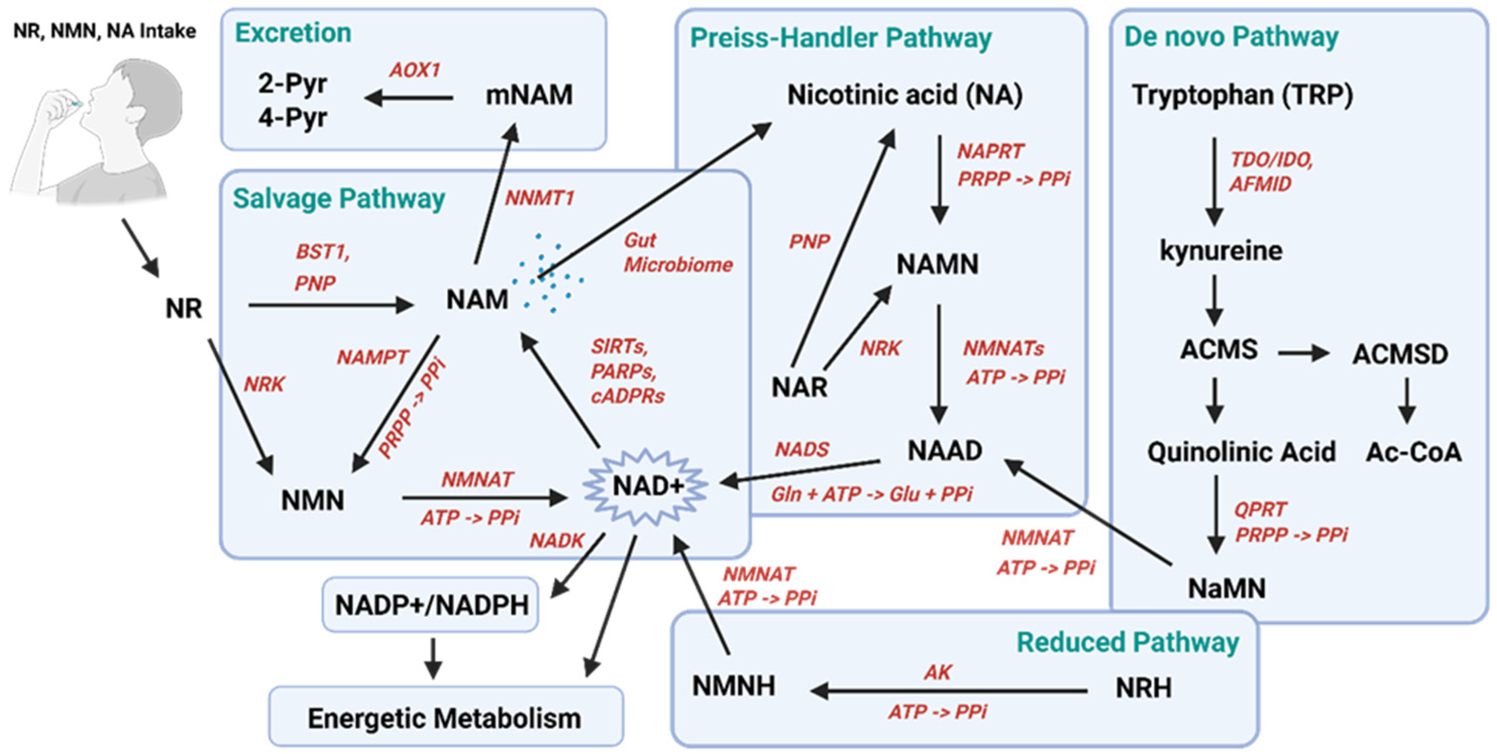
2. NAM and Nicotinamide Adenine Dinucleotide (NAD+) Metabolism
2.1. NAD+ and Energy Production
2.2. NAD+-Consuming Enzymes
3. Biological Role of NAM and NAD+ Precursors in the Skin
4. Clinical Evidence: NAM and NAD+ Precursors in Aging
4.1. Systemic NAM and NAD+ Precursors and Anti-Aging Effects

| Compound | Study/Design | Population | Intervention | Main Findings | Statistical Significance | Main Limitations | Ref. |
|---|---|---|---|---|---|---|---|
| NR | Double-blind RCT, crossover pharmacokinetic study | 12 healthy adults (30–55 y) | NR up to 1000 mg/day for several weeks | Increased whole-blood and PBMC NAD+ (≈1.3–2.7 fold) | Significant dose-dependent metabolite changes | Small sample, short duration | [75] |
| NMN | Double-blind RCT, placebo-controlled metabolic study (jRCTs041200034) | 30 healthy adults (20–65 y) | 250 mg/day for 12 weeks | Increased NAD+ pathway markers (blood NAD+, NAMN); no clinically meaningful safety concerns. | Significant increase in NAD+ markers (p < 0.001) | No clinical aging endpoints | [77] |
| NMN | Double-blind RCT, placebo-controlled study (UMIN000036321) | 42 older men (≥65 y) | 250 mg/day for 12 weeks | Increased circulating NAD+ metabolites; acceptable tolerability; improved gait speed, grip strength | Significant increase in NAD+ metabolites (p < 0.05 to p < 0.001), improvement in gait speed (p = 0.033) and left grip strength (p = 0.019) | Male-only cohort; short follow-up; no clinical aging or long-term safety endpoints | [78] |
| NMN | Double-blind RCT, placebo-controlled crossover trial (NCT03151239) | 25 postmenopausal prediabetic women (55–75 y) | 250 mg/day for 10 weeks | Increased NAD+ and metabolites, improved skeletal muscle insulin sensitivity associated with enhanced muscle gene expression related to metabolism | Significant improvement in muscle insulin sensitivity (p < 0.05) | Highly specific population, short duration, no skin endpoints | [86] |
| NMN | Phase I safety study (UMIN000021309) | 10 healthy men (40–60 y) | Single doses 100–500 mg; assessment over 5 h | Well tolerated; increased NAD-related metabolites (2-Py, 4 Py) | Significant dose-dependent metabolite changes (p < 0.05, p < 0.01) | Very short-term exposure | [80] |
| NMN | Double-blind RCT, placebo-controlled study | 32 overweight/obese adults (55–80 y) | 1000–2000 mg/day for 14 days | Increased NAD+ metabolites; safe | Significant metabolite increases with dose-related effect; no significant safety signal | Very short intervention | [81] |
| NMN | Double-blind RCT, placebo-controlled study (UMIN000043084) | 41 healthy adults (20–65 y) | 1250 mg/day for 4 weeks | NMN was well tolerated with no clinically relevant adverse events; no significant abnormalities in hematological or biochemical parameters | No significant safety-related changes reported (p > 0.05 across safety endpoints) | Designed for safety only; short duration; no efficacy or aging-related endpoints | [82] |
| NMN | Double-blind RCT, placebo-controlled study (NCT04228640) | 66 healthy adults (40–65 y) | 300 mg/day for 60 days | Improved physical performance (e.g., walking distance, muscle endurance) and quality-of-life related measures; increases in NAD+ biomarkers | Significant improvements in selected physical performance and metabolic endpoints (p < 0.05 for primary/selected outcomes; some secondary endpoints not significant) | Short duration, no long-term aging outcomes | [84] |
| NMN | Double-blind RCT, placebo-controlled study (UMIN000038097) | 48 overweight/obese older adults (≥65 y) | 250 mg/day for 12 weeks | Improved sleep quality, reduced fatigue, and modest improvements in physical performance measures in selected subgroups | Significant improvements in selected sleep/fatigue and physical performance endpoints (p < 0.05 for several outcomes; not all endpoints significant) | Heterogeneous outcomes, reliance on self-reported measures, no long-term aging endpoints | [83] |
| NMN | Double-blind RCT, placebo-controlled study (ChiCTR2000035138) | 48 recreational runners (27–50 y) | 300–1200 mg/day for 6 weeks | Improved aerobic capacity and ventilatory threshold with dose trend; no major adverse symptoms/ECG issues. | Significant improvements vs.placebo in selected ventilatory-threshold parameters (p < 0.05); no significant change in VO2max or body composition | Small sample, short duration, heterogeneous dose response, no aging or metabolic endpoints | [85] |
| NMN | Open-label pilot (NCT04228640) | 8 healthy men (45–60 y) | 300 mg/day for 30–90 days | Increased telomere length in PBMCs | Significant increase in PBMC telomere length (p < 0.05/0.01) | Exploratory finding due to open-label design and very small sample size. | [86] |
| NAM | Acute supplementation study | 5 healthy adults | Single oral NAM dose (100 or 500 mg) | 500 mg transiently increased blood NAD+ levels and altered circulating lipidomic profile; well tolerated | Significant increase in blood NAD+ at 12 h with 500 mg (p < 0.05); significant lipidomic changes reported | Very small sample size, acute exposure only, no clinically relevant endpoints | [87] |
4.2. Topical NAM and Skin Anti-Aging Effects
| Study Design | Population | Treatment | Main Outcomes | Statistical Significance | Main Limitations | Ref. |
|---|---|---|---|---|---|---|
| Double-blind, vehicle-controlled, split-face (left–right randomized) trial | 50 women | 5% NAM moisturizer vs.vehicle twice daily for 12 weeks | Reduced wrinkles, hyperpigmented spots, red blotchiness, skin yellowing, texture alterations and skin elasticity | Significant skin appearance improvements vs.vehicle for fine lines/wrinkles, hyperpigmented spots, red blotchiness, and skin sallowness. Elasticity, measured via cutometry, was improved. | Predominantly investigator-assessed clinical outcomes | [89,90] |
| Randomized double-blind placebo-controlled split-face trial | 52 women | 0.03% kinetin + 4% niacinamide (group 1) vs.niacinamide 4%(group 2) twice daily for 12 weeks | Combination treatment produced greater skin improvements than niacinamide alone | Group 1: persistent and significant reductions in spot, pore, wrinkle, and evenness counts were found at weeks 8 and 12 in group 1. A significant increase in corneal hydration status was also evident at week 12, whereas persistent decreases in erythema index were apparent at 8 and 12 weeks. Group 2: more modest improvements with significant reductions in pore and evenness counts at week 8 and wrinkle counts at week 12 were noted. | Short-term study with predominantly investigator-assessed clinical outcomes and limited generalizability. | [91] |
| Randomized, vehicle-controlled, split-face trial | 30 women | 4% niacinamide lotion vs.vehicle lotion once daily for 8 weeks | Reduced wrinkles and skin roughness | Wrinkle grade significantly decreased vs.baseline and vs.vehicle (p < 0.001 for key comparisons). Surface roughness improved vs.baseline (p < 0.01) and vs.control (p < 0.05). | Short follow-up duration | [92] |
| Multi-arm translational package: (i) clinical facial study + (ii) treated-vs-untreated arm | 44 women (face); 30 women (arm biopsies) | Topical formula centered on 6% niacinamide plus hyaluronic acid fractions once daily for 8 weeks | Significant improvements in wrinkles/fine lines and skin quality scores; radiance +44% at 2 months, smoothness score +39% on average; fine lines −15% on average; transcriptomics consistent with “senomorphic” signaling (down-modulation of multiple SASP genes, such as MMP12/CXCL9 and S100A8/9). | Highly significant improvements in radiance, smoothness, homogeneity, and plumpness at 1 and 2 months (p < 0.0001 for most endpoints). Radiance increased significantly at 1 and 2 months (p < 0.0001). Skin smoothness improvements were significant at both time points (p < 0.0001). Reductions in fine lines were significant (p < 0.004), as were reductions in wrinkles (p < 0.004). Twenty-four aging-/SASP-related genes were significantly modulated (p < 0.05), with predominant downregulation (20 downregulated vs. 4 upregulated). | Effects confounded by multi-ingredient formulation | [97] |
| Randomized, parallel-group facial appearance study | 99 (test)/97 (control) subjects | Multi-product regimen: SPF30 day lotion (5% niacinamide + peptides) + night cream (niacinamide + peptides, NPP) + wrinkle treatment (niacinamide + peptides + 0.3% retinyl propionate) vs.comparator regimen incl. 0.02% tretinoin (emollient base) + SPF30. Wrinkle treatment twice daily; day lotion + night cream daily; 8 weeks (subset continued +16 weeks) | Improved facial wrinkle appearance and photodamage parameters; NPP regimen showed greater responder rates and superior self-assessed improvements vs.tretinoin. TEWL remained stable in NPP but increased with tretinoin; erythema and dryness were higher with tretinoin. | Significant improvement in wrinkle appearance vs.baseline (p ≤ 0.05) in both groups; NPP superior to tretinoin for wrinkle improvement (p < 0.01). Higher responder rates with NPP (p = 0.03–0.02). Significant between-group differences favoring NPP in self-assessment outcomes at 4 and 8 weeks (p < 0.05). Significant between-group difference in TEWL change (p < 0.01). | Effects confounded by multi-active formulation | [98] |
| Randomized, double-blind, vehicle-controlled, split-face trial | 40 women | 2% gold silk sericin + 5% niacinamide + 0.1% Signaline™ vs.simple O/W emulsion twice daily for 12 weeks | Improved hydration, elasticity, and barrier-related biophysical parameters | Significant improvements versus vehicle in hydration, elasticity, barrier function, and surface topography (p< 0.05 for all measured biophysical parameters). | Multi-component formulation | [99] |
| Open-label pilot study | 25 women | Night formulation: 0.5% retinol + 4.4% niacinamide + 1% resveratrol + 1.1% hexylresorcinol for 10 weeks | Improved pigmentation and wrinkle appearance from week 4 to week 10 | Statistical interpretation is limited by the open-label, uncontrolled design | Uncontrolled study design | [100] |
| Double-blind, randomized, split-face, vehicle-controlled (post-procedure) | 24 patients | 2% adipocyte-derived MSC-conditioned medium + 2% niacinamide vs.vehicle for 3 weeks after fractional ablative CO2 laser therapy | Reduced wrinkles and pigmentation after laser treatment | Reduced wrinkle index (p = 0.036) and melanin index (p = 0.043) vs.control | Small sample size and short follow-up; multi-component formulation | [101] |
| Randomized, double-blind, placebo-controlled clinical program (photodamaged skin; multi-study report) | 96 subjects | Myristyl nicotinate (MN) topical (1–5% across studies) vs.placebo formulations daily or twice-daily applications for ~12 weeks | Increased epidermal NAD levels (+25%), stratum corneum thickness (~+70%) and epidermal thickness (~+20%) vs.placebo; reduced TEWL (~20%) on cheeks and arms | Significant increases in skin NAD (p = 0.001), stratum corneum thickness (p = 0.0001), epidermal thickness (p = 0.001), epidermal renewal (p ≤ 0.003), and significant TEWL reductions on cheeks (p = 0.012) and arms (p = 0.017) | Evaluated NAM derivative | [96] |
| Controlled clinical studies | 18 women with hyperpigmentation; 120 women with facial tanning | Topical 2–5% NAM formulations for 4 weeks | Reduced hyperpigmentation and improved skin lightness in treated groups; inhibition of melanosome transfer was demonstrated mechanistically | Significant improvements in pigmentation parameters versus baseline and/or control conditions after 4 weeks (p < 0.05 where reported) | Heterogeneous study populations and endpoints; short treatment duration; limited mechanistic–clinical correlation; | [102] |
| Double-blind randomized clinical trial | 27 patients with melasma | 4% NAM vs.4% hydroquinone (HQ) for 8 weeks | Comparable improvement in melasma severity, reduction in inflammatory infiltrate, and solar elastosis, with better tolerability for NAM | No significant difference NAM vs.HQ; fewer adverse effects with NAM (18% with NAM vs.29% with HQ). | Small sample size and short treatment duration | [104] |
5. Clinical Evidence: NAM in NMSC Prevention
5.1. Systemic NAM and Chemoprevention
| Study Type | Population | Treatment | Main Outcomes | Statistical Significance | Main Limitations | Reference |
|---|---|---|---|---|---|---|
| Phase III randomized, placebo-controlled trial (ONTRAC) | 386 high-risk patients with previous NMSC | Oral NAM 500 mg twice daily for 12 months | Reduced incidence of new non-melanoma skin cancers, mainly SCC and AK | 23% reduction in NMSC (95% CI: 4–38%, p = 0.02); stronger effect for SCC (95% CI: 0–51%, p = 0.05); AK reduction (0.001 < p < 0.01) | No long-term post-intervention follow-up after discontinuation | [106] |
| Phase II double-blinded randomized controlled trial | Patients with AKs (n ≈ 74–114 across dosing arms, depending on protocol phase) | Oral NAM 500 mg once or twice daily | Dose-dependent reduction in AKs, favoring twice-daily dosing | Greater AK reduction with BID dosing (p = 0.005) than OD dosing (p = 0.05) | Small sample size; short duration; surrogate endpoint (AK rather than cancer incidence) | [107] |
| Retrospective cohort study | >33,000 patients with a prior history of NMSC | Oral NAM 500 mg twice daily | Reduced risk of subsequent non-melanoma skin cancers in a real-world setting | 14% overall reduction (HR 0.86; 95% CI 0.82–0.89); 54% reduction when started after first NMSC (HR 0.47; 95% CI 0.23–0.97); reduction for SCC (HR 0.78; 95% CI 0.75–0.82) and BCC (HR 1.00; 95% CI 0.96–1.05) | Observational design; residual confounding; lack of randomization | [109] |
| Systematic review and meta-analysis | High-risk patients/organ transplant recipients (multiple pooled studies) | Oral NAM | Reduced incidence of BCC and SCC; increased gastrointestinal (GI) adverse events | Significant reduction in BCC (RR 0.46; 95% CI 0.22–0.95) and SCC (RR 0.48; 95% CI 0.26–0.88); ↑ GI adverse effects (RR 1.78; 95% CI 1.30–2.45) | Variable study quality; pooled populations with differing baseline risk | [110] |
| Systematic review and meta-analysis | Mixed populations (multiple RCTs and observational datasets) | Oral NAM | No significant reduction in SCC, BCC, or total NMSC | No significant reduction in SCC (RR 0.81; 95% CI 0.48–1.37), BCC (RR 0.88; 95% CI 0.50–1.55), or total NMSC (RR 0.82; 95% CI 0.61–1.12) | Clinical and methodological heterogeneity; differences in included study designs and populations | [111] |
| Phase III randomized, placebo-controlled trial (solid organ transplant recipients; ONTRANS) | 158 solid organ transplant recipients | Oral nicotinamide 500 mg twice daily vs.placebo for 12 months | No significant reduction in NMSC cancers, including SCC, BCC, or AK, in immunosuppressed transplant recipients | No significant difference between NAM and placebo for SCC, BCC, or total NMSC (rate ratio ~1.0; p not significant) | Early trial termination; underpowered study; immunosuppressed population limiting comparability with prior RCTs | [112] |
5.2. Topical NAM and Local Photoprotection
6. Discussion
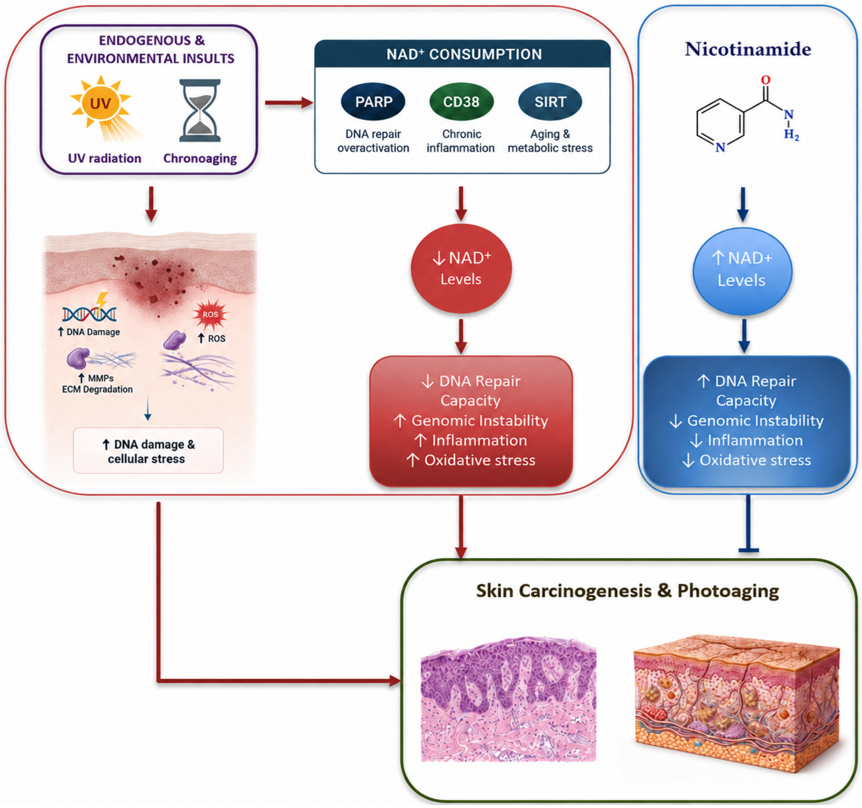
6.1. A Shared Metabolic Mechanism: NAD+ as a Central Node
6.2. Clinical Evidence: NAM Selective Topical Effects and Systemic Benefit
6.3. Limitations, Safety Considerations, and Future Directions
7. Conclusions
Author Contributions
Funding
Institutional Review Board Statement
Data Availability Statement
Conflicts of Interest
References
- Fania, L.; Didona, D.; Di Pietro, F.R.; Verkhovskaia, S.; Morese, R.; Paolino, G.; Donati, M.; Ricci, F.; Coco, V.; Ricci, F.; et al. Cutaneous Squamous Cell Carcinoma: From Pathophysiology to Novel Therapeutic Approaches. Biomedicines 2021, 9, 171. [Google Scholar] [CrossRef]
- Fania, L.; Abeni, D.; Esposito, I.; Spagnoletti, G.; Citterio, F.; Romagnoli, J.; Castriota, M.; Ricci, F.; Moro, F.; Perino, F.; et al. Behavioral and demographic factors associated with occurrence of non-melanoma skin cancer in organ transplant recipients. G. Ital. Dermatol. Venereol. 2020, 155, 669–675. [Google Scholar] [CrossRef]
- Fania, L.; Mazzanti, C.; Campione, E.; Candi, E.; Abeni, D.; Dellambra, E. Role of Nicotinamide in Genomic Stability and Skin Cancer Chemoprevention. Int. J. Mol. Sci. 2019, 20, 5946. [Google Scholar] [CrossRef] [PubMed]
- Dotto, G.P. Multifocal Epithelial Tumors and Field Cancerization: Stroma as a Primary Determinant. J. Clin. Investig. 2014, 124, 1446–1453. [Google Scholar] [CrossRef]
- Pellacani, G.; Lim, H.W.; Stockfleth, E.; Sibaud, V.; Brugués, A.O.; Saint Aroman, M. Photoprotection: Current Developments and Controversies. J. Eur. Acad. Dermatol. Venereol. 2024, 38, 12–20. [Google Scholar] [CrossRef] [PubMed]
- Camillo, L.; Zavattaro, E.; Savoia, P. Nicotinamide: A Multifaceted Molecule in Skin Health and Beyond. Medicina 2025, 61, 254. [Google Scholar] [CrossRef] [PubMed]
- Hyeraci, M.; Papanikolau, E.S.; Grimaldi, M.; Ricci, F.; Pallotta, S.; Monetta, R. Systemic Photoprotection in Melanoma and Non-Melanoma Skin Cancer. Biomolecules 2023, 13, 1067. [Google Scholar] [CrossRef]
- Chen, A.C.; Martin, A.J.; Choy, B.; Fernández-Peñas, P.; Dalziell, R.A.; McKenzie, C.A.; Scolyer, R.A.; Dhillon, H.M.; Vardy, J.L.; Kricker, A.; et al. A Phase 3 Randomized Trial of Nicotinamide for Skin-Cancer Chemoprevention. N. Engl. J. Med. 2015, 373, 1618–1626. [Google Scholar] [CrossRef]
- Searle, T.; Ali, F.R.; Al-Niaimi, F. Systemic photoprotection in 2021. Clin. Exp. Dermatol. 2021, 46, 1189–1204. [Google Scholar] [CrossRef]
- Malesu, R.; Martin, A.J.; Lyons, J.G.; Scolyer, R.A.; Chen, A.C.; McKenzie, C.A.; Madore, J.; Hallidaya, G.M.; Damian, D.L. Nicotinamide for Skin Cancer Chemoprevention: Effects of Nicotinamide on Melanoma In Vitro and In Vivo. Photochem. Photobiol. Sci. 2020, 19, 171–179. [Google Scholar] [CrossRef]
- Di Guardo, A.; Trovato, F.; Cantisani, C.; Ricci, F.; Di Lella, G.; Moro, F.; Morese, R.; Panebianco, A.; Nisticò, S.P.; Pellacani, G.; et al. Advanced Cutaneous Squamous Cell Carcinoma: Biology, Immunotherapy, and Evolving Prognostic Factors. Biomedicines 2025, 13, 3010. [Google Scholar] [CrossRef]
- Damgaard, M.V.; Treebak, J.T. What Is Really Known about the Effects of Nicotinamide Riboside Supplementation in Humans. Sci. Adv. 2023, 9, eadi4862. [Google Scholar] [CrossRef]
- Yusri, K.; Jose, S.; Vermeulen, K.S.; Tan, T.C.M.; Sorrentino, V. The Role of NAD+ Metabolism and Its Modulation of Mitochondria in Aging and Disease. npj Metab. Health Dis. 2025, 3, 26. [Google Scholar] [CrossRef]
- Revollo, J.R.; Grimm, A.A.; Imai, S. The NAD Biosynthesis Pathway Mediated by Nicotinamide Phosphoribosyltransferase Regulates Sir2 Activity in Mammalian Cells. J. Biol. Chem. 2004, 279, 50754–50763. [Google Scholar] [CrossRef]
- Imai, S.; Johnson, S. NAD+ Biosynthesis, Aging, and Disease. F1000Research 2018, 7, 132. [Google Scholar] [CrossRef] [PubMed]
- Yaku, K.; Palikhe, S.; Izumi, H.; Yoshida, T.; Hikosaka, K.; Hayat, F.; Karim, M.; Iqbal, T.; Nitta, Y.; Sato, A.; et al. BST1 regulates nicotinamide riboside metabolism via its glycohydrolase and base-exchange activities. Nat. Commun. 2021, 12, 6767. [Google Scholar] [CrossRef]
- Alegre, G.F.S.; Pastore, G.M. NAD+ Precursors Nicotinamide Mononucleotide (NMN) and Nicotinamide Riboside (NR): Potential Dietary Contribution to Health. Curr. Nutr. Rep. 2023, 12, 445–464. [Google Scholar] [CrossRef]
- Ratajczak, J.; Joffraud, M.; Trammell, S.A.; Ras, R.; Canela, N.; Boutant, M.; Kulkarni, S.S.; Rodrigues, M.; Redpath, P.; Migaud, M.E.; et al. NRK1 controls nicotinamide mononucleotide and nicotinamide riboside metabolism in mammalian cells. Nat. Commun. 2016, 7, 13103. [Google Scholar] [CrossRef]
- Lee, K.J.; Chokshi, S.; Joshi, T.; Cummings, M.; Lyons, C.E.; Singleton, M.H.; Catranis, E.; Madiera da Silva, L.; Hayat, F.; Migaud, M.; et al. NAD+ Boosting Through NRH Supplementation Enhances Treatment Efficacy in EOC In Vitro. Int. J. Mol. Sci. 2025, 26, 1719. [Google Scholar] [CrossRef] [PubMed]
- Xie, N.; Zhang, L.; Gao, W.; Huang, C.; Huber, P.E.; Zhou, X.; Li, C.; Shen, G.; Zou, B. NAD+ metabolism: Pathophysiologic mechanisms and therapeutic potential. Signal Transduct. Target. Ther. 2020, 5, 227. [Google Scholar] [CrossRef] [PubMed]
- Ji, X.; Zheng, M.; Yu, T.; Kang, J.; Fan, T.; Xu, B. NAD+-Consuming Enzymes in Stem Cell Homeostasis. Oxid. Med. Cell. Longev. 2023, 2023, 4985726. [Google Scholar] [CrossRef]
- Zhang, W.; Ren, H.; Chen, W.; Hu, B.; Feng, C.; Li, P.; Shi, Y.; Fang, J. Nicotinamide phosphoribosyltransferase in NAD+ metabolism: Physiological and pathophysiological implications. Cell Death Discov. 2025, 11, 371. [Google Scholar] [CrossRef]
- Bursch, K.L.; Goetz, C.J.; Smith, B.C. Current Trends in Sirtuin Activator and Inhibitor Development. Molecules 2024, 29, 1185. [Google Scholar] [CrossRef]
- Rifaï, K.; Judes, G.; Idrissou, M.; Daures, M.; Bignon, Y.J.; Penault-Llorca, F.; Bernard-Gallon, D. SIRT1-dependent epigenetic regulation of H3 and H4 histone acetylation in human breast cancer. Oncotarget 2018, 9, 30661–30678. [Google Scholar] [CrossRef] [PubMed]
- Yang, Y.; Liu, Y.; Wang, Y.; Chao, Y.; Zhang, J.; Jia, Y.; Tie, J.; Hu, D. Regulation of SIRT1 and its roles in inflammation. Front. Immunol. 2022, 13, 831168. [Google Scholar] [CrossRef] [PubMed]
- Jackson, M.D.; Schmidt, M.T.; Oppenheimer, N.J.; Denu, J.M. Mechanism of Nicotinamide Inhibition and Transglycosidation by Sir2 Histone/Protein Deacetylases. J. Biol. Chem. 2003, 278, 50985–50998. [Google Scholar] [CrossRef] [PubMed]
- Imai, S.; Guarente, L. It Takes Two to Tango: NAD+ and Sirtuins in Aging/Longevity Control. npj Aging Mech. Dis. 2016, 2, 16017. [Google Scholar] [CrossRef]
- Anderson, K.A.; Madsen, A.S.; Olsen, C.A.; Hirschey, M.D. Metabolic Control by Sirtuins and Other Enzymes That Sense NAD+, NADH, or Their Ratio. Biochim. Biophys. Acta Bioenerg. 2017, 1858, 991–998. [Google Scholar] [CrossRef]
- Garcia-Peterson, L.M.; Wilking-Busch, M.J.; Ndiaye, M.A.; Philippe, C.G.A.; Setaluri, V.; Ahmad, N. Sirtuins in Skin and Skin Cancers. Skin Pharmacol. Physiol. 2017, 30, 216–224. [Google Scholar] [CrossRef]
- Li, D.; Li, W.; Liao, X.; Jiang, S.; Ma, M.; Hu, Z.; Lin, K.; Yu, W.; Sun, X.; Fan, Y.; et al. NAD+-dependent Sirt6 is a key regulator involved in telomere shortening of in vitro-cultured preimplantation embryos. Commun. Biol. 2025, 8, 1275. [Google Scholar] [CrossRef]
- Wu, Q.J.; Zhang, T.N.; Chen, H.H.; Yu, X.F.; Lv, J.L.; Liu, Y.Y.; Liu, Y.S.; Zheng, G.; Zhao, J.Q.; Wei, Y.F.; et al. The sirtuin family in health and disease. Signal Transduct. Target. Ther. 2022, 7, 402. [Google Scholar] [CrossRef]
- Lei, P.; Li, W.; Luo, J.; Xu, N.; Wang, Y.; Xie, D.; Guan, H.; Huang, B.; Huang, X.; Zhou, P. PARP (poly ADP-ribose polymerase) family in health and disease. MedComm 2025, 6, e70314. [Google Scholar] [CrossRef]
- Cohen, M.S. Interplay between Compartmentalized NAD+ Synthesis and Consumption: A Focus on the PARP Family. Genes Dev. 2020, 34, 254–262. [Google Scholar] [CrossRef]
- Hurtado-Bagès, S.; Knobloch, G.; Ladurner, A.G.; Buschbeck, M. The Taming of PARP1 and Its Impact on NAD+ Metabolism. Mol. Metab. 2020, 38, 100950. [Google Scholar] [CrossRef]
- Langelier, M.-F.; Zandarashvili, L.; Aguiar, P.M.; Black, B.E.; Pascal, J.M. NAD+ Analog Reveals PARP-1 Substrate-Blocking Mechanism and Allosteric Communication from Catalytic Center to DNA-Binding Domains. Nat. Commun. 2018, 9, 844. [Google Scholar] [CrossRef]
- Hopp, A.-K.; Grüter, P.; Hottiger, M.O. Regulation of Glucose Metabolism by NAD+ and ADP-Ribosylation. Cells 2019, 8, 890, Erratum in Cells 2019, 8, 1371. https://doi.org/10.3390/cells8111371. [Google Scholar] [CrossRef] [PubMed]
- Martínez-Morcillo, F.J.; Cantón-Sandoval, J.; Martínez-Navarro, F.J.; Cabas, I.; Martínez-Vicente, I.; Armistead, J.; Hatzold, J.; López-Muñoz, A.; Martínez-Menchón, T.; Corbalán-Vélez, R.; et al. NAMPT-derived NAD+ fuels PARP1 to promote skin inflammation through parthanatos cell death. PLoS Biol. 2021, 19, e3001455. [Google Scholar] [CrossRef] [PubMed]
- Khan, J.A.; Forouhar, F.; Tao, X.; Tong, L. Nicotinamide Adenine Dinucleotide Metabolism as an Attractive Target for Drug Discovery. Expert Opin. Ther. Targets 2007, 11, 695–705. [Google Scholar] [CrossRef]
- Kennedy, B.E.; Sharif, T.; Martell, E.; Dai, C.; Kim, Y.; Lee, P.W.K.; Gujar, S.A. NAD+ salvage pathway in cancer metabolism and therapy. Pharmacol. Res. 2016, 114, 274–283. [Google Scholar] [CrossRef] [PubMed]
- Katayoshi, T.; Nakajo, T.; Tsuji-Naito, K. Restoring NAD+ by NAMPT Is Essential for the SIRT1/p53-Mediated Survival of UVA- and UVB-Irradiated Epidermal Keratinocytes. J. Photochem. Photobiol. B 2021, 221, 112238. [Google Scholar] [CrossRef]
- Zhang, T.; Berrocal, J.G.; Frizzell, K.M.; Gamble, M.J.; DuMond, M.E.; Krishnakumar, R.; Yang, T.; Sauve, A.A.; Kraus, W.L. Regulation of poly(ADP-ribose) polymerase-1-dependent gene expression through promoter-directed recruitment of a nuclear NAD+ synthase. J. Biol. Chem. 2012, 287, 12405–12416. [Google Scholar] [CrossRef]
- Konen, J.M.; Fradette, J.J.; Gibbons, D.L. The Good, the Bad and the Unknown of CD38 in the Metabolic Microenvironment and Immune Cell Functionality of Solid Tumors. Cells 2019, 9, 52. [Google Scholar] [CrossRef]
- Chini, C.C.S.; Hogan, K.A.; Warner, G.M.; Tarragó, M.G.; Peclat, T.R.; Tchkonia, T.; Kirkland, J.L.; Chini, E.N. The NADase CD38 is induced by factors secreted from senescent cells providing a potential link between senescence and age-related cellular NAD+ decline. Biochem. Biophys. Res. Commun. 2019, 513, 486–493. [Google Scholar] [CrossRef]
- Covarrubias, A.J.; Kale, A.; Perrone, R.; Lopez-Dominguez, J.A.; Pisco, A.O.; Kasler, H.G.; Schmidt, M.S.; Heckenbach, I.; Kwok, R.; Wiley, C.D.; et al. Senescent cells promote tissue NAD+ decline during ageing via the activation of CD38+ macrophages. Nat. Metab. 2020, 2, 1265–1283, Erratum in Nat. Metab. 2021, 3, 120–121. https://doi.org/10.1038/s42255-020-00328-w. [Google Scholar] [CrossRef] [PubMed]
- Van Deursen, J.M. The Role of Senescent Cells in Ageing. Nature 2014, 509, 439–446. [Google Scholar] [CrossRef]
- Katsyuba, E.; Auwerx, J. Modulating NAD+ Metabolism, from Bench to Bedside. EMBO J. 2017, 36, 2670–2683. [Google Scholar] [CrossRef] [PubMed]
- Ministrini, S.; Liberale, L.; Erle, H.-E.; Percoco, G.; Tfayli, A.; Assi, A.; Kapitonov, I.; Greiner, I.; Camici, G.G. A liposomal formulation enhances the anti-senescence properties of nicotinamide adenine-dinucleotide (NAD+) in endothelial cells and keratinocytes. Curr. Issues Mol. Biol. 2025, 47, 722. [Google Scholar] [CrossRef] [PubMed]
- Surjana, D.; Halliday, G.M.; Damian, D.L. Nicotinamide Enhances Repair of Ultraviolet Radiation-Induced DNA Damage in Human Keratinocytes and Ex Vivo Skin. Carcinogenesis 2013, 34, 1144–1149. [Google Scholar] [CrossRef]
- Ma, T.; He, J.; Long, Q.; Wang, Y.; Chen, F.; Chen, S.; Xu, K.; Cao, Y. Orientin attenuates UVB-induced skin photodamage by inhibiting ROS generation via the AMPK/Nrf2 axis. Int. Immunopharmacol. 2025, 155, 114655. [Google Scholar] [CrossRef]
- Damian, D.L. Nicotinamide for Skin Cancer Chemoprevention. Australas. J. Dermatol. 2017, 161, 1140–1147. [Google Scholar] [CrossRef]
- Pihl, C.; Andersen, F.; Bjerring, P.; Haedersdal, M.; Lerche, C.M. Efficacy of Combinational Treatment versus Nicotinamide Monotherapy in the Prevention of Ultraviolet Radiation-Induced Skin Cancer. Dermatology 2024, 240, 453–461. [Google Scholar] [CrossRef]
- Tan, C.Y.R.; Tan, C.L.; Chin, T.; Morenc, M.; Ho, C.Y.; Rovito, H.A. Nicotinamide Prevents UVB- and Oxidative Stress-Induced Photoaging in Human Primary Keratinocytes. J. Investig. Dermatol. 2022, 142, 1670–1681. [Google Scholar] [CrossRef]
- Camillo, L.; Gironi, L.C.; Zavattaro, E.; Esposto, E.; Savoia, P. Nicotinamide Attenuates UV-Induced Stress Damage in Human Primary Keratinocytes from Cancerization Fields. J. Investig. Dermatol. 2022, 142, 1466–1477.e1. [Google Scholar] [CrossRef]
- Mahajan, A.S.; Arikatla, V.S.; Thyagarajan, A.; Zhelay, T.; Sahu, R.P.; Kemp, M.G.; Spandau, D.F.; Travers, J.B. Creatine and nicotinamide prevent oxidant-induced senescence in human fibroblasts. Nutrients 2021, 13, 4102. [Google Scholar] [CrossRef]
- Oblong, J.E.; Bowman, A.; Rovito, H.A.; Jarrold, B.B.; Sherrill, J.D.; Black, M.R.; Nelson, G.; Kimball, A.B.; Birch-Machin, M. Metabolic dysfunction in human skin: Restoration of mitochondrial integrity and metabolic output by nicotinamide (niacinamide) in primary dermal fibroblasts from older aged donors. Aging Cell 2020, 19, e13248. [Google Scholar] [CrossRef]
- Tao, Z.; Jin, Z.; Wu, J.; Cai, G.; Yu, X. Sirtuin family in autoimmune diseases. Front. Immunol. 2023, 14, 1186231. [Google Scholar] [CrossRef]
- Wang, T.; Qin, Y.; Qiao, J.; Liu, Y.; Wang, L.; Zhang, X. Overexpression of SIRT6 Regulates NRF2/HO-1 and NF-κB Signaling Pathways to Alleviate UVA-Induced Photoaging in Skin Fibroblasts. J. Photochem. Photobiol. B 2023, 249, 112801. [Google Scholar] [CrossRef]
- Lee, I.H.; Cao, L.; Mostoslavsky, R. A Role for the NAD-Dependent Deacetylase SIRT1 in the Regulation of Autophagy. Proc. Natl. Acad. Sci. USA 2008, 105, 3374–3379. [Google Scholar] [CrossRef]
- Jang, S.Y.; Kang, H.T.; Hwang, E.S. Nicotinamide-Induced Mitophagy: Event Mediated by High NAD+/NADH Ratio and SIRT1 Protein Activation. J. Biol. Chem. 2012, 287, 19304–19314. [Google Scholar] [CrossRef]
- McDaniel, M.; Lee, L.H.; Lin, S.J. NAD+ Homeostasis and Autophagy: Integrated Control through Nutrient Signaling in Yeast and Mammals. Cells 2025, 14, 1495. [Google Scholar] [CrossRef]
- Qian, S.; Dai, S.; Guo, C.; Wang, W.; Pang, J.; Shen, Y.; Xu, M.; Hu, J.; Cui, W.; Sun, X.; et al. Apoptotic bodies restore NAD and mitochondrial homeostasis in fibroblasts. Adv. Sci. 2025, 12, e15691. [Google Scholar] [CrossRef]
- Kang, S.; Park, J.; Cho, E.; Kim, D.; Ye, S.; Jeong, E.T.; Jun, S.-H.; Kang, N.-G. Distinctive gene expression profiles and biological responses of skin fibroblasts to nicotinamide mononucleotide: Implications for longevity effects on skin. Biomedicines 2025, 13, 2395. [Google Scholar] [CrossRef]
- Sun, Z.; Li, J.; Zheng, Y.; Zhang, J.; Bai, W.; Deng, X.; Wu, Z.; Xu, X.; Ding, W.; Qian, H.; et al. Small extracellular vesicles derived from mesenchymal stromal cells loaded with β-nicotinamide mononucleotide activate NAD+/SIRT3 signaling pathway-mediated mitochondrial autophagy to delay skin aging. Stem Cell Res. Ther. 2025, 16, 339. [Google Scholar] [CrossRef]
- Wang, H.; Sun, Y.; Pi, C. Nicotinamide Mononucleotide Supplementation Improves Mitochondrial Dysfunction and Rescues Cellular Senescence by NAD+/Sirt3 Pathway in Mesenchymal Stem Cells. Int. J. Mol. Sci. 2022, 23, 14739. [Google Scholar] [CrossRef]
- Monfrecola, G.; Gaudiello, F.; Cirillo, T.; Fabbrocini, G.; Balato, A.; Lembo, S. Nicotinamide Downregulates Gene Expression of Interleukin-6, Interleukin-10, Monocyte Chemoattractant Protein-1, and Tumour Necrosis Factor-α in HaCaT Keratinocytes after Ultraviolet B Irradiation. Clin. Exp. Dermatol. 2013, 38, 185–188. [Google Scholar] [CrossRef]
- Elhassan, Y.S.; Kluckova, K.; Fletcher, R.S.; Schmidt, M.S.; Garten, A.; Doig, C.L.; Cartwright, D.M.; Oakey, L.; Burley, C.V.; Jenkinson, N.; et al. Nicotinamide riboside augments the aged human skeletal muscle NAD+ metabolome and induces transcriptomic and anti-inflammatory signatures. Cell Rep. 2019, 28, 1717–1728.e6. [Google Scholar] [CrossRef]
- Kang, S.; Park, J.; Cheng, Z.; Ye, S.; Jun, S.-H.; Kang, N.-G. Novel Approach to Skin Anti-Aging: Boosting Pharmacological Effects of Exogenous Nicotinamide Adenine Dinucleotide (NAD+) by Synergistic Inhibition of CD38 Expression. Cells 2024, 13, 1799. [Google Scholar] [CrossRef]
- Philips, N.; Chalensouk-Khaosaat, J.; Gonzalez, S. Simulation of the Elastin and Fibrillin in Non-Irradiated or UVA-Radiated Fibroblasts, and Direct Inhibition of Elastase or Matrix Metalloproteinases Activity by Nicotinamide or Its Derivatives. J. Cosmet. Sci. 2018, 69, 47–56. [Google Scholar]
- Tanno, O.; Ota, Y.; Kitamura, N.; Katsube, T.; Inoue, S. Nicotinamide Increases Biosynthesis of Ceramides as Well as Other Stratum Corneum Lipids to Improve the Epidermal Permeability Barrier. Br. J. Dermatol. 2000, 143, 524–531. [Google Scholar] [CrossRef]
- Song, Q.; Zhou, X.; Xu, K.; Liu, S.; Zhu, X.; Yang, J. The Safety and Antiaging Effects of Nicotinamide Mononucleotide in Human Clinical Trials: An Update. Adv. Nutr. 2023, 14, 1416–1435. [Google Scholar] [CrossRef]
- Conlon, N.J. The Role of NAD+ in Regenerative Medicine. Plast. Reconstr. Surg. 2022, 150, 41S–48S. [Google Scholar] [CrossRef]
- Chini, C.C.S.; Cordeiro, H.S.; Tran, N.L.K.; Chini, E.N. NAD Metabolism: Role in Senescence Regulation and Aging. Aging Cell 2024, 23, e13920. [Google Scholar] [CrossRef] [PubMed]
- Zapata-Pérez, R.; Wanders, R.J.A.; van Karnebeek, C.D.M.; Houtkooper, R.H. NAD+ Homeostasis in Human Health and Disease. EMBO Mol. Med. 2021, 13, e13943. [Google Scholar] [CrossRef]
- Snaidr, V.A.; Damian, D.L.; Halliday, G.M. Nicotinamide for Photoprotection and Skin Cancer Chemoprevention: A Review of Efficacy and Safety. Exp. Dermatol. 2019, 28, 15–22. [Google Scholar] [CrossRef]
- Trammell, S.A.J.; Schmidt, M.S.; Weidemann, B.J.; Redpath, P.; Jaksch, F.; Dellinger, R.W.; Li, Z.; Abel, E.D.; Migaud, M.E.; Brenner, C. Nicotinamide riboside is uniquely and orally bioavailable in mice and humans. Nat. Commun. 2016, 7, 12948. [Google Scholar] [CrossRef]
- Migaud, M.E.; Ziegler, M.; Baur, J.A. Regulation of and Challenges in Targeting NAD+ Metabolism. Nat. Rev. Mol. Cell Biol. 2024, 25, 822–840. [Google Scholar] [CrossRef]
- Okabe, K.; Yaku, K.; Uchida, Y.; Fukamizu, Y.; Sato, T.; Sakurai, T.; Tobe, K.; Nakagawa, T. Oral administration of nicotinamide mononucleotide is safe and efficiently increases blood nicotinamide adenine dinucleotide levels in healthy subjects. Front. Nutr. 2022, 9, 868640. [Google Scholar] [CrossRef] [PubMed]
- Igarashi, M.; Nakagawa-Nagahama, Y.; Miura, M.; Kashiwabara, K.; Yaku, K.; Sawada, M.; Sekine, R.; Fukamizu, Y.; Sato, T.; Sakurai, T.; et al. Chronic nicotinamide mononucleotide supplementation elevates blood nicotinamide adenine dinucleotide levels and alters muscle function in healthy older men. npj Aging 2022, 8, 5. [Google Scholar] [CrossRef]
- Yoshino, M.; Yoshino, J.; Kayser, B.D.; Patti, G.J.; Franczyk, M.P.; Mills, K.F.; Sindelar, M.; Pietka, T.; Patterson, B.W.; Imai, S.I.; et al. Nicotinamide mononucleotide increases muscle insulin sensitivity in prediabetic women. Science 2021, 372, 1224–1229. [Google Scholar] [CrossRef]
- Irie, J.; Inagaki, E.; Fujita, M.; Nakaya, H.; Mitsuishi, M.; Yamaguchi, S.; Yamashita, K.; Shigaki, S.; Ono, T.; Yukioka, H.; et al. Effect of oral administration of nicotinamide mononucleotide on clinical parameters and nicotinamide metabolite levels in healthy Japanese men. Endocr. J. 2020, 67, 153–160. [Google Scholar] [CrossRef] [PubMed]
- Pencina, K.M.; Lavu, S.; Dos Santos, M.; Beleva, Y.M.; Cheng, M.; Livingston, D.; Bhasin, S. MIB-626, an oral formulation of a microcrystalline unique polymorph of β-nicotinamide mononucleotide, increases circulating nicotinamide adenine dinucleotide and its metabolome in middle-aged and older adults. J. Gerontol. A Biol. Sci. Med. Sci. 2023, 78, 90–96. [Google Scholar] [CrossRef]
- Fukamizu, Y.; Uchida, Y.; Shigekawa, A.; Sato, T.; Kosaka, H.; Sakurai, T. Safety Evaluation of β-Nicotinamide Mononucleotide Oral Administration in Healthy Adult Men and Women. Sci. Rep. 2022, 12, 14442. [Google Scholar] [CrossRef] [PubMed]
- Kim, M.; Seol, J.; Sato, T.; Fukamizu, Y.; Sakurai, T.; Okura, T. Effect of 12-Week Intake of Nicotinamide Mononucleotide on Sleep Quality, Fatigue, and Physical Performance in Older Japanese Adults: A Randomized, Double-Blind Placebo-Controlled Study. Nutrients 2022, 14, 755. [Google Scholar] [CrossRef] [PubMed]
- Huang, H. A Multicentre, Randomised, Double Blind, Parallel Design, Placebo Controlled Study to Evaluate the Efficacy and Safety of Uthever (NMN Supplement), an Orally Administered Supplementation in Middle Aged and Older Adults. Front. Aging 2022, 3, 851698. [Google Scholar] [CrossRef]
- Liao, B.; Zhao, Y.; Wang, D.; Zhang, X.; Hao, X.; Hu, M. Nicotinamide Mononucleotide Supplementation Enhances Aerobic Capacity in Amateur Runners: A Randomized, Double-Blind Study. J. Int. Soc. Sports Nutr. 2021, 18, 54. [Google Scholar] [CrossRef]
- Niu, K.M.; Bao, T.T.; Gao, L.M.; Ru, M.; Li, Y.M.; Jiang, L. The Impacts of Short-Term NMN Supplementation on Serum Metabolism, Fecal Microbiota, and Telomere Length in Pre-Aging Phase. Front. Nutr. 2021, 8, 756243. [Google Scholar] [CrossRef] [PubMed]
- Ito, T.K.; Sato, T.; Takanashi, Y.; Tamannaa, Z.; Kitamoto, T.; Odagiri, K.; Setou, M. A single oral supplementation of nicotinamide within the daily tolerable upper level increases blood NAD+ levels in healthy subjects. Transl. Med. Aging 2021, 5, 43–51. [Google Scholar] [CrossRef]
- Boo, Y.C. Mechanistic Basis and Clinical Evidence for the Applications of Nicotinamide (Niacinamide) to Control Skin Aging and Pigmentation. Antioxidants 2021, 10, 1315. [Google Scholar] [CrossRef]
- Bissett, D.L.; Miyamoto, K.; Sun, P.; Li, J.; Berge, C.A. Topical Niacinamide Reduces Yellowing, Wrinkling, Red Blotchiness, and Hyperpigmented Spots in Aging Facial Skin. Int. J. Cosmet. Sci. 2004, 26, 231–238. [Google Scholar] [CrossRef]
- Bissett, D.L.; Oblong, J.E.; Berge, C.A. Niacinamide: A B Vitamin That Improves Aging Facial Skin Appearance. Dermatol. Surg. 2005, 31, 860–866. [Google Scholar] [CrossRef]
- Chiu, P.C.; Chan, C.C.; Lin, H.M.; Chiu, H.C. The Clinical Anti-Aging Effects of Topical Kinetin and Niacinamide in Asians: A Randomized, Double-Blind, Placebo-Controlled, Split-Face Comparative Trial. J. Cosmet. Dermatol. 2007, 6, 243–249. [Google Scholar] [CrossRef]
- Kawada, A.; Konishi, N.; Oiso, N.; Kawara, S.; Date, A. Evaluation of Anti-Wrinkle Effects of a Novel Cosmetic Containing Niacinamide. J. Dermatol. 2008, 35, 637–642. [Google Scholar] [CrossRef]
- Cole, M.A.; Quan, T.; Voorhees, J.J.; Fisher, G.J. Extracellular Matrix Regulation of Fibroblast Function: Redefining Our Perspective on Skin Aging. J. Cell Commun. Signal. 2018, 12, 35–43. [Google Scholar] [CrossRef] [PubMed]
- Imokawa, G.; Ishida, K. Biological Mechanisms Underlying the Ultraviolet Radiation-Induced Formation of Skin Wrinkling and Sagging I: Reduced Skin Elasticity, Highly Associated with Enhanced Dermal Elastase Activity, Triggers Wrinkling and Sagging. Int. J. Mol. Sci. 2015, 16, 7753–7775. [Google Scholar] [CrossRef] [PubMed]
- Choi, E.H. Aging of the Skin Barrier. Clin. Dermatol. 2019, 37, 336–345. [Google Scholar] [CrossRef]
- Jacobson, E.L.; Kim, H.; Kim, M.; Williams, J.D.; Coyle, D.L.; Coyle, W.R.; Grove, G.; Rizer, R.L.; Stratton, M.S.; Jacobson, M.K. A topical lipophilic niacin derivative increases NAD, epidermal differentiation and barrier function in photodamaged skin. Exp. Dermatol. 2007, 16, 490–499. [Google Scholar] [CrossRef] [PubMed]
- Bogdanowicz, P.; Bensadoun, P.; Noizet, M.; Béganton, B.; Philippe, A.; Alvarez-Georges, S.; Doat, G.; Tourette, A.; Bessou-Touya, S.; Lemaitre, J.-M.; et al. Senomorphic activity of a combination of niacinamide and hyaluronic acid: Correlation with clinical improvement of skin aging. Sci. Rep. 2024, 14, 16321. [Google Scholar] [CrossRef]
- Fu, J.J.J.; Hillebrand, G.G.; Raleigh, P.; Li, J.; Marmor, M.J.; Bertucci, V.; Grimes, P.E.; Mandy, S.H.; Perez, M.I.; Weinkle, S.H.; et al. A randomized, controlled comparative study of the wrinkle reduction benefits of a cosmetic niacinamide/peptide/retinyl propionate product regimen versus a prescription 0.02% tretinoin product regimen. Br. J. Dermatol. 2010, 162, 647–654. [Google Scholar] [CrossRef]
- Berardesca, E.; Ardigo, M.; Cameli, N.; Mariano, M.; Agozzino, M.; Matts, P.J. Randomized, Double-Blinded, Vehicle-Controlled, Split-Face Study to Evaluate the Effects of Topical Application of a Gold Silk Sericin/Niacinamide/Signaline Complex on Biophysical Parameters Related to Skin Ageing. Int. J. Cosmet. Sci. 2015, 37, 606–612. [Google Scholar] [CrossRef]
- Farris, P.; Zeichner, J.; Berson, D. Efficacy and Tolerability of a Skin Brightening/Anti-Aging Cosmeceutical Containing Retinol 0.5%, Niacinamide, Hexylresorcinol, and Resveratrol. J. Drugs Dermatol. 2016, 15, 863–868. [Google Scholar]
- Lee, Y.I.; Kim, S.; Kim, J.; Kim, J.; Chung, K.B.; Lee, J.H. Randomized Controlled Study for the Anti-Aging Effect of Human Adipocyte-Derived Mesenchymal Stem Cell Media Combined with Niacinamide after Laser Therapy. J. Cosmet. Dermatol. 2021, 20, 1774–1781. [Google Scholar] [CrossRef] [PubMed]
- Hakozaki, T.; Minwalla, L.; Zhuang, J.; Chhoa, M.; Matsubara, A.; Miyamoto, K.; Greatens, A.; Hillebrand, G.G.; Bissett, D.L.; Boissy, R.E. The effect of niacinamide on reducing cutaneous pigmentation and suppression of melanosome transfer. Br. J. Dermatol. 2002, 147, 20–31. [Google Scholar] [CrossRef]
- Greatens, A.; Hakozaki, T.; Koshoffer, A.; Epstein, H.; Schwemberger, S.; Babcock, G.; Bissett, D.; Takiwaki, H.; Arase, S.; Wickett, R.R.; et al. Effective inhibition of melanosome transfer to keratinocytes by lectins and niacinamide is reversible. Exp. Dermatol. 2005, 14, 498–508. [Google Scholar] [CrossRef]
- Navarrete-Solís, J.; Castanedo-Cázares, J.P.; Torres-Álvarez, B.; Oros-Ovalle, C.; Fuentes-Ahumada, C.; González, F.J.; Martínez-Ramírez, J.D.; Moncada, B. A double-blind, randomized clinical trial of niacinamide 4% versus hydroquinone 4% in the treatment of melasma. Dermatol. Res. Pract. 2011, 2011, 379173. [Google Scholar] [CrossRef] [PubMed]
- Wohlrab, J.; Kreft, D. Niacinamide—Mechanisms of Action and Its Topical Use in Dermatology. Skin Pharmacol. Physiol. 2014, 27, 311–315. [Google Scholar] [CrossRef]
- Chen, A.C.; Damian, D.L.; Halliday, G.M. Oral and systemic photoprotection. Photodermatol. Photoimmunol. Photomed. 2014, 30, 102–111. [Google Scholar] [CrossRef]
- Surjana, D.; Halliday, G.M.; Martin, A.J.; Moloney, F.J.; Damian, D.L. Oral Nicotinamide Reduces Actinic Keratoses in Phase II Double-Blinded Randomized Controlled Trials. J. Investig. Dermatol. 2012, 132, 1497–1500. [Google Scholar] [CrossRef]
- Belardi, R.; Pacifici, F.; Cosio, T.; Lambiase, S.; Shumak, R.G.; Artosi, F.; Rivieccio, A.; Cavalloro, D.; Dellambra, E.; Bianchi, L.; et al. Role of Nicotinamide in the Pathogenesis of Actinic Keratosis: Implications for NAD+/SIRT1 Pathway. Biomolecules 2024, 14, 1512. [Google Scholar] [CrossRef]
- Breglio, K.F.; Knox, K.M.; Hwang, J.; Weiss, R.; Maas, K.; Zhang, S. Nicotinamide for Skin Cancer Chemoprevention. JAMA Dermatol. 2025, 161, 1140–1147. [Google Scholar] [CrossRef]
- Mainville, L.; Smilga, A.S.; Fortin, P.R. Effect of Nicotinamide in Skin Cancer and Actinic Keratoses Chemoprophylaxis, and Adverse Effects Related to Nicotinamide: A Systematic Review and Meta-Analysis. J. Cutan. Med. Surg. 2022, 26, 297–308. [Google Scholar] [CrossRef] [PubMed]
- Tosti, G.; Pepe, F.; Gnagnarella, P.; Silvestri, F.; Gaeta, A.; Queirolo, P. The Role of Nicotinamide as Chemo-Preventive Agent in NMSCs: A Systematic Review and Meta-Analysis. Nutrients 2023, 16, 100. [Google Scholar] [CrossRef]
- Allen, N.C.; Martin, A.J.; Snaidr, V.A.; Eggins, R.; Chong, A.H.; Fernandéz-Peñas, P.; Gin, D.; Sidhu, S.; Paddon, V.L.; Banney, L.A.; et al. Nicotinamide for skin-cancer chemoprevention in transplant recipients. N. Engl. J. Med. 2023, 388, 804–812. [Google Scholar] [CrossRef] [PubMed]
- Ferrell, M.; Wang, Z.; Anderson, J.T.; Li, X.S.; Witkowski, M.; DiDonato, J.A.; Hilser, J.R.; Hartiala, J.A.; Haghikia, A.; Cajka, T.; et al. A terminal metabolite of niacin promotes vascular inflammation and contributes to cardiovascular disease risk. Nat. Med. 2024, 30, 424–434, Erratum in Nat. Med. 2024, 30, 424–434. [Google Scholar] [CrossRef] [PubMed]
- Sivapirabu, G.; Yiasemides, E.; Halliday, G.M.; Park, J.; Damian, D.L. Topical Nicotinamide Modulates Cellular Energy Metabolism and Provides Broad-Spectrum Protection against Ultraviolet Radiation-Induced Immunosuppression in Humans. Br. J. Dermatol. 2009, 161, 1357–1364. [Google Scholar] [CrossRef]
- Thanos, S.M.; Halliday, G.M.; Damian, D.L. Nicotinamide Reduces Photodynamic Therapy-Induced Immunosuppression in Humans. Br. J. Dermatol. 2012, 167, 631–636. [Google Scholar] [CrossRef]
- Moloney, F.; Vestergaard, M.; Radojkovic, B.; Damian, D. Randomized, Double-Blinded, Placebo Controlled Study to Assess the Effect of Topical 1% Nicotinamide on Actinic Keratoses. Br. J. Dermatol. 2010, 162, 1138–1139. [Google Scholar] [CrossRef]
- Sacks, D.; Baxter, B.; Campbell, B.C.V.; Carpenter, J.S.; Cognard, C.; Dippel, D.; Eesa, M.; Fischer, U.; Hausegger, K.; Hirsch, J.A.; et al. Multisociety consensus quality improvement revised consensus statement for endovascular therapy of acute ischemic stroke. Int. J. Stroke 2018, 13, 612–632. [Google Scholar] [CrossRef] [PubMed]
- Seviiri, M.; Law, M.H.; Ong, J.-S.; Gharahkhani, P.; Fontanillas, P.; 23andMe Research Team; Olsen, C.M.; Whiteman, D.C.; MacGregor, S. A multi-phenotype analysis reveals 19 susceptibility loci for basal cell carcinoma and 15 for squamous cell carcinoma. Nat. Commun. 2022, 13, 7650. [Google Scholar] [CrossRef]
- Nan, H.; Xu, M.; Kraft, P.; Qureshi, A.A.; Chen, C.; Guo, Q.; Hu, F.B.; Curhan, G.; Amos, C.I.; Wang, L.-E.; et al. Genome-wide association study identifies novel alleles associated with risk of cutaneous basal cell carcinoma and squamous cell carcinoma. Hum. Mol. Genet. 2011, 20, 3718–3724. [Google Scholar] [CrossRef]
- Campana, I.; Minafò, Y.A.; Scaglione, G.L.; Giovanardi, G.; Cosio, T.; Lanna, C.; Artosi, F.; Lambiase, S.; Valente, C.; Bartolocci, V.; et al. Genetic markers of tumor multiplicity in non-melanoma skin cancer: Associations of 19 SNPs in an Italian cohort. Dermatol. Ther. 2026, 1–19. [Google Scholar] [CrossRef]
- Covarrubias, A.J.; Perrone, R.; Grozio, A.; Verdin, E. NAD+ Metabolism and Its Roles in Cellular Processes during Ageing. Nat. Rev. Mol. Cell Biol. 2020, 22, 119–141. [Google Scholar] [CrossRef]


| Study Type | Population | Treatment | Main Outcomes | Statistical Significance | Main Limitations | Reference |
|---|---|---|---|---|---|---|
| Randomized, double-blind, vehicle-controlled UV irradiation studies | Healthy Mantoux-positive volunteers (four studies) | Topical NAM 0.2–5% before and/or after solar-simulated UV exposure | Reduced UV-induced suppression of delayed-type hypersensitivity responses by ~50%; attenuation of both UVB- and UVA-induced immunosuppression; increased expression of oxidative phosphorylation-related enzymes in UV-exposed keratinocytes | Significant reduction in UV-induced immunosuppression compared with vehicle (approximately 50%; p < 0.05 across experimental conditions) | Experimental short-term UV model; surrogate immunologic endpoints rather than clinical cancer outcomes | [114] |
| Randomized controlled study using methyl aminolevulinate photodynamic therapy (PDT) model | 20 healthy Mantoux-positive volunteers | Topical 5% NAM vs.vehicle after PDT | Reduced PDT-induced immunosuppression by ~59%; effects comparable to oral NAM | Significant reduction in immunosuppression versus control (p < 0.0001) | Short-term experimental model; no long-term clinical endpoints | [115] |
| Randomized, double-blind, placebo-controlled trial | 30 sun-damaged patients with actinic keratoses (AKs) | Topical 1% NAM lotion twice daily for 6 months | Reduced AK counts on face, scalp, and forearms | AK reduction: 22% with NAM vs.10% with vehicle (p = 0.04) | Small sample size; surrogate endpoint (AK rather than NMSC incidence) | [116] |
Disclaimer/Publisher’s Note: The statements, opinions and data contained in all publications are solely those of the individual author(s) and contributor(s) and not of MDPI and/or the editor(s). MDPI and/or the editor(s) disclaim responsibility for any injury to people or property resulting from any ideas, methods, instructions or products referred to in the content. |
© 2026 by the authors. Licensee MDPI, Basel, Switzerland. This article is an open access article distributed under the terms and conditions of the Creative Commons Attribution (CC BY) license.
Share and Cite
Moro, F.; Panebianco, A.S.I.; Bartolocci, V.; Capone, A.; Di Guardo, A.; Hyeraci, M.; Gemma, G.P.A.; Di Lella, G.; Colonna, L.; Ricci, F.; et al. Skin Cancer Prevention and Antiaging: Role of Nicotinamide. Int. J. Mol. Sci. 2026, 27, 4918. https://doi.org/10.3390/ijms27114918
Moro F, Panebianco ASI, Bartolocci V, Capone A, Di Guardo A, Hyeraci M, Gemma GPA, Di Lella G, Colonna L, Ricci F, et al. Skin Cancer Prevention and Antiaging: Role of Nicotinamide. International Journal of Molecular Sciences. 2026; 27(11):4918. https://doi.org/10.3390/ijms27114918
Chicago/Turabian StyleMoro, Francesco, Annarita Silvia Irene Panebianco, Valeria Bartolocci, Alessio Capone, Antonio Di Guardo, Mariafrancesca Hyeraci, Giuseppe Paolo Antonio Gemma, Giovanni Di Lella, Laura Colonna, Francesco Ricci, and et al. 2026. "Skin Cancer Prevention and Antiaging: Role of Nicotinamide" International Journal of Molecular Sciences 27, no. 11: 4918. https://doi.org/10.3390/ijms27114918
APA StyleMoro, F., Panebianco, A. S. I., Bartolocci, V., Capone, A., Di Guardo, A., Hyeraci, M., Gemma, G. P. A., Di Lella, G., Colonna, L., Ricci, F., Dellambra, E., & Fania, L. (2026). Skin Cancer Prevention and Antiaging: Role of Nicotinamide. International Journal of Molecular Sciences, 27(11), 4918. https://doi.org/10.3390/ijms27114918