Article

Molecular Characterization and Functional Analysis of Amhr2 in Sex Differentiation and Gonadal Development of Blotched Snakehead (Channa maculata)

1
China (Guangxi)-ASEAN Key Laboratory of Comprehensive Exploitation and Utilization of Aquatic Germplasm Resources, Ministry of Agriculture and Rural Affairs, Key Laboratory of Aquaculture Genetic and Breeding and Healthy Aquaculture of Guangxi, Guangxi Academy of Fishery Sciences, Nanning 530021, China
2
Key Laboratory of Tropical and Subtropical Fishery Resources Application and Cultivation, Ministry of Agriculture and Rural Affairs, Pearl River Fisheries Research Institute, Chinese Academy of Fishery Sciences, Guangzhou 510380, China
3
School of Life and Health Sciences, Hunan University of Science and Technology, Xiangtan 411201, China
4
College of Animal Science and Technology, Yangzhou University, Yangzhou 225000, China
*
Authors to whom correspondence should be addressed.
These authors contributed equally to this work.
Int. J. Mol. Sci. 2026, 27(11), 4884; https://doi.org/10.3390/ijms27114884 (registering DOI)
Submission received: 20 April 2026 / Revised: 19 May 2026 / Accepted: 26 May 2026 / Published: 28 May 2026

Abstract

The anti-Müllerian hormone type II receptor (Amhr2) is a critical component of the transforming growth factor-β (TGF-β) signaling pathway and plays essential roles in sex determination and gonadal development in teleosts. However, its function in the blotched snakehead (Channa maculata), an economically important fish in China, remains unexplored. In this study, we cloned and characterized the Amhr2 ortholog from C. maculata, designated CmAmhr2. The gene encodes a 443-amino acid protein containing a conserved STYKc kinase domain. Sequence and phylogenetic analyses revealed that CmAmhr2 is homologous to autosomal Amhr2 in other teleosts. Spatiotemporal expression analyses showed that CmAmhr2 was predominantly expressed in testes, particularly during critical windows of gonadal differentiation. In situ hybridization localized CmAmhr2 transcripts mainly in spermatogonia, with weaker signals in primary spermatocytes, Sertoli cells, and early-stage oocytes (oogonia and primary oocytes). Dietary administration of 30 mg/kg 17β-estradiol (E2) from 15 to 45 days post-fertilization (dpf) for 30 days induced male-to-female sex reversal, producing neofemales (XY-F) and intersex individuals (XY-I). CmAmhr2 expression levels progressively declined with the degree of gonadal feminization: highest in normal XY male (XY-M) testes, intermediate in XY-I ovotestes, and lowest in fully feminized XY-F and normal XX female (XX-F) ovaries. Furthermore, CRISPR/Cas9-mediated mutagenesis of CmAmhr2 generated frameshift mutations predicted to disrupt the kinase domain. These findings suggest that CmAmhr2 is involved in male sex differentiation and testis development in C. maculata, providing novel molecular insights and a foundation for future sex-control research in aquaculture.

1. Introduction

Unlike the relatively conserved genetic sex determination (GSD) systems in mammals, fish exhibit remarkable diversity, ranging from strict GSD to environmental sex determination (ESD) influenced by temperature, pH, or exogenous hormones, often through complex genotype–environment interactions [1]. This plasticity positions fish as valuable models for investigating gonadal fate regulation. Sex determination is initiated by a signaling cascade triggered by sex-determining (SD) genes on sex chromosomes, directing bipotential gonads toward either ovarian or testicular development [2]. Among the key pathways, the transforming growth factor-β (TGF-β) superfamily, particularly Anti-Müllerian hormone (Amh) and its cognate type II receptor (Amhr2), constitutes a key regulatory module for testicular development across teleosts [3].
The Amh/Amhr2 pathway is evolutionarily conserved in vertebrate sex differentiation but has undergone functional diversification. In mammals, Amh drives Müllerian duct regression, a process essential for male reproductive tract development [4]. As the dedicated type II receptor, Amhr2 binds Amh, recruits and activates a type I receptor, and transduces signals via Smad proteins to modulate downstream gene expression [5]. Although teleosts lack Müllerian ducts, the elevated gonadal expression of both Amh and Amhr2 suggests the co-option of this pathway for lineage-specific functions [3]. For instance, Amhr2 functions as an SD gene in several pufferfish (Takifugu rubripes, T. pardalis, and T. poecilonotus) [6,7,8]. Conversely, certain cyprinids, including zebrafish (Danio rerio), grass carp (Ctenopharyngodon idella), common carp (Cyprinus carpio), and bluntnose black bream (Megalobrama amblycephala), have lost Amhr2 during evolution, with its function possibly taken over by another type II receptor such as bone morphogenetic protein receptor type 2 (Bmpr2) [9]. In contrast, a Y-chromosome duplicate, Amhr2y, has emerged as a master male-determining gene in a wide range of species, including yellow perch (Perca flavescens) [10], Ayu (Plecoglossus altivelis) [11], common seadragon (Phyllopteryx taeniolatus), alligator pipefish (Syngnathoides biaculeatus) [12], southern catfish (Silurus meridionalis) [13], Pangasiidae catfish [14], Amur catfish (S. asotus) [15], Midas cichlids [16], and spotted knifejaw (Oplegnathus punctatus) [17]. These observations underscore the remarkable diversity and evolutionary flexibility of sex determination pathways in fish.
Functional studies using CRISPR/Cas9 gene editing have further elucidated the roles of this pathway. In medaka (Oryzias latipes), Amhr2 mutation (the hotei mutant) leads to excessive germ cell proliferation and impaired sex differentiation [18]. In Nile tilapia (Oreochromis niloticus), the Y-chromosome Amhy functions as a master SD gene; the disruption of either Amhy or Amhr2 causes complete male-to-female sex reversal in XY individuals [19]. Likewise, in Japanese flounder (Paralichthys olivaceus), loss of Amhr2 results in gonadal reversal in XY males, with evidence suggesting that the pathway promotes masculinization through suppression of estrogen synthesis [20]. Collectively, these findings establish the Amh/Amhr2 signaling pathway as a central regulator of male sex determination and differentiation across diverse fish species, despite substantial variation in genomic configuration and precise roles among teleost lineages.
Blotched snakehead (Channa maculata) is an economically important fish in China, valued for its excellent meat quality, high nutritional content, and few intermuscular bones. In 2024, its annual production reached 595,498 tons [21]. It also serves as a valuable model for studying sex determination and sexual dimorphism, as males exhibit consistently superior growth rates and larger body size than females [22]. Previous genetic analyses have confirmed an XX/XY sex-determination system in C. maculata, with LG2 identified as the sex chromosome [22]. However, the underlying mechanisms of sex determination and differentiation in this species remain unclear. In this study, to investigate the molecular cascade of male gonadal differentiation in C. maculata, we cloned Amhr2 gene in C. maculata (designated CmAmhr2), a key regulator of germ cell differentiation and gonadal development, analyzed its expression patterns in different adult tissues and across gonadal development stages, examined its response to exogenous 17β-estradiol (E2) treatment, and generated mosaic P0 mutants using CRISPR/Cas9 for further functional characterization. This work provides fundamental insights into the molecular regulation of sex differentiation and gonadal development in C. maculata and establishes a theoretical basis for sex-controlled breeding in this species.

2. Results

2.1. Identification and Molecular Characterization of CmAmhr2

A 1913 bp cDNA sequence encoding Amhr2 was obtained from C. maculata and designated CmAmhr2 (GenBank accession No. PZ213752). The sequence contains a 272 bp 5′-UTR, a 1332 bp ORF encoding a 443-amino acid protein, and a 309 bp 3′-UTR (Figure S1). Domain analysis identified a signal peptide (residues 1~21) and a STYKc catalytic domain (residues 140~434) that constitutes the kinase core of AMHR2 (Figure S1).
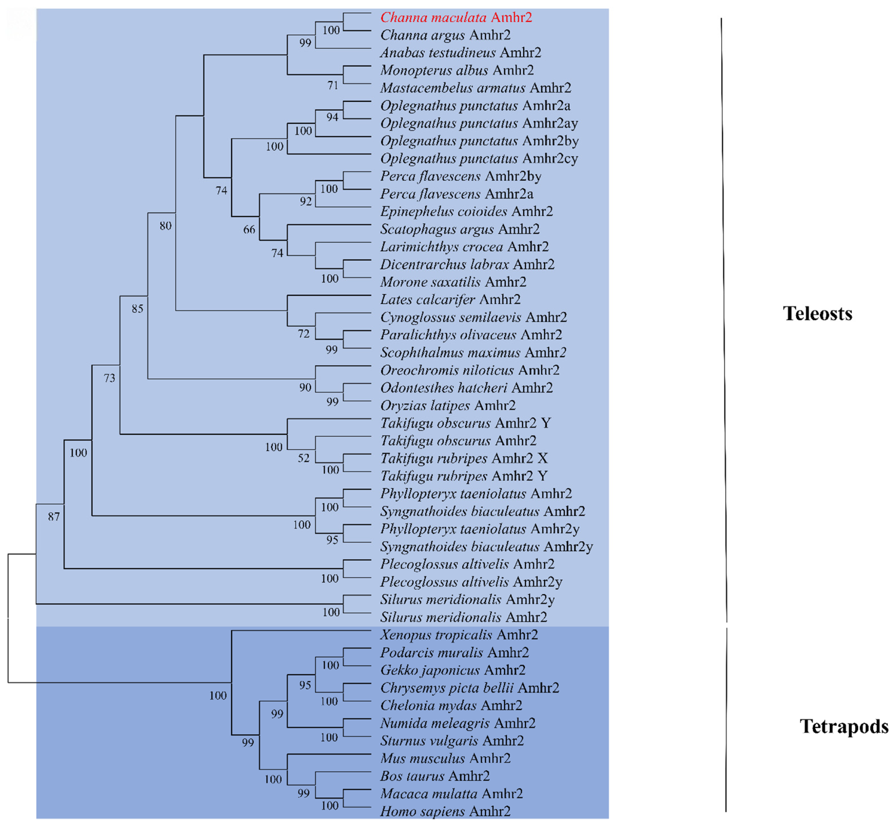
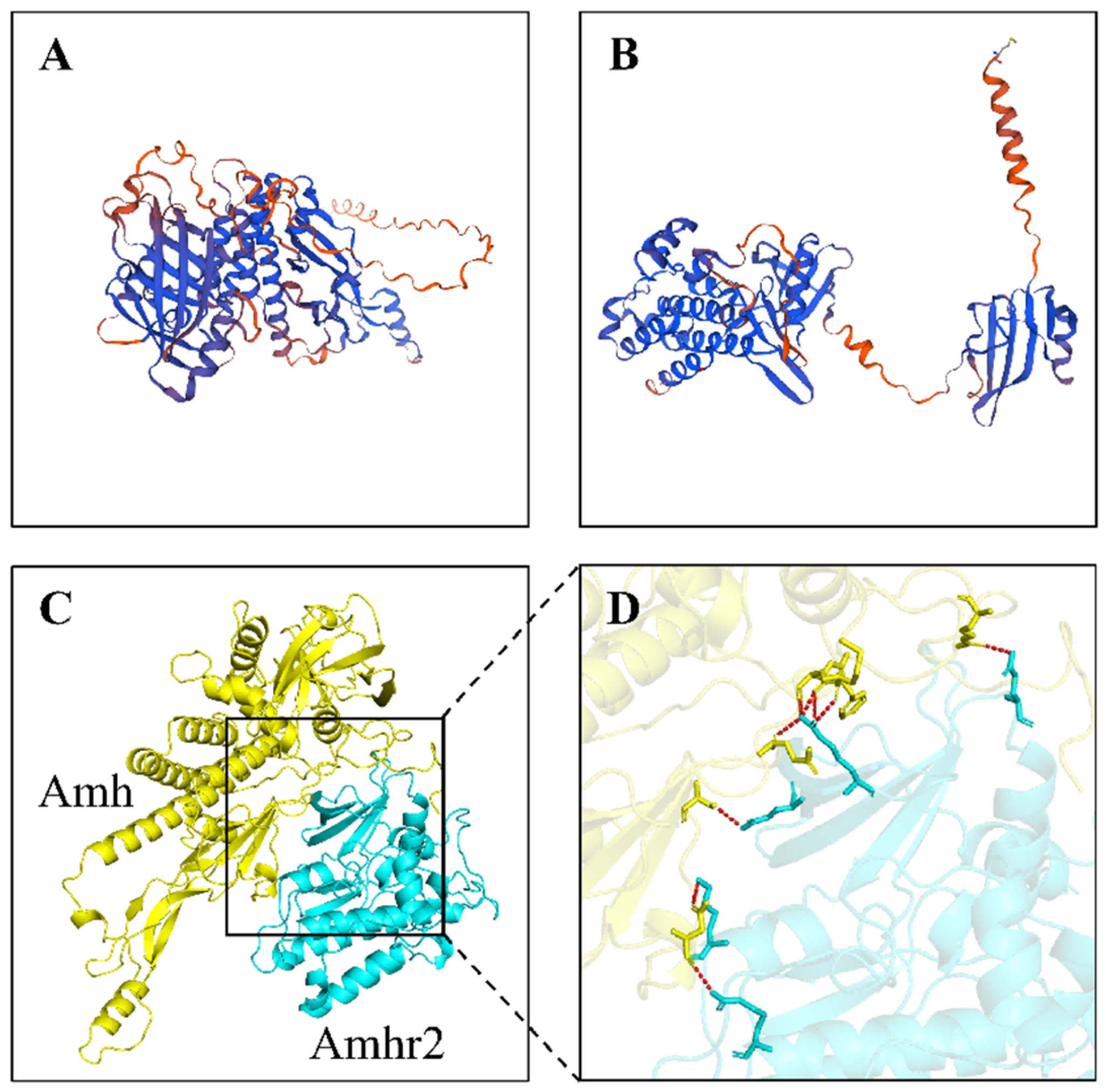
CmAmhr2 shared high sequence identity with teleost orthologs (87.58% with C. argus, 65.53% with E. coioides), but lower identity with bird (Numida meleagris, 24.28%) and mammalian (Mus musculus, 24.08%) (Figure S2). A neighbor-joining (NJ) phylogenetic tree placed CmAmhr2 within the teleost clade, separate from reptile, amphibian, bird, and mammalian sequences (Figure 1). Predicted three-dimensional structures of CmAmh and CmAmhr2 via SWISS-MODEL revealed characteristic α-helices, β-sheets, and random coils, and molecular docking further revealed a strong predicted interaction between the two proteins (Figure 2). These results confirm that the cloned sequence represents the authentic Amhr2 ortholog in C. maculata.

2.2. Genomic Structure Analysis of CmAmhr2

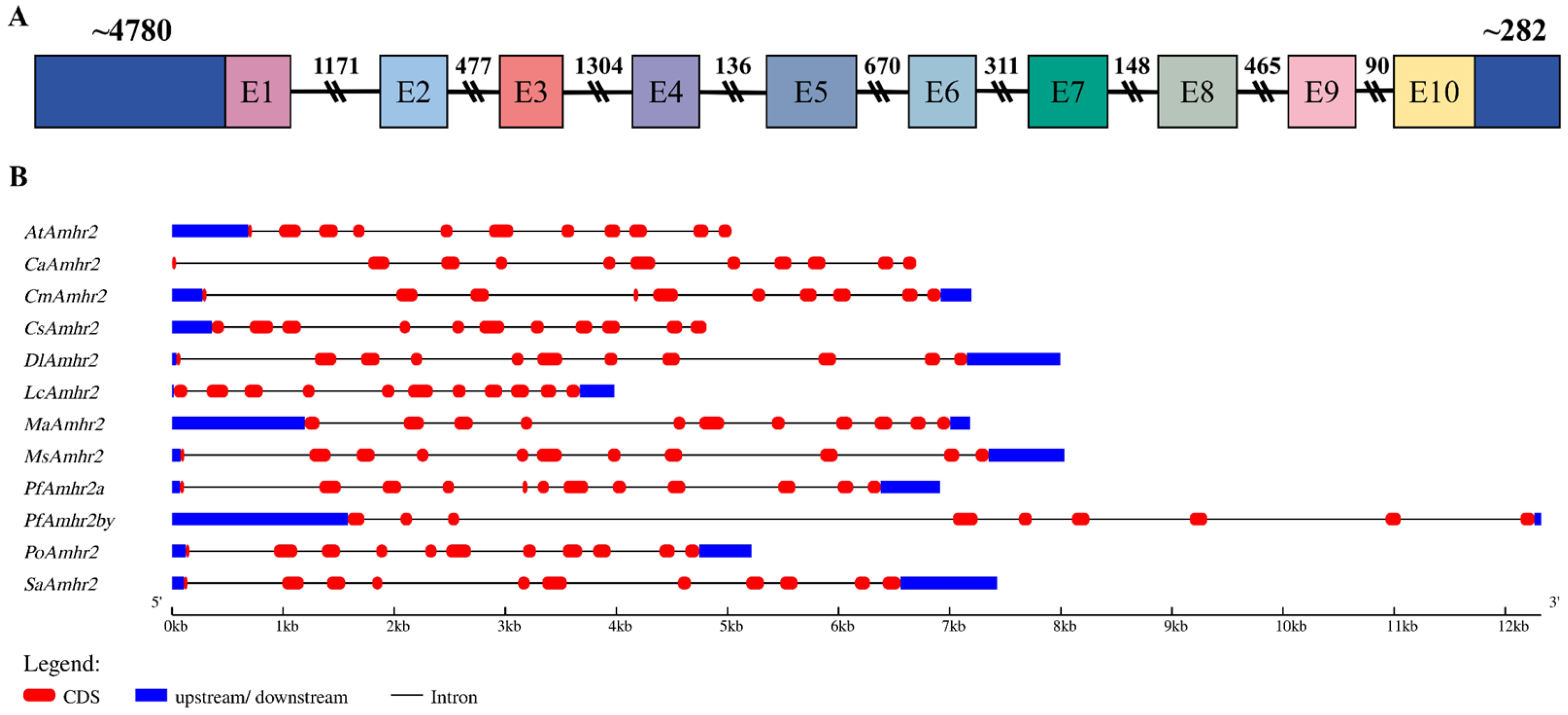
The full-length genomic sequence of CmAmhr2 spans 11,706 bp and comprises ten exons (E1~E10) of 37, 189, 165, 41, 222, 115, 152, 157, 137, and 117 bp, respectively, interspersed with nine introns of 1171, 477, 1304, 136, 670, 311, 148, 465, and 90 bp. All exon-intron boundaries conformed to the canonical GT-AG splice donor-acceptor rule (Figure 3A). Comparative analysis across teleosts showed that most species possess eleven exons and ten introns, with introns substantially longer than exons. However, PfAmhr2a (Amhr2 from Perca flavescens) contains twelve exons (Figure 3B).
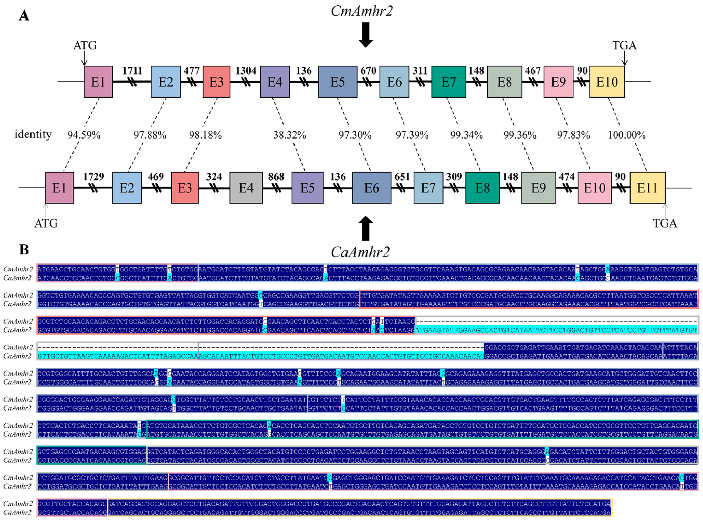
Comparison with the closely related C. argus (CaAmhr2) revealed that CmAmhr2 contains ten exons (E1~E10), whereas CaAmhr2 has eleven (E1~E11) (Figure 4). Most exons are highly conserved, with identity ranging from 94.59% to 100.00%. The additional exon in CaAmhr2 (E4) corresponds to the region encoding the transmembrane domain of the AMHR2 protein (Figure S3). Notably, exon 5 (E5) of CaAmhr2 shows substantially lower identity (38.32%) with exon 4 (E4) of CmAmhr2, suggesting potential functional divergence in this region. Subcellular localization predictions showed that CmAmhr2 was located in the endoplasmic reticulum, whereas CaAmhr2 was predicted to be a transmembrane protein localized at the cell membrane (Figure S4).
Additionally, a 4780 bp 5′-flanking region and a 282 bp 3′-flanking region of CmAmhr2 were obtained. Multiple putative TFBS were predicted within the promoter region (−1800~+1), including motifs for AR, Sp1, WT1, DMRT1, Smad2, estrogen receptor (ER), CCAAT/enhancer protein (C/EBP), GATA transcription factor (GATA-1), glucocorticoid receptor (GR), activating protein 1 (AP-1), Sox9, Oct-1, Foxl2, MyoD and Smad3, among others (Figure S5).

2.3. Expression Profiles of CmAmhr2 in Tissues and During Gonadal Development

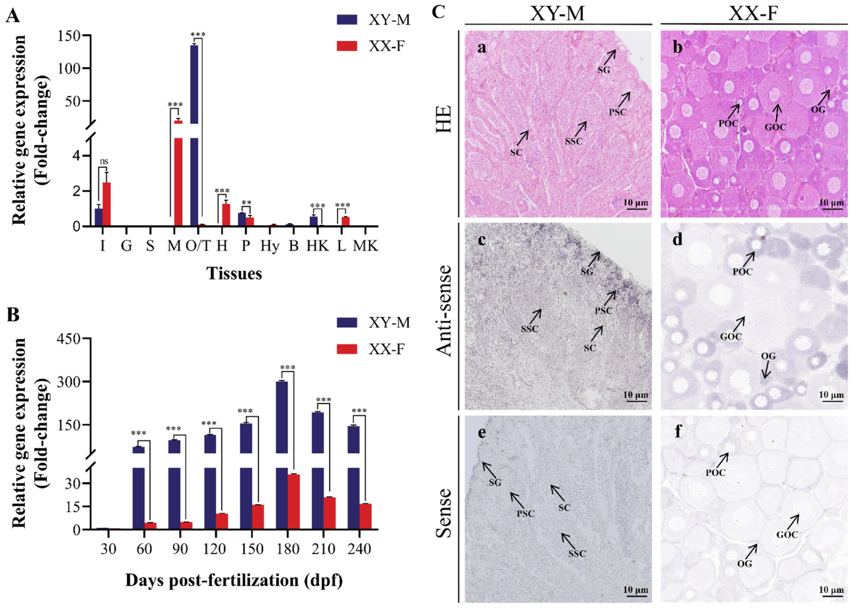
Tissue-specific expression of CmAmhr2 was examined in XY-M and XX-F individuals using qRT-PCR (Figure 5A). Pronounced sexual dimorphism was observed, most notably in the gonads: expression in XY-M testes reached 134.72 ± 2.48-fold (relative to intestinal baseline), whereas expression in XX-F ovaries was only 0.11 ± 0.01-fold (p < 0.001). Moderate, dimorphic expression was also detected in muscle (0.04 ± 0.00-fold in XY-M versus 19.63 ± 3.45-fold in XX-F) and intestines. Low expression levels were found in gills, spleen, hypothalamus, brain, and middle kidney in both sexes.
Expression levels during gonadal development were examined from 30 to 240 dpf (Figure 5B). Transcript levels were minimal and similar between sexes at 30 dpf (1.21 ± 0.02-fold in XY-M testes vs. 1.00 ± 0.01-fold in XX-F ovaries, p > 0.05). From 60 dpf onward, expression in XY-M testes progressively increased relative to XX-F ovaries. In XY-M testes, CmAmhr2 expression increased progressively from 60 dpf (96.51 ± 1.03-fold), peaked at 180 dpf (299.59 ± 3.66-fold), and subsequently declined by 240 dpf (145.27 ± 2.93-fold). In contrast, ovarian expression remained relatively stable throughout development, ranging from 1.00 ± 0.01-fold at 30 dpf to 16.60 ± 0.32-fold at 240 dpf. Between 60~240 dpf, testicular expression was 8.38- to 16.36-fold higher than ovarian expression (p < 0.001). Thus, CmAmhr2 exhibits a male-biased expression pattern that becomes prominent during gonadal differentiation.

2.4. Cellular Localization of CmAmhr2 Transcripts in Gonads

Spatial distribution of CmAmhr2 transcripts was examined by ISH on gonadal sections from 120 dpf XY-M and XX-F individuals (Figure 5C). Adjacent sections stained with HE confirmed the typical gonadal composition. In XY-M testes, spermatogonia (SG), primary spermatocytes (PSC), and secondary spermatocytes (SSC) were clearly distinguishable (Figure 5C-a). XX-F ovaries contained oogonia (OG), primary oocytes (POC), and growing oocytes (GOC) (Figure 5C-b). Upon hybridization with the antisense probe, positive signals in XY-M testes were predominantly localized to SG, with weaker signals in PSC and Sertoli cells (Figure 5C-c). In XX-F ovaries, CmAmhr2 transcripts were mainly detected in early oocytes, including OG and POC (Figure 5C-d). No specific signals were observed with the sense probe in either testes (Figure 5C-e) or ovaries (Figure 5C-f).

2.5. Expression Dynamics of CmAmhr2 Following Exogenous E2 Treatment

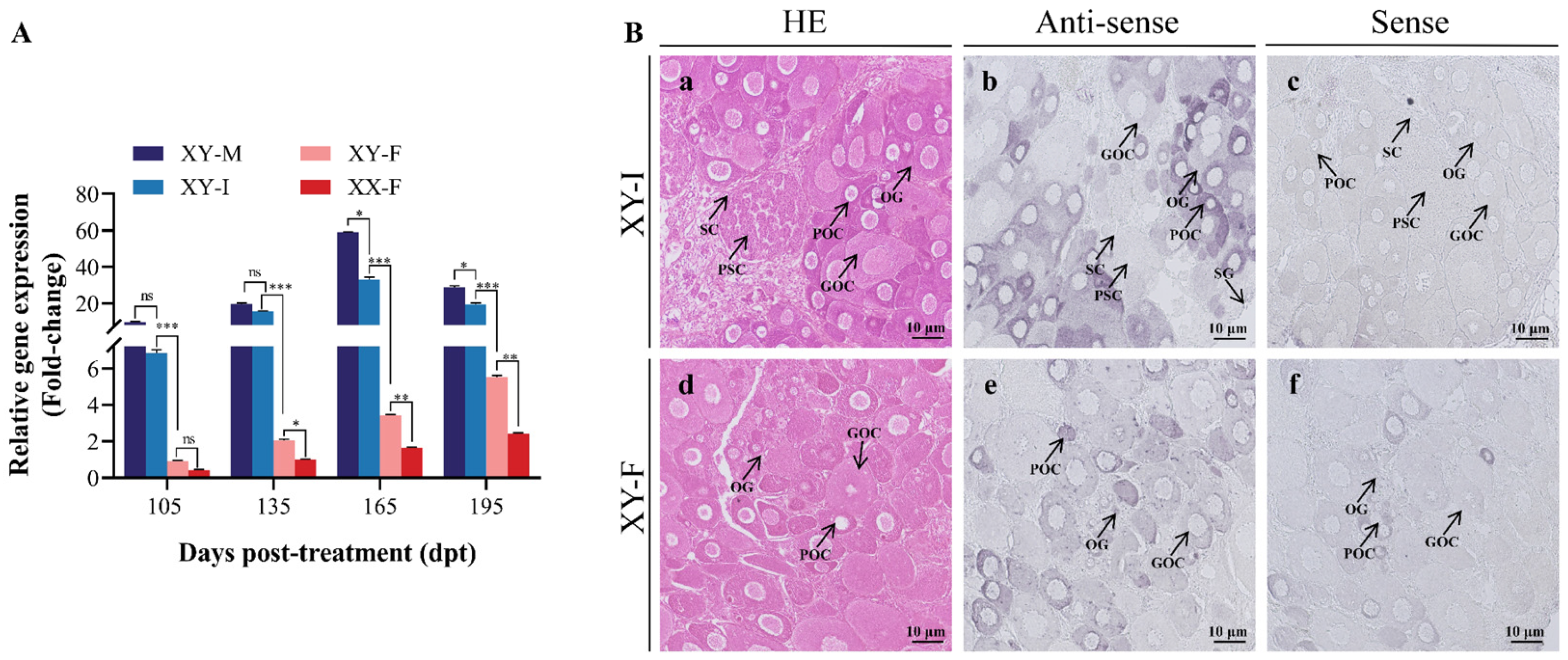
The effects of E2 treatment on the gonadal development of XY-genotype C. maculata were systematically investigated through histological examination and genetic sex identification. At 105 dpt (120 dpf), the sex reversal rate was 64.7%, normal testis, sex-reversed ovaries, and sex-reversed ovotestis were found in the 30 mg/kg E2-treated groups (Table 1). Additionally, transcript levels of CmAmhr2 were assessed in gonads of four phenotypic groups (XY-M, XY-F, XY-I, and XX-F) at 105, 135, 165, and 195 dpt (Figure 6A). Throughout the sampling period, CmAmhr2 expression remained significantly higher in XY-M testes than in XX-F ovaries (p < 0.001). Notably, expression levels declined progressively with the degree of gonadal feminization: highest in XY-M testes, intermediate in XY-I ovotestes, and lowest in fully feminized XY-F and XX-F ovaries. At 105 dpt, expression was highest in XY-M testes (9.73 ± 0.24-fold), followed by XY-I ovotestes (6.84 ± 0.18-fold), with no significant difference between these two groups (p > 0.05). In contrast, XY-F and XX-F ovaries showed substantially lower levels (0.92 ± 0.04-fold and 0.44 ± 0.02-fold, respectively). By 135 dpt, expression surged in XY-M testes (19.62 ± 0.47-fold) and XY-I ovotestes (15.69 ± 0.16-fold), while remaining low in XY-F (2.05 ± 0.07-fold) and XX-F ovaries (1.0 ± 0.03-fold). This divergence intensified at 165 dpt, with peak expression in XY-M testes (59.03 ± 0.18-fold) and XY-I ovotestes (33.04 ± 1.40-fold), versus low ovarian levels (XY-F: 3.44 ± 0.03-fold; XX-F: 1.65 ± 0.04-fold). Despite modest declines being noted by 195 dpt (XY-M: 28.67 ± 0.89-fold; XY-I: 19.51 ± 0.65-fold), testicular expression remained significantly higher than ovarian levels (p < 0.001) (XY-F: 5.12 ± 0.21-fold; XX-F: 2.34 ± 0.09-fold).
Cellular localization of CmAmhr2 transcripts following E2 treatment was examined by ISH on gonadal sections from 105 dpt individuals (Figure 6B). Adjacent HE-stained sections revealed the cellular composition of each gonadal phenotype. In XY-I ovotestes, both ovarian (OG, POC, and GOC) and testicular (SG, PSC) structures were present (Figure 6B-a), with CmAmhr2 transcripts detected in early oocytes (OG, POC) and SG (Figure 6B-b). In XY-F individuals, gonads consisted exclusively of ovarian components (OG, POC, and GOC) (Figure 6B-d), with positive signals primarily localized to OG and POC (Figure 6B-e). No specific signals were observed with sense probes in either XY-I (Figure 6B-c) or XY-F (Figure 6B-f) gonads.

2.6. Disruption of CmAmhr2 Using CRISPR/Cas9

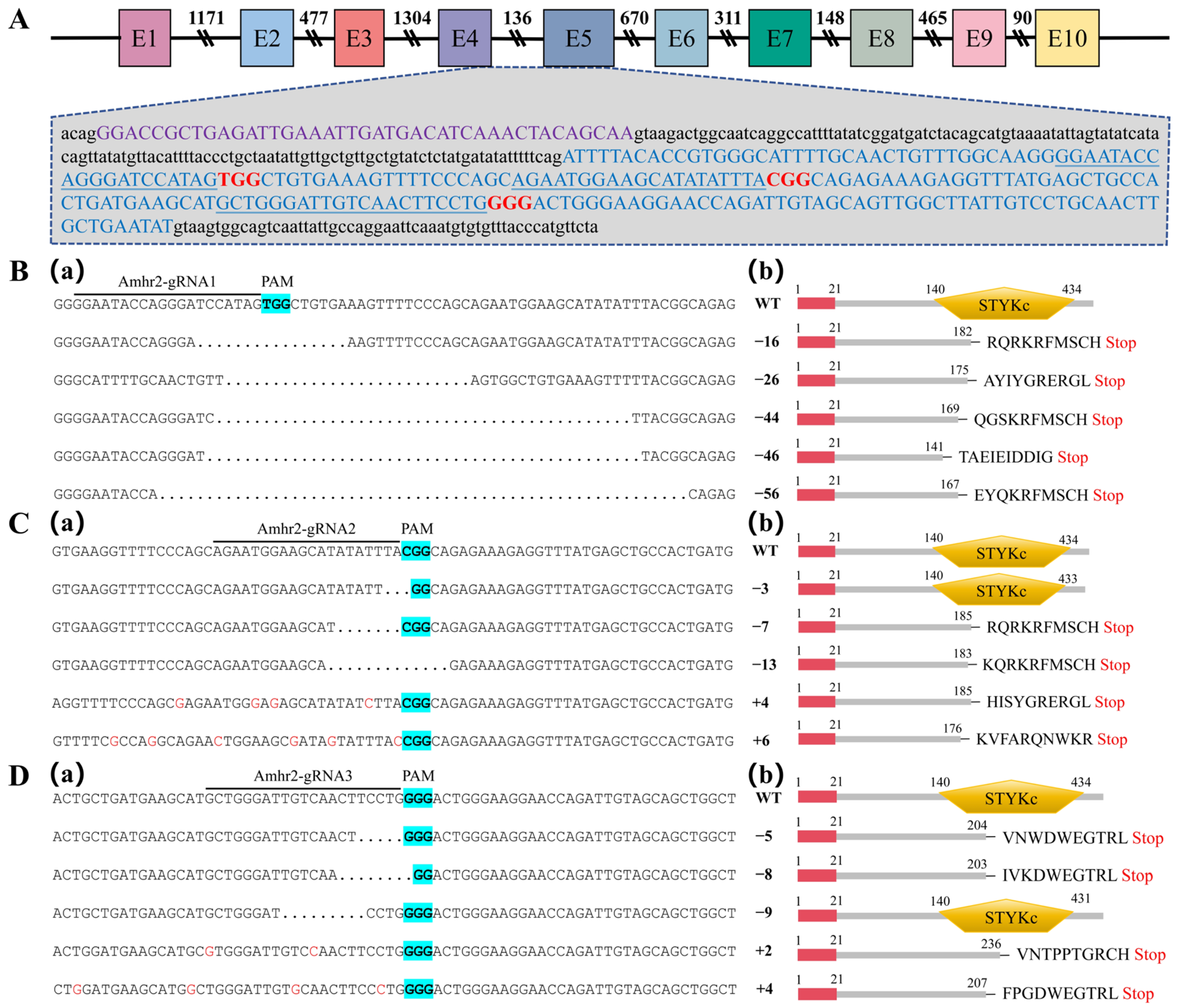
To investigate the functional role of CmAmhr2 in sex determination and differentiation, CRISPR/Cas9 editing was performed targeting exon 5 of CmAmhr2. Three gRNAs were designed adjacent to protospacer adjacent motif (PAM) sequences (Figure 7A). Zygotes at the 1~4 cell stage were microinjected within ~60 min post-fertilization with 1 nL of a solution containing gRNA and Cas9 protein. Fertilization rates were 42.3 ± 3.1%, 41.4 ± 1.4%, 42.7 ± 2.0%, and 41.0 ± 1.2% for the 0.75% NaCl-injected, Amhr2-gRNA1, -gRNA2, and -gRNA3 groups, respectively, with no significant differences among injected groups (p > 0.05). All were significantly lower than the untreated control (80.7 ± 2.0%, p < 0.05). Similarly, hatching rates were 34.6 ± 1.1%, 34.6 ± 2.0%, 34.2 ± 1.2%, and 33.7 ± 1.5% for the four injected groups, respectively, showing no intergroup differences (p > 0.05), but significantly reduced compared to the untreated control (73.1 ± 1.2%, p < 0.05). Survival to the flat swimming stage was also markedly affected: from 900 embryos per group, 577 fry survived in the untreated control, whereas only 256, 234, 211, and 219 fry survived in the 0.75% NaCl, Amhr2-gRNA1, -gRNA2, and -gRNA3-injected groups (Table 2, Figure S6), respectively, representing significantly reduced survival relative to untreated control (p < 0.05).
Mutation analysis at the target sites was conducted by PCR amplification, subcloning, and sequencing of 48 hpf fry. Mutagenesis frequencies were 50.0%, 43.3%, and 40.0% for the Amhr2-gRNA1, -gRNA2, and -gRNA3-injected groups, respectively (Table 3). Sequencing alignment identified multiple insertions and deletions (indels) induced by the CRISPR/Cas9 system (Figure 7). In the Amhr2-gRNA1 injected group, five deletions (16, 26, 44, 46, and 56 bp) were identified (Figure 7B-a), all non-triplet and predicted to cause frameshifts (Figure 7B-b). In the Amhr2-gRNA2 group, three deletions (3, 7, and 13 bp) and two insertions (4 and 6 bp) were observed (Figure 7C-a). Aside from the 3-bp deletion, all were expected to induce frameshifts (Figure 7C-b). In the Amhr2-gRNA3 group, mutations comprised deletions (5, 8, and 9 bp) and insertions (2 and 4 bp) (Figure 7D-a). The 9-bp triplet deletion led to a three-amino-acid loss without frameshift, whereas all other indels resulted in frameshifts (Figure 7D-b). Given the limited sample size and the mosaic nature of F0 mutants, these mutation frequencies provide only a preliminary indication of genome editing activity, not precise estimates of mutagenesis efficiency. Collectively, these results confirm that all three gRNAs effectively cleaved the genomic CmAmhr2 locus, with most indels introducing frameshifts and premature termination codons, likely generating truncated, nonfunctional protein products.

3. Discussion

As a key member of the TGF-β receptor superfamily, Amhr2 was identified and characterized in C. maculata (designated CmAmhr2). The gene encodes a single transcript containing the conserved STYKc domain characteristic of type II TGF-β receptor, consistent with our previous localization of a single Amhr2 copy on LG05 [23]. Phylogenetic analysis placed CmAmhr2 within the teleost clade, and molecular docking predicted its direct interaction with CmAmh, suggesting a conserved Amh/Amhr2 signaling axis. Recent large-scale surveys indicate that, due to gene duplication or mutation, Amh and Amhr2 homologs serve as SD genes in 34 and 43 teleost species, respectively, accounting for ~50% of reported fish species with identified SD genes [24]. This recurrent recruitment underscores the evolutionary significance of this pathway in teleost sex determination and differentiation, although species-specific variations exist. For instance, many fish, such as Anabas testudineus (XM_026347020.1), Cynoglossus semilaevis (XM_025060061.1), and Dicentrarchus labrax (JQ801443.1), possess a single copy. Notably, comparative genomic analysis revealed that, unlike the C. argus ortholog (CaAmhr2), which includes an additional exon encoding the transmembrane domain, CmAmhr2 lacks this feature, consistent with its predicted localization as a soluble protein in the endoplasmic reticulum rather than a membrane-bound receptor. Despite this structural divergence, CmAmhr2 retains the ability to bind CmAmh, implying potential unconventional signaling mechanisms. This finding aligns with evidence of teleost-specific adaptations, such as the use of Bmpr2a as an alternative type II receptor for Amh in D. rerio [25]. Such structural variation, possibly arising from lineage-specific events following whole-genome duplication, warrants further functional investigation in C. maculata.
Although teleosts lack Müllerian ducts, the Amh/Amhr2 pathway is widely involved in gonadal development, with Amhr2 predominantly expressed in testes across diverse species, such as largemouth bass (Micropterus salmoides) [26], O. niloticus [19], T. rubripes [6], P. olivaceus [20], E. coioides [27]. The present study yielded similar results, showing high gonadal expression of CmAmhr2 with a male bias. However, some cyprinid species, such as D. rerio, C. carpio, and C. idella, have lost Amhr2, with its function possibly replaced by another type II receptor, such as Bmpr2 [9,25]. In certain catfishes, an additional Y-chromosome copy, Amhr2y, is testis-specific in S. meridionalis [13] and S. asotus [15], whereas the autosomal Amhr2 is expressed in both sexes (higher in testes). Extra-gonadal expression of Amhr2 has also been reported, including in the head kidney in striped catfish (Pangasianodon hypophthalmus) [14], kidney, gill, and spleen in S. asotus [15], and kidney, intestine, and heart in male O. punctatus [17]. Similarly, CmAmhr2 exhibited relatively high expression in muscle, and we identified a MyoD transcription factor binding site in its promoter. MyoD, a myogenic regulatory factor specifically expressed in myoblasts and skeletal muscle cells, plays a key role in muscle development and growth [28]. This binding site suggests a potential regulatory link between CmAmhr2 and muscle growth in C. maculata.
During gonadal development, CmAmhr2 expression was consistently higher in testes than in ovaries, showing an initial increase followed by a decline. Expression was low at 30 dpf (prior to testicular differentiation), increased sharply at 60 dpf (coinciding with the appearance of primary spermatocytes), and peaked at 180 dpf (when testes contained abundant sperm), indicating a close association with testicular differentiation and development in C. maculata [25]. Sexually dimorphic expression of Amhr2 varies among teleosts. In O. punctatus, male-biased Amhr2 expression persists throughout development, with peaks at 6 and 180 days post-hatching (dph) [17]. In contrast, O. latipes shows female-biased expression during early development (0~39 dph), switching to male-biased after 60 dph [29]. In S. asotus and S. meridionalis, the autosomal Amhr2 is expressed in both sexes but significantly higher in XY gonads at all stages, whereas the Y-linked Amhr2y is testis-specific, peaking early and then declining [13,15]. ISH revealed that CmAmhr2 transcripts in testes were predominantly localized in SG, with weaker signals in PSC and Sertoli cells; in ovaries, low expression was observed in early oocytes. Expression patterns of Amhr2 vary among teleost species. In O. niloticus, Amhr2 is expressed in SG and Sertoli cells of the testis, as well as in OG, oocytes, and granulosa cells of the ovary [19,30]. In S. chuatsi, expression is detected in previtellogenic oocytes and Sertoli cells [31]. In O. punctatus, Amhr2 signals are predominantly localized in PSC and SSC, stronger in PSC than in SSC, with weaker signals in the Sertoli cells, and no ovarian signals [17]. Collectively, these findings suggest that Amhr2 plays a significant role in testis development and may also contribute to ovarian function in certain species.
Exogenous steroid hormones are widely used to induce sex reversal, during which the expression patterns of sex-related genes often change accordingly. In this study, treatment with 30 mg/kg E2 successfully induced male-to-female sex reversal in C. maculata, accompanied by a significant reduction in CmAmhr2 expression. Since Amhr2 is closely associated with testicular differentiation, its downregulation under E2 treatment is consistent with the suppression of the male pathway and the promotion of ovarian differentiation. Notably, the extent of CmAmhr2 suppression appeared to correlate with the degree of feminization observed in gonadal phenotypes, suggesting that reduced CmAmhr2 expression may contribute to, but not solely determine, sex reversal efficiency. Similar inhibitory effects of E2 on testicular Amhr2 expression have been reported in Anguilla japonica [32], T. rubripes [33], and black porgy (Acanthopagrus schlegeli) [34]. In contrast, in the hermaphroditic E. coioides, both Amh and Amhr2 are significantly upregulated during female-to-male sex reversal [27], further supporting the conserved role of the Amh/Amhr2 signaling pathway in male differentiation across teleosts. However, complete sex reversal was not achieved in this study (64.7%), and a proportion of individuals exhibited intersex gonads containing both male (SG and PSC) and female (OG, POC, and GOC) germ cells. This incomplete reversal suggests that while E2 treatment effectively suppresses CmAmhr2, the degree of suppression may be insufficient in some individuals to fully inhibit testicular development. Several factors may contribute to this variability. First, the administered dose (30 mg/kg) may be too low to induce complete sex reversal in all individuals. Second, uneven hormone intake due to differences in feeding behavior or metabolism could lead to variable internal E2 exposure. A similar phenomenon has been reported in S. chuatsi [35], where inconsistent hormone uptake resulted in partial sex reversal. Therefore, the occurrence of intersex individuals likely reflects incomplete suppression of the male pathway, highlighting that both the level and consistency of exogenous hormone exposure are critical determinants of sex reversal efficiency. Future studies optimizing dosage, treatment duration, and delivery methods may help achieve more stable and complete sex reversal.
Microinjection is widely used to deliver reagents into early fish embryos but can cause mechanical injury and reduce hatching rates, as reported in several species, such as channel catfish (Ictalurus punctatus) [36], sterlet (Acipenser ruthenus) [37], P. hypophthalmus [38], and large yellow croaker (Larimichthys crocea) [39], though not in fathead minnow (Pimephales promelas) [40]. In this study, we microinjected sgRNA/Cas9 complexes into C. maculata embryos at the 1~4 cell stage to generate Amhr2 mutants. Both saline-injected and gRNA/Cas9-injected groups exhibited lower hatching rates than uninjected controls, with no significant difference between the two injected groups, indicating that the injection procedure itself, not the gRNA complexes, reduced hatching. All three designed gRNAs efficiently targeted CmAmhr2, with mutagenesis efficiencies of 40~50%. The observed mutations were predominantly frameshifts leading to premature stop codons and disruption of the kinase domain, a region essential for Amhr2 phosphorylation and signaling [5]. Thus, the generated CmAmhr2 mutants are predicted to exhibit complete loss of Amh/Amhr2 function. Loss-of-function Amhr2 mutants have been generated in several fish species, including O. latipes [18], O. niloticus [19], P. altivelis [11], P. olivaceus [20], and S. meridionalis [13]. In O. latipes and O. niloticus, Amhr2-deficient XY individuals showed male-to-female sex reversal at rates exceeding 50% and 100%, respectively [18,19]. In P. olivaceus, loss of Amhr2 also caused sex reversal, which could be rescued by an aromatase inhibitor, suggesting that the Amh/Amhr2 system influences male differentiation by suppressing estrogen synthesis [20]. Similarly, loss of the Y-linked Amhr2y results in complete gonadal sex reversal in XY mutants of P. altivelis [11] and S. meridionalis [13]. Collectively, these findings underscore the critical role of Amhr2 in male sex determination and differentiation, gonadal development, and early germ cell regulation in fish. However, because the knockout fish generated in this study are still immature and require two generations to produce homozygous CmAmhr2−/− mutants, the effects of CmAmhr2 deletion on gonadal development and potential sex reversal remain to be determined. Future work will prioritize elucidating the functional role of CmAmhr2 and the specific molecular mechanisms of its signal transduction.

4. Materials and Methods

4.1. Experimental Fish and Sampling

Blotched snakeheads were reared at the Fangcun Experiment Station of Pearl River Fisheries Research Institute (Guangzhou, China). Adult XY-M and XX-F were identified using a sex-specific molecular marker and gonadal histology [22]. Tissue samples, including liver (L), gills (G), middle kidney (MK), spleen (S), head kidney (HK), intestines (I), muscle (M), heart (H), gonads (ovaries or testes (O/T)), brain (B), hypothalamus (Hy), and pituitary (P), were collected from one-year-old XX-F and XY-M individuals (n = 3 per sex) following anesthesia with tricaine methanesulfonate (MS-222, Sigma-Aldrich, Burlington, MA, USA) at a dose of 1.25 g/mL. These tissues were used for gene cloning and expression analysis.
To investigate Amhr2 expression during gonadal differentiation, gonads were harvested from randomly selected XX-F and XY-M fish (n = 3 per sex per time point) at 30, 60, 90, 120, 150, 180, 210, and 240 dpf. The methods for genetic and physiological sex identification were described above. All samples were immediately frozen in liquid nitrogen and stored at −80 °C for subsequent quantitative real-time PCR (qRT-PCR). Additionally, gonads from 120 dpf XX-F and XY-M individuals were fixed in Bouin’s solution (MedChemExpress, Monmouth Junction, NJ, USA) for hematoxylin and eosin (HE) staining and in situ hybridization (ISH). All experimental procedures were conducted in accordance with animal welfare guidelines and approved by the Animal Ethics Committee of the Pearl River Fisheries Research Institute, Chinese Academy of Fishery Sciences.

4.2. Gene Cloning and Sequence Analysis

Total RNA extraction and cDNA synthesis were performed following established protocols [41]. Gene-specific primers (Table 4) for amplifying the Amhr2 coding sequence were designed based on the blotched snakehead genome (SRA Accession No. PRJNA730430) [42]. The open reading frame (ORF) was amplified from gonadal cDNA via PCR. Products of the expected size were gel-purified using a Gel Rapid Extraction Kit (CWBIO, Taizhou, China), ligated into the pMD18-T vector (Takara, Kusatsu, Japan), and transformed into Trans5α chemically competent cells (TransGen Biotech, Beijing, China). Positive clones were selected and sequenced (Tsingke Biotech, Beijing, China).
Homologous Amhr2 protein sequences from other vertebrates were retrieved from the NCBI database (accession numbers listed in Table 5). Multiple sequence alignment was conducted using DNAMAN (version 6.0.3.99). A phylogenetic tree was constructed with MEGA 11.0 using the neighbor-joining method with 1000 bootstrap replicates. Conserved domains were identified via the Simple Modular Architecture Research Tool (SMART) (http://smart.embl-heidelberg.de/) (accessed on 30 October 2025). The predicted coding sequences of Amh and Amhr2 were obtained from the blotched snakehead genome (SRA Accession No. PRJNA730430) and the northern snakehead genome (SRA Accession No. PRJNA731586), respectively [42]. Three-dimensional protein structures were predicted via SWISS-MODEL (https://swissmodel.expasy.org/) (accessed on 30 October 2025), and molecular docking simulations were conducted using HDOCK (http://hdock.phys.hust.edu.cn/) (accessed on 30 October 2025), with results visualized in PyMOL (version 3.0.3).

4.3. Genomic Sequence Cloning and Structure Analysis

Genomic DNA was extracted from tail tissue using a Universal Genomic DNA Kit (CWBIO, Taizhou, China). The putative Amhr2 genomic sequence was obtained from the blotched snakehead genome (SRA Accession No. PRJNA730430) [42]. Primers (Table 6) were designed to validate this sequence, and PCR products were cloned and sequenced as described in Section 4.2. The exon-intron structure was determined by aligning genomic and cDNA sequences following the GT/AG splice site rule.
For comparative analysis, Amhr2 coding and genomic sequences from other teleosts were retrieved from the NCBI database (accession numbers listed in Table 7). Genomic structures were visualized using the Gene Structure Display Server 2.0 (http://gsds.cbi.pku.edu.cn) (accessed on 30 October 2025). Promoter core elements (PCE) and transcript start sites (TSS) were predicted with NNPP (http://www.fruitfly.org/seq_tools/promoter.html) (accessed on 30 October 2025). Putative transcription factor binding sites (TFBS) in the promoter region were predicted using AliBaba 2.1 (http://generegulation.com/pub/programs/alibaba2/index.html) (accessed on 30 October 2025) and Animal TFDB (https://guolab.wchscu.cn/AnimalTFDB4/) (accessed on 30 October 2025). Signal peptides and cleavage sites were predicted using SignalP-6.0 (https://services.healthtech.dtu.dk/services/SignalP-6.0/) (accessed on 30 October 2025), and subcellular localization with DeepLoc-2.1 (https://services.healthtech.dtu.dk/services/DeepLoc-2.1/) (accessed on 30 October 2025).

4.4. Estrogen Treatment

From 15 to 45 dpf, fry were fed for 30 days a diet supplemented with E2 (Macklin, Shanghai, China). The treatment window (15~45 dpf) was selected based on the critical period of gonadal sex differentiation in C. maculata, as demonstrated by Zhang et al. [25]. E2 was first dissolved in absolute ethanol, then evenly sprayed onto a commercial compound feed (Rongchuan, Guangzhou, China) and thoroughly mixed to ensure homogeneous distribution. The treated feed was air-dried in the shade to allow complete evaporation of ethanol before use. Based on our previous study [43], which reported a 70% sex reversal efficiency and the development of functional ovaries with this concentration, the experimental group received feed containing 30 mg E2 per kg. The control group was fed the same diet treated with ethanol only (0 mg/kg). Each treatment was conducted in triplicate tanks with equal stocking densities. During the experimental period, fish were reared in an open recirculating aquaculture system under controlled conditions: water temperature 26~28 °C, dissolved oxygen 6~8 mg/L, ammonia nitrogen < 0.5 mg/L, nitrite < 0.01 mg/L, and pH 7.0~7.5. After the treatment period, all fish were reared under the same standard conditions until sexual maturity.
At 105, 135, 165, and 195 days post-treatment (dpt), corresponding to 120, 150, 180, and 210 dpf, 30 fish were randomly sampled from each replicate of the E2-treated and control groups following MS-222 anesthesia. These four time points were selected based on the typical gonadal developmental cycle of C. maculata and the objective of assessing the stability of E2-induced sex reversal. In our previous study [43], gonads at 60~90 dpf were not fully reversed into ovaries. At 120 dpf, the ovaries of sex-reversed fish were essentially formed, and by 240 dpf, the ovaries contained abundant mature oocytes. The selected time points thus allowed us to determine whether the E2-induced gene expression pattern exhibits long-term stability. Using the sex-specific molecular marker and gonadal histology [22], individuals were classified as XY-M, XX-F, XY-I, and XY-F. Individuals were first classified into different sex categories using molecular markers and histological examination, and then randomly selected from each category (n = 3 per sex per time point) for expression analysis at the indicated dpt. Additionally, gonads from 105 dpt XX-F, XY-M, XY-I, and XY-F individuals were fixed in Bouin’s solution for HE staining and ISH.

4.5. Quantitative Real-Time PCR (qRT-PCR)

qRT-PCR primers are listed in Table 4, with β-actin as the reference gene, consistent with our previous study [44]. Reactions were performed in triplicate on a StepOnePlus™ Real-Time PCR System (ABI, Foster City, CA, USA) using SYBR® Green Master Mix (Toyobo, Osaka, Japan) according to the manufacturer’s instructions. Each 20 μL reaction mixture contained 10 μL SYBR® Green Master Mix, 0.8 μL each of forward and reverse primers, 7.4 μL ddH2O, and 1 μL cDNA. The thermal cycling protocol was as follows: 95 °C for 2 min; 39 cycles of 95 °C for 15 s, 50 °C for 15 s, and 72 °C for 30 s; followed by a melting curve acquisition step (60 °C for 5 s, then 95 °C for 5 s) using the instrument’s default settings. Relative expression levels of Amhr2 were calculated using the 2−∆∆Ct method. For tissue distribution, expression in the intestines (I) of XX-F individuals was set as the baseline (1.0). For gonadal development analysis, expression in the gonads of 30 dpf XX-F fish served as the baseline (1.0). In the estrogen treatment experiment, expression levels were normalized to those in 120 dpf XX-F individuals.

4.6. In Situ Hybridization (ISH)

Gonads from 120 dpf individuals were dissected, fixed in Bouin’s solution for 24 h at 4 °C, dehydrated, paraffin-embedded, and serially sectioned at 5 μm thickness. Consecutive sections were processed for HE staining and ISH following previously described procedures. For riboprobe synthesis, target gene fragments were amplified using gene-specific primers (Table 4). Digoxigenin (DIG)-labeled sense and antisense RNA probes were generated using a DIG RNA Labeling Kit (Roche, Mannheim, Germany) and purified via LiCl/ethanol precipitation.
Tissue sections were deparaffinized, rehydrated, and digested with 10 μg/mL proteinase K (CWBIO, Taizhou, China) at 37 °C for 15 min. Hybridization was performed with sense or antisense probes at 65 °C for 16 h. Signals were detected using an alkaline phosphatase-conjugated anti-DIG antibody (Roche, Mannheim, Germany; 1:1000 dilution) and visualized with NBT/BCIP (Roche, Mannheim, Germany). Sections were washed in 1×PBST, mounted with gelatin glycerin (Servicebio, Wuhan, China), and examined under a Nikon Eclipse Ti-U microscope (Tokyo, Japan).

4.7. Knockout of the Amhr2 Gene in C. maculata

CRISPR/Cas9 was used to generate Amhr2 knockout mutants. Three guide RNAs (Amhr2-gRNA1, -gRNA2, and -gRNA3; Table 8) targeting the Amhr2 gene were designed using CRISPRscan (https://www.crisprscan.org/) (accessed on 30 October 2025). The gRNAs were synthesized with a MAXIscript T7 In Vitro Transcription Kit (Invitrogen, Carlsbad, CA, USA) and purified via LiCl/ethanol precipitation. Prior to microinjection, each gRNA was mixed with TrueCut Cas9 Protein v2 (Invitrogen, Carlsbad, CA, USA). A mixture containing 200 pg gRNA and 300 pg Cas9 protein was co-injected into embryos at the 1~4 cell stage. Two control groups were included: 0.75% NaCl-injected and untreated. Each treatment was performed in triplicate with 300 embryos per replicate (900 embryos per group). Survival rates were assessed at 12 h post-fertilization (hpf; gastrula stage), 36 hpf (hatching stage), and 120 hpf (flat swimming stage). Five fry per group were collected at 48 hpf for genomic DNA extraction using a Universal Genomic DNA Kit. Given that F0 individuals are typically mosaic, early-stage screening using a small number of randomly selected embryos (commonly 5~10) is standard practice for confirming successful mutagenesis. Mutation detection used the primer pair Amhr2-JC-F/R (Table 4). Artificial insemination, microinjection, mutagenesis analysis, and fish rearing followed previously established protocols [41].

4.8. Statistical Analysis

All data are presented as mean ± standard deviation (SD). Statistical comparisons between groups were performed using Student’s t-test or one-way ANOVA followed by Dunnett’s post hoc test in SPSS version 26.0 (SPSS Inc., Chicago, IL, USA). Significance levels were set at p < 0.05, p < 0.01, and p < 0.001.

5. Conclusions

In this study, we successfully cloned and characterized the Amhr2 ortholog in C. maculata and demonstrated its essential role in male sex differentiation and testis development. CmAmhr2 exhibited male-biased expression in developing gonads and localized to germ cells, implicating it in early spermatogenesis. Its down-regulation following E2 treatment, concomitant with sex reversal, supports hormonal modulation of CmAmhr2 expression. CRISPR/Cas9-mediated knockout confirmed that loss-of-function mutations disrupt the kinase domain critical for Amhr2 function, providing a foundation for future mechanistic studies. These findings advance our understanding of sex differentiation pathways in C. maculata and establish a valuable molecular target for sex-controlled breeding in aquaculture.

Supplementary Materials

The following supporting information can be downloaded at: https://www.mdpi.com/article/10.3390/ijms27114884/s1.

Author Contributions

Conceptualization, J.S. and X.Z.; Methodology, Y.Z. and Y.S.; Validation, Y.Z., J.S. and S.F.; Investigation, M.O., Y.Z. and J.Q.; Resources, M.O., J.Z. and J.Q.; Data curation, M.O.; Writing—original draft preparation, M.O. and Y.Z.; Writing—review and editing, J.Z. and J.Q.; Visualization, Q.L. and H.L.; Supervision, J.Z. and J.Q.; Project administration, J.Z.; Funding acquisition, M.O., J.Z. and J.Q. All authors have read and agreed to the published version of the manuscript.

Funding

This work was supported by Guangxi Science and Technology Innovation Platform Plan (GuiKe-LT2600640018), the Basic and Applied Basic Research Foundation of Guangdong Province (2024A1515030165), the Guangdong Special Support Program (2024TQ08A617), the China Agriculture Research System of MOF and MARA (CARS-46), the National Natural Science Foundation of China (32373127), the Guangdong Provincial Special Fund for Modern Agriculture Industry Technology Innovation Teams (2024CXTD26), the Science and Technology Program of Guangzhou (2025A04J5171), the Central Public-interest Scientific Institution Basal Research Fund, CAFS (2023TD37; 2026ZX21), China-ASEAN Maritime Cooperation Fund (CAMC-2018F).

Institutional Review Board Statement

All experimental procedures were conducted in accordance with animal welfare guidelines and approved by the Animal Ethics Committee of the Pearl River Fisheries Research Institute, Chinese Academy of Fishery Sciences (Approval No. LAEC-PRFRI-2024-02-02, on 10 February 2024).

Informed Consent Statement

Not applicable.

Data Availability Statement

All the data related to this project is available from the corresponding author and will be provided on request.

Conflicts of Interest

The authors declare no conflicts of interest.

References

  1. Kitano, J.; Ansai, S.; Takehana, Y.; Yamamoto, Y. Diversity and Convergence of Sex-Determination Mechanisms in Teleost Fish. Annu. Rev. Anim. Biosci. 2024, 12, 233–259. [Google Scholar] [CrossRef] [PubMed]
  2. Oliveira, M.A.; Filho, A.S.S.; Araújo, F.E. TGF-β Superfamily: An Overview of Amh Signaling into Sex Determination and Differentiation in Fish. Braz. Arch. Biol. Technol. 2023, 66, e23220371. [Google Scholar]
  3. Yu, H.; Du, X.; Chen, X.; Liu, L.; Wang, X. Transforming growth factor-β (TGF-β): A master signal pathway in teleost sex determination. Gen. Comp. Endocrinol. 2024, 355, 114561. [Google Scholar] [CrossRef]
  4. Howard, J.A.; Hart, K.N.; Thompson, T.B. Molecular Mechanisms of AMH Signaling. Front. Endocrinol. 2022, 13, 927824. [Google Scholar] [CrossRef]
  5. Belville, C.; Marechal, J.-D.; Pennetier, S.; Carmillo, P.; Masgrau, L.; Messika-Zeitoun, L.; Galey, J.; Machado, G.; Treton, D.; Gonzales, J.; et al. Natural mutations of the anti-Mullerian hormone type II receptor found in persistent Mullerian duct syndrome affect ligand binding, signal transduction and cellular transport. Hum. Mol. Genet. 2009, 18, 3002–3013. [Google Scholar] [CrossRef]
  6. Kamiya, T.; Kai, W.; Tasumi, S.; Oka, A.; Matsunaga, T.; Mizuno, N.; Fujita, M.; Suetake, H.; Suzuki, S.; Hosoya, S.; et al. A Trans-Species Missense SNP in Amhr2 Is Associated with Sex Determination in the Tiger Pufferfish, Takifugu rubripes (Fugu). PLoS Genet. 2012, 8, e1002798. [Google Scholar] [CrossRef]
  7. Gao, F.-X.; Shi, Y.; Duan, W.; Lu, W.-J.; Huang, W.; Zhang, X.-J.; Zhao, Z.; Zhou, L.; Gui, J.-F. A rapid and reliable method for identifying genetic sex in obscure pufferfish (Takifugu obscurus). Aquaculture 2020, 519, 734749. [Google Scholar] [CrossRef]
  8. Duan, W.; Gao, F.-X.; Chen, Z.; Gao, Y.; Gui, J.-F.; Zhao, Z.; Shi, Y. A sex-linked SNP mutation in amhr2 is responsible for male differentiation in obscure puffer (Takifugu obscurus). Mol. Biol. Rep. 2021, 48, 6035–6046. [Google Scholar] [CrossRef]
  9. Zheng, S.; Long, J.; Liu, Z.; Tao, W.; Wang, D. Identification and Evolution of TGF-β Signaling Pathway Members in Twenty-Four Animal Species and Expression in Tilapia. Int. J. Mol. Sci. 2018, 19, 1154. [Google Scholar] [CrossRef]
  10. Feron, R.; Zahm, M.; Cabau, C.; Klopp, C.; Roques, C.; Bouchez, O.; Eché, C.; Valière, S.; Donnadieu, C.; Haffray, P.; et al. Characterization of a Y-specific duplication/insertion of the anti-Mullerian hormone type II receptor gene based on a chromosome-scale genome assembly of yellow perch, Perca flavescens. Mol. Ecol. Resour. 2020, 20, 531–543. [Google Scholar] [CrossRef] [PubMed]
  11. Nakamoto, M.; Uchino, T.; Koshimizu, E.; Kuchiishi, Y.; Sekiguchi, R.; Wang, L.; Sudo, R.; Endo, M.; Guiguen, Y.; Schartl, M.; et al. A Y-linked anti-Müllerian hormone type-II receptor is the sex-determining gene in ayu, Plecoglossus altivelis. PLoS Genet. 2021, 17, e1009705. [Google Scholar] [CrossRef]
  12. Qu, M.; Liu, Y.; Zhang, Y.; Wan, S.; Ravi, V.; Qin, G.; Jiang, H.; Wang, X.; Zhang, H.; Zhang, B.; et al. Seadragon genome analysis provides insights into its phenotype and sex determination locus. Sci. Adv. 2021, 7, eabg5196. [Google Scholar] [CrossRef] [PubMed]
  13. Zheng, S.; Tao, W.; Yang, H.; Kocher, T.D.; Wang, Z.; Peng, Z.; Jin, L.; Pu, D.; Zhang, Y.; Wang, D. Identification of sex chromosome and sex-determining gene of southern catfish (Silurus meridionalis) based on XX, XY and YY genome sequencing. Proc. R. Soc. B Biol. Sci. 2022, 289, 20212645. [Google Scholar] [CrossRef]
  14. Wen, M.; Pan, Q.; Jouanno, E.; Montfort, J.; Zahm, M.; Cabau, C.; Klopp, C.; Iampietro, C.; Roques, C.; Bouchez, O.; et al. An ancient truncated duplication of the anti-Müllerian hormone receptor type 2 gene is a potential conserved master sex determinant in the Pangasiidae catfish family. Mol. Ecol. Resour. 2022, 22, 2411–2428. [Google Scholar] [CrossRef] [PubMed]
  15. Zheng, S.; Tao, W.; Tao, H.; Yang, H.; Wu, L.; Shao, F.; Wang, Z.; Jin, L.; Peng, Z.; Wang, D.; et al. Characterization of the male-specific region containing the candidate sex-determining gene in Amur catfish (Silurus asotus) using third-generation- and pool-sequencing data. Int. J. Biol. Macromol. 2023, 248, 125908. [Google Scholar] [CrossRef] [PubMed]
  16. Nacif, C.L.; Kratochwil, C.F.; Kautt, A.F.; Nater, A.; Machado-Schiaffino, G.; Meyer, A.; Henning, F. Molecular parallelism in the evolution of a master sex-determining role for the anti-Mullerian hormone receptor 2 gene (amhr2) in Midas cichlids. Mol. Ecol. 2023, 32, 1398–1410. [Google Scholar] [CrossRef]
  17. Zhao, H.; Li, J.; Xiao, Z.; Xiao, Y. Dose-dependent role of AMH and AMHR2 signaling in male differentiation and regulation of sex determination in Spotted knifejaw (Oplegnathus punctatus) with X1X1X2X2/X1X2Y chromosome system. Cell Commun. Signal. 2025, 23, 59. [Google Scholar] [CrossRef]
  18. Morinaga, C.; Saito, D.; Nakamura, S.; Sasaki, T.; Asakawa, S.; Shimizu, N.; Mitani, H.; Furutani-Seiki, M.; Tanaka, M.; Kondoh, H. The hotei mutation of medaka in the anti-Müllerian hormone receptor causes the dysregulation of germ cell and sexual development. Proc. Natl. Acad. Sci. USA 2007, 104, 9691–9696. [Google Scholar] [CrossRef]
  19. Li, M.; Sun, Y.; Zhao, J.; Shi, H.; Zeng, S.; Ye, K.; Jiang, D.; Zhou, L.; Sun, L.; Tao, W.; et al. A Tandem Duplicate of Anti-Müllerian Hormone with a Missense SNP on the Y Chromosome Is Essential for Male Sex Determination in Nile Tilapia, Oreochromis niloticus. PLoS Genet. 2015, 11, e1005678. [Google Scholar] [CrossRef]
  20. Yamaguchi, T.; Kitano, T. Amh/Amhr2 Signaling Causes Masculinization by Inhibiting Estrogen Synthesis during Gonadal Sex Differentiation in Japanese Flounder (Paralichthys olivaceus). Int. J. Mol. Sci. 2023, 24, 2480. [Google Scholar] [CrossRef]
  21. Ministry of Agriculture and Rural Affairs, China. China Fishery Statistical Year Book; China Agriculture Press: Beijing, China, 2025. (In Chinese) [Google Scholar]
  22. Yang, C.; Huang, R.; Ou, M.; Gui, B.; Zhao, J.; He, L.; Li, Y.; Liao, L.; Chen, K.; Wang, Y. A rapid method of sex-specific marker discovery based on NGS and determination of the XX/XY sex-determination system in Channa maculata. Aquaculture 2020, 528, 735499. [Google Scholar] [CrossRef]
  23. Zhang, X.; Wu, Y.; Zhang, Y.; Zhang, J.; Chu, P.; Chen, K.; Liu, H.; Luo, Q.; Fei, S.; Zhao, J.; et al. Histological observations and transcriptome analyses reveal the dynamic changes in the gonads of the blotched snakehead (Channa maculata) during sex differentiation and gametogenesis. Biol. Sex Differ. 2024, 15, 70. [Google Scholar] [CrossRef] [PubMed]
  24. Zhou, L.; Wang, D.; Liu, X. Amh/Amhr2 homologs: The predominant recurrent sex determining genes in teleosts. Water Biol. Secur. 2026, 5, 100492. [Google Scholar] [CrossRef]
  25. Wu, K.; Yue, Y.; Zhou, L.; Zhang, Z.; Shan, H.; He, H.; Ge, W. Disrupting Amh and androgen signaling reveals their distinct roles in zebrafish gonadal differentiation and gametogenesis. Commun. Biol. 2025, 8, 371. [Google Scholar] [CrossRef]
  26. Zhang, X.; Ruan, Z.; Sun, C.; Hu, C.; Huang, Y.; You, X.; Wang, X.; Xu, J.; Liu, H.; Liu, X.; et al. Genome-Wide Identification, Sequence Alignment, and Transcription of Five Sex-Related Genes in Largemouth Bass (Micropterus Salmoides). Front. Biosci.-Landmark 2024, 29, 63. [Google Scholar] [CrossRef]
  27. Wu, G.-C.; Li, H.-W.; Tey, W.-G.; Lin, C.-J.; Chang, C.-F. Expression profile of amh/Amh during bi-directional sex change in the protogynous orange-spotted grouper Epinephelus coioides. PLoS ONE 2017, 12, e0185864. [Google Scholar] [CrossRef]
  28. Wang, R.; Chen, F.; Chen, Q.; Wan, X.; Shi, M.; Chen, A.K.; Ma, Z.; Li, G.; Wang, M.; Ying, Y.; et al. MyoD is a 3D genome structure organizer for muscle cell identity. Nat. Commun. 2022, 13, 205. [Google Scholar] [CrossRef] [PubMed]
  29. Klüver, N.; Pfennig, F.; Pala, I.; Storch, K.; Schlieder, M.; Froschauer, A.; Gutzeit, H.O.; Schartl, M. Differential expression of anti-Müllerian hormone (amh) and anti-Müllerian hormone receptor type II (amhrII) in the teleost medaka. Dev. Dyn. 2007, 236, 271–281. [Google Scholar] [CrossRef] [PubMed]
  30. Liu, X.; Xiao, H.; Jie, M.; Dai, S.; Wu, X.; Li, M.; Wang, D. Amh regulate female folliculogenesis and fertility in a dose-dependent manner through Amhr2 in Nile tilapia. Mol. Cell. Endocrinol. 2020, 499, 110593. [Google Scholar] [CrossRef]
  31. Liu, S.; Han, C.; Huang, J.; Li, M.; Yang, J.; Li, G.; Lin, H.; Li, S.; Zhang, Y. Genome-wide identification, evolution and expression of TGF-β signaling pathway members in mandarin fish (Siniperca chuatsi). Int. J. Biol. Macromol. 2023, 253, 126949. [Google Scholar] [CrossRef]
  32. Lin, C.-J.; Jeng, S.-R.; Lei, Z.-Y.; Yueh, W.-S.; Dufour, S.; Wu, G.-C.; Chang, C.-F. Involvement of Transforming Growth Factor Beta Family Genes in Gonadal Differentiation in Japanese Eel, Anguilla japonica, According to Sex-Related Gene Expressions. Cells 2021, 10, 3007. [Google Scholar] [CrossRef] [PubMed]
  33. Shen, X.; Yan, H.; Li, W.; Zhou, H.; Wang, J.; Zhang, Q.; Zhang, L.; Liu, Q.; Liu, Y. Estrodiol-17β and aromatase inhibitor treatment induced alternations of genome-wide DNA methylation pattern in Takifugu rubripes gonads. Gene 2023, 882, 147641. [Google Scholar] [CrossRef] [PubMed]
  34. Wu, G.-C.; Chiu, P.-C.; Lyu, Y.-S.; Chang, C.-F. The Expression of amh and amhr2 Is Associated with the Development of Gonadal Tissue and Sex Change in the Protandrous Black Porgy, Acanthopagrus schlegeli. Biol. Reprod. 2010, 83, 443–453. [Google Scholar] [CrossRef][Green Version]
  35. Liu, S.; Li, M.; Han, C.; Zhang, W.; Jiang, Y.; Yang, M.; Zhang, Y.; Zhang, Y.; Li, S. Production of neofemale by 17β-estradiol and YY super-male breeding in mandarin fish (Siniperca chuatsi). Aquaculture 2024, 581, 740479. [Google Scholar] [CrossRef]
  36. Coogan, M.; Alston, V.; Su, B.; Khalil, K.; Elaswad, A.; Khan, M.; Simora, R.M.C.; Johnson, A.; Xing, D.; Li, S.; et al. CRISPR/Cas-9 induced knockout of myostatin gene improves growth and disease resistance in channel catfish (Ictalurus punctatus). Aquaculture 2022, 557, 738290. [Google Scholar] [CrossRef]
  37. Baloch, A.R.; Franěk, R.; Tichopád, T.; Fučíková, M.; Rodina, M.; Pšenička, M. Dnd1 Knockout in Sturgeons by CRISPR/Cas9 Generates Germ Cell Free Host for Surrogate Production. Animals 2019, 9, 174. [Google Scholar] [CrossRef]
  38. Booncherd, K.; Sreebun, S.; Pasomboon, P.; Boonanuntanasarn, S. Effects of CRISPR/Cas9-mediated dnd1 knockout impairs gonadal development in striped catfish. Animal 2024, 18, 101039. [Google Scholar] [CrossRef]
  39. Li, Q.; Shao, G.; Ding, Y.; Xu, L.; Shao, J.; Ao, J.; Chen, X. Effective CRISPR/Cas9-based genome editing in large yellow croaker (Larimichthys crocea). Aquac. Fish. 2023, 8, 26–32. [Google Scholar] [CrossRef]
  40. Maki, J.A.; Cavallin, J.E.; Lott, K.G.; Saari, T.W.; Ankley, G.T.; Villeneuve, D.L. A method for CRISPR/Cas9 mutation of genes in fathead minnow (Pimephales promelas). Aquat. Toxicol. 2020, 222, 105464. [Google Scholar] [CrossRef]
  41. Ou, M.; Huang, R.; Yang, C.; Gui, B.; Luo, Q.; Zhao, J.; Li, Y.; Liao, L.; Zhu, Z.; Wang, Y.; et al. Chromosome-level genome assemblies of Channa argus and Channa maculata and comparative analysis of their temperature adaptability. GigaScience 2021, 10, giab070. [Google Scholar] [CrossRef]
  42. Ou, M.; Zhang, X.; Zhang, Y.; Lu, Y.; Yang, Z.; Chen, K.; Liu, H.; Luo, Q.; Fei, S.; Zhao, J. Histological observations and transcriptome analyses reveal the gonadal changes in blotched snakehead (Channa maculata) during sex reversal induced by 17β-estradiol. Aquac. Rep. 2025, 45, 103161. [Google Scholar] [CrossRef]
  43. Mao, H.; Chen, K.; Zhu, X.; Luo, Q.; Zhao, J.; Li, W.; Wu, X.; Xu, H. Identification of suitable reference genes for quantitative real-time PCR normalization in blotched snakehead Channa maculata. J. Fish Biol. 2017, 90, 2312–2322. [Google Scholar] [CrossRef]
  44. Ou, M.; Wang, F.; Li, K.; Wu, Y.; Huang, S.; Luo, Q.; Liu, H.; Zhang, X.; Fei, S.; Chen, K.; et al. Generation of myostatin gene-edited blotched snakehead (Channa maculata) using CRISPR/Cas9 system. Aquaculture 2023, 563, 738988. [Google Scholar] [CrossRef]
Figure 1. Phylogenetic analysis of Amhr2 amino acid sequences from various vertebrate species. The evolutionary history was inferred using the NJ method. Bootstrap values (1000 replicates) are indicated at branch nodes. Amhr2 from C. maculata is highlighted in bold red.
Ijms 27 04884 g001
Figure 2. Predicted three-dimensional structures and molecular docking of Amh and Amhr2 from C. maculata. (A) Amh protein structure. (B) Amhr2 protein structure. (C) Docking prediction of the Amh-Amhr2 complex. (D) Magnified view of the interaction interface from (C). In (A,B), α-helices are shown in orange/red, β-sheets in blue, and random coils in light shades (e.g., pink, purple). In (C,D), red dashed lines indicate hydrogen bonds between the two protein chains.
Ijms 27 04884 g002
Figure 3. Genomic structure analysis of Amhr2 orthologs from C. maculata and other selected teleosts. (A) Schematic diagram of the CmAmhr2 genomic sequence. Colored boxes represent exons, numbers between boxes indicate intron lengths (in base pairs). (B) Comparative structural organization of the Amhr2 gene across different fish species. Exons are depicted as red rectangles, UTRs as blue rectangles, and introns as connecting lines. Species abbreviations: At (A. testudineus), Ca (C. argus), Cm (C. maculata), Cs (C. semilaevis), Dl (D. labrax), Lc (Lates calcarifer), Ma (Monopterus albus), Ms (Morone saxatilis), Pf (P. flavescens), Po (P. olivaceus), Sa (Scatophagus argus).
Ijms 27 04884 g003
Figure 4. Comparative analysis of the Amhr2 gene structure and coding sequences between C. maculata and C. argus. (A) Schematic representation of the genomic organization of CmAmhr2 and CaAmhr2. Different colored boxes represent distinct exons, numbers between boxes indicate intron lengths (in base pairs). (B) Alignment of CDS of CmAmhr2 and CaAmhr2. Different colored boxes indicate different exons. Identical nucleotide residues between the two sequences are highlighted in dark blue.
Ijms 27 04884 g004
Figure 5. Expression patterns of CmAmhr2 transcripts. (A) Relative expression in different adult tissues, normalized to expression in the intestines (I). (B) Relative expression at different developmental stages, normalized to expression at 30 dpf. In (A,B), β-actin was used as the reference gene. Data are mean ± SD (n = 3). “ns”: not significant (p > 0.05), “**”: p < 0.01; “***”: p < 0.001. (C) Localization of CmAmhr2 in XY-M testes and XX-F ovaries. (a,b) HE staining of testes and ovaries, respectively. (c,d) Location using antisense probes in the testes and ovaries, respectively. (e,f) Negative controls using sense probes in testes and ovaries, respectively. Positive signals with the antisense probe were indicated in purple. SG: spermatogonia; PSC: primary spermatocyte; SSC: secondary spermatocyte; SC: Sertoli cell; OG: oogonia; POC: primary oocyte; GOC: growing oocyte. Scale bar = 10 µm.
Ijms 27 04884 g005
Figure 6. Response of CmAmhr2 expression to exogenous E2. (A) Expression levels following E2 treatment at different developmental stages, normalized to expression at 105 dpt in XX-F ovaries. “*”: p < 0.05, “**”: p < 0.01, “***”: p < 0.001, ns, not significant. (B) Localization of CmAmhr2 in XY-I ovotestes and XY-F ovaries at 105 dpt. (a,d) HE staining of XY-I ovotestes and XY-F ovaries, respectively. (b,e) Location using antisense probes in XY-I ovotestes and XY-F ovaries, respectively. (c,f) Negative controls using sense probes in XY-I ovotestes and XY-F ovaries, respectively. SG: spermatogonia; PSC: primary spermatocyte; SC: Sertoli cell; OG: oogonia; POC: primary oocytes; GOC: growing oocytes. Scale bar = 10 µm.
Ijms 27 04884 g006
Figure 7. Analysis of Amhr2 mutation in C. maculata. (A) CRISPR/Cas9 target sites in the CmAmhr2 gene. The gRNA target site is underlined, and the PAM is shown in bold red. (BD) Mutation types in Amhr2-gRNA1, -gRNA2, and -gRNA3-injected groups, respectively. (a) Sequences of CmAmhr2 showing mutations induced by co-delivered gRNA and Cas9 protein. Wild-type (wt) sequences are shown at the top; the gRNA target site is indicated by an overline. The PAM has a blue background. Deletions are marked by “−, insertions by “+”, and numbers to the right indicate the number of missing or added bases. (b) Schematic diagrams showing the predicted intact Amhr2 protein in WT and the truncated Amhr2 protein in mosaic P0 C.maculata (see (a)). Numbers indicate amino acid residue positions. Frameshift reading produces amino acid sequences after incomplete domains, resulting in a premature termination codon (shown in bold red).
Ijms 27 04884 g007
Table 1. Statistical summary of genetic sex ratios and gonadal phenotypes in fish exposed to varying E2 concentrations at 120 dpf (105 dpt).
E2 Concentration (mg/kg)Genetic SexGonadal PhenotypeSex-Reversal Rate (%)
TestisOvotestisOvary
0 (Control)XX1600270.0
XY143300
30XX13001364.7
XY172411
Table 2. Fertilization, hatching, and survival rates of C. maculata embryos and fry in control and gRNA/Cas9-injected groups.
GroupFertilized Eggs (N)Embryos at Gastrula Stage (N)Fertilization Rate (%)Embryos at Hatching Stage (N)Hatching Rate (%)Fry at Flat Swimming Stage (N)
Amhr2-gRNA1900373 a41.4 ± 1.4% a311 a34.6 ± 2.0% a234 a
Amhr2-gRNA2900384 a42.7 ± 2.0% a308 a34.2 ± 1.2% a211 a
Amhr2-gRNA3900369 a41.0 ± 1.2% a303 a33.7 ± 1.5% a219 a
0.75% NaCl900380 a42.3 ± 3.1% b312 a34.6 ± 1.1% b256 a
Untreated control900726 b80.7 ± 2.0% b658 b73.1 ± 1.2% b577 b
Note: Fertilization rate (%) = (number of embryos at gastrula stage)/(number of fertilized eggs) × 100%, hatching rate (%) = (number of embryos at hatching stage)/(number of fertilized eggs) × 100%. Values are presented as mean ± SD (n = 3). Different superscript letters within the same column indicate significant differences (p < 0.05), identical superscript letters indicate no significant difference (p > 0.05).
Table 3. Mutation types and efficiency of Amhr2 editing in F0 mosaic individuals.
Injected gRNAMutation TypeNumber of Clone Carrying Mutated Amhr2Mutation Rate
Amhr2-gRNA1WT1550.0%
−564
−453
−443
−262
−163
Amhr2-gRNA2WT1743.3%
−33
−73
−132
+44
+61
Amhr2-gRNA3WT1840.0%
−53
−84
−93
+21
+41
Note: Deletions are denoted by “−” and insertions by “+”. Mutation rate (%) = (number of clones carrying mutated Amhr2)/(total examined clone) × 100%.
Table 4. Primers used for cDNA cloning, qRT-PCR, ISH, and knockout detection.
Primer NameSequences (5′-3′)Application
Amhr2-F1CCTAAGAGACGGTGTGCGPartial sequence obtaining
Amhr2-R1TCAAATAAATCAGAGCAGCG
Amhr2-5′F-outCACACCGTCTCTTAGG5′-Race PCR amplification
Amhr2-5′R-inATCAGCCACCACAGTTGC
Amhr2-3′F-outGTGGAGGGTCATACTCAGATGGGCAC3′-Race PCR amplification
Amhr2-3′R-inGCAGGGAGACATCTATTCTTTGGGAC
Amhr2-F2GAGTTAACATGAACCTGCORF qualifying
Amhr2-R2AATGTCAAGTCAACTGAA
Amhr2-qFGAGACGGTGTGCGTTCAAAG qRT-PCR
Amhr2-qRATCGGGACAAGACTTTTCAACTA
β-actin-qFGCAAGCAGGAGTATGATGAG
β-actin-qRTTGGGATTGTTTCAGTCAGT
Amhr2-ISH-FTATGAGCTGCCACTGATGAAIn situ hybridization
Amhr2-ISH-RCCAAAGAATAGATGTCTCCCT
Amhr2-ISH-zy-FTAATACGACTCACTATAGGGTATGAGCTGCCACTGATGAA
Amhr2-ISH-zy-RCCAAAGAATAGATGTCTCCCT
Amhr2-ISH-fy-FTATGAGCTGCCACTGATGAA
Amhr2-ISH-fy-RTAATACGACTCACTATAGGGCCAAAGAATAGATGTCTCCCT
Amhr2-JC-FACAGGGACCGCTGAGATKnockout detection
Amhr2-JC-RTAGAACATGGGTAAACACACAT
Table 5. Accession numbers of Amhr2 amino acid sequences used for phylogenetic tree construction (NJ method).
SpeciesGeneAccession NumberSpeciesGeneAccession Number
Anabas testudineusAmhr2XP_026202805.1Oplegnathus punctatusAmhr2cy[17]
Bos taurusAmhr2NP_001192257.1Oreochromis niloticusAmhr2XP_003448346.2
Channa argusAmhr2XP_067361660.1Oryzias latipesAmhr2ABF59994.1
Channa maculataAmhr2PZ213752Perca flavescensAmhr2aXP_028432776.1
Chelonia mydasAmhr2XP_027683906.1Perca flavescensAmhr2byXP_028443898.1
Chrysemys pictaAmhr2XP_023965667.1Paralichthys olivaceusAmhr2XP_019952149.1
Cynoglossus semilaevisAmhr2XP_024915829.1Phyllopteryx taeniolatusAmhr2[12]
Dicentrarchus labraxAmhr2AGB07595.1Phyllopteryx taeniolatusAmhr2y[12]
Epinephelus coioidesAmhr2AXQ39882.1Plecoglossus altivelisAmhr2BBP93678.1
Gekko japonicusAmhr2XP_015277694.1Plecoglossus altivelisAmhr2-YBBP93677.1
Homo sapiensAmhr2NP_065434.1Podarcis muralisAmhr2XP_028577368.1
Larimichthys croceaAmhr2XP_027135385.1Scatophagus argusAmhr2AYN77823.1
Lates calcariferAmhr2XP_018534460.1Scophthalmus maximusAmhr2XP_035487290.1
Macaca mulattaAmhr2XP_001105261.1Silurus meridionalisAmhr2[13]
Mastacembelus armatusAmhr2XP_026162084.1Silurus meridionalisAmhr2y[13]
Monopterus albusAmhr2XP_020445009.1Sturnus vulgarisAmhr2XP_014748582.1
Morone saxatilisAmhr2XP_035517801.1Syngnathoides biaculeatusAmhr2[12]
Mus musculusAmhr2NP_001343504.1Syngnathoides biaculeatusAmhr2y[12]
Numida meleagrisAmhr2XP_021238106.1Takifugu obscurusAmhr2QGU34108.1
Odontesthes hatcheriAmhr2AWK67621.1Takifugu obscurusAmhr2-YQAT98470.1
Oplegnathus punctatusAmhr2a[17]Takifugu rubripesAmhr2-XXP_011612373.2
Oplegnathus punctatusAmhr2ay[17]Takifugu rubripesAmhr2-YXP_011612373.2
Oplegnathus punctatusAmhr2by[17]Xenopus tropicalisAmhr2XP_031753061.1
Table 6. Primers are used for genomic sequence cloning.
Primer NameSequences (5′-3′)Length (bp)
Amhr2-gDNA-F1TCTGTGTGTGACTAATGTGCCAA1718
Amhr2-gDNA-R1TCCCCTACAAGCCCACCAG
Amhr2-gDNA-F2GCTTCTCTCTTTTGTCTTCTGAT1577
Amhr2-gDNA-R2AAACCACATCTAAACAATCTGCT
Amhr2-gDNA-F3CAAAATACTGGAGATTACTGTGTGTG1453
Amhr2-gDNA-R3GATGACTCCTGACTTTGGTGTTC
Amhr2-gDNA-F4CCCAGATTGACCTCTCGTGTA1110
Amhr2-gDNA-R4TGCAGGTTCATGTTAACTCTAAGC
Amhr2-gDNA-F5TGGTCCATTTACAACTGCCT1697
Amhr2-gDNA-R5TTATCATTATTCTGACTTCTGTT
Amhr2-gDNA-F6CAGAAGTCAGAATAATGATAAGAAGA1453
Amhr2-gDNA-R6AGTGGCTTCCAATACTTCATCT
Amhr2-gDNA-F7CAACAGGAACATCTCTTGGACC1419
Amhr2-gDNA-R7CAGTCTTACTTGCTGTAGTTTGATGTC
Amhr2-gDNA-F8TTCACATTGCCCTCACTGATTTT1135
Amhr2-gDNA-R8AGGTTCATCACACTGCCATTA
Amhr2-gDNA-F9CTAATCATCCAGTCTATCCTAACG837
Amhr2-gDNA-R9GCCCATCTGAGTATGACCCTA
Amhr2-gDNA-F10GTCCTGTGTGATTTTGGATGCTC1474
Amhr2-gDNA-R10CTGAAAATGTGGTTTCATTTGTATTC
Table 7. Accession numbers of genomic DNA (gDNA) and coding DNA sequence (CDS) for Amhr2 orthologs in various species.
SpeciesGeneCDS Accession NumbergDNA Accession Number
Anabas testudineusAmhr2XM_026347020.1CM015722.1
Channa argusAmhr2XM_067505559.1NC_090201.1
Cynoglossus semilaevisAmhr2XM_025060061.1NC_024317.1
Dicentrarchus labraxAmhr2JQ801443.1NW_026136711.1
Lates calcariferAmhr2XM_018678944.2NC_066844.1
Monopterus albusAmhr2XM_020589353.1NW_018127881.1
Morone saxatilisAmhr2XM_035661908.1NW_023339740.1
Paralichthys olivaceusAmhr2XM_020096590.2NC_091094.1
Perca fluviatilisAmhr2aXM_028576975.1NC_041334.1
Perca fluviatilisAmhr2byXM_028588097.1NC_041339.1
Scatophagus argusAmhr2MH238356.1NC_058495.1
Table 8. gRNA and primer sequences for targeting Amhr2 in C. maculata.
Primer NameSequences (5′-3′)Locus
Amhr2-gRNA1TAATACGACTCACTATAGGAATACCAGGGATCCATAGGTTTTAGAGCTAGAAATAGCExon 5
Amhr2-gRNA2TAATACGACTCACTATAGAGAATGGAAGCATATATTTAGTTTTAGAGCTAGAAATAGCExon 5
Amhr2-gRNA3TAATACGACTCACTATAGCTGGGATTGTCAACTTCCTGGTTTTAGAGCTAGAAATAGCExon 5
Common reverseAAAAAAAGCACCGACTCGGT/
Note: Target sites are double-underlined, and bold text denotes the plasmid scaffold sequence.
Disclaimer/Publisher’s Note: The statements, opinions and data contained in all publications are solely those of the individual author(s) and contributor(s) and not of MDPI and/or the editor(s). MDPI and/or the editor(s) disclaim responsibility for any injury to people or property resulting from any ideas, methods, instructions or products referred to in the content.