Review

Prognostic and Predictive Significance of Selected Gene Mutations in Pancreatic and Intestinal Neuroendocrine Tumors


Department of Endocrinology, Metabolism and Internal Diseases, Poznan University of Medical Sciences, Przybyszewskiego 49, 60-355 Poznań, Poland
*
Authors to whom correspondence should be addressed.
Int. J. Mol. Sci. 2026, 27(11), 4874; https://doi.org/10.3390/ijms27114874
Submission received: 15 April 2026 / Revised: 14 May 2026 / Accepted: 26 May 2026 / Published: 28 May 2026

Abstract

Neuroendocrine tumors (NETs) constitute a heterogeneous and predominantly malignant group of neuroendocrine neoplasms that arise from endocrine cells dispersed throughout the body. Their clinical presentation, biological behavior, prognosis, and therapeutic management vary considerably depending on the primary tumor location and hormonal activity. Despite substantial progress in understanding the biology of NETs, identifying reliable molecular biomarkers for diagnosis, prognosis, and prediction of treatment response remains a major challenge. Increasing attention has therefore been devoted to the molecular characterization of NETs, with particular focus on recurrent genetic alterations that may contribute to tumor initiation and progression. In this review, we summarize current knowledge and recent findings referring to certain genes involved in the tumorigenesis of pancreatic and intestinal neuroendocrine tumors. We chose the genes based on data from the COSMIC (Catalogue of Somatic Mutations in Cancer) database, which compiles somatic mutations identified across numerous human cancers. We outline the biological functions of these changes and discuss their potential prognostic and predictive role as molecular markers. We also discuss their clinical relevance in both sporadic and familial forms of NETs, alongside their implications for future research and personalized management strategies.

1. Introduction

Neuroendocrine tumors (NETs) constitute a heterogeneous group of neuroendocrine neoplasms (NENs) with variable malignant potential that differentiate from neuroendocrine cells dispersed throughout the body. Historically, they were named carcinoids; however, this term has been replaced and is now mainly used for tumors in the bronchopulmonary and thymic locations [1]. The incidence of neuroendocrine tumors has been increasing in recent years and is estimated at approximately 5.86–9.39/100.000 population/year [2,3]. Although they are classified as rare diseases, they attract considerable attention from physicians of various specialties due to their complexity, heterogeneity, and diverse clinical presentation. The 2022 World Health Organization (WHO) criteria for NENs separate well-differentiated NETs from poorly differentiated neuroendocrine carcinomas (NECs). The degree of malignancy, or grade (G1, G2, and G3) of these tumors, depends on the Ki-67 index and the mitotic count [4]. The more common G1 and G2 NETs are characterized by slower growth and a less aggressive course than G3. However, they are often diagnosed at an advanced and metastatic stage, which impacts patients’ quality and length of life and necessitates long-term treatment.
From an embryological point of view, NETs can be categorized according to their origin from the foregut, midgut, or hindgut [5]. They are most commonly found in the gastrointestinal tract and pancreas, where they are known as gastroenteropancreatic NETs (GEP-NETs). The second largest group consists of pulmonary carcinoids [6]. NETs may have the ability to produce hormones, although the majority of them are hormonally inactive. The secretion of serotonin and other biogenic amines, characteristic of tumors originating from the intestine and lungs, leads to carcinoid syndrome (CS). CS is a set of symptoms such as abdominal pain, diarrhea, and facial flushing. It has a negative impact on the course of the disease and may lead to carcinoid heart disease, which should be actively screened for [7]. Pancreatic neuroendocrine tumors (panNETs) may also, in up to 20% of cases, produce specific hormones such as insulin, glucagon, vasoactive intestinal peptide, somatostatin or gastrin [8]. In these situations, the clinical presentation and symptoms will depend on the type of hormone released. The secretion of these hormones may lead to earlier symptom onset and thus an earlier diagnosis of NETs [9].
One of the greatest challenges in NETs is the lack of sensitive and specific markers, whether diagnostic, prognostic, or predictive [10]. Despite the passage of time, chromogranin A still remains one of the most commonly used biochemical markers. Its sensitivity and specificity are unsatisfactory, and its role is currently being reduced mainly to disease monitoring and the assessment of recurrence or progression [9,10,11]. The impact of others, such as pancreatic polypeptide, neuron-specific enolase, beta-human chorionic gonadotropin, or alpha-fetoprotein, is limited only to selected clinical situations [11,12,13,14]. Promising results in this area have been shown for visfatin, but studies on this marker require further exploration [15].
In NETs, liquid biopsy whole-exome and whole-genome sequencing studies based on the analysis of genetic alterations have also been investigated. A liquid biopsy refers to the analysis of a blood sample that carries tumor-derived information (circulating tumor cells, DNA and RNA) and can be obtained non-invasively compared with a conventional tissue biopsy. The best-known and most standardized tool is the NETest, which was reported to have very high sensitivity and specificity [16,17]. Its limitations, however, are its high cost and restricted availability, which prevent its wider dissemination. Moreover, the NETest has not received Food and Drug Administration (FDA) approval. Its clinical validity remains debated due to limited independent validation and the lack of full transparency of the proprietary algorithm. Despite that, data regarding genetic alterations contributing to the development of NETs are steadily increasing. Understanding these changes, analyzing them, and conducting prospective studies may, in the future, allow us to fill the gap associated with the limitations of biomarkers.
In recent years, intensive research has been ongoing to identify suitable prognostic and predictive markers in NETs. Particular attention has been directed toward genetic alterations, including those detectable through liquid biopsy, as well as mutation profiling in these tumors [18]. The aim of our article is to review the current existing knowledge and latest findings on selected gene alterations in the most commonly localized (pancreatic and intestinal) NETs, as well as their potential use as molecular markers. We would also like to discuss their prognostic and predictive value in clinical practice in both familial and sporadic NETs.

2. Literature Search Strategy

We conducted a structured literature search in PubMed to identify studies relevant to the topic. The search focused on well-differentiated neuroendocrine tumors (WD-NETs, WHO Grades 1–3) of the pancreas, small intestine, and large intestine, explicitly excluding poorly differentiated NECs due to their distinct clinical behavior. This was a narrative review with a systematic literature search strategy, aiming to provide a comprehensive description of genetic alterations in selected NET primary locations. In order to maintain a systematic approach to this narrative review, the search and screening processes were conducted in several phases.

2.1. Gene Selection

Key genes recurrently altered in NETs were identified through a preliminary analysis of genes contributing to NET tumorigenesis according to the COSMIC (Catalogue of Somatic Mutations In Cancer) database. The selection of genes was primarily based on their frequency of reporting in the COSMIC database. Priority was given to genes identified in both pancreatic and intestinal localization datasets. Chosen genes included: MEN1, ATRX, DAXX, TSC2, TP53, ARID1A, VHL, PTEN, MTOR, CDKN1B, SMAD4, RB1, APC, CTNNB1, and NF1. These genes formed the basis of the search query and were also evaluated for their prognostic and predictive utility.

2.2. Search Query

The PubMed search combined tumor terminology (taking into account multiple spelling versions and plural forms), NET primary locations, preselected genes, and biomarker-related terms using Boolean operators. The following search string was used:
  • (“neuroendocrine” OR NET OR NETs OR NEN OR NENs)
  • AND
  • (tumor OR tumors OR tumour OR tumours OR neoplasm OR neoplasms)
  • AND
  • (pancreas OR “small intestine” OR “large intestine” OR midgut)
  • AND
  • (MEN1 OR ATRX OR DAXX OR TSC2 OR TP53 OR ARID1A OR VHL OR PTEN OR MTOR OR CDKN1B OR SMAD4 OR RB1 OR APC OR CTNNB1 OR NF1)
  • AND
  • (biomarker OR biomarkers OR prognostic OR predictive OR marker OR markers)

2.3. Study Selection

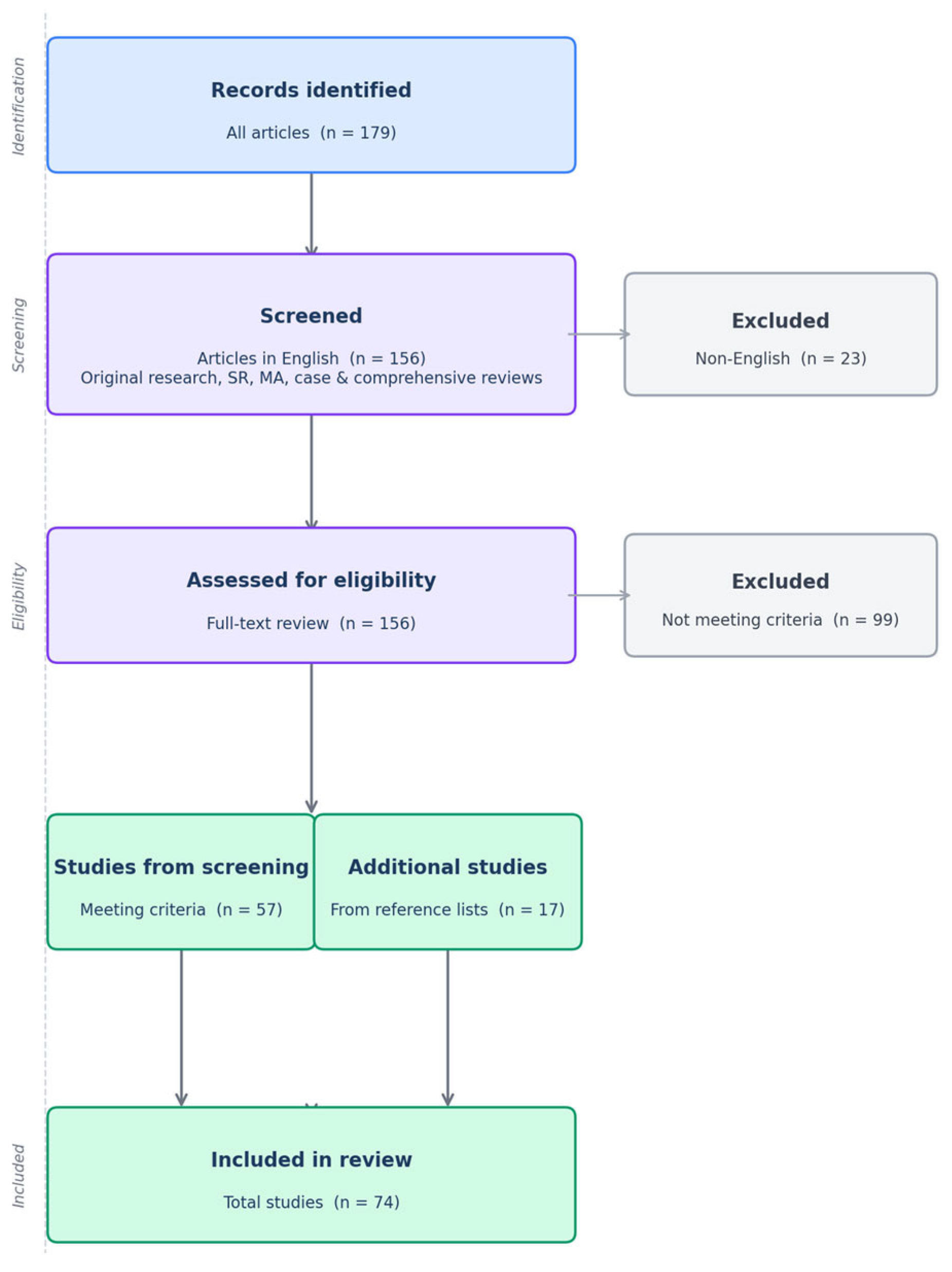
Eligible studies included original research articles, systematic reviews, meta-analyses, case reports, and comprehensive reviews. Letters to the editor, editorials, commentaries, conference abstracts, and book chapters were excluded. Titles and abstracts were screened independently by two reviewers (AA and JS) for their adherence to the inclusion criteria and relevance to the study topic. We included studies published between January 2010 and 15 October 2025. In cases of discrepancy, final decisions were made with input from PK and JM. Only articles in English were selected. After the completion of the study selection process, 57 articles were chosen to be included in this review, with 17 additional studies retrieved from the references of the source materials. The complete flow chart is presented in Figure 1.

3. Discussion

3.1. Pancreatic NETs

Pancreatic neuroendocrine tumors are characterized by a complex genetic landscape that is more diverse and more frequently altered than in NETs of the small or large intestine. Whole-genome analysis of NETs has revealed driver somatic mutations in recurrent genes that impact four major pathways: (1) chromatin remodeling, (2) DNA damage repair, (3) mTOR signaling activation, and (4) telomere maintenance [19,20]. Likewise, genes involved in cell cycle regulation, the Wnt signaling pathway, and tumor suppressors have also been identified [20]. An overview of the above-mentioned mechanisms and their related genes selected in our review is provided in Table 1.
The majority of panNETs occur sporadically, while others develop in the context of genetic syndromes such as multiple endocrine neoplasia type 1 (MEN1) and 4 (MEN4), tuberous sclerosis complex (TSC), neurofibromatosis type 1 (NF1), and von Hippel–Lindau disease (VHL) [21,22,23]. Those linked to genetic syndromes are generally regarded as less aggressive and tend to have a more favorable prognosis [8].

3.1.1. MEN1

Multiple endocrine neoplasia type 1 (MEN1) syndrome is caused by an inherited or de novo germline autosomal dominant mutation in a tumor suppressor gene located on chromosome 11q13, which encodes a 610-amino-acid protein called menin [22]. The function of menin is associated with the regulation of cell growth, apoptosis, and DNA repair [24]. More than 1100 mutations have been detected across the entire coding region [25]. MEN1 syndrome shows high penetrance, and its clinical manifestations vary widely [26]. Patients diagnosed with MEN1 syndrome exhibit a significantly increased risk of developing multiple endocrine neoplasms at an early age, with over 90% affected by the age of 40 years [27]. Tumors most often occur as parathyroid gland adenomas (90%), followed by pancreatic or duodenal NETs (50%) and anterior pituitary adenomas (40%) [28]. PanNETs appear to be the most challenging and constitute the leading cause of death in patients with MEN1 (40–50%) [24,29].
In panNETs, sporadic mutations in the MEN1 gene occur even more frequently, with their prevalence estimated at up to 44% [30]. Notably, these alterations appear to be restricted to panNETs and are absent in pancreatic NECs [31]. Considering the frequency of these mutations, it is justified to determine their impact on the disease course.
Chiloiro et al. compared the clinical, histological, and morphological aspects of panNETs in patients with and without MEN1 syndrome [32]. They found that the frequency of liver and lymph node metastases was higher in MEN1 wild-type patients. Moreover, the mutation-positive group showed a more favorable prognosis, but this difference was not statistically significant. It can be partly explained by earlier diagnosis and improved screening due to active surveillance of these tumors in first-degree relatives.
These findings are in line with those of Gleeson et al., who demonstrated that patients harboring a MEN1 variant developed pancreatic NETs of smaller size and were more frequently diagnosed at an early disease stage [33]. Other studies also emphasize a more favorable disease course and slower progression in patients with these mutations [19,21,24,27,34]. It should be noted, however, that within the MEN1 group, prognostically unfavorable genetic factors may also occur. One such factor is a mutation in c-MET, a proto-oncogene encoding the c-MET tyrosine kinase receptor for hepatocyte growth factor. Ghosh et al. demonstrated that overexpression of c-MET is associated with a more aggressive disease course and poorer prognosis, similar to what is observed in other malignancies associated with mutations in this gene [35].

3.1.2. VHL

Von Hippel–Lindau (VHL) syndrome is an inherited cancer predisposition disorder with autosomal dominant transmission, caused by a germline mutation in the VHL gene located on the short arm of chromosome 3 [36]. The VHL gene is responsible for regulating the cellular oxygen-sensing pathway by controlling the stability of hypoxia-inducible factors (HIFs), and its inactivation results in aberrant activation of genes involved in angiogenesis, cell proliferation, and metabolism [26]. Mutations in this gene lead to the development of clear cell tumors affecting multiple organs, including hemangioblastomas of the central nervous system and retina, renal cell carcinoma, pheochromocytomas, panNETs, and adrenal cortical adenomas [37]. In addition to germline mutations, the VHL gene is also frequently inactivated in up to 25% of sporadic panNETs, mostly by non-mutational mechanisms [30].
PanNETs occur in 10–17% of patients with VHL syndrome and are usually small, non-functioning (NF), well- or moderately differentiated tumors with high expression of somatostatin receptor analogs [34,38]. The majority are identified at a relatively young age, with a mean age at diagnosis of 26–38 years, and typically present as multifocal lesions distributed throughout the pancreas [27,38]. Malignant transformation of these tumors is relatively uncommon [24]. Compared with sporadic panNETs, VHL-associated tumors are far less often high-grade or metastatic and have better long-term outcomes [27,39]. However, it should be noted that several risk factors associated with panNET metastasis in VHL disease have been identified, including tumor diameter exceeding 3 cm, blood type 0, a tumor doubling time of less than 500 days, and missense mutations located in exon 3 of the VHL gene [36,38].
Considering the above, the presence of a VHL mutation should be regarded as a positive prognostic factor. This also applies to the majority of cases in which 18F-FDG PET scans show increased glucose uptake. This phenomenon may be explained not by high tumor grade but rather by the mechanism of VHL gene inactivation, which leads to accumulation of HIF protein, a pseudohypoxic state, and direct regulation of glucose transporter 1, responsible for cellular glucose uptake [40]. This phenomenon applies to both sporadic VHL mutations and VHL disease-related panNETs.

3.1.3. DAXX and ATRX

Among sporadic panNETs, approximately 45% present inactivating somatic mutations in mutually exclusive tumor suppressor genes: DAXX (death-domain-associated protein, located on chromosome 6p21.3) or ATRX (alpha-thalassemia/mental retardation syndrome X-linked, located on chromosome Xq21.1). Both genes encode proteins involved in chromatin remodeling. Their mutations result in loss of expression of the corresponding protein, as assessed by immunohistochemistry (IHC) [21,34,41,42]. In the Hechtman study, the sensitivity of translating DAXX alterations into IHC was 95% [43]. The consequence of these mutations is the activation of the alternative lengthening of telomeres (ALT) mechanism, used by several types of malignancies, including panNETs. This mechanism allows neoplasms to maintain telomere length, supporting sustained tumor growth and cell proliferation [42,44]. Mutations in the DAXX and ATRX genes represent the second most common alterations in panNETs after MEN1. Similar to MEN1 gene alterations, DAXX and ATRX mutations occur exclusively in panNETs and have not been reported in panNECs to date [31].
Based on the association between an activated ALT mechanism and DAXX/ATRX mutations, Luchini et al. suggested the determination of ALT as a potential prognostic biomarker and demonstrated that its presence was associated with poorer prognosis in panNETs [44]. These findings were supported by Marinoni et al., who showed a significant association between ALT activation and T stage and reported that tumors positive for ALT were characterized by significantly shorter relapse-free survival and reduced disease-specific survival (DSS) [45]. Similar conclusions were described in the study by McGovern et al., in which panNETs were assessed preoperatively using CT scans, and negative prognostic features were strongly correlated with ALT-positive status [46]. Of interest are also the studies by Pea and Hackeng, in which they demonstrated that ALT-positive patients have an increased risk of liver metastases in the panNET < 3 cm and insulinoma groups, respectively [47,48]. Moreover, Singhi et al. reported that patients with ALT-positive panNETs have a 40% 5-year disease-free survival (DFS), compared with 96% in ALT-negative patients [49].
In addition to its prognostic role, ALT status may also serve as a marker of pancreatic origin, supporting the diagnostic process in patients with neuroendocrine metastases from an unknown primary [44]. However, DAXX and ATRX mutations, together with their associated consequences, such as activation of the ALT mechanism, may also be observed, although less frequently, in extrapancreatic NETs [1,42,50].
The implications of ALT activation overlap with findings associated with mutations in the DAXX and ATRX genes. In the aforementioned study by Gleeson et al., the authors demonstrated that tumors harboring DAXX and/or ATRX mutations were larger at diagnosis, exhibited more malignant features, and were more frequently observed at advanced disease stages [33]. Similar conclusions were reached in many other studies [42,50,51,52,53]. However, in the study by Chen et al., in which loss of these proteins was correlated with a higher Ki-67 index and higher WHO grade of NETs, only DAXX loss reached statistical significance [50]. These correlations with more aggressive tumor potential also extend to functioning panNETs, such as glucagonomas, as demonstrated in the study by Mattiolo et al., in which ATRX or DAXX mutations were detected in the majority of cases [54].
Additional evidence for a worse prognosis in patients harboring DAXX/ATRX mutations comes from the study by Hong et al., where the presence of these mutations was associated with significantly shorter recurrence-free survival (RFS) [55]. This was also confirmed in the study by Hackeng, in which the combination of ALT-positive and/or ATRX/DAXX-negative NF-panNETs was correlated with markedly shorter RFS. This association was also observed in tumors < 2 cm [52]. Similarly, in extrapancreatic NETs, including those of the duodenum, appendix, and colon, overall survival (OS) was lower in patients who had lost ATRX or DAXX expression [20]. It should also be noted that an opposite correlation has been observed in patients with established metastatic panNETs, in whom loss of DAXX/ATRX was identified directly in metastases. This factor resulted in a better prognosis in several studies [20]. Taken together, the available evidence supports the recognition of DAXX and ATRX mutations and ALT activation (altered in primary origin) as independent negative prognostic factors.

3.1.4. MTOR

The MTOR gene (the mammalian target of rapamycin), located on chromosome 1p36, encodes a serine–threonine kinase complex that serves as a central regulator of cell growth, proliferation, metabolism, and survival through the integration of nutrient and growth factor signaling. It becomes activated through phosphorylation, resulting in the formation of phosphorylated mTOR (p-mTOR) [56]. In panNETs, genomic alterations affecting components of the PI3K/AKT/mTOR pathway are observed in approximately 15% of cases and can be reflected by immunohistochemical staining of phosphorylated proteins of the pathway, contributing to its hyperactivation [34,56,57].
The expression of mTOR in panNETs has been evaluated as a potential prognostic marker. In the study by Lamberti et al., the prognostic significance of p-mTOR protein expression was assessed by calculating a quantitative score (QS) based on the percentage of positive cells and staining intensity during immunostaining evaluation. A high p-mTOR QS was shown to correlate with more advanced staging (IIIB–IV vs. I–II) and the presence of metastatic disease at diagnosis. Similarly, higher p-mTOR QS values were associated with disease relapse and shorter disease-free survival (DFS) [57]. In another study on panNETs conducted by Komori et al., elevated p-mTOR expression was likewise associated with more aggressive tumor behavior and adverse clinical characteristics, including larger tumor size, higher histological grade, and more advanced stage at diagnosis [56]. However, no significant association with DFS was observed, although the results showed a trend toward such a relationship (p < 0.1). The prognostic significance of MTOR has also been investigated in NETs of other locations, including the small intestine. In a study by Qian et al., mTOR expression was associated with a higher Ki-67 index, as well as shorter DFS and overall survival (OS) [58].
Clinically, aberrant mTOR signaling has predictive therapeutic relevance, as pharmacologic inhibition using mTOR inhibitors such as everolimus has demonstrated a significant improvement in progression-free survival (PFS) in patients with advanced pancreatic and extrapancreatic NETs [6]. In the study by Gelsomino et al., the expression of phosphorylated mTOR was examined during everolimus treatment and correlated with patient outcomes, as the mTOR-positive group had significantly better PFS and OS [59].
Overall, the presence of MTOR gene mutations appears to act as a positive predictive marker for response to everolimus therapy, while concurrently serving as a negative prognostic indicator for disease progression and clinical outcome.

3.1.5. PTEN

Phosphatase and tensin homolog (PTEN) is a well-known tumor suppressor gene located on chromosome 10q23, a region commonly affected by somatic deletions in various tumors. It encodes a 403-amino-acid protein (PTEN) and plays an important role in controlling cell growth, apoptosis, cell adhesion, and cell migration [60]. In NETs, PTEN regulates the PI3K/Akt/mTOR pathway.
So far, its role as a prognostic marker in NETs has been well documented. Krausch et al. found in their research that loss of PTEN correlates with tumor progression and is associated with poorer outcomes and survival [35]. This is in line with the study by Missiaglia et al., in which reduced PTEN expression was linked to a more aggressive tumor phenotype. In addition, they demonstrated that low cytoplasmic PTEN expression was associated with a shorter time to disease progression and decreased disease-free survival [61]. Another observation was made by Estrella et al., confirming the significant negative prognostic role of PTEN alterations in NETs [62].

3.1.6. TSC2

Tuberous sclerosis complex gene 2 (TSC2) is a tumor suppressor gene encoding tuberin and located on chromosome 16p13.3. Its germline mutation, together with the TSC1 gene (encoding hamartin), results in the development of tuberous sclerosis complex, a rare inherited autosomal dominant syndrome. The molecular pathogenesis of the disease is driven by disruption of the normal activity of the hamartin–tuberin complex, leading to dysregulation of the mTOR pathway and resulting in unregulated cell proliferation and tumorigenesis [63]. The phenotypic manifestations of this disorder encompass the development of multiple hamartomatous lesions, a spectrum of neurological abnormalities—most prominently epilepsy and intellectual disability—and characteristic cutaneous features, including hypomelanotic macules, facial angiofibromas, shagreen patches, and ungual fibromas [27].
Existing case reports on TSC2 as a molecular prognostic marker in NETs suggest that some younger patients may remain asymptomatic. These observations may be influenced by more intensive screening in individuals with tuberous sclerosis, similar to MEN1 syndrome [63,64]. In contrast to these reports, stronger evidence comes from the study by Gleeson et al., in which alterations in TSC2 were linked to increased disease progression and worse overall survival (OS) [33]. Similar conclusions were reported by Missiaglia et al., who demonstrated that patients with reduced TSC2 expression showed poorer outcomes, including shorter OS, progression-free survival (PFS), and disease-free survival (DFS) [61]. Considering these findings, mutations in the TSC2 gene should be regarded as a negative prognostic factor in patients with NETs.

3.1.7. NF1

Neurofibromatosis type 1 (NF1), previously known as von Recklinghausen disease, is an inherited autosomal dominant condition resulting from pathogenic variants in the NF1 gene. It encodes the tumor suppressor protein neurofibromin and is located on chromosome region 17q11.2 [27,30]. Inactivation of the NF1 gene leads to constitutive upregulation of RAS signaling pathways, promoting the development of hallmark clinical and neoplastic features of the syndrome, including neurofibromas, café-au-lait spots, Lisch nodules, intertriginous freckling, and optic pathway gliomas [27,30].
In patients with NF1, neuroendocrine tumors can develop in approximately 10% of cases and are almost exclusively duodenal somatostatinomas of the periampullary region and, more rarely, nonfunctional panNETs [24,27,30,65]. Malignant forms occur in about 30% of cases [24]. The presence of sporadic NF1 mutations in NETs is occasional, and there are currently insufficient data to determine their clinical significance [30]. According to the cited studies, duodenal and pancreatic somatostatinomas associated with NF1 differ from their sporadic pancreatic counterparts in multiple respects: syndromic tumors are identified at a smaller size, rarely induce a clinical hypersecretory state, and have a lower incidence of metastatic disease at the time of diagnosis [27]. To sum up, the presence of an inherited NF1 gene mutation should be considered a positive prognostic marker, indicative of a more favorable clinical outcome and disease course.

3.1.8. RB1

The RB1 (retinoblastoma 1) gene is located on chromosome 13q14 and encodes the tumor suppressor retinoblastoma protein involved in cell cycle regulation. Inactivation of RB1 through loss or mutation impairs this checkpoint mechanism, promoting uncontrolled cell division and contributing to the development of retinoblastoma as well as numerous other malignancies, including NENs. In NETs, RB1 gene mutations are considered rare and are mainly observed in NET G3 metastases rather than in primary tumors [65]. Their presence is therefore indicative of advanced, disseminated disease. Conversely, in NECs, the prevalence of somatic mutations is high and estimated to be up to 74% of cases [21,34,37]. This may be a valuable supportive tool in the differential diagnosis of these types of NENs [1,66,67].
According to our review, there are no data on the predictive value of RB1 in NETs. However, it is worth noting that in the subgroup of panNECs, there are reports indicating that loss of Rb protein expression associated with RB1 gene mutations represents a positive predictive factor for response to first-line platinum-based chemotherapy, as the response rate to this treatment is higher than in the group without Rb loss.
On the other hand, this group is characterized by shorter OS, which confirms a more aggressive disease course [68]. Similar observations were made by Hijioka et al. [69]. Therefore, RB1 gene mutations should be considered a negative prognostic factor (for NETs and panNECs) and a positive predictive factor (for platinum-based chemotherapy in panNECs).

3.1.9. TP53

TP53 is a tumor suppressor gene located on chromosome 17p13.1 that encodes the p53 protein, a key regulator of cell cycle arrest, DNA repair, senescence, and apoptosis in response to cellular stress. TP53 mutations lead to abnormal p53 staining on immunohistochemistry (for example, in 100% of small cell and 90% of large cell neuroendocrine carcinomas) [21,34,37,67]. In the context of NENs, TP53 alterations are rare in well-differentiated panNETs but occur frequently in poorly differentiated panNECs [1,66,68,70]. Similar to the RB1 gene, the presence of TP53 mutations should guide the diagnosis toward NEC [1,66,71].
There are numerous reports describing the impact of TP53 mutations on the course of disease in panNETs. Ail et al. showed in their research that a mutated p53 pattern in patients with gastroenteropancreatic neuroendocrine neoplasms was associated with worse OS and DFS, irrespective of tumor grade [72]. Comparable conclusions have been reported by others [33]. Moreover, Kasajima et al. demonstrated an association between sudden and severe deterioration in all NET G3 patients’ condition and immunohistochemical overexpression of p53, accompanied by a corresponding mutation in the TP53 gene [73]. Additional characteristics of TP53 are presented in the intestinal NETs section.

3.1.10. SMAD4

The mothers against decapentaplegic homolog 4 (SMAD4) gene encodes a key transcriptional mediator of the TGF-β signaling pathway and acts as a tumor suppressor by regulating cell proliferation, differentiation, and apoptosis. The gene is located on chromosome 18q21, and its alterations result in reduced or absent SMAD4 protein expression.
SMAD4 alterations in pancreatic malignancies are primarily associated with pancreatic adenocarcinoma. Some studies indicate that SMAD4 mutations are infrequently observed in pancreatic NECs and do not appear to play a significant role in these tumors [68,74]. However, these data are inconclusive, as conflicting evidence has been reported in other studies [31,75].
In gastrointestinal NETs, loss or inactivation of SMAD4 is uncommon but has been associated with tumor progression, aggressive behavior, and poorer prognosis [76]. Further evidence supporting this hypothesis from the Martin et al. report indicates that SMAD4 mutations are clinically significant and likely contribute to high-grade transformation in WD-panNETs [77].

3.1.11. ARID1A

ARID1A (AT-rich interactive domain-containing protein 1A) is a tumor suppressor gene encoding a core subunit of the SWI/SNF (switch/sucrose nonfermentable) ATP-dependent chromatin-remodeling complex and plays a fundamental role in the regulation of chromatin accessibility, transcriptional programs, cell cycle progression, and DNA damage response. Recurrent loss-of-function alterations in ARID1A have been documented across a broad spectrum of human malignancies, including gastrointestinal, ovarian, and colorectal cancers and panNETs. The gene is localized to chromosome 1p36.11, and its inactivating mutations are associated with loss of the corresponding protein [51,78].
In the Han et al. study, ARID1A transcript levels were markedly downregulated in NF-panNETs compared with those in the adjacent normal pancreas. Among panNETs, two subgroups were identified based on ARID1A expression levels: a high-expression group and a low-expression group. Patients with reduced ARID1A expression more frequently exhibited regional lymph node metastases, larger tumor size, higher Ki-67 indices, and higher tumor grade [78]. These findings are consistent with the results reported by Roy et al., in which, among the evaluated biomarkers, loss of ARID1A was associated with more aggressive features of pancreatic neuroendocrine tumors and the presence of distant metastases. Moreover, patients in this subgroup exhibited significantly shorter DFS and DSS, underscoring the negative prognostic impact of ARID1A loss [51].

3.2. Intestinal NETs

The molecular landscape of intestinal NETs is characterized predominantly by chromosomal alterations and epigenetic changes, including DNA methylation, rather than by single somatic mutations [79,80,81]. Loss of chromosome 18 is considered the most frequent molecular event, occurring in approximately 63–82% of small intestinal NETs (siNETs), and is thought to be an early event in tumor development [80,82,83,84]. The overall somatic mutation burden is consistently reported to be noticeably lower than in panNETs. Depending on the cohort and methodology, in siNETs, driver mutations are detected in up to 34.6% of cases and are mostly located in tumor suppressor genes (CDKN1B, TP53) or, less frequently, in proto-oncogenes (KRAS, NRAS, MET) [82,85]. However, these alterations are typically non-recurrent and occur at lower frequencies. Driver mutations are detected more frequently in patients with a higher Ki-67 index [82]. Among colorectal NENs, the largest proportion consists of NECs, which are frequently characterized by mutations in APC, KRAS, BRAF, and TP53 and often exhibit microsatellite instability [5].

3.2.1. CDKN1B

The cyclin-dependent kinase inhibitor 1B gene (CDKN1B) is a tumor suppressor gene located on chromosome 12p13. The CDKN1B gene encodes the p27 protein, which is a key regulator of cell cycle progression [86]. Loss-of-function alterations or reduced expression of p27 contribute to dysregulated cellular proliferation and are considered early events in tumorigenesis [86]. Germline mutations in the CDKN1B gene are associated with MEN4 syndrome, a phenotype characterized by tumors affecting the parathyroid glands, pituitary, and pancreas, closely resembling the clinical presentation observed in MEN1 [21,86]. CDKN1B alterations can act as driver events in about 7–20% of sporadic siNETs [80,82,85,86,87]. Therefore, it is assumed to be the main somatic mutation observed in this group of NETs.
Data on the prognostic/predictive value of CDKN1B in intestinal NETs are very limited, with most studies focusing on its pathogenetic role. According to Crona et al., there is no clear agreement on the influence of these mutations on clinical characteristics and survival [86]. Similar conclusions were reached by Simbolo et al. in their study [85]. This is also partially supported by the study by Elias et al., who stated that the presence of CDKN1B gene mutations does not appear to be essential for metastasis formation [88]. On the other hand, Scarpa underlines that DFS analysis showed a progressively poorer prognosis in patients harboring CDKN1B alterations, although the results were mainly related to copy number variations (CNVs) rather than somatic mutations [79]. In summary, CDKN1B gene mutations appear to have unclear prognostic and predictive relevance so far.

3.2.2. APC

The APC (adenomatous polyposis coli) gene is a tumor suppressor gene located on chromosome 5q21–q22 [89]. It encodes a multifunctional APC protein that negatively regulates the Wnt/β-catenin signaling pathway, controlling cell proliferation, differentiation, and genomic stability. The APC gene is well known for its role in familial and sporadic colon cancer, with a reported frequency of approximately 70% [89]. In less common cases, it has also been detected in tumors arising from the liver, stomach, and lung (notably squamous cell carcinoma and small cell carcinoma), as well as in breast cancer and cerebellar medulloblastoma [89].
One of the attempts to investigate the occurrence and impact of APC alterations in intestinal NETs was conducted by Bottarelli et al., who investigated 30 siNETs and reported that 23% harbored APC mutations and 15% displayed loss of one gene copy [89]. In a study by Simbolo et al., APC alterations were found in 7.7% of cases [85]. However, no clear correlation with clinical course and prognosis has been found so far [85,89]. Due to the limited availability of existing data, the prognostic and predictive significance of APC alterations in NETs remains largely undefined.

3.2.3. CTNNB1

The CTNNB1 (catenin beta 1) gene, located on chromosome 3p22.1, encodes β-catenin, which plays a crucial role in the canonical Wnt signaling pathway. Activating mutations in CTNNB1 lead to stabilization and nuclear accumulation of β-catenin, promoting transcription of Wnt target genes involved in proliferation and tumor progression. It is most frequently associated with colorectal cancer but is also detected in other malignancies.
In our review, only a single study showed that in intestinal NETs, CTNNB1 methylation was increased in metastases [79]. None of the other studies specifically investigated mutations affecting this gene. Similar to the APC gene, there is currently no evidence regarding the prognostic or predictive significance of CTNNB1 alterations in intestinal NETs.

3.2.4. TP53

The introduction and molecular characteristics of the TP53 gene have been presented in Section 3.1.9.
In intestinal neuroendocrine NENs, TP53 mutations, similar to those in pancreatic NENs, are markedly more frequent in NECs than in NETs and may aid in distinguishing between these subgroups [71]. Data regarding the prognostic role of TP53 in intestinal NETs remain limited; however, they appear consistent with observations in pancreatic NETs. In both studies by Ail et al. and Cubiella et al., an immunohistochemical p53 expression pattern indicative of TP53 mutation, as well as mutation status assessed directly, was associated with poorer prognosis and reduced overall survival [71,72]. Therefore, TP53 mutation is considered a negative prognostic factor in intestinal NETs, similar to the pancreatic group.

3.3. Conclusions and Future Directions

For many years, numerous attempts have been made to identify reliable biomarkers for NETs. Recent advances in genetics have improved our understanding of the mechanisms underlying tumorigenesis. In addition, minimally invasive approaches, such as liquid biopsy, now allow targeted profiling of these tumors, providing valuable insights into their biology. Genetic profiling may play a pivotal role in early diagnosis, prognostic assessment, detection of recurrence, and guiding individualized therapeutic strategies, particularly through the identification of novel molecular targets.
Our review highlights that, despite progress in understanding the prognostic value of genetic alterations in panNETs, their application as predictive biomarkers remains limited. In intestinal NETs, which exhibit markedly different tumorigenic mechanisms, available data are too limited to draw firm conclusions. Significant gaps in this research area persist, underscoring the need for further studies to fully realize their clinical potential. A detailed summary presenting the prognostic and predictive relevance of individual gene mutations is provided below in Table 2.
To conclude, gene mutations as molecular markers have diverse applications, including diagnostic differentiation between NETs and NECs, identification of unknown primary tumor sites, stratification of patients at higher risk of progression and poorer prognosis, and selection of appropriate therapies. Collectively, these applications contribute to a personalized decision-making process, which is expected to become increasingly valuable in modern medicine. However, clinical implementation of these biomarkers remains constrained by several significant limitations. One of them is the restricted availability of advanced molecular diagnostic techniques, particularly in low-resource healthcare centers. An additional challenge is the lack of standardized detection methods and insufficient clinical validation of many potential biomarkers in large, multicenter studies. Moreover, the heterogeneity of NETs within locations and the biological differences between locations further complicate the interpretation of results. Therefore, further studies are required to evaluate the actual clinical utility of these mutations (such as a risk stratification model or diagnostic workflow) and to facilitate their integration into current diagnostic and therapeutic algorithms. Additionally, there is a need for further investigation of emerging and less-studied biomarkers, as this research gap still exists.

3.4. Review Limitations

Our review focuses primarily on somatic mutations in NETs and does not consider epigenetics, transcriptomics, or other multi-omics approaches. Moreover, some evidence supporting the significance of specific genes comes from limited data, such as small patient cohorts or isolated case reports, which may reduce their clinical relevance. Finally, we searched the literature using a single database (PubMed only), which may introduce a potential risk of selection bias.

Author Contributions

Conceptualization, J.M. (Jan Musiałkiewicz) and B.B.; methodology, J.M. (Jan Musiałkiewicz) and P.K.; validation, P.G. and M.R.; formal analysis, B.B.; investigation, J.M. (Jan Musiałkiewicz) and A.M.; resources, M.R.; data curation, J.M. (Jan Musiałkiewicz), A.M., A.A., J.M. (Joanna Maciejewska) and P.K.; writing—original draft preparation, J.M. (Jan Musiałkiewicz), A.M., J.M. (Joanna Maciejewska) and A.A.; writing—review and editing, J.M. (Jan Musiałkiewicz), P.G., M.R. and B.B.; visualization, J.M. (Joanna Maciejewska) and A.A.; supervision, B.B., P.G., M.R.; project administration, J.M. (Jan Musiałkiewicz) and M.R.; funding acquisition, M.R. All authors have read and agreed to the published version of the manuscript.

Funding

This research received no external funding.

Institutional Review Board Statement

Not applicable.

Informed Consent Statement

Not applicable.

Data Availability Statement

No new data were created or analyzed in this study. Data sharing is not applicable to this article.

Conflicts of Interest

The authors declare no conflicts of interest.

Abbreviations

The following abbreviations are used in this manuscript:
NETNeuroendocrine tumor
NENNeuroendocrine neoplasm
WHOWorld Health Organization
NECNeuroendocrine carcinoma
GEP-NETsGastroenteropancreatic NETs
CSCarcinoid syndrome
PanNETsPancreatic neuroendocrine tumors
WDWell-differentiated
COSMICCatalogue of Somatic Mutations In Cancer
MENMultiple endocrine neoplasia
TSCTuberous sclerosis complex
NF1Neurofibromatosis type 1
VHLVon Hippel–Lindau
HIFHypoxia-inducible factor
NFNon-functioning
18F-FDG PETPositron emission tomography using fluorine-18 labeled glucose
DAXXDeath-domain associated protein
ATRXAlpha-thalassemia/mental retardation syndrome X-linked
IHCImmunohistochemistry
ALTAlternative lengthening of telomeres
RFSRecurrence-free survival
DFSDisease-free survival
DSSDisease-specific survival
OSOverall survival
mTORmammalian target of rapamycin
QSQuantitative score
PFSProgression-free survival
PTENPhosphatase and tensin homolog
SMAD4Mothers against decapentaplegic homolog 4
ARID1AAT-rich interactive domain-containing protein 1A
SWI/SNFSwitch/sucrose nonfermentable
SiNETsSmall intestine neuroendocrine tumors
CDKN1BCyclin-dependent kinase inhibitor 1B
APCAdenomatous polyposis coli
CTNNB1Catenin beta 1

References

  1. Rindi, G.; Mete, O.; Uccella, S.; Basturk, O.; La Rosa, S.; Brosens, L.A.A.; Ezzat, S.; de Herder, W.W.; Klimstra, D.S.; Papotti, M.; et al. Overview of the 2022 WHO Classification of Neuroendocrine Neoplasms. Endocr. Pathol. 2022, 33, 115–154. [Google Scholar] [CrossRef]
  2. Zhu, Y. Incidence trend of neuroendocrine tumors and disparities by sex and race/ethnicity in adults from the United States, 2000–2020. Eur. J. Cancer Prev. 2024, 33, 475–484. [Google Scholar] [CrossRef]
  3. Hallet, J.; Law, C.H.; Cukier, M.; Saskin, R.; Liu, N.; Singh, S. Exploring the rising incidence of neuroendocrine tumors: A population-based analysis of epidemiology, metastatic presentation, and outcomes. Cancer 2015, 121, 589–597. [Google Scholar] [CrossRef] [PubMed]
  4. Imamura, M. Recent standardization of treatment strategy for pancreatic neuroendocrine tumors. World J. Gastroenterol. 2010, 16, 4519–4525. [Google Scholar] [CrossRef]
  5. Oronsky, B.; Ma, P.C.; Morgensztern, D.; Carter, C.A. Nothing But NET: A Review of Neuroendocrine Tumors and Carcinomas. Neoplasia 2017, 19, 991–1002. [Google Scholar] [CrossRef] [PubMed]
  6. Yao, J.C.; Fazio, N.; Singh, S.; Buzzoni, R.; Carnaghi, C.; Wolin, E.; Tomasek, J.; Raderer, M.; Lahner, H.; Voi, M.; et al. Everolimus for the treatment of advanced, non-functional neuroendocrine tumours of the lung or gastrointestinal tract (RADIANT-4): A randomised, placebo-controlled, phase 3 study. Lancet 2016, 387, 968–977. [Google Scholar] [CrossRef] [PubMed]
  7. Grozinsky-Glasberg, S.; Davar, J.; Hofland, J.; Dobson, R.; Prasad, V.; Pascher, A.; Denecke, T.; Tesselaar, M.E.T.; Panzuto, F.; Albåge, A.; et al. European Neuroendocrine Tumor Society (ENETS) 2022 Guidance Paper for Carcinoid Syndrome and Carcinoid Heart Disease. J. Neuroendocrinol. 2022, 34, e13146. [Google Scholar] [CrossRef]
  8. Tacelli, M.; Gentiluomo, M.; Biamonte, P.; Castano, J.P.; Berković, M.C.; Cives, M.; Kapitanović, S.; Marinoni, I.; Marinovic, S.; Nikas, I.; et al. Pancreatic neuroendocrine neoplasms (pNENs): Genetic and environmental biomarkers for risk of occurrence and prognosis. Semin. Cancer Biol. 2025, 112, 112–125. [Google Scholar] [CrossRef]
  9. Kos-Kudła, B.; Foltyn, W.; Malczewska, A.; Bednarczuk, T.; Bolanowski, M.; Borowska, M.; Chmielik, E.; Ćwikła, J.B.; Gisterek, I.; Handkiewicz-Junak, D.; et al. Update of the diagnostic and therapeutic guidelines for gastro-entero-pancreatic neuroendocrine neoplasms (recommended by the Polish Network of Neuroendocrine Tumours). Endokrynol. Pol. 2022, 73, 387–454. [Google Scholar] [CrossRef]
  10. Gierach, M.; Łazor, N.; Witkowska, W.; Kaczmarek, M.; Łukasiewicz, D.; Junik, R. Severe secretory diarrhea due to VIPoma. Endokrynol. Pol. 2025, 76, 682–683. [Google Scholar] [CrossRef]
  11. Komarnicki, P.; Musiałkiewicz, J.; Stańska, A.; Maciejewski, A.; Gut, P.; Mastorakos, G.; Ruchała, M. Circulating Neuroendocrine Tumor Biomarkers: Past, Present and Future. J. Clin. Med. 2022, 11, 5542. [Google Scholar] [CrossRef] [PubMed]
  12. Fuksiewicz, M.; Kowalska, M.; Kolasinska-Cwikla, A.; Kotowicz, B. Serum levels of neuron-specific enolase as a prognostic factor for disease progression in patients with GET/NEN in the pancreas and the small intestine. Endocr. Connect. 2022, 11, e210647. [Google Scholar] [CrossRef]
  13. Komarnicki, P.; Gut, P.; Cieślewicz, M.; Musiałkiewicz, J.; Maciejewski, A.; Czupińska, M.; Mastorakos, G.; Ruchała, M. Serum β-hCG as a Biomarker in Pancreatic Neuroendocrine Tumors: Rethinking Single-Analyte Approach. Cancers 2024, 16, 2060. [Google Scholar] [CrossRef]
  14. Musiałkiewicz, J.; Ruchała, M.; Komarnicki, P.; Maciejewski, A.; Sienkiewicz, J.; Sirisachadecha, P.; Budny, B.; Gut, P. Longitudinal analysis of serum alpha-fetoprotein in stable and progressive metastatic pancreatic neuroendocrine tumours. Endocr. Connect. 2025, 14, e250350. [Google Scholar] [CrossRef] [PubMed]
  15. Komarnicki, P.; Maciejewski, A.; Musiałkiewicz, J.; Czupińska, M.; Mastorakos, G.; Ruchała, M.; Gut, P. Serum Visfatin/eNAMPT as a Biomarker in Pancreatic and Small Intestine Neuroendocrine Tumors: A Cross-Sectional Study and Future Perspectives. Cancers 2025, 17, 2343. [Google Scholar] [CrossRef]
  16. Puliani, G.; Di Vito, V.; Feola, T.; Sesti, F.; Centello, R.; Pandozzi, C.; Tarsitano, M.G.; Verrico, M.; Lenzi, A.; Isidori, A.M.; et al. NETest: A Systematic Review Focusing on the Prognostic and Predictive Role. Neuroendocrinology 2022, 112, 523–536. [Google Scholar] [CrossRef]
  17. Malczewska, A.; Kos-Kudła, B.; Kidd, M.; Drozdov, I.; Bodei, L.; Matar, S.; Oberg, K.; Modlin, I.M. The clinical applications of a multigene liquid biopsy (NETest) in neuroendocrine tumors. Adv. Med. Sci. 2020, 65, 18–29. [Google Scholar] [CrossRef]
  18. Almeida, C.; Gervaso, L.; Frigè, G.; Spada, F.; Benini, L.; Cella, C.A.; Mazzarella, L.; Fazio, N. The Role of Liquid Biopsy in Gastroenteropancreatic Neuroendocrine Neoplasms. Cancers 2024, 16, 3349. [Google Scholar] [CrossRef] [PubMed]
  19. Challis, B.G.; Casey, R.T.; Grossman, A.; Newell-Price, J.; Newey, P.; Thakker, R.V. What is the appropriate management of nonfunctioning pancreatic neuroendocrine tumours disclosed on screening in adult patients with multiple endocrine neoplasia type 1? Clin. Endocrinol. 2019, 91, 708–715. [Google Scholar] [CrossRef]
  20. Boons, G.; Vandamme, T.; Peeters, M.; Van Camp, G.; Op de Beeck, K. Clinical applications of (epi)genetics in gastroenteropancreatic neuroendocrine neoplasms: Moving towards liquid biopsies. Rev. Endocr. Metab. Disord. 2019, 20, 333–351. [Google Scholar] [CrossRef]
  21. Reid, M.D.; Balci, S.; Saka, B.; Adsay, N.V. Neuroendocrine tumors of the pancreas: Current concepts and controversies. Endocr. Pathol. 2014, 25, 65–79. [Google Scholar] [CrossRef]
  22. Thakker, R.V. Multiple endocrine neoplasia type 1 (MEN1). Best Pract. Res. Clin. Endocrinol. Metab. 2010, 24, 355–370. [Google Scholar] [CrossRef] [PubMed]
  23. Woo, C.G.; Choi, S.Y.; Kwak, J.J.; Chin, S.; Kim, H.K. Clear cell neuroendocrine tumor of the pancreas in von Hippel-Lindau disease: A case report and literature review. Neuro Endocrinol. Lett. 2017, 38, 83–86. [Google Scholar] [PubMed]
  24. Mauriello, C.; Napolitano, S.; Gambardella, C.; Candela, G.; De Vita, F.; Orditura, M.; Sciascia, V.; Tartaglia, E.; Lanza, M.; Santini, L.; et al. Conservative management and parenchyma-sparing resections of pancreatic neuroendocrine tumors: Literature review. Int. J. Surg. 2015, 21, S10–S14. [Google Scholar] [CrossRef]
  25. Perrier, N.D. From Initial Description by Wermer to Present-Day MEN1: What have We Learned? World J. Surg. 2018, 42, 1031–1035. [Google Scholar] [CrossRef]
  26. Welsch, C.; Flügel, A.K.; Rondot, S.; Schulze, E.; Sircar, I.; Nußbaumer, J.; Bojunga, J. Distinct clinical phenotypes in a family with a novel truncating MEN1 frameshift mutation. BMC Endocr. Disord. 2022, 22, 64. [Google Scholar] [CrossRef]
  27. Guilmette, J.M.; Nosé, V. Neoplasms of the Neuroendocrine Pancreas: An Update in the Classification, Definition, and Molecular Genetic Advances. Adv. Anat. Pathol. 2019, 26, 13–30. [Google Scholar] [CrossRef]
  28. Effraimidis, G.; Knigge, U.; Rossing, M.; Oturai, P.; Rasmussen, Å.K.; Feldt-Rasmussen, U. Multiple endocrine neoplasia type 1 (MEN-1) and neuroendocrine neoplasms (NENs). Semin. Cancer Biol. 2022, 79, 141–162. [Google Scholar] [CrossRef]
  29. Ito, T.; Igarashi, H.; Uehara, H.; Berna, M.J.; Jensen, R.T. Causes of death and prognostic factors in multiple endocrine neoplasia type 1: A prospective study: Comparison of 106 MEN1/Zollinger-Ellison syndrome patients with 1613 literature MEN1 patients with or without pancreatic endocrine tumors. Medicine 2013, 92, 135–181. [Google Scholar] [CrossRef]
  30. Mafficini, A.; Scarpa, A. Genomic landscape of pancreatic neuroendocrine tumours: The International Cancer Genome Consortium. J. Endocrinol. 2018, 236, R161–R167. [Google Scholar] [CrossRef] [PubMed]
  31. Kim, H.; An, S.; Lee, K.; Ahn, S.; Park, D.Y.; Kim, J.H.; Kang, D.W.; Kim, M.J.; Chang, M.S.; Jung, E.S.; et al. Pancreatic High-Grade Neuroendocrine Neoplasms in the Korean Population: A Multicenter Study. Cancer Res. Treat. 2020, 52, 263–276. [Google Scholar] [CrossRef]
  32. Chiloiro, S.; Lanza, F.; Bianchi, A.; Schinzari, G.; Brizi, M.G.; Giampietro, A.; Rufini, V.; Inzani, F.; Giordano, A.; Rindi, G.; et al. Pancreatic neuroendocrine tumors in MEN1 disease: A mono-centric longitudinal and prognostic study. Endocrine 2018, 60, 362–367. [Google Scholar] [CrossRef] [PubMed]
  33. Gleeson, F.C.; Voss, J.S.; Kipp, B.R.; Kerr, S.E.; Van Arnam, J.S.; Mills, J.R.; Marcou, C.A.; Schneider, A.R.; Tu, Z.J.; Henry, M.R.; et al. Assessment of pancreatic neuroendocrine tumor cytologic genotype diversity to guide personalized medicine using a custom gastroenteropancreatic next-generation sequencing panel. Oncotarget 2017, 8, 93464–93475. [Google Scholar] [CrossRef] [PubMed]
  34. Reid, M.D.; Saka, B.; Balci, S.; Goldblum, A.S.; Adsay, N.V. Molecular genetics of pancreatic neoplasms and their morphologic correlates: An update on recent advances and potential diagnostic applications. Am. J. Clin. Pathol. 2014, 141, 168–180. [Google Scholar] [CrossRef] [PubMed]
  35. Ghosh, R.; Akbulut, D.; Simonds, W.F.; Weinstein, L.S.; Sadowski, S.M.; Blau, J.E.; Quezado, M.; Agarwal, S.K.; Jha, S. MEN1-Related Neuroendocrine Tumors Show c-MET Overexpression. J. Endocr. Soc. 2025, 9, bvaf147. [Google Scholar] [CrossRef]
  36. Tirosh, A.; El Lakis, M.; Green, P.; Nockel, P.; Patel, D.; Nilubol, N.; Gara, S.K.; Keutgen, X.M.; Linehan, W.M.; Kebebew, E. In silico VHL Gene Mutation Analysis and Prognosis of Pancreatic Neuroendocrine Tumors in von Hippel-Lindau Disease. J. Clin. Endocrinol. Metab. 2018, 103, 1631–1638. [Google Scholar] [CrossRef]
  37. Kim, J.Y.; Hong, S.M. Recent Updates on Neuroendocrine Tumors From the Gastrointestinal and Pancreatobiliary Tracts. Arch. Pathol. Lab. Med. 2016, 140, 437–448. [Google Scholar] [CrossRef]
  38. Penitenti, F.; Landoni, L.; Scardoni, M.; Piredda, M.L.; Cingarlini, S.; Scarpa, A.; D’Onofrio, M.; Girelli, D.; Davi, M.V. Clinical presentation, genotype-phenotype correlations, and outcome of pancreatic neuroendocrine tumors in Von Hippel-Lindau syndrome. Endocrine 2021, 74, 180–187. [Google Scholar] [CrossRef]
  39. Laks, S.; van Leeuwaarde, R.; Patel, D.; Keutgen, X.M.; Hammel, P.; Nilubol, N.; Links, T.P.; Halfdanarson, T.R.; Daniels, A.B.; Tirosh, A.; et al. Management recommendations for pancreatic manifestations of von Hippel-Lindau disease. Cancer 2022, 128, 435–446. [Google Scholar] [CrossRef]
  40. Bucau, M.; Laurent-Bellue, A.; Poté, N.; Hentic, O.; Cros, J.; Mikail, N.; Rebours, V.; Ruszniewski, P.; Lebtahi, R.; Couvelard, A. 18F-FDG Uptake in Well-Differentiated Neuroendocrine Tumors Correlates with Both Ki-67 and VHL Pathway Inactivation. Neuroendocrinology 2018, 106, 274–282. [Google Scholar] [CrossRef]
  41. Hadano, A.; Hirabayashi, K.; Yamada, M.; Kawanishi, A.; Takanashi, Y.; Kawaguchi, Y.; Nakagohri, T.; Nakamura, N.; Mine, T. Molecular alterations in sporadic pancreatic neuroendocrine microadenomas. Pancreatology 2016, 16, 411–415. [Google Scholar] [CrossRef]
  42. Heaphy, C.M.; Singhi, A.D. The diagnostic and prognostic utility of incorporating DAXX, ATRX, and alternative lengthening of telomeres to the evaluation of pancreatic neuroendocrine tumors. Hum. Pathol. 2022, 129, 11–20. [Google Scholar] [CrossRef] [PubMed]
  43. Hechtman, J.F.; Klimstra, D.S.; Nanjangud, G.; Frosina, D.; Shia, J.; Jungbluth, A.A. Performance of DAXX Immunohistochemistry as a Screen for DAXX Mutations in Pancreatic Neuroendocrine Tumors. Pancreas 2019, 48, 396–399. [Google Scholar] [CrossRef]
  44. Luchini, C.; Lawlor, R.T.; Bersani, S.; Vicentini, C.; Paolino, G.; Mattiolo, P.; Pea, A.; Cingarlini, S.; Milella, M.; Scarpa, A. Alternative Lengthening of Telomeres (ALT) in Pancreatic Neuroendocrine Tumors: Ready for Prime-Time in Clinical Practice? Curr. Oncol. Rep. 2021, 23, 106. [Google Scholar] [CrossRef]
  45. Marinoni, I.; Kurrer, A.S.; Vassella, E.; Dettmer, M.; Rudolph, T.; Banz, V.; Hunger, F.; Pasquinelli, S.; Speel, E.J.; Perren, A. Loss of DAXX and ATRX are associated with chromosome instability and reduced survival of patients with pancreatic neuroendocrine tumors. Gastroenterology 2014, 146, 453–460.e5. [Google Scholar] [CrossRef]
  46. McGovern, J.M.; Singhi, A.D.; Borhani, A.A.; Furlan, A.; McGrath, K.M.; Zeh, H.J., 3rd; Bahary, N.; Dasyam, A.K. CT Radiogenomic Characterization of the Alternative Lengthening of Telomeres Phenotype in Pancreatic Neuroendocrine Tumors. AJR Am. J. Roentgenol. 2018, 211, 1020–1025. [Google Scholar] [CrossRef]
  47. Pea, A.; Yu, J.; Marchionni, L.; Noe, M.; Luchini, C.; Pulvirenti, A.; de Wilde, R.F.; Brosens, L.A.; Rezaee, N.; Javed, A.; et al. Genetic Analysis of Small Well-differentiated Pancreatic Neuroendocrine Tumors Identifies Subgroups With Differing Risks of Liver Metastases. Ann. Surg. 2020, 271, 566–573. [Google Scholar] [CrossRef] [PubMed]
  48. Hackeng, W.M.; Schelhaas, W.; Morsink, F.H.M.; Heidsma, C.M.; van Eeden, S.; Valk, G.D.; Vriens, M.R.; Heaphy, C.M.; Nieveen van Dijkum, E.J.M.; Offerhaus, G.J.A.; et al. Alternative Lengthening of Telomeres and Differential Expression of Endocrine Transcription Factors Distinguish Metastatic and Non-metastatic Insulinomas. Endocr. Pathol. 2020, 31, 108–118. [Google Scholar] [CrossRef] [PubMed]
  49. Singhi, A.D.; Liu, T.C.; Roncaioli, J.L.; Cao, D.; Zeh, H.J.; Zureikat, A.H.; Tsung, A.; Marsh, J.W.; Lee, K.K.; Hogg, M.E.; et al. Alternative Lengthening of Telomeres and Loss of DAXX/ATRX Expression Predicts Metastatic Disease and Poor Survival in Patients with Pancreatic Neuroendocrine Tumors. Clin. Cancer Res. 2017, 23, 600–609. [Google Scholar] [CrossRef]
  50. Chen, S.F.; Kasajima, A.; Yazdani, S.; Chan, M.S.; Wang, L.; He, Y.Y.; Gao, H.W.; Sasano, H. Clinicopathologic significance of immunostaining of α-thalassemia/mental retardation syndrome X-linked protein and death domain-associated protein in neuroendocrine tumors. Hum. Pathol. 2013, 44, 2199–2203. [Google Scholar] [CrossRef]
  51. Roy, S.; LaFramboise, W.A.; Liu, T.C.; Cao, D.; Luvison, A.; Miller, C.; Lyons, M.A.; O’Sullivan, R.J.; Zureikat, A.H.; Hogg, M.E.; et al. Loss of Chromatin-Remodeling Proteins and/or CDKN2A Associates With Metastasis of Pancreatic Neuroendocrine Tumors and Reduced Patient Survival Times. Gastroenterology 2018, 154, 2060–2063.e8. [Google Scholar] [CrossRef]
  52. Hackeng, W.M.; Brosens, L.A.A.; Kim, J.Y.; O’Sullivan, R.; Sung, Y.N.; Liu, T.C.; Cao, D.; Heayn, M.; Brosnan-Cashman, J.; An, S.; et al. Non-functional pancreatic neuroendocrine tumours: ATRX/DAXX and alternative lengthening of telomeres (ALT) are prognostically independent from ARX/PDX1 expression and tumour size. Gut 2022, 71, 961–973. [Google Scholar] [CrossRef] [PubMed]
  53. Mastrosimini, M.G.; Manfrin, E.; Remo, A.; De Bellis, M.; Parisi, A.; Pedron, S.; Luchini, C.; Brunelli, M.; Ammendola, S.; Bernardoni, L.; et al. Endoscopic ultrasound fine-needle biopsy to assess DAXX/ATRX expression and alternative lengthening of telomeres status in non-functional pancreatic neuroendocrine tumors. Pancreatology 2023, 23, 429–436. [Google Scholar] [CrossRef]
  54. Mattiolo, P.; Bevere, M.; Mafficini, A.; Verschuur, A.V.D.; Calicchia, M.; Hackeng, W.M.; Simbolo, M.; Paiella, S.; Dreijerink, K.M.A.; Landoni, L.; et al. Glucagon-producing pancreatic neuroendocrine tumors (glucagonomas) are enriched in aggressive neoplasms with ARX and PDX1 co-expression, DAXX/ATRX mutations, and ALT (alternative lengthening of telomeres). Endocr. Pathol. 2024, 35, 354–361. [Google Scholar] [CrossRef]
  55. Hong, X.; Qiao, S.; Li, F.; Wang, W.; Jiang, R.; Wu, H.; Chen, H.; Liu, L.; Peng, J.; Wang, J.; et al. Whole-genome sequencing reveals distinct genetic bases for insulinomas and non-functional pancreatic neuroendocrine tumours: Leading to a new classification system. Gut 2020, 69, 877–887. [Google Scholar] [CrossRef] [PubMed]
  56. Komori, Y.; Yada, K.; Ohta, M.; Uchida, H.; Iwashita, Y.; Fukuzawa, K.; Kashima, K.; Yokoyama, S.; Inomata, M.; Kitano, S. Mammalian target of rapamycin signaling activation patterns in pancreatic neuroendocrine tumors. J. Hepatobiliary Pancreat. Sci. 2014, 21, 288–295. [Google Scholar] [CrossRef]
  57. Lamberti, G.; Ceccarelli, C.; Brighi, N.; Maggio, I.; Santini, D.; Mosconi, C.; Ricci, C.; Biasco, G.; Campana, D. Determination of Mammalian Target of Rapamycin Hyperactivation as Prognostic Factor in Well-Differentiated Neuroendocrine Tumors. Gastroenterol. Res. Pract. 2017, 2017, 7872519. [Google Scholar] [CrossRef] [PubMed]
  58. Qian, Z.R.; Ter-Minassian, M.; Chan, J.A.; Imamura, Y.; Hooshmand, S.M.; Kuchiba, A.; Morikawa, T.; Brais, L.K.; Daskalova, A.; Heafield, R.; et al. Prognostic significance of MTOR pathway component expression in neuroendocrine tumors. J. Clin. Oncol. 2013, 31, 3418–3425. [Google Scholar] [CrossRef]
  59. Gelsomino, F.; Casadei-Gardini, A.; Caputo, F.; Rossi, G.; Bertolini, F.; Petrachi, T.; Spallanzani, A.; Pettorelli, E.; Kaleci, S.; Luppi, G. mTOR Pathway Expression as Potential Predictive Biomarker in Patients with Advanced Neuroendocrine Tumors Treated with Everolimus. Cancers 2020, 12, 1201. [Google Scholar] [CrossRef]
  60. Krausch, M.; Raffel, A.; Anlauf, M.; Schott, M.; Willenberg, H.; Lehwald, N.; Hafner, D.; Cupisti, K.; Eisenberger, C.F.; Knoefel, W.T. Loss of PTEN expression in neuroendocrine pancreatic tumors. Horm. Metab. Res. 2011, 43, 865–871. [Google Scholar] [CrossRef]
  61. Missiaglia, E.; Dalai, I.; Barbi, S.; Beghelli, S.; Falconi, M.; della Peruta, M.; Piemonti, L.; Capurso, G.; Di Florio, A.; delle Fave, G.; et al. Pancreatic endocrine tumors: Expression profiling evidences a role for AKT-mTOR pathway. J. Clin. Oncol. 2010, 28, 245–255. [Google Scholar] [CrossRef]
  62. Estrella, J.S.; Broaddus, R.R.; Mathews, A.; Milton, D.R.; Yao, J.C.; Wang, H.; Rashid, A. Progesterone receptor and PTEN expression predict survival in patients with low- and intermediate-grade pancreatic neuroendocrine tumors. Arch. Pathol. Lab. Med. 2014, 138, 1027–1036. [Google Scholar] [CrossRef]
  63. Bombardieri, R.; Moavero, R.; Roberto, D.; Cerminara, C.; Curatolo, P. Pancreatic neuroendocrine tumor in a child with a tuberous sclerosis complex 2 (TSC2) mutation. Endocr. Pract. 2013, 19, e124–e128. [Google Scholar] [CrossRef]
  64. Larson, A.M.; Hedgire, S.S.; Deshpande, V.; Stemmer-Rachamimov, A.O.; Harisinghani, M.G.; Ferrone, C.R.; Shah, U.; Thiele, E.A. Pancreatic neuroendocrine tumors in patients with tuberous sclerosis complex. Clin. Genet. 2012, 82, 558–563. [Google Scholar] [CrossRef]
  65. Klöppel, G. Neuroendocrine Neoplasms: Dichotomy, Origin and Classifications. Visc. Med. 2017, 33, 324–330. [Google Scholar] [CrossRef]
  66. Kasajima, A.; Konukiewitz, B.; Schlitter, A.M.; Weichert, W.; Klöppel, G. An analysis of 130 neuroendocrine tumors G3 regarding prevalence, origin, metastasis, and diagnostic features. Virchows Arch. 2022, 480, 359–368. [Google Scholar] [CrossRef]
  67. Tanaka, H.; Hijioka, S.; Hosoda, W.; Ueno, M.; Kobayashi, N.; Ikeda, M.; Ito, T.; Kodama, Y.; Morizane, C.; Notohara, K.; et al. Pancreatic neuroendocrine carcinoma G3 may be heterogeneous and could be classified into two distinct groups. Pancreatology 2020, 20, 1421–1427. [Google Scholar] [CrossRef]
  68. Konukiewitz, B.; Schlitter, A.M.; Jesinghaus, M.; Pfister, D.; Steiger, K.; Segler, A.; Agaimy, A.; Sipos, B.; Zamboni, G.; Weichert, W.; et al. Somatostatin receptor expression related to TP53 and RB1 alterations in pancreatic and extrapancreatic neuroendocrine neoplasms with a Ki67-index above 20%. Mod. Pathol. 2017, 30, 587–598. [Google Scholar] [CrossRef]
  69. Hijioka, S.; Hosoda, W.; Matsuo, K.; Ueno, M.; Furukawa, M.; Yoshitomi, H.; Kobayashi, N.; Ikeda, M.; Ito, T.; Nakamori, S.; et al. Rb Loss and KRAS Mutation Are Predictors of the Response to Platinum-Based Chemotherapy in Pancreatic Neuroendocrine Neoplasm with Grade 3: A Japanese Multicenter Pancreatic NEN-G3 Study. Clin. Cancer Res. 2017, 23, 4625–4632. [Google Scholar] [CrossRef]
  70. Konukiewitz, B.; Jesinghaus, M.; Steiger, K.; Schlitter, A.M.; Kasajima, A.; Sipos, B.; Zamboni, G.; Weichert, W.; Pfarr, N.; Klöppel, G. Pancreatic neuroendocrine carcinomas reveal a closer relationship to ductal adenocarcinomas than to neuroendocrine tumors G3. Hum. Pathol. 2018, 77, 70–79. [Google Scholar] [CrossRef]
  71. Cubiella, T.; Celada, L.; San-Juan-Guardado, J.; Rodríguez-Aguilar, R.; Suárez-Priede, Á.; Poch, M.; Dominguez, F.; Fernández-Vega, I.; Montero-Pavón, P.; Fraga, M.F.; et al. PCDHGC3 hypermethylation as a potential biomarker of intestinal neuroendocrine carcinomas. J. Pathol. 2024, 263, 418–428. [Google Scholar] [CrossRef]
  72. Ail, D.A.; Paulose, R.R. Prognostic and predictive significance of p53 and ATRX in neuroendocrine neoplasms of GIT and pancreas and their utility as an adjunct to accurate diagnosis-An eight-year retrospective study. Indian J. Gastroenterol. 2025, 44, 95–102. [Google Scholar] [CrossRef]
  73. Kasajima, A.; Pfarr, N.; Mayr, E.M.; Ura, A.; Moser, E.; von Werder, A.; Agaimy, A.; Pavel, M.; Klöppel, G. Rapid evolution of metastases in patients with treated G3 neuroendocrine tumors associated with NEC-like transformation and TP53 mutation. Endocr. Pathol. 2024, 35, 313–324. [Google Scholar] [CrossRef]
  74. Tang, L.H.; Basturk, O.; Sue, J.J.; Klimstra, D.S. A practical approach to the classification of WHO grade 3 (G3) well-differentiated neuroendocrine tumor (WD-NET) and poorly differentiated neuroendocrine carcinoma (PD-NEC) of the pancreas. Am. J. Surg. Pathol. 2016, 40, 1192–1202. [Google Scholar] [CrossRef]
  75. Heymann, J.J.; Siddiqui, M.T. Ancillary techniques in cytologic specimens obtained from solid lesions of the pancreas: A review. Acta Cytol. 2020, 64, 103–123. [Google Scholar] [CrossRef]
  76. Roland, C.L.; Starker, L.F.; Kang, Y.; Chatterjee, D.; Estrella, J.; Rashid, A.; Katz, M.H.; Aloia, T.A.; Lee, J.E.; Dasari, A.; et al. Loss of DPC4/SMAD4 expression in primary gastrointestinal neuroendocrine tumors is associated with cancer-related death after resection. Surgery 2017, 161, 753–759. [Google Scholar] [CrossRef]
  77. Martin, D.R.; LaBauve, E.; Pomo, J.M.; Chiu, V.K.; Hanson, J.A.; Gullapalli, R.R. Site-specific genomic alterations in a well-differentiated pancreatic neuroendocrine tumor with high-grade progression. Pancreas 2018, 47, 502–510. [Google Scholar] [CrossRef]
  78. Han, X.; Chen, W.; Chen, P.; Zhou, W.; Rong, Y.; Lv, Y.; Li, J.A.; Ji, Y.; Chen, W.; Lou, W.; et al. Aberration of ARID1A is associated with the tumorigenesis and prognosis of sporadic nonfunctional pancreatic neuroendocrine tumors. Pancreas 2020, 49, 514–523. [Google Scholar] [CrossRef]
  79. Scarpa, A. The landscape of molecular alterations in pancreatic and small intestinal neuroendocrine tumours. Ann. Endocrinol. 2019, 80, 153–158. [Google Scholar] [CrossRef]
  80. Karpathakis, A.; Dibra, H.; Pipinikas, C.; Feber, A.; Morris, T.; Francis, J.; Oukrif, D.; Mandair, D.; Pericleous, M.; Mohmaduvesh, M.; et al. Prognostic impact of novel molecular subtypes of small intestinal neuroendocrine tumor. Clin. Cancer Res. 2016, 22, 250–258. [Google Scholar] [CrossRef]
  81. Fotouhi, O.; Adel Fahmideh, M.; Kjellman, M.; Sulaiman, L.; Höög, A.; Zedenius, J.; Hashemi, J.; Larsson, C. DNA hypomethylation and promoter methylation in small intestinal neuroendocrine tumors: An in vivo and in vitro study. Epigenetics 2014, 9, 987–997. [Google Scholar] [CrossRef]
  82. Samsom, K.G.; Levy, S.; Van Veenendaal, L.M.; Roepman, P.; Kodach, L.L.; Steeghs, N.; Valk, G.D.; Dercksen, M.W.; Kuhlmann, K.F.; Verbeek, W.H.; et al. Driver mutations occur frequently in metastases of well-differentiated small intestine neuroendocrine tumours. Histopathology 2021, 78, 556–566. [Google Scholar] [CrossRef]
  83. Francis, J.M.; Kiezun, A.; Ramos, A.H.; Serra, S.; Pedamallu, C.S.; Qian, Z.R.; Banck, M.S.; Kanwar, R.; Kulkarni, A.A.; Karpathakis, A.; et al. Somatic mutation of CDKN1B in small intestine neuroendocrine tumors. Nat. Genet. 2013, 45, 1483–1486. [Google Scholar] [CrossRef] [PubMed]
  84. Postel, M.D.; Darabi, S.; Howe, J.R.; Liang, W.S.; Craig, D.W.; Demeure, M.J. Multiomic sequencing of paired primary and metastatic small bowel carcinoids. F1000Research 2023, 12, 417. [Google Scholar] [CrossRef] [PubMed]
  85. Simbolo, M.; Vicentini, C.; Mafficini, A.; Fassan, M.; Pedron, S.; Corbo, V.; Mastracci, L.; Rusev, B.; Pedrazzani, C.; Landoni, L.; et al. Mutational and copy number asset of primary sporadic neuroendocrine tumors of the small intestine. Virchows Arch. 2018, 473, 709–717. [Google Scholar] [CrossRef]
  86. Crona, J.; Gustavsson, T.; Norlén, O.; Edfeldt, K.; Åkerström, T.; Westin, G.; Hellman, P.; Björklund, P.; Stålberg, P. Somatic mutations and genetic heterogeneity at the CDKN1B locus in small intestinal neuroendocrine tumors. Ann. Surg. Oncol. 2015, 22, 1428–1435. [Google Scholar] [CrossRef]
  87. Maxwell, J.E.; Sherman, S.K.; Li, G.; O’Dorisio, T.M.; Howe, J.R. Somatic alterations of CDKN1B are associated with small bowel neuroendocrine tumors. J. Am. Coll. Surg. 2014, 219, S126–S127. [Google Scholar] [CrossRef]
  88. Elias, E.; Ardalan, A.; Lindberg, M.; Reinsbach, S.E.; Muth, A.; Nilsson, O.; Arvidsson, Y.; Larsson, E. Independent somatic evolution underlies clustered neuroendocrine tumors in the human small intestine. Nat. Commun. 2021, 12, 6367. [Google Scholar] [CrossRef] [PubMed]
  89. Bottarelli, L.; Azzoni, C.; Pizzi, S.; D’Adda, T.; Silini, E.M.; Bordi, C.; Rindi, G. Adenomatous polyposis coli gene involvement in ileal enterochromaffin cell neuroendocrine neoplasms. Hum. Pathol. 2013, 44, 2736–2742. [Google Scholar] [CrossRef]
Figure 1. Flow chart illustrating study selection.
Ijms 27 04874 g001
Table 1. Gene mutations and their regulatory mechanisms in neuroendocrine tumors.
MechanismGene MutatedLocalization
Chromatin remodelingMEN1Pancreas, small intestine
ARID1APancreas
mTOR signaling activationMTORPancreas, small intestine
NF1Pancreas, small intestine
PTENPancreas
TSC2Pancreas
Tumor suppressorTP53Pancreas, large intestine
RB1Pancreas, large intestine
VHLPancreas
SMAD4Pancreas
CDKN1BSmall intestine
Wnt signalingAPCSmall and large intestine
CTNNB1Small intestine
Telomere maintenance
(ALT pathway)
DAXXPancreas
ATRXPancreas
Table 2. Summary of the prognostic and predictive relevance of individual genes in NETs according to tumor location.
GeneTumor LocationRelevanceMutation EffectReferences
MEN1PancreasPrognosticPositiveChallis et al. 2019 [19]
Reid et al. 2014 [21]
Mauriello et al. 2015 [24]
Guilmette et al. 2019 [27]
Chiloiro et al. 2018 [32]
Gleeson et al. 2017 [33]
Reid et al. 2014 [34]
ATRXPancreasPrognosticNegative Luchini et al. 2021 [44]
Rindi et al. 2022 [1]
Marinoni et al. 2014 [45]
McGovern et al. 2018 [46]
Singhi et al. 2017 [49]
Hong et al. 2020 [55]
Hackeng et al. 2022 [52]
Mattiolo et al. 2024 [54]
Gleeson et al. 2017 [33]
Mastrosimini et al. 2023 [53]
Heaphy & Singhi 2022 [42]
DAXXPancreasPrognosticNegativeLuchini et al. 2021 [44]
Rindi et al. 2022 [1]
Marinoni et al. 2014 [45]
McGovern et al. 2018 [46]
Singhi et al. 2017 [49]
Hong et al. 2020 [55]
Hackeng et al. 2022 [52]
Mattiolo et al. 2024 [54]
Chen et al. 2013 [50]
Gleeson et al. 2017 [33]
Mastrosimini et al. 2023 [53]
Heaphy & Singhi 2022 [42]
TSC2PancreasPrognosticNegativeMissiaglia et al. 2010 [61]
Gleeson et al. 2017 [33]
TP53Pancreas, small and
large intestine
PrognosticNegativeCubiella et al. 2024 [71]
Ail & Paulose 2025 [72]
Kasajima et al. 2024 [73]
Gleeson et al. 2017 [33]
ARID1APancreasPrognosticNegativeHan et al. 2020 [78]
Roy et al. 2018 [51]
VHLPancreasPrognosticPositiveGuilmette & Nosé 2019 [27]
Laks et al. 2022 [39]
Reid et al. 2014 [34]
Penitenti et al. 2021 [38]
PTENPancreasPrognosticNegativeKrausch et al. 2011 [60]
Missiaglia et al. 2010 [61]
Estrella et al. 2014 [62]
MTORPancreas, small intestinePrognosticNegativeLamberti et al. 2017 [57]
Komori et al. 2014 [56]
Qian et al. 2013 [58]
PredictivePositiveGelsomino et al. 2020 [59]
CDKN1BPancreas, small intestineUnclearUnclearSimbolo et al. 2018 [85]
Crona et al. 2015 [86]
Elias et al. 2021 [88]
SMAD4Pancreas, small and
large intestine
PrognosticNegativeMartin et al. 2018 [77]
Roland et al. 2017 [76]
RB1Pancreas, large intestinePrognosticNegativeKonukiewitz et al. 2017 [68]
PredictivePositiveHijioka et al. 2017 [69]
APCSmall and large intestineNo evidenceNo evidenceSimbolo et al. 2018 [85]
Bottarelli et al. 2013 [89]
CTNNB1Small intestineNo evidenceNo evidenceScarpa 2019 [79]
NF1Pancreas, small intestine PrognosticPositiveGuilmette et al. 2019 [27]
Disclaimer/Publisher’s Note: The statements, opinions and data contained in all publications are solely those of the individual author(s) and contributor(s) and not of MDPI and/or the editor(s). MDPI and/or the editor(s) disclaim responsibility for any injury to people or property resulting from any ideas, methods, instructions or products referred to in the content.