Communication

Genetic and Clinical Heterogeneity of Polish Patients with Congenital Stationary Night Blindness (CSNB)


by
Lukasz Kuszel
1,*,
Anna Wawrocka
1,†,
Joanna Walczak-Sztulpa
1,†,
Anna Skorczyk-Werner
1 and
Maciej R. Krawczynski
1,2
1
Department of Medical Genetics, Poznan University of Medical Sciences, 8 Rokietnicka Str., 60-806 Poznan, Poland
2
GENESIS Diagnostics, 77A Dabrowskiego Str., 60-529 Poznan, Poland
*
Author to whom correspondence should be addressed.
These authors contributed equally to this work.
Int. J. Mol. Sci. 2026, 27(11), 4855; https://doi.org/10.3390/ijms27114855 (registering DOI)
Submission received: 17 April 2026 / Revised: 22 May 2026 / Accepted: 26 May 2026 / Published: 28 May 2026
(This article belongs to the Section Molecular Genetics and Genomics)

Abstract

Congenital stationary night blindness (CSNB) is a rare, genetically and clinically heterogeneous group of non-progressive inherited retinal diseases characterized by night blindness, myopia, nystagmus, and decreased visual acuity, for which comprehensive genetic characterization remains essential to enable accurate diagnosis and future gene therapy development. In this study, we performed a clinical and genetic analysis of twenty-one Polish families diagnosed with CSNB using next-generation sequencing (NGS)-based targeted gene panels and, in one case, whole-exome sequencing (WES), complemented by Sanger sequencing for variant validation and segregation analysis. Pathogenic variants were identified in six genes: GPR179 and NYX were the most frequently affected (six families each), followed by CACNA1F (three families), GRM6, TRPM1, and SLC24A1 (two families each). The complete Schubert–Bornschein form predominated in our cohort, in contrast to previous reports indicating higher prevalence of the incomplete form. Notably, ten previously unreported variants were identified in CACNA1F, GRM6, and NYX, expanding the known mutational spectrum of CSNB. Certain variants appear enriched in the Polish population. These findings underscore the value of NGS-based approaches for precise molecular diagnosis of CSNB and contribute to the broader understanding of its genetic architecture.

1. Introduction

Congenital stationary night blindness (CSNB) belongs to a group of rare non-syndromic, non-progressive inherited retinal diseases. It is a genetically and clinically heterogeneous disorder. CSNB is characterized primarily by night blindness, myopia, nystagmus, and decreased visual acuity. Affected individuals may also exhibit strabismus and fundus abnormalities. CSNB symptoms show a childhood onset. Clinical features are associated with defective signal processing from photoreceptor cells to bipolar cells in the retina. CSNB is associated with various forms of inheritance, including autosomal dominant (AD), autosomal recessive (AR), and the most common X-linked (XL) form of CSNB.
An accurate diagnosis of this disorder requires a fundoscopic examination and non-invasive full-field electroretinography (ffERG), which is crucial for determining the CSNB subtype. CSNB can be classified into two groups: CSNB with a normal fundus image and CSNB with fundus abnormalities [1,2,3].
Fundus albipunctatus (OMIM #136880) and Oguchi disease (OMIM #258100 type 1 and OMIM #613411 type 2) belong to CSNB with fundus defects, and both are inherited in an autosomal recessive manner.
CSNB with no fundus anomalies can be diagnosed as Schubert–Bornschein type and Riggs type. Moreover, the Schubert–Bornschein type can be further divided into two subtypes: complete CSNB (cCSNB, CSNB1) and incomplete CSNB (icCSNB, CSNB2) [3,4].
The Schubert–Bornschein type is the most common form of CSNB and can be inherited in an AR or X-linked manner.
The Riggs type can be associated with an AD or AR mode of inheritance. The Riggs type of CSNB is characterized by dysfunction in rod photoreceptors [2,5].
To date, 18 genes are known to cause CSNB without fundus abnormalities [6].
X-linked CSNB is caused by pathogenic variants in the CACNA1F gene (OMIM*300071) and the NYX gene (OMIM*300278), known to be related to icCSNB type and cCSNB type, respectively. The phenotype is associated with optic disc tilt, optic nerve atrophy, and morning glory syndrome. Nystagmus can also be present.
So far, three genes have been associated with the autosomal dominant form of CSNB, namely GNAT1 (OMIM*610444), PDE6B (OMIM*163500), and RHO (OMIM*300071). Patients with this form of inheritance have a normal fundus with no nystagmus.
An autosomal recessive mode of inheritance of this disorder is caused by thirteen genes. These include CABP4 (OMIM*608965), GNAT1 (OMIM*616389), GUY2D (OMIM*618555), GNB3 (OMIM*617024), GPR179 (OMIM*614515), GRK1 (OMIM*613411), GRM6 (OMIM*257270), LRIT3 (OMIM*615058), RDH5 (OMIM*601617), SAG (OMIM*181031), SLC24A1 (OMIM*613830), and TRPM1 (OMIM*603576) [4,6,7].
In recent years, research projects focusing on gene therapies for retinal genetic disorders have made significant progress. CSNB is a non-progressive disease, making it an interesting candidate for gene therapy treatment. From another site, finding a therapeutic solution for CSNB is challenging due to the narrow therapeutic window, as most patients with CSNB exhibit poor vision in infancy [2]. Recently, several animal models have been developed for CSNB, including those of mice, horses, dogs, rats, zebrafish, and flies. Nevertheless, therapies for CSNB have not been discovered so far. Designing specific gene therapies for inherited eye disorders remains challenging, as it involves a significant amount of time for the entire process, encompassing genetic diagnosis, designing the therapy, drug production, and preclinical testing [3,7,8]. Therefore, genetic diagnostics and understanding the molecular pathogenesis of CSNB-related genes will provide opportunities for developing gene therapy and improving the quality of life for affected individuals.
Here, we report a clinical and genetic analysis of a cohort of twenty-one Polish families diagnosed with CSNB. These include nine families with X-linked CSNB and twelve families with the autosomal recessive form of CSNB. This is the first study on a Polish group of patients with congenital stationary night blindness. So far, data concerning Central and Eastern European populations have been limited. Our findings show that the prevalence of genes related to CSNB in our population differs from populations associated with the high frequency of consanguinity [1].

2. Results

2.1. Clinical Features

A cohort of twenty-one patients with a confirmed diagnosis of congenital stationary night blindness and twenty-nine family members was enrolled in the study. Patients exhibit the classical manifestations of CSNB. Almost all patients develop high myopia (no data available for patients P48, P52, and P55). The pathological alterations primarily affect the rod photoreceptors, which are responsible for scotopic vision. In contrast, cone function is typically normal or only slightly reduced. Night blindness was identified in 16 patients, whereas 2 patients did not report impaired scotopic vision (P36, P54). Reduced visual acuity was observed in the majority of patients. Normal visual acuity was documented only in patients P23 and P55. The detailed ophthalmological findings of the patients are summarized in Table 1.

2.2. Molecular Results

Comprehensive molecular investigations employing NGS-based targeted gene panels and whole-exome sequencing (WES) revealed variants in six genes implicated in this disorder. Ten of the identified variants have not been previously described in the literature in association with CSNB. In the studies performed, the most prevalent gene defects were variants in GPR179 (CSNB1E; 28.6%) and NYX (CSNB1A; 28.6%), followed by CACNA1F (CSNB2A; 14.3%), GRM6 (CSNB1B; 9.5%), SLC24A1 (CSNB1D; 9.5%), and TRPM1 (CSNB1C; 9.5%). The identified genetic variants are presented and summarized in Table 2.
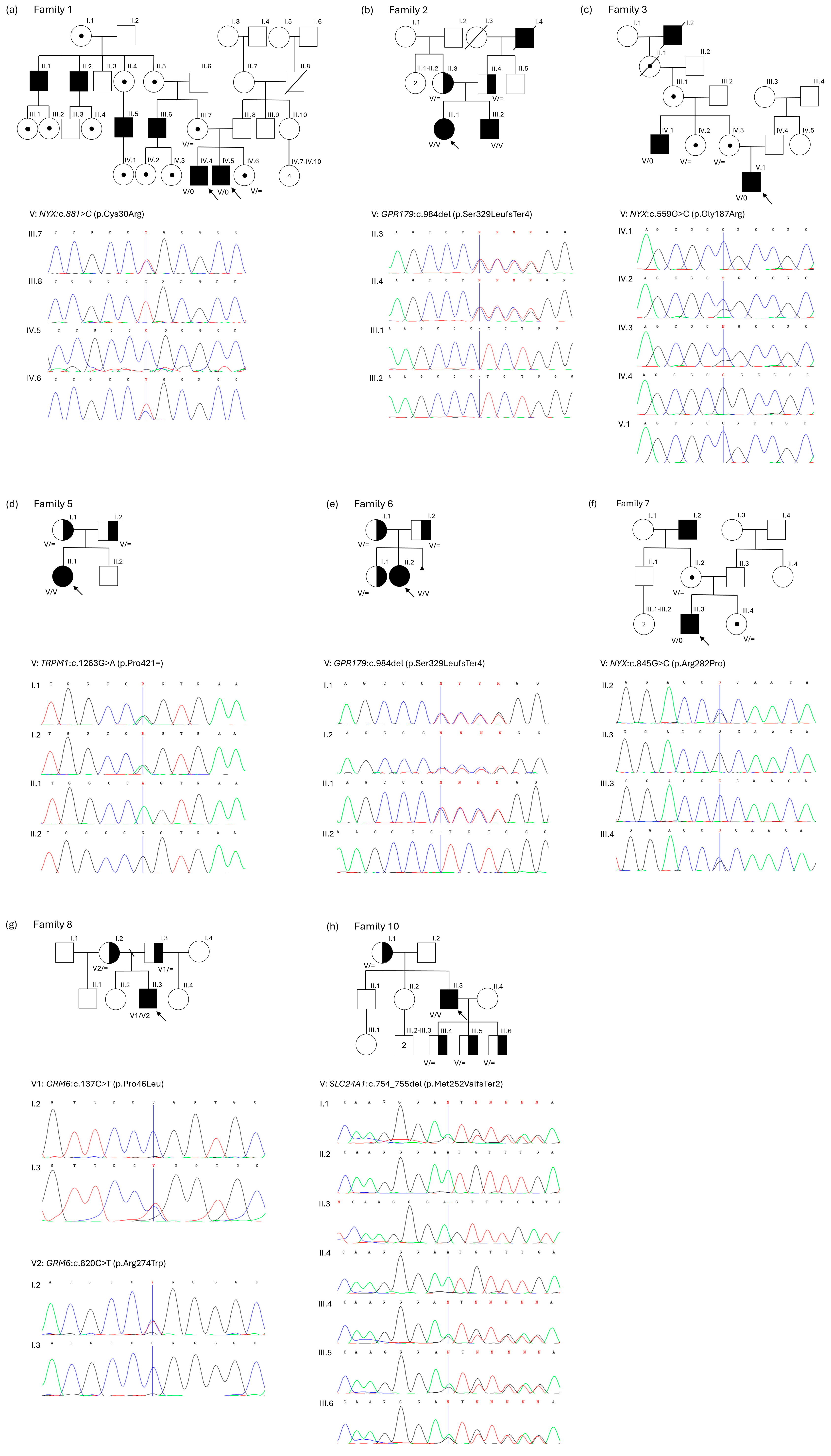
Subsequently, invitations to participate in the segregation analysis of the identified variants were sent to 21 probands’ families. Among those invited, eight families agreed to participate in the study. In these families, pedigree analyses were carried out alongside molecular characterization of the detected variants. The outcomes of these investigations are summarized in Figure 1.

3. Discussion

Congenital stationary night blindness comprises a group of disorders characterized by marked heterogeneity in inheritance patterns, causative genes, and clinical and electrophysiological manifestations. Establishing a precise diagnosis based exclusively on a standard ophthalmological examination, in the absence of a family history of the disease, is frequently exceedingly challenging. Recent advances in high-throughput sequencing technologies have enabled accurate and highly precise diagnosis of CSNB.
NGS analyses enabled unequivocal confirmation of the CSNB type in 21 examined patients. The complete form of the Schubert–Bornschein type (cCSNB) was identified in 15 patients (P1, P7, P10, P15, P20, P23, P27, P31, P46, P47, P49, P50, P51, P52, P55), incomplete form (iCSNB) was identified in 3 patients (P36, P54, P57), and the Riggs type was confirmed in two patients (P39 and P48). According to the literature, the incomplete Schubert–Bornschein form is reported to be the most common, which is inconsistent with our findings in this patient’s cohort. In our study, the cCSNB form clearly predominated. The incomplete form is characterized by less pronounced impairment of night vision. In fact, approximately half of the affected patients do not report complaints related to night vision [7]. In our cohort, patients with the iCSNB P36 and P54 did not experience night vision difficulties, whereas patient P57 did. The complete Schubert–Bornschein form is typically associated with night blindness, reduced visual acuity to approximately 0.5, and moderate to high myopia. In the studied group, patients reported symptoms of night blindness, and a marked reduction in visual acuity was observed in the majority of cases. Exceptions were patients P15, P23, and P55, in whom visual acuity remained within the normal range. Strabismus and nystagmus are also frequently observed in patients with cCSNB type, with nystagmus tending to diminish with age [4]. Among the 15 patients included in the study with cCSNB, strabismus was identified in 5 cases (P7, P20, P27, P47, P49) and nystagmus in 6 cases (P7, P10, P27, P31, P47, P50); both conditions co-occurred in 3 patients (P7, P27, and P47). In the Riggs form, night blindness is observed, whereas visual acuity is typically unimpaired [9]. Both patients, P39 and P48, reported difficulties with night vision. In patient P48, visual acuity was close to normal, whereas patient P39 exhibited a markedly reduced visual acuity (Table 1).
The highest prevalence was observed for GPR179 and NYX (six families each), whereas alterations in CACNA1F were identified in three families, and alterations in GRM6, TRPM1, and SLC24A1 were identified in two families each.
The most frequently identified variant in our cohort was p.Ser329LeufsTer4 in the GPR179 gene associated with the autosomal recessive complete form of congenital stationary night blindness (Schubert–Bornschein type; CSNB1E). This change has been found in all six GPR179-affected individuals. Four out of six patients were homozygous, and two were compound heterozygotes for this gene alteration. This variant in a homozygous state has also been found in one out of three CSNB Polish patients described by Durajczyk et al. [6], indicating that the frequency of p.Ser329LeufsTer4 change might be relatively higher in the Polish population as compared to the general population. These findings are within the range of allele frequency in the Polgenom database estimated at 0.4%, and the allele frequency described in the general population in the GnomAD database, reported as 0.03493%. However, large studies described by AItalbishi et al., including 161 CSNB patients from 76 families, showed that defects in the TRPM1 gene are the most identified in the Palestinian and Israeli populations with autosomal recessive CSNB [10].
Similar results have been reported by Sundaramurthy et al., who identified the TRPM1 gene as the most prevalent gene alteration in the Indian cohort [11]. The TRPM1 gene was also the most frequent cause of CSNB in the article described by Huang et al. [4]. TRPM1 variants have also been identified in two out of six CSNB families reported by Kim et al. [2]. Additionally, genetic analysis in a cohort of CSNB patients from Saudi Arabia showed the recessive variants in TRPM1 and CABP4 accounted for the majority of affected individuals. These findings might be associated with the high frequency of consanguinity in the populations analyzed [1].
Interestingly, recent large studies reported by Katta et al. performed in 122 CSNB individuals (107 CSNB families) revealed the presence of variants in 50 families in the CACNA1F gene, NYX variants in 22 families, TRPM1 defects in 19 families, GRM6 changes in 13 families, GPR179 in 2 families, and only 1 patient has been found to have the CABP4 gene variant [7]. Other reports also indicate the frequent involvement of CACNA1F, NYX, and GRM6 genes in the pathogenesis of CSNB [4,11].
Another variant, p.Met252ValfsTer2 in SLC24A1, was homozygous in two families with CSNB in our cohort. The frequency of this alteration in the general population, based on GnomAD, is estimated at 0.0176% and in the Polgenom database at 0.79%, suggesting that this change is more common in the Polish population.
In the present study, we identified 10 previously unreported variants associated with CSNB in three genes: CACNA1F, GRM6, and NYX, each encoding a protein essential for signal transmission from photoreceptors to bipolar cells in the retina. These findings expand the known mutational spectrum of CSNB and reinforce its genetic heterogeneity.
Three novel variants were detected in CACNA1F. Two of these, c.4588+1G>A and c.3942+1G>C, are located at the donor splice site of their respective introns, disrupting the canonical splice donor consensus sequence. According to ACMG criteria, variants at this position constitute very strong evidence of pathogenicity. Although splice-site mutations in CACNA1F have been previously reported, these specific variants have not been described in association with CSNB. The third CACNA1F variant, c.1018C>T (p.Gln340Ter), is a nonsense substitution that converts a glutamine codon to a premature termination codon at position 340 of the Cav1.4 protein (voltage-gated calcium channel). The p.Gln340Ter variant truncates the protein within the first repeat domain (domain I), eliminating the vast majority of the transmembrane segments (S1–S6 of domains II–IV), the voltage-sensing apparatus, the pore-forming regions, and the extensive C-terminal regulatory domain [12,13,14].
Two previously unreported variants were identified in GRM6. The first, c.152_174dup (p.Gln59AlafsTer10), is a duplication within the N-terminal extracellular ligand-binding domain that results in a frameshift and premature termination after ten aberrant amino acids. This region is critical for glutamate binding, and disease-associated mutations in the ligand-binding domain have been shown to abolish proper protein trafficking to the cell surface, with mutant mGluR6 being retained in the endoplasmic reticulum [12,15]. This variant is cataloged in the gnomAD population database and has been assigned an rs identifier, indicating that it has been observed in the general population. However, to date, c.152_174dup has not been reported in association with CSNB or any other retinal disorder in the published literature. Its presence in gnomAD does not preclude pathogenicity, as autosomal recessive CSNB requires biallelic mutations, and heterozygous carriers are expected to be phenotypically normal. The second GRM6 variant, c.445_453del (p.Val149_Ala151del), is an in-frame deletion removing three amino acids within the ligand-binding domain. The variant also has an rs id but has not been associated with CSNB so far. Although such deletions are less frequently reported than frameshift or missense variants, removal of residues from the glutamate-binding region is likely to alter receptor conformation and impair ligand binding or protein folding. Previous studies have demonstrated that even single amino acid changes in the ligand-binding domain can abolish mGluR6 surface localization [15].
Finally, five novel missense variants were identified in the NYX gene: c.559G>C (p.Gly187Arg), c.845G>C (p.Arg282Pro), c.565A>T (p.Ile189Phe), c.788G>T (p.Gly263Val), and c.446T>C (p.Leu149Pro). All identified variants are located within the leucine-rich repeat (LRR) region of the protein. The LRR domain, situated in the extracellular space, forms a curved solenoid structure that mediates key protein–protein interactions and contributes to the assembly of a macromolecular signaling complex required for ON-bipolar cell function. In particular, this domain enables direct interactions with TRPM1 and mGluR6, which are essential components of the ON-bipolar signaling pathway [16,17,18]. Variants affecting conserved residues within the LRR region are therefore likely to disrupt the domain’s structural integrity and impair its interactions with downstream signaling partners.
Collectively, these findings expand the allelic spectrum of CSNB and support the need for comprehensive genetic screening of all known CSNB-associated genes to enable accurate molecular diagnosis. However, due to cohort size and limited access for segregation analysis in families described (eight out of twenty-one) further investigations and observations are necessary to increase our knowledge concerning CSNB. Only in this way the visual prognosis, mode of inheritance, and optimal management can be unequivocally determined.

4. Materials and Methods

4.1. Clinical Examination

Twenty-one patients affected with CSNB and twenty-nine family members were recruited from twenty-one families. Both the patients and all family members were of Polish origin. In the study group, eight cases were familial and thirteen sporadic.
The patients were previously referred for ophthalmologic examination, including measurement of central visual acuity (18 patients), full-field electroretinography (ERG; 18 patients), and optical coherence tomography (OCT; 15 patients). Patients were provided with genetic counseling in conjunction with a comprehensive medical and family history analysis.
The study was conducted in accordance with the ethical principles outlined in the Declaration of Helsinki and its subsequent amendments and was approved by the local Bioethics Committee at the Poznan University of Medical Sciences (approval no. 485/24). Written informed consent was obtained from the patients and, for patients under 18 years, from their parents.

4.2. Molecular Analysis

Genomic DNA used for molecular NGS testing was extracted from peripheral blood samples collected from probands. Blood was drawn using a vacuum collection system into EDTA-containing tubes, and DNA was subsequently isolated from peripheral blood leukocytes with the MagCore® HF16 Automated Nucleic Acid Extractor and the Genomic DNA Large Volume Whole Blood Kit (RBC Bioscience Corp., New Taipei, Taiwan).
Molecular testing was conducted in each proband using next-generation sequencing (NGS) with a targeted gene panel for inherited ocular disorders. In one case, whole-exome sequencing (WES) was additionally performed. NGS analyses were outsourced to external laboratories, including Genomed (Warsaw, Poland) and Asper Biogene (Tartu, Estonia). The technical specifications of these procedures are summarized and included in the Supplementary Materials (Table S1). Bioinformatic analyses were performed according to the protocol described by Matczynska et al. [19]. Molecular analyses were performed between 2017 and 2025, during which the gene panel underwent several modifications, leading to differences in certain parameters. The NGS gene panels differed mainly in the total number of analyzed genes, reflecting the ongoing development and expansion of diagnostic panels over time. However, all genes known to be associated with congenital stationary night blindness (CSNB) were included in each version of the panel; therefore, these modifications did not affect the detection of variants in established CSNB-related genes. Due to the large number of analyzed genes and the evolving composition of the panels, detailed gene lists are available from the authors upon reasonable request.
The decision to use diagnostic whole-exome sequencing (WES) in one patient was based on the patient’s young age (2 years at the time of testing), progressive myopia, and the presence of additional clinical features, including mild developmental delay and a cavernous hemangioma involving the left side of the face. Owing to the patient’s young age, several ophthalmological procedures could not be performed, which precluded a complete ophthalmic evaluation. WES analysis was conducted according to the routine diagnostic pipeline, with particular emphasis placed on genes included in ophthalmic gene panels as well as genes selected on the basis of the extraocular manifestations, thereby enabling a broader diagnostic assessment.
Validation of identified variants in probands and segregation analyses in selected families was performed using Sanger sequencing of PCR-amplified products. For these procedures, genomic DNA was obtained from buccal swabs following the manufacturer’s protocol (NucleoSpin Tissue Kit; Macherey-Nagel, Düren, Germany). PCR primers were designed with Primer3Plus software (version 3.3.0, available online). Primer sequences are available upon request. For variant classification according to the American College of Medical Genetics (ACMG) guidelines, three online tools were used: Varsome (varsome.com; version 13.15.1.0; access date: 16 April 2026) [20], Franklin by Genoos (franklin.genoox.com; version 93; access date: 16 April 2026), and GeneBe (genebe.net; beta version; access date: 16 April 2026) [21].

Supplementary Materials

The following supporting information can be downloaded at: https://www.mdpi.com/article/10.3390/ijms27114855/s1.

Author Contributions

Conceptualization, L.K.; methodology, J.W.-S. and A.W.; validation, J.W.-S., A.S.-W. and L.K.; investigation, L.K., A.W. and J.W.-S.; resources, L.K.; writing—original draft preparation, L.K., A.W. and J.W.-S.; writing—review and editing, M.R.K.; visualization, L.K.; supervision, M.R.K.; project administration, A.W. and J.W.-S. All authors have read and agreed to the published version of the manuscript.

Funding

This research received no external funding.

Institutional Review Board Statement

The study was conducted in accordance with the Declaration of Helsinki and approved by the local Bioethics Committee at the Poznan University of Medical Sciences (approval no. 485/24, approved on 27 June 2024).

Informed Consent Statement

Informed consent was obtained from all subjects involved in the study.

Data Availability Statement

The original contributions presented in this study are included in the article/Supplementary Material. Further inquiries can be directed to the corresponding author.

Conflicts of Interest

Author Maciej R. Krawczynski was employed by the company GENESIS Diagnostics, Poznan, Poland, with which the Poznan University of Medical Sciences has scientific cooperation. The remaining authors declare that the research was conducted in the absence of any commercial or financial relationships that could be construed as a potential conflict of interest.

References

  1. Almutairi, F.; Almeshari, N.; Ahmad, K.; Magliyah, M.S.; Schatz, P. Congenital stationary night blindness: An update and review of the disease spectrum in Saudi Arabia. Acta Ophthalmol. 2021, 99, 581–591. [Google Scholar] [CrossRef] [PubMed]
  2. Kim, A.H.; Liu, P.K.; Chang, Y.H.; Kang, E.Y.; Wang, H.H.; Chen, N.; Tseng, Y.J.; Seo, G.H.; Lee, H.; Liu, L.; et al. Congenital Stationary Night Blindness: Clinical and Genetic Features. Int. J. Mol. Sci. 2022, 23, 14965. [Google Scholar] [CrossRef] [PubMed]
  3. Zhang, Y.; Lin, S.; Yu, L.; Lin, X.; Qu, S.; Ye, Q.; Yu, M.; Chen, W.; Wu, W. Gene therapy shines light on congenital stationary night blindness for future cures. J. Transl. Med. 2025, 23, 392. [Google Scholar] [CrossRef] [PubMed]
  4. Huang, L.; Bai, X.; Xie, Y.; Zhou, Y.; Wu, J.; Li, N. Clinical and genetic studies for a cohort of patients with congenital stationary night blindness. Orphanet J. Rare Dis. 2024, 19, 101. [Google Scholar] [CrossRef]
  5. Krawczynski, M.R.; Niedziela-Schwartz, Z.; Skorczyk-Werner, A.; Wawrocka, A. Oftalmogenetyka, 1st ed.; PZWL Wydawnictwo Lekarskie: Warszawa, Polska, 2023; p. 540. [Google Scholar]
  6. Durajczyk, M.; Lubiński, W. Congenital Stationary Night Blindness (CSNB)-Case Reports and Review of Current Knowledge. J. Clin. Med. 2025, 14, 1238. [Google Scholar] [CrossRef] [PubMed]
  7. Katta, M.; de Guimaraes, T.A.C.; Fujinami-Yokokawa, Y.; Fujinami, K.; Georgiou, M.; Mahroo, O.A.; Webster, A.R.; Michaelides, M. Congenital Stationary Night Blindness: Structure, Function and Genotype-Phenotype Correlations in a Cohort of 122 Patients. Ophthalmol. Retin. 2024, 8, 932–941. [Google Scholar] [CrossRef] [PubMed]
  8. Georgiou, M.; Fujinami, K.; Michaelides, M. Inherited retinal diseases: Therapeutics, clinical trials and end points—A review. Clin. Exp. Ophthalmol. 2021, 49, 270–288. [Google Scholar] [CrossRef] [PubMed]
  9. Zeitz, C.; Robson, A.G.; Audo, I. Congenital stationary night blindness: An analysis and update of genotype-phenotype correlations and pathogenic mechanisms. Prog. Retin. Eye Res. 2015, 45, 58–110. [Google Scholar] [CrossRef] [PubMed]
  10. AlTalbishi, A.; Zelinger, L.; Zeitz, C.; Hendler, K.; Namburi, P.; Audo, I.; Sheffer, R.; Yahalom, C.; Khateb, S.; Banin, E.; et al. TRPM1 Mutations are the Most Common Cause of Autosomal Recessive Congenital Stationary Night Blindness (CSNB) in the Palestinian and Israeli Populations. Sci. Rep. 2019, 9, 12047. [Google Scholar] [CrossRef] [PubMed]
  11. Sundaramurthy, S.; Malaichamy, S.; Sen, P.; Sachidanandam, R.; Audo, I.; Zeitz, C.; Sarangapani, S.; Soumittra, N. Genetic analysis of congenital stationary night blindness and Oguchi disease in an Indian cohort. Acta Ophthalmol. 2025, 103, e496–e510. [Google Scholar] [CrossRef] [PubMed]
  12. Consortium, U. UniProt: The Universal Protein Knowledgebase in 2025. Nucleic Acids Res. 2025, 53, D609–D617. [Google Scholar] [CrossRef] [PubMed]
  13. Catterall, W.A.; Perez-Reyes, E.; Snutch, T.P.; Striessnig, J. International Union of Pharmacology. XLVIII. Nomenclature and structure-function relationships of voltage-gated calcium channels. Pharmacol. Rev. 2005, 57, 411–425. [Google Scholar] [CrossRef] [PubMed]
  14. Catterall, W.A.; Lenaeus, M.J.; Gamal El-Din, T.M. Structure and Pharmacology of Voltage-Gated Sodium and Calcium Channels. Annu. Rev. Pharmacol. Toxicol. 2020, 60, 133–154. [Google Scholar] [CrossRef] [PubMed]
  15. Zeitz, C.; Forster, U.; Neidhardt, J.; Feil, S.; Kälin, S.; Leifert, D.; Flor, P.J.; Berger, W. Night blindness-associated mutations in the ligand-binding, cysteine-rich, and intracellular domains of the metabotropic glutamate receptor 6 abolish protein trafficking. Hum. Mutat. 2007, 28, 771–780. [Google Scholar] [CrossRef] [PubMed]
  16. Bojang, P.; Gregg, R.G. Topological analysis of small leucine-rich repeat proteoglycan nyctalopin. PLoS ONE 2012, 7, e33137. [Google Scholar] [CrossRef] [PubMed]
  17. Pearring, J.N.; Bojang, P.; Shen, Y.; Koike, C.; Furukawa, T.; Nawy, S.; Gregg, R.G. A role for nyctalopin, a small leucine-rich repeat protein, in localizing the TRP melastatin 1 channel to retinal depolarizing bipolar cell dendrites. J. Neurosci. 2011, 31, 10060–10066. [Google Scholar] [CrossRef] [PubMed]
  18. Cao, Y.; Posokhova, E.; Martemyanov, K.A. TRPM1 forms complexes with nyctalopin in vivo and accumulates in postsynaptic compartment of ON-bipolar neurons in mGluR6-dependent manner. J. Neurosci. 2011, 31, 11521–11526. [Google Scholar] [CrossRef] [PubMed]
  19. Matczyńska, E.; Beć-Gajowniczek, M.; Sivitskaya, L.; Gregorczyk, E.; Łyszkiewicz, P.; Szymańczak, R.; Jędrzejowska, M.; Wylęgała, E.; Krawczyński, M.R.; Teper, S.; et al. Optimised, Broad NGS Panel for Inherited Eye Diseases to Diagnose 1000 Patients in Poland. Biomedicines 2024, 12, 1355. [Google Scholar] [CrossRef] [PubMed]
  20. Kopanos, C.; Tsiolkas, V.; Kouris, A.; Chapple, C.E.; Albarca Aguilera, M.; Meyer, R.; Massouras, A. VarSome: The human genomic variant search engine. Bioinformatics 2019, 35, 1978–1980. [Google Scholar] [CrossRef] [PubMed]
  21. Stawiński, P.; Płoski, R. Genebe.net: Implementation and validation of an automatic ACMG variant pathogenicity criteria assignment. Clin. Genet. 2024, 106, 119–126. [Google Scholar] [CrossRef]
Figure 1. The pedigrees and segregation of the variants associated with the disease in patients’ families. (a)—family 1; (b)—family 2; (c)—family 3; (d)—family 5; (e)—family 6; (f)—family 7; (g)—family 8; (h)—family 10. The identified variants are shown below the pedigrees. Symbols used in pedigrees: square—male; circle—female; empty symbol—unaffected individual; filled/black symbol—affected by the trait or disease; half-shaded symbol—carrier (for recessive traits); dot inside a circle—female carrier of an X-linked trait; diagonal slash through symbol—deceased; the arrow indicates the proband; small triangle—miscarriage; diagonal slash through partner line—divorce; generations are labeled with Roman numerals and individuals within a generation are numbered left to right; V/V—homozygote; V/=—heterozygote; V/0—hemizygote.
Ijms 27 04855 g001
Table 1. Clinical symptoms of the patients with CSNB.
Family/Patient IDCurrent Age/GenderCSNB TypeOphthalmic Symptoms
Night Blindness Family HistoryERG ResultsMyopiaBCVA
RE/LE
(VIS?)
VF RestrictionOCTAdditional Ophthalmological Symptoms
F1/P120/MCSNB1A++Scotopic—extinguished
Photopic—normal
−15.0D0.4–0.5Irregular visual field defectsThinning of the retina-
F2/P713/FCSNB1E++Scotopic—decreased
Photopic—reduced amplitudes
−7.0D
+astigmatism
0.3–0.25NDThinning of the retinaBilateral strabismus that resolved spontaneously; color vision impairment; mild nystagmus
F3/P103/MCSNB1A++Scotopic—extinguished
Photopic—normal
−3.0D
+astigmatism (−1.5D)
NDNDNDHorizontal nystagmus
F4/P157/FCSNB1B+Scotopic—extinguished−9.0D
+astigmatism
0.9–1.0NDNormal-
F5/P206/FCSNB1C+Scotopic—extinguished
Photopic—reduced amplitudes
−7.0D0.3–0.4NDNormalOblique strabismus
F6/P2317/FCSNB1E+Scotopic—extinguishedMyopic astigmatism1.0NDND-
F7/P279/MCSNB1A++Scotopic—extinguished−8.0D0.4NDNormalNystagmus, strabismus
F8/P319/MCSNB1B+Scotopic—extinguished
Photopic—reduced amplitudes
from −3.0D to
−4.0D
+ myopic astigmatism
0.2–0.3Narrowing of the visual fieldThinning of the retinaInfantile nystagmus
F9/P3610/MCSNB2AScotopic—extinguished−5.5D
+myopic astigmatism
(−2.5D)
0.5-Normal-
F10/P3939/MCSNB1D+NDMixed astigmatism0.2–0.3Narrowing of the visual field to approx. 15 degreesPerifoveal photoreceptor atrophy-
F11/P463/MCSNB1ENDND−5.0DNDNDThinning of the retina-
F12/P479/MCSNB1A++Scotopic—extinguished
Photopic—normal
Myopic astigmatism0.8–0.25NDNormalMild nystagmus and divergent strabismus
F13/P4847/MCSNB1D++Scotopic—decreased
Photopic—decreased
ND0.7–1.0Narrowing of the visual field to approx. 20 degreesPerifoveal photoreceptor atrophy
RE—normal macula, LE—lamellar macular hole
-
F14/P4916/MCSNB1A+Scotopic—extinguished
Photopic—decreased
−6.5D0.6–0.8NDNormalConvergent strabismus
F15/P508/MCSNB1ENDScotopic—extinguished
Photopic—within the lower limits of normal
Myopic astigmatism0.6–0.8NDNDHorizontal nystagmus
F16/P5151/MCSNB1C+Scotopic—decreased
Photopic—normal
Myopic astigmatism0.2–0.8Irregular visual field defectsPhotoreceptor atrophy-
F17/P5214/MCSNB1E+-Scotopic—extinguished
Photopic—reduced amplitudes
Slight myopia0.6–0.8NDNormal-
F18/P5411/MCSNB2A+Scotopic—extinguished with the b-wave decreased
Photopic -with the b-wave decreased
Abnormal oscillatory potentials
−9.0D
+ myopic astigmatism
(−2.0D)
0.4–0.3NDNormal-
F19/P5550/FCSNB1E+Scotopic—decreased
Photopic—disturbed morphology without electronegative recording
ND1.0Irregular visual field defectsBorderline of normal-
F20/P567/MCSNB1A++Scotopic—extinguished
Photopic—reduced
Myopia 0.6–0.7Normal ND-
F21/P577/MCSNB2A+Scotopic—extinguished
Photopic—reduced amplitudes
Myopic astigmatism0.4–0.5-Abnormal profile of the fovea fundus-
BCVA—best-corrected visual acuity; RE—right eye; LE—left eye; M—male; F—female; VF- visual field; ND—no data; +present, −absent.
Table 2. DNA variants identified by NGS in patients.
Patient/FamilyGeneTranscriptVariant ClassificationInheritanceACMG ClassificationClinVarMolecular Method of Searching the VariantsGnomAD v4.1.0
Total Allele Frequency
NucleotideProteinVarsomeFranklinGeneBe
F9/P36CACNA1FNM_001256789.3c.4588+1G>ASplice donor variantXLRLPLPPNot reportedNGS panel-
F18/P54CACNA1FNM_001256789.3c.3942+1G>CSplice donor variantXLRLPLPVUSNot reportedNGS panel-
F21/P57CACNA1FNM_001256789.3c.1018C>Tp.Gln340TerXLRLPLPPNot reportedNGS panel-
F2/P7GPR179NM_001004334.4c.984del
rs770066665
p.Ser329LeufsTer4ARPPPP/LPNGS panel0.0003493
F6/P23GPR179NM_001004334.4c.984del
rs770066665
p.Ser329LeufsTer4ARPPPP/LPNGS panel0.0003493
F11/P46GPR179NM_001004334.4c.984del
rs770066665
p.Ser329LeufsTer4ARPPPP/LPWES0.0003493
F15/P50GPR179NM_001004334.4c.984del
rs770066665
c.1368del
rs1435030978
p.Ser329LeufsTer4

p.Phe456LeufsTer30
ARP

P
P

P
P

P
P/LP

P
NGS panel

NGS panel
0.0003493

0.00001921
F17/P52GPR179NM_001004334.4c.984del
rs770066665
c.1141C>T
rs749683775
p.Ser329LeufsTer4

p.Arg381Trp
ARP

VUS
P

VUS
P

VUS
P/LP

VUS
NGS panel

NGS panel
0.0003493

0.00001555
F19/P55GPR179NM_001004334.4c.984del
rs770066665
p.Ser329LeufsTer4ARPPPP/LPNGS panel0.0003493
F4/P15GRM6NM_000843.4c.152_174dup
rs1760743459
c.445_453del
rs2480407301
p.Gln59AlafsTer10

p.Val149_Ala151del
ARP

VUS
P

VUS
P

VUS
P

Not reported
NGS panel

NGS panel
0.000001466

0.000001373
F8/P31GRM6NM_000843.4c.137C>T
rs62638197
c.820C>T
rs577125911
p.Pro46Leu

p.Arg274Trp
ARLP

VUS
LP

VUS
P

VUS
LP

VUS
NGS panel

NGS panel
0.0001538

0.000008676
F1/P1NYXNM_001378477.3c.88T>C
rs1292184180
p.Cys30ArgXLRVUSVUSLPVUSNGS panel0.000
F3/P10NYXNM_001378477.3c.559G>Cp.Gly187ArgXLRVUSVUSVUSNot reportedNGS panel-
F7/P27NYXNM_001378477.3c.845G>Cp.Arg282ProXLRVUSVUSLPNot reportedNGS panel-
F12/P47NYXNM_001378477.3c.565A>Tp.Ile189PheXLRVUSVUSVUSNot reportedNGS panel-
F14/P49NYXNM_022567.2c.788G>Tp.Gly263ValXLRVUSVUSVUSNot reportedNGS panel-
F20/P56NYXNM_001378477.3c.446T>Cp.Leu149ProXLRVUSVUSVUSNot reportedNGS panel-
F10/P39SLC24A1NM_004727.3c.754_755del
rs777989874
p.Met252ValfsTer2ARPPPP/LPNGS panel0.0001760
F13/P48SLC24A1NM_004727.3c.754_755del
rs777989874
p.Met252ValfsTer2ARPPPP/LPNGS panel0.0001760
F5/P20TRPM1NM_001252024.2c.1263G>A
rs768701595
p.Pro421=ARLPLPVUSConflicting classificationsNGS panel0.000008056
F16/P51TRPM1NM_001252020.1c.332A>G
rs200514769
c.3397C>T
rs779371614
p.Tyr111Cys

p.Arg1133 *
ARLP

LP
P

LP
VUS

P
Conflicting classifications
Not reported
NGS panel

NGS panel
0.0001887

0.000004008
XLR—X-linked recessive, AR—autosomal recessive, LP—likely pathogenic, P—pathogenic, VUS—uncertain significance, NGS—next generation sequencing, WES—whole-exome sequencing. American College of Medical Genetics (ACMG) classification was obtained through the Varsome, Franklin, and GeneBe online available tools. New variants identified in this study are bolded. * stop codon.
Disclaimer/Publisher’s Note: The statements, opinions and data contained in all publications are solely those of the individual author(s) and contributor(s) and not of MDPI and/or the editor(s). MDPI and/or the editor(s) disclaim responsibility for any injury to people or property resulting from any ideas, methods, instructions or products referred to in the content.