Review

Sulfur-Containing Amino Acids: The Conversion Process from Product to Substrate

1
College of Life Sciences, China Jiliang University, Hangzhou 310018, China
2
Zhejiang Provincial Engineering Research Center of New Technologies and Applications for Targeted Therapy of Major Diseases, Zhejiang Provincial Key Laboratory of Drug Discovery and Development for Metabolic Diseases, College of Life Sciences and Medicine, Zhejiang Sci-Tech University, Hangzhou 310018, China
*
Author to whom correspondence should be addressed.
Int. J. Mol. Sci. 2026, 27(11), 4771; https://doi.org/10.3390/ijms27114771
Submission received: 8 April 2026 / Revised: 10 May 2026 / Accepted: 15 May 2026 / Published: 26 May 2026
(This article belongs to the Section Molecular Biology)

Abstract

Methionine and cysteine are the principal sulfur-containing proteinogenic amino acids, playing pivotal roles in protein structure, function, and cellular metabolic regulation. The biosynthetic machinery of these amino acids is intricate and exhibits distinct evolutionary divergence across species. This review comprehensively summarizes the biosynthesis of cysteine and methionine in model organisms ranging from Escherichia coli and yeast to plants and Homo sapiens. Specifically, we examine the metabolic interconversion and the transition of roles between these two amino acids during de novo synthesis. Furthermore, we dissect the physiological significance of the transsulfuration pathway in mammals, which utilizes methionine as a precursor for cysteine biosynthesis.

1. Introduction

Among the classical amino acids that constitute proteins, cysteine and methionine hold a unique status as the sole sulfur-containing members, acting as critical sinks for reactive species due to their specific atomic composition [1]. The remarkable biological versatility of cysteine stems directly from the chemical properties of its thiol group, which is highly nucleophilic and redox-active, allowing it to function as an effective molecular switch in response to cellular stimuli [2]. This reactivity is fundamental to its direct participation in the active sites of numerous enzymes, such as cysteine cathepsins, where it performs vital catalytic functions ranging from protein turnover to antigen presentation [3]. Furthermore, the ability of cysteine residues to form covalent disulfide bonds is crucial for maintaining the structural integrity of proteins, a feature that can be mapped with high precision to understand protein stability [4]. Cysteine is also a primary target for a host of post-translational modifications, including S-glutathionylation, a key mechanism for regulating protein function and achieving antioxidant defense through redox signaling [5]. Beyond its role in protein architecture, cysteine serves as the rate-limiting precursor for the synthesis of glutathione (GSH), and its availability is tightly linked to cell death pathways like ferroptosis when regulation fails [6].
Concurrently, methionine fulfills two equally indispensable roles, primarily serving as the conserved initiating amino acid for the synthesis of nearly all eukaryotic proteins, mediated by specific quality control mechanisms [7]. In the realm of cellular metabolism, its more profound function is as the exclusive precursor to S-adenosylmethionine (SAM), a vital metabolite that links nutritional status to autophagy and cell growth [8]. SAM acts as the universal methyl donor, participating in the enzymatic methylation of a wide array of biological substrates, including DNA and histones, thereby playing a central role in the epigenetic regulation of gene expression [9]. Moreover, SAM is a critical metabolic node, providing aminopropyl groups for polyamine synthesis and acting as an allosteric regulator to coordinate sulfur flux through the transsulfuration pathway [10].
The profound biological importance of cysteine and methionine necessitates robust mechanisms in all organisms to ensure their continuous and regulated supply, often integrating these pathways with carbon and nitrogen metabolism [11]. However, the strategies employed to achieve this metabolic homeostasis reveal a fundamental evolutionary divergence, particularly regarding assimilatory sulfate reduction pathways [12]. Mammals exhibit a pronounced metabolic dependency. Because they lack the capacity for strictly de novo methionine biosynthesis, it is classified as a nutritionally essential amino acid that must be primarily obtained from the diet to maintain post-translational regulatory networks [13].
In mammals, cysteine is considered conditionally essential. This classification arises because, although endogenous synthesis is generally sufficient for healthy adults, it becomes critically inadequate under specific physiological or pathological conditions, such as rapid neonatal growth, severe metabolic stress, or critical illness. In such scenarios, the organism’s metabolic demand significantly outpaces its biosynthetic capacity, making adequate dietary supplementation of cysteine strictly indispensable [14]. Under normal conditions, however, adequate cysteine is maintained via diet or the irreversible transsulfuration pathway, which concurrently generates hydrogen sulfide. This pathway channels the sulfur atom from dietary methionine through the intermediate homocysteine into a serine backbone to generate cysteine. The process is governed by complex enzymatic regulators, most notably cystathionine β-synthase [15].
In stark contrast, plants and many microorganisms display significant metabolic autonomy. They generally retain complex and energetically costly de novo biosynthetic pathways, allowing them to synthesize both cysteine and methionine from simple inorganic sulfur sources [16]. This metabolic dichotomy separating organisms capable of primary sulfur assimilation from those dependent on dietary transsulfuration, represents a key evolutionary divide with far-reaching implications for nutrition, toxicology, and disease pathology.
In this review, we first conduct a comparative analysis of the anabolic processes of cysteine and methionine across diverse model organisms, ranging from E. coli and yeast to plants and humans. Subsequently, we explore the evolutionary plasticity that has shaped these diverse metabolic networks and the intricate mechanisms regulating their function.

2. The Synthesis of Cysteine and Methionine in Escherichia coli

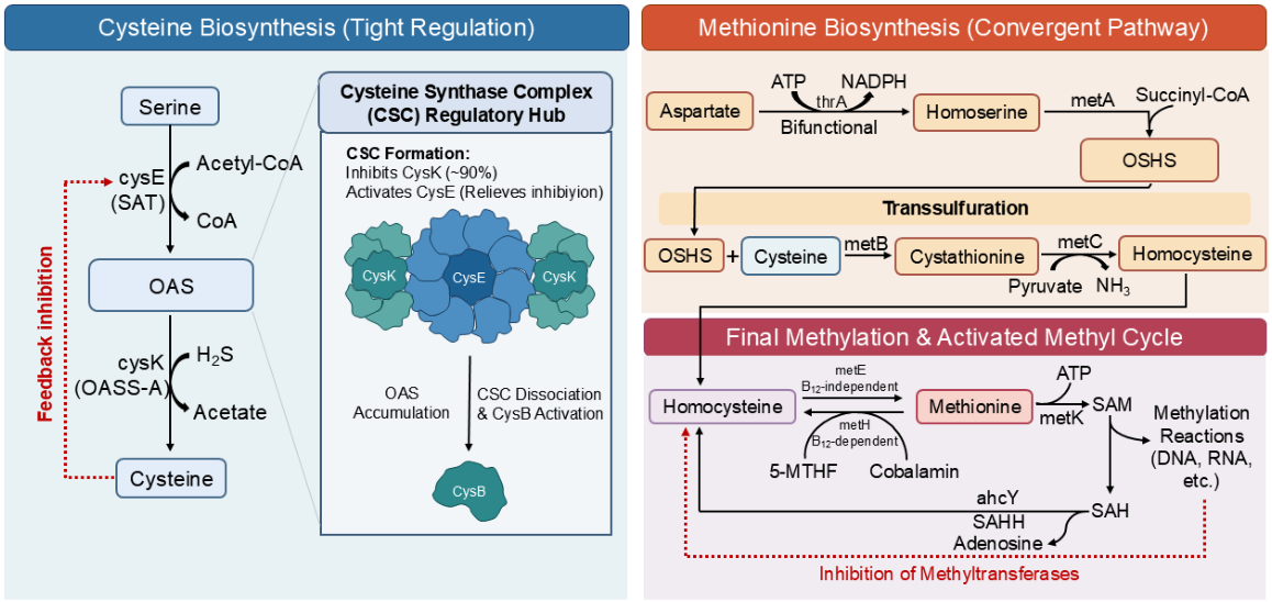
Sulfur-containing amino acids (SCAAs), primarily cysteine and methionine, play indispensable roles in cellular physiology [1]. They are not only fundamental building blocks for protein synthesis but also serve as central players in redox homeostasis, one-carbon metabolism, and the synthesis of numerous biomolecules [1,17]. As a model organism in microbiological research, E. coli provides a classic paradigm for understanding the synthesis and regulatory networks of these critical amino acids [18]. This review will delve into the biosynthetic pathways of cysteine and methionine in E. coli, focusing on the key enzymatic steps, intricate regulatory mechanisms, and the tight interplay between these two metabolic routes (Figure 1).

2.1. De Novo Biosynthesis of Cysteine: A Tightly Regulated Two-Step Pathway

In E. coli, the de novo biosynthesis of cysteine is an efficient and tightly regulated two-step process that converts serine, a product of central carbon metabolism, into the sulfur-containing amino acid cysteine, thereby integrating inorganic sulfur into biomolecules [19].

2.1.1. Initial Step: Acetylation Catalyzed by Serine Acetyltransferase (CysE)

Cysteine biosynthesis begins with the activation of L-serine, a reaction catalyzed by Serine O-acetyltransferase (SAT), the product of the cysE gene [19]. This enzyme utilizes acetyl-coenzyme A (acetyl-CoA) as the acetyl group donor to acetylate the hydroxyl group of L-serine, yielding the intermediate O-acetylserine (OAS) [20]. This reaction serves as the committed and primary regulatory point of the entire pathway, as its activity is subject to potent allosteric feedback inhibition by the final product, L-cysteine [21,22]. This inhibition involves a dual mechanism: cysteine not only acts as a competitive inhibitor with respect to serine by binding to the active site but also induces a conformational change in the enzyme’s C-terminal tail, which physically blocks the binding of acetyl-CoA [23]. This ensures a rapid and complete shutdown of the pathway when cysteine is abundant [21,23]. Such stringent regulation is crucial to prevent the excessive accumulation of intracellular cysteine, as high concentrations can fuel the Fenton reaction, producing cytotoxic hydroxyl radicals that cause oxidative damage to the cell [24].

2.1.2. Final Step: Sulfhydration Catalyzed by Cysteine Synthase (CysK)

The second and final step of the biosynthetic pathway involves the integration of a sulfur atom into the carbon backbone [22]. This reaction is catalyzed by O-acetylserine sulfhydrylase, specifically the isozyme A (OASS-A) encoded by the cysK gene [21]. CysK is a pyridoxal 5′-phosphate (PLP)-dependent enzyme that catalyzes a β-replacement reaction, substituting the acetoxy group of OAS with sulfide (H2S) to ultimately produce L-cysteine and acetate [23].

2.1.3. The Cysteine Synthase Complex (CSC): A Central Regulatory Hub for Sulfur Metabolism

In addition to their independent catalytic functions, CysE and CysK can form a multi-enzyme complex through high-affinity protein–protein interactions, known as the Cysteine Synthase Complex (CSC) [25]. In E. coli, the formation of the CSC is mediated by the insertion of the flexible C-terminal tail of CysE into the active site of CysK [26]. This interaction has profound regulatory effects, primarily activating CysE by relieving its feedback inhibition by L-cysteine and substrate inhibition by L-serine. This ensures a continuous supply of the intermediate OAS. While the complex formation may modulate CysK activity, its primary role in bacteria is often associated with substrate channeling and metabolic efficiency. This sophisticated regulation allows the cell to fine-tune cysteine biosynthesis. When sulfur is scarce, OAS accumulates, acting as a signaling molecule to activate the transcriptional regulator CysB, which upregulates genes for sulfate uptake and reduction [19].

2.2. Biosynthesis of Methionine: A Convergent Pathway Integrating Carbon, Sulfur, and Methyl Sources

In contrast to the metabolic pathway in mammals, the sulfur atom of methionine in E. coli is derived from cysteine, representing a reversal of the flow observed in mammals where methionine serves as the sulfur donor for cysteine [27]. The biosynthesis of methionine is a pathway that converges distinct metabolic precursors, with its carbon skeleton, sulfur atom, and methyl group originating from aspartate, cysteine, and the one-carbon pool, respectively [28].

2.2.1. Source of the Carbon Skeleton: The Aspartate Pathway

The carbon skeleton of methionine is derived from aspartate through a series of reactions in the aspartate family amino acid biosynthetic pathway [29]. In E. coli, the first and third steps in the conversion of aspartate to the key intermediate homoserine are catalyzed by a unique multifunctional enzyme, aspartokinase I/homoserine dehydrogenase I, encoded by the thrA gene [30]. This bifunctional enzyme serves as a critical metabolic control point, its activity is allosterically feedback-inhibited by L-threonine, an end product of the aspartate pathway, thereby precisely controlling the carbon flux into this metabolic network [31]. Homoserine sits at a metabolic branch point and can be converted to either threonine or methionine [32]. The first committed step into the methionine-specific branch is the succinylation of homoserine, catalyzed by homoserine O-succinyltransferase (encoded by the metA gene), to produce O-succinyl-L-homoserine (OSHS) [33]. This step activates the γ-carbon of homoserine, preparing it for the subsequent transfer of the sulfur atom.

2.2.2. Transsulfuration: Transfer of the Sulfur Atom from Cysteine to Homocysteine

The transfer of the sulfur atom from cysteine to the methionine precursor is accomplished through a process known as transsulfuration [34]. This pathway is catalyzed by two sequential PLP-dependent enzymes [35]. First, cystathionine γ-synthase, encoded by the metB gene, catalyzes a γ-replacement reaction between OSHS and L-cysteine to form the thioether intermediate, cystathionine [34]. Subsequently, cystathionine β-lyase, encoded by the metC gene, cleaves cystathionine via an α, β-elimination reaction, releasing homocysteine, the direct precursor of methionine, along with pyruvate and ammonia [36].

2.2.3. Final Methylation: Homocysteine to Methionine

The final step in methionine biosynthesis is the transfer of a methyl group to the thiol group of homocysteine [37]. This methyl group is derived from 5-MTHF [38]. To adapt to varying environmental conditions, E. coli has evolved two functionally distinct but mechanistically related methionine synthases [39]. In the absence of cobalamin (vitamin B12), the cell relies on the cobalamin-independent methionine synthase, encoded by the metE gene [40]. This enzyme transfers the methyl group directly from 5-MTHF to homocysteine, ensuring survival in B12-deficient environments, albeit with relatively low catalytic efficiency [41]. Conversely, when vitamin B12 is available, E. coli preferentially utilizes the cobalamin-dependent methionine synthase, encoded by the metH gene [39]. MetH exhibits a catalytic efficiency approximately 40- to 100-fold higher than that of MetE. By utilizing the cobalamin cofactor as an intermediate methyl carrier, MetH significantly accelerates methionine synthesis, providing a substantial growth advantage in nutrient-rich environments [42]. Recent studies have further shown that E. coli can adapt via evolutionary mutations to enhance the uptake of suboptimal cobalamin analogs, thereby maximizing the high catalytic potential of MetH [43].

2.3. The Activated Methyl Cycle: Methionine Regeneration and Metabolic Regulation

In E. coli, newly synthesized methionine serves not only as a building block for proteins but also as the entry point into a central metabolic loop known as the activated methyl cycle [44]. This cycle is responsible for generating the universal methyl donor for all cellular methylation reactions and maintaining methylation homeostasis [37].

2.3.1. Synthesis and Function of S-Adenosylmethionine (SAM)

One of the primary metabolic functions of methionine is to serve as the precursor for the synthesis of SAM [45]. The synthesis of SAM is catalyzed by S-adenosylmethionine synthetase (MAT), encoded by the metK gene, in a reaction that consumes one molecule of methionine and one molecule of ATP [46]. As the principal methyl donor in the cell, SAM is involved in the methylation of various biological macromolecules, including DNA, RNA, proteins, and lipids [47]. It plays an essential role in key life activities such as the epigenetic regulation of gene expression, ribosome biogenesis, and the synthesis of secondary metabolites [48].

2.3.2. Regulatory Role of the Methylation Byproduct S-Adenosylhomocysteine (SAH)

Upon donating its methyl group, SAM is converted into the byproduct SAH. Far from being inert, SAH acts as a potent competitive inhibitor of most SAM-dependent methyltransferases [49]. The accumulation of SAH competes with SAM for enzyme active sites, thereby significantly impairing cellular methylation capacity [50]. Consequently, the intracellular SAM/SAH ratio is widely regarded as a critical indicator of the cellular methylation potential.

2.3.3. Closing the Cycle: Regeneration of Homocysteine

To maintain the cell’s methylation capacity and recycle the sulfur-containing backbone, SAH must be rapidly removed [51]. This critical step is catalyzed by S-adenosylhomocysteine hydrolase (SAHH), encoded by the ahcY gene [52], which mediates the reversible hydrolysis of SAH into adenosine and homocysteine. The resulting homocysteine reenters the methionine synthesis pathway, where it is re-methylated into methionine by either MetE or MetH, thereby closing the activated methyl cycle. This hydrolysis is crucial for preventing the accumulation of inhibitory SAH and serves as a central element in regulating cellular methylation homeostasis [53]. Furthermore, this cycle exemplifies the principle of metabolic economy, by regenerating homocysteine, it maximizes the utilization of sulfur-containing metabolites and avoids the significant energy expenditure required for the de novo synthesis of methionine [54]. The central importance of this cycle also renders it a vulnerable metabolic hub [55]. For instance, the T3 bacteriophage has evolved a strategy to directly inhibit the host’s MetK enzyme, thereby dismantling the entire methylation system, a finding that highlights the cycle’s critical role in host-pathogen interactions [56].

3. The Synthesis of Cysteine and Methionine in Yeast

Yeasts, particularly Schizosaccharomyces pombe and Saccharomyces cerevisiae, are considered powerful model organisms for studying conserved cellular processes in eukaryotes. These two evolutionarily distant species provide a unique perspective for the comparative analysis of fundamental metabolic pathways, such as the biosynthesis of sulfur-containing amino acids [57]. This article will focus on haploid strains of Schizosaccharomyces pombe and both haploid and diploid states of Saccharomyces cerevisiae to illustrate their distinct metabolic strategies for synthesizing cysteine and methionine [58]. Although both utilize aspartate as a common precursor, they exhibit significant differences in the core mechanisms for sulfur incorporation and interconversion between these critical amino acids, reflecting their respective unique evolutionary trajectories.

3.1. De Novo Synthesis Pathway in Schizosaccharomyces pombe

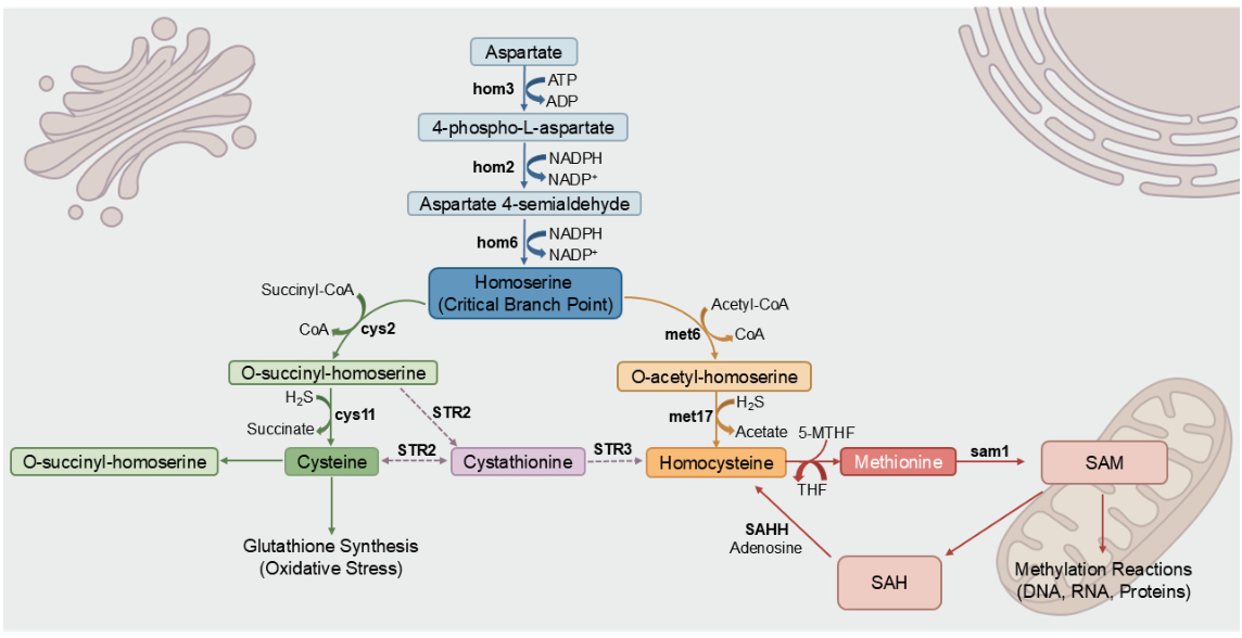
In Schizosaccharomyces pombe, the biosynthesis of cysteine and methionine originates from aspartate and inorganic sulfur, assimilated as H2S, following a pathway characterized by linear and parallel branches (Figure 2). The upstream portion of this pathway represents a conserved metabolic module where aspartate is converted to homoserine through three enzymatic steps. This sequence generates a critical branch-point intermediate for the biosynthesis of both sulfur-containing amino acids and threonine [59]. The subpathway is initiated by aspartate kinase, encoded by the hom3 gene, which utilizes ATP to phosphorylate aspartate into 4-phospho-L-aspartate [60]. Subsequently, aspartate-semialdehyde dehydrogenase, which is encoded by hom2, reduces 4-phospho-L-aspartate to aspartate 4-semialdehyde, using NAD(P)H as a cofactor [59]. Finally, homoserine dehydrogenase, which is encoded by hom6, catalyzes the reduction of aspartate 4-semialdehyde to yield homoserine.
The pathway bifurcates from homoserine into two parallel branches dedicated to the de novo synthesis of cysteine and homocysteine [57]. In the cysteine branch, Cys2-mediated succinylation converts homoserine into O-succinyl-homoserine using succinyl-CoA. Subsequently, Cys11 catalyzes the condensation of O-succinyl-homoserine with hydrogen sulfide to yield cysteine and succinate [61]. Analogously, the methionine branch initiates with the acetylation of homoserine by Met6. The resulting O-acetyl-homoserine is then converted into homocysteine and acetate by Met17 in the presence of hydrogen sulfide [62]. Such a parallel architecture enables the independent regulation of metabolic flux, allowing cells to balance demands for cysteine and methionine [57].
In addition to de novo synthesis from O-acetyl-homoserine, homocysteine can be derived from cystathionine via an alternative route. This pathway initiates with O-succinyl-homoserine and cysteine, mediated by the STR2 and STR3 enzymes [63]. Once synthesized, homocysteine enters the final stage of methionine biosynthesis. This step is catalyzed by 5-methyltetrahydropteroyltriglutamate-homocysteine methyltransferase, encoded by the met26 gene, which transfers a methyl group from 5-MTHF to homocysteine to form methionine [64].
Methionine subsequently plays a pivotal role in the methionine cycle, where it is converted into SAM by S-adenosylmethionine synthase, encoded by the sam1 gene [65]. As the primary methyl donor in the cell, SAM participates in the methylation of diverse biomolecules, including DNA, RNA, and proteins. This process is critical for the regulation of gene expression and cell signaling [66]. Following methyl transfer, SAM is converted to SAH. SAH is subsequently hydrolyzed by SAHH into homocysteine and adenosine, thereby completing the cycle and regenerating the precursor for methionine synthesis [51].

3.2. Homocysteine-Centric Metabolic Hub in Saccharomyces cerevisiae

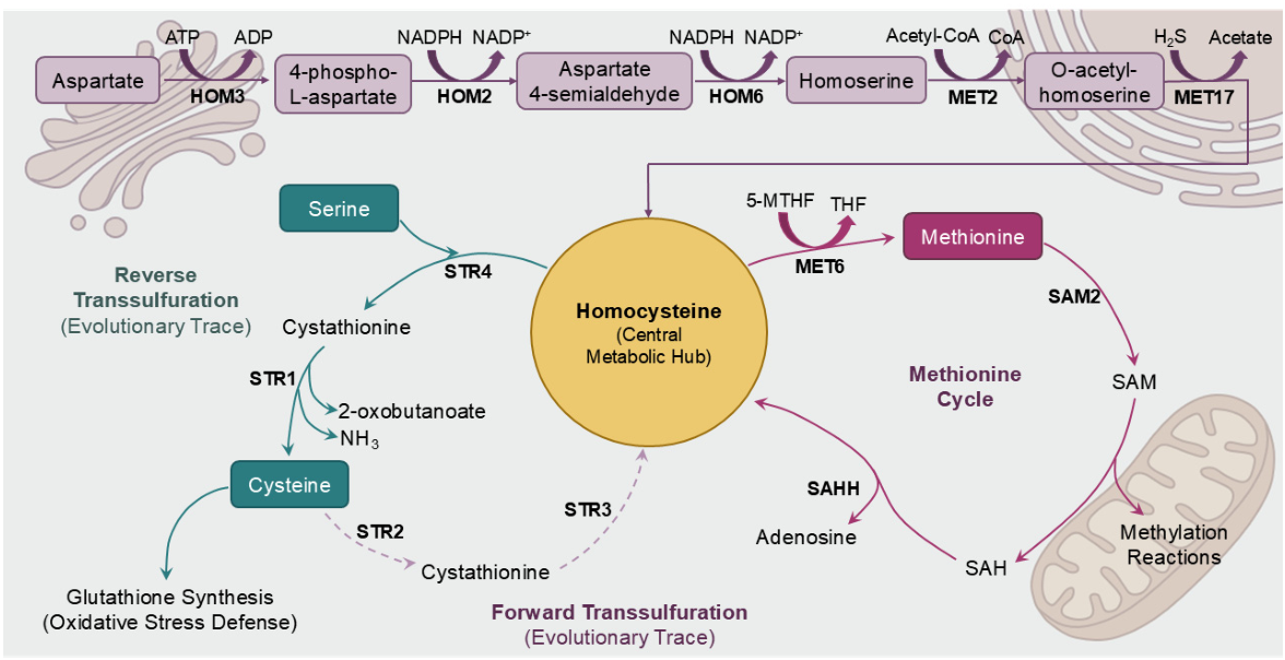
In contrast to S. pombe, the pathway for homocysteine synthesis from aspartate is the key upstream route for the synthesis of both cysteine and methionine in Saccharomyces cerevisiae, making homocysteine a central metabolic hub (Figure 3). Here, the sulfur atom of homocysteine is transferred for cysteine synthesis, while its carbon skeleton is methylated to form methionine [67]. Cysteine synthesis in S. cerevisiae relies exclusively on the so-called reverse transsulfuration pathway [68]. The first step of this pathway is catalyzed by cystathionine β-synthase, encoded by the STR4 gene, which condenses serine and homocysteine to form cystathionine [69]. Subsequently, cystathionine γ-lyase, encoded by the STR1 gene, catalyzes the cleavage of cystathionine to produce cysteine, 2-oxobutanoate, and ammonia [70]. This pathway is critical for maintaining cellular cysteine levels, as cysteine serves as a precursor for glutathione synthesis, which plays a central role in combating oxidative stress [71].
Concurrently, another portion of homocysteine is utilized for methionine synthesis. This process is catalyzed by 5-methyltetrahydropteroyltriglutamate-homocysteine methyltransferase, which is encoded by the MET6 gene. The enzyme transfers a methyl group from 5-MTHF [72]. Metabolic engineering of MET6 and SAM2 can significantly enhance intracellular SAM production, highlighting the importance of this step [73]. Furthermore, Saccharomyces cerevisiae retains the ability to synthesize homocysteine from cysteine via cystathionine γ-synthase and cystathionine β-lyase [74]. In contrast to the cyclic methionine cycle, the conversion between homocysteine and cysteine mediated by STR1, STR2, STR3 and STR4 is not a cyclic process [75]. Instead, the forward transsulfuration pathway catalyzed by STR2 and STR3 does not serve as a primary anabolic route in S. cerevisiae, it resembles an evolutionary remnant, possibly reflecting the organism’s ancestral reliance on this pathway for sulfur metabolism [76].

4. The Synthesis of Cysteine and Methionine in Arabidopsis thaliana

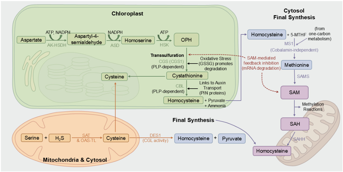
In plants, the de novo synthesis of methionine is fundamentally distinct from the pathway in mammals, as it relies on cysteine to serve as the essential sulfur donor through a process known as the transsulfuration pathway [77]. This metabolic route represents a critical intersection of carbon, nitrogen, and sulfur assimilation, requiring coordinated input from multiple primary metabolic pathways (Figure 4). The two key substrates, O-phosphohomoserine (OPH) and cysteine, are derived from separate biosynthetic origins [78,79]. OPH is synthesized from aspartate within the chloroplast through a series of reactions catalyzed by the bifunctional aspartate kinase-homoserine dehydrogenase (AK-HSDH), aspartate semialdehyde dehydrogenase (ASD), and homoserine kinase (HSK) [80]. Concurrently, cysteine is produced in the cytosol, mitochondria, and plastids from serine and inorganic sulfide via the sequential action of SAT and O-acetylserine (thiol) lyase (OAS-TL) [81]. This compartmentalization, with early steps occurring in the chloroplast and the final methylation step in the cytosol, necessitates inter-organellar transport of intermediates and allows for complex, multilayered regulation that links photosynthetic activity directly to the production of this essential amino acid [82].
The first committed and primary regulatory step in the methionine-specific branch is the formation of the thioether intermediate, cystathionine, from OPH and cysteine [83]. This γ-replacement reaction is catalyzed by the chloroplastic enzyme cystathionine γ-synthase (CGS), which is encoded by the CGS1 gene. As a PLP-dependent enzyme, Arabidopsis CGS is subject to sophisticated regulation that integrates signals related to both metabolic homeostasis and cellular stress [84]. The principal control mechanism is a post-transcriptional feedback loop mediated by SAM, the main downstream product of methionine metabolism [85]. An excess of SAM induces translational arrest of the CGS1 mRNA, which subsequently triggers its degradation, thereby rapidly downregulating enzyme synthesis to match cellular demand for methylation reactions [86]. Furthermore, CGS activity is modulated by the cellular redox state. Under oxidative stress, elevated levels of oxidized glutathione (GSSG) can bind to and accelerate the degradation of the CGS protein, effectively rerouting the limited cysteine pool away from methionine synthesis and towards the more immediate need for glutathione production for antioxidant defense [87].
Following its synthesis in the chloroplast, cystathionine is cleaved in the penultimate step of the pathway to yield homocysteine, pyruvate, and ammonia. This α, β-elimination reaction is catalyzed by another plastid-localized, PLP-dependent enzyme, cystathionine β-lyase (CBL) [88]. The essentiality of CBL is highlighted by studies showing that its inhibition or genetic knockout leads to methionine starvation, resulting in either a lethal phenotype or severely stunted plant growth. The catalytic mechanism of CBL relies on the PLP cofactor to facilitate the Cβ-Sγ bond cleavage of cystathionine through the formation of a stabilized quinoid intermediate [89]. Beyond its canonical role in methionine biosynthesis, recent evidence has linked CBL function to broader developmental processes. For instance, disruption of CBL activity in Arabidopsis has been shown to reduce the abundance of PIN-FORMED (PIN) proteins, which are critical transporters for the phytohormone auxin, thereby impairing auxin-responsive gene expression and connecting sulfur metabolism directly to hormonal regulation of plant architecture [90].
The final step of de novo methionine synthesis, the methylation of homocysteine, occurs in the cytosol, requiring the export of homocysteine from the chloroplast [91]. This reaction is catalyzed by the enzyme 5-methyltetrahydropteroyltriglutamate-homocysteine methyltransferase 1 (MS1), a cobalamin-independent methionine synthase. MS1 transfers a methyl group from 5-MTHF, a derivative of the folate-mediated one-carbon metabolism pathway, to the thiol group of homocysteine to form methionine [92]. This cytosolic localization is crucial, as the methionine produced is the immediate precursor for SAM, the universal methyl donor for virtually all cellular methylation reactions. The activity of MS1 is therefore critical for maintaining the cellular methylation potential, often measured as the SAM-to-SAH ratio [93]. Indeed, mutations in the ATMS1 gene cause a significant decrease in this ratio, leading to genome-wide reductions in DNA methylation and histone H3 lysine 9 dimethylation. These epigenetic changes, in turn, affect chromatin silencing, gene expression, and key developmental processes such as seed germination [94].
While the primary metabolic flux in plants is directed towards methionine synthesis, the existence of the bifunctional enzyme L-cysteine desulfhydrase 1 (DES1) introduces a layer of metabolic plasticity. The principal and well-established role of DES1 is the cytosolic degradation of cysteine into pyruvate, ammonia, and the gaseous signaling molecule H2S [95]. This function is integral to cellular signaling, particularly in the abscisic acid (ABA)-dependent pathway for stomatal closure [96]. However, DES1 has also been shown to possess cystathionine γ-lyase (CGL) activity, the key enzymatic function of the reverse transsulfuration pathway from methionine to cysteine that is dominant in mammals [97]. This CGL activity suggests that Arabidopsis retains the enzymatic capacity to convert cystathionine back to cysteine. While not a primary route, this bifunctionality may allow the plant to salvage sulfur from the methionine pathway or fine-tune the balance of sulfur-containing amino acids under specific metabolic conditions, thereby providing a mechanism for homeostatic regulation at the cystathionine metabolic node [98].

5. The Synthesis of Cysteine and Methionine in Homo sapiens

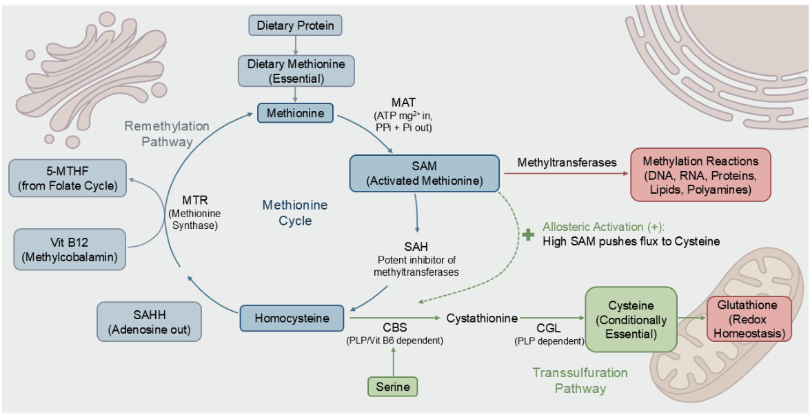
Before diving into the specific metabolic limitations of Homo sapiens, it is crucial to consider the evolutionary context of this pathway in other multicellular heterotrophs, such as the nematode Caenorhabditis elegans and the fruit fly Drosophila melanogaster. Unlike autotrophic plants, these invertebrate models, much like mammals, heavily rely on dietary sources of methionine as the primary precursor to drive the energetically costly biosynthesis of cysteine. In these organisms, the transsulfuration pathway is highly conserved, representing a fundamental evolutionary strategy to manage the intense energetic costs associated with sulfur amino acid interconversion before the emergence of highly specialized mammalian networks. Building upon this conserved foundation, in H. sapiens, the metabolism of sulfur-containing amino acids differs significantly from that of other organisms due to these specific evolutionary and biochemical constraints. To elucidate this complex process, the figure below summarizes the key enzymes involved in methionine and cysteine metabolism and their properties (Figure 5).

5.1. Metabolic Limitations and the Essentiality of Methionine in Humans

Unlike E. coli and higher plants, which synthesize methionine de novo from precursors such as aspartate, mammals lack this pathway [99]. Consequently, methionine is an essential amino acid dependent on dietary intake [100], serving as the ultimate sulfur donor for all sulfur-containing amino acids in humans [101]. Conversely, cysteine is classified as a conditionally non-essential amino acid [102]. Its synthesis relies exclusively on the transsulfuration pathway, which converts methionine-derived homocysteine (Hcy) and serine into cysteine [103]. Thus, cysteine synthesis is strictly contingent upon methionine availability, and insufficient methionine intake renders cysteine essential [104]. This dependency positions dietary methionine at a critical metabolic nexus, influencing not only protein synthesis but also cellular epigenetic potential and redox homeostasis [105].

5.2. The Methionine Cycle: The Sole Endogenous Source of Homocysteine

Homocysteine, a non-proteinogenic sulfur-containing amino acid, serves as the central hub linking methionine and cysteine metabolism. Not derived from the diet, homocysteine is formed exclusively as an intermediate within the methionine cycle [106]. This cycle, initiated by methionine activation, positions homocysteine at a critical metabolic branch point where it faces two competing fates: remethylation back to methionine or irreversible catabolism via the transsulfuration pathway to synthesize cysteine [103]. Given that the metabolic flux of both essential methionine and conditionally essential cysteine converges at this node, the precise regulation of homocysteine is vital for cellular homeostasis. Consequently, plasma homocysteine concentration acts as a sensitive biomarker of one-carbon metabolic status, and elevated levels typically reflect enzymatic disruptions or vitamin deficiencies rather than direct dietary intake [107].

5.3. S-Adenosylmethionine (SAM): Synthesis and Its Role as a Universal Methyl Donor

Beyond its role as a fundamental building block for proteins, the majority of methionine in the human body is utilized for the synthesis of SAM, a high-energy compound often referred to as activated methionine [108]. The biosynthesis of SAM is an ATP-dependent process catalyzed by MAT, which is encoded by the MAT1A and MAT2A genes in humans [109]. This enzymatic reaction is unique in that it transfers the entire adenosyl group from an ATP molecule to the sulfur atom of methionine, followed by the hydrolysis of the resulting triphosphate, making the reaction virtually irreversible under physiological conditions [110]. The most critical function of SAM is to serve as the universal methyl donor for the vast majority of methylation reactions within the cell [9]. These reactions, catalyzed by methyltransferases, are fundamental to cellular regulation, encompassing a wide range of vital activities from the epigenetic modification of DNA and histones to the synthesis of neurotransmitters [111]. Furthermore, SAM also serves as a donor of aminopropyl groups for the synthesis of polyamines, which are essential molecules for cell growth and proliferation [112].

5.4. The Path to Homocysteine: SAH Formation and Hydrolysis

Following its role as a methyl donor, SAM is converted into SAH, a potent feedback inhibitor of most methyltransferases [113]. The accumulation of SAH would halt cellular methylation reactions. Therefore, its efficient removal is crucial for maintaining methylation capacity [114]. This task is executed by SAHH, which catalyzes the reversible hydrolysis of SAH into adenosine and homocysteine [115]. Although the equilibrium of this reaction theoretically favors SAH synthesis, in vivo it proceeds toward hydrolysis because the products adenosine and homocysteine are rapidly scavenged by downstream pathways. This step completes the methionine cycle, releasing homocysteine back into the metabolic pool for further metabolism. Consequently, the intracellular SAM: SAH ratio often referred to as the methylation index, is considered a more accurate indicator of the cell’s methylation capacity than SAM levels alone [116].

5.5. Regulation at the Crossroads: The Metabolic Fate of Homocysteine

Homocysteine occupies a critical metabolic branch point, where it can be directed into two distinct pathways [117]. The first is remethylation to methionine, a process that conserves both the sulfur atom and the carbon skeleton [118]. This reaction is primarily catalyzed by methionine synthase (MS), which utilizes methylcobalamin as a coenzyme and 5-MTHF as the methyl donor, thereby coupling the methionine cycle with folate metabolism [119]. The second pathway is the irreversible transsulfuration pathway [120]. The initial step, catalyzed by cystathionine β-synthase (CBS), involves the condensation of homocysteine with serine to form cystathionine, requiring PLP as a cofactor [121].
The metabolic fate of homocysteine is predominantly regulated by SAM [122,123]. Elevated intracellular SAM levels, indicative of methionine abundance, act as an allosteric activator of CBS [124]. This feed-forward mechanism ensures that excess homocysteine is diverted into the transsulfuration pathway for the synthesis of cysteine, glutathione, and other sulfur-containing compounds [125]. Conversely, low SAM levels reduce CBS activity, thereby channeling the limited homocysteine pool toward remethylation to regenerate the essential amino acid methionine [126].

6. Conclusions and Perspectives

Throughout cysteine and methionine metabolism, H2S serves as the primary source of sulfur [127]. A key distinction lies in their functional groups: the sulfur atom in cysteine enables the regulation of redox balance, whereas the sulfur atom in methionine facilitates its role as a methyl carrier [128]. In the methionine cycle, methionine synthase catalyzes the synthesis of methionine from homocysteine. Subsequently, methionine-derived SAM donates a methyl group in methylation reactions and is converted to SAH. SAH is then hydrolyzed to regenerate homocysteine [129]. Notably, in organisms capable of de novo methionine synthesis, this pathway is often referred to as the homocysteine cycle, as homocysteine marks the starting point [130].
Beyond methylation, the homocysteine cycle demonstrates metabolic economy. In mammals, cysteine is classified as a non-essential amino acid because it can be endogenously synthesized from methionine-derived homocysteine and serine via the transsulfuration pathway, catalyzed by CBS and cystathionine γ-lyase (CTH) [15]. Consequently, mammalian cysteine synthesis differs from that in other organisms [131]. Although the primary function of the transsulfuration pathway is cysteine biosynthesis, it has been discovered that this pathway also contributes to endogenous H2S production through the enzymatic activities of CBS and CTH [132]. This suggests a potential regulatory role of the transsulfuration pathway in methionine metabolism [133]. From a broader perspective, dietary methionine consumed by mammals originates from plant synthesis. The sulfur, initially assimilated as H2S or cysteine in plants, is released back into the metabolic pool as H2S and cysteine in mammals via the actions of CBS and CTH [134].
From a biosynthetic perspective, mammals, including humans have lost the capacity for de novo methionine synthesis, rendering it an essential amino acid. The ancestral pathway of methionine biosynthesis from aspartate is energetically costly and inefficient, making dietary acquisition a more economical strategy given the abundance of food sources [135,136]. Consequently, several amino acids, including methionine, have become essential due to the loss of their synthetic pathways [137]. In contrast, the de novo synthesis of cysteine presents a paradox [138]. While the direct synthesis of cysteine from serine and H2S is theoretically straightforward, mammals primarily rely on the transsulfuration pathway, which uses methionine as a sulfur donor. This pathway is metabolically costly and inefficient [139,140]. Notably, in non-small cell lung cancer (NSCLC) cell lines, serine fails to contribute to cysteine synthesis via the transsulfuration pathway [141]. Furthermore, the conversion of methionine to cysteine is limited by SAM-mediated methylation regulation. Intriguingly, the two key enzymes in this pathway, CBS and CTH, exhibit higher activity in catalyzing H2S production from cysteine than in generating cysteine [142].
In summary, while metabolic evolution generally favors efficiency and economy, the retention of an energetically costly cysteine biosynthetic pathway in mammals suggests a compromise, possibly for the sake of regulatory control or H2S signaling. While the underlying evolutionary rationale remains an open question, elucidating these complex metabolic networks extends far beyond fundamental physiology. A comprehensive understanding of sulfur amino acid conversion is crucially important for industrial biotechnology and pharmaceutical manufacturing. Specifically, mapping these metabolic pathways is essential for optimizing the precursor supply required for the biosynthesis of antibiotics and other active compounds. As highlighted in recent studies on industrial microorganisms like Streptomyces, strategically manipulating sulfur metabolism significantly enhances the yield of valuable secondary metabolites [143]. Ultimately, translating this metabolic knowledge into targeted metabolic engineering offers highly promising avenues for advanced drug production.

Author Contributions

P.Z. and C.X. wrote the manuscript draft; P.Z. and C.X. drew the figures; Y.J. and C.W. revised and proofread the manuscript. All authors have read and approved the article. All authors have read and agreed to the published version of the manuscript.

Funding

Zhejiang Provincial Natural and Science Foundation (LQN25C070001), the 18th Special Funding Program of the China Postdoctoral Science Foundation (2025T180752), and Zhejiang Sci-Tech University Research Start-up Fund (24042191-Y).

Institutional Review Board Statement

Not applicable.

Informed Consent Statement

Not applicable.

Data Availability Statement

No new data were created or analyzed in this study. Data sharing is not applicable to this article.

Conflicts of Interest

The authors declare no conflicts of interest.

Abbreviations

The following abbreviations are used in this manuscript:
GSHGlutathione
SAMS-adenosylmethionine
SCAAsSulfur-containing amino acids
SATSerine O-acetyltransferase
Acetyl-CoAAcetyl-coenzyme A
OASO-acetylserine
PLPPyridoxal 5′-phosphate
H2SSulfide
CSCCysteine Synthase Complex
OSHSO-succinyl-L-homoserine
5-MTHF5-methyltetrahydrofolate
MATS-adenosylmethionine synthetase
SAHS-Adenosylhomocysteine
SAHHS-adenosylhomocysteine hydrolase
OPHO-phosphohomoserine
AK-HSDHAspartate kinase–homoserine dehydrogenase
ASDAspartate semialdehyde dehydrogenase
HSKHomoserine kinase
OAS-TLO-acetylserine (thiol) lyase
CGSCystathionine γ-synthase
GSSGOxidized glutathione
CBLCystathionine β-lyase
PINPIN-FORMED
MS1Methyltransferase 1
DES1Desulfhydrase 1
ABAAbscisic acid
CGLCystathionine γ-lyase
HcyHomocysteine
MSMethionine synthase
CBSCystathionine β-synthase
CTHCystathionine γ-lyase
NSCLCNon-small cell lung cancer

References

  1. Laatsch, B.F.; Ali, B.A.; Berthiaume, A.R.; Cunningham, G.C.; Duncan, S.M.; Hau, N.; Ho, A.D.; Loomis, M.A.; Lowater, H.R.; Mcnally, B.L.; et al. A Review of Methionine and Cysteine: Their Roles as Sinks for Reactive Oxygen and Nitrogen Species, Including Bioinformatic Analysis of Their Percent Compositions in Metabolic Proteins. Indian J. Clin. Biochem. 2025, 40, 551–562. [Google Scholar] [CrossRef]
  2. Li, H.X.; Wang, X.Y.; Liu, Y.; Zhang, P.Y.; Chen, F.Y.; Zhang, N.; Zhao, B.; Guo, Y.D. Cysteine Thiol-Based Oxidative Post-Translational Modifications Fine-Tune Protein Functions in Plants. Agronomy 2024, 14, 2757. [Google Scholar] [CrossRef]
  3. Turk, V.; Stoka, V.; Vasiljeva, O.; Renko, M.; Sun, T.; Turk, B.; Turk, D. Cysteine cathepsins: From structure, function and regulation to new frontiers. Biochim. Biophys. Acta-Proteins Proteom. 2012, 1824, 68–88. [Google Scholar] [CrossRef]
  4. Heissel, S.; He, Y.; Jankevics, A.; Shi, Y.Q.; Molina, H.; Viner, R.; Scheltema, R.A. Fast and Accurate Disulfide Bridge Detection. Mol. Cell. Proteom. 2024, 23, 100759. [Google Scholar] [CrossRef]
  5. Musaogullari, A.; Chai, Y.C. Redox Regulation by Protein S-Glutathionylation: From Molecular Mechanisms to Implications in Health and Disease. Int. J. Mol. Sci. 2020, 21, 8113. [Google Scholar] [CrossRef]
  6. Sun, X.; Xie, L.; Wang, S.; Zeng, S.; Wu, L.; Tang, X.; Zhu, J.; Lin, S.; Hu, T.; Jia, L.; et al. S-glutathionylation modification of proteins and the association with cellular death (Review). Med. Int. 2025, 5, 64. [Google Scholar] [CrossRef]
  7. Lee, C.S.; Kim, S.Y.; Lee, H.; Roh, T.Y.; Hwang, C.S. Formyl-methionine-mediated eukaryotic ribosome quality control pathway for cold adaptation. Mol. Cell 2025, 85, 602–619. [Google Scholar] [CrossRef]
  8. Ouyang, Y.; Wu, Q.; Li, J.J.; Sun, S.; Sun, S.R. S-adenosylmethionine: A metabolite critical to the regulation of autophagy. Cell Prolif. 2020, 53, e12891. [Google Scholar] [CrossRef]
  9. Fernández-Ramos, D.; Lopitz-Otsoa, F.; Lu, S.C.; Mato, J.M. S-Adenosylmethionine: A Multifaceted Regulator in Cancer Pathogenesis and Therapy. Cancers 2025, 17, 535. [Google Scholar] [CrossRef] [PubMed]
  10. Marton, L.J.; Pegg, A.E. Polyamines as targets for therapeutic intervention. Annu. Rev. Pharmacol. Toxicol. 1995, 35, 55–91. [Google Scholar] [CrossRef] [PubMed]
  11. Jobe, T.O.; Zenzen, I.; Rahimzadeh Karvansara, P.; Kopriva, S. Integration of sulfate assimilation with carbon and nitrogen metabolism in transition from C3 to C4 photosynthesis. J. Exp. Bot. 2019, 70, 4211–4221. [Google Scholar] [CrossRef]
  12. Wang, S.Q.; Lu, Q.H.; Liang, Z.W.; Yu, X.X.; Lin, M.; Mai, B.X.; Qiu, R.L.; Shu, W.S.; He, Z.L.; Wall, J.D. Generation of zero-valent sulfur from dissimilatory sulfate reduction in sulfate-reducing microorganisms. Proc. Natl. Acad. Sci. USA 2023, 120, e2220725120. [Google Scholar] [CrossRef]
  13. Pajares, M.Á. Posttranslational Regulation of Mammalian Sulfur Amino Acid Metabolism. Int. J. Mol. Sci. 2025, 26, 2488. [Google Scholar] [CrossRef]
  14. McPherson, V.N.; Hardy, G. Clinical and nutritional benefits of cysteine-enriched protein supplements. Curr. Opin. Clin. Nutr. Metab. Care 2011, 14, 562–568. [Google Scholar] [CrossRef]
  15. Sbodio, J.I.; Snyder, S.H.; Paul, B.D. Regulators of the transsulfuration pathway. Br. J. Pharmacol. 2019, 176, 583–593. [Google Scholar] [CrossRef]
  16. Koprivova, A.; Kopriva, S. Molecular mechanisms of regulation of sulfate assimilation: First steps on a long road. Front. Plant Sci. 2014, 5, 589. [Google Scholar] [CrossRef]
  17. Olsen, T.; Elshorbagy, A.; Johnson, J.E.; Nichenametla, S.N.; Dong, Z.; Sambamurti, K.; Richie, J.P.; Vinknes, K.J. Sulfur amino acids, metabolic health and beyond: Recent advances, translational implications, and future research considerations. Anal. Biochem. 2026, 709, 116008. [Google Scholar] [CrossRef]
  18. Smirnova, G.; Tyulenev, A.; Sutormina, L.; Kalashnikova, T.; Muzyka, N.; Ushakov, V.; Samoilova, Z.; Oktyabrsky, O. Regulation of Cysteine Homeostasis and Its Effect on Escherichia coli Sensitivity to Ciprofloxacin in LB Medium. Int. J. Mol. Sci. 2024, 25, 4424. [Google Scholar] [CrossRef]
  19. Hicks, J.L.; Oldham, K.E.A.; McGarvie, J.; Walker, E.J. Combatting antimicrobial resistance via the cysteine biosynthesis pathway in bacterial pathogens. Biosci. Rep. 2022, 42, BSR20220368. [Google Scholar] [CrossRef]
  20. Liang, B.; Sun, G.N.; Zhang, X.P.; Nie, Q.J.; Zhao, Y.K.; Yang, J.M. Recent advances, challenges and metabolic engineering strategies in the biosynthesis of 3-hydroxypropionic acid. Biotechnol. Bioeng. 2022, 119, 2639–2668. [Google Scholar] [CrossRef]
  21. Tao, Y.; Zheng, D.D.; Zou, W.; Guo, T.; Liao, G.J.; Zhou, W. Targeting the cysteine biosynthesis pathway in microorganisms: Mechanism, structure, and drug discovery. Eur. J. Med. Chem. 2024, 271, 116461. [Google Scholar] [CrossRef]
  22. Oldham, K.E.A.; Jiao, W.T.; Prentice, E.; Hicks, J.L. Identification of novel inhibitors targeting serine acetyltransferase from Neisseria gonorrhoeae. Comput. Struct. Biotechnol. J. 2025, 27, 682–691. [Google Scholar] [CrossRef]
  23. Oldham, K.E.A.; Prentice, E.J.; Summers, E.L.; Hicks, J.L. Serine acetyltransferase from Neisseria gonorrhoeae; structural and biochemical basis of inhibition. Biochem. J. 2022, 479, 57–74. [Google Scholar] [CrossRef]
  24. Yang, H.; Zhang, B.; Wu, Z.D.; Xiu, X.L.; Liu, Z.Q.; Zheng, Y.G. Metabolic Reprogramming in Escherichia coli for Efficient L-Cysteine Production by Metabolic Node Engineering. Acs Sustain. Chem. Eng. 2025, 13, 4778–4789. [Google Scholar] [CrossRef]
  25. Rosa, B.; Dickinson, E.R.; Marchetti, M.; Campanini, B.; Pioselli, B.; Bettati, S.; Rand, K.D. Revealing the Dynamic Allosteric Changes Required for Formation of the Cysteine Synthase Complex by Hydrogen-Deuterium Exchange MS. Mol. Cell. Proteom. 2021, 20, 100098. [Google Scholar] [CrossRef]
  26. Pederick, J.L.; Vandborg, B.C.; George, A.; Bovermann, H.; Boyd, J.M.; Freundlich, J.S.; Bruning, J.B. Identification of cysteine metabolism regulator (CymR)-derived pentapeptides as nanomolar inhibitors of Staphylococcus aureus O-acetyl-ʟ-serine sulfhydrylase (CysK). ACS Infect. Dis. 2025, 11, 238–248. [Google Scholar] [CrossRef]
  27. Jin, S.; Wijerathne, C.U.B.; Siow, Y.L.; O, K. Disruption of the transsulfuration pathway by acute kidney injury causes intestinal damage. iScience 2025, 28, 113511. [Google Scholar] [CrossRef]
  28. Harting, C.; Teleki, A.; Braakmann, M.; Jankowitsch, F.; Takors, R. Systemic intracellular analysis for balancing complex biosynthesis in a transcriptionally deregulated Escherichia coli l-Methionine producer. Microb. Biotechnol. 2024, 17, e14433. [Google Scholar] [CrossRef] [PubMed]
  29. Han, M.; Zhang, C.; Suglo, P.; Sun, S.; Wang, M.; Su, T. l-Aspartate: An Essential Metabolite for Plant Growth and Stress Acclimation. Molecules 2021, 26, 1887. [Google Scholar] [CrossRef] [PubMed]
  30. Hao, R.; Wang, S.; Jin, X.; Yang, X.; Qi, Q.; Liang, Q. Dynamic and balanced regulation of the thrABC operon gene for efficient synthesis of L-threonine. Front. Bioeng. Biotechnol. 2023, 11, 1118948. [Google Scholar] [CrossRef]
  31. Sun, Y.J.; Wu, J.P.; Xu, J.Q.; Yang, L.R. Metabolic Engineering of Escherichia coli for the Production of l-Homoserine. Chem. Bio Eng. 2024, 1, 223–230. [Google Scholar] [CrossRef]
  32. Jin, X.; Wang, S.; Wang, Y.; Qi, Q.; Liang, Q. Metabolic engineering strategies for L-Homoserine production in Escherichia coli. Microb. Cell Fact. 2024, 23, 338. [Google Scholar] [CrossRef]
  33. Gruzdev, N.; Hacham, Y.; Haviv, H.; Stern, I.; Gabay, M.; Bloch, I.; Amir, R.; Gal, M.; Yadid, I. Conversion of methionine biosynthesis in Escherichia coli from trans- to direct-sulfurylation enhances extracellular methionine levels. Microb. Cell Fact. 2023, 22, 151. [Google Scholar] [CrossRef]
  34. Brewster, J.L.; Pachl, P.; McKellar, J.L.O.; Selmer, M.; Squire, C.J.; Patrick, W.M. Structures and kinetics of Thermotoga maritima MetY reveal new insights into the predominant sulfurylation enzyme of bacterial methionine biosynthesis. J. Biol. Chem. 2021, 296, 100797. [Google Scholar] [CrossRef]
  35. Tramonti, A.; Nardella, C.; di Salvo, M.L.; Barile, A.; D’Alessio, F.; de Crécy-Lagard, V.; Contestabile, R. Knowns and Unknowns of Vitamin B6 Metabolism in Escherichia coli. EcoSal Plus 2021, 9, eESP-0004-2021. [Google Scholar] [CrossRef]
  36. Kelley, B.R.; Callahan, S.M.; Johnson, J.G. Transcription of Cystathionine β-Lyase (MetC) Is Repressed by HeuR in Campylobacter jejuni, and Methionine Biosynthesis Facilitates Colonocyte Invasion. J. Bacteriol. 2021, 203, e0016421. [Google Scholar] [CrossRef]
  37. Mohr, M.K.F.; Satanowski, A.; Lindner, S.N.; Erb, T.J.; Andexer, J.N. Rewiring Escherichia coli to transform formate into methyl groups. Microb. Cell Fact. 2025, 24, 55. [Google Scholar] [CrossRef]
  38. Sullivan, M.R.; Darnell, A.M.; Reilly, M.F.; Kunchok, T.; Joesch-Cohen, L.; Rosenberg, D.; Ali, A.; Rees, M.G.; Roth, J.A.; Lewis, C.A.; et al. Methionine synthase is essential for cancer cell proliferation in physiological folate environments. Nat. Metab. 2021, 3, 1500–1511. [Google Scholar] [CrossRef]
  39. Rao, D.; Füssy, Z.; Brisbin, M.M.; McIlvin, M.R.; Moran, D.M.; Allen, A.E.; Follows, M.J.; Saito, M.A. Flexible B12 ecophysiology of Phaeocystis antarctica due to a fusion B12-independent methionine synthase with widespread homologues. Proc. Natl. Acad. Sci. USA 2024, 121, e2204075121. [Google Scholar] [CrossRef]
  40. Price, M.N.; Deutschbauer, A.M.; Arkin, A.P. Four families of folate-independent methionine synthases. PLoS Genet. 2021, 17, e1009342. [Google Scholar] [CrossRef]
  41. Wu, C.; Mori, M.; Abele, M.; Banaei-Esfahani, A.; Zhang, Z.; Okano, H.; Aebersold, R.; Ludwig, C.; Hwa, T. Enzyme expression kinetics by Escherichia coli during transition from rich to minimal media depends on proteome reserves. Nat. Microbiol. 2023, 8, 347–359. [Google Scholar] [CrossRef]
  42. Mendoza, J.; Purchal, M.; Yamada, K.; Koutmos, M. Structure of full-length cobalamin-dependent methionine synthase and cofactor loading captured in crystallo. Nat. Commun. 2023, 14, 6365. [Google Scholar] [CrossRef]
  43. Mok, K.C.; Hallberg, Z.F.; Procknow, R.R.; Taga, M.E. Laboratory evolution of E. coli with a natural vitamin B12 analog reveals roles for cobamide uptake and adenosylation in methionine synthase-dependent growth. J. Bacteriol. 2025, 207, e0028424. [Google Scholar] [CrossRef]
  44. Li, Z.; Wen, X.; Bolotova, S.B.; Seebeck, F.P. Short-Circuiting the SAM-Cycle in Escherichia coli. J. Am. Chem. Soc. 2025, 147, 47690–47700. [Google Scholar] [CrossRef] [PubMed]
  45. Shafqat, N.; Muniz, J.R.; Pilka, E.S.; Papagrigoriou, E.; von Delft, F.; Oppermann, U.; Yue, W.W. Insight into S-adenosylmethionine biosynthesis from the crystal structures of the human methionine adenosyltransferase catalytic and regulatory subunits. Biochem. J. 2013, 452, 27–36. [Google Scholar] [CrossRef]
  46. Schlesier, J.; Siegrist, J.; Gerhardt, S.; Erb, A.; Blaesi, S.; Richter, M.; Einsle, O.; Andexer, J.N. Structural and functional characterisation of the methionine adenosyltransferase from Thermococcus kodakarensis. BMC Struct. Biol. 2013, 13, 22. [Google Scholar] [CrossRef]
  47. Rudenko, A.Y.; Mariasina, S.S.; Bolikhova, A.K.; Nikulin, M.V.; Ozhiganov, R.M.; Vasil’ev, V.G.; Ikhalaynen, Y.A.; Khandazhinskaya, A.L.; Khomutov, M.A.; Sergiev, P.V.; et al. Organophosphorus S-adenosyl-L-methionine mimetics: Synthesis, stability, and substrate properties. Front. Chem. 2024, 12, 1448747. [Google Scholar] [CrossRef]
  48. Ero, R.; Leppik, M.; Reier, K.; Liiv, A.; Remme, J. Ribosomal RNA modification enzymes stimulate large ribosome subunit assembly in E. coli. Nucleic Acids Res. 2024, 52, 6614–6628. [Google Scholar] [CrossRef]
  49. Vizán, P.; Di Croce, L.; Aranda, S. Functional and Pathological Roles of AHCY. Front. Cell Dev. Biol. 2021, 9, 654344. [Google Scholar] [CrossRef]
  50. Tehlivets, O.; Malanovic, N.; Visram, M.; Pavkov-Keller, T.; Keller, W. S-adenosyl-L-homocysteine hydrolase and methylation disorders: Yeast as a model system. Biochim. Biophys. Acta 2013, 1832, 204–215. [Google Scholar] [CrossRef]
  51. Stanhope, S.C.; Singhal, K.; Morato, N.M.; Feng, Y.; Meng, G.; Marlin, M.N.; Kotanko, C.C.; Jarrett, M.M.; Mesecar, A.D.; Cooks, R.G.; et al. Drosophila Ahcy is a redox sensor that modulates gene expression to protect against light stress-induced retinal degeneration. Proc. Natl. Acad. Sci. USA 2025, 122, e2511388122. [Google Scholar] [CrossRef]
  52. Shi, D.; Zhang, Y.; Wang, J.; Ren, W.; Zhang, J.; Mbadianya, J.I.; Zhu, Y.; Chen, C.; Ma, H. S-adenosyl-L-homocysteine hydrolase FgSah1 is required for fungal development and virulence in Fusarium graminearum. Virulence 2021, 12, 2171–2185. [Google Scholar] [CrossRef]
  53. Sadeeshkumar, H.; Balaji, A.; Sutherland, A.G.; Mootien, S.; Anthony, K.G.; Breaker, R.R. Screening for small molecule inhibitors of SAH nucleosidase using an SAH riboswitch. Anal. Biochem. 2023, 666, 115047. [Google Scholar] [CrossRef]
  54. Korenblum, E.; Massalha, H.; Aharoni, A. Plant-microbe interactions in the rhizosphere via a circular metabolic economy. Plant Cell 2022, 34, 3168–3182. [Google Scholar] [CrossRef]
  55. Simon-Baram, H.; Kleiner, D.; Shmulevich, F.; Zarivach, R.; Zalk, R.; Tang, H.; Ding, F.; Bershtein, S. SAMase of Bacteriophage T3 Inactivates Escherichia coli’s Methionine S-Adenosyltransferase by Forming Heteropolymers. mBio 2021, 12, e0124221. [Google Scholar] [CrossRef]
  56. Andriianov, A.; Trigüis, S.; Drobiazko, A.; Sierro, N.; Ivanov, N.V.; Selmer, M.; Severinov, K.; Isaev, A. Phage T3 overcomes the BREX defense through SAM cleavage and inhibition of SAM synthesis by SAM lyase. Cell Rep. 2023, 42, 112972. [Google Scholar] [CrossRef]
  57. Ohtsuka, H.; Shimasaki, T.; Aiba, H. Response to sulfur in Schizosaccharomyces pombe. FEMS Yeast Res. 2021, 21, foab041. [Google Scholar] [CrossRef]
  58. Guo, L.; Ganguly, A.; Sun, L.; Suo, F.; Du, L.L.; Russell, P. Global Fitness Profiling Identifies Arsenic and Cadmium Tolerance Mechanisms in Fission Yeast. G3 (Bethesda) 2016, 6, 3317–3333. [Google Scholar] [CrossRef]
  59. Kuplińska, A.; Rząd, K. Molecular targets for antifungals in amino acid and protein biosynthetic pathways. Amino Acids 2021, 53, 961–991. [Google Scholar] [CrossRef]
  60. Chik, J.K.; Su, X.B.; Klepin, S.; Raygoza, J.; Pillus, L. Non-canonical chromatin-based functions for the threonine metabolic pathway. Sci. Rep. 2024, 14, 22629. [Google Scholar] [CrossRef]
  61. Zhang, X.; Xin, Y.; Chen, Z.; Xia, Y.; Xun, L.; Liu, H. Sulfide-quinone oxidoreductase is required for cysteine synthesis and indispensable to mitochondrial health. Redox Biol. 2021, 47, 102169. [Google Scholar] [CrossRef] [PubMed]
  62. Rij, M.; Wendland, J. Use of the Saccharomycopsis schoenii MET17 promoter for regulated heterologous gene expression. Curr. Genet. 2024, 70, 9. [Google Scholar] [CrossRef]
  63. Yu, J.S.L.; Heineike, B.M.; Hartl, J.; Aulakh, S.K.; Correia-Melo, C.; Lehmann, A.; Lemke, O.; Agostini, F.; Lee, C.T.; Demichev, V.; et al. Inorganic sulfur fixation via a new homocysteine synthase allows yeast cells to cooperatively compensate for methionine auxotrophy. PLoS Biol. 2022, 20, e3001912. [Google Scholar] [CrossRef]
  64. Zhang, X.; Sun, W.; Chen, D.; Murchie, A.I.H. Interactions between SAM and the 5’ UTR mRNA of the sam1 gene regulate translation in S. pombe. RNA 2022, 26, 150–161. [Google Scholar] [CrossRef]
  65. Hoffert, K.M.; Higginbotham, K.S.P.; Gibson, J.T.; Oehrle, S.; Strome, E.D. Mutations in the S-Adenosylmethionine Synthetase Genes SAM1 and SAM2 Differentially Affect Genome Stability in Saccharomyces cerevisiae. Genetics 2019, 213, 97–112. [Google Scholar] [CrossRef]
  66. He, H.; Zhang, Q.; Fan, Z.; Duan, H.; Wang, Y.; Luo, B.; Li, Q.; Liu, J.; Li, D.; Fang, S.; et al. Autophagy dictates PHGDH-mediated serine metabolism in a timely manner to support oocyte development. Autophagy 2026, 22, 65–84. [Google Scholar] [CrossRef]
  67. D’Souza, S.W.; Glazier, J.D. Homocysteine Metabolism in Pregnancy and Developmental Impacts. Front. Cell Dev. Biol. 2022, 10, 802285. [Google Scholar] [CrossRef] [PubMed]
  68. Rząd, K.; Kuplińska, A.; Gabriel, I. Fungal L-Methionine Biosynthesis Pathway Enzymes and Their Applications in Various Scientific and Commercial Fields. Biomolecules 2024, 14, 1315. [Google Scholar] [CrossRef] [PubMed]
  69. Chang, W.; Zhang, M.; Jin, X.; Zhang, H.; Zheng, H.; Zheng, S.; Qiao, Y.; Yu, H.; Sun, B.; Hou, X.; et al. Inhibition of fungal pathogenicity by targeting the H2S-synthesizing enzyme cystathionine β-synthase. Sci. Adv. 2022, 8, eadd5366. [Google Scholar] [CrossRef]
  70. Gu, Z.; Sun, Y.; Wu, F.; Wu, X. Mechanism of Growth Regulation of Yeast Involving Hydrogen Sulfide From S-Propargyl-Cysteine Catalyzed by Cystathionine-γ-Lyase. Front. Microbiol. 2021, 12, 679563, Erratum in Front Microbiol. 2021, 12, 761240. https://doi.org/10.3389/fmicb.2021.761240. [Google Scholar] [CrossRef]
  71. Xi, C.; Pang, J.; Xue, W.; Cui, Y.; Jiang, N.; Zhi, W.; Shi, H.; Horuzsko, A.; Pace, B.S.; Zhu, X. Transsulfuration pathway activation attenuates oxidative stress and ferroptosis in sickle primary erythroblasts and transgenic mice. Commun. Biol. 2025, 8, 15. [Google Scholar] [CrossRef]
  72. Lehner, S.; Boles, E. Development of vitamin B12 dependency in Saccharomyces cerevisiae. FEMS Yeast Res. 2023, 23, foad020. [Google Scholar] [CrossRef]
  73. Tan, L.; Zhang, Y.; Liu, P.; Wu, Y.; Huang, Z.; Hu, Z.; Liu, Z.; Wang, Y.; Zheng, Y. System metabolic engineering modification of Saccharomyces cerevisiae to increase SAM production. Bioresour. Bioprocess. 2025, 12, 19. [Google Scholar] [CrossRef] [PubMed]
  74. Remines, M.; Schoonover, M.; Knox, Z.; Kenwright, K.; Hoffert, K.M.; Coric, A.; Mead, J.; Ampfer, J.; Seye, S.; Strome, E.D. Profiling the Compendium of Changes in Saccharomyces cerevisiae Due to Mutations That Alter Availability of the Main Methyl Donor S-Adenosylmethionine. G3 Genes Genomes Genet. 2023, 14, jkae002. [Google Scholar] [CrossRef]
  75. Panmanee, W.; Tran, M.T.H.; Seye, S.N.; Strome, E.D. Altered S-AdenosylMethionine availability impacts dNTP pools in Saccharomyces cerevisiae. Yeast 2024, 41, 513–524. [Google Scholar] [CrossRef]
  76. Lombardi, L.; Salzberg, L.I.; Cinnéide, E.Ó.; O’Brien, C.; Morio, F.; Turner, S.A.; Byrne, K.P.; Butler, G. Alternative sulphur metabolism in the fungal pathogen Candida parapsilosis. Nat. Commun. 2024, 15, 9190. [Google Scholar] [CrossRef]
  77. Elango, R. Methionine Nutrition and Metabolism: Insights from Animal Studies to Inform Human Nutrition. J. Nutr. 2020, 150, 2518S–2523S. [Google Scholar] [CrossRef] [PubMed]
  78. Garneau, M.G.; Lu, M.Z.; Grant, J.; Tegeder, M. Role of source-to-sink transport of methionine in establishing seed protein quantity and quality in legumes. Plant Physiol. 2021, 187, 2134–2155. [Google Scholar] [CrossRef]
  79. Kopriva, S.; Rahimzadeh Karvansara, P.; Takahashi, H. Adaptive modifications in plant sulfur metabolism over evolutionary time. J. Exp. Bot. 2024, 75, 4697–4711. [Google Scholar] [CrossRef]
  80. Niu, K.; Gao, L.; Ge, L.; Liu, Z.; Zheng, Y. Advances in the biosynthesis of L-homoserine and its derivatives by metabolic engineering of Escherichia coli. Sheng Wu Gong Cheng Xue Bao 2022, 38, 4385–4402. [Google Scholar] [CrossRef] [PubMed]
  81. de Bont, L.; Donnay, N.; Couturier, J.; Rouhier, N. Redox regulation of enzymes involved in sulfate assimilation and in the synthesis of sulfur-containing amino acids and glutathione in plants. Front. Plant Sci. 2022, 13, 958490. [Google Scholar] [CrossRef]
  82. Jardine, K.J.; Honeker, L.K.; Zhang, Z.; Kwatcho Kengdo, S.; Yang, Y.; Roscioli, J.; Riley, W.J. Evolutionary and functional relationships between plant and microbial C1 metabolism in terrestrial ecosystems. New Phytol. 2025, 248, 1132–1153. [Google Scholar] [CrossRef]
  83. Girija, A.; Hacham, Y.; Dvir, S.; Panda, S.; Lieberman-Lazarovich, M.; Amir, R. Cystathionine γ-synthase expression in seeds alters metabolic and DNA methylation profiles in Arabidopsis. Plant Physiol. 2023, 193, 595–610. [Google Scholar] [CrossRef] [PubMed]
  84. Farkas, P.; Fitzpatrick, T.B. Two pyridoxal phosphate homeostasis proteins are essential for management of the coenzyme pyridoxal 5’-phosphate in Arabidopsis. Plant Cell 2024, 36, 3689–3708. [Google Scholar] [CrossRef]
  85. Tanaka, M.; Sotta, N.; Duncan, S.; Chiba, Y.; Onouchi, H.; Marée, A.F.M.; Naito, S.; Grieneisen, V.A.; Fujiwara, T. Ribosome stalling-induced NIP5;1 mRNA decay triggers ARGONAUTE1-dependent transcription downregulation. Nucleic Acids Res. 2025, 53, gkaf159. [Google Scholar] [CrossRef]
  86. Hamano, T.; Nagumo, Y.; Umehara, T.; Hirono, K.; Fujiwara, K.; Taguchi, H.; Chadani, Y.; Doi, N. STALL-seq: mRNA-display selection of bacterial and eukaryotic translational arrest sequences from large random-sequence libraries. J. Biol. Chem. 2024, 300, 107978. [Google Scholar] [CrossRef] [PubMed]
  87. Hacham, Y.; Kaplan, A.; Cohen, E.; Gal, M.; Amir, R. Sulfur metabolism under stress: Oxidized glutathione inhibits methionine biosynthesis by destabilizing the enzyme cystathionine γ-synthase. J. Integr. Plant Biol. 2025, 67, 87–100. [Google Scholar] [CrossRef] [PubMed]
  88. Wang, Z.; He, F.; Mu, Y.; Zhang, L.; Liu, Z.; Liu, D.; Yang, J.; Jin, Z.; Pei, Y. Identification and functional characterization of a cystathionine β-lyase (CBL) enzyme for H2S production in Arabidopsis thaliana. Plant Physiol. Biochem. 2022, 182, 76–89. [Google Scholar] [CrossRef]
  89. Cordoza, J.L.; Chen, P.Y.; Blaustein, L.R.; Lima, S.T.; Fiore, M.F.; Chekan, J.R.; Moore, B.S.; McKinnie, S.M.K. Mechanistic and Structural Insights into a Divergent PLP-Dependent l-Enduracididine Cyclase from a Toxic Cyanobacterium. ACS Catal. 2023, 13, 9817–9828. [Google Scholar] [CrossRef]
  90. Zhao, Q.; Zhao, P.X.; Wu, Y.; Zhong, C.Q.; Liao, H.; Li, C.Y.; Fu, X.D.; Fang, P.; Xu, P.; Xiang, C.B. SUE4, a novel PIN1-interacting membrane protein, regulates acropetal auxin transport in response to sulfur deficiency. New Phytol. 2023, 237, 78–87. [Google Scholar] [CrossRef]
  91. Chen, X.; Xu, J.; Wong, N.K.; Zhong, S.; Yang, M.; Liu, Z.; Lu, Y.; Li, W.; Zhou, Y. Chemoproteomic Profiling of Cobalamin-Independent Methionine Synthases in Plants with a Covalent Probe. J. Agric. Food Chem. 2020, 68, 8050–8056. [Google Scholar] [CrossRef]
  92. Ju, C.; Kong, D.; Lee, Y.; Ge, G.; Song, Y.; Liu, J.; Kwak, J.M. Methionine synthase 1 provides methionine for activation of the GLR3.5 Ca2+ channel and regulation of germination in Arabidopsis. J. Exp. Bot. 2020, 71, 178–187. [Google Scholar] [CrossRef]
  93. Rudolf, E.E.; Hüther, P.; Forné, I.; Georgii, E.; Han, Y.; Hell, R.; Wirtz, M.; Imhof, A.; Becker, C.; Durner, J.; et al. GSNOR Contributes to Demethylation and Expression of Transposable Elements and Stress-Responsive Genes. Antioxidants 2021, 10, 1128. [Google Scholar] [CrossRef]
  94. Geng, Z.K.; Ma, L.; Rong, Y.L.; Li, W.J.; Yao, G.F.; Zhang, H.; Hu, K.D. A Hydrogen-Sulfide-Repressed Methionine Synthase SlMS1 Acts as a Positive Regulator for Fruit Ripening in Tomato. Int. J. Mol. Sci. 2022, 23, 12239. [Google Scholar] [CrossRef]
  95. Scuffi, D.; Pantaleno, R.; Schiel, P.; Niemeier, J.O.; Costa, A.; Schwarzländer, M.; Laxalt, A.M.; García-Mata, C. Hydrogen sulfide modulates flagellin-induced stomatal immunity. Plant Physiol. 2025, 199, kiaf561. [Google Scholar] [CrossRef]
  96. Zhang, J.; Zhou, H.; Zhou, M.; Ge, Z.; Zhang, F.; Foyer, C.H.; Yuan, X.; Xie, Y. The coordination of guard-cell autonomous ABA synthesis and DES1 function in situ regulates plant water deficit responses. J. Adv. Res. 2020, 27, 191–197. [Google Scholar] [CrossRef]
  97. Liu, H.; Wang, J.; Liu, J.; Liu, T.; Xue, S. Hydrogen sulfide (H2S) signaling in plant development and stress responses. aBIOTECH 2021, 2, 32–63. [Google Scholar] [CrossRef]
  98. Liu, T.; Chen, H.; Luo, S.; Xue, S. Hydrogen sulphide alleviates root growth inhibition induced by phosphate starvation. Plant Cell Environ. 2024, 47, 5265–5279. [Google Scholar] [CrossRef]
  99. Espe, M.; Adam, A.C.; Saito, T.; Skjærven, K.H. Methionine: An Indispensable Amino Acid in Cellular Metabolism and Health of Atlantic Salmon. Aquac. Nutr. 2023, 2023, 5706177. [Google Scholar] [CrossRef]
  100. Fang, H.; Stone, K.P.; Wanders, D.; Forney, L.A.; Gettys, T.W. The Origins, Evolution, and Future of Dietary Methionine Restriction. Annu. Rev. Nutr. 2022, 42, 201–226. [Google Scholar] [CrossRef]
  101. Hansen, A.W.; Venkatachalam, K.V. Sulfur-Element containing metabolic pathways in human health and crosstalk with the microbiome. Biochem. Biophys. Rep. 2023, 35, 101529. [Google Scholar] [CrossRef]
  102. Mahmoudi Ghehsareh, M.; Bakhtiari, S.; Asri, N.; Rezaei-Tavirani, M.; Jahani-Sherafat, S.; Masotti, A.; Rostami-Nejad, M. Impacts of conditionally essential amino acids, cysteine, and tyrosine on intestinal inflammation in celiac disease. Amino Acids 2025, 58, 1. [Google Scholar] [CrossRef] [PubMed]
  103. Flori, L.; Veneziano, S.; Martelli, A.; Piragine, E.; Calderone, V. Transsulfuration Pathway Products and H2S-Donors in Hyperhomocysteinemia: Potential Strategies Beyond Folic Acid. Int. J. Mol. Sci. 2025, 26, 6430. [Google Scholar] [CrossRef]
  104. Tada, K.; Mitsuyama, K.; Nishizawa, H.; Shima, H.; Muto, A.; Wada, M.; Saigusa, D.; Igarashi, K. Cysteine Restriction Induces Ferroptosis Depending on the S-adenosylmethionine and Polyamine Biosynthetic Pathways in Hepatic Cancer Cells. Gastro Hep Adv. 2025, 5, 100817. [Google Scholar] [CrossRef] [PubMed]
  105. Yan, C.; He, B.; Wang, C.; Li, W.; Tao, S.; Chen, J.; Wang, Y.; Yang, L.; Wu, Y.; Wu, Z.; et al. Methionine in embryonic development: Metabolism, redox homeostasis, epigenetic modification and signaling pathway. Crit. Rev. Food Sci. Nutr. 2025, 65, 8051–8074. [Google Scholar] [CrossRef]
  106. Jakubowski, H.; Witucki, Ł. Homocysteine Metabolites, Endothelial Dysfunction, and Cardiovascular Disease. Int. J. Mol. Sci. 2025, 26, 746. [Google Scholar] [CrossRef]
  107. Joshi, S.M.; Jadavji, N.M. Deficiencies in one-carbon metabolism led to increased neurological disease risk and worse outcome: Homocysteine is a marker of disease state. Front. Nutr. 2024, 11, 1285502. [Google Scholar] [CrossRef]
  108. Rudenko, A.Y.; Mariasina, S.S.; Ozhiganov, R.M.; Sergiev, P.V.; Polshakov, V.I. Enzymatic Reactions of S-Adenosyl-L-Methionine: Synthesis and Applications. Biochemistry 2025, 90, S105–S134. [Google Scholar] [CrossRef]
  109. Wang, Y.; Muylaert, C.; Wyns, A.; Vlummens, P.; De Veirman, K.; Vanderkerken, K.; Zaal, E.; Berkers, C.; Moreaux, J.; De Bruyne, E.; et al. S-adenosylmethionine biosynthesis is a targetable metabolic vulnerability in multiple myeloma. Haematologica 2024, 109, 256–271. [Google Scholar] [CrossRef] [PubMed]
  110. Niland, C.N.; Ghosh, A.; Cahill, S.M.; Schramm, V.L. Mechanism and Inhibition of Human Methionine Adenosyltransferase 2A. Biochemistry 2021, 60, 791–801. [Google Scholar] [CrossRef]
  111. Bekdash, R.A. Methyl Donors, Epigenetic Alterations, and Brain Health: Understanding the Connection. Int. J. Mol. Sci. 2023, 24, 2346. [Google Scholar] [CrossRef]
  112. Yerlikaya, A. Spermine synthase in Snyder-Robinson syndrome and cancer. Mol. Biol. Rep. 2025, 53, 40. [Google Scholar] [CrossRef] [PubMed]
  113. Hammerstad, M.; Steinvik, E.; Hersleth, H.P. The methyltransferase NmbA methylates the low-molecular weight thiol bacillithiol, and displays a specific structural architecture. Redox Biol. 2025, 88, 103937. [Google Scholar] [CrossRef]
  114. Pham, H.; Kumar, M.; Martinez, A.R.; Ali, M.; Lowery, R.G. Development and validation of a generic methyltransferase enzymatic assay based on an SAH riboswitch. SLAS Discov. 2024, 29, 100161. [Google Scholar] [CrossRef]
  115. Koeppl, L.H.; Popadić, D.; Saleem-Batcha, R.; Germer, P.; Andexer, J.N. Structure, function and substrate preferences of archaeal S-adenosyl-L-homocysteine hydrolases. Commun. Biol. 2024, 7, 380. [Google Scholar] [CrossRef]
  116. Yalley, N.N.; Armasu, S.M.; Fan, W.Z.; Yan, I.K.; Ahmed, F.Y.; Stål, P.; Roberts, L.R.; Patel, T.; Antwi, S.O. Associations of S-Adenosylmethionine and S-Adenosylhomocysteine with Hepatocellular Carcinoma. Metabolites 2025, 15, 740. [Google Scholar] [CrossRef]
  117. Tassinari, V.; Jia, W.; Chen, W.L.; Candi, E.; Melino, G. The methionine cycle and its cancer implications. Oncogene 2024, 43, 3483–3488. [Google Scholar] [CrossRef] [PubMed]
  118. Zarembska, E.; Ślusarczyk, K.; Wrzosek, M. The Implication of a Polymorphism in the Methylenetetrahydrofolate Reductase Gene in Homocysteine Metabolism and Related Civilisation Diseases. Int. J. Mol. Sci. 2023, 25, 193. [Google Scholar] [CrossRef]
  119. Guéant, J.L.; Guéant-Rodriguez, R.M.; Kosgei, V.J.; Coelho, D. Causes and consequences of impaired methionine synthase activity in acquired and inherited disorders of vitamin B12 metabolism. Crit. Rev. Biochem. Mol. Biol. 2022, 57, 133–155. [Google Scholar] [CrossRef]
  120. Araki, S.; Takata, T.; Yoon, S.; Kasamatsu, S.; Ihara, H.; Nakagawa, H.; Akaike, T.; Tsuchiya, Y.; Watanabe, Y. The Supersulfide-Producing Activity of Rat Cystathionine γ-Lyase Is Irreversibly Inactivated by L-CysNO but Not by L-GSNO. Antioxidants 2025, 14, 1113. [Google Scholar] [CrossRef] [PubMed]
  121. Echizen, H.; Hanaoka, K.; Shimamoto, K.; Hibi, R.; Toma-Fukai, S.; Ohno, H.; Sasaki, E.; Komatsu, T.; Ueno, T.; Tsuchiya, Y.; et al. Discovery of a cystathionine γ-lyase (CSE) selective inhibitor targeting active-site pyridoxal 5’-phosphate (PLP) via Schiff base formation. Sci. Rep. 2023, 13, 16456. [Google Scholar] [CrossRef]
  122. Caracausi, M.; Ramacieri, G.; Catapano, F.; Cicilloni, M.; Lajin, B.; Pelleri, M.C.; Piovesan, A.; Vitale, L.; Locatelli, C.; Pirazzoli, G.L.; et al. The functional roles of S-adenosyl-methionine and S-adenosyl-homocysteine and their involvement in trisomy 21. Biofactors 2024, 50, 709–724. [Google Scholar] [CrossRef]
  123. Li, X.H.; Lee, S.H.; Lu, Q.Y.; Zhan, C.L.; Lee, G.H.; Kim, J.D.; Sim, J.M.; Song, H.J.; Cui, X.S. MAT2A is essential for zygotic genome activation by maintaining of histone methylation in porcine embryos. Theriogenology 2024, 230, 81–90. [Google Scholar] [CrossRef]
  124. Conter, C.; Núñez-Franco, R.; Al-Sadeq, D.W.; Fernández-Rodríguez, C.; Goikoetxea-Usandizaga, N.; Nasrallah, G.K.; Nomikos, M.; Martinez-Chantar, M.L.; Astegno, A.; Jiménez-Osés, G.; et al. The disease-linked R336C mutation in cystathionine β-synthase disrupts communication with the PLP cofactor, yet maintains the enzyme’s overall structural integrity. FEBS J. 2025, 292, 4933–4954. [Google Scholar] [CrossRef]
  125. Zuhra, K.; Petrosino, M.; Gupta, B.; Panagaki, T.; Cecconi, M.; Myrianthopoulos, V.; Schneiter, R.; Mikros, E.; Majtan, T.; Szabo, C. Epigallocatechin gallate is a potent inhibitor of cystathionine beta-synthase: Structure-activity relationship and mechanism of action. Nitric Oxide 2022, 128, 12–24. [Google Scholar] [CrossRef]
  126. Yang, H.; Ji, X.; Zhong, H.; Yang, X.; Hu, D.; Cai, G.; Wu, Z. CRISPR screening identifies protein methylation and ubiquitination modifications that modulate aflatoxin B1 cytotoxicity. Sci. China Life Sci. 2025, 68, 2121–2136. [Google Scholar] [CrossRef] [PubMed]
  127. Li, Z.G.; Fang, J.R.; Bai, S.J. Hydrogen sulfide signaling in plant response to temperature stress. Front. Plant Sci. 2024, 15, 1337250. [Google Scholar] [CrossRef]
  128. Bonifácio, V.D.B.; Pereira, S.A.; Serpa, J.; Vicente, J.B. Cysteine metabolic circuitries: Druggable targets in cancer. Br. J. Cancer 2021, 124, 862–879. [Google Scholar] [CrossRef] [PubMed]
  129. Lionaki, E.; Ploumi, C.; Tavernarakis, N. One-Carbon Metabolism: Pulling the Strings behind Aging and Neurodegeneration. Cells 2022, 11, 214. [Google Scholar] [CrossRef] [PubMed]
  130. Vione, B.; Lajin, B.; Antonaros, F.; Cicilloni, M.; Catapano, F.; Locatelli, C.; Pelleri, M.C.; Piovesan, A.; Vitale, L.; Pirazzoli, G.L.; et al. Metabolic and genetic imbalance of the homocysteine-methionine cycle in trisomy 21. Physiol. Genom. 2025, 57, 566–574. [Google Scholar] [CrossRef]
  131. Finkelstein, J.D. Inborn errors of sulfur-containing amino acid metabolism. J. Nutr. 2006, 136, 1750S–1754S. [Google Scholar] [CrossRef]
  132. Wilkie, S.E.; Borland, G.; Carter, R.N.; Morton, N.M.; Selman, C. Hydrogen sulfide in ageing, longevity and disease. Biochem. J. 2021, 478, 3485–3504. [Google Scholar] [CrossRef]
  133. Hou, Y.; Lv, B.; Du, J.; Ye, M.; Jin, H.; Yi, Y.; Huang, Y. Sulfide regulation and catabolism in health and disease. Signal Transduct. Target. Ther. 2025, 10, 174. [Google Scholar] [CrossRef] [PubMed]
  134. Francioso, A.; Baseggio Conrado, A.; Mosca, L.; Fontana, M. Chemistry and Biochemistry of Sulfur Natural Compounds: Key Intermediates of Metabolism and Redox Biology. Oxid. Med. Cell Longev. 2020, 2020, 8294158. [Google Scholar] [CrossRef]
  135. Hou, C. Energetic cost of biosynthesis is a missing link between growth and longevity in mammals. Proc. Natl. Acad. Sci. USA 2024, 121, e2315921121. [Google Scholar] [CrossRef] [PubMed]
  136. Mota-Martorell, N.; Jové, M.; Berdún, R.; Òbis, È.; Barja, G.; Pamplona, R. Methionine Metabolism Is Down-Regulated in Heart of Long-Lived Mammals. Biology 2022, 11, 1821. [Google Scholar] [CrossRef]
  137. Leese, H.J.; McKeegan, P.J.; Sturmey, R.G. Amino Acids and the Early Mammalian Embryo: Origin, Fate, Function and Life-Long Legacy. Int. J. Environ. Res. Public Health 2021, 18, 9874. [Google Scholar] [CrossRef]
  138. Chen, Y.; Betenbaugh, M.J. Reconstruction of reverse transsulfuration pathway enables cysteine biosynthesis and enhances resilience to oxidative stress in Chinese Hamster Ovary cells. Metab. Eng. 2023, 76, 204–214. [Google Scholar] [CrossRef]
  139. Agrawal, R.; Pal, V.K.; K S, S.; Menon, G.J.; Singh, I.R.; Malhotra, N.; C S, N.; Ganesh, K.; Rajmani, R.S.; Narain Seshasayee, A.S.; et al. Hydrogen sulfide (H2S) coordinates redox balance, carbon metabolism, and mitochondrial bioenergetics to suppress SARS-CoV-2 infection. PLoS Pathog. 2025, 21, e1013164. [Google Scholar] [CrossRef] [PubMed]
  140. Yoon, S.J.; Combs, J.A.; Falzone, A.; Prieto-Farigua, N.; Caldwell, S.; Ackerman, H.D.; Flores, E.R.; DeNicola, G.M. Comprehensive Metabolic Tracing Reveals the Origin and Catabolism of Cysteine in Mammalian Tissues and Tumors. Cancer Res. 2023, 83, 1426–1442, Correction in Cancer Res. 2024, 84, 1372. https://doi.org/10.1158/0008-5472.CAN-24-0459. [Google Scholar] [CrossRef]
  141. Zhu, J.; Berisa, M.; Schwörer, S.; Qin, W.; Cross, J.R.; Thompson, C.B. Transsulfuration Activity Can Support Cell Growth upon Extracellular Cysteine Limitation. Cell Metab. 2019, 30, 865–876.e5. [Google Scholar] [CrossRef] [PubMed]
  142. Jeitner, T.M.; Azcona, J.A.; Ables, G.P.; Cooke, D.; Horowitz, M.C.; Singh, P.; Kelly, J.M.; Cooper, A.J.L. Cystine rather than cysteine is the preferred substrate for β-elimination by cystathionine γ-lyase: Implications for dietary methionine restriction. Geroscience 2024, 46, 3617–3634. [Google Scholar] [CrossRef]
  143. Krysenko, S.; Wohlleben, W. Role of Carbon, Nitrogen, Phosphate and Sulfur Metabolism in Secondary Metabolism Precursor Supply in Streptomyces spp. Microorganisms 2024, 12, 1571. [Google Scholar] [CrossRef] [PubMed]
Figure 1. Escherichia coli Cysteine and Methionine Biosynthesis. Abbreviations: Acetyl-CoA, Acetyl-coenzyme A; SAT, Serine O-acetyltransferase; OAS, O-acetylserine; OASS-A, O-acetylserine sulfhydrylase isozyme A; OSHS, O-succinyl-L-homoserine; 5-MTHF, 5-methyltetrahydrofolate; SAM, S-adenosylmethionine; SAH, S-Adenosylhomocysteine; SAHH, S-adenosylhomocysteine hydrolase.
Ijms 27 04771 g001
Figure 2. The synthesis of cysteine and methionine in Schizosaccharomyces pombe. Abbreviations: Acetyl-CoA, Acetyl-coenzyme A; 5-MTHF, 5-methyltetrahydrofolate; THF, Tetrahydrofolate; SAM, S-adenosylmethionine; SAH, S-Adenosylhomocysteine; SAHH, S-adenosylhomocysteine hydrolase.
Ijms 27 04771 g002
Figure 3. The synthesis of cysteine and methionine in Saccharomyces cerevisiae. Abbreviations: Acetyl-CoA, Acetyl-coenzyme A; 5-MTHF, 5-methyltetrahydrofolate; THF, Tetrahydrofolate; SAM, S-adenosylmethionine; SAH, S-Adenosylhomocysteine; SAHH, S-adenosylhomocysteine hydrolase.
Ijms 27 04771 g003
Figure 4. The synthesis of cysteine and methionine in Arabidopsis thaliana. Abbreviations: AK-HSDH, Aspartate kinase-homoserine dehydrogenase; ASD, Aspartate semialdehyde dehydrogenase; HSK, Homoserine kinase; OPH, O-phosphohomoserine; CGS, Cystathionine γ-synthase; GSSG, Oxidized glutathione; CBL, Cystathionine β-lyase; H2S, sulfide; SAT, Serine O-acetyltransferase; OAS-TL, O-acetylserine (thiol) lyase; DES1, Desulfhydrase 1; CGL, Cystathionine γ-lyase; 5-MTHF, 5-methyltetrahydrofolate; MS1, Methyltransferase 1; SAMS, S-Adenosylmethionine Synthase; SAM, S-adenosylmethionine; SAH, S-Adenosylhomocysteine; SAHH, S-adenosylhomocysteine hydrolase.
Ijms 27 04771 g004
Figure 5. The synthesis of cysteine and methionine in Homo sapiens. Abbreviations: 5-MTHF, 5-methyltetrahydrofolate; SAHH, S-adenosylhomocysteine hydrolase; CBS, Cystathionine β-synthase; SAH, S-Adenosylhomocysteine; SAM, S-adenosylmethionine; MAT, S-adenosylmethionine synthetase; CGL, Cystathionine γ-lyase.
Ijms 27 04771 g005
Disclaimer/Publisher’s Note: The statements, opinions and data contained in all publications are solely those of the individual author(s) and contributor(s) and not of MDPI and/or the editor(s). MDPI and/or the editor(s) disclaim responsibility for any injury to people or property resulting from any ideas, methods, instructions or products referred to in the content.