Article

Downregulating Nrl Expression and Rod Photoreceptor Protection

1
Bascom Palmer Eye Institute, Department of Ophthalmology, University of Miami, Miller School of Medicine, Miami, FL 33136, USA
2
Department of Biomedical Engineering, Florida International University, Miami, FL 33174, USA
3
QOL Medical, LLC, Vero Beach, FL 32963, USA
*
Author to whom correspondence should be addressed.
Int. J. Mol. Sci. 2026, 27(11), 4683; https://doi.org/10.3390/ijms27114683
Submission received: 26 December 2025 / Revised: 11 May 2026 / Accepted: 15 May 2026 / Published: 22 May 2026

Abstract

Retinitis pigmentosa (RP) is a genetically heterogeneous group of inherited retinal degenerations with primary degeneration of rod photoreceptors followed by secondary cone loss. We investigated whether downregulating Nrl (neural retina leucine zipper), a key transcription factor specifying rod fate, can reprogram rods into a more resilient state. In a transgenic NrlN/N mouse in which Nrl was markedly downregulated, the rod phenotype became more like a rod precursor, particularly in the inferior retina. Crossing NrlN/N mice with two rod degeneration models, rd1 (Pde6brd1/rd1) and rhodopsin P23H knock-in (RhoP23H/P23H) mice, showed significantly improved photoreceptor survival in double-mutant mice. In addition, AAV-mediated delivery of shRNA targeting Nrl mRNA substantially enhanced photoreceptor survival in rd10 (Pde6brd10/rd10) mice. These findings demonstrate that downregulation of Nrl reprograms rods and confers broad resistance to degeneration across multiple RP models. AAV-mediated Nrl knockdown represents a promising mutation-independent therapeutic strategy for autosomal recessive and dominant forms of RP.

1. Introduction

Retinitis pigmentosa (RP) comprises a clinically and genetically heterogeneous group of inherited retinal dystrophies affecting more than 1.5 million patients worldwide [1]. The disease is characterized by primary degeneration of rod photoreceptors, followed by secondary cone loss. RP patients experience night blindness (nyctalopia) and progressive constriction of the peripheral visual field. The decrease in visual field is exponential at 2.6–13.5% per year in the remaining visual-field area, ultimately resulting in tunnel vision and total blindness [2,3]. To date, pathogenic variants in more than 100 genes have been implicated in RP [4]. Despite significant research progress, no approved disease-modifying therapy exists to halt or slow the photoreceptor degeneration.
Rod photoreceptors differentiate from rod precursor cells, S-cones. Nrl (neural retina leucine zipper) is a master transcription factor that not only controls rod differentiation but also maintains rod phenotype [5]. In humans, autosomal recessive Nrl mutations are associated with retinal dystrophy [6], and a loss-of-function mutation causes enhanced S-cone syndrome [7]. Also, autosomal dominant variants of Nrl Nr are linked to an autosomal dominant retinal degeneration known as RP 27 [8,9].
Since rods are the target of RP-related retinal degeneration, a strategy to protect rods is to alter the phenotype of rods by reprogramming rod photoreceptors. Nrl has been the focus of such research. Studies showed that genetic knockout of Nrl prevents rod precursors from differentiating into rod photoreceptors, and the cells remain as S-cones [10,11,12]. Matured rods in adult animals could also be reprogrammed by conditional knockout or gene editing to neutralize the Nrl gene, and the reprogrammed cells were resistant to degeneration [13,14,15,16].
The studies mentioned above, neutralizing the Nrl gene by conditional knockout and in vivo gene editing, demonstrated that manipulating the Nrl gene could reprogram rods and make them resistant to degeneration, and provided the proof of concept for targeting Nrl as a promising therapeutic avenue for RP [17]. The technical challenge is how to knock out Nrl in human rod cells. Conditional knockout of Nrl is obviously not practical for direct clinical treatment in human patients, as it requires complex, multi-step engineering, including homologous recombination in embryonic stem cells to insert recombinase recognition sites (e.g., loxP sites for Cre recombinase) flanking the target sequence [18]. To develop Nrl targeting as a clinically viable therapy requires a major technical breakthrough to inhibit Nrl expression safely and effectively in the adult human retina.
We hypothesized that downregulating the expression of Nrl could reprogram rod photoreceptors and protect them from degeneration without gene knockout. In the present work, we show that downregulating Nrl protects photoreceptors from degeneration. Reprogramming rod photoreceptors was demonstrated in a transgenic NrlN/N mouse line in which Nrl expression was markedly reduced. When NrlN/N mice were crossed with two mouse models of RP, rd1 (Pde6brd1/rd1) and rhodopsin P23H knock-in (RhoP23H/P23H) mice, photoreceptor survival was significantly enhanced in the double-mutant mice. We further investigated downregulating Nrl with short hairpin RNA (shRNA) targeting Nrl mRNA. Adeno-associated virus (AAV)-delivered shRNA conferred significant photoreceptor protection against degeneration in rd10 (Pde6brd10/rd10) mice. The present work represents a major technical advance and establishes Nrl downregulation as a promising mutation-independent therapeutic strategy for autosomal dominant and recessive RP.

2. Results

2.1. Downregulation of Nrl Expression in NrlN/N Mice

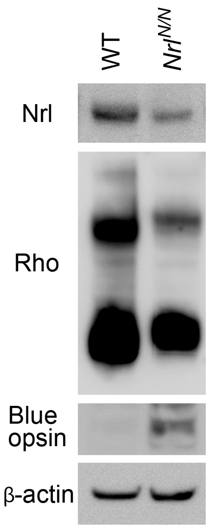
A transgenic NrlN/N mouse line was generated by inserting a PGK-Neo cassette flanked with two FRT sites (NrlN) into the intron between exons 3 and 4 of the Nrl locus (Figure 1). The PGK-Neo cassette is known to influence the expression of genes [19,20]. Mice homozygous for the NrlN allele are viable, fertile, and grossly normal. Western blot analysis showed that Nrl protein in retinas of NrlN/N mice was significantly reduced (Figure 2), accompanied by an increase in S-opsin and a decrease in rhodopsin protein (Figure 2). These results confirmed that Nrl expression was downregulated in NrlN/N mice, and rod differentiation was attenuated.
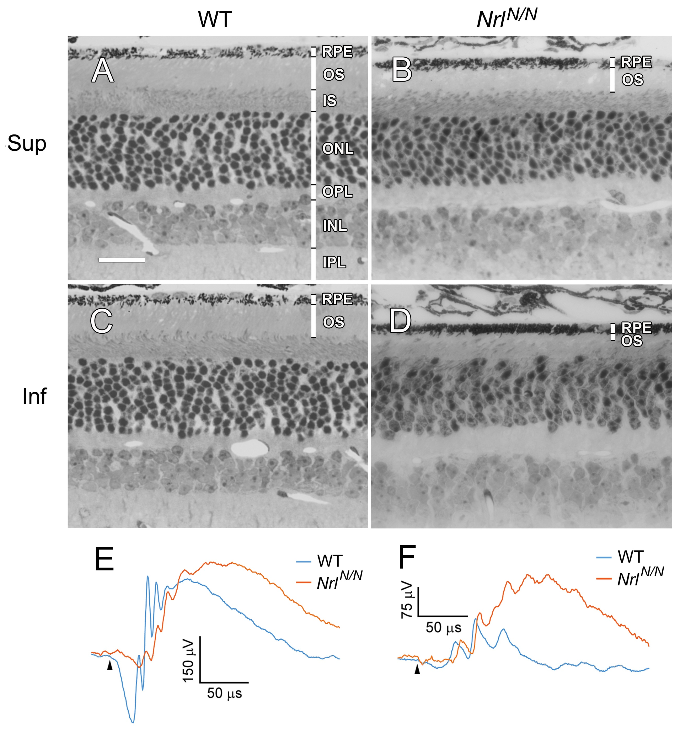
Morphologically, the retinas of NrlN/N animals (Figure 3B,D) exhibited normal retinal layers with normal laminar organization compared with the retinas of WT mice (Figure 3A,C), although in the NrlN/N mice, the ONL (outer nuclear layer) was one row of nuclei less than that of the wild-type mice. Photoreceptor outer segments (OSs) were shorter in NrlN/N mice (Figure 3B,D), especially in the inferior retina (Figure 3D). No retinal disorganization or rosette was observed in NrlN/N mice (Figure 3B,D), unlike in Nrl-knockout mice, in which retinal disorganization and rosette are common [10].
Dark-adapted ERGs (electroretinograms) from NrlN/N mice showed a very small a-wave, consistent with short photoreceptor outer segments (Figure 3E). The b-wave in the dark-adapted ERG was smaller than that from the WT mice (Figure 3E). In the light-adapted ERG, the b-wave from NrlN/N mice was larger than that from WT mice, consistent with more cone-like cells (Figure 3F, see below).
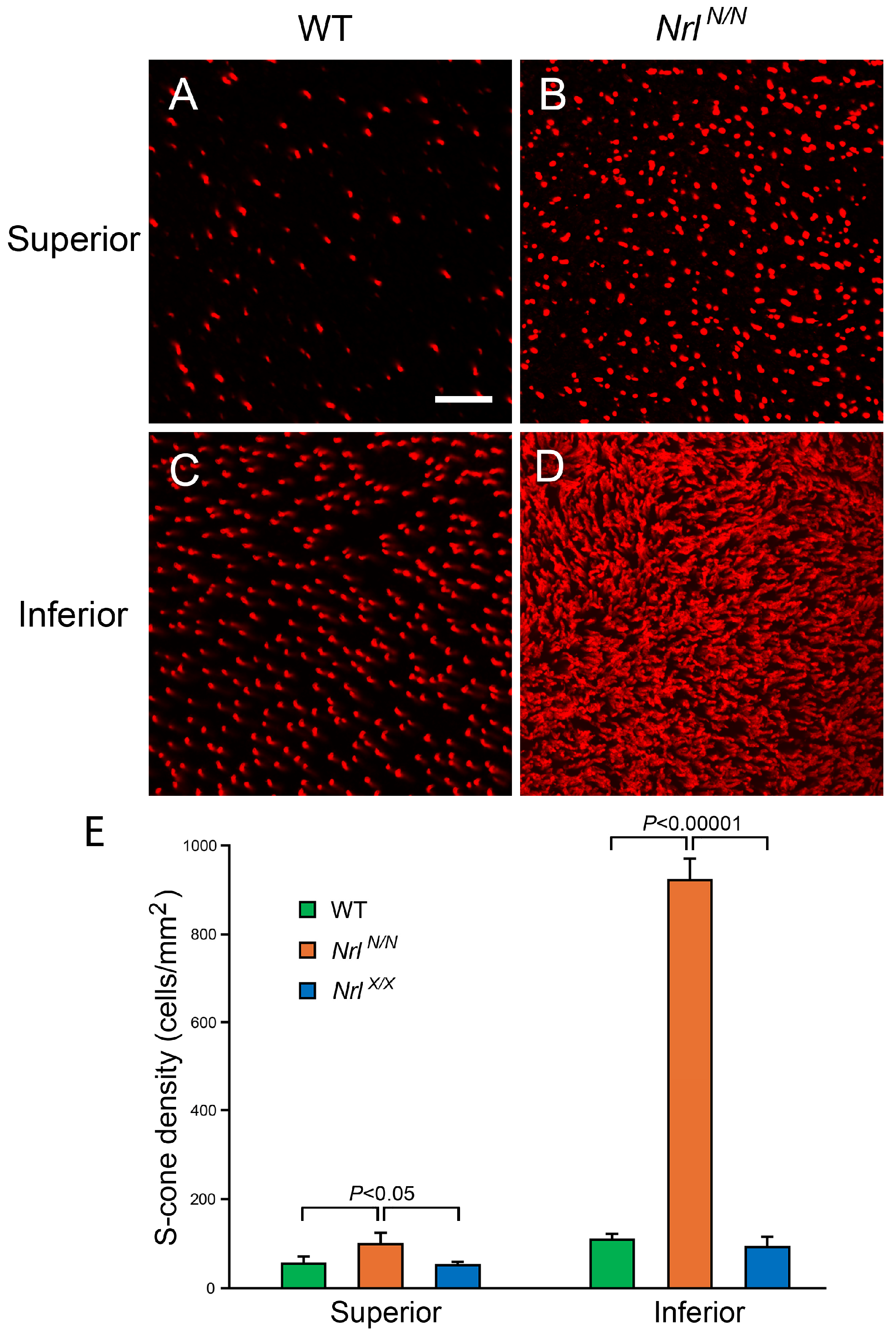
NrlN/N animals had significantly more S-opsin-positive cells in the retina (Figure 4B,D) than in wild-type mice (Figure 4A,C), especially in the inferior retina (Figure 4D). The average density of S-opsin-positive cells in the inferior retinas of NrlN/N mice was 8.3 times the density in the inferior retinas of WT mice (Figure 4E; Table 1). When the PGK-Neo cassette was removed from NrlN/N mice by crossing them with R26FLPe mice, the density of S-opsin-positive cells in the Nrl X/X mice was not significantly different from that in WT retinas (Figure 4E). R26FLPe mice express FLPe (an enhanced version of flippase) [21] in multiple organs, including the retina.
These results indicate that downregulation of Nrl expression reprograms rod photoreceptors, and cells in the inferior retina are more sensitive to Nrl downregulation than cells in the superior retina.
It is worth noticing that the density of S-opsin-positive cells in the inferior retinas in WT mice was significantly higher than that in the superior retinas (Figure 4E; Table 1). The densities of S-opsin-positive cells were higher in the inferior retinas in NrlN/N and NrlX/X mice (Figure 4E; Table 1).

2.2. Nrl Downregulation and Photoreceptor Survival

To investigate the effects of Nrl downregulation on photoreceptor degeneration, we crossed NrlN/N mice with two rod degeneration models: the rd1 (Pde6brd1/rd1) mouse and the Rho P23H knock-in mouse (RhoP23H/P23H). The Rd1 mouse carries the rd1 mutation in the Pde6b gene and is a model widely used in RP research [22]. The RhoP23H/P23H mouse is a transgenic mouse harboring the P23H mutation in the Rho gene [23]. The Rho P23H mutation is the most common mutation in autosomal dominant RP patients [3,23].
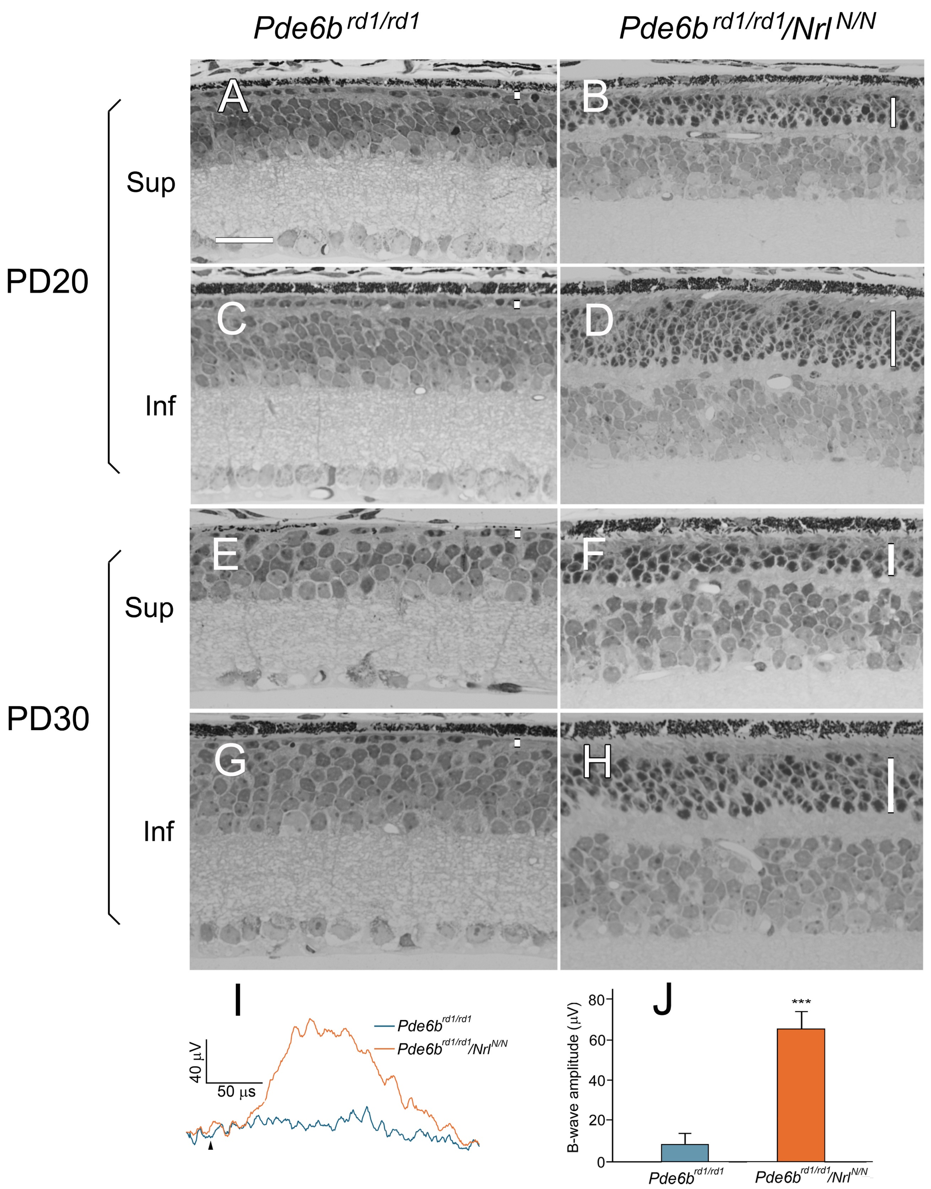
Photoreceptors in rd1 mice underwent rapid degeneration (Figure 5A,C,E,G). The ONL had one row of nuclei at PD 20 (postnatal day 20) (Figure 5A,C) and less than one row by PD 30 (Figure 5E,G). In double-mutant Pde6brd1/rd1/NrlN/N mice, the ONL had 3–4 rows of nuclei in the superior retina and 5–6 rows in the inferior retina at PD 20 (Figure 5B,D). At PD 30, the ONL in the superior retina had three rows of photoreceptor nuclei in the ONL and five in the inferior retina (Figure 5F,H). The ERG from the rd1 mouse was flat at PD 30, whereas the ERG b-wave from the Pde6brd1/rd1/NrlN/N mouse was significantly larger (Figure 5I,J).
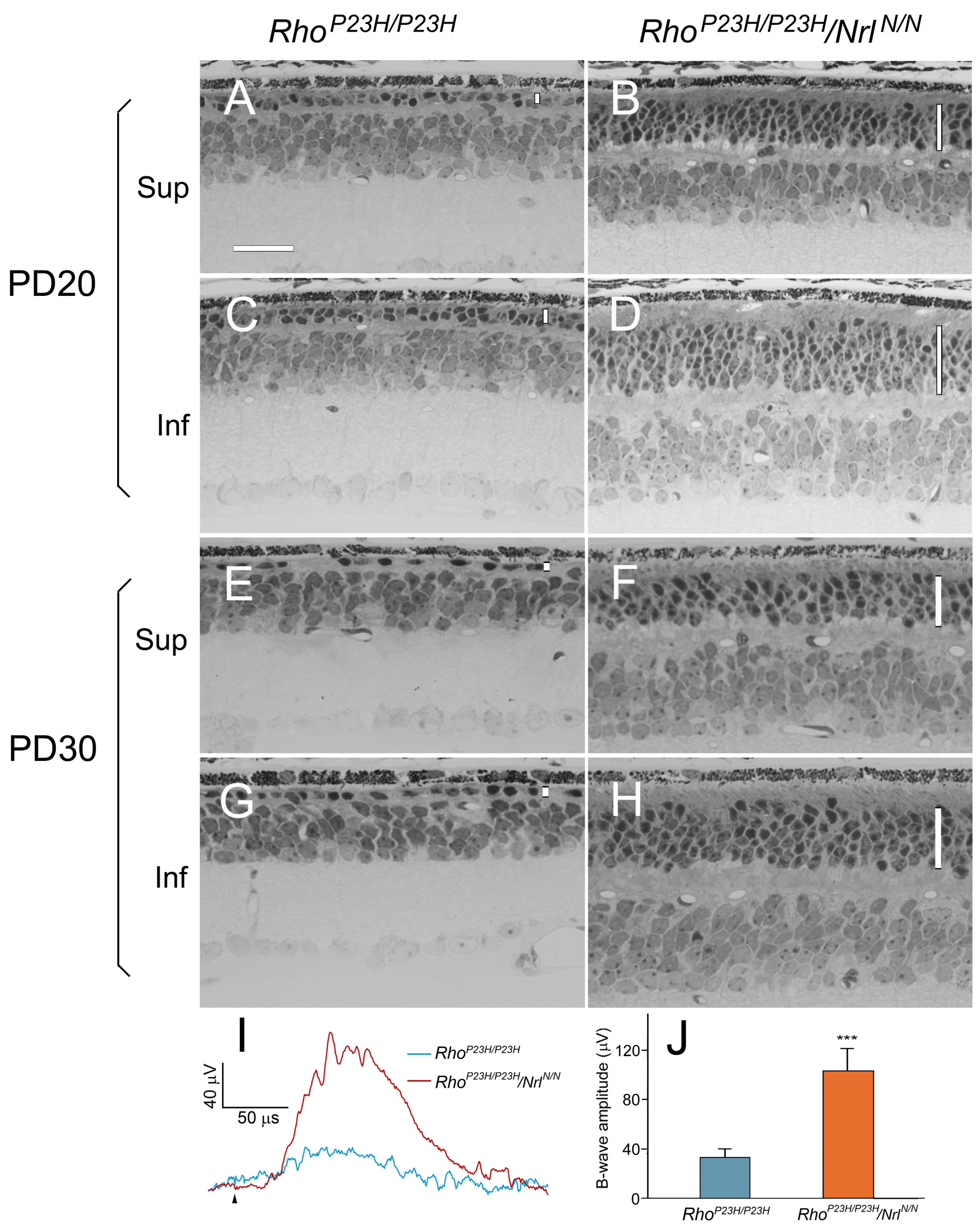
Retinal degeneration was also fast in RhoP23H/P23H mice. At PD 20, the ONL had 1–2 rows of nuclei in the superior retina and two rows in the inferior retina (Figure 6A,C). By PD 30, the ONL had less than one row of nuclei in the superior retina and one row in the inferior retina (Figure 6E,G). In RhoP23H/P23H/NrlN/N mice, however, the ONL had four rows of photoreceptor nuclei in the superior retina (Figure 6B) and six rows in the inferior retina at P20 (Figure 6D). And by PD 30, the ONL still had 3–4 rows of nuclei in the superior retina (Figure 6F) and 5–6 rows in the inferior retina (Figure 6H). The ERG from the RhoP23H/P23H mouse was almost flat at PD 30. In contrast, the ERG b-wave from the RhoP23H/P23H/NrlN/N mouse was significantly larger (Figure 6I,J).
These results indicate that reprogrammed photoreceptors are resistant to RP-associated degeneration.

2.3. Nrl Downregulation with shRNA Targeting Nrl mRNA

We next assessed the downregulation of Nrl expression by shRNA. ShRNA targeting different regions of the ORF (open reading frame) of mouse Nrl mRNA were predicted by two online algorithms (see Materials and Methods). Sequences of shRNA that effectively downregulated Nrl expression were selected, and a full shRNA was created by inserting an shRNA-targeting sequence into the mirE backbone [24] and fused to the 3′ UTR (untranslated region) of a small fluorescent protein, CagFbFP [25].
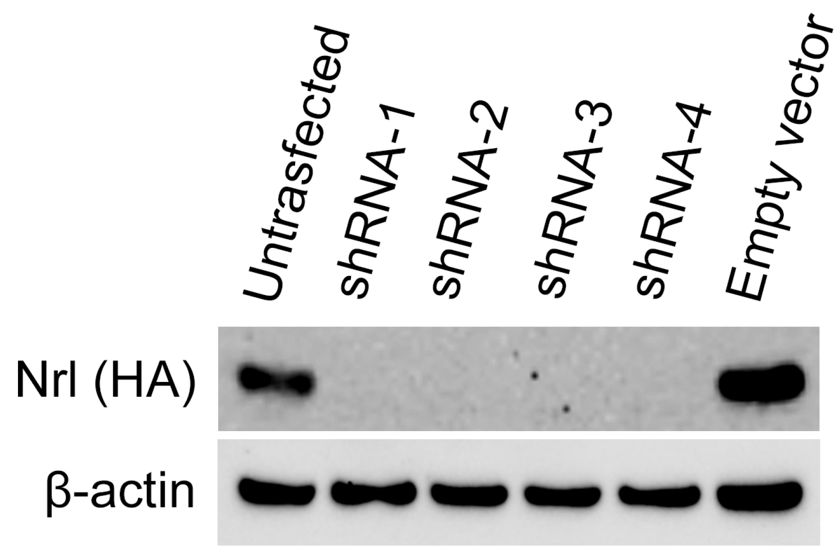
The capability of a given shRNA to downregulate Nrl expression was evaluated in 293-Nrl cells that stably overexpressed mouse Nrl. A plasmid containing a given shRNA* (pRVS-CagFbFP-shRNA*) was transfected into 293-Nrl cells, and the transfected cells were harvested 72 h later. Western blot analysis showed Nrl expression was blocked by each shRNA effectively (Figure 7). shRNA not targeting mouse Nrl had no effects on Nrl expression.
Two shRNAs (shRNA-2 and -4) shown in Figure 7 were selected and packaged in AAV for experiments with rd10 mice, a retinal degeneration model for experimental therapy [22]. The right eye of an rd10 mouse was injected with 1.5 µL of either AAV-shRNA-2 or AAV-shRNA-4 into the subretinal space at PD 14, and the left eye was injected with 1.5 µL of control AAV-GFP. Eyes were collected by PD 35 for morphological analysis. ERGs were recorded before eye collection.
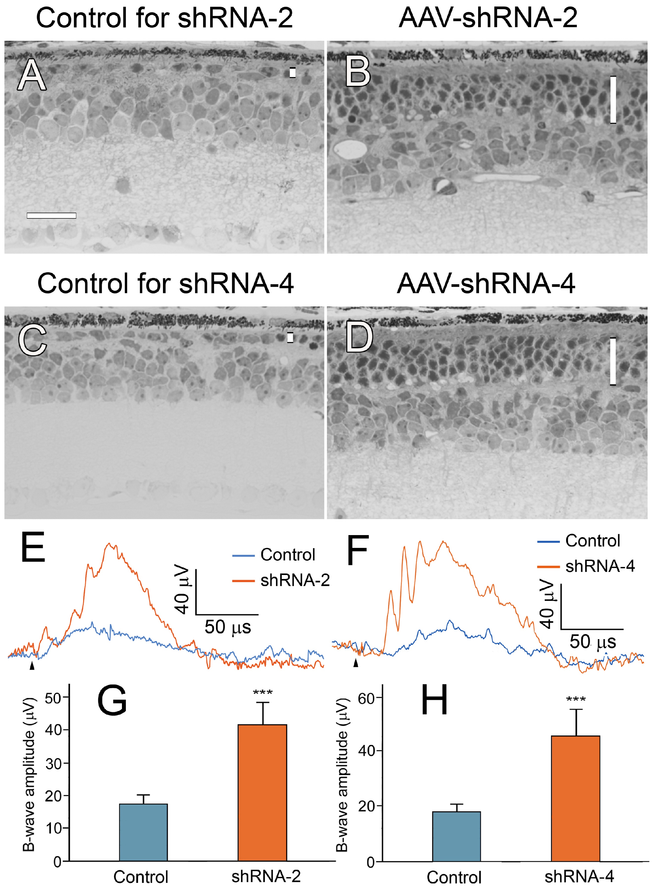
In the eyes treated with AAV-shRNA-2 (Figure 8B) or AAV-shRNA-4 (Figure 8D), the ONL in the injected area had 4–5 rows of nuclei, compared with one row of nuclei in the control eyes (Figure 8A,C). The ERG b-wave amplitudes from the eyes injected with either AAV-shRNA-2 or AAV-shRNA-4 were larger than those from control eyes (Figure 8E–H).

3. Discussion

We showed that the rod photoreceptor phenotype can be reprogrammed by downregulating Nrl expression. In the transgenic NrlN/N mouse, Nrl expression was effectively downregulated by a PGK-Neo cassette inserted into the intron between exons 3 and 4 of the Nrl gene. The PGK-Neo cassette is known to downregulate nearby genes [19,20].
The most important finding from experiments with the NrlN/N mouse is that rod photoreceptors can be reprogrammed by reducing Nrl expression, indicating that the function of Nrl is a graded regulator of rod phenotype rather than an all-or-none determinant of rod differentiation, as previously assumed. The discovery of reprogramming rod photoreceptors by reducing Nrl expression also represents a major advance toward developing mutation-independent therapies for RP.
Targeting Nrl by genomically neutralizing the Nrl gene, as shown by previous studies [13,14,15,16], poses substantial hurdles for clinical translation. In contrast, downregulation of Nrl expression is technically straightforward and clinically feasible. Our results showed that shRNA-mediated suppression of Nrl mRNA effectively enhanced photoreceptor survival when delivered by an AAV. These findings provide compelling preclinical evidence for an AAV-shRNA-based, mutation-independent therapeutic strategy for RP.
shRNA is a powerful tool for knocking down gene expression by targeting specific mRNA sequences. A typical shRNA consists of a target-specific stem and a loop, with the full shRNA sequence usually under 100 bp. In the present work, we designed a compact all-in-one expression cassette (<2.5 kb) that includes a tissue-specific promoter, the ORF of a small fluorescent reporter (CagFbFP), the shRNA sequence embedded in the 3′ UTR, and flanking AAV inverted terminal repeats (ITRs). The size was small enough to be packaged into a double-stranded AAV (dsAAV) vector [26]. Unlike a single-stranded AAV (ssAAV) that requires rate-limiting second-strand synthesis in host cells, a dsAAV delivers a transcription-ready double-stranded genome, resulting in a faster onset and higher transgene expression [26]. Moreover, the small footprint of each shRNA allows multiple shRNAs targeting different regions of the same mRNA to be combined within a single vector, thereby enhancing the overall knockdown efficiency.
A striking feature of the NrlN/N mouse was the marked dorsoventral difference in rod photoreceptor differentiation. Photoreceptor outer segments in the inferior retina were substantially shorter (Figure 3), and the number of S-opsin-positive cells was ~9-fold higher than in the superior retina (Figure 4; Table 1). Thus, rods in the inferior retina were considerably more sensitive to reduced Nrl expression than those in the superior retina. These findings indicate that rods in the inferior retina are less differentiated than those in the superior retina in NrlN/N mice. In wild-type mice, the density of S-opsin-positive cells in the inferior retina was also significantly higher than that in the superior retina (Figure 4, Table 1), suggesting similar dorsoventral asymmetry in rod differentiation.
It is surprising to notice that rods in the superior retina were well-preserved in both Pde6brd1/rd1/NrlN/N (Figure 5B) and RhoP23H/P23H/NrlN/N mice (Figure 6B), even though rods in this region showed limited reprogramming in the NrlN/N background (as discussed above). Thus, a substantial increase in rod survival was achieved with a low level of rod reprogramming, suggesting that therapeutic strategies aimed at downregulating Nrl for RP could have a broad therapeutic dose window. A successful clinical outcome may not require complete or a high level of Nrl suppression.
Our results from NrlN/N mice indicate that Nrl is essential for maintaining normal retinal layers and laminal organization, even at a low level. Retinal disorganization and rosette formation are common in Nrl-knockout (Nrl-/-) mice when Nrl expression is absent [10,13]. No such structural disruption or retinal disorganization was observed in NrlN/N mice (Figure 3A). When considering targeting Nrl as a therapy for RP, downregulating Nrl expression is likely a safer approach than gene knockout, as the latter may carry the risk of inducing retinal disorganization and rosette formation as unintended side effects.
Rod photoreceptor reprogramming would reduce retinal light sensitivity, thereby decreasing overall scotopic vision in treated eyes. In modern environments with ubiquitous artificial lighting, however, this is unlikely to cause meaningful functional difficulty. The benefit of halting the progressive retinal degeneration and preserving high-acuity vision by downregulating Nrl should far outweigh the modest trade-off in low-light sensitivity.
In summary, we demonstrated that the rod photoreceptor phenotype can be reprogrammed by downregulating the expression of Nrl, and our AAV-shRNA experiments provide pre-clinical evidence supporting AAV-shRNA as a mutation-independent therapeutic approach for both recessive and dominant RP.

4. Materials and Methods

4.1. Animals

Procedures involving animals were approved by the Institutional Animal Care and Use Committee (IACUC) of the University of Miami, the Miller School of Medicine. All methods were carried out in accordance with relevant guidelines and regulations, including adherence to the ARRIVE (Animal Research: Reporting of In Vivo Experiments) guidelines and the ARVO (Association for Research in Vision and Ophthalmology) Statement for the Use of Animals in Ophthalmic and Vision Research.
C57/6, Pde6brd1, RhoP23H, Pde6brd10 and R26FLPe mice were purchased from Jackson Labs (Bar Harbor, ME, USA). The animals were kept in a 12 h light/dark cycle at an in-cage illumination of <50 lux. The room temperature was kept at 20–22 °C.

4.2. Transgenic NrlN/N Mouse Generation

A transgenic NrlN/N mouse line was generated in the Transgenic Mouse Facility, the University of Miami School of Medicine. The transgene (NrlN) DNA construct contained a PGK-Neo cassette flanked by a pair of FRT sites inserted into the intron between exons 3 and 4 of the mouse Nrl gene (Figure 1). The DNA construct was electroporated into mouse embryonic stem cells. Correctly targeted cells were selected and used to generate chimeric animals. Transgenic chimeric mice were bred to obtain animals homozygous for the NrlN allele.

4.3. shRNA Design

Sequences of shRNA targeting different regions of mouse Nrl mRNA ORF were predicted by 2 algorithms online, the GPP Web Portal of Broad Institute [27] and the SplashRNA of Memorial Sloan Kettering Cancer Center [28], and tested for their efficacy in downregulating Nrl expression. A full shRNA was made by placing a targeting sequence in the optimized mirE backbone [24], which was embedded in the 3′ UTR of a small fluorescent protein, CagFbFP [24,25], to create CagFbFP-shRNA. The DNA construct was cloned into the vector pRc/RSV (Invitrogen, Carlsbad, CA, USA) to create the plasmid pRSV-CagFbFP-shRNA. The plasmid containing a given shRNA was tested for downregulating mouse Nrl expression in cells overexpressing mouse Nrl (see below).

4.4. Nrl-Expressing Cells and shRNA Evaluation

A cell line overexpressing mouse Nrl was created for testing the capacity of a given shRNA to downregulate mouse Nrl expression. A plasmid (pcDNA-mNrl) expressing mouse Nrl was created by subcloning the mouse Nrl cDNA sequence (ORF plus a C-terminus HA-tag) into pcDNA3.1-puro (Thermo Fisher Scientific, Waltham, MA, USA) and transfected into HEK293T cells (CRL-3211, ATCC, Manassas, VA, USA) using the jetPRIME transfection kit (Avantor, Radnor, PA, USA). Cells were cultured in DMEM (Dulbecco’s modified Eagle’s medium) with 10% fetal calf serum at 37 °C and maintained at 37 °C in 5% CO2/95% air. Stably transfected cells were selected with 1 µg/mL of puromycin. A cell colony expressing a high level of Nrl was selected as 293-Nrl and expanded.
To evaluate the Nrl-downregulating capability of a given shRNA*, a plasmid containing the shRNA (pRVS-CagFbFP-shRNA*; see above) was transfected into 293-Nrl cells. The level of mouse Nrl protein was examined 72 h later. Untransfected 293-Nrl cells and cells transfected with the empty vector served as controls.

4.5. AAV-shRNA Construction

AAV-shRNA was created by subcloning the DNA construct of a given shRNA* (CagFbFP-shRNA*) into pscAAV, downstream of the human rhodopsin promoter. AAV-shRNA* was then packaged as double-stranded AAV (pscAAV-shRNA*) into serotype AAV2.7m8 for photoreceptor expression [26,29]. AAV-shRNA and AAV-GFP (>2 × 10¹³ GC/mL in PBS) were produced by Vector Builder (Chicago, IL, USA).

4.6. Subretinal Injection

To inject AAV-shRNA, an rd10 mouse was anesthetized with intraperitoneal injections of ketamine (80 mg/kg) and xylazine (10 mg/kg). AAV-shRNA was injected into the subretinal space via a 33-gauge blunt-ended needle connected to a 10 µL microsyringe (Hamilton, Reno, NV, USA) under a surgical microscope. The right eye of the mouse was injected with 1.5 µL of AAV-shRNA, and the left eye was injected with the control viral vector AAV-GFP (1.5 µL).

4.7. Western Blotting

Retinas were harvested after animals were euthanized using CO2 overdose. Cell samples were collected after transfection. Retinal or cell samples were homogenized in the RIPA lysis buffer, and the total protein concentration of a sample was determined by the BCA (bicinchoninic acid) protein assay (Bio-Rad Labs, Hercules, CA, USA). Western blotting was performed using primary antibodies (anti-Nrl ABN1712, Millipore Sigma, Burlington, MA, USA; anti-HA 26183, Thermo Fisher Scientific, Waltham, MA, USA; anti-β actin sc-47778, Santa Cruz Biotechnology, Santa Cruz, CA, USA; anti-S-opsin AB5407, Millipore Sigma; and anti-rhodopsin, B6-30, gift of Dr. Jeremy Nathans) followed by appropriate secondary antibody conjugated to horseradish peroxidase. Antigen signals were detected with a Chemiluminescent Substrate Kit (Thermo Fisher Scientific, Waltham, MA, USA) and imaged on a luminescent image analyzer (ImageQuant LAS-4000, GE Healthcare Life Sciences, Chicago, IL, USA).

4.8. Histology

For histological evaluation of the retinas, animals were euthanized using CO2 overdose and immediately perfused with mixed aldehydes, as described previously [30,31]. Eyes were removed, semi-sectioned along the vertical meridian, and embedded in an Epon/Araldite mixture [30,31]. Semi-thin (1 µm thick) sections were cut to display the entire retina along the vertical meridian, or through the injected region, and stained with toluidine blue [30,31]. Retinal sections were examined by light microscopy.

4.9. Immunocytochemistry

Immunocytochemical staining was employed to identify S-opsin-positive cells in whole-mount retinas. Tissue samples were prepared as described previously [32]. Briefly, the animals were perfused with PBS after being euthanized using CO2 overdose. The eyes were collected, and retina–lens preparations were made by removing the corneas and then the sclera–choroid–RPE. The retina–lens preparations were fixed in 4% paraformaldehyde, washed with PBS, and incubated with primary anti-S-opsin antibodies (anti-S-opsin AB5407, MilliporeSigma) and then secondary antibodies conjugated with Cy3 (Jackson ImmunoResearch, West Grove, PA USA). After antibody incubation, the lenses were removed, and the retinas were flat-mounted photoreceptor-side up on slides. Stained whole-mount retinas were examined by confocal microscopy, and cone densities were quantified.

4.10. Electroretinogram (ERG)

ERGs were recorded with a UTAS system (LKC Technologies, Gaithersburg, MD, USA). For dark-adapted ERG, animals were dark-adapted for more than 3 h before recording. Light-adapted ERGs were recorded after animals were light-adapted to room light for >30 min.
An animal was put on the animal holder with a heat pad to maintain body temperature at 37 °C after being anesthetized with intraperitoneal injections of ketamine (80 mg/kg) and xylazine (10 mg/kg). A contact lens electrode was placed on the cornea of each eye after the pupils were dilated with 0.1% atropine and 0.1% phenylephrine HCl. A differential electrode was placed under the skin of the forehead, and a ground electrode under the skin at the base of the tail. Full-field ERGs were elicited by 1 ms white flashes generated by white LEDs in the Ganzfeld sphere. The inter-stimulus intervals were 10 s. Each recording was the average of 10 responses.

4.11. Statistical Analysis

Statistical analyses were performed using the Prism 10 (GraphPad Software, Boston, MA, USA). Data were evaluated by Student’s t-test for comparisons between two experimental groups or ANOVA (analysis of variance) followed by Tukey comparison among three or more groups.

Author Contributions

Conceptualization: Y.L. and R.W.; Investigation: Y.L., S.J., W.T. and R.W.; Writing: Y.L., S.J., W.T. and R.W. All authors have read and agreed to the published version of the manuscript.

Funding

This work was supported by NIH grants R01EY015289, R01EY018586, R01EY031492, R01EY026643, A Scientific Award to Dr. Wen from Hope for Vision, grants from the James and Esther King Biomedical Research Program of the State of Florida (Grant 08KN-09, 2KF02), and a private donation to Dr. Wen for Retinal Degeneration Research. It was also supported by NIH Core Grants P30EY14801 and P30EY002162 to Bascom Palmer Eye Institute, and an unrestricted grant from Research to Prevent Blindness Inc. to Bascom Palmer Eye Institute.

Institutional Review Board Statement

Procedures involving animals were approved by the Institutional Animal Care and Use Committee (IACUC) of the University of Miami, the Miller School of Medicine (Protocol #23-108, approval date 12 July 2023).

Informed Consent Statement

Not applicable.

Data Availability Statement

The data are presented in this article.

Conflicts of Interest

Author Weng Tao was employed by the company QOL Medical. The remaining authors declare that the research was conducted in the absence of any commercial or financial relationships that could be construed as a potential conflict of interest.

Abbreviations

The following abbreviations are used in this manuscript:
RPRetinitis pigmentosa
AAVAdeno-associated virus
dsAAVDouble-stranded adeno-associated virus
ssAAVSingle-stranded adeno-associated virus
NrlNeural retina leucine zipper
DHDDSDehydrodolichyl diphosphate synthase
shRNAShort hairpin RNA
HAHemagglutinin
ERGElectroretinogram
PGK–NeoPhosphoglycerate kinase I promoter–neomycin phosphotransferase gene
FLPFlippase recombinase
FRTFlippase recognition target
PBSPhosphate-buffered saline
RPERetinal pigment epithelium
OSOuter segment
ISInner segment
ONLOuter nuclear layer
OPLOuter plexiform layer
INLInner nuclear layer
IPLInner plexiform layer
WTWild type
ORFOpen reading frame
UTRUntranslated region

References

  1. Verbakel, S.K.; van Huet, R.A.C.; Boon, C.J.F.; den Hollander, A.I.; Collin, R.W.J.; Klaver, C.C.W.; Hoyng, C.B.; Roepman, R.; Klevering, B.J. Non-syndromic retinitis pigmentosa. Prog. Retin. Eye Res. 2018, 66, 157–186. [Google Scholar] [CrossRef]
  2. Delyfer, M.N.; Leveillard, T.; Mohand-Said, S.; Hicks, D.; Picaud, S.; Sahel, J.A. Inherited retinal degenerations: Therapeutic prospects. Biol. Cell 2004, 96, 261–269. [Google Scholar] [CrossRef]
  3. Hartong, D.T.; Berson, E.L.; Dryja, T.P. Retinitis pigmentosa. Lancet 2006, 368, 1795–1809. [Google Scholar] [CrossRef]
  4. RetNet. Summaries of Genes and Loci Causing Retinal Diseases. Waltham, MA, USA. Available online: https://retnet.org/summaries (accessed on 25 October 2025).
  5. Swaroop, A.; Xu, J.Z.; Pawar, H.; Jackson, A.; Skolnick, C.; Agarwal, N. A conserved retina-specific gene encodes a basic motif/leucine zipper domain. Proc. Natl. Acad. Sci. USA 1992, 89, 266–270. [Google Scholar] [CrossRef]
  6. Raza, M.; Cornish, E.E.; Ovens, C.; Nash, B.M.; McGaughran, J.; Jamieson, R.V.; Grigg, J.R. NRL-associated autosomal recessive retinopathy: Novel variants expanding the phenotype, natural history and a comprehensive literature search. Ophthalmic Genet. 2026, 47, 22–34. [Google Scholar] [CrossRef]
  7. Littink, K.W.; Stappers, P.T.Y.; Riemslag, F.C.C.; Talsma, H.E.; van Genderen, M.M.; Cremers, F.P.M.; Collin, R.W.J.; van den Born, L.I. Autosomal recessive nrl mutations in patients with enhanced s-cone syndrome. Genes 2018, 9, 68, Correction in Genes 2018, 9, 145. [Google Scholar] [CrossRef] [PubMed]
  8. Bessant, D.A.; Payne, A.M.; Mitton, K.P.; Wang, Q.L.; Swain, P.K.; Plant, C.; Bird, A.C.; Zack, D.J.; Swaroop, A.; Bhattacharya, S.S. A mutation in NRL is associated with autosomal dominant retinitis pigmentosa. Nat. Genet. 1999, 21, 355–356. [Google Scholar] [CrossRef]
  9. Bessant, D.A.; Holder, G.E.; Fitzke, F.W.; Payne, A.M.; Bhattacharya, S.S.; Bird, A.C. Phenotype of retinitis pigmentosa associated with the Ser50Thr mutation in the NRL gene. Arch. Ophthalmol. 2003, 121, 793–802. [Google Scholar] [CrossRef] [PubMed]
  10. Mears, A.J.; Kondo, M.; Swain, P.K.; Takada, Y.; Bush, R.A.; Saunders, T.L.; Sieving, P.A.; Swaroop, A. Nrl is required for rod photoreceptor development. Nat. Genet. 2001, 29, 447–452. [Google Scholar] [CrossRef] [PubMed]
  11. Mustafi, D.; Engel, A.H.; Palczewski, K. Structure of cone photoreceptors. Prog. Retin. Eye Res. 2009, 28, 289–302. [Google Scholar] [CrossRef]
  12. Oh, E.C.; Khan, N.; Novelli, E.; Khanna, H.; Strettoi, E.; Swaroop, A. Transformation of cone precursors to functional rod photoreceptors by bZIP transcription factor NRL. Proc. Natl. Acad. Sci. USA 2007, 104, 1679–1684. [Google Scholar] [CrossRef]
  13. Montana, C.L.; Kolesnikov, A.V.; Shen, S.Q.; Myers, C.A.; Kefalov, V.J.; Corbo, J.C. Reprogramming of adult rod photoreceptors prevents retinal degeneration. Proc. Natl. Acad. Sci. USA 2013, 110, 1732–1737. [Google Scholar] [CrossRef]
  14. Yu, W.; Mookherjee, S.; Chaitankar, V.; Hiriyanna, S.; Kim, J.W.; Brooks, M.; Ataeijannati, Y.; Sun, X.; Dong, L.; Li, T.; et al. Nrl knockdown by AAV-delivered CRISPR/Cas9 prevents retinal degeneration in mice. Nat. Commun. 2017, 8, 14716. [Google Scholar] [CrossRef]
  15. Zhu, J.; Ming, C.; Fu, X.; Duan, Y.; Hoang, D.A.; Rutgard, J.; Zhang, R.; Wang, W.; Hou, R.; Zhang, D.; et al. Gene and mutation independent therapy via CRISPR-Cas9 mediated cellular reprogramming in rod photoreceptors. Cell Res. 2017, 27, 830–833, Correction in Cell Res. 2019, 29, 337. [Google Scholar] [CrossRef]
  16. Liu, Z.; Chen, S.; Lo, C.H.; Wang, Q.; Sun, Y. All-in-one AAV-mediated Nrl gene inactivation rescues retinal degeneration in Pde6a mice. J. Clin. Investig. 2024, 9, e178159. [Google Scholar] [CrossRef]
  17. Moore, S.M.; Skowronska-Krawczyk, D.; Chao, D.L. Targeting of the nrl pathway as a therapeutic strategy to treat retinitis pigmentosa. J. Clin. Med. 2020, 9, 2224. [Google Scholar] [CrossRef] [PubMed]
  18. Mak, T.W.; Penninger, J.M.; Ohashi, P.S. Knockout mice: A paradigm shift in modern immunology. Nat. Rev. Immunol. 2001, 1, 11–19. [Google Scholar] [CrossRef] [PubMed]
  19. Olson, E.N.; Arnold, H.H.; Rigby, P.W.; Wold, B.J. Know your neighbors: Three phenotypes in null mutants of the myogenic bHLH gene MRF4. Cell 1996, 85, 1–4. [Google Scholar] [CrossRef] [PubMed]
  20. Pham, C.T.; MacIvor, D.M.; Hug, B.A.; Heusel, J.W.; Ley, T.J. Long-range disruption of gene expression by a selectable marker cassette. Proc. Natl. Acad. Sci. USA 1996, 93, 13090–13095. [Google Scholar] [CrossRef]
  21. Buchholz, F.; Angrand, P.O.; Stewart, A.F. Improved properties of FLP recombinase evolved by cycling mutagenesis. Nat. Biotechnol. 1998, 16, 657–662. [Google Scholar] [CrossRef]
  22. Chang, B.; Hawes, N.L.; Hurd, R.E.; Davisson, M.T.; Nusinowitz, S.; Heckenlively, J.R. Retinal degeneration mutants in the mouse. Vis. Res. 2002, 42, 517–525. [Google Scholar] [CrossRef] [PubMed]
  23. Sakami, S.; Maeda, T.; Bereta, G.; Okano, K.; Golczak, M.; Sumaroka, A.; Roman, A.J.; Cideciyan, A.V.; Jacobson, S.G.; Palczewski, K. Probing mechanisms of photoreceptor degeneration in a new mouse model of the common form of autosomal dominant retinitis pigmentosa due to P23H opsin mutations. J. Biol. Chem. 2011, 286, 10551–10567. [Google Scholar] [CrossRef] [PubMed]
  24. Fellmann, C.; Hoffmann, T.; Sridhar, V.; Hopfgartner, B.; Muhar, M.; Roth, M.; Lai, D.Y.; Barbosa, I.A.; Kwon, J.S.; Guan, Y.; et al. An optimized microRNA backbone for effective single-copy RNAi. Cell Rep. 2013, 5, 1704–1713. [Google Scholar] [CrossRef]
  25. Nazarenko, V.V.; Remeeva, A.; Yudenko, A.; Kovalev, K.; Dubenko, A.; Goncharov, I.M.; Kuzmichev, P.; Rogachev, A.V.; Buslaev, P.; Borshchevskiy, V.; et al. A thermostable flavin-based fluorescent protein from Chloroflexus aggregans: A framework for ultra-high resolution structural studies. Photochem. Photobiol. Sci. 2019, 18, 1793–1805. [Google Scholar] [CrossRef]
  26. Wang, Z.; Ma, H.I.; Li, J.; Sun, L.; Zhang, J.; Xiao, X. Rapid and highly efficient transduction by double-stranded adeno-associated virus vectors in vitro and in vivo. Gene Ther. 2003, 10, 2105–2111. [Google Scholar] [CrossRef]
  27. GPP Web Portal. Broad Institute 2022. Available online: https://portals.broadinstitute.org/gpp/public/gene/search (accessed on 25 October 2025).
  28. SplashRNA. Memorial Sloan Kettering Cancer Center. 2022. Available online: http://splashrna.mskcc.org/ (accessed on 25 October 2025).
  29. Dalkara, D.; Byrne, L.C.; Klimczak, R.R.; Visel, M.; Yin, L.; Merigan, W.H.; Flannery, J.G.; Schaffer, D.V. In vivo-directed evolution of a new adeno-associated virus for therapeutic outer retinal gene delivery from the vitreous. Sci. Transl. Med. 2013, 5, 189ra176. [Google Scholar] [CrossRef]
  30. LaVail, M.M.; Battelle, B.A. Influence of eye pigmentation and light deprivation on inherited retinal dystrophy in the rat. Exp. Eye Res. 1975, 21, 167–192. [Google Scholar] [CrossRef] [PubMed]
  31. Song, Y.; Zhao, L.; Tao, W.; Laties, A.M.; Luo, Z.; Wen, R. Photoreceptor protection by cardiotrophin-1 in transgenic rats with the rhodopsin mutation s334ter. Investig. Ophthalmol. Vis. Sci. 2003, 44, 4069–4075. [Google Scholar] [CrossRef]
  32. Li, Y.; Tao, W.; Luo, L.; Huang, D.; Kauper, K.; Stabila, P.; Lavail, M.M.; Laties, A.M.; Wen, R. CNTF induces regeneration of cone outer segments in a rat model of retinal degeneration. PLoS ONE 2010, 5, e9495. [Google Scholar] [CrossRef] [PubMed]
Figure 1. A schematic of the NrlN DNA construct. The PGK-Neo cassette (red), flanked by two FRT sites (yellow), was inserted into the intron between exons 3 and 4 (gray) of the mouse Nrl gene. The PGK-Neo cassette is removable in the presence of recombinase flippase (FLP). Exon 4 is flanked by two loxP sites (blue).
Ijms 27 04683 g001
Figure 2. Nrl downregulation in NrlN/N mice. Retinas were collected at PD (postnatal day) 60 from NrlN/N and WT (wildtype C57) mice. Protein levels were examined by Western blot analysis. In NrlN/N mice, the level of Nrl was significantly reduced compared with that in the WT control. The levels of rhodopsin (Rho) were also reduced, whereas the level of S-opsin was increased in NrlN/N mice. The levels of β-actin served as loading controls.
Ijms 27 04683 g002
Figure 3. Retinal morphology and ERG of NrlN/N mice. Eyes were collected from PD 60 NrlN/N and WT mice. The retina of the NrlN/N mouse had normal layers in laminar organization (B,D) compared with that of the WT mouse (A,C), with 1 row of nuclei in the ONL thinner than the WT. The rod outer segments (OSs) were shorter in the NrlN/N mouse (B,D) than in the control retina (A,C), especially in the inferior retina (D). At PD 60, the dark-adapted ERG from NrlN/N mouse had a very small a-wave, and the b-wave amplitude was smaller than that of the WT mouse ((E); elicited by −0.402 log cd·s/m2 white flashes). The light-adapted b-wave from NrlN/N mice was larger than that from WT mice ((F); elicited by 0.998 log cd·s/m2 flashes with 30 cd/m2 white background). Black arrowheads in (E,F) indicate ERG flash onset. Retinal layers are indicated by white bars in panel A. RPE: retinal pigment epithelium; OS: outer segment; IS: inner segment; ONL: outer nuclear layer; OPL: outer plexiform layer; INL: inner nuclear layer; IPL: inner plexiform layer. Sup: superior; Inf: inferior. Scale bar: 25 μm.
Ijms 27 04683 g003
Figure 4. S-opsin-positive cells in the retina. Eyes were collected at PD 60 from NrlN/N and WT mice. Immunocytochemical analysis in flat-mounted retinas showed many S-opsin-positive cells in the retina of the NrlN/N mouse (B,D), especially in the inferior retina (D), compared with the superior (A) and inferior retina (C) of the WT (wild-type) mouse. The average density of S-opsin-positive cells in the superior retinas of NrlN/N mice was significantly higher than that in either WT mice (p < 0.05; n = 3) or NrlX/Xmice in which the PGK-Neor cassette was removed (p < 0.05; n = 3) (E). In the inferior retina, the density of S-opsin-positive cells in NrlN/N mice was much higher than in the WT and NrlX/X -N/-N mice (p < 0.0001; n = 3) (E). The S-opsin-positive cell densities in the NrlX/X mouse in the superior or the inferior retina were comparable to those in the WT mouse (E). In addition, the density of S-opsin-positive cells in the inferior retina in WT mice was significantly higher than in the superior retina (p < 0.005; n = 3), as well as in NrlN/N mice (p < 0.0001; n = 3) and NrlX/X mice (p < 0.05; n = 3). Sup: superior; Inf: inferior. Scale bar: 20 µm.
Ijms 27 04683 g004
Figure 5. Photoreceptor preservation in mice with Pde6brd1/rd1/NrlN/Ndouble mutants. Retinal degeneration in the rd1 (Pde6brd1/rd1) mouse is rapid. The ONL had 1 row of nuclei at PD 20 in both the superior (A) and inferior retina (C), and less than 1 row of nuclei by PD 30 (E,G). In the Pde6brd1/rd1/NrlN/N mouse, photoreceptors were well-preserved. At PD 20, the ONL had 3–4 rows of nuclei in the superior retina, and 5–6 rows in the inferior retina (B,D), and by PD 30, the ONL still had 3 rows in the superior retina, and 5 rows in the inferior retina (F,H). The ERG (elicited by 0.398 log cd·s/m2 white flashes with a 30 cd/m2 white background) from the Pde6brd1/rd1 mouse was flat, whereas the b-wave from the Pde6brd1/rd1/NrlN/N mouse was significantly larger at PD 30 ((I,J); *** p < 0.0001; n = 3). Black arrowhead indicates flash onset. The ONL in each retinal section is indicated by a vertical white bar (AD). Sup: superior; Inf: inferior. Scale bar: 25 µm.
Ijms 27 04683 g005
Figure 6. Photoreceptor preservation in mice with RhoP23H/P23H/NrlN/N double mutants. Retinal degeneration in RhoP23H/P23H mouse was so rapid that the ONL had less than 1–2 rows of nuclei in the superior retina (A) and 2 rows of nuclei in the inferior retina (C) at PD 20, and by PD 30, the ONL had less than 1 row of nuclei in the superior retina (E) and 1 row in the inferior retina (G). In the RhoP23H/P23H/NrlN/N mouse, the ONL had 4 rows of nuclei in the superior retina (B) and 6 rows in inferior retina (D) at P20. By PD 30, the ONL still had 3–4 rows of nuclei in the superior retina (F) and 5–6 rows in the inferior retina (H). The ERG at PD 30, elicited by 0.398 log cd·s/m2 white flashes with a 30 cd·s/m2 white background, showed that the b-wave from the RhoP23H/P23H mouse was very small, but the b-wave from the Pde6brd1/rd1/NrlN/N mouse was significantly larger ((I,J); *** p < 0.001; n = 3). Black arrowhead indicates flash onset. ONL in each retinal section is indicated by a vertical white bar (AD). Sup: superior; Inf: inferior. Scale bar: 25 µm.
Ijms 27 04683 g006
Figure 7. Nrl downregulation by shRNA. 293-Nrl cells stably expressing mouse Nrl were used to evaluate the capability of downregulating Nrl expression by shRNA. Cells were transfected with a plasmid containing a given shRNA and harvested 72 h after transfection. Western blots showed that 4 shRNAs successfully downregulated Nrl expression. Untransfected 293-Nrl cells and 293-Nrl cells transfected with the empty vector served as controls. The levels of β-actin in each sample served as loading controls.
Ijms 27 04683 g007
Figure 8. Photoreceptor protection by shRNA in mice with Pde6brd10/rd10mutation. The right eyes of rd10 mice were injected with AAV-shRNA-2 or AAV-shRNA-4 into the subretinal space at PD 14, and the left eyes were injected with AAV-GFP as injection controls. Eyes were collected at PD 35. The retinas in the control eyes had 1 row of nuclei in the ONL (A,C). In the eyes injected with AAV-shRNA-2 (B) or AAV-shRNA-4 (D), the ONL had 4 rows of nuclei in the injected area (B,D). The ERG b-waves from the control eyes were very small (E,F), but the b-waves from eyes injected with AAV-shRNA-2 (E,G) or AAV-shRNA-4 (F,H) were significantly larger (*** p < 0.001; n = 3 for (G,H)) (ERGs were elicited by 0.398 log cd·s/m2 white flashes with a 30 cd/m2 white background). Black arrowheads indicate flash onset. The ONL in each retinal section is indicated by a vertical white bar (AD). Scale bar: 25 µm.
Ijms 27 04683 g008
Table 1. Densities of S-opsin-positive cells *.
WTNrlN/NNrlX/X
Superior57 ± 11100 ± 2352 ± 4
Inferior111 ± 9925 ± 4594 ± 20
p-value
Inferior vs superior
<0.005<0.0001<0.05
* Data are presented as cells per 0.006 mm2; mean ± SD; n = 3.
Disclaimer/Publisher’s Note: The statements, opinions and data contained in all publications are solely those of the individual author(s) and contributor(s) and not of MDPI and/or the editor(s). MDPI and/or the editor(s) disclaim responsibility for any injury to people or property resulting from any ideas, methods, instructions or products referred to in the content.