The Molecular Mechanisms of Metformin’s Action on Blood Lipid Profile in Diabetic Patients

Abstract
1. Introduction
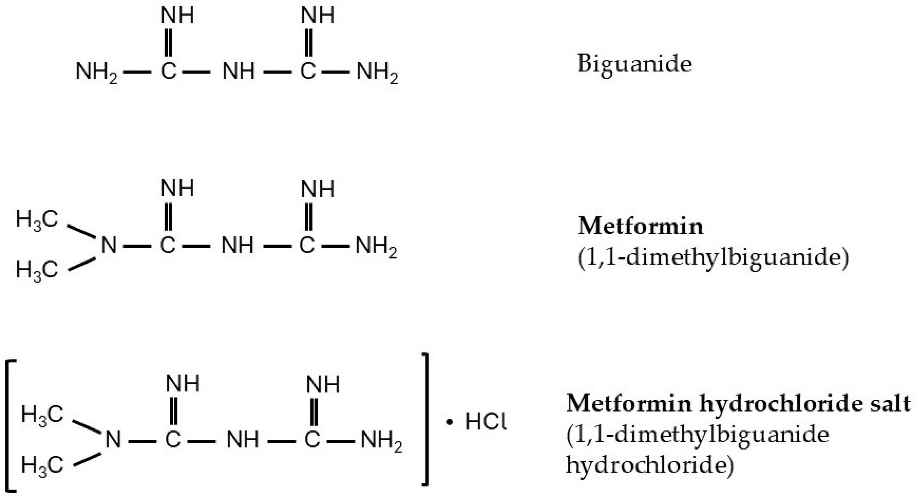
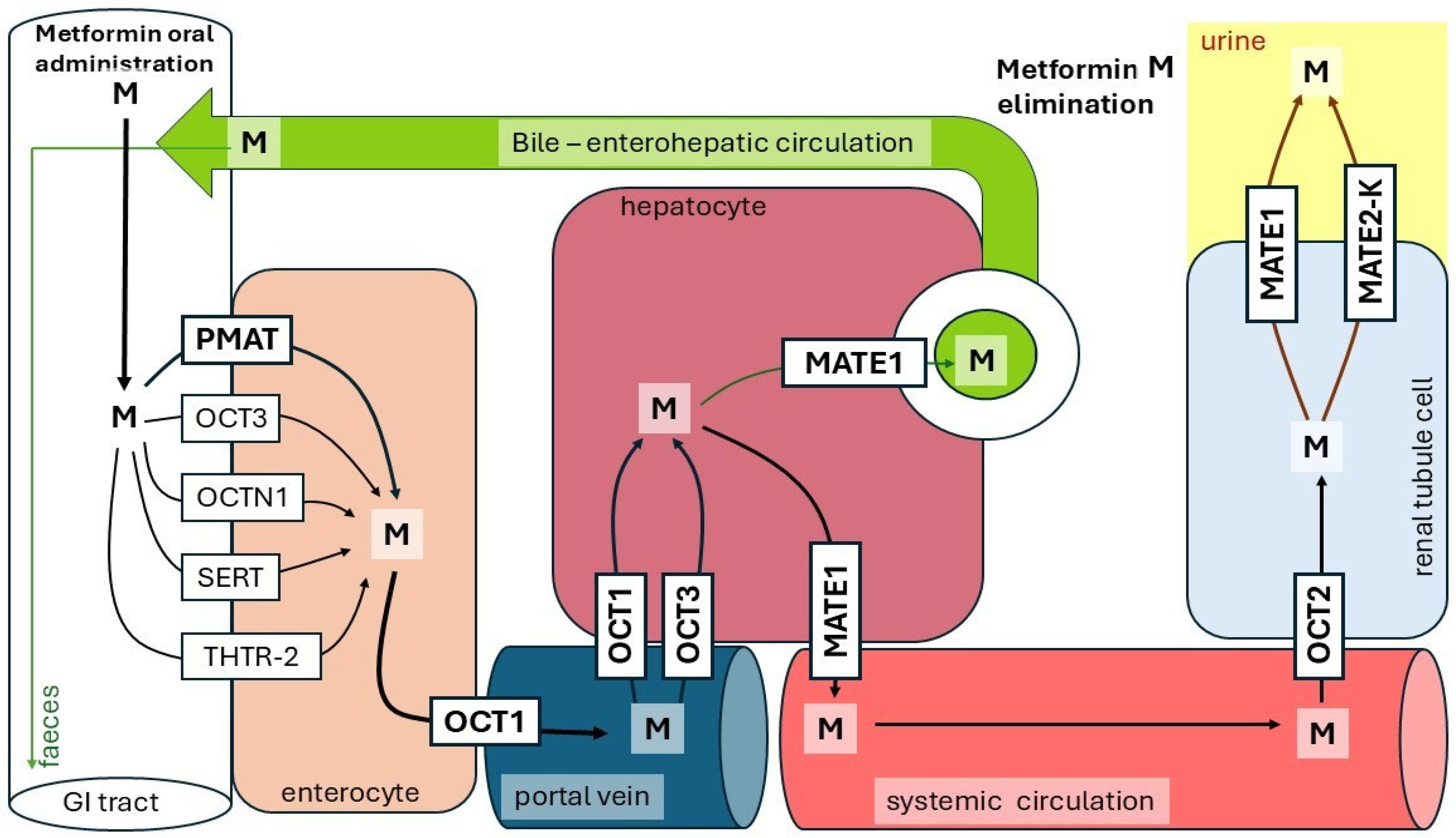
2. Metformin Absorption and Disposal
3. Metformin as an Antihyperglycemic Drug
4. Metformin as a Blood Lipid-Lowering Agent in Patients with T2D
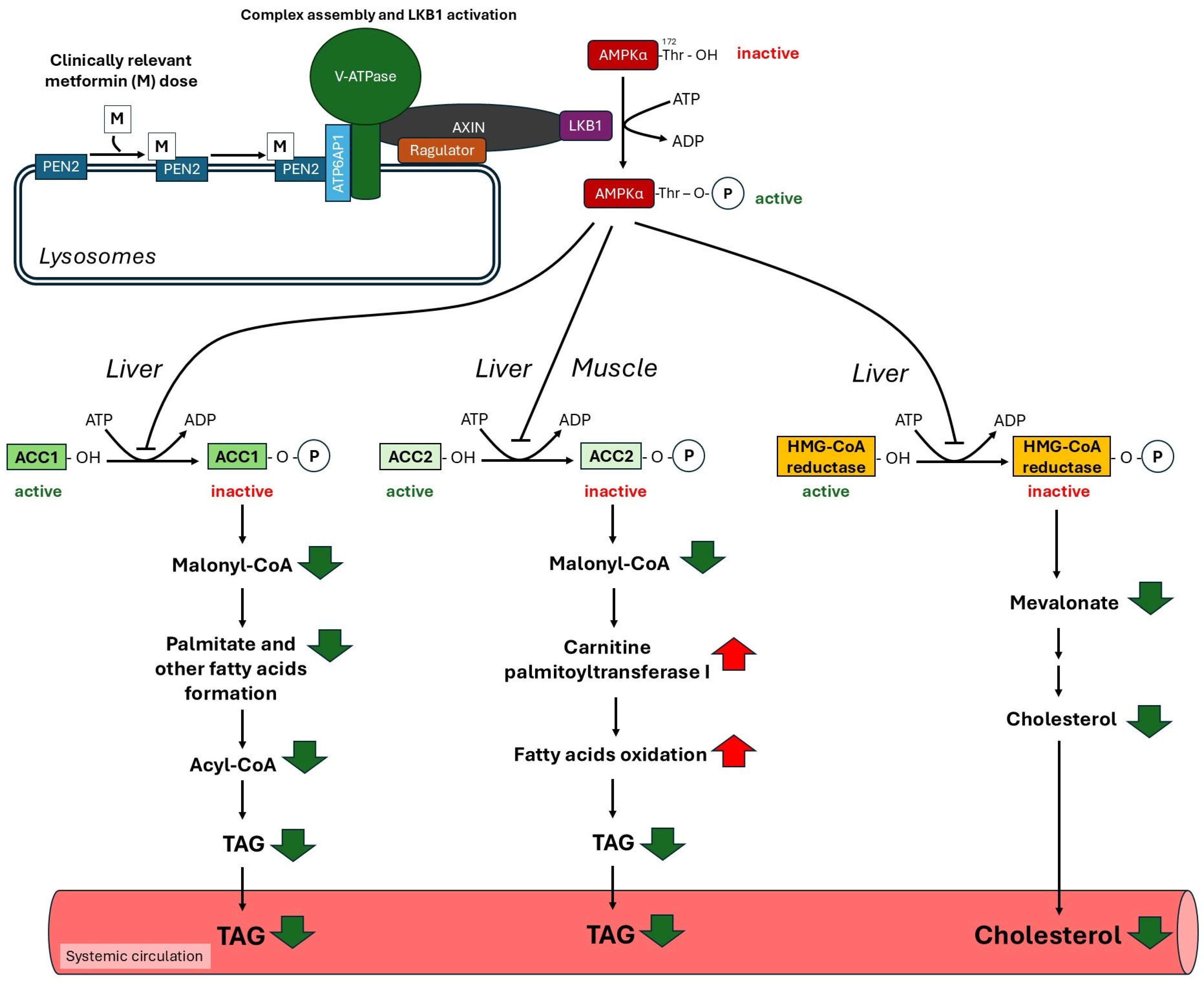
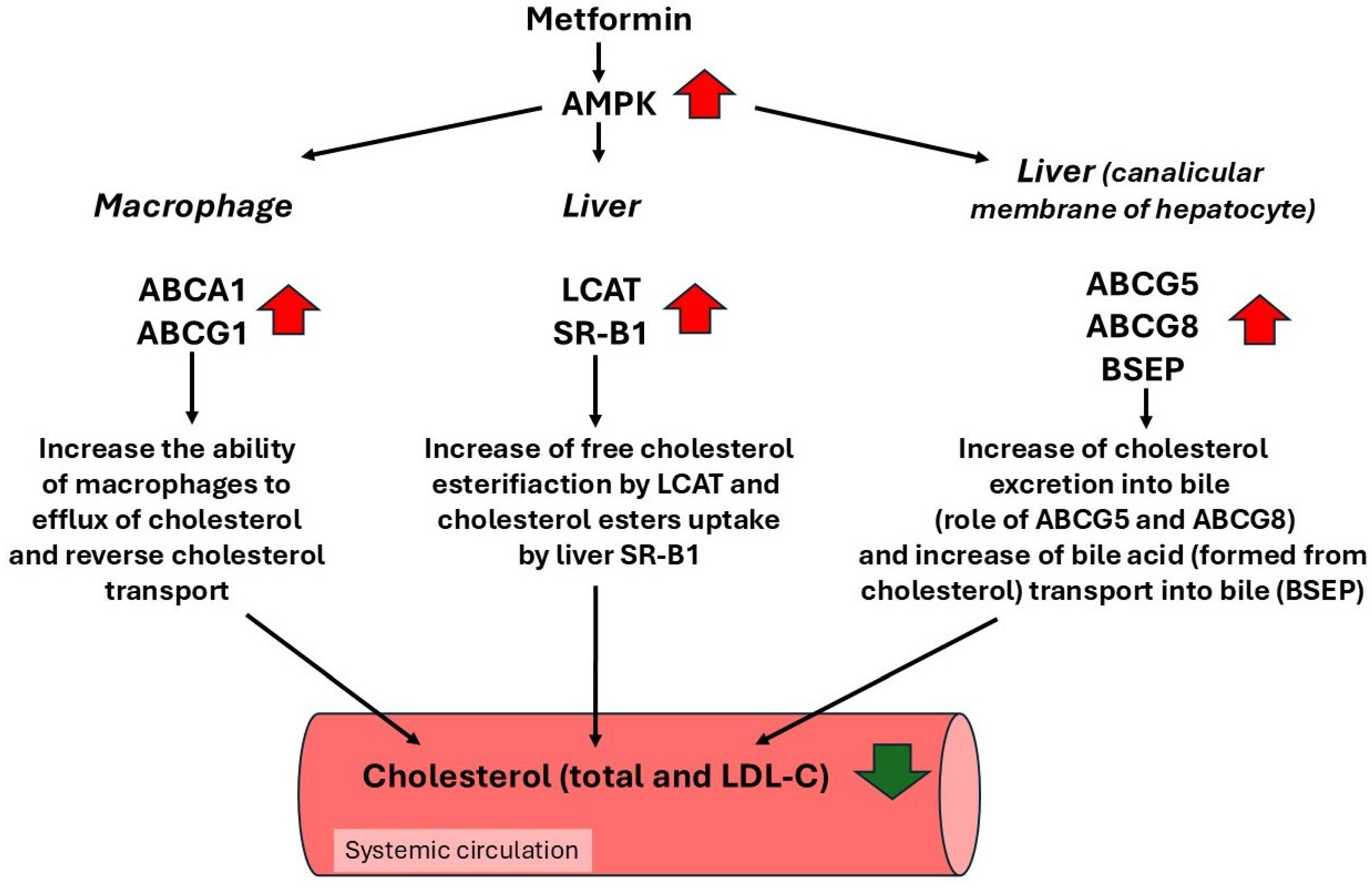
4.1. Potential Role of AMP-Activated Protein Kinase Activation by Metformin in the Regulation of Lipid Metabolism
4.2. Metformin Decreases Plasma Triacylglycerol Concentration via Promoting VLDL-Triacylglycerol Clearance by Brown Adipose Tissue (BAT)
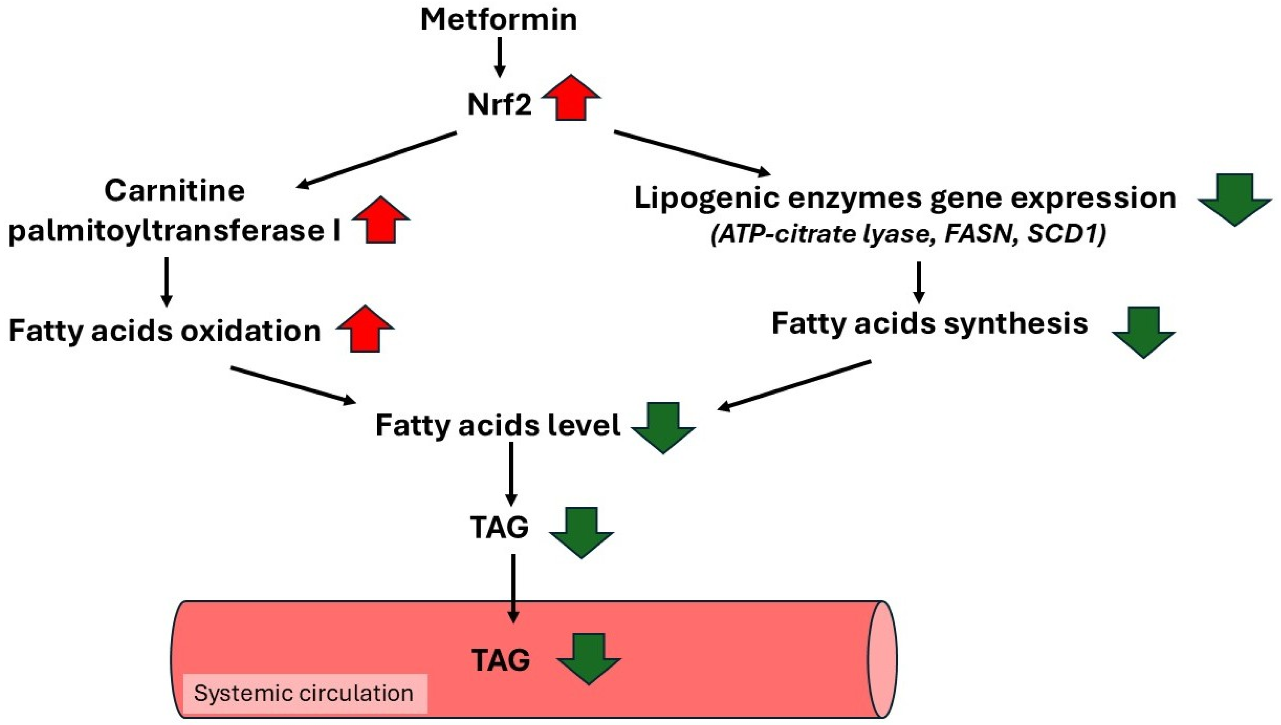
4.3. Metformin, via an Increase in the Nuclear Factor Erythroid 2-Related Factor 2 (Nrf2) Expression, Could Exert a Beneficial Effect on Lipid Profile in Diabetic Patients
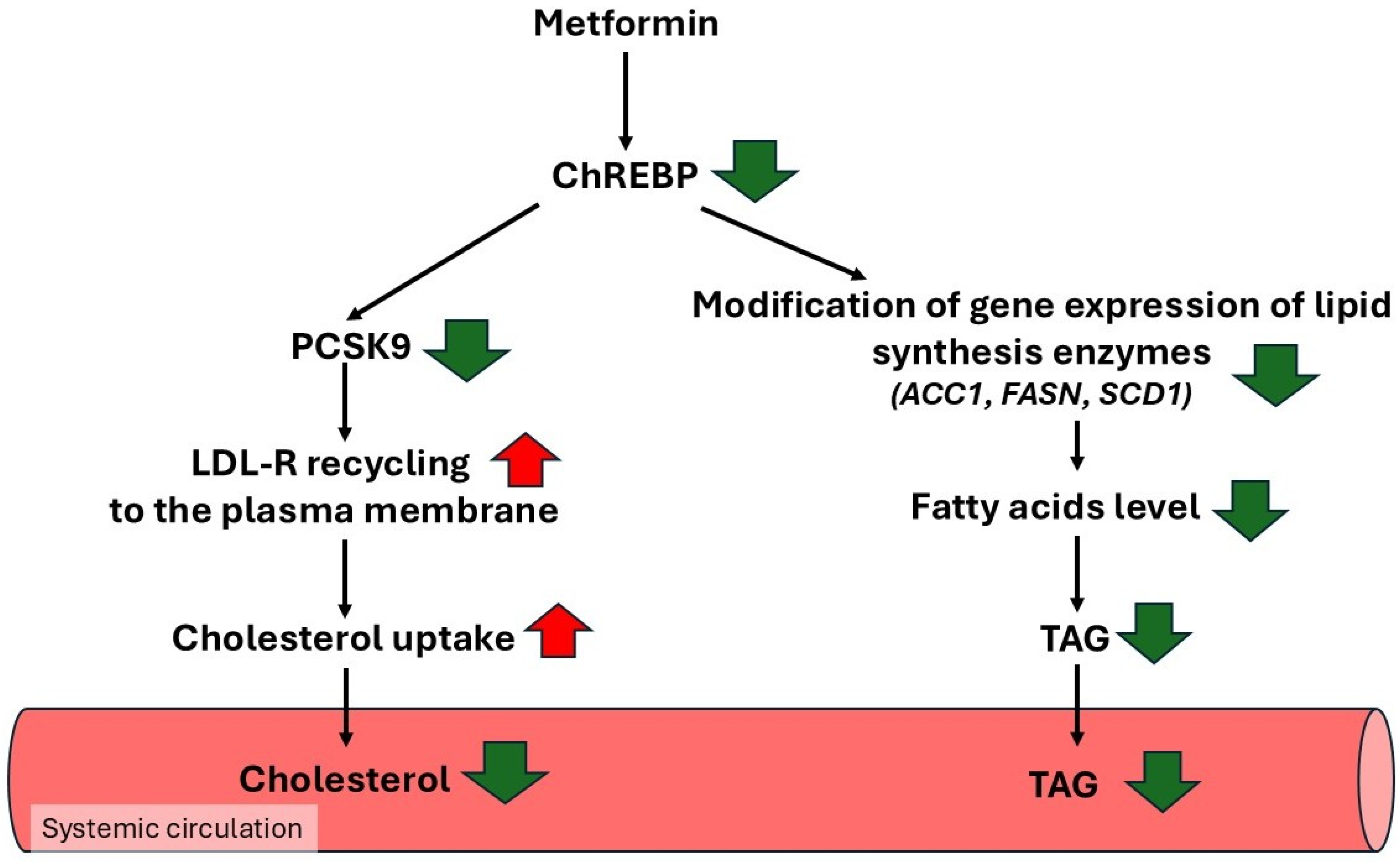
4.4. Metformin Reduces Blood Total Cholesterol and LDL-C Concentration via Suppressing ChREBP, Resulting in Decreased Gene Expression Encoding PCSK9
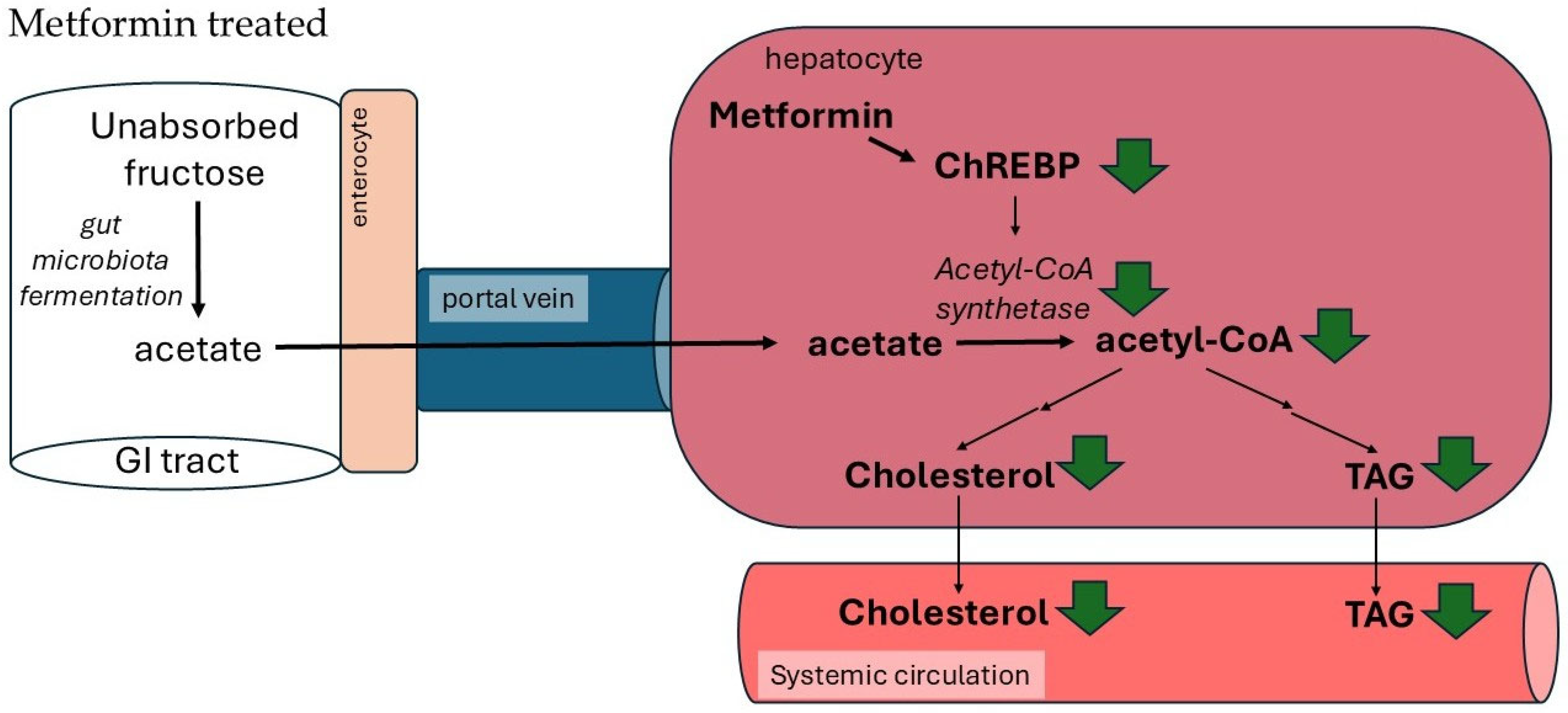
4.5. Metformin Reduces Blood TAG Concentration via Suppressing ChREBP, Resulting in Decreased Gene Expression Encoding Lipogenic Enzymes
4.6. Metformin Reduces Blood Triacylglycerol and Cholesterol Concentrations via Suppressing Acetyl-CoA Synthetase, Which Plays a Key Role in Liver TAG and Cholesterol Biosynthesis from Acetate Formed by Gut Microbiota
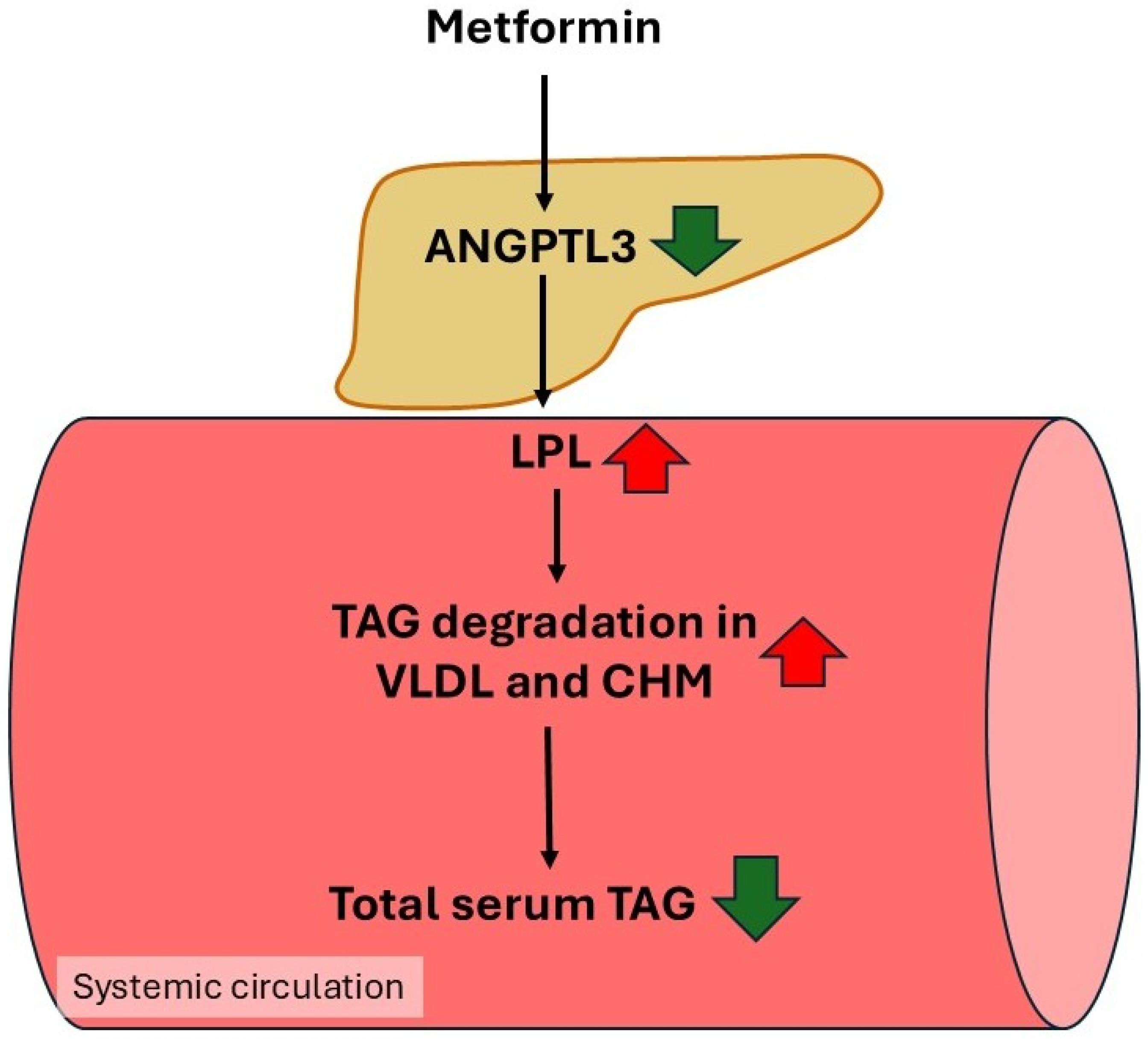
4.7. Inhibition of Angiopoetin-like 3 Protein (ANGPTL3) Gene Expression by Metformin—A Potential Mechanism for Lowering Plasma TAG Concentrations
4.8. Metformin as an AMPK Activator Enhances Reverse Cholesterol Transport
4.9. Metformin May Have a Beneficial Effect on Dyslipidemia in T2D Patients via Activation of AMPK, Which Inhibits LXRα Activity
4.10. Serum Lipid Alterations After 1 Dose of Metformin Intake by Healthy Subjects Analyzed by Mass Spectrometry
5. Conclusions
Author Contributions
Funding
Institutional Review Board Statement
Data Availability Statement
Conflicts of Interest
Abbreviations
| ABCA1 | ATP-binding cassette A1 |
| ABCG1 | ATP-binding cassette G1 |
| ABCG5 | ATP-binding cassette transporter G5 |
| ABCG8 | ATP-binding cassette transporter G8 |
| ACC1 | Acetyl-CoA carboxylase isoform I |
| ACC2 | Acetyl-CoA carboxylase isoform II |
| ADP | Adenosine diphosphate |
| AMP | Adenosine monophosphate |
| AMPK | AMP-activated protein kinase |
| ANGPTL3 | Angiopoietin-like 3 protein |
| APH-1 | Anterior pharynx defective 1 |
| Apo CIII | Apolipoprotein CIII |
| ApoB100 | Apolipoprotein B100 |
| APOE3 | Apolipoprotein E3 |
| ASCVD | Atherosclerotic cardiovascular disease |
| ATF1 | Activating transcription factor 1 |
| ATP | Adenosine triphosphate |
| ATP6AP1 | ATPase H+ Transporting Accessory Protein 1 |
| AXIN | Axis inhibitor |
| BAT | Brown adipose tissue |
| BCAA | Branched-chain amino acids |
| BSEP | Bile salt transporter |
| bZIP | Basic leucine zipper protein |
| CAD | Coronary artery disease |
| CaMKK2 | Ca2+/calmodulin dependent protein kinase 2 |
| cAMP | Cyclic adenosine monophosphate |
| CETP | Cholesteryl ester transfer protein |
| CHL | Cholesterol |
| ChREBP | Carbohydrate response element binding protein |
| CKD | Chronic kidney disease |
| CNC | Cap‘n’Collar transcription factor |
| COVID-19 | Coronavirus disease 2019 |
| CPT1 | Carnitine palmitoyl-CoA transferase 1 |
| CRE | cAMP Response Element |
| CREBP | cAMP response element binding protein |
| CVD | Cardiovascular disease |
| CYPs | Cytochrome P450 enzymes |
| EL | Endothelial lipase |
| FASN | Fatty acids synthase |
| FFA | Free fatty acids |
| GDF15 | Growth differentiation factor 15 |
| GIP | Glucose-dependent insulinotropic peptide |
| GLP-1 | Glucagon-like peptide 1 |
| GSH | Reduced glutathione |
| GSSG | Oxidized glutathione |
| HDL | High density lipoprotein |
| HDL-C | High density lipoprotein cholesterol |
| HMG-CoA | 3-hydroxy-3-methylglutaryl-CoA |
| HSL | Hormone-sensitive lipase |
| Km | Michaelis–Menten constant |
| LCAT | Lecithin cholesterol acyltransferase |
| LDL | Low density lipoprotein |
| LDL-R | Low density lipoprotein receptor |
| LKB1 | Liver kinase B1 |
| LoF | Loss-of-function mutations |
| Lp(a) | Lipoprotein a |
| LPL | Lipoprotein lipase |
| LXRα | Liver X receptor alfa |
| M | Metformin |
| MATE-1 | Multi-drug and toxin extrusion-1 transporter |
| MATE2-K | Multi-drug and toxin extrusion2-K transporter |
| mGPDH | Mitochondrial glycerol 3-phosphate dehydrogenase |
| NAD | Nicotinamide adenine dinucleotide |
| NADH | Nicotinamide adenine dinucleotide reduced |
| NAFLD | Non-alcoholic fatty liver disease |
| NASH | Non-alcoholic steatohepatitis |
| NKX3.1 | Androgen regulated gene encoding transcription factor |
| Nrf2 | Nuclear factor erythroid 2-related factor 2 |
| OCT1 | Organic cation transporter 1 |
| OCT2 | Organic cation transporter 2 |
| OCT3 | Organic cation transporter 3 |
| OCTN1 | Carnitine/organic cation transporter |
| PCSK9 | Proprotein convertase subtilisin/kexin 9 |
| PEN2 | Presenilin enhancer 2 |
| Pi | Phosphate |
| PKA | Protein kinase A |
| PMAT | Plasma membrane monoamine transporter |
| PPi | Pyrophosphate |
| Rap 1 | Ras-related protein 1 |
| SCD1 | Stearoyl-CoA desaturase 1 |
| SERT | Serotonin reuptake transporter |
| SGLT2 | Sodium/glucose cotransporter 2 |
| SLC19A3 | Solute carrier family 19 member 3 |
| SLC22A1 | Solute carrier family 22 member 1 |
| SLC22A4 | Solute carrier family 22 member 4 |
| SLC29A4 | Solute carrier family 29 member 4 |
| SLC6A4 | Solute carrier family 6 member 4 |
| SR-B1 | Scavenger receptor class B type 1 |
| SREBP-1c | Sterol regulatory element-binding protein 1c |
| SREBP2 | Sterol regulatory element-inding protein 2 |
| STK 11 (AMPK) | Serine/threonine kinase 11 |
| T2DM | Type 2 diabetes |
| TAG | Triacylglycerol |
| THTR-2 | Human thiamine transporter 2 |
| v-ATPase | Vacuolar adenosine triphosphatase |
| VLDL | Very low-density lipoprotein |
References
- Reitman, M.L.; Schadt, E.E. Pharmacogenetics of Metformin Response: A Step in the Path toward Personalized Medicine. J. Clin. Investig. 2007, 117, 1226–1229. [Google Scholar] [CrossRef]
- Schernthaner, G.; Schernthaner, G.-H. The Right Place for Metformin Today. Diabetes Res. Clin. Pract. 2020, 159, 107946. [Google Scholar] [CrossRef] [PubMed]
- Scheen, A.J. Clinical Pharmacokinetics of Metformin. Clin. Pharmacokinet. 1996, 30, 359–371. [Google Scholar] [CrossRef] [PubMed]
- Scarpello, J.H.B.; Howlett, H.C.S. Metformin Therapy and Clinical Uses. Diabetes Vasc. Dis. Res. 2008, 5, 157–167. [Google Scholar] [CrossRef]
- Stumvoll, M.; Nurjhan, N.; Perriello, G.; Dailey, G.; Gerich, J.E. Metabolic Effects of Metformin in Non-Insulin-Dependent Diabetes Mellitus. N. Engl. J. Med. 1995, 333, 550–554. [Google Scholar] [CrossRef]
- Diabetes Prevention Program Research Group. Long-Term Safety, Tolerability, and Weight Loss Associated with Metformin in the Diabetes Prevention Program Outcomes Study. Diabetes Care 2012, 35, 731–737. [Google Scholar] [CrossRef]
- Fullerton, M.D.; Galic, S.; Marcinko, K.; Sikkema, S.; Pulinilkunnil, T.; Chen, Z.-P.; O’Neill, H.M.; Ford, R.J.; Palanivel, R.; O’Brien, M.; et al. Single Phosphorylation Sites in Acc1 and Acc2 Regulate Lipid Homeostasis and the Insulin-Sensitizing Effects of Metformin. Nat. Med. 2013, 19, 1649–1654. [Google Scholar] [CrossRef] [PubMed]
- Day, E.A.; Ford, R.J.; Smith, B.K.; Mohammadi-Shemirani, P.; Morrow, M.R.; Gutgesell, R.M.; Lu, R.; Raphenya, A.R.; Kabiri, M.; McArthur, A.G.; et al. Metformin-Induced Increases in GDF15 Are Important for Suppressing Appetite and Promoting Weight Loss. Nat. Metab. 2019, 1, 1202–1208. [Google Scholar] [CrossRef]
- Coll, A.P.; Chen, M.; Taskar, P.; Rimmington, D.; Patel, S.; Tadross, J.A.; Cimino, I.; Yang, M.; Welsh, P.; Virtue, S.; et al. GDF15 Mediates the Effects of Metformin on Body Weight and Energy Balance. Nature 2020, 578, 444–448, Correction in Nature 2020, 578, E24. [Google Scholar] [CrossRef]
- Effect of Intensive Blood-Glucose Control with Metformin on Complications in Overweight Patients with Type 2 Diabetes (UKPDS 34). Lancet 1998, 352, 854–865. [CrossRef]
- Triggle, C.R.; Mohammed, I.; Bshesh, K.; Marei, I.; Ye, K.; Ding, H.; MacDonald, R.; Hollenberg, M.D.; Hill, M.A. Metformin: Is It a Drug for All Reasons and Diseases? Metabolism 2022, 133, 155223. [Google Scholar] [CrossRef]
- Malaekeh-Nikouei, A.; Shokri-Naei, S.; Karbasforoushan, S.; Bahari, H.; Baradaran Rahimi, V.; Heidari, R.; Askari, V.R. Metformin beyond an Anti-Diabetic Agent: A Comprehensive and Mechanistic Review on Its Effects against Natural and Chemical Toxins. Biomed. Pharmacother. 2023, 165, 115263. [Google Scholar] [CrossRef]
- Kulkarni, A.S.; Gubbi, S.; Barzilai, N. Benefits of Metformin in Attenuating the Hallmarks of Aging. Cell Metab. 2020, 32, 15–30. [Google Scholar] [CrossRef]
- Cory, T.J.; Emmons, R.S.; Yarbro, J.R.; Davis, K.L.; Pence, B.D. Metformin Suppresses Monocyte Immunometabolic Activation by SARS-CoV-2 Spike Protein Subunit 1. Front. Immunol. 2021, 12, 733921. [Google Scholar] [CrossRef] [PubMed]
- Gordon, D.E.; Jang, G.M.; Bouhaddou, M.; Xu, J.; Obernier, K.; White, K.M.; O’Meara, M.J.; Rezelj, V.V.; Guo, J.Z.; Swaney, D.L.; et al. A SARS-CoV-2 Protein Interaction Map Reveals Targets for Drug Repurposing. Nature 2020, 583, 459–468. [Google Scholar] [CrossRef] [PubMed]
- Libby, G.; Donnelly, L.A.; Donnan, P.T.; Alessi, D.R.; Morris, A.D.; Evans, J.M.M. New Users of Metformin Are at Low Risk of Incident Cancer: A Cohort Study among People with Type 2 Diabetes. Diabetes Care 2009, 32, 1620–1625. [Google Scholar] [CrossRef]
- Currie, C.J.; Poole, C.D.; Jenkins-Jones, S.; Gale, E.A.M.; Johnson, J.A.; Morgan, C.L. Mortality after Incident Cancer in People with and without Type 2 Diabetes: Impact of Metformin on Survival. Diabetes Care 2012, 35, 299–304. [Google Scholar] [CrossRef]
- Noto, H.; Goto, A.; Tsujimoto, T.; Noda, M. Cancer Risk in Diabetic Patients Treated with Metformin: A Systematic Review and Meta-Analysis. PLoS ONE 2012, 7, e33411. [Google Scholar] [CrossRef] [PubMed]
- Zakikhani, M.; Dowling, R.; Fantus, I.G.; Sonenberg, N.; Pollak, M. Metformin Is an AMP Kinase-Dependent Growth Inhibitor for Breast Cancer Cells. Cancer Res. 2006, 66, 10269–10273. [Google Scholar] [CrossRef]
- Evans, J.M.M.; Donnelly, L.A.; Emslie-Smith, A.M.; Alessi, D.R.; Morris, A.D. Metformin and Reduced Risk of Cancer in Diabetic Patients. BMJ 2005, 330, 1304–1305. [Google Scholar] [CrossRef]
- Saydullaeva, I.; Butuner, B.D.; Korkmaz, K.S. NKX3.1 Expression Contributes to Epithelial–Mesenchymal Transition of Prostate Cancer Cells. ACS Omega 2023, 8, 32580–32592. [Google Scholar] [CrossRef]
- Papachristodoulou, A.; Heidegger, I.; Virk, R.K.; Di Bernardo, M.; Kim, J.Y.; Laplaca, C.; Picech, F.; Schäfer, G.; De Castro, G.J.; Hibshoosh, H.; et al. Metformin Overcomes the Consequences of NKX3.1 Loss to Suppress Prostate Cancer Progression. Eur. Urol. 2024, 85, 361–372. [Google Scholar] [CrossRef]
- Hirst, J.A.; Farmer, A.J.; Ali, R.; Roberts, N.W.; Stevens, R.J. Quantifying the Effect of Metformin Treatment and Dose on Glycemic Control. Diabetes Care 2012, 35, 446–454. [Google Scholar] [CrossRef]
- LaMoia, T.E.; Shulman, G.I. Cellular and Molecular Mechanisms of Metformin Action. Endocr. Rev. 2021, 42, 77–96. [Google Scholar] [CrossRef] [PubMed]
- Pentikäinen, P.J.; Neuvonen, P.J.; Penttilä, A. Pharmacokinetics of Metformin after Intravenous and Oral Administration to Man. Eur. J. Clin. Pharmacol. 1979, 16, 195–202. [Google Scholar] [CrossRef] [PubMed]
- Sambol, N.C.; Chiang, J.; Lin, E.T.; Goodman, A.M.; Liu, C.Y.; Benet, L.Z.; Cogan, M.G. Kidney Function and Age Are Both Predictors of Pharmacokinetics of Metformin. J. Clin. Pharmacol. 1995, 35, 1094–1102. [Google Scholar] [CrossRef]
- Klepser, T.B.; Kelly, M.W. Metformin Hydrochloride: An Antihyperglycemic Agent. Am. J. Health-Syst. Pharm. 1997, 54, 893–903. [Google Scholar] [CrossRef]
- Foretz, M.; Guigas, B.; Bertrand, L.; Pollak, M.; Viollet, B. Metformin: From Mechanisms of Action to Therapies. Cell Metab. 2014, 20, 953–966. [Google Scholar] [CrossRef] [PubMed]
- Zhou, M.; Xia, L.; Wang, J. Metformin Transport by a Newly Cloned Proton-Stimulated Organic Cation Transporter (Plasma Membrane Monoamine Transporter) Expressed in Human Intestine. Drug Metab. Dispos. Biol. Fate Chem. 2007, 35, 1956–1962. [Google Scholar] [CrossRef]
- Liang, X.; Giacomini, K.M. Transporters Involved in Metformin Pharmacokinetics and Treatment Response. J. Pharm. Sci. 2017, 106, 2245–2250. [Google Scholar] [CrossRef]
- Chen, E.C.; Liang, X.; Yee, S.W.; Geier, E.G.; Stocker, S.L.; Chen, L.; Giacomini, K.M. Targeted Disruption of Organic Cation Transporter 3 Attenuates the Pharmacologic Response to Metformin. Mol. Pharmacol. 2015, 88, 75–83. [Google Scholar] [CrossRef]
- Nakamichi, N.; Shima, H.; Asano, S.; Ishimoto, T.; Sugiura, T.; Matsubara, K.; Kusuhara, H.; Sugiyama, Y.; Sai, Y.; Miyamoto, K.-I.; et al. Involvement of Carnitine/Organic Cation Transporter OCTN1/SLC22A4 in Gastrointestinal Absorption of Metformin. J. Pharm. Sci. 2013, 102, 3407–3417. [Google Scholar] [CrossRef]
- Han, T.K.; Proctor, W.R.; Costales, C.L.; Cai, H.; Everett, R.S.; Thakker, D.R. Four Cation-Selective Transporters Contribute to Apical Uptake and Accumulation of Metformin in Caco-2 Cell Monolayers. J. Pharmacol. Exp. Ther. 2015, 352, 519–528. [Google Scholar] [CrossRef]
- Liang, X.; Chien, H.-C.; Yee, S.W.; Giacomini, M.M.; Chen, E.C.; Piao, M.; Hao, J.; Twelves, J.; Lepist, E.-I.; Ray, A.S.; et al. Metformin Is a Substrate and Inhibitor of the Human Thiamine Transporter, THTR-2 (SLC19A3). Mol. Pharm. 2015, 12, 4301–4310. [Google Scholar] [CrossRef]
- Müller, J.; Lips, K.S.; Metzner, L.; Neubert, R.H.H.; Koepsell, H.; Brandsch, M. Drug Specificity and Intestinal Membrane Localization of Human Organic Cation Transporters (OCT). Biochem. Pharmacol. 2005, 70, 1851–1860. [Google Scholar] [CrossRef] [PubMed]
- Estudante, M.; Morais, J.G.; Soveral, G.; Benet, L.Z. Intestinal Drug Transporters: An Overview. Adv. Drug Deliv. Rev. 2013, 65, 1340–1356. [Google Scholar] [CrossRef] [PubMed]
- He, L.; Wondisford, F.E. Metformin Action: Concentrations Matter. Cell Metab. 2015, 21, 159–162. [Google Scholar] [CrossRef]
- Pernicova, I.; Korbonits, M. Metformin—Mode of Action and Clinical Implications for Diabetes and Cancer. Nat. Rev. Endocrinol. 2014, 10, 143–156. [Google Scholar] [CrossRef] [PubMed]
- Shu, Y.; Brown, C.; Castro, R.A.; Shi, R.J.; Lin, E.T.; Owen, R.P.; Sheardown, S.A.; Yue, L.; Burchard, E.G.; Brett, C.M.; et al. Effect of Genetic Variation in the Organic Cation Transporter 1, OCT1, on Metformin Pharmacokinetics. Clin. Pharmacol. Ther. 2008, 83, 273–280. [Google Scholar] [CrossRef]
- Gong, L.; Goswami, S.; Giacomini, K.M.; Altman, R.B.; Klein, T.E. Metformin Pathways: Pharmacokinetics and Pharmacodynamics. Pharmacogenet. Genom. 2012, 22, 820–827. [Google Scholar] [CrossRef]
- Wang, D.-S.; Jonker, J.W.; Kato, Y.; Kusuhara, H.; Schinkel, A.H.; Sugiyama, Y. Involvement of Organic Cation Transporter 1 in Hepatic and Intestinal Distribution of Metformin. J. Pharmacol. Exp. Ther. 2002, 302, 510–515. [Google Scholar] [CrossRef] [PubMed]
- Mofo Mato, E.P.; Guewo-Fokeng, M.; Essop, M.F.; Owira, P.M.O. Genetic Polymorphisms of Organic Cation Transporter 1 (OCT1) and Responses to Metformin Therapy in Individuals with Type 2 Diabetes: A Systematic Review. Medicine 2018, 97, e11349. [Google Scholar] [CrossRef]
- Nies, A.T.; Koepsell, H.; Winter, S.; Burk, O.; Klein, K.; Kerb, R.; Zanger, U.M.; Keppler, D.; Schwab, M.; Schaeffeler, E. Expression of Organic Cation Transporters OCT1 (SLC22A1) and OCT3 (SLC22A3) Is Affected by Genetic Factors and Cholestasis in Human Liver. Hepatology 2009, 50, 1227–1240. [Google Scholar] [CrossRef]
- Mahrooz, A.; Parsanasab, H.; Hashemi-Soteh, M.B.; Kashi, Z.; Bahar, A.; Alizadeh, A.; Mozayeni, M. The Role of Clinical Response to Metformin in Patients Newly Diagnosed with Type 2 Diabetes: A Monotherapy Study. Clin. Exp. Med. 2015, 15, 159–165. [Google Scholar] [CrossRef]
- Chaker, F.; Kallel, A.; Khessairi, N.; Yazidi, M.; Oueslati, I.; Chatti, H.A.; Feki, M.; Chihaoui, M. Metformin Efficacy and Tolerance According to Genetic Polymorphisms of Organic Cation Transporter 1 in Tunisian Patients with Type 2 Diabetes. Front. Endocrinol. 2025, 16, 1536402. [Google Scholar] [CrossRef]
- Jin, H.-E.; Hong, S.-S.; Choi, M.-K.; Maeng, H.-J.; Kim, D.-D.; Chung, S.-J.; Shim, C.-K. Reduced Antidiabetic Effect of Metformin and Down-Regulation of Hepatic Oct1 in Rats with Ethynylestradiol-Induced Cholestasis. Pharm. Res. 2009, 26, 549–559. [Google Scholar] [CrossRef]
- Wilcock, C.; Bailey, C.J. Accumulation of Metformin by Tissues of the Normal and Diabetic Mouse. Xenobiotica Fate Foreign Compd. Biol. Syst. 1994, 24, 49–57. [Google Scholar] [CrossRef] [PubMed]
- Choi, Y.H.; Lee, M.G. Effects of Enzyme Inducers and Inhibitors on the Pharmacokinetics of Metformin in Rats: Involvement of CYP2C11, 2D1 and 3A1/2 for the Metabolism of Metformin. Br. J. Pharmacol. 2006, 149, 424–430. [Google Scholar] [CrossRef] [PubMed]
- Tucker, G.; Casey, C.; Phillips, P.; Connor, H.; Ward, J.; Woods, H. Metformin Kinetics in Healthy Subjects and in Patients with Diabetes Mellitus. Br. J. Clin. Pharmacol. 1981, 12, 235–246. [Google Scholar] [CrossRef]
- Graham, G.G.; Punt, J.; Arora, M.; Day, R.O.; Doogue, M.P.; Duong, J.K.; Furlong, T.J.; Greenfield, J.R.; Greenup, L.C.; Kirkpatrick, C.M.; et al. Clinical Pharmacokinetics of Metformin. Clin. Pharmacokinet. 2011, 50, 81–98. [Google Scholar] [CrossRef]
- Ito, S.; Kusuhara, H.; Yokochi, M.; Toyoshima, J.; Inoue, K.; Yuasa, H.; Sugiyama, Y. Competitive Inhibition of the Luminal Efflux by Multidrug and Toxin Extrusions, but Not Basolateral Uptake by Organic Cation Transporter 2, Is the Likely Mechanism Underlying the Pharmacokinetic Drug-Drug Interactions Caused by Cimetidine in the Kidney. J. Pharmacol. Exp. Ther. 2012, 340, 393–403. [Google Scholar] [CrossRef] [PubMed]
- Jonker, J.W.; Wagenaar, E.; Van Eijl, S.; Schinkel, A.H. Deficiency in the Organic Cation Transporters 1 and 2 (Oct1/Oct2 [Slc22a1/Slc22a2]) in Mice Abolishes Renal Secretion of Organic Cations. Mol. Cell. Biol. 2003, 23, 7902–7908. [Google Scholar] [CrossRef]
- Matsushima, S.; Maeda, K.; Inoue, K.; Ohta, K.; Yuasa, H.; Kondo, T.; Nakayama, H.; Horita, S.; Kusuhara, H.; Sugiyama, Y. The Inhibition of Human Multidrug and Toxin Extrusion 1 Is Involved in the Drug-Drug Interaction Caused by Cimetidine. Drug Metab. Dispos. Biol. Fate Chem. 2009, 37, 555–559. [Google Scholar] [CrossRef]
- Tsuda, M.; Terada, T.; Mizuno, T.; Katsura, T.; Shimakura, J.; Inui, K. Targeted Disruption of the Multidrug and Toxin Extrusion 1 (Mate1) Gene in Mice Reduces Renal Secretion of Metformin. Mol. Pharmacol. 2009, 75, 1280–1286. [Google Scholar] [CrossRef]
- Yonezawa, A.; Inui, K. Importance of the Multidrug and Toxin Extrusion MATE/SLC47A Family to Pharmacokinetics, Pharmacodynamics/Toxicodynamics and Pharmacogenomics. Br. J. Pharmacol. 2011, 164, 1817–1825. [Google Scholar] [CrossRef]
- Masuda, S.; Terada, T.; Yonezawa, A.; Tanihara, Y.; Kishimoto, K.; Katsura, T.; Ogawa, O.; Inui, K. Identification and Functional Characterization of a New Human Kidney-Specific H+/Organic Cation Antiporter, Kidney-Specific Multidrug and Toxin Extrusion 2. J. Am. Soc. Nephrol. 2006, 17, 2127–2135. [Google Scholar] [CrossRef]
- Kekuda, R.; Prasad, P.D.; Wu, X.; Wang, H.; Fei, Y.J.; Leibach, F.H.; Ganapathy, V. Cloning and Functional Characterization of a Potential-Sensitive, Polyspecific Organic Cation Transporter (OCT3) Most Abundantly Expressed in Placenta. J. Biol. Chem. 1998, 273, 15971–15979. [Google Scholar] [CrossRef]
- Bleasby, K.; Castle, J.C.; Roberts, C.J.; Cheng, C.; Bailey, W.J.; Sina, J.F.; Kulkarni, A.V.; Hafey, M.J.; Evers, R.; Johnson, J.M.; et al. Expression Profiles of 50 Xenobiotic Transporter Genes in Humans and Pre-Clinical Species: A Resource for Investigations into Drug Disposition. Xenobiotica Fate Foreign Compd. Biol. Syst. 2006, 36, 963–988. [Google Scholar] [CrossRef]
- Lee, N.; Duan, H.; Hebert, M.F.; Liang, C.J.; Rice, K.M.; Wang, J. Taste of a Pill: Organic Cation Transporter-3 (OCT3) Mediates Metformin Accumulation and Secretion in Salivary Glands. J. Biol. Chem. 2014, 289, 27055–27064. [Google Scholar] [CrossRef] [PubMed]
- Rena, G.; Hardie, D.G.; Pearson, E.R. The Mechanisms of Action of Metformin. Diabetologia 2017, 60, 1577–1585. [Google Scholar] [CrossRef] [PubMed]
- Hundal, R.S.; Krssak, M.; Dufour, S.; Laurent, D.; Lebon, V.; Chandramouli, V.; Inzucchi, S.E.; Schumann, W.C.; Petersen, K.F.; Landau, B.R.; et al. Mechanism by Which Metformin Reduces Glucose Production in Type 2 Diabetes. Diabetes 2000, 49, 2063–2069. [Google Scholar] [CrossRef]
- Johnson, A.B.; Webster, J.M.; Sum, C.F.; Heseltine, L.; Argyraki, M.; Cooper, B.G.; Taylor, R. The Impact of Metformin Therapy on Hepatic Glucose Production and Skeletal Muscle Glycogen Synthase Activity in Overweight Type II Diabetic Patients. Metabolism 1993, 42, 1217–1222. [Google Scholar] [CrossRef] [PubMed]
- Meyer, F.; Ipaktchi, M.; Clauser, H. Specific Inhibition of Gluconeogenesis by Biguanides. Nature 1967, 213, 203–204. [Google Scholar] [CrossRef] [PubMed]
- Cusi, K.; Consoli, A.; DeFronzo, R.A. Metabolic Effects of Metformin on Glucose and Lactate Metabolism in Noninsulin-Dependent Diabetes Mellitus. J. Clin. Endocrinol. Metab. 1996, 81, 4059–4067. [Google Scholar][Green Version]
- Natali, A.; Ferrannini, E. Effects of Metformin and Thiazolidinediones on Suppression of Hepatic Glucose Production and Stimulation of Glucose Uptake in Type 2 Diabetes: A Systematic Review. Diabetologia 2006, 49, 434–441. [Google Scholar] [CrossRef] [PubMed]
- Zang, M.; Zuccollo, A.; Hou, X.; Nagata, D.; Walsh, K.; Herscovitz, H.; Brecher, P.; Ruderman, N.B.; Cohen, R.A. AMP-Activated Protein Kinase Is Required for the Lipid-Lowering Effect of Metformin in Insulin-Resistant Human HepG2 Cells. J. Biol. Chem. 2004, 279, 47898–47905. [Google Scholar] [CrossRef]
- Foretz, M.; Hébrard, S.; Leclerc, J.; Zarrinpashneh, E.; Soty, M.; Mithieux, G.; Sakamoto, K.; Andreelli, F.; Viollet, B. Metformin Inhibits Hepatic Gluconeogenesis in Mice Independently of the LKB1/AMPK Pathway via a Decrease in Hepatic Energy State. J. Clin. Investig. 2010, 120, 2355–2369. [Google Scholar] [CrossRef]
- Geerling, J.J.; Boon, M.R.; van der Zon, G.C.; van den Berg, S.A.A.; van den Hoek, A.M.; Lombès, M.; Princen, H.M.G.; Havekes, L.M.; Rensen, P.C.N.; Guigas, B. Metformin Lowers Plasma Triglycerides by Promoting VLDL-Triglyceride Clearance by Brown Adipose Tissue in Mice. Diabetes 2014, 63, 880–891. [Google Scholar] [CrossRef]
- Agius, L.; Ford, B.E.; Chachra, S.S. The Metformin Mechanism on Gluconeogenesis and AMPK Activation: The Metabolite Perspective. Int. J. Mol. Sci. 2020, 21, 3240. [Google Scholar] [CrossRef]
- Ma, T.; Tian, X.; Zhang, B.; Li, M.; Wang, Y.; Yang, C.; Wu, J.; Wei, X.; Qu, Q.; Yu, Y.; et al. Low-Dose Metformin Targets the Lysosomal AMPK Pathway through PEN2. Nature 2022, 603, 159–165. [Google Scholar] [CrossRef]
- Foretz, M.; Guigas, B.; Viollet, B. Understanding the Glucoregulatory Mechanisms of Metformin in Type 2 Diabetes Mellitus. Nat. Rev. Endocrinol. 2019, 15, 569–589. [Google Scholar] [CrossRef]
- Foretz, M.; Guigas, B.; Viollet, B. Metformin: Update on Mechanisms of Action and Repurposing Potential. Nat. Rev. Endocrinol. 2023, 19, 460–476. [Google Scholar] [CrossRef]
- Sirtori, C.R.; Castiglione, S.; Pavanello, C. Metformin: From Diabetes to Cancer to Prolongation of Life. Pharmacol. Res. 2024, 208, 107367. [Google Scholar] [CrossRef] [PubMed]
- Hardie, D.G.; Schaffer, B.E.; Brunet, A. AMPK: An Energy-Sensing Pathway with Multiple Inputs and Outputs. Trends Cell Biol. 2016, 26, 190–201. [Google Scholar] [CrossRef] [PubMed]
- Lin, S.-C.; Hardie, D.G. AMPK: Sensing Glucose as Well as Cellular Energy Status. Cell Metab. 2018, 27, 299–313. [Google Scholar] [CrossRef]
- Jeon, S.-M. Regulation and Function of AMPK in Physiology and Diseases. Exp. Mol. Med. 2016, 48, e245. [Google Scholar] [CrossRef] [PubMed]
- Johanns, M.; Hue, L.; Rider, M.H. AMPK Inhibits Liver Gluconeogenesis: Fact or Fiction? Biochem. J. 2023, 480, 105–125. [Google Scholar] [CrossRef]
- Hardie, D.G. Metformin-Acting through Cyclic AMP as Well as AMP? Cell Metab. 2013, 17, 313–314. [Google Scholar] [CrossRef]
- Zhang, C.-S.; Li, M.; Ma, T.; Zong, Y.; Cui, J.; Feng, J.-W.; Wu, Y.-Q.; Lin, S.-Y.; Lin, S.-C. Metformin Activates AMPK through the Lysosomal Pathway. Cell Metab. 2016, 24, 521–522. [Google Scholar] [CrossRef]
- Sugawara, K.; Ogawa, W. New Mechanism of Metformin Action Mediated by Lysosomal Presenilin Enhancer 2. J. Diabetes Investig. 2023, 14, 12–14. [Google Scholar] [CrossRef]
- Liu, L.; Patnana, P.K.; Nimmagadda, S.C. Low-Dose Metformin and PEN2-Dependent Lysosomal AMPK Activation: Benefits Outnumber Side Effects. Signal Transduct. Target. Ther. 2022, 7, 178. [Google Scholar] [CrossRef]
- Rossetti, L.; Smith, D.; Shulman, G.I.; Papachristou, D.; DeFronzo, R.A. Correction of Hyperglycemia with Phlorizin Normalizes Tissue Sensitivity to Insulin in Diabetic Rats. J. Clin. Investig. 1987, 79, 1510–1515. [Google Scholar] [CrossRef] [PubMed]
- Kalkan, A.T.; Yorulmaz, G.; Akalin, A.; Dinleyici, E.C. Intestinal Microbiota Composition in Patients with Type 2 Diabetes and Effects of Oral Antidiabetics. J. Clin. Med. 2025, 14, 2786. [Google Scholar] [CrossRef] [PubMed]
- Szymczak-Pajor, I.; Drzewoski, J.; Kozłowska, M.; Krekora, J.; Śliwińska, A. The Gut Microbiota-Related Antihyperglycemic Effect of Metformin. Pharmaceuticals 2025, 18, 55. [Google Scholar] [CrossRef]
- Klein, A.B.; Nicolaisen, T.S.; Johann, K.; Fritzen, A.M.; Mathiesen, C.V.; Gil, C.; Pilmark, N.S.; Karstoft, K.; Blond, M.B.; Quist, J.S.; et al. The GDF15-GFRAL Pathway Is Dispensable for the Effects of Metformin on Energy Balance. Cell Rep. 2022, 40, 111258. [Google Scholar] [CrossRef]
- Aguilar-Recarte, D.; Barroso, E.; Zhang, M.; Rada, P.; Pizarro-Delgado, J.; Peña, L.; Palomer, X.; Valverde, Á.M.; Wahli, W.; Vázquez-Carrera, M. A Positive Feedback Loop between AMPK and GDF15 Promotes Metformin Antidiabetic Effects. Pharmacol. Res. 2023, 187, 106578. [Google Scholar] [CrossRef]
- Rittig, N.; Aagaard, N.K.; Sundelin, E.; Villadsen, G.E.; Sandahl, T.D.; Holst, J.J.; Hartmann, B.; Brøsen, K.; Grønbaek, H.; Jessen, N. Metformin Stimulates Intestinal Glycolysis and Lactate Release: A Single-Dose Study of Metformin in Patients With Intrahepatic Portosystemic Stent. Clin. Pharmacol. Ther. 2021, 110, 1329–1336. [Google Scholar] [CrossRef]
- Klein, A.B.; Kleinert, M.; Richter, E.A.; Clemmensen, C. GDF15 in Appetite and Exercise: Essential Player or Coincidental Bystander? Endocrinology 2022, 163, bqab242. [Google Scholar] [CrossRef]
- Spilker, C.; Kreutz, M.R. RapGAPs in Brain: Multipurpose Players in Neuronal Rap Signalling. Eur. J. Neurosci. 2010, 32, 1–9. [Google Scholar] [CrossRef]
- Kaneko, K.; Xu, P.; Cordonier, E.L.; Chen, S.S.; Ng, A.; Xu, Y.; Morozov, A.; Fukuda, M. Neuronal Rap1 Regulates Energy Balance, Glucose Homeostasis, and Leptin Actions. Cell Rep. 2016, 16, 3003–3015. [Google Scholar] [CrossRef] [PubMed]
- Lin, H.-Y.; Lu, W.; He, Y.; Fu, Y.; Kaneko, K.; Huang, P.; De la Puente-Gomez, A.B.; Wang, C.; Yang, Y.; Li, F.; et al. Low-Dose Metformin Requires Brain Rap1 for Its Antidiabetic Action. Sci. Adv. 2025, 11, eadu3700. [Google Scholar] [CrossRef]
- Zubirán, R.; Cruz-Bautista, I.; Aguilar-Salinas, C.A. Interaction Between Primary Hyperlipidemias and Type 2 Diabetes: Therapeutic Implications. Diabetes Ther. Res. Treat. Educ. Diabetes Relat. Disord. 2024, 15, 1979–2000. [Google Scholar] [CrossRef]
- Drexel, H.; Festa, A.; Schmidt, T.A.; Rocca, B.; Dobrev, D.; Agewall, S.; Tamargo, J.; Kaser, S. Management of Dyslipidaemia in Patients with Comorbidities: Facing the Challenge: Type 2 Diabetes Mellitus. Eur. Heart J. Cardiovasc. Pharmacother. 2025, 11, 698–711. [Google Scholar] [CrossRef]
- Stern, M.P. Diabetes and Cardiovascular Disease. The “Common Soil” Hypothesis. Diabetes 1995, 44, 369–374. [Google Scholar] [CrossRef]
- Wu, L.; Parhofer, K.G. Diabetic Dyslipidemia. Metabolism 2014, 63, 1469–1479. [Google Scholar] [CrossRef]
- Schofield, J.D.; Liu, Y.; Rao-Balakrishna, P.; Malik, R.A.; Soran, H. Diabetes Dyslipidemia. Diabetes Ther. Res. Treat. Educ. Diabetes Relat. Disord. 2016, 7, 203–219. [Google Scholar] [CrossRef]
- Rains, S.G.; Wilson, G.A.; Richmond, W.; Elkeles, R.S. The Effect of Glibenclamide and Metformin on Serum Lipoproteins in Type 2 Diabetes. Diabet. Med. J. Br. Diabet. Assoc. 1988, 5, 653–658. [Google Scholar] [CrossRef] [PubMed]
- Rains, S.G.; Wilson, G.A.; Richmond, W.; Elkeles, R.S. The Reduction of Low Density Lipoprotein Cholesterol by Metformin Is Maintained with Long-Term Therapy. J. R. Soc. Med. 1989, 82, 93–94. [Google Scholar] [CrossRef][Green Version]
- Xu, T.; Brandmaier, S.; Messias, A.C.; Herder, C.; Draisma, H.H.M.; Demirkan, A.; Yu, Z.; Ried, J.S.; Haller, T.; Heier, M.; et al. Effects of Metformin on Metabolite Profiles and LDL Cholesterol in Patients with Type 2 Diabetes. Diabetes Care 2015, 38, 1858–1867. [Google Scholar] [CrossRef] [PubMed]
- DeFronzo, R.A.; Goodman, A.M.; The Multicenter Metformin Study Group. Efficacy of Metformin in Patients with Non-Insulin-Dependent Diabetes Mellitus. N. Engl. J. Med. 1995, 333, 541–549. [Google Scholar] [CrossRef] [PubMed]
- Mouasher, W.Z.Y.; Alqura’an, B.A.A.; Alhawamdeh, A.F.F.; Alraggad, O.J.H.; Aisheh, H.H.H.A. Effect of Metformin on Lipid Profiles of T2DM Mellitus. Sch. Acad. J. Pharm. 2025, 14, 37–41. [Google Scholar] [CrossRef]
- Gillani, S.W.; Ghayedi, N.; Roosta, P.; Seddigh, P.; Nasiri, O. Effect of Metformin on Lipid Profiles of Type 2 Diabetes Mellitus: A Meta-Analysis of Randomized Controlled Trials. J. Pharm. Bioallied Sci. 2021, 13, 76–82. [Google Scholar] [CrossRef]
- Wulffelé, M.G.; Kooy, A.; de Zeeuw, D.; Stehouwer, C.D.A.; Gansevoort, R.T. The Effect of Metformin on Blood Pressure, Plasma Cholesterol and Triglycerides in Type 2 Diabetes Mellitus: A Systematic Review. J. Intern. Med. 2004, 256, 1–14. [Google Scholar] [CrossRef]
- Lin, S.H.; Cheng, P.C.; Tu, S.T.; Hsu, S.R.; Cheng, Y.C.; Liu, Y.H. Effect of Metformin Monotherapy on Serum Lipid Profile in Statin-Naïve Individuals with Newly Diagnosed Type 2 Diabetes Mellitus: A Cohort Study. PeerJ 2018, 6, e4578. [Google Scholar] [CrossRef]
- Salpeter, S.R.; Buckley, N.S.; Kahn, J.A.; Salpeter, E.E. Meta-Analysis: Metformin Treatment in Persons at Risk for Diabetes Mellitus. Am. J. Med. 2008, 121, 149–157.e2. [Google Scholar] [CrossRef]
- Zaheer, I.; Naim, H.; Balakrishna, J.; Bandela, P.V. Effect of Metformin on Lipid Profile of Type II Diabetes Patients. Afr. J. Biomed. Res. 2024, 27, 1751–1754. [Google Scholar] [CrossRef]
- Koç, Z. Evaluation of Low Dose Metformin Response and Dyslipidemia Levels in Patients with Pre-Diabetes. South. Clin. Istanb. Eurasia 2022, 33, 399–405. [Google Scholar]
- Weng, S.; Luo, Y.; Zhang, Z.; Su, X.; Peng, D. Effects of Metformin on Blood Lipid Profiles in Nondiabetic Adults: A Meta-Analysis of Randomized Controlled Trials. Endocrine 2020, 67, 305–317. [Google Scholar] [CrossRef]
- Kashi, Z.; Mahrooz, A.; Kianmehr, A.; Alizadeh, A. The Role of Metformin Response in Lipid Metabolism in Patients with Recent-Onset Type 2 Diabetes: HbA1c Level as a Criterion for Designating Patients as Responders or Nonresponders to Metformin. PLoS ONE 2016, 11, e0151543. [Google Scholar]
- Dai, X.; Wang, H.; Jing, Z.; Fu, P. The Effect of a Dual Combination of Noninsulin Antidiabetic Drugs on Lipids: A Systematic Review and Network Meta-Analysis. Curr. Med. Res. Opin. 2014, 30, 1777–1786. [Google Scholar] [CrossRef]
- Kooy, A.; de Jager, J.; Lehert, P.; Bets, D.; Wulffelé, M.G.; Donker, A.J.M.; Stehouwer, C.D.A. Long-Term Effects of Metformin on Metabolism and Microvascular and Macrovascular Disease in Patients with Type 2 Diabetes Mellitus. Arch. Intern. Med. 2009, 169, 616–625. [Google Scholar] [CrossRef]
- Linfoot, P.; Bergstrom, C.; Ipp, E. Pathophysiology of Ketoacidosis in Type 2 Diabetes Mellitus. Diabet. Med. J. Br. Diabet. Assoc. 2005, 22, 1414–1419. [Google Scholar] [CrossRef]
- Buse, J.B.; Tan, M.H.; Prince, M.J.; Erickson, P.P. The Effects of Oral Anti-Hyperglycaemic Medications on Serum Lipid Profiles in Patients with Type 2 Diabetes. Diabetes Obes. Metab. 2004, 6, 133–156. [Google Scholar] [CrossRef]
- Green, C.J.; Marjot, T.; Walsby-Tickle, J.; Charlton, C.; Cornfield, T.; Westcott, F.; Pinnick, K.E.; Moolla, A.; Hazlehurst, J.M.; McCullagh, J.; et al. Metformin Maintains Intrahepatic Triglyceride Content through Increased Hepatic de Novo Lipogenesis. Eur. J. Endocrinol. 2022, 186, 367–377. [Google Scholar] [CrossRef] [PubMed]
- Gormsen, L.C.; Søndergaard, E.; Christensen, N.L.; Jakobsen, S.; Nielsen, E.H.T.; Munk, O.L.; Tolbod, L.P.; Jessen, N.; Nielsen, S. Metformin Does Not Affect Postabsorptive Hepatic Free Fatty Acid Uptake, Oxidation or Resecretion in Humans: A 3-Month Placebo-Controlled Clinical Trial in Patients with Type 2 Diabetes and Healthy Controls. Diabetes Obes. Metab. 2018, 20, 1435–1444. [Google Scholar] [CrossRef]
- Sauriasari, R.; Syawalia, F.; Azizahwati, A. Effect of Metformin and Metformin-Sulfonylurea on Lipid Profile of Type 2 Diabetes Mellitus Patients: A Cross-Sectional Study. İstanb. J. Pharm. 2022, 52, 114–120. [Google Scholar] [CrossRef]
- Nair, S.; Diehl, A.M.; Wiseman, M.; Farr, G.H.; Perrillo, R.P. Metformin in the Treatment of Non-Alcoholic Steatohepatitis: A Pilot Open Label Trial. Aliment. Pharmacol. Ther. 2004, 20, 23–28. [Google Scholar] [CrossRef]
- Marchesini, G.; Brizi, M.; Bianchi, G.; Tomassetti, S.; Zoli, M.; Melchionda, N. Metformin in Non-Alcoholic Steatohepatitis. Lancet 2001, 358, 893–894. [Google Scholar] [CrossRef] [PubMed]
- Schwimmer, J.B.; Middleton, M.S.; Deutsch, R.; Lavine, J.E. A Phase 2 Clinical Trial of Metformin as a Treatment for Non-Diabetic Paediatric Non-Alcoholic Steatohepatitis. Aliment. Pharmacol. Ther. 2005, 21, 871–879. [Google Scholar] [CrossRef]
- Jiang, W.-L.; Cai, D.-B.; Yin, F.; Zhang, L.; Zhao, X.-W.; He, J.; Ng, C.H.; Ungvari, G.S.; Sim, K.; Hu, M.-L.; et al. Adjunctive Metformin for Antipsychotic-Induced Dyslipidemia: A Meta-Analysis of Randomized, Double-Blind, Placebo-Controlled Trials. Transl. Psychiatry 2020, 10, 117. [Google Scholar] [CrossRef]
- Wu, R.-R.; Zhang, F.-Y.; Gao, K.-M.; Ou, J.-J.; Shao, P.; Jin, H.; Guo, W.-B.; Chan, P.K.; Zhao, J.-P. Metformin Treatment of Antipsychotic-Induced Dyslipidemia: An Analysis of Two Randomized, Placebo-Controlled Trials. Mol. Psychiatry 2016, 21, 1537–1544. [Google Scholar]
- De Hert, M.; Kalnicka, D.; van Winkel, R.; Wampers, M.; Hanssens, L.; Van Eyck, D.; Scheen, A.; Peuskens, J. Treatment with Rosuvastatin for Severe Dyslipidemia in Patients with Schizophrenia and Schizoaffective Disorder. J. Clin. Psychiatry 2006, 67, 1889–1896. [Google Scholar] [CrossRef] [PubMed]
- Mamputu, J.C.; Wiernsperger, N.F.; Renier, G. Antiatherogenic Properties of Metformin: The Experimental Evidence. Diabetes Metab. 2003, 29, 6S71–6S76. [Google Scholar] [CrossRef] [PubMed]
- Hu, D.; Guo, Y.; Wu, R.; Shao, T.; Long, J.; Yu, B.; Wang, H.; Luo, Y.; Lu, H.; Zhang, J.; et al. New Insight Into Metformin-Induced Cholesterol-Lowering Effect Crosstalk Between Glucose and Cholesterol Homeostasis via ChREBP (Carbohydrate-Responsive Element-Binding Protein)-Mediated PCSK9 (Proprotein Convertase Subtilisin/Kexin Type 9) Regulation. Arterioscler. Thromb. Vasc. Biol. 2021, 41, e208–e223. [Google Scholar] [CrossRef] [PubMed]
- Marquié, G. Effect of Metformin on Lipid Metabolism in the Rabbit Aortic Wall. Atherosclerosis 1978, 30, 165–175. [Google Scholar] [CrossRef]
- Zhou, G.; Myers, R.; Li, Y.; Chen, Y.; Shen, X.; Fenyk-Melody, J.; Wu, M.; Ventre, J.; Doebber, T.; Fujii, N.; et al. Role of AMP-Activated Protein Kinase in Mechanism of Metformin Action. J. Clin. Investig. 2001, 108, 1167–1174. [Google Scholar] [CrossRef]
- Hardie, D.G.; Scott, J.W.; Pan, D.A.; Hudson, E.R. Management of Cellular Energy by the AMP-Activated Protein Kinase System. FEBS Lett. 2003, 546, 113–120. [Google Scholar]
- Viollet, B.; Guigas, B.; Garcia, N.S.; Leclerc, J.; Foretz, M.; Andreelli, F. Cellular and Molecular Mechanisms of Metformin: An Overview. Clin. Sci. 2012, 122, 253–270. [Google Scholar] [CrossRef]
- Hardie, D.G.; Carling, D. The AMP-Activated Protein Kinase—Fuel Gauge of the Mammalian Cell? Eur. J. Biochem. 1997, 246, 259–273. [Google Scholar]
- Hayashi, T.; Hirshman, M.F.; Kurth, E.J.; Winder, W.W.; Goodyear, L.J. Evidence for 5’ AMP-Activated Protein Kinase Mediation of the Effect of Muscle Contraction on Glucose Transport. Diabetes 1998, 47, 1369–1373. [Google Scholar] [CrossRef]
- Nogalska, A.; Sucajtys-Szulc, E.; Swierczynski, J. Leptin Decreases Lipogenic Enzyme Gene Expression through Modification of SREBP-1c Gene Expression in White Adipose Tissue of Aging Rats. Metabolism 2005, 54, 1041–1047. [Google Scholar] [CrossRef]
- Stelmanska, E.; Sucajtys-Szulc, E.; Korczynska, J.; Adrych, K.; Swierczynski, J. Diversity of SREBP-1 Gene Expression in Rat Adipose Tissue Depots in Response to Refeeding after Food Restriction. Biochim. Biophys. Acta 2005, 1733, 130–136. [Google Scholar] [CrossRef]
- Korczynska, J.; Stelmanska, E.; Nogalska, A.; Szolkiewicz, M.; Goyke, E.; Swierczynski, J.; Rutkowski, B. Upregulation of Lipogenic Enzymes Genes Expression in White Adipose Tissue of Rats with Chronic Renal Failure Is Associated with Higher Level of Sterol Regulatory Element Binding Protein-1. Metabolism 2004, 53, 1060–1065. [Google Scholar] [CrossRef]
- Kawaguchi, T.; Osatomi, K.; Yamashita, H.; Kabashima, T.; Uyeda, K. Mechanism for Fatty Acid “Sparing” Effect on Glucose-Induced Transcription: Regulation of Carbohydrate-Responsive Element-Binding Protein by AMP-Activated Protein Kinase. J. Biol. Chem. 2002, 277, 3829–3835. [Google Scholar] [CrossRef] [PubMed]
- Kim, E.; Liu, N.-C.; Yu, I.-C.; Lin, H.-Y.; Lee, Y.-F.; Sparks, J.D.; Chen, L.-M.; Chang, C. Metformin Inhibits Nuclear Receptor TR4-Mediated Hepatic Stearoyl-CoA Desaturase 1 Gene Expression with Altered Insulin Sensitivity. Diabetes 2011, 60, 1493–1503. [Google Scholar] [CrossRef]
- Turyn, J.; Stojek, M.; Swierczynski, J. Up-Regulation of Stearoyl-CoA Desaturase 1 and Elongase 6 Genes Expression in Rat Lipogenic Tissues by Chronic Food Restriction and Chronic Food Restriction/Refeeding. Mol. Cell. Biochem. 2010, 345, 181–188. [Google Scholar] [CrossRef] [PubMed]
- Burg, J.S.; Espenshade, P.J. Regulation of HMG-CoA Reductase in Mammals and Yeast. Prog. Lipid Res. 2011, 50, 403–410. [Google Scholar] [CrossRef] [PubMed]
- El-Mir, M.Y.; Nogueira, V.; Fontaine, E.; Avéret, N.; Rigoulet, M.; Leverve, X. Dimethylbiguanide Inhibits Cell Respiration via an Indirect Effect Targeted on the Respiratory Chain Complex I. J. Biol. Chem. 2000, 275, 223–228. [Google Scholar] [CrossRef]
- Madiraju, A.K.; Erion, D.M.; Rahimi, Y.; Zhang, X.-M.; Braddock, D.T.; Albright, R.A.; Prigaro, B.J.; Wood, J.L.; Bhanot, S.; MacDonald, M.J.; et al. Metformin Suppresses Gluconeogenesis by Inhibiting Mitochondrial Glycerophosphate Dehydrogenase. Nature 2014, 510, 542–546. [Google Scholar] [CrossRef]
- Cao, J.; Meng, S.; Chang, E.; Beckwith-Fickas, K.; Xiong, L.; Cole, R.N.; Radovick, S.; Wondisford, F.E.; He, L. Low Concentrations of Metformin Suppress Glucose Production in Hepatocytes through AMP-Activated Protein Kinase (AMPK). J. Biol. Chem. 2014, 289, 20435–20446. [Google Scholar] [CrossRef]
- Zhang, C.-S.; Hawley, S.A.; Zong, Y.; Li, M.; Wang, Z.; Gray, A.; Ma, T.; Cui, J.; Feng, J.-W.; Zhu, M.; et al. Fructose-1,6-Bisphosphate and Aldolase Mediate Glucose Sensing by AMPK. Nature 2017, 548, 112–116. [Google Scholar] [CrossRef] [PubMed]
- Zhang, Y.-L.; Guo, H.; Zhang, C.-S.; Lin, S.-Y.; Yin, Z.; Peng, Y.; Luo, H.; Shi, Y.; Lian, G.; Zhang, C.; et al. AMP as a Low-Energy Charge Signal Autonomously Initiates Assembly of AXIN-AMPK-LKB1 Complex for AMPK Activation. Cell Metab. 2013, 18, 546–555. [Google Scholar] [CrossRef]
- Zhang, C.-S.; Jiang, B.; Li, M.; Zhu, M.; Peng, Y.; Zhang, Y.-L.; Wu, Y.-Q.; Li, T.Y.; Liang, Y.; Lu, Z.; et al. The Lysosomal V-ATPase-Ragulator Complex Is a Common Activator for AMPK and mTORC1, Acting as a Switch between Catabolism and Anabolism. Cell Metab. 2014, 20, 526–540. [Google Scholar] [CrossRef]
- Ning, G.; Fan, X.; Juan, D.; Wenxue, Z.; Sijia, W.; Meinei, C.; Xiaolong, D.; Yiming, Q. The Manipulator behind “Scissors”: γ -Secretase and Its Modulators in Alzheimer’s Disease. Front. Aging Neurosci. 2025, 17, 1637671. [Google Scholar] [CrossRef]
- Gutierrez, E.; Lütjohann, D.; Kerksiek, A.; Fabiano, M.; Oikawa, N.; Kuerschner, L.; Thiele, C.; Walter, J. Importance of γ-Secretase in the Regulation of Liver X Receptor and Cellular Lipid Metabolism. Life Sci. Alliance 2020, 3, e201900521. [Google Scholar] [CrossRef] [PubMed]
- Francis, R.; McGrath, G.; Zhang, J.; Ruddy, D.A.; Sym, M.; Apfeld, J.; Nicoll, M.; Maxwell, M.; Hai, B.; Ellis, M.C.; et al. Aph-1 and Pen-2 Are Required for Notch Pathway Signaling, Gamma-Secretase Cleavage of betaAPP, and Presenilin Protein Accumulation. Dev. Cell 2002, 3, 85–97. [Google Scholar] [CrossRef]
- Bu, T.; Sun, Z.; Pan, Y.; Deng, X.; Yuan, G. Glucagon-Like Peptide-1: New Regulator in Lipid Metabolism. Diabetes Metab. J. 2024, 48, 354–372. [Google Scholar] [CrossRef] [PubMed]
- Hori, M.; Hasegawa, Y.; Hayashi, Y.; Nakagami, T.; Harada-Shiba, M. Acute Cholesterol-Lowering Effect of Exendin-4 in Ldlr−/− and C57BL/6J Mice. J. Atheroscler. Thromb. 2023, 30, 74–86. [Google Scholar] [CrossRef]
- Frias, J.P.; Nauck, M.A.; Van, J.; Kutner, M.E.; Cui, X.; Benson, C.; Urva, S.; Gimeno, R.E.; Milicevic, Z.; Robins, D.; et al. Efficacy and Safety of LY3298176, a Novel Dual GIP and GLP-1 Receptor Agonist, in Patients with Type 2 Diabetes: A Randomised, Placebo-Controlled and Active Comparator-Controlled Phase 2 Trial. Lancet 2018, 392, 2180–2193. [Google Scholar] [CrossRef]
- Rizvi, A.A.; Rizzo, M. The Emerging Role of Dual GLP-1 and GIP Receptor Agonists in Glycemic Management and Cardiovascular Risk Reduction. Diabetes Metab. Syndr. Obes. Targets Ther. 2022, 15, 1023–1030. [Google Scholar] [CrossRef]
- Luo, F.; Das, A.; Chen, J.; Wu, P.; Li, X.; Fang, Z. Metformin in Patients with and without Diabetes: A Paradigm Shift in Cardiovascular Disease Management. Cardiovasc. Diabetol. 2019, 18, 54. [Google Scholar] [CrossRef]
- Westerterp, M.; van der Hoogt, C.C.; de Haan, W.; Offerman, E.H.; Dallinga-Thie, G.M.; Jukema, J.W.; Havekes, L.M.; Rensen, P.C.N. Cholesteryl Ester Transfer Protein Decreases High-Density Lipoprotein and Severely Aggravates Atherosclerosis in APOE*3-Leiden Mice. Arterioscler. Thromb. Vasc. Biol. 2006, 26, 2552–2559. [Google Scholar] [CrossRef]
- Cypess, A.M.; Lehman, S.; Williams, G.; Tal, I.; Rodman, D.; Goldfine, A.B.; Kuo, F.C.; Palmer, E.L.; Tseng, Y.-H.; Doria, A.; et al. Identification and Importance of Brown Adipose Tissue in Adult Humans. N. Engl. J. Med. 2009, 360, 1509–1517. [Google Scholar] [CrossRef] [PubMed]
- van Marken Lichtenbelt, W.D.; Vanhommerig, J.W.; Smulders, N.M.; Drossaerts, J.M.A.F.L.; Kemerink, G.J.; Bouvy, N.D.; Schrauwen, P.; Teule, G.J.J. Cold-Activated Brown Adipose Tissue in Healthy Men. N. Engl. J. Med. 2009, 360, 1500–1508, Correction in N. Engl. J. Med. 2009, 360, 1917. [Google Scholar] [CrossRef]
- Virtanen, K.A.; Lidell, M.E.; Orava, J.; Heglind, M.; Westergren, R.; Niemi, T.; Taittonen, M.; Laine, J.; Savisto, N.-J.; Enerbäck, S.; et al. Functional Brown Adipose Tissue in Healthy Adults. N. Engl. J. Med. 2009, 360, 1518–1525, Correction in N. Engl. J. Med. 2009, 361, 1123. [Google Scholar] [CrossRef]
- Ravussin, E.; Galgani, J.E. The Implication of Brown Adipose Tissue for Humans. Annu. Rev. Nutr. 2011, 31, 33–47. [Google Scholar] [CrossRef] [PubMed]
- Seale, P.; Kajimura, S.; Spiegelman, B.M. Transcriptional Control of Brown Adipocyte Development and Physiological Function--of Mice and Men. Genes Dev. 2009, 23, 788–797. [Google Scholar] [CrossRef] [PubMed]
- Ouellet, V.; Labbé, S.M.; Blondin, D.P.; Phoenix, S.; Guérin, B.; Haman, F.; Turcotte, E.E.; Richard, D.; Carpentier, A.C. Brown Adipose Tissue Oxidative Metabolism Contributes to Energy Expenditure during Acute Cold Exposure in Humans. J. Clin. Investig. 2012, 122, 545–552. [Google Scholar] [CrossRef]
- Du, J.; Zhu, M.; Li, H.; Liang, G.; Li, Y.; Feng, S. Metformin Attenuates Cardiac Remodeling in Mice through the Nrf2/Keap1 Signaling Pathway. Exp. Ther. Med. 2020, 20, 838–845. [Google Scholar] [CrossRef]
- Mthembu, S.X.H.; Mazibuko-Mbeje, S.E.; Ziqubu, K.; Silvestri, S.; Orlando, P.; Nkambule, B.B.; Muller, C.J.F.; Tiano, L.; Dludla, P.V. Pharmacological Activation of Nrf2: Cardioprotective Effects of Statins and Metformin against Dyslipidemia-Induced Oxidative Stress. Atherosclerosis 2025, 409, 120474. [Google Scholar] [CrossRef] [PubMed]
- Moi, P.; Chan, K.; Asunis, I.; Cao, A.; Kan, Y.W. Isolation of NF-E2-Related Factor 2 (Nrf2), a NF-E2-like Basic Leucine Zipper Transcriptional Activator That Binds to the Tandem NF-E2/AP1 Repeat of the Beta-Globin Locus Control Region. Proc. Natl. Acad. Sci. USA 1994, 91, 9926–9930. [Google Scholar] [CrossRef]
- Yamamoto, M.; Kensler, T.W.; Motohashi, H. The KEAP1-NRF2 System: A Thiol-Based Sensor-Effector Apparatus for Maintaining Redox Homeostasis. Physiol. Rev. 2018, 98, 1169–1203. [Google Scholar] [CrossRef]
- Baird, L.; Dinkova-Kostova, A.T. The Cytoprotective Role of the Keap1-Nrf2 Pathway. Arch. Toxicol. 2011, 85, 241–272. [Google Scholar] [CrossRef]
- Esteras, N.; Abramov, A.Y. Nrf2 as a Regulator of Mitochondrial Function: Energy Metabolism and Beyond. Free Radic. Biol. Med. 2022, 189, 136–153. [Google Scholar] [CrossRef] [PubMed]
- Luchkova, A.; Mata, A.; Cadenas, S. Nrf2 as a Regulator of Energy Metabolism and Mitochondrial Function. FEBS Lett. 2024, 598, 2092–2105. [Google Scholar] [CrossRef]
- Sykiotis, G.P.; Habeos, I.G.; Samuelson, A.V.; Bohmann, D. The Role of the Antioxidant and Longevity-Promoting Nrf2 Pathway in Metabolic Regulation. Curr. Opin. Clin. Nutr. Metab. Care 2011, 14, 41–48. [Google Scholar] [CrossRef] [PubMed]
- Yates, M.S.; Tran, Q.T.; Dolan, P.M.; Osburn, W.O.; Shin, S.; McCulloch, C.C.; Silkworth, J.B.; Taguchi, K.; Yamamoto, M.; Williams, C.R.; et al. Genetic versus Chemoprotective Activation of Nrf2 Signaling: Overlapping yet Distinct Gene Expression Profiles between Keap1 Knockout and Triterpenoid-Treated Mice. Carcinogenesis 2009, 30, 1024–1031. [Google Scholar] [CrossRef]
- Kitteringham, N.R.; Abdullah, A.; Walsh, J.; Randle, L.; Jenkins, R.E.; Sison, R.; Goldring, C.E.P.; Powell, H.; Sanderson, C.; Williams, S.; et al. Proteomic Analysis of Nrf2 Deficient Transgenic Mice Reveals Cellular Defence and Lipid Metabolism as Primary Nrf2-Dependent Pathways in the Liver. J. Proteom. 2010, 73, 1612–1631. [Google Scholar] [CrossRef] [PubMed]
- Tanaka, Y.; Ikeda, T.; Yamamoto, K.; Ogawa, H.; Kamisako, T. Dysregulated Expression of Fatty Acid Oxidation Enzymes and Iron-Regulatory Genes in Livers of Nrf2-Null Mice. J. Gastroenterol. Hepatol. 2012, 27, 1711–1717. [Google Scholar] [CrossRef]
- Chowdhry, S.; Nazmy, M.H.; Meakin, P.J.; Dinkova-Kostova, A.T.; Walsh, S.V.; Tsujita, T.; Dillon, J.F.; Ashford, M.L.J.; Hayes, J.D. Loss of Nrf2 Markedly Exacerbates Nonalcoholic Steatohepatitis. Free Radic. Biol. Med. 2010, 48, 357–371. [Google Scholar] [CrossRef]
- Sugimoto, H.; Okada, K.; Shoda, J.; Warabi, E.; Ishige, K.; Ueda, T.; Taguchi, K.; Yanagawa, T.; Nakahara, A.; Hyodo, I.; et al. Deletion of Nuclear Factor-E2-Related Factor-2 Leads to Rapid Onset and Progression of Nutritional Steatohepatitis in Mice. Am. J. Physiol. Gastrointest. Liver Physiol. 2010, 298, G283–G294. [Google Scholar] [CrossRef]
- Ludtmann, M.H.R.; Angelova, P.R.; Zhang, Y.; Abramov, A.Y.; Dinkova-Kostova, A.T. Nrf2 Affects the Efficiency of Mitochondrial Fatty Acid Oxidation. Biochem. J. 2014, 457, 415–424. [Google Scholar] [CrossRef]
- Pang, S.; Lynn, D.A.; Lo, J.Y.; Paek, J.; Curran, S.P. SKN-1 and Nrf2 Couples Proline Catabolism with Lipid Metabolism during Nutrient Deprivation. Nat. Commun. 2014, 5, 5048. [Google Scholar] [CrossRef]
- Ryan, D.G.; Knatko, E.V.; Casey, A.M.; Hukelmann, J.L.; Dayalan Naidu, S.; Brenes, A.J.; Ekkunagul, T.; Baker, C.; Higgins, M.; Tronci, L.; et al. Nrf2 Activation Reprograms Macrophage Intermediary Metabolism and Suppresses the Type I Interferon Response. iScience 2022, 25, 103827. [Google Scholar] [CrossRef] [PubMed]
- Meakin, P.J.; Chowdhry, S.; Sharma, R.S.; Ashford, F.B.; Walsh, S.V.; McCrimmon, R.J.; Dinkova-Kostova, A.T.; Dillon, J.F.; Hayes, J.D.; Ashford, M.L.J. Susceptibility of Nrf2-Null Mice to Steatohepatitis and Cirrhosis upon Consumption of a High-Fat Diet Is Associated with Oxidative Stress, Perturbation of the Unfolded Protein Response, and Disturbance in the Expression of Metabolic Enzymes but Not with Insulin Resistance. Mol. Cell. Biol. 2014, 34, 3305–3320. [Google Scholar] [PubMed]
- Turner, R.C.; Millns, H.; Neil, H.A.; Stratton, I.M.; Manley, S.E.; Matthews, D.R.; Holman, R.R. Risk Factors for Coronary Artery Disease in Non-Insulin Dependent Diabetes Mellitus: United Kingdom Prospective Diabetes Study (UKPDS: 23). BMJ 1998, 316, 823–828. [Google Scholar] [CrossRef]
- Preis, S.R.; Pencina, M.J.; Hwang, S.-J.; D’Agostino, R.B.; Savage, P.J.; Levy, D.; Fox, C.S. Trends in Cardiovascular Disease Risk Factors in Individuals with and without Diabetes Mellitus in the Framingham Heart Study. Circulation 2009, 120, 212–220. [Google Scholar] [CrossRef]
- Petrie, J.R.; Chaturvedi, N.; Ford, I.; Brouwers, M.C.G.J.; Greenlaw, N.; Tillin, T.; Hramiak, I.; Hughes, A.D.; Jenkins, A.J.; Klein, B.E.K.; et al. Cardiovascular and Metabolic Effects of Metformin in Patients with Type 1 Diabetes (REMOVAL): A Double-Blind, Randomised, Placebo-Controlled Trial. Lancet Diabetes Endocrinol. 2017, 5, 597–609, Correction in Lancet Diabetes Endocrinol. 2017, 5, e5. [Google Scholar] [CrossRef]
- Nițu, E.-T.; Jianu, N.; Merlan, C.; Foica, D.; Sbârcea, L.; Buda, V.; Suciu, M.; Lombrea, A.; Movilă, D.E. A Comprehensive Review of the Latest Approaches to Managing Hypercholesterolemia: A Comparative Analysis of Conventional and Novel Treatments: Part I. Life 2025, 15, 1185. [Google Scholar] [CrossRef] [PubMed]
- Sacks, F.M.; Carey, V.J.; Fruchart, J.-C. Combination Lipid Therapy in Type 2 Diabetes. N. Engl. J. Med. 2010, 363, 692–694; author reply 694–695. [Google Scholar]
- Jeon, H.; Blacklow, S.C. Structure and Physiologic Function of the Low-Density Lipoprotein Receptor. Annu. Rev. Biochem. 2005, 74, 535–562. [Google Scholar] [CrossRef]
- Soutar, A.K.; Naoumova, R.P. Mechanisms of Disease: Genetic Causes of Familial Hypercholesterolemia. Nat. Clin. Pract. Cardiovasc. Med. 2007, 4, 214–225. [Google Scholar]
- Kotowski, I.K.; Pertsemlidis, A.; Luke, A.; Cooper, R.S.; Vega, G.L.; Cohen, J.C.; Hobbs, H.H. A Spectrum of PCSK9 Alleles Contributes to Plasma Levels of Low-Density Lipoprotein Cholesterol. Am. J. Hum. Genet. 2006, 78, 410–422. [Google Scholar] [CrossRef]
- Seidah, N.G.; Awan, Z.; Chrétien, M.; Mbikay, M. PCSK9: A Key Modulator of Cardiovascular Health. Circ. Res. 2014, 114, 1022–1036. [Google Scholar] [CrossRef] [PubMed]
- Sucajtys-Szulc, E.; Szolkiewicz, M.; Swierczynski, J.; Rutkowski, B. Up-Regulation of Liver Pcsk9 Gene Expression as a Possible Cause of Hypercholesterolemia in Experimental Chronic Renal Failure. Mol. Cell. Biochem. 2016, 411, 281–287. [Google Scholar] [PubMed]
- Lagace, T.A.; Curtis, D.E.; Garuti, R.; McNutt, M.C.; Park, S.W.; Prather, H.B.; Anderson, N.N.; Ho, Y.K.; Hammer, R.E.; Horton, J.D. Secreted PCSK9 Decreases the Number of LDL Receptors in Hepatocytes and in Livers of Parabiotic Mice. J. Clin. Investig. 2006, 116, 2995–3005. [Google Scholar] [CrossRef]
- Horton, J.D.; Cohen, J.C.; Hobbs, H.H. PCSK9: A Convertase That Coordinates LDL Catabolism. J. Lipid Res. 2009, 50, S172–S177. [Google Scholar] [CrossRef]
- Rudenko, G.; Henry, L.; Henderson, K.; Ichtchenko, K.; Brown, M.S.; Goldstein, J.L.; Deisenhofer, J. Structure of the LDL Receptor Extracellular Domain at Endosomal pH. Science 2002, 298, 2353–2358. [Google Scholar] [CrossRef] [PubMed]
- Beglova, N.; Jeon, H.; Fisher, C.; Blacklow, S.C. Cooperation between Fixed and Low pH-Inducible Interfaces Controls Lipoprotein Release by the LDL Receptor. Mol. Cell 2004, 16, 281–292. [Google Scholar] [CrossRef]
- Zhang, D.-W.; Lagace, T.A.; Garuti, R.; Zhao, Z.; McDonald, M.; Horton, J.D.; Cohen, J.C.; Hobbs, H.H. Binding of Proprotein Convertase Subtilisin/Kexin Type 9 to Epidermal Growth Factor-like Repeat A of Low Density Lipoprotein Receptor Decreases Receptor Recycling and Increases Degradation. J. Biol. Chem. 2007, 282, 18602–18612. [Google Scholar] [CrossRef]
- Benjannet, S.; Rhainds, D.; Essalmani, R.; Mayne, J.; Wickham, L.; Jin, W.; Asselin, M.-C.; Hamelin, J.; Varret, M.; Allard, D.; et al. NARC-1/PCSK9 and Its Natural Mutants: Zymogen Cleavage and Effects on the Low Density Lipoprotein (LDL) Receptor and LDL Cholesterol. J. Biol. Chem. 2004, 279, 48865–48875. [Google Scholar] [CrossRef] [PubMed]
- Zhang, D.-W.; Garuti, R.; Tang, W.-J.; Cohen, J.C.; Hobbs, H.H. Structural Requirements for PCSK9-Mediated Degradation of the Low-Density Lipoprotein Receptor. Proc. Natl. Acad. Sci. USA 2008, 105, 13045–13050. [Google Scholar] [CrossRef]
- Stoekenbroek, R.M.; Lambert, G.; Cariou, B.; Hovingh, G.K. Inhibiting PCSK9—Biology beyond LDL Control. Nat. Rev. Endocrinol. 2018, 15, 52–62. [Google Scholar] [CrossRef]
- Burke, A.C.; Dron, J.S.; Hegele, R.A.; Huff, M.W. PCSK9: Regulation and Target for Drug Development for Dyslipidemia. Annu. Rev. Pharmacol. Toxicol. 2017, 57, 223–244. [Google Scholar] [CrossRef]
- Sabatine, M.S.; Leiter, L.A.; Wiviott, S.D.; Giugliano, R.P.; Deedwania, P.; De Ferrari, G.M.; Murphy, S.A.; Kuder, J.F.; Gouni-Berthold, I.; Lewis, B.S.; et al. Cardiovascular Safety and Efficacy of the PCSK9 Inhibitor Evolocumab in Patients with and without Diabetes and the Effect of Evolocumab on Glycaemia and Risk of New-Onset Diabetes: A Prespecified Analysis of the FOURIER Randomised Controlled Trial. Lancet Diabetes Endocrinol. 2017, 5, 941–950. [Google Scholar] [CrossRef]
- Ray, K.K.; Colhoun, H.M.; Szarek, M.; Baccara-Dinet, M.; Bhatt, D.L.; Bittner, V.A.; Budaj, A.J.; Diaz, R.; Goodman, S.G.; Hanotin, C.; et al. Effects of Alirocumab on Cardiovascular and Metabolic Outcomes after Acute Coronary Syndrome in Patients with or without Diabetes: A Prespecified Analysis of the ODYSSEY OUTCOMES Randomised Controlled Trial. Lancet Diabetes Endocrinol. 2019, 7, 618–628, Correction in Lancet Diabetes Endocrinol. 2019, 7, e20. [Google Scholar] [CrossRef]
- Filippatos, T.D.; Kei, A.; Rizos, C.V.; Elisaf, M.S. Effects of PCSK9 Inhibitors on Other than Low-Density Lipoprotein Cholesterol Lipid Variables. J. Cardiovasc. Pharmacol. Ther. 2018, 23, 3–12. [Google Scholar] [CrossRef]
- Do, R.Q.; Vogel, R.A.; Schwartz, G.G. PCSK9 Inhibitors: Potential in Cardiovascular Therapeutics. Curr. Cardiol. Rep. 2013, 15, 345. [Google Scholar] [CrossRef]
- Abdullah, F.E.; Ahmed, S.N. Serum Lipoprotein(a), and Plasminogen Activator Inhibitor-1 in Uncomplicated Type 2 Diabetes Mellitus: A Case-Control Study. Ann. Med. Surg. 2023, 85, 3801–3805. [Google Scholar] [CrossRef] [PubMed]
- Postic, C.; Dentin, R.; Denechaud, P.-D.; Girard, J. ChREBP, a Transcriptional Regulator of Glucose and Lipid Metabolism. Annu. Rev. Nutr. 2007, 27, 179–192. [Google Scholar] [CrossRef] [PubMed]
- Abdul-Wahed, A.; Guilmeau, S.; Postic, C. Sweet Sixteenth for ChREBP: Established Roles and Future Goals. Cell Metab. 2017, 26, 324–341. [Google Scholar] [CrossRef]
- Denechaud, P.-D.; Bossard, P.; Lobaccaro, J.-M.A.; Millatt, L.; Staels, B.; Girard, J.; Postic, C. ChREBP, but Not LXRs, Is Required for the Induction of Glucose-Regulated Genes in Mouse Liver. J. Clin. Investig. 2008, 118, 956–964. [Google Scholar] [CrossRef]
- Iizuka, K.; Takao, K.; Yabe, D. ChREBP-Mediated Regulation of Lipid Metabolism: Involvement of the Gut Microbiota, Liver, and Adipose Tissue. Front. Endocrinol. 2020, 11, 587189. [Google Scholar] [CrossRef]
- Postic, C.; Girard, J. Contribution of de Novo Fatty Acid Synthesis to Hepatic Steatosis and Insulin Resistance: Lessons from Genetically Engineered Mice. J. Clin. Investig. 2008, 118, 829–838. [Google Scholar] [CrossRef]
- Iizuka, K.; Bruick, R.K.; Liang, G.; Horton, J.D.; Uyeda, K. Deficiency of Carbohydrate Response Element-Binding Protein (ChREBP) Reduces Lipogenesis as Well as Glycolysis. Proc. Natl. Acad. Sci. USA 2004, 101, 7281–7286. [Google Scholar] [CrossRef]
- Bray, G.A.; Nielsen, S.J.; Popkin, B.M. Consumption of High-Fructose Corn Syrup in Beverages May Play a Role in the Epidemic of Obesity. Am. J. Clin. Nutr. 2004, 79, 537–543, Correction in Am. J. Clin. Nutr. 2004, 80, 1090. [Google Scholar] [CrossRef]
- Hannou, S.A.; Haslam, D.E.; McKeown, N.M.; Herman, M.A. Fructose Metabolism and Metabolic Disease. J. Clin. Investig. 2018, 128, 545–555. [Google Scholar] [CrossRef]
- Zhao, S.; Jang, C.; Liu, J.; Uehara, K.; Gilbert, M.; Izzo, L.; Zeng, X.; Trefely, S.; Fernandez, S.; Carrer, A.; et al. Dietary Fructose Feeds Hepatic Lipogenesis via Microbiota-Derived Acetate. Nature 2020, 579, 586–591. [Google Scholar] [CrossRef]
- Kim, M.; Astapova, I.I.; Flier, S.N.; Hannou, S.A.; Doridot, L.; Sargsyan, A.; Kou, H.H.; Fowler, A.J.; Liang, G.; Herman, M.A. Intestinal, but Not Hepatic, ChREBP Is Required for Fructose Tolerance. JCI Insight 2017, 2, e96703. [Google Scholar] [CrossRef]
- Kato, T.; Iizuka, K.; Takao, K.; Horikawa, Y.; Kitamura, T.; Takeda, J. ChREBP-Knockout Mice Show Sucrose Intolerance and Fructose Malabsorption. Nutrients 2018, 10, 340. [Google Scholar] [CrossRef]
- Jang, C.; Hui, S.; Lu, W.; Cowan, A.J.; Morscher, R.J.; Lee, G.; Liu, W.; Tesz, G.J.; Birnbaum, M.J.; Rabinowitz, J.D. The Small Intestine Converts Dietary Fructose into Glucose and Organic Acids. Cell Metab. 2018, 27, 351–361.e3. [Google Scholar] [CrossRef]
- Ferraris, R.P.; Choe, J.-Y.; Patel, C.R. Intestinal Absorption of Fructose. Annu. Rev. Nutr. 2018, 38, 41–67. [Google Scholar] [CrossRef] [PubMed]
- Douard, V.; Ferraris, R.P. The Role of Fructose Transporters in Diseases Linked to Excessive Fructose Intake. J. Physiol. 2013, 591, 401–414. [Google Scholar] [CrossRef]
- Hato, T.; Tabata, M.; Oike, Y. The Role of Angiopoietin-like Proteins in Angiogenesis and Metabolism. Trends Cardiovasc. Med. 2008, 18, 6–14. [Google Scholar] [CrossRef] [PubMed]
- Camenisch, G.; Pisabarro, M.T.; Sherman, D.; Kowalski, J.; Nagel, M.; Hass, P.; Xie, M.-H.; Gurney, A.; Bodary, S.; Liang, X.H.; et al. ANGPTL3 Stimulates Endothelial Cell Adhesion and Migration via Integrin Alpha Vbeta 3 and Induces Blood Vessel Formation In Vivo. J. Biol. Chem. 2002, 277, 17281–17290. [Google Scholar]
- Köster, A.; Chao, Y.B.; Mosior, M.; Ford, A.; Gonzalez-DeWhitt, P.A.; Hale, J.E.; Li, D.; Qiu, Y.; Fraser, C.C.; Yang, D.D.; et al. Transgenic Angiopoietin-like (Angptl)4 Overexpression and Targeted Disruption of Angptl4 and Angptl3: Regulation of Triglyceride Metabolism. Endocrinology 2005, 146, 4943–4950. [Google Scholar] [CrossRef] [PubMed]
- Lupski, J.R. Genomic Rearrangements and Sporadic Disease. Nat. Genet. 2007, 39, S43–S47. [Google Scholar] [CrossRef]
- Shimizugawa, T.; Ono, M.; Shimamura, M.; Yoshida, K.; Ando, Y.; Koishi, R.; Ueda, K.; Inaba, T.; Minekura, H.; Kohama, T.; et al. ANGPTL3 Decreases Very Low Density Lipoprotein Triglyceride Clearance by Inhibition of Lipoprotein Lipase. J. Biol. Chem. 2002, 277, 33742–33748. [Google Scholar] [CrossRef]
- Yoshida, K.; Shimizugawa, T.; Ono, M.; Furukawa, H. Angiopoietin-like Protein 4 Is a Potent Hyperlipidemia-Inducing Factor in Mice and Inhibitor of Lipoprotein Lipase. J. Lipid Res. 2002, 43, 1770–1772. [Google Scholar]
- Kersten, S. Physiological Regulation of Lipoprotein Lipase. Biochim. Biophys. Acta (BBA)—Mol. Cell Biol. Lipids 2014, 1841, 919–933. [Google Scholar] [CrossRef]
- Brown, W.V.; Goldberg, I.J.; Young, S.G. JCL Roundtable: Hypertriglyceridemia Due to Defects in Lipoprotein Lipase Function. J. Clin. Lipidol. 2015, 9, 274–280. [Google Scholar] [CrossRef]
- Zhang, R. The ANGPTL3-4-8 Model, a Molecular Mechanism for Triglyceride Trafficking. Open Biol. 2016, 6, 150272. [Google Scholar] [CrossRef] [PubMed]
- Ono, M.; Shimizugawa, T.; Shimamura, M.; Yoshida, K.; Noji-Sakikawa, C.; Ando, Y.; Koishi, R.; Furukawa, H. Protein Region Important for Regulation of Lipid Metabolism in Angiopoietin-like 3 (ANGPTL3): ANGPTL3 Is Cleaved and Activated In Vivo. J. Biol. Chem. 2003, 278, 41804–41809. [Google Scholar] [CrossRef]
- Lee, E.-C.; Desai, U.; Gololobov, G.; Hong, S.; Feng, X.; Yu, X.-C.; Gay, J.; Wilganowski, N.; Gao, C.; Du, L.-L.; et al. Identification of a New Functional Domain in Angiopoietin-like 3 (ANGPTL3) and Angiopoietin-like 4 (ANGPTL4) Involved in Binding and Inhibition of Lipoprotein Lipase (LPL). J. Biol. Chem. 2009, 284, 13735–13745. [Google Scholar] [CrossRef] [PubMed]
- Musunuru, K.; Kathiresan, S. Genetics of Coronary Artery Disease. Annu. Rev. Genom. Hum. Genet. 2010, 11, 91–108. [Google Scholar] [CrossRef]
- Bauer, R.C.; Stylianou, I.M.; Rader, D.J. Functional Validation of New Pathways in Lipoprotein Metabolism Identified by Human Genetics. Curr. Opin. Lipidol. 2011, 22, 123. [Google Scholar] [CrossRef]
- Mattijssen, F.; Kersten, S. Regulation of Triglyceride Metabolism by Angiopoietin-like Proteins. Biochim. Biophys. Acta 2012, 1821, 782–789. [Google Scholar] [CrossRef] [PubMed]
- Romeo, S.; Yin, W.; Kozlitina, J.; Pennacchio, L.A.; Boerwinkle, E.; Hobbs, H.H.; Cohen, J.C. Rare Loss-of-Function Mutations in ANGPTL Family Members Contribute to Plasma Triglyceride Levels in Humans. J. Clin. Investig. 2009, 119, 70–79. [Google Scholar] [CrossRef]
- Musunuru, K.; Pirruccello, J.P.; Do, R.; Peloso, G.M.; Guiducci, C.; Sougnez, C.; Garimella, K.V.; Fisher, S.; Abreu, J.; Barry, A.J.; et al. Exome Sequencing, ANGPTL3 Mutations, and Familial Combined Hypolipidemia. N. Engl. J. Med. 2010, 363, 2220–2227. [Google Scholar] [CrossRef]
- Dewey, F.E.; Gusarova, V.; Dunbar, R.L.; O’Dushlaine, C.; Schurmann, C.; Gottesman, O.; McCarthy, S.; Hout, C.V.V.; Bruse, S.; Dansky, H.M.; et al. Genetic and Pharmacologic Inactivation of ANGPTL3 and Cardiovascular Disease. N. Engl. J. Med. 2017, 377, 211–221. [Google Scholar] [CrossRef]
- Graham, M.J.; Lee, R.G.; Brandt, T.A.; Tai, L.-J.; Fu, W.; Peralta, R.; Yu, R.; Hurh, E.; Paz, E.; McEvoy, B.W.; et al. Cardiovascular and Metabolic Effects of ANGPTL3 Antisense Oligonucleotides. N. Engl. J. Med. 2017, 377, 222–232. [Google Scholar] [CrossRef]
- Shan, L.; Yu, X.-C.; Liu, Z.; Hu, Y.; Sturgis, L.T.; Miranda, M.L.; Liu, Q. The Angiopoietin-like Proteins ANGPTL3 and ANGPTL4 Inhibit Lipoprotein Lipase Activity through Distinct Mechanisms. J. Biol. Chem. 2009, 284, 1419–1424. [Google Scholar] [CrossRef]
- Koishi, R.; Ando, Y.; Ono, M.; Shimamura, M.; Yasumo, H.; Fujiwara, T.; Horikoshi, H.; Furukawa, H. Angptl3 Regulates Lipid Metabolism in Mice. Nat. Genet. 2002, 30, 151–157. [Google Scholar] [CrossRef]
- Noto, D.; Arca, M.; Tarugi, P.; Cefalù, A.B.; Barbagallo, C.M.; Averna, M.R. Association between Familial Hypobetalipoproteinemia and the Risk of Diabetes. Is This the Other Side of the Cholesterol-Diabetes Connection? A Systematic Review of Literature. Acta Diabetol. 2017, 54, 111–122. [Google Scholar] [CrossRef]
- Giammanco, A.; Spina, R.; Cefalù, A.B.; Averna, M. APOC-III: A Gatekeeper in Controlling Triglyceride Metabolism. Curr. Atheroscler. Rep. 2023, 25, 67–76. [Google Scholar] [CrossRef] [PubMed]
- Ooi, E.M.M.; Chan, D.T.; Watts, G.F.; Chan, D.C.; Ng, T.W.K.; Dogra, G.K.; Irish, A.B.; Barrett, P.H.R. Plasma Apolipoprotein C-III Metabolism in Patients with Chronic Kidney Disease. J. Lipid Res. 2011, 52, 794–800. [Google Scholar] [CrossRef]
- Barbagallo, C.M.; Cefalù, A.B.; Giammanco, A.; Noto, D.; Caldarella, R.; Ciaccio, M.; Averna, M.R.; Nardi, E. Lipoprotein Abnormalities in Chronic Kidney Disease and Renal Transplantation. Life 2021, 11, 315. [Google Scholar] [CrossRef] [PubMed]
- Jørgensen, A.B.; Frikke-Schmidt, R.; Nordestgaard, B.G.; Tybjærg-Hansen, A. Loss-of-Function Mutations in APOC3 and Risk of Ischemic Vascular Disease. N. Engl. J. Med. 2014, 371, 32–41. [Google Scholar] [CrossRef]
- Geach, T. Genetics. APOC3 Mutations Lower CVD Risk. Nat. Rev. Cardiol. 2014, 11, 496. [Google Scholar] [CrossRef]
- Koren, E.; Corder, C.; Mueller, G.; Centurion, H.; Hallum, G.; Fesmire, J.; McConathy, W.D.; Alaupovic, P. Triglyceride Enriched Lipoprotein Particles Correlate with the Severity of Coronary Artery Disease. Atherosclerosis 1996, 122, 105–115. [Google Scholar] [CrossRef] [PubMed]
- Taskinen, M.-R.; Borén, J. Why Is Apolipoprotein CIII Emerging as a Novel Therapeutic Target to Reduce the Burden of Cardiovascular Disease? Curr. Atheroscler. Rep. 2016, 18, 59. [Google Scholar] [CrossRef]
- Reeskamp, L.F.; Tromp, T.R.; Stroes, E.S.G. The next Generation of Triglyceride-Lowering Drugs: Will Reducing Apolipoprotein C-III or Angiopoietin like Protein 3 Reduce Cardiovascular Disease? Curr. Opin. Lipidol. 2020, 31, 140–146. [Google Scholar] [CrossRef]
- Pessentheiner, A.R.; Ramms, B.; Gordts, P.L.S.M. ANGPTL3 Targeting: The Power of Versatile Lipid-Lowering. Atherosclerosis 2018, 268, 185–187. [Google Scholar] [CrossRef]
- Gusarova, V.; Alexa, C.A.; Wang, Y.; Rafique, A.; Kim, J.H.; Buckler, D.; Mintah, I.J.; Shihanian, L.M.; Cohen, J.C.; Hobbs, H.H.; et al. ANGPTL3 Blockade with a Human Monoclonal Antibody Reduces Plasma Lipids in Dyslipidemic Mice and Monkeys. J. Lipid Res. 2015, 56, 1308–1317. [Google Scholar] [CrossRef] [PubMed]
- Wang, Y.; Gusarova, V.; Banfi, S.; Gromada, J.; Cohen, J.C.; Hobbs, H.H. Inactivation of ANGPTL3 Reduces Hepatic VLDL-Triglyceride Secretion. J. Lipid Res. 2015, 56, 1296–1307. [Google Scholar] [CrossRef] [PubMed]
- Lin, L.; Burke, J.; Venkatesh, S.; Sadana, P. AMPK-SIRT1-Independent Inhibition of ANGPTL3 Gene Expression Is a Potential Lipid-Lowering Mechanism of Metformin. J. Pharm. Pharmacol. 2019, 71, 1421–1428. [Google Scholar] [CrossRef]
- Shimamura, M.; Matsuda, M.; Yasumo, H.; Okazaki, M.; Fujimoto, K.; Kono, K.; Shimizugawa, T.; Ando, Y.; Koishi, R.; Kohama, T.; et al. Angiopoietin-like Protein3 Regulates Plasma HDL Cholesterol through Suppression of Endothelial Lipase. Arterioscler. Thromb. Vasc. Biol. 2007, 27, 366–372. [Google Scholar] [CrossRef]
- Knapp, M.; Gorski, J. Endothelial Lipase: Regulation and Biological Function. J. Physiol. Pharmacol. 2022, 73, 329–336. [Google Scholar]
- Riederer, M.; Lechleitner, M.; Hrzenjak, A.; Koefeler, H.; Desoye, G.; Heinemann, A.; Frank, S. Endothelial Lipase (EL) and EL-Generated Lysophosphatidylcholines Promote IL-8 Expression in Endothelial Cells. Atherosclerosis 2011, 214, 338–344. [Google Scholar] [CrossRef]
- Kojma, Y.; Hirata, K.-I.; Ishida, T.; Shimokawa, Y.; Inoue, N.; Kawashima, S.; Quertermous, T.; Yokoyama, M. Endothelial Lipase Modulates Monocyte Adhesion to the Vessel Wall. A Potential Role in Inflammation. J. Biol. Chem. 2004, 279, 54032–54038. [Google Scholar] [CrossRef]
- Yu, X.; Lu, J.; Li, J.; Guan, W.; Deng, S.; Deng, Q.; Ye, H.; Han, W.; Yu, Y.; Zhang, R. Serum Triglyceride Lipase Concentrations Are Independent Risk Factors for Coronary Artery Disease and In-Stent Restenosis. J. Atheroscler. Thromb. 2019, 26, 762–774. [Google Scholar] [CrossRef]
- Wan, X.; Huo, Y.; Johns, M.; Piper, E.; Mason, J.C.; Carling, D.; Haskard, D.O.; Boyle, J.J. 5′-AMP-Activated Protein Kinase-Activating Transcription Factor 1 Cascade Modulates Human Monocyte-Derived Macrophages to Atheroprotective Functions in Response to Heme or Metformin. Arterioscler. Thromb. Vasc. Biol. 2013, 33, 2470–2480, Correction in Arterioscler. Thromb. Vasc. Biol. 2016, 36, e80. [Google Scholar] [CrossRef] [PubMed]
- Rehfuss, R.P.; Walton, K.M.; Loriaux, M.M.; Goodman, R.H. The cAMP-Regulated Enhancer-Binding Protein ATF-1 Activates Transcription in Response to cAMP-Dependent Protein Kinase A. J. Biol. Chem. 1991, 266, 18431–18434. [Google Scholar] [CrossRef]
- Boyle, J.J.; Johns, M.; Kampfer, T.; Nguyen, A.T.; Game, L.; Schaer, D.J.; Mason, J.C.; Haskard, D.O. Activating Transcription Factor 1 Directs Mhem Atheroprotective Macrophages through Coordinated Iron Handling and Foam Cell Protection. Circ. Res. 2012, 110, 20–33. [Google Scholar] [CrossRef] [PubMed]
- Salt, I.P.; Nunes, J.R.C.; Fullerton, M.D. Metformin Again? Atheroprotection Mediated by Macrophage AMPK and ATF1. Cardiovasc. Res. 2021, 117, 1233–1234. [Google Scholar] [CrossRef]
- Ma, A.; Wang, J.; Yang, L.; An, Y.; Zhu, H. AMPK Activation Enhances the Anti-Atherogenic Effects of High Density Lipoproteins in apoE−/− Mice. J. Lipid Res. 2017, 58, 1536–1547. [Google Scholar] [CrossRef]
- Kennedy, M.A.; Barrera, G.C.; Nakamura, K.; Baldán, A.; Tarr, P.; Fishbein, M.C.; Frank, J.; Francone, O.L.; Edwards, P.A. ABCG1 Has a Critical Role in Mediating Cholesterol Efflux to HDL and Preventing Cellular Lipid Accumulation. Cell Metab. 2005, 1, 121–131. [Google Scholar] [CrossRef]
- Wang, S.; Smith, J.D. ABCA1 and Nascent HDL Biogenesis. BioFactors 2014, 40, 547–554. [Google Scholar] [CrossRef] [PubMed]
- Shen, W.-J.; Azhar, S.; Kraemer, F.B. SR-B1: A Unique Multifunctional Receptor for Cholesterol Influx and Efflux. Annu. Rev. Physiol. 2018, 80, 95–116. [Google Scholar] [CrossRef]
- Rousset, X.; Shamburek, R.; Vaisman, B.; Amar, M.; Remaley, A.T. Lecithin Cholesterol Acyltransferase: An Anti- or pro-Atherogenic Factor? Curr. Atheroscler. Rep. 2011, 13, 249–256. [Google Scholar] [CrossRef]
- Fullerton, M.D.; Ford, R.J.; McGregor, C.P.; LeBlond, N.D.; Snider, S.A.; Stypa, S.A.; Day, E.A.; Lhoták, Š.; Schertzer, J.D.; Austin, R.C.; et al. Salicylate Improves Macrophage Cholesterol Homeostasis via Activation of Ampk. J. Lipid Res. 2015, 56, 1025–1033. [Google Scholar] [CrossRef]
- Galic, S.; Fullerton, M.D.; Schertzer, J.D.; Sikkema, S.; Marcinko, K.; Walkley, C.R.; Izon, D.; Honeyman, J.; Chen, Z.-P.; van Denderen, B.J.; et al. Hematopoietic AMPK Β1 Reduces Mouse Adipose Tissue Macrophage Inflammation and Insulin Resistance in Obesity. J. Clin. Investig. 2011, 121, 4903–4915. [Google Scholar] [CrossRef]
- Berge, K.E.; Tian, H.; Graf, G.A.; Yu, L.; Grishin, N.V.; Schultz, J.; Kwiterovich, P.; Shan, B.; Barnes, R.; Hobbs, H.H. Accumulation of Dietary Cholesterol in Sitosterolemia Caused by Mutations in Adjacent ABC Transporters. Science 2000, 290, 1771–1775. [Google Scholar] [CrossRef]
- Yu, L.; Hammer, R.E.; Li-Hawkins, J.; Von Bergmann, K.; Lutjohann, D.; Cohen, J.C.; Hobbs, H.H. Disruption of Abcg5 and Abcg8 in Mice Reveals Their Crucial Role in Biliary Cholesterol Secretion. Proc. Natl. Acad. Sci. USA 2002, 99, 16237–16242. [Google Scholar] [CrossRef] [PubMed]
- Yu, L.; Li-Hawkins, J.; Hammer, R.E.; Berge, K.E.; Horton, J.D.; Cohen, J.C.; Hobbs, H.H. Overexpression of ABCG5 and ABCG8 Promotes Biliary Cholesterol Secretion and Reduces Fractional Absorption of Dietary Cholesterol. J. Clin. Investig. 2002, 110, 671–680. [Google Scholar] [CrossRef] [PubMed]
- Kathiresan, S.; Willer, C.J.; Peloso, G.M.; Demissie, S.; Musunuru, K.; Schadt, E.E.; Kaplan, L.; Bennett, D.; Li, Y.; Tanaka, T.; et al. Common Variants at 30 Loci Contribute to Polygenic Dyslipidemia. Nat. Genet. 2009, 41, 56–65. [Google Scholar] [CrossRef]
- Teslovich, T.M.; Musunuru, K.; Smith, A.V.; Edmondson, A.C.; Stylianou, I.M.; Koseki, M.; Pirruccello, J.P.; Ripatti, S.; Chasman, D.I.; Willer, C.J.; et al. Biological, Clinical and Population Relevance of 95 Loci for Blood Lipids. Nature 2010, 466, 707–713. [Google Scholar] [CrossRef]
- Teupser, D.; Baber, R.; Ceglarek, U.; Scholz, M.; Illig, T.; Gieger, C.; Holdt, L.M.; Leichtle, A.; Greiser, K.H.; Huster, D.; et al. Genetic Regulation of Serum Phytosterol Levels and Risk of Coronary Artery Disease. Circ. Cardiovasc. Genet. 2010, 3, 331–339. [Google Scholar] [CrossRef] [PubMed]
- Molusky, M.M.; Hsieh, J.; Lee, S.X.; Ramakrishnan, R.; Tascau, L.; Haeusler, R.A.; Accili, D.; Tall, A.R. Metformin and AMP Kinase Activation Increase Expression of the Sterol Transporters ABCG5/8 (ATP-Binding Cassette Transporter G5/G8) with Potential Antiatherogenic Consequences. Arterioscler. Thromb. Vasc. Biol. 2018, 38, 1493–1503. [Google Scholar] [CrossRef]
- Trauner, M.; Boyer, J.L. Bile Salt Transporters: Molecular Characterization, Function, and Regulation. Physiol. Rev. 2003, 83, 633–671. [Google Scholar] [CrossRef]
- Janowski, B.A.; Willy, P.J.; Devi, T.R.; Falck, J.R.; Mangelsdorf, D.J. An Oxysterol Signalling Pathway Mediated by the Nuclear Receptor LXR Alpha. Nature 1996, 383, 728–731. [Google Scholar] [CrossRef]
- Schultz, J.R.; Tu, H.; Luk, A.; Repa, J.J.; Medina, J.C.; Li, L.; Schwendner, S.; Wang, S.; Thoolen, M.; Mangelsdorf, D.J.; et al. Role of LXRs in Control of Lipogenesis. Genes Dev. 2000, 14, 2831–2838. [Google Scholar] [CrossRef]
- Joseph, S.B.; Laffitte, B.A.; Patel, P.H.; Watson, M.A.; Matsukuma, K.E.; Walczak, R.; Collins, J.L.; Osborne, T.F.; Tontonoz, P. Direct and Indirect Mechanisms for Regulation of Fatty Acid Synthase Gene Expression by Liver X Receptors. J. Biol. Chem. 2002, 277, 11019–11025. [Google Scholar] [CrossRef]
- Kirchgessner, T.G.; Sleph, P.; Ostrowski, J.; Lupisella, J.; Ryan, C.S.; Liu, X.; Fernando, G.; Grimm, D.; Shipkova, P.; Zhang, R.; et al. Beneficial and Adverse Effects of an LXR Agonist on Human Lipid and Lipoprotein Metabolism and Circulating Neutrophils. Cell Metab. 2016, 24, 223–233. [Google Scholar] [CrossRef]
- Kovrov, O.; Landfors, F.; Saar-Kovrov, V.; Näslund, U.; Olivecrona, G. Lipoprotein Size Is a Main Determinant for the Rate of Hydrolysis by Exogenous LPL in Human Plasma. J. Lipid Res. 2022, 63, 100144. [Google Scholar] [CrossRef] [PubMed]
- Griffett, K.; Burris, T.P. Development of LXR Inverse Agonists to Treat MAFLD, NASH, and Other Metabolic Diseases. Front. Med. 2023, 10, 1102469. [Google Scholar] [CrossRef]
- Batchuluun, B.; Pinkosky, S.L.; Steinberg, G.R. Lipogenesis Inhibitors: Therapeutic Opportunities and Challenges. Nat. Rev. Drug Discov. 2022, 21, 283–305. [Google Scholar] [CrossRef] [PubMed]
- Yap, F.; Craddock, L.; Yang, J. Mechanism of AMPK Suppression of LXR-Dependent Srebp-1c Transcription. Int. J. Biol. Sci. 2011, 7, 645–650. [Google Scholar] [CrossRef] [PubMed]
- Yang, J.; Craddock, L.; Hong, S.; Liu, Z.-M. AMP-Activated Protein Kinase Suppresses LXR-Dependent Sterol Regulatory Element-Binding Protein-1c Transcription in Rat Hepatoma McA-RH7777 Cells. J. Cell. Biochem. 2009, 106, 414–426. [Google Scholar] [CrossRef]
- Lin, M.-J.; Dai, W.; Scott, M.J.; Li, R.; Zhang, Y.-Q.; Yang, Y.; Chen, L.-Z.; Huang, X.-S. Metformin Improves Nonalcoholic Fatty Liver Disease in Obese Mice via Down-Regulation of Apolipoprotein A5 as Part of the AMPK/LXRα Signaling Pathway. Oncotarget 2017, 8, 108802–108809. [Google Scholar] [CrossRef]
- Ma, C.; Zhang, W.; Yang, X.; Liu, Y.; Liu, L.; Feng, K.; Zhang, X.; Yang, S.; Sun, L.; Yu, M.; et al. Functional Interplay between Liver X Receptor and AMP-Activated Protein Kinase α Inhibits Atherosclerosis in Apolipoprotein E-Deficient Mice—A New Anti-Atherogenic Strategy. Br. J. Pharmacol. 2018, 175, 1486–1503. [Google Scholar] [CrossRef]
- Dahabiyeh, L.A.; Mujammami, M.; AlMalki, R.H.; Arafat, T.; Benabdelkamel, H.; Alfadda, A.A.; Abdel Rahman, A.M. Lipids Alterations Associated with Metformin in Healthy Subjects: An Investigation Using Mass Spectrometry Shotgun Approach. Int. J. Mol. Sci. 2022, 23, 11478. [Google Scholar] [CrossRef] [PubMed]
- Maceyka, M.; Harikumar, K.B.; Milstien, S.; Spiegel, S. Sphingosine-1-Phosphate Signaling and Its Role in Disease. Trends Cell Biol. 2012, 22, 50–60. [Google Scholar] [CrossRef] [PubMed]
- Dahabiyeh, L.A.; Mujammami, M.; Arafat, T.; Benabdelkamel, H.; Alfadda, A.A.; Abdel Rahman, A.M. A Metabolic Pattern in Healthy Subjects Given a Single Dose of Metformin: A Metabolomics Approach. Front. Pharmacol. 2021, 12, 705932. [Google Scholar] [CrossRef]
- Kim, H.W. Metabolomic Approaches to Investigate the Effect of Metformin: An Overview. Int. J. Mol. Sci. 2021, 22, 10275. [Google Scholar] [CrossRef] [PubMed]
- Siavash, M.; Tabbakhian, M.; Sabzghabaee, A.M.; Razvi, N. Severity of Gastrointestinal Side Effects of Metformin Tablet Compared to Metformin Capsule in Type 2 Diabetes Mellitus Patients. J. Res. Pharm. Pract. 2017, 6, 73–76. [Google Scholar] [CrossRef]



Disclaimer/Publisher’s Note: The statements, opinions and data contained in all publications are solely those of the individual author(s) and contributor(s) and not of MDPI and/or the editor(s). MDPI and/or the editor(s) disclaim responsibility for any injury to people or property resulting from any ideas, methods, instructions or products referred to in the content. |
© 2026 by the authors. Licensee MDPI, Basel, Switzerland. This article is an open access article distributed under the terms and conditions of the Creative Commons Attribution (CC BY) license.
Share and Cite
Dettlaff-Pokora, A.; Swierczynski, J. The Molecular Mechanisms of Metformin’s Action on Blood Lipid Profile in Diabetic Patients. Int. J. Mol. Sci. 2026, 27, 4635. https://doi.org/10.3390/ijms27104635
Dettlaff-Pokora A, Swierczynski J. The Molecular Mechanisms of Metformin’s Action on Blood Lipid Profile in Diabetic Patients. International Journal of Molecular Sciences. 2026; 27(10):4635. https://doi.org/10.3390/ijms27104635
Chicago/Turabian StyleDettlaff-Pokora, Agnieszka, and Julian Swierczynski. 2026. "The Molecular Mechanisms of Metformin’s Action on Blood Lipid Profile in Diabetic Patients" International Journal of Molecular Sciences 27, no. 10: 4635. https://doi.org/10.3390/ijms27104635
APA StyleDettlaff-Pokora, A., & Swierczynski, J. (2026). The Molecular Mechanisms of Metformin’s Action on Blood Lipid Profile in Diabetic Patients. International Journal of Molecular Sciences, 27(10), 4635. https://doi.org/10.3390/ijms27104635