A Case of Sudden Unexpected Infant Death with Presumptive SARS-CoV-2 Infection

 , ,
, ,
Abstract
1. Introduction
2. Case Presentation
2.1. Patient
2.2. Materials and Methods
3. Results
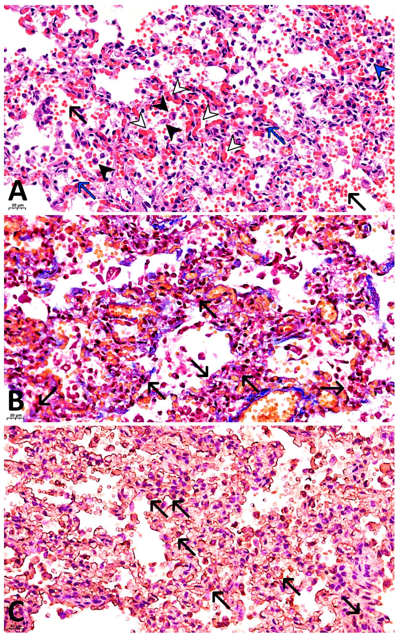
- With antibodies against fibrinogen: focal fibrin deposits are observed as strands along the inner surface of the alveoli, along the apical portion of bronchial epithelial cells, as well as fibrin thrombi in the capillaries of the interalveolar septa (Figure 1C).
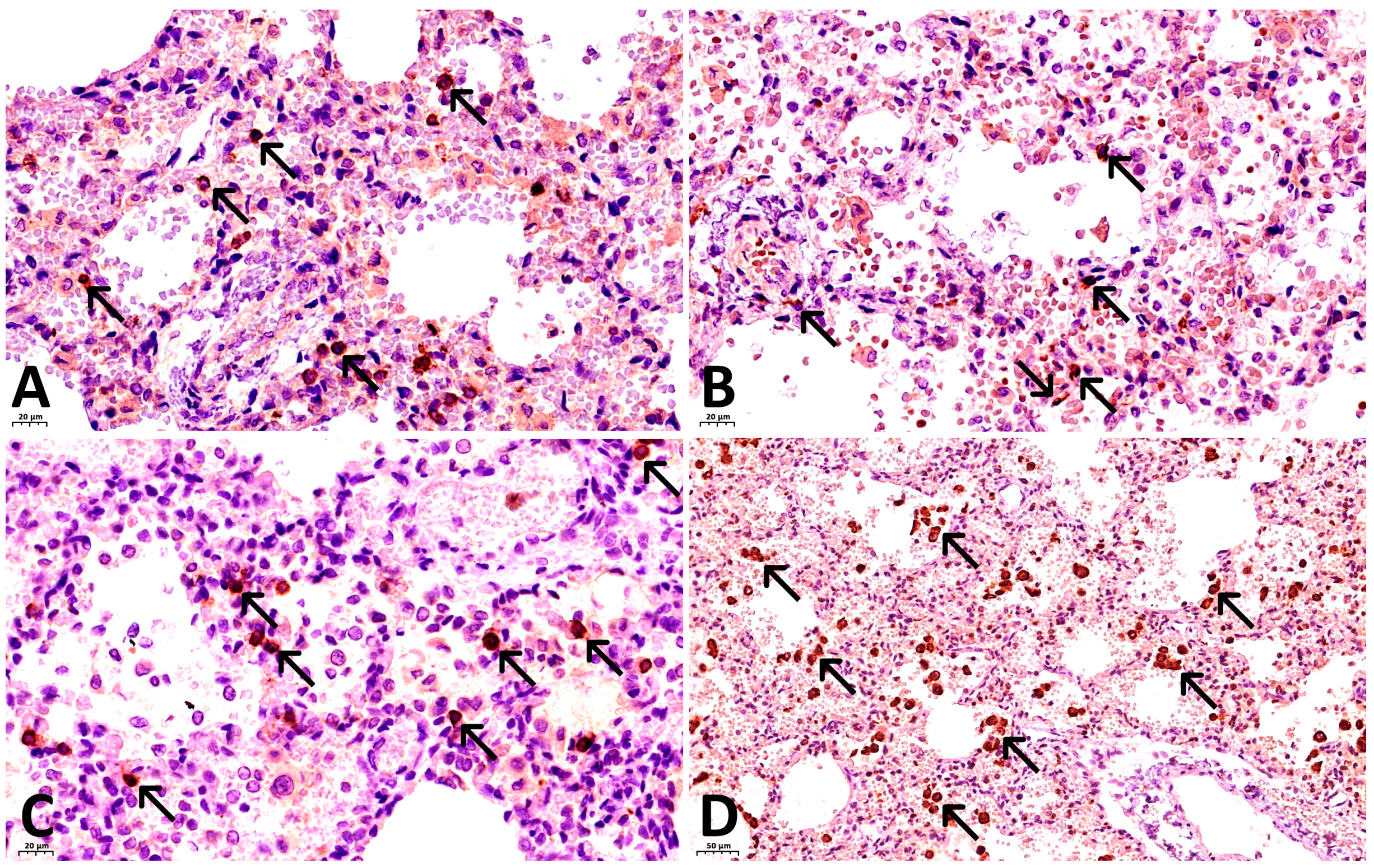
- With antibodies against CD68: marked expression of the marker is observed, with 30–40 macrophages visualized per high-power field (×400), and their number increasing to 50 or more in clusters (Figure 2D).
- With antibodies against CD8: 10–15 T lymphocytes are identified per high power field (×400), with their concentration increasing to 30–35 cells within lymphocytic aggregates (Figure 2A).
- With antibodies against CD20: 15–20 B lymphocytes are identified per high power field (×400), increasing to 30–35 or more cells within lymphocytic aggregates (Figure 2C).
- With antibodies against CD4: 3–6 T lymphocytes are identified per high power field (×400), with focal increase to 8–10 cells (Figure 2B).
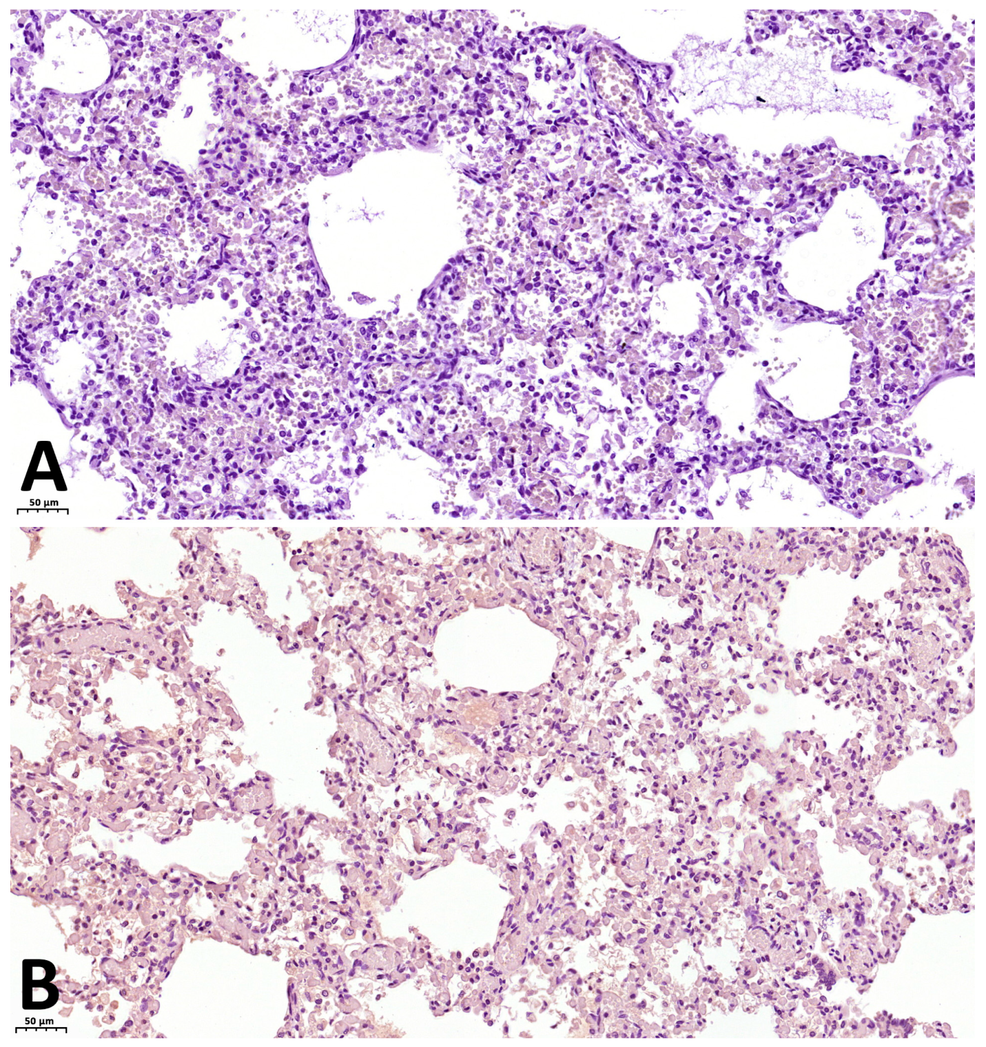
- As a first negative control for SARS-CoV-2 nucleocapsid staining, phosphate buffer (pH 7.6) was applied to the paraffin section of the lung from the investigated case instead of antibodies against this antigen. No expression was observed on these sections (Figure 3A). For the second negative control, lung tissue from a child with viral pneumonia who died from causes other than COVID-19 was used. In this specimen, expression was likewise absent (Figure 3B).
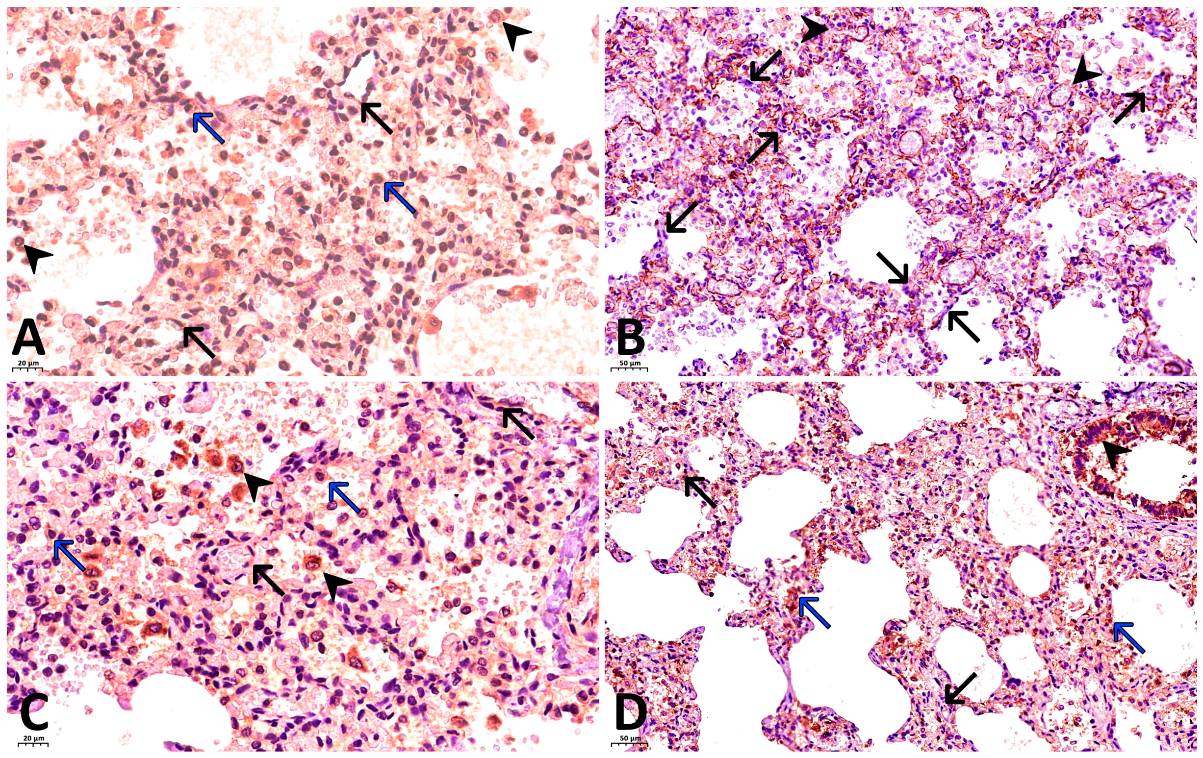
- With antibodies against COVID 19 nucleocapsid protein: markedly pronounced expression (+++) was detected in the endothelium of microcirculatory vessels, bronchial epithelium, alveolar macrophages, and alveolocytes (Figure 4A).
- With antibodies against IL-6: no expression was detected (negative, confirmed twice).
- With antibodies against TNF-α: no expression was detected (negative, confirmed twice).
- With antibodies against CD31: the capillary network demonstrates a discontinuous pattern, with focal loss of marker expression in the endothelium of some capillaries, and areas of absent expression fields are noted. At the same time, marked expression (+++) is focally observed in the endothelium of microcirculatory vessels (Figure 4B).
- With antibodies against IFN-γ: marked expression (+++) is observed in the endothelium of microcirculatory vessels, bronchial epithelium, alveolocytes, and alveolar macrophages (25–30 cells per high power field at ×400 magnification) (Figure 4C).
- With antibodies against CD95: moderately pronounced expression (++) is observed in the endothelium of some microcirculatory vessels, as well as marked expression (+++) in the bronchial epithelium, alveolar macrophages, and alveolocytes (Figure 4D).
4. Discussion
5. Conclusions
Author Contributions
Funding
Institutional Review Board Statement
Informed Consent Statement
Data Availability Statement
Conflicts of Interest
Abbreviations
| SUID | Sudden Unexpected Infant Death |
| SIDS | Sudden Infant Death Syndrome |
| IL-6 | Interleukin-6 |
| TNF-α | Tumor Necrosis Factor-α |
| IFN-γ | Interferon-γ |
| PCR | Polymerase Chain Reaction |
| DNA | Deoxyribonucleic Acid |
| IHC | Immunohistochemical |
| CD | Cluster of Differentiation |
| CRP | C-Reactive Protein |
| ICAM-1 | Intercellular Adhesion Molecule 1 |
| H&E | Hematoxylin and eosin |
| DIC | Disseminated intravascular coagulation |
References
- Pereira de Godoy, A.C.; Bulgarelli Bestetti, R. Hospitalization and Mortality in Brazilian Children and Adolescents Due to COVID-19: Retrospective Study. JMIR Pediatr. Parent. 2025, 8, e67546. [Google Scholar] [CrossRef] [Scilit] [PubMed]
- Malloy, M.H.; MacDorman, M. Changes in the classification of sudden unexpected infant deaths: United States, 1992–2001. Pediatrics 2005, 115, 1247–1253. [Google Scholar] [CrossRef] [Scilit] [PubMed]
- Park, S.; Han, J.H.; Hwang, J.; Yon, D.K.; Lee, S.W.; Kim, J.H.; Koyanagi, A.; Jacob, L.; Oh, H.; Kostev, K.; et al. The global burden of sudden infant death syndrome from 1990 to 2019: A systematic analysis from the Global Burden of Disease study 2019. QJM Int. J. Med. 2022, 115, 735–744. [Google Scholar] [CrossRef] [Scilit]
- Guare, E.G.; Zhao, R.; Ssentongo, P.; Batra, E.K.; Chinchilli, V.M.; Paules, C.I. Rates of Sudden Unexpected Infant Death Before and During the COVID-19 Pandemic. JAMA Netw. Open 2024, 7, e2435722. [Google Scholar] [CrossRef] [Scilit]
- Lisman, D.; Zielińska, G.; Drath, J.; Łaszczewska, A.; Savochka, I.; Parafiniuk, M.; Ossowski, A. Molecular Diagnosis of COVID-19 Sudden and Unexplained Deaths: The Insidious Face of the Pandemic. Diagnostics 2023, 13, 2980. [Google Scholar] [CrossRef] [Scilit]
- Shapiro-Mendoza, C.K.; Woodworth, K.R.; Cottengim, C.R.; Erck Lambert, A.B.; Harvey, E.M.; Monsour, M.; Parks, S.E.; Barfield, W.D. Sudden Unexpected Infant Deaths: 2015–2020. Pediatrics 2023, 151, e2022058820. [Google Scholar] [CrossRef] [Scilit]
- Benny, M.; Bandstra, E.S.; Saad, A.G.; Lopez-Alberola, R.; Saigal, G.; Paidas, M.J.; Jayakumar, A.R.; Duara, S. Maternal SARS-CoV-2, Placental Changes and Brain Injury in 2 Neonates. Pediatrics 2023, 151, e2022058271. [Google Scholar] [CrossRef] [Scilit]
- Zheng, X.; Liu, M.; Zhao, X.; Xu, X.; Tao, W.; Wu, L.; Sun, W.; Dong, Y.; Xi, Y. Global, regional, and national burden of sudden infant death syndrome and the impact of COVID-19: A trend and health inequality analysis based on the global burden of disease study 2021. Front. Pediatr. 2025, 13, 1623238. [Google Scholar] [CrossRef] [Scilit] [PubMed]
- Sacco, M.A.; Gualtieri, S.; Verrina, M.C.; Aquila, V.R.; Tarda, L.; Tarallo, A.P.; Aquila, I. A Narrative Overview of Fatal Myocarditis in Infant with Focus on Sudden Unexpected Death and Forensic Implications. J. Clin. Med. 2025, 14, 4340. [Google Scholar] [CrossRef] [Scilit]
- Grasmeyer, S.; Madea, B. Immunohistochemical diagnosis of myocarditis on (infantile) autopsy material: Does it improve the diagnosis? Forensic Sci. Med. Pathol. 2015, 11, 168–176. [Google Scholar] [CrossRef] [Scilit]
- Berezovskiy, D.P.; Fedulova, M.V.; Gornostaev, D.V.; Avanesyan, H.A.; Borshchevskaya, V.N.; Pigolkin, Y.I. Morphofunctional characteristics of the heart in sudden death of children under 1 year of age in the forensic medical aspect. Sud.-Meditsinskaia Ekspertiza 2022, 65, 5–8. (In Russian) [Google Scholar] [CrossRef] [Scilit] [PubMed]
- Pryce, J.W.; Bamber, A.R.; Ashworth, M.T.; Klein, N.J.; Sebire, N.J. Immunohistochemical expression of inflammatory markers in sudden infant death; ancillary tests for identification of infection. J. Clin. Pathol. 2014, 67, 1044–1051. [Google Scholar] [CrossRef] [Scilit]
- Lavezzi, A.M.; Weese-Mayer, D.E.; Yu, M.Y.; Jennings, L.J.; Corna, M.F.; Casale, V.; Oneda, R.; Matturri, L. Developmental alterations of the respiratory human retrotrapezoid nucleus in sudden unexplained fetal and infant death. Auton. Neurosci. Basic Clin. 2012, 170, 12–19. [Google Scholar] [CrossRef] [Scilit] [PubMed]
- Lavezzi, A.M.; Mehboob, R.; Piscioli, F.; Pusiol, T. New Step in Understanding the Pathogenetic Mechanism of Sudden Infant Death Syndrome: Involvement of the Pontine Reticular Gigantocellular Nucleus. Int. J. Mol. Sci. 2024, 25, 6920. [Google Scholar] [CrossRef] [Scilit] [PubMed]
- Lavezzi, A.M.; Corna, M.; Mingrone, R.; Matturri, L. Study of the human hypoglossal nucleus: Normal development and morpho-functional alterations in sudden unexplained late fetal and infant death. Brain Dev. 2010, 32, 275–284. [Google Scholar] [CrossRef] [Scilit]
- Lavezzi, A.M.; Mehboob, R. The Mesencephalic Periaqueductal Gray, a Further Structure Involved in Breathing Failure Underlying Sudden Infant Death Syndrome. ASN Neuro 2021, 13, 17590914211048260. [Google Scholar] [CrossRef] [Scilit]
- Nasyrov, R.A.; Ivanov, D.O.; Krasnogorskaya, O.L.; Timchenko, V.N.; Fedotova, E.P.; Chepelev, A.S.; Galichina, V.A.; Sidorova, N.A.; Anichkov, N.M. COVID-19 in Children: Molecular Profile and Pathological Features. Int. J. Mol. Sci. 2023, 24, 16750. [Google Scholar] [CrossRef] [Scilit]
- Kinloch, N.N.; Ritchie, G.; Brumme, C.J.; Dong, W.; Dong, W.; Lawson, T.; Jones, R.B.; Montaner, J.S.G.; Leung, V.; Romney, M.G.; et al. Suboptimal Biological Sampling as a Probable Cause of False-Negative COVID-19 Diagnostic Test Results. J. Infect. Dis. 2020, 222, 899–902, Erratum in J. Infect. Dis. 2021, 224, 184. [Google Scholar] [CrossRef] [Scilit]
- Woloshin, S.; Patel, N.; Kesselheim, A.S. False Negative Tests for SARS-CoV-2 Infection—Challenges and Implications. N. Engl. J. Med. 2020, 383, e38. [Google Scholar] [CrossRef] [Scilit]
- Green, D.A.; Zucker, J.; Westblade, L.F.; Whittier, S.; Rennert, H.; Velu, P.; Craney, A.; Cushing, M.; Liu, D.; Sobieszczyk, M.E.; et al. Clinical Performance of SARS-CoV-2 Molecular Tests. J. Clin. Microbiol. 2020, 58, e00995-20. [Google Scholar] [CrossRef] [Scilit]
- Zhao, J.; Yuan, Q.; Wang, H.; Liu, W.; Liao, X.; Su, Y.; Wang, X.; Yuan, J.; Li, T.; Li, J.; et al. Antibody Responses to SARS-CoV-2 in Patients with Novel Coronavirus Disease 2019. Clin. Infect. Dis. 2020, 71, 2027–2034. [Google Scholar] [CrossRef] [Scilit] [PubMed]
- Sessa, F.; Bertozzi, G.; Cipolloni, L.; Baldari, B.; Cantatore, S.; D’Errico, S.; Di Mizio, G.; Asmundo, A.; Castorina, S.; Salerno, M.; et al. Clinical-Forensic Autopsy Findings to Defeat COVID-19 Disease: A Literature Review. J. Clin. Med. 2020, 9, 2026. [Google Scholar] [CrossRef] [Scilit]
- Keaney, D.; Whelan, S.; Finn, K.; Lucey, B. Misdiagnosis of SARS-CoV-2: A Critical Review of the Influence of Sampling and Clinical Detection Methods. Med. Sci. 2021, 9, 36. [Google Scholar] [CrossRef] [Scilit]
- Çubukçu, H.C.; Coşkun, A. False negative results and tolerance limits of SARS-CoV-2 laboratory tests. Pathog. Glob. Health 2021, 115, 137–138. [Google Scholar] [CrossRef] [Scilit] [PubMed]
- Mosscrop, L.; Watber, P.; Elliot, P.; Cooke, G.; Barclay, W.; Freemont, P.S.; Rosadas, C.; Taylor, G.P. Evaluation of the impact of pre-analytical conditions on sample stability for the detection of SARS-CoV-2 RNA. J. Virol. Methods 2022, 309, 114607. [Google Scholar] [CrossRef] [Scilit]
- Pan, Y.; Long, L.; Zhang, D.; Yuan, T.; Cui, S.; Yang, P.; Wang, Q.; Ren, S. Potential False-Negative Nucleic Acid Testing Results for Severe Acute Respiratory Syndrome Coronavirus 2 from Thermal Inactivation of Samples with Low Viral Loads. Clin. Chem. 2020, 66, 794–801. [Google Scholar] [CrossRef] [Scilit]
- Wölfel, R.; Corman, V.M.; Guggemos, W.; Seilmaier, M.; Zange, S.; Müller, M.A.; Niemeyer, D.; Jones, T.C.; Vollmar, P.; Rothe, C.; et al. Virological assessment of hospitalized patients with COVID-2019. Nature 2020, 581, 465–469, Erratum in Nature 2020, 588, E35. [Google Scholar] [CrossRef] [Scilit] [PubMed]
- Kucirka, L.M.; Lauer, S.A.; Laeyendecker, O.; Boon, D.; Lessler, J. Variation in False-Negative Rate of Reverse Transcriptase Polymerase Chain Reaction-Based SARS-CoV-2 Tests by Time Since Exposure. Ann. Intern. Med. 2020, 173, 262–267. [Google Scholar] [CrossRef] [Scilit]
- Harms, P.W.; Frankel, T.L.; Moutafi, M.; Rao, A.; Rimm, D.L.; Taube, J.M.; Thomas, D.; Chan, M.P.; Pantanowitz, L. Multiplex Immunohistochemistry and Immunofluorescence: A Practical Update for Pathologists. Mod. Pathol. 2023, 36, 100197. [Google Scholar] [CrossRef] [Scilit]
- Nasyrov, R.A.; Galichina, V.A.; Drobintseva, A.O.; Tonkonog, D.V.; Kalinina, E.Y.; Agafonnikova, A.A. Microvascular Genesis of Diseases: From Hypothesis to Theory. Life 2026, 16, 314. [Google Scholar] [CrossRef] [Scilit]
- Mansoor, S.; Butt, A.R.; Bibi, A.; Mushtaq, S.; Ullah, I.; Alshahrani, F.; Khan, A.; Mansoor, A. Expression of IFN-Gamma is significantly reduced during severity of COVID-19 infection in hospitalized patients. PLoS ONE 2023, 18, e0291332. [Google Scholar] [CrossRef] [Scilit]
- Ye, Q.; Wang, B.; Mao, J. The pathogenesis and treatment of the ‘Cytokine Storm’ in COVID-19. J. Infect. 2020, 80, 607–613. [Google Scholar] [CrossRef] [Scilit]
- Iannaccone, G.; Scacciavillani, R.; Del Buono, M.G.; Camilli, M.; Ronco, C.; Lavie, C.J.; Abbate, A.; Crea, F.; Massetti, M.; Aspromonte, N. Weathering the Cytokine Storm in COVID-19: Therapeutic Implications. Cardiorenal Med. 2020, 10, 277–287. [Google Scholar] [CrossRef] [Scilit]
- Manjili, R.H.; Zarei, M.; Habibi, M.; Manjili, M.H. COVID-19 as an Acute Inflammatory Disease. J. Immunol. 2020, 205, 12–19. [Google Scholar] [CrossRef] [Scilit]
- Del Valle, D.M.; Kim-Schulze, S.; Huang, H.H.; Beckmann, N.D.; Nirenberg, S.; Wang, B.; Lavin, Y.; Swartz, T.H.; Madduri, D.; Stock, A.; et al. An inflammatory cytokine signature predicts COVID-19 severity and survival. Nat. Med. 2020, 26, 1636–1643. [Google Scholar] [CrossRef] [Scilit]
- Hasanvand, A. COVID-19 and the role of cytokines in this disease. Inflammopharmacology 2022, 30, 789–798. [Google Scholar] [CrossRef] [Scilit] [PubMed]
- Levy, D.E.; García-Sastre, A. The virus battles: IFN induction of the antiviral state and mechanisms of viral evasion. Cytokine Growth Factor Rev. 2001, 12, 143–156. [Google Scholar] [CrossRef] [Scilit] [PubMed]
- Sun, D.; Li, H.; Lu, X.X.; Xiao, H.; Ren, J.; Zhang, F.R.; Liu, Z.S. Clinical features of severe pediatric patients with coronavirus disease 2019 in Wuhan: A single center’s observational study. World J. Pediatr. 2020, 16, 251–259. [Google Scholar] [CrossRef] [Scilit] [PubMed]
- Gadotti, A.C.; de Castro Deus, M.; Telles, J.P.; Wind, R.; Goes, M.; Garcia Charello Ossoski, R.; de Padua, A.M.; de Noronha, L.; Moreno-Amaral, A.; Baena, C.P.; et al. IFN-γ is an independent risk factor associated with mortality in patients with moderate and severe COVID-19 infection. Virus Res. 2020, 289, 198171. [Google Scholar] [CrossRef] [Scilit]
- Artamonov, A.A.; Nikitin, Y.V.; Meshkova, M.E.; Ivanov, A.M. Role of IFNγ in pathogenesis of SARS-CoV-2 infection. Med. Immunol. 2022, 24, 903–910. (In Russian) [Google Scholar] [CrossRef] [Scilit]
- Toro, A.; Lage-Vickers, S.; Bizzotto, J.; Vilicich, F.; Sabater, A.; Pascual, G.; Ledesma-Bazan, S.; Sanchis, P.; Ruiz, M.S.; Arevalo, A.P.; et al. Pin-Pointing the Key Hubs in the IFN-γ Pathway Responding to SARS-CoV-2 Infection. Viruses 2022, 14, 2180. [Google Scholar] [CrossRef] [Scilit]
- Bastard, P.; Gervais, A.; Le Voyer, T.; Rosain, J.; Philippot, Q.; Manry, J.; Michailidis, E.; Hoffmann, H.H.; Eto, S.; Garcia-Prat, M.; et al. Autoantibodies neutralizing type I IFNs are present in ~4% of uninfected individuals over 70 years old and account for ~20% of COVID-19 deaths. Sci. Immunol. 2021, 6, eabl4340. [Google Scholar] [CrossRef] [Scilit] [PubMed]
- Xiong, X.; Chua, G.T.; Chi, S.; Kwan, M.Y.W.; Wong, W.H.S.; Zhou, A.; Shek, C.C.; Tung, K.T.S.; Qin, H.; Wong, R.S.; et al. Haematological and immunological data of Chinese children infected with coronavirus disease 2019. Data Brief. 2020, 31, 105953. [Google Scholar] [CrossRef] [Scilit] [PubMed]
- Morhart, P.; Kehl, S.; Schuh, W.; Hermes, K.; Meltendorf, S.; Neubert, A.; Schneider, M.; Brunner-Weinzierl, M.; Schneider, H.; Lingel, H. Age-related Differences in Immune Reactions to SARS-CoV-2 Spike and Nucleocapsid Antigens. In Vivo 2023, 37, 70–78. [Google Scholar] [CrossRef] [Scilit]
- Goenka, A.; Halliday, A.; Gregorova, M.; Milodowski, E.; Thomas, A.; Williamson, M.K.; Baum, H.; Oliver, E.; Long, A.E.; Knezevic, L.; et al. Young infants exhibit robust functional antibody responses and restrained IFN-γ production to SARS-CoV-2. Cell Rep. Med. 2021, 2, 100327. [Google Scholar] [CrossRef] [Scilit]
- Shimizu, J.; Sasaki, T.; Ong, G.H.; Koketsu, R.; Samune, Y.; Nakayama, E.E.; Nagamoto, T.; Yamamoto, Y.; Miyazaki, K.; Shioda, T. IFN-γ derived from activated human CD4+ T cells inhibits the replication of SARS-CoV-2 depending on cell-type and viral strain. Sci. Rep. 2024, 14, 26660. [Google Scholar] [CrossRef] [Scilit]
- Karki, R.; Sharma, B.R.; Tuladhar, S.; Williams, E.P.; Zalduondo, L.; Samir, P.; Zheng, M.; Sundaram, B.; Banoth, B.; Malireddi, R.K.S.; et al. Synergism of TNF-α and IFN-γ Triggers Inflammatory Cell Death, Tissue Damage, and Mortality in SARS-CoV-2 Infection and Cytokine Shock Syndromes. Cell 2021, 184, 149–168.e17. [Google Scholar] [CrossRef] [Scilit] [PubMed]

Disclaimer/Publisher’s Note: The statements, opinions and data contained in all publications are solely those of the individual author(s) and contributor(s) and not of MDPI and/or the editor(s). MDPI and/or the editor(s) disclaim responsibility for any injury to people or property resulting from any ideas, methods, instructions or products referred to in the content. |
© 2026 by the authors. Licensee MDPI, Basel, Switzerland. This article is an open access article distributed under the terms and conditions of the Creative Commons Attribution (CC BY) license.
Share and Cite
Galichina, V.A.; Nasyrov, R.A.; Davydova, Z.V.; Gabaraev, S.E.; Yagmurov, O.D. A Case of Sudden Unexpected Infant Death with Presumptive SARS-CoV-2 Infection. Int. J. Mol. Sci. 2026, 27, 4604. https://doi.org/10.3390/ijms27104604
Galichina VA, Nasyrov RA, Davydova ZV, Gabaraev SE, Yagmurov OD. A Case of Sudden Unexpected Infant Death with Presumptive SARS-CoV-2 Infection. International Journal of Molecular Sciences. 2026; 27(10):4604. https://doi.org/10.3390/ijms27104604
Chicago/Turabian StyleGalichina, Veronika A., Ruslan A. Nasyrov, Zlata V. Davydova, Simon E. Gabaraev, and Orasmurad D. Yagmurov. 2026. "A Case of Sudden Unexpected Infant Death with Presumptive SARS-CoV-2 Infection" International Journal of Molecular Sciences 27, no. 10: 4604. https://doi.org/10.3390/ijms27104604
APA StyleGalichina, V. A., Nasyrov, R. A., Davydova, Z. V., Gabaraev, S. E., & Yagmurov, O. D. (2026). A Case of Sudden Unexpected Infant Death with Presumptive SARS-CoV-2 Infection. International Journal of Molecular Sciences, 27(10), 4604. https://doi.org/10.3390/ijms27104604