Article

Role of the IRE1α-XBP1 Axis in IgE-Dependent Activation of Mast Cells

Department of Biological Science and Technology, Faculty of Advanced Engineering, Tokyo University of Science, 6-3-1 Niijuku, Katsushika-ku, Tokyo 125-8585, Japan
*
Author to whom correspondence should be addressed.
Int. J. Mol. Sci. 2026, 27(10), 4532; https://doi.org/10.3390/ijms27104532
Submission received: 17 April 2026 / Revised: 7 May 2026 / Accepted: 12 May 2026 / Published: 18 May 2026
(This article belongs to the Special Issue Molecular Mechanisms of Allergy and Asthma: 4th Edition)

Abstract

The IRE1α-XBP1 axis is the most conserved of the three major unfolded protein response (UPR) branches triggered by the endoplasmic reticulum (ER) stress. Although the transcription factor XBP1 is involved in the development and function of several hematopoietic lineages, its role in the activation of mast cells (MCs), which are critical in allergic responses, remains largely unknown. We identified salicylaldehyde, which suppresses IRE1α nuclease activity that is essential for XBP1 production, as an inhibitor of MC activation in our previous screening; therefore, we herein investigated the effects of additional IRE1α inhibitors, 3-methyl-6-bromo-salichylaldehyde (MBSA) and KIRA6, targeting the nuclease domain and kinase domain, respectively, on MC activation. MBSA and KIRA6 suppressed IgE-dependent degranulation of bone marrow-derived MCs (BMMCs) but did not inhibit Ca2+ ionophore- or compound48/80-induced degranulation. Treatments with inhibitors of two other branches of UPR, the PERK and ATF6 pathways, did not affect the IgE-induced activation of BMMCs. The intraperitoneal administration of MBSA or KIRA6 significantly suppressed IgE-induced passive anaphylaxis in mice. Furthermore, to examine the effects of XBP1, siRNA-mediated knockdown was performed. The results obtained confirmed that Xbp1 siRNA introduction reduced the IgE-dependent degranulation of BMMCs in parallel with the knockdown level of Xbp1 mRNA. Therefore, the IRE1α-XBP1 axis plays a significant role in IgE-dependent and MC-mediated allergic responses and is considered to be a therapeutic target of allergic diseases.

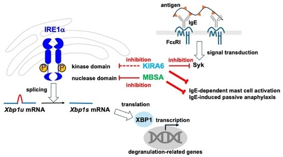
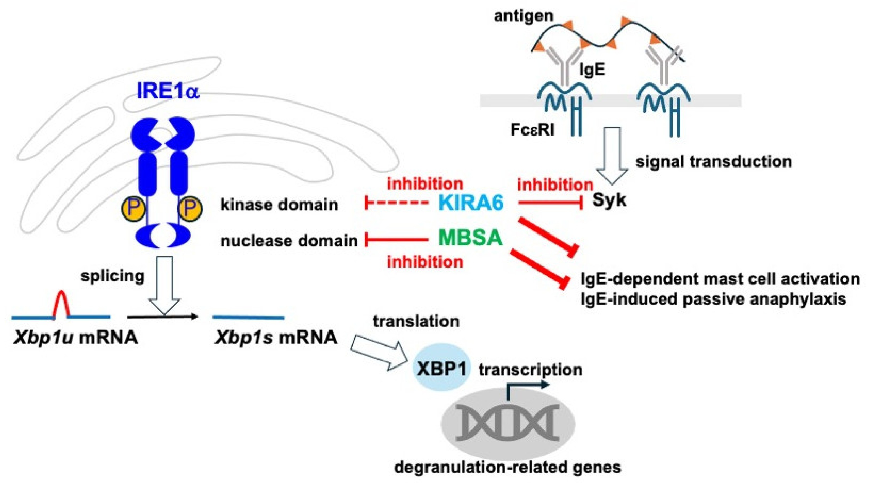
Graphical Abstract

1. Introduction

Mast cells (MCs) play important roles in IgE-mediated immune responses, including the pathogenesis of allergic diseases and defenses against parasite infections. In the case of allergic patients carrying hyper IgE, FcεRI expressed on MCs is occupied with antigen-specific IgE, and the cross-linking of multiple IgEs on FcεRI with allergens rapidly activates MCs, resulting in degranulation, eicosanoid release, and cytokine production. In addition, several receptors, such as ST2, MRGPRX2, TLR4, and P2X7, which recognize IL-33, a chemical compound, LPS, and ATP, respectively, are involved in the IgE-independent activation of MCs. Current anti-allergic drugs, such as inhibitors of histamine receptors, leukotriene receptors, and anti-human IgE, target events related to MC activation, demonstrating that the regulation of MC activation is effective for the prevention of allergic diseases [1].
We previously performed screening to identify novel anti-allergic compounds using mouse bone marrow-derived MCs (BMMCs), and selected salicylaldehyde (SA) as the most effective inhibitor of MC activation [2]. The administration of SA suppressed IgE-dependent passive anaphylaxis in mice; however, the molecular mechanisms by which SA inhibit the IgE-dependent activation of MCs in vivo and in vitro remain unclear.
In the present study, we investigated the effects of the IRE1α-XBP1 axis, the most conserved arm of the unfolded protein response (UPR) in MC activation, because it is considered to be a target of SA [3,4,5]. The UPR occurred in response to the endoplasmic reticulum (ER) stress induced by the accumulation of misfolded proteins within the ER lumen. The following events are induced under the UPR condition to maintain cellular homeostasis: the degradation of unfolded proteins is promoted, further protein synthesis is inhibited, and repair mechanisms are facilitated [6]. IRE1α is an enzyme that exhibits both kinase and endoribonuclease activities, and homodimerized and phosphorylated IRE1α creates spliced mature XBP1 mRNA by cleaving out 26 nucleotides, resulting in the translation of the transcription factor XBP1, which up-regulates the expression of UPR-related genes. XBP1 also regulates several cellular events, such as protein secretion, lipid biosynthesis, glycosylation, and autophagy [6]. XBP1 is involved in various immune system functions, including the differentiation of plasma cells [7], the development and survival of dendritic cells [8] and eosinophils [9], the TLR-mediated cytokine production of macrophages [10], the regulation of type I interferon production in chronically activated pDC [11], and the anti-tumor activity of dendritic cells [12]. On the other hand, the role of XBP1 and/or UPR in MC activation associated with cellular stress remains unclear. Although recent studies examined the role of XBP1 in MC function, the findings obtained were controversial [13,14]. The present study demonstrated a positive role for the IRE1α-XBP1 axis in the IgE-induced activation of MCs.

2. Results

2.1. Effects of the SA-Related Compound 3-Methyl-6-Bromo-Salichylaldehyde (MBSA) and the XBP1 Splicing Inhibitor KIRA6 on the IgE-Induced Degranulation of BMMCs

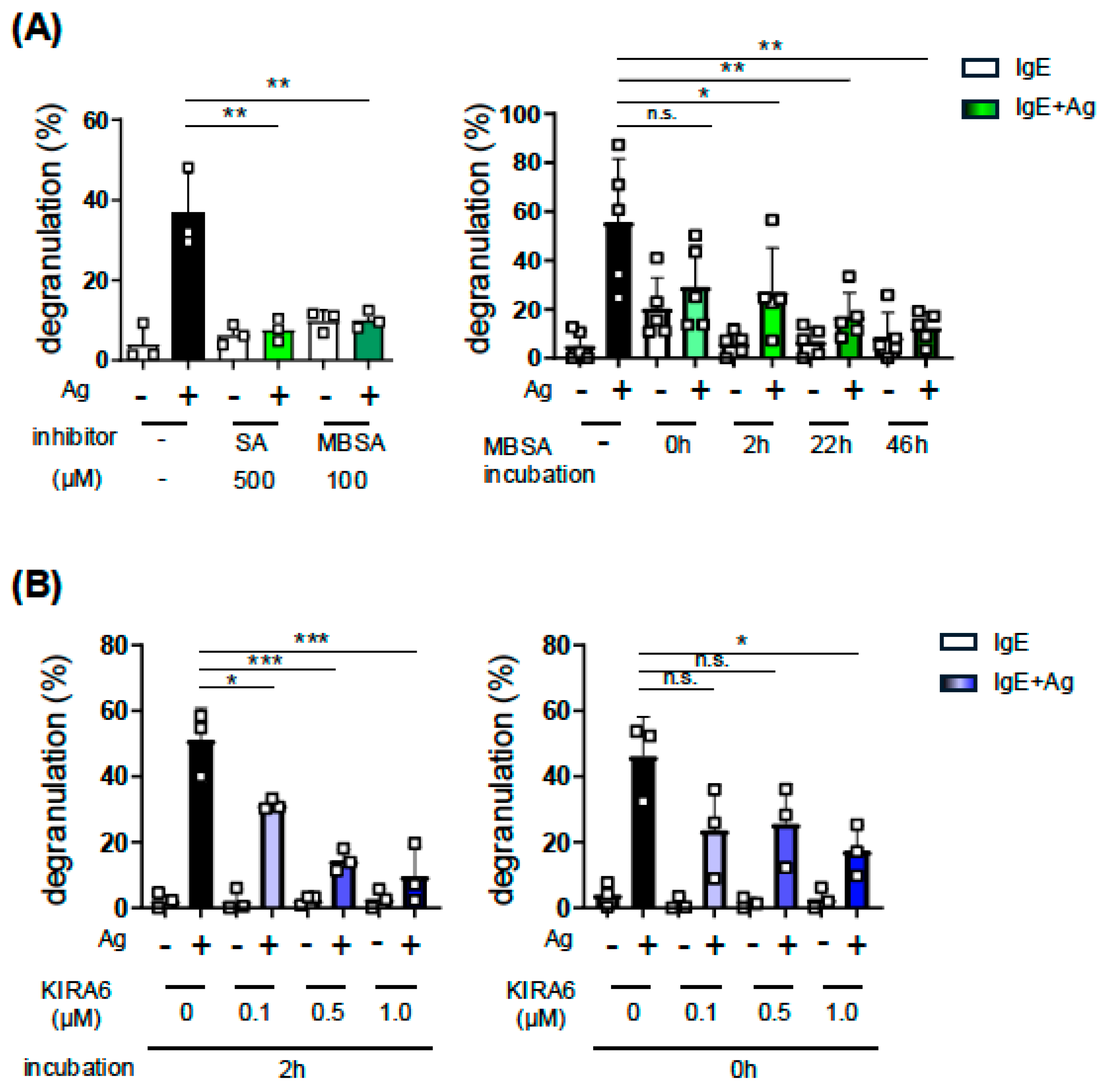
We identified SA as an inhibitor of MC activation through the screening of a chemical/natural compound library in our previous study [2]. Since SA has been used to inhibit Xbp1 mRNA splicing by IRE1α in keratinocytes [3] and melanoma [4], we herein investigated the role of XBP1 and XBP1-related cellular events, including UPR, in MC activation. We examined the effect of SA-related compound MBSA on MC activation, which exhibits higher inhibitory activity against XBP1 mRNA splicing than SA [5]. As shown in Figure 1A, left, the treatment with MBSA for 48 h suppressed the IgE-induced degranulation of BMMCs, even at a lower dose than SA, and an inhibitory effect was observed under the condition that MBSA was added to culture media 2 h before or just when the FcεRI-mediated stimulation was induced (Figure 1A, right). We then investigated the involvement of the IRE1α-XBP1 pathway in the IgE-induced activation of MCs by using another inhibitor, KIRA6, which suppresses XBP1 activation by targeting the kinase domain of IRE1α [15], and found that KIRA6 significantly suppressed IgE-induced degranulation (Figure 1B).
Collectively, these results suggest that the inhibition of the IRE1α-XBP1 pathway suppressed the IgE-induced degranulation of MCs.

2.2. Effects of MBSA and KIRA6 on IgE-Induced Cytokine Production and IgE-Independent Degranulation of BMMCs

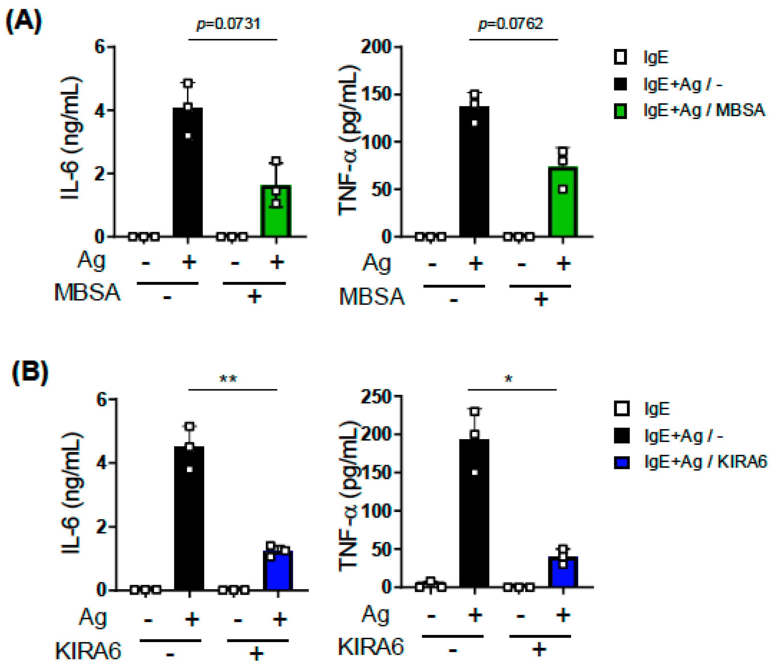
Upon IgE-dependent activation, MCs cause not only degranulation as the immediate reaction but also release of inflammatory cytokines involved in the late-phase reaction. To examine the effects of XBP1 inhibition on cytokine release, we assessed the amounts of IL-6 and TNF-α produced from IgE-stimulated BMMCs using ELISAs. The results obtained showed that KIRA6 significantly suppressed the IgE-induced secretion of IL-6 and TNF-α from BMMCs (Figure 2B), whereas the effects of MBSA on the release of these cytokines were not significant (Figure 2A).
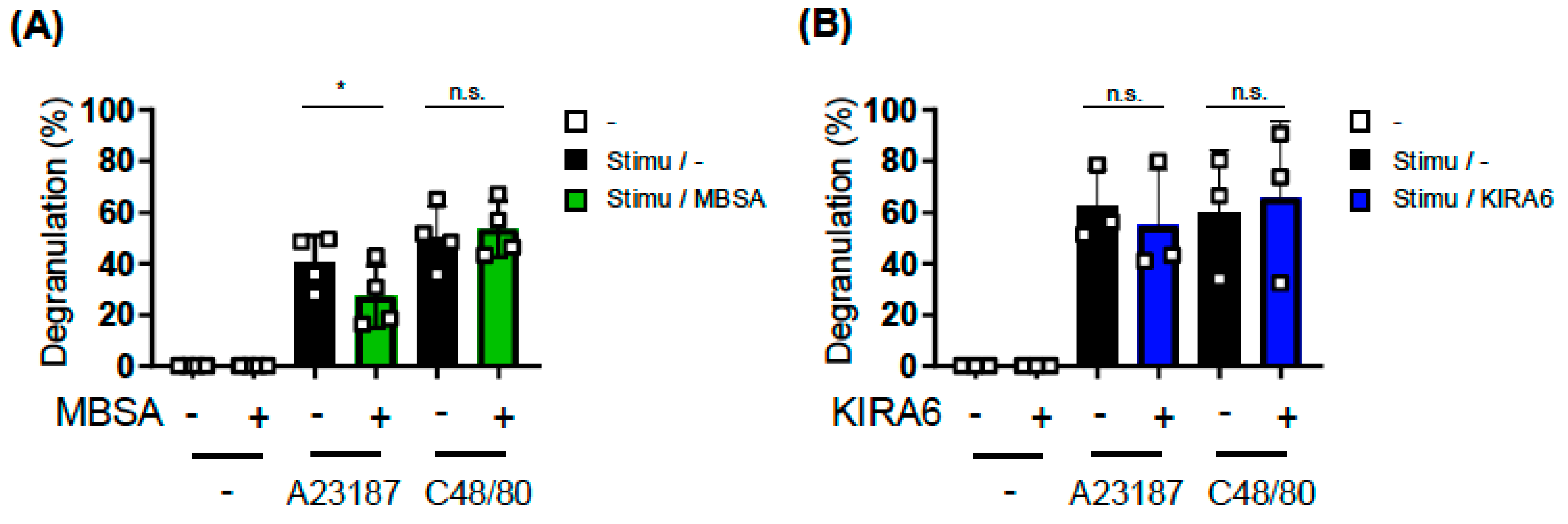
Furthermore, we investigated the effects of inhibitors on the IgE-independent stimulation of BMMCs. Using a b-hexosaminidase assay, we found that compound 48/80-induced degranulation was not affected by the pretreatment with MBSA, whereas Ca2+ ionophore-induced degranulation was significantly inhibited by MBSA (Figure 3A). In contrast, KIRA6 did not suppress the IgE-independent degranulation of BMMCs (Figure 3B).

2.3. Analysis of the Involvement of Two Other UPR Pathways on MC Activation Using Specific Inhibitors

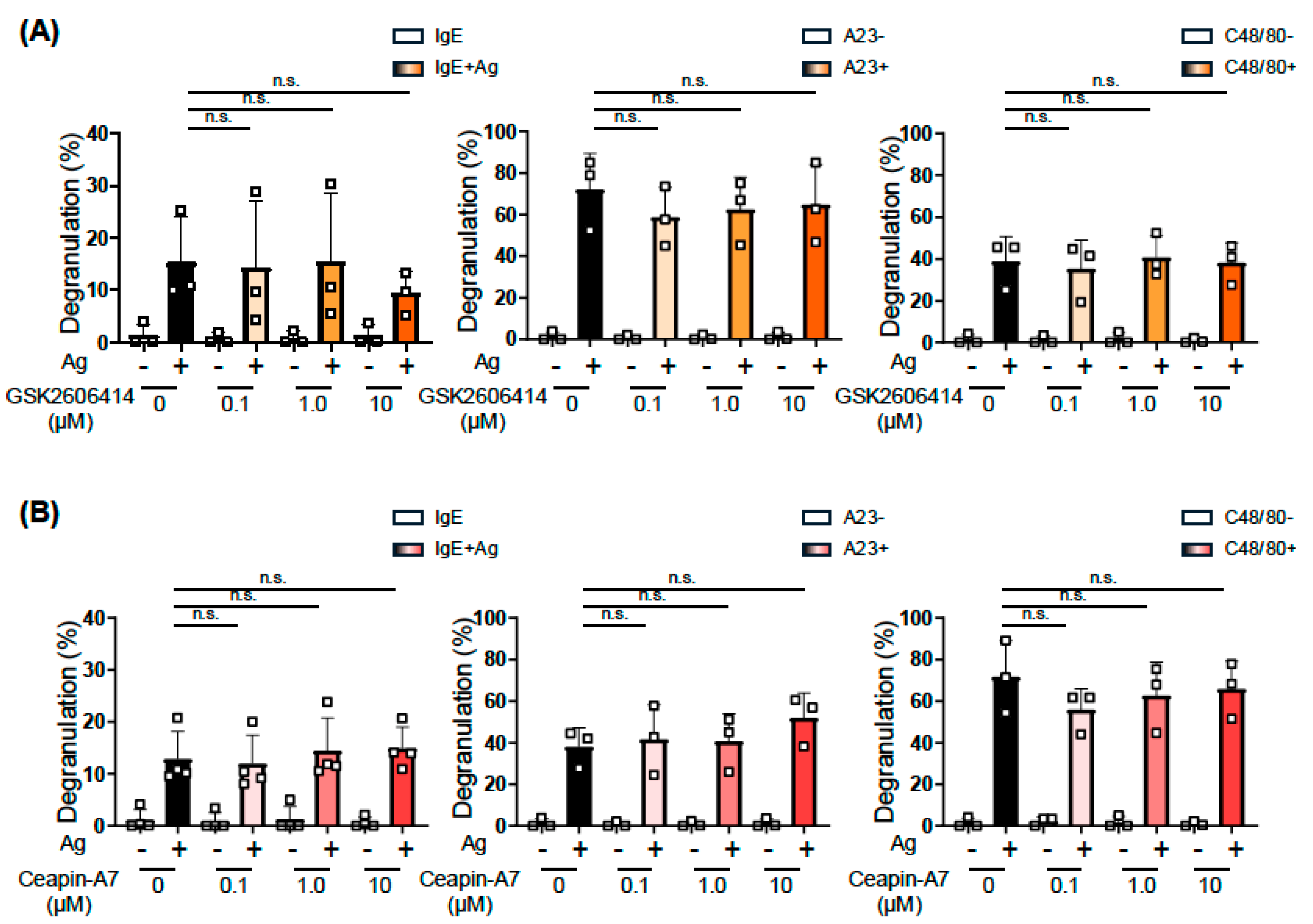
In addition to the IRE1α-XBP1 pathway, the PERK-ATF4 and ATF6 pathways are involved in the UPR. Therefore, we examined the effects of GSK2606414 and CeapinA-7, which inhibit the activation of PERK [17] and the trafficking of ATF6 [18], respectively, on MC activation. We pretreated BMMCs with 0.1–10 μM of GSK2606414 or CeapinA-7 for 24 h based on previously reported experimental conditions. Neither GSK2606414 (Figure 4A) nor CeapinA-7 (Figure 4B) exerted apparent effects on the IgE-, A23187-, or Compound 48/40-induced degranulation of BMMCs.
These results suggest the IRE1α-XBP1-dependent and PERK-ATF4/ATF6-independent manners of the IgE-induced activation of MCs, as observed in other events related to prominent protein synthesis and immuno-responses [7,19,20].

2.4. Effects of Tunicamycin on MC Activation

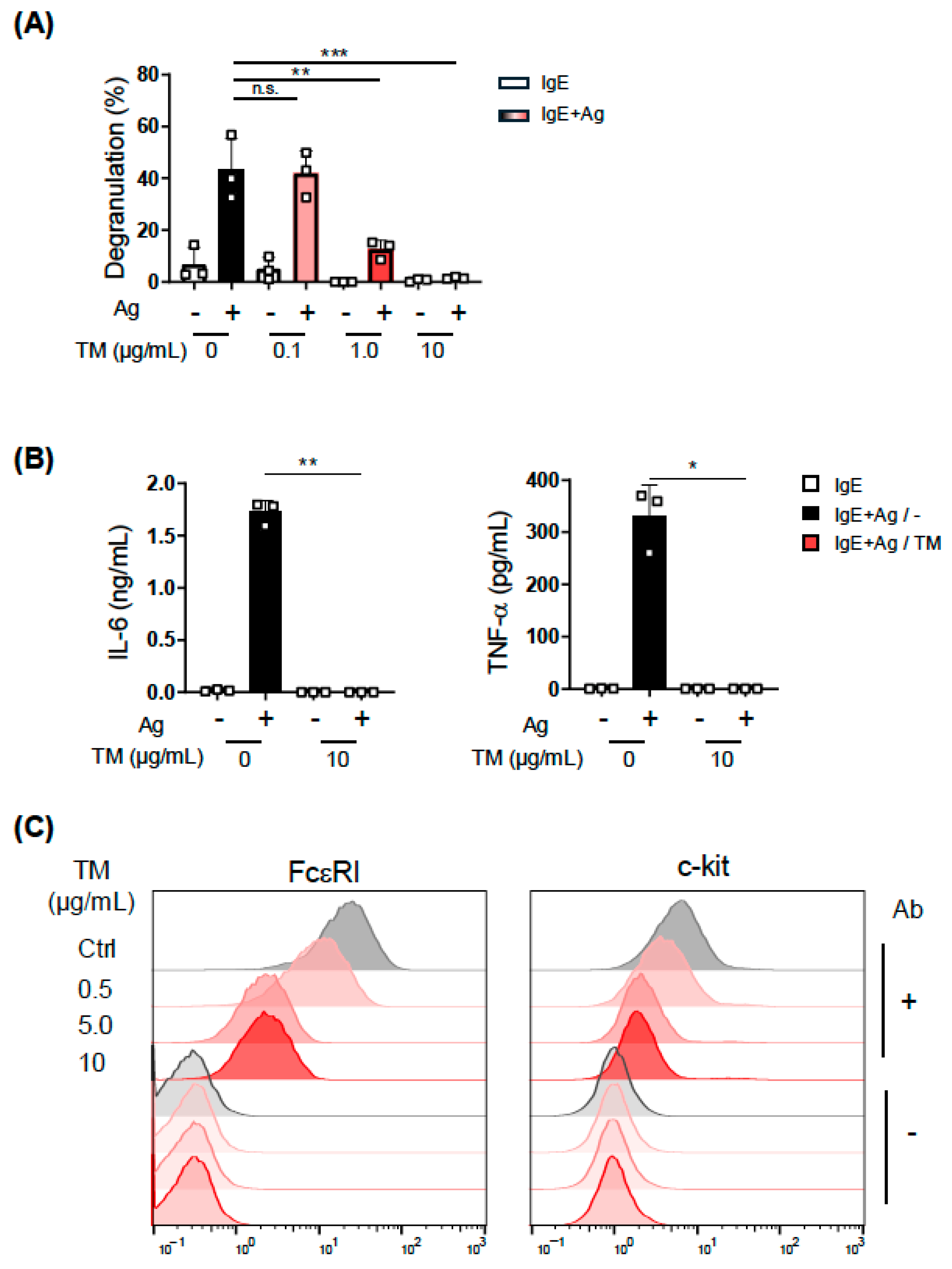
Tunicamycin, which inhibits the N-linked glycosylation of proteins, induces the accumulation of unfolded proteins in the ER and subsequent ER stress. In a recent study on the relationship of ER stress with MC degranulation, the intraperitoneal (i.p.) administration of tunicamycin increased the amount of serum Mcpt1 in mice [13], which is a hallmark of MC activation. We then treated BMMCs with tunicamycin to investigate whether the induction of ER stress up-regulates the IgE-induced activation of MCs. As shown in Figure 5A, the tunicamycin pretreatment suppressed the IgE-induced degranulation of BMMCs in a dose-dependent manner. The treatment of BMMCs with 10 μg/mL tunicamycin, which significantly reduced degranulation, completely inhibited the IgE-dependent secretion of IL-6 and TNF-α (Figure 5B). We also revealed that the surface expression of FcεRI on BMMCs was markedly reduced by the treatment with tunicamycin (Figure 5C).
Since N-glycosylation is required for multiple processes in MC function, including the transport of FcεRI from the ER to the cell surface [21], these results suggest that tunicamycin did not specifically induce the UPR as predicted in a previous study [13] but inhibited MC activation by instead exerting a broad effect on MCs.

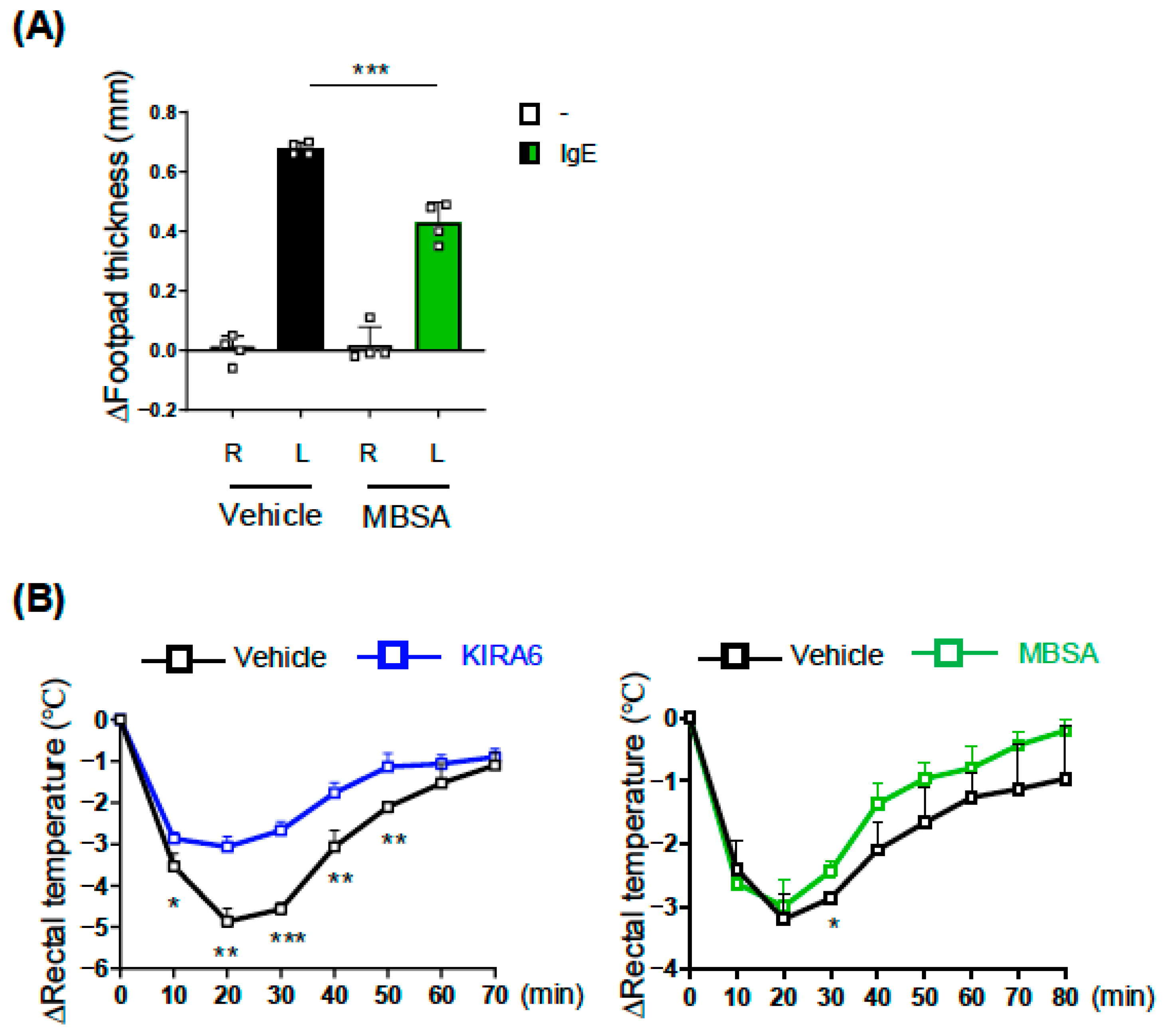
2.5. MBSA and KIRA6 Suppressed IgE-Dependent Anaphylaxis in Mice

The above-mentioned results of in vitro experiments using BMMCs demonstrated that inhibitors of the IRE1α-XBP1 pathway suppressed the IgE-induced activation of MCs. To examine the effects of these inhibitors on the IgE-dependent allergic response in vivo, we utilized passive anaphylaxis models. As shown in Figure 6A, the i.p. administration of MBSA significantly suppressed ear swelling caused by an i.v. injection of TNP-BSA into IgE-preinjected mice in the passive cutaneous anaphylaxis (PCA) model. We also confirmed that the body temperature reduction induced following the antigen injection in the passive systemic anaphylaxis (PSA) model was attenuated in mice administered MBSA or KIRA6 (Figure 6B).
These results indicate the biological significance of the IRE1α-XBP1 pathway in the IgE-dependent anaphylaxis.

2.6. XBP1 Is Involved in IgE-Induced Responses of MCs

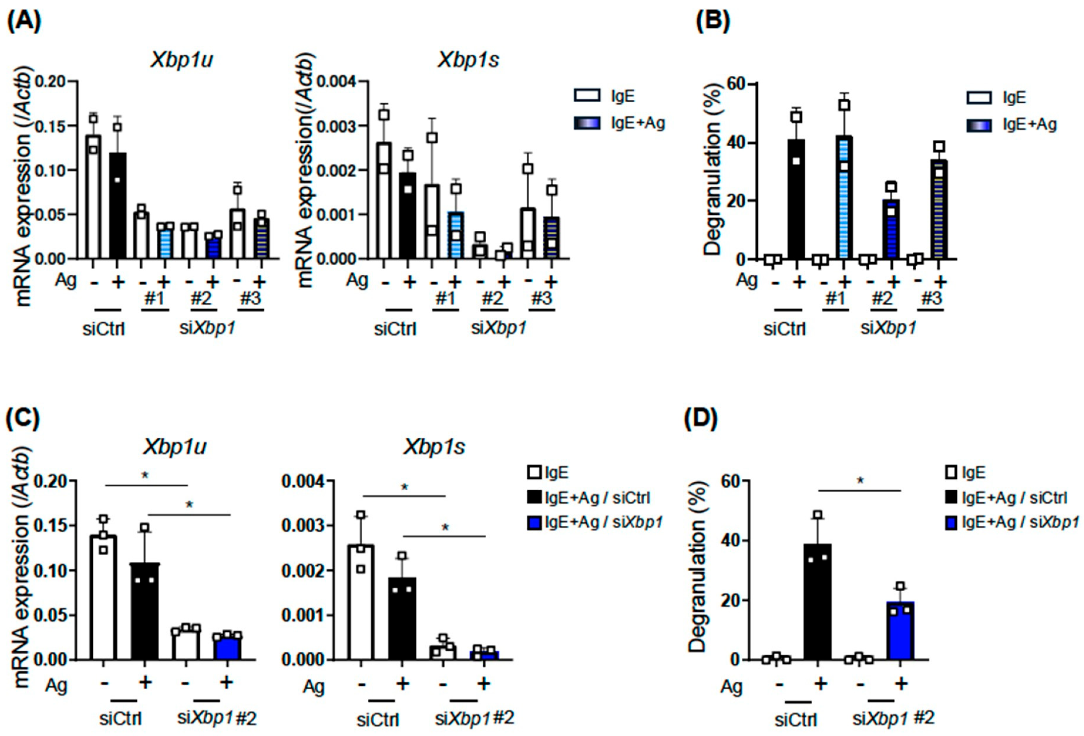
Two studies on the effects of KIRA6 on MC activation were recently published [14,23]. These studies claimed that KIRA6 suppressed MC activation by inhibiting kinases downstream of FcεRI, such as Lyn and/or Fyn, rather than IRE1α. These conclusions prompted us to investigate the involvement of XBP1 itself in the IgE-induced activation of MCs. To clarify the role of XBP1 in MC activation, we performed a knockdown experiment using siRNA transfection. Among three clones of Xbp1 siRNAs, Xbp1-2 siRNA most effectively reduced the mRNA levels of spliced Xbp1 (Xbp1s), whereas the knockdown efficiency of Xbp1-1 and -3 siRNAs was moderate (Figure 7A). A β-hexosaminidase assay on IgE-dependently activated BMMCs revealed that the extent of the degranulation of Xbp1 siRNA-transfected BMMCs decreased in parallel with the knocked down levels of Xbp1 mRNA (Figure 7B). We confirmed that the levels of the Xbp1 mRNA reduction (Figure 7C) and degranulation suppression (Figure 7D) by Xbp1-2 siRNA were significant.
Taken together, these results demonstrated that XBP1 was required for the IgE-dependent activation of MCs.

3. Discussion

We previously identified SA as the most effective compound that inhibited the IgE-dependent activation of MCs in a screening study aimed at the development of novel anti-allergic drugs [2]. Since SA inhibits the splicing of Xbp1 mRNA by IRE1α [3,4], which is required for the translation of a functional XBP1 protein, we investigated the involvement of the IRE1α-XBP1 axis in the IgE-dependent activation of MCs in the present study. We revealed that two other IRE1α inhibitors of endoribonuclease and kinase activities, the SA-related compound MBSA [5] and KIRA6 [15], respectively, suppressed the IgE-dependent degranulation of MCs in vitro and IgE-induced passive anaphylaxis in vitro, whereas the inhibitors targeting PERK and ATF6, which are also involved in the UPR as well as IRE1α-XBP1, did not affect the activation of MCs. Furthermore, the knockdown of Xbp1 mRNA reduced the IgE-dependent degranulation of MCs. Based on these results, we concluded that XBP1 plays a positive role in the IgE-dependent activation of MCs, which may be a therapeutic target of allergic diseases.
XBP1 transactivates the genes related to the secretion of proteins and lipid mediators, including insulin processing protease in the β cells [20], antibody production in plasma cells [7], and prostaglandin synthesis-related enzymes in dendritic cells [24]. Therefore, the genes expressed in activated MCs, which secrete various mediators such as proteases, eicosanoids, and cytokines, may be targets of XBP1. We intended to conduct further detailed analyses of the target genes of XBP1 in MCs.
During the preparation of this manuscript, two studies reporting the anti-allergic effects of KIRA6 were published [14,23]. Wunderle et al. demonstrated that KIRA6 suppressed the Ag-stimulated signaling of MCs by binding to kinases, LYN, FYN, and KIT [23]. The other study claimed that KIRA6 inhibited the kinase activity of Lyn in an IRE1α-independent manner because the knockout of the Ern1 gene (encoding IRE1α) by genome editing did not affect the Ag-induced activation of RBL-2H3, a rat basophilic cell line [14]. These findings prompted us to perform the knockdown of Xbp1 in BMMCs in order to reveal the role of XBP1 in the IgE-dependent activation of BMMCs, because an experiment revealing the direct effects of XBP1 is required and primary cells rather than leukemia cells needed to be used. The result showing that the extent of the degranulation of BMMCs decreased in parallel with Xbp1 mRNA levels indicated a positive role for XBP1 in the activation of MCs. Previous studies on the conditional knockout of Xbp1 demonstrated the requirement for XBP1 in the development and specific gene expression of hematopoietic cells [8,24,25]. XBP1 maintains the function of immune cells, which is not restricted in the UPR.
A recent study investigated the effects of tunicamycin on MC function [13]. The findings obtained showed that an i.p. injection of tunicamycin for 3 days increased the serum concentration of Mcpt1 in mice without inducing any allergic model, and also that the exposure of BMMCs to tunicamycin for 3 h increased the phosphorylation of kinases, Lyn, Syk, and PLCγ1. Based on these findings, it was concluded that the induction of ER stress by tunicamycin activated MCs. Since the effects of tunicamycin on the IgE-induced activation of MCs was not examined in this study, it is unclear whether the enhanced phosphorylation of kinases leads to the stronger activation of MCs and also which type of stimulation induced Mcpt1 secretion from MCs in tunicamycin-treated mice. However, under our experimental conditions, the tunicamycin treatment markedly suppressed the IgE-dependent activation of BMMCs (Figure 5). This result contrasted with our prediction, based on the previous study, that tunicamycin would enhance the IgE-dependent activation of MCs by inducing ER stress. A marked reduction in cell-surface FcεRI levels in tunicamycin-treated BMMCs is considered to be the primary cause of the responsiveness defect to FcεRI-mediated activation. However, we also recognize that the block of protein glycosylation by tunicamycin broadly affected MC activation-related events because A23187-induced activation was also suppressed in tunicamycin-treated BMMCs.
In the present study, we incubated BMMCs with IRE1α inhibitors for 0~48 h, and found that MBSA exerted stronger inhibitory effects with longer incubation times, whereas KIRA6 added just before the stimulation was the most effective for the suppression of MC activation. This difference may partly reflect the off-target effects of KIRA6 against kinases, Lyn, and/or Fyn [14,23]. In these studies, KIRA6 added 30 min [26] or 1 h [23] before the stimulation inhibited the IgE-dependent activation of MCs. The stronger inhibitory effect of KIRA6 than that of MBSA observed in PSA and cytokine production under our experimental conditions may be due to the additional target sites of KIRA6. However, we do not exclude the role of KIRA6 against IRE1α because Xbp1s mRNA levels in Ag-stimulated BMMCs were generally reduced in the presence of 1 mM KIRA6 [23]. Our preliminary experiment showed that mRNA levels of Xbp1s in BMMCs decreased 2 to 48 h after MBSA treatment; however, further experiments are needed to clarify how Xbp1s mRNA levels change over time following KIRA6 treatment under our experimental conditions. To evaluate the IRE1α-XBP1-dependency of the inhibitory effects of MBSA and KIRA6, experiments could be conducted using MCs lacking XBP1. This point could potentially be resolved by performing a degranulation assay of BMMCs generated from Xbp1-deficient mice to determine whether MBSA and KIRA6 exert additional inhibitory effects on Xbp1-knockout BMMCs. Furthermore, the use of Xbp1-knockout MCs may demonstrate that XBP1 is involved in the inhibitory effect of MBSA on A23187-induced MC activation.
The involvement of the IRE1α-XBP1 axis in the IgE-dependent activation of MCs has been controversial: an IRE1α deficiency did not affect the IgE-induced activation of RBL-2H3 cells [14], whereas IgE-dependent Syk phosphorylation and calcium mobilization were completely inhibited in Xbp1 knockout mouse MCs [13]. The results obtained using IRE1α inhibitors and Xbp1-targeting siRNA suggest that this pathway plays a positive role in IgE-dependent activation; however, further studies are needed to resolve this issue. A previous study using Ern1-knockout BM dendritic cells have suggested that IRE1α-XBP1 signaling plays an essential role in prostaglandin biosynthesis [24]. Furthermore, we have demonstrated that prostanoids, particularly PGE2, suppressed IgE-dependent activation of MCs [22,27]. Recently, we observed increased levels of Ptgs mRNAs in MBSA-treated or Xbp1 siRNA-transfected BMMCs. It may be interesting to evaluate the involvement of XBP1 in PG-related genes in MCs. We are currently attempting to identify the target gene(s) of XBP1 in MCs, which we expect to provide insights into the role of XBP1 in MC activation.
Finally, it is important to note that because XBP1 is involved in multiple processes across various organs and cells, it is necessary to consider the potential side effects that may arise from systemic administration of the inhibitors of XBP1. Identifying the target gene(s) of XBP1 in MCs could lead to development of specific treatments for allergic diseases.

4. Materials and Methods

4.1. Mice and Cells

C57BL/6J male and female mice (Japan SLC, Hamamatsu, Japan) housed under specific pathogen-free conditions were utilized as a passive anaphylaxis model (6–8 weeks old) or were sacrificed to generate BMMCs (6–10 weeks old). BMMCs were prepared under recombinant mouse IL-3 (BioLegend, San Diego, CA, USA) supplemented conditions as previously described [28]. Mice were maintained under specific pathogen-free-conditions, and all experiments using mice were performed following the guidelines of the Institutional Review Board of Tokyo University of Science. The present study was approved by the Animal Care and Use Committees of Tokyo University of Science: K22005.

4.2. Reagents

Anti-TNP mouse IgE (clone IgE-3, BD Bioscience, San Jose, CA, USA), TNP-BSA (LSL, Tokyo, Japan), 4-nitrophenyl-N-acetyl-β-D-glucosaminide (#N0866, Tokyo Chemical Industry, Tokyo, Japan), SA (#S0004, Tokyo Chemical Industry), MBSA (#B1487, Tokyo Chemical Industry), KIRA6 (#S8658, Selleck Chemicals, Houston, TX, USA), GSK2606414 (#T2614, TargetMol, Boston, MA, USA), Ceapin-A7 (#T9110, TargetMol), A23187 (#11016, Funakoshi, Tokyo, Japan), compound 48/80 (#C2313, Sigma-Aldrich, St. Louis, MO, USA), tunicamycin (#ab120296, Abcam, Cambridge, UK), and DMSO (07-4860-5, Sigma-Aldrich) were purchased from the indicated sources.

4.3. Degranulation of MCs

A degranulation assay was performed using a previously described method [16] by sensitizing BMMCs (5.0 × 10⁵) with 0.2 μg/mL anti-TNP mouse IgE in 1 mL medium at 37 °C for 2 h, and then stimulating these cells with 3 ng/mL TNP-BSA in 200 μL Tyrode’s buffer after washing with Tyrode’s buffer. After a 30 min stimulation, culture supernatants were harvested for the β-hexosaminidase assay [16].

4.4. Quantification of mRNAs

RNAzol RT Reagent (#RN190, Cosmo Bio, Tokyo, Japan) and ReverTraAce qPCR RT Master Mix (#FSQ-201, TOYOBO, Osaka, Japan) were used for the extraction of total RNA and for the synthesis of cDNA, respectively. Quantitative real-time PCR was performed on a StepOne Real-Time PCR System (Applied Biosystems, Waltham, MA, USA) with THUNDERBIRD SYBR qPCR Mix (#QPS201, TOYOBO) using the following primers.
Xbp1u-F; 5′-GACAGAGAGTCAAACTAACGTGG-3′
Xbp1u-R; 5′-GTCCAGCAGGCAAGAAGGT-3′
Xbp1s-F; 5′-AAGAACACGCTTGGGAATGG-3′
Xbp1s-R; 5′-CTGCACCTGCTGCGGAC-3′
Actb-F; 5′-AGATGACCCAGATCATGTTTGAGA-3′
Actb-R; 5′-CACAGCCTGGATGGCTACGTA-3′

4.5. ELISA

The concentrations of mouse IL-6 and TNF-α were assessed using ELISA MAX Standard/Deluxe Set-series ELISA kits (#431316 for IL-6; #430901 and #430916 for TNF-α).

4.6. Flow Cytometric Analysis

BMMCs pretreated with 1 mg/mL Fc block (clone 93, BioLegend) were stained with 2.5 mg/mL DAPI (#11034-56, Nacalai Tesque, Kyoto, Japan), PE anti-mouse FceRI (clone MAR1, BioLegend, 1:500), FITC anti-mouse c-kit (clone 2B, BioLegend, 1:200), and APC anti-mouse c-kit (clone ACK2, BioLegend, 1:200). Fluorescence was detected by a FACSLyric Analyzer (BD Pharmingen, San Diego, CA, USA) or a MACS Quant Analyzer (Miltenyi Biotec, Tubingen, Germany), and data were processed with FlowJo software v10.10.0 (Tomy Digital Biology, Tokyo, Japan).

4.7. Transfection of siRNA

Small interfering RNAs for mouse Xbp1; Xbp1-1 (Xbp1-MSS278861), Xbp1-2 (Xbp1-MSS278862), Xbp1-3 (Xbp1-MSS278863), and Stealth RNAi siRNA Negative Control MedGC (#12935-300) were purchased from Invitrogen (Carlsbad, CA, USA). BMMCs (2.0–6.0 × 10⁶) were transfected with 10 mL of 20 mmol/L siRNA using the Neon transfection system (Invitrogen) as previously described [28].

4.8. Passive Anaphylaxis

PSA and PCA were introduced into C57BL/6 mice by the administration of anti-TNP mouse IgE and TNP-BSA as described in our previous study [22]. MBSA (50–100 mg/kg), KIRA6 (2.5 mg/kg), or vehicle (saline:PEG:Tween-80:DMSO = 10:8:1:1 for MBSA, saline:DMSO:Tween-80 = 18:1:1 for KIRA6) was administered via an i.p. injection once per day.

4.9. Statistical Analysis

A two-tailed Student’s t-test was used to compare two samples. To compare more than three samples, a one-way ANOVA followed by Dunnett’s multiple comparison test was performed. p-values < 0.05 were considered to be significant.

Author Contributions

Conceptualization, C.N.; investigation—the majority of experiments and data analysis were performed by H.K. with assistance from K.N. and R.S.; writing—original draft preparation, review, and editing, C.N.; supervision, C.N.; project administration, K.N., and C.N.; funding acquisition, K.N., and C.N. All authors have read and agreed to the published version of the manuscript.

Funding

This work was supported by Grants-in-Aid for Scientific Research (B) 23K26860 (CN), and 23H02167 (CN); a Grant-in-Aid for Early-Career Scientists 24K17872 (KN); a Tokyo University of Science Grant for President’s Research Promotion (CN); the Tojuro Iijima Foundation for Food Science and Technology (CN); a Research Grant from the Mishima Kaiun Memorial Foundation (CN); and a Research Grant from the Takeda Science Foundation (CN).

Institutional Review Board Statement

The animal study protocol was approved by the Institutional Review Board of Tokyo University of Science. The present study was approved by the Animal Care and Use Committees of Tokyo University of Science: K22005, approval date 26 April 2022.

Informed Consent Statement

Not applicable.

Data Availability Statement

The raw data supporting the conclusions of this article will be made available by the authors, without undue reservation.

Acknowledgments

We thank members of the Laboratory of Molecular Biology and Immunology, Department of Biological Science and Technology, Tokyo University of Science for their constructive discussions and technical support. We greatly appreciate the consideration from Yayoi Yasuda, Masako Yasuda, and Kimihiko Yasuda.

Conflicts of Interest

The authors declare no conflicts of interest.

Abbreviations

The following abbreviations are used in this manuscript:
BMMCbone marrow-derived mast cell
ERendoplasmic reticulum
MBSA3-methyl-6-bromo-salichylaldehyde
MCMast cell
SAsalicylaldehyde
UPRunfolded protein response

References

  1. Kolkhir, P.; Elieh-Ali-Komi, D.; Metz, M.; Siebenhaar, F.; Maurer, M. Understanding human mast cells: Lesson from therapies for allergic and non-allergic diseases. Nat. Rev. Immunol. 2022, 22, 294–308. [Google Scholar] [CrossRef]
  2. Ashikari, T.; Hachisu, M.; Nagata, K.; Ando, D.; Iizuka, Y.; Ito, N.; Ito, K.; Ikeda, Y.; Matsubara, H.; Yashiro, T.; et al. Salicylaldehyde Suppresses IgE-Mediated Activation of Mast Cells and Ameliorates Anaphylaxis in Mice. Int. J. Mol. Sci. 2022, 23, 8826. [Google Scholar] [CrossRef] [PubMed]
  3. Li, S.; Zhu, G.; Yang, Y.; Jian, Z.; Guo, S.; Dai, W.; Shi, Q.; Ge, R.; Ma, J.; Liu, L.; et al. Oxidative stress drives CD8. J. Allergy Clin. Immunol. 2017, 140, 177–189.e9. [Google Scholar] [CrossRef] [PubMed]
  4. Croft, A.; Tay, K.H.; Boyd, S.C.; Guo, S.T.; Jiang, C.C.; Lai, F.; Tseng, H.-Y.; Jin, L.; Rizos, H.; Hersey, P.; et al. Oncogenic activation of MEK/ERK primes melanoma cells for adaptation to endoplasmic reticulum stress. J. Investig. Dermatol. 2014, 134, 488–497. [Google Scholar] [CrossRef] [PubMed]
  5. Volkmann, K.; Lucas, J.L.; Vuga, D.; Wang, X.; Brumm, D.; Stiles, C.; Kriebel, D.; Der-Sarkissian, A.; Krishnan, K.; Schweitzer, C.; et al. Potent and selective inhibitors of the inositol-requiring enzyme 1 endoribonuclease. J. Biol. Chem. 2011, 286, 12743–12755. [Google Scholar] [CrossRef]
  6. Bettigole, S.E.; Glimcher, L.H. Endoplasmic reticulum stress in immunity. Annu. Rev. Immunol. 2015, 33, 107–138. [Google Scholar] [CrossRef]
  7. Reimold, A.M.; Iwakoshi, N.N.; Manis, J.; Vallabhajosyula, P.; Szomolanyi-Tsuda, E.; Gravallese, E.M.; Friend, D.; Grusby, M.J.; Alt, F.; Glimcher, L.H. Plasma cell differentiation requires the transcription factor XBP-1. Nature 2001, 412, 300–307. [Google Scholar] [CrossRef]
  8. Iwakoshi, N.N.; Pypaert, M.; Glimcher, L.H. The transcription factor XBP-1 is essential for the development and survival of dendritic cells. J. Exp. Med. 2007, 204, 2267–2275. [Google Scholar] [CrossRef]
  9. Bettigole, S.E.; Lis, R.; Adoro, S.; Lee, A.-H.; Spencer, L.A.; Weller, P.F.; Glimcher, L.H. The transcription factor XBP1 is selectively required for eosinophil differentiation. Nat. Immunol. 2015, 16, 829–837. [Google Scholar] [CrossRef]
  10. Martinon, F.; Chen, X.; Lee, A.H.; Glimcher, L.H. TLR activation of the transcription factor XBP1 regulates innate immune responses in macrophages. Nat. Immunol. 2010, 11, 411–418. [Google Scholar] [CrossRef]
  11. Chaudhary, V.; Kioon, M.D.A.; Hwang, S.-M.; Mishra, B.; Lakin, K.; Kirou, K.A.; Zhang-Sun, J.; Wiseman, R.L.; Spiera, R.F.; Crow, M.K.; et al. Chronic activation of pDCs in autoimmunity is linked to dysregulated ER stress and metabolic responses. J. Exp. Med. 2022, 219, e20221085. [Google Scholar] [CrossRef]
  12. Cubillos-Ruiz, J.R.; Silberman, P.C.; Rutkowski, M.R.; Chopra, S.; Perales-Puchalt, A.; Song, M.; Zhang, S.; Bettigole, S.E.; Gupta, D.; Holcomb, K.; et al. ER Stress Sensor XBP1 Controls Anti-tumor Immunity by Disrupting Dendritic Cell Homeostasis. Cell 2015, 161, 1527–1538. [Google Scholar] [CrossRef]
  13. Fan, J.; Ma, L.; Xie, B.; Qiu, S.; Song, S.; Tang, Z.; Wu, Y.; Huangfu, H.; Feng, Y.; Luo, X.; et al. Modulating endoplasmic reticulum stress attenuates mast cell degranulation. Int. Immunopharmacol. 2024, 126, 111336, Corrected in Int. Immunopharmacol. 2024, 137, 112497. https://doi.org/10.1016/j.intimp.2024.112497. [Google Scholar] [CrossRef]
  14. Matsushima, G.; Matsui, Y.; Okamoto, H.; Umeda, N.; Kakimoto, M.; Mino, M.; Nakagawa, T.; Ishii, K.; Matsuo, Y.; Matsubara, D.; et al. Unique compound with anti-allergic action: Inhibition of Lyn kinase activity by KIRA6. Front. Pharmacol. 2025, 16, 1625798. [Google Scholar] [CrossRef] [PubMed]
  15. Ghosh, R.; Wang, L.; Wang, E.S.; Perera, B.G.K.; Igbaria, A.; Morita, S.; Prado, K.; Thamsen, M.; Caswell, D.; Macias, H.; et al. Allosteric inhibition of the IRE1α RNase preserves cell viability and function during endoplasmic reticulum stress. Cell 2014, 158, 534–548. [Google Scholar] [CrossRef]
  16. Inage, E.; Kasakura, K.; Yashiro, T.; Suzuki, R.; Baba, Y.; Nakano, N.; Hara, M.; Tanabe, A.; Oboki, K.; Matsumoto, K.; et al. Critical Roles for PU.1, GATA1, and GATA2 in the expression of human FcεRI on mast cells: PU.1 and GATA1 transactivate FCER1A, and GATA2 transactivates FCER1A and MS4A2. J. Immunol. 2014, 192, 3936–3946. [Google Scholar] [CrossRef] [PubMed]
  17. Axten, J.M.; Medina, J.R.; Feng, Y.; Shu, A.; Romeril, S.P.; Grant, S.W.; Li, W.H.H.; Heerding, D.A.; Minthorn, E.; Mencken, T.; et al. Discovery of 7-methyl-5-(1-{[3-(trifluoromethyl)phenyl]acetyl}-2,3-dihydro-1H-indol-5-yl)-7H-pyrrolo[2,3-d]pyrimidin-4-amine (GSK2606414), a potent and selective first-in-class inhibitor of protein kinase R (PKR)-like endoplasmic reticulum kinase (PERK). J. Med. Chem. 2012, 55, 7193–7207. [Google Scholar] [CrossRef]
  18. Torres, S.E.; Gallagher, C.M.; Plate, L.; Gupta, M.; Liem, C.R.; Guo, X.; Tian, R.; Stroud, R.M.; Kampmann, M.; Weissman, J.S.; et al. Ceapins block the unfolded protein response sensor ATF6α by inducing a neomorphic inter-organelle tether. eLife 2019, 8, e46595. [Google Scholar] [CrossRef]
  19. Kaser, A.; Lee, A.-H.; Franke, A.; Glickman, J.N.; Zeissig, S.; Tilg, H.; Nieuwenhuis, E.E.; Higgins, D.E.; Schreiber, S.; Glimcher, L.H.; et al. XBP1 links ER stress to intestinal inflammation and confers genetic risk for human inflammatory bowel disease. Cell 2008, 134, 743–756. [Google Scholar] [CrossRef]
  20. Lee, A.H.; Chu, G.C.; Iwakoshi, N.N.; Glimcher, L.H. XBP-1 is required for biogenesis of cellular secretory machinery of exocrine glands. EMBO J. 2005, 24, 4368–4380. [Google Scholar] [CrossRef] [PubMed]
  21. Albrecht, B.; Woisetschläger, M.; Robertson, M.W. Export of the high affinity IgE receptor from the endoplasmic reticulum depends on a glycosylation-mediated quality control mechanism. J. Immunol. 2000, 165, 5686–5694. [Google Scholar] [CrossRef]
  22. Nagata, K.; Ando, D.; Ashikari, T.; Ito, K.; Miura, R.; Fujigaki, I.; Goto, Y.; Ando, M.; Ito, N.; Kawazoe, H.; et al. Butyrate, Valerate, and Niacin Ameliorate Anaphylaxis by Suppressing IgE-Dependent Mast Cell Activation: Roles of GPR109A, PGE2, and Epigenetic Regulation. J. Immunol. 2024, 212, 771–784. [Google Scholar] [CrossRef] [PubMed]
  23. Wunderle, V.; Wilhelm, T.; Boukeileh, S.; Goßen, J.; Margreiter, M.A.; Sakurov, R.; Capellmann, S.; Schwoerer, M.; Ahmed, N.; Bronneberg, G.; et al. KIRA6 is an Effective and Versatile Mast Cell Inhibitor of IgE-mediated Activation. Eur. J. Immunol. 2025, 55, e202451348. [Google Scholar] [CrossRef] [PubMed]
  24. Chopra, S.; Giovanelli, P.; Alvarado-Vazquez, P.A.; Alonso, S.; Song, M.; Sandoval, T.A.; Chae, C.-S.; Tan, C.; Fonseca, M.M.; Gutierrez, S.; et al. IIRE1α-XBP1 signaling in leukocytes controls prostaglandin biosynthesis and pain. Science 2019, 365, eaau6499. [Google Scholar] [CrossRef] [PubMed]
  25. Tavernier, S.J.; Osorio, F.; Vandersarren, L.; Vetters, J.; Vanlangenakker, N.; Van Isterdael, G.; Vergote, K.; De Rycke, R.; Parthoens, E.; van de Laar, L.; et al. Regulated IRE1-dependent mRNA decay sets the threshold for dendritic cell survival. Nat. Cell Biol. 2017, 19, 698–710. [Google Scholar] [CrossRef]
  26. Hirose, E.; Matsushima, M.; Takagi, K.; Ota, Y.; Ishigami, K.; Hirayama, T.; Hayashi, Y.; Nakamura, T.; Hashimoto, N.; Imaizumi, K.; et al. Involvement of heme oxygenase-1 in kaempferol-induced anti-allergic actions in RBL-2H3 cells. Inflammation 2009, 32, 99–108. [Google Scholar] [CrossRef]
  27. Nagata, K.; Kasakura, K.; Miura, R.; Yashiro, T.; Nishiyama, C. Suppressive role of PPARγ in the IgE-dependent activation of mast cells. Int. Immunol. 2020, 32, 143–150. [Google Scholar] [CrossRef]
  28. Oda, Y.; Kasakura, K.; Fujigaki, I.; Kageyama, A.; Okumura, K.; Ogawa, H.; Yashiro, T.; Nishiyama, C. The effect of PU.1 knockdown on gene expression and function of mast cells. Sci. Rep. 2018, 8, 2005. [Google Scholar] [CrossRef]
Figure 1. Effects of IRE1α inhibitors, SA, MBSA, and KIRA6, on the IgE-induced degranulation of BMMCs. (A) IgE-induced degranulation of BMMCs treated with SA or MBSA (right); BMMCs were treated with 500 μM SA or 100 μM MBSA for 48 h, (left); BMMCs were treated with 100 μM MBSA for the indicated time. After the treatment with SA or MBSA, BMMCs (5 × 10⁵ cells/mL) were incubated with 200 ng/mL of mouse IgE for 2 h. After washing BMMCs with culture medium twice, BMMCs suspended in Tyrode’s buffer were stimulated with 3 ng/mL of TNP-BSA as an antigen (Ag) for 30 min. Beta-hexosaminidase activities in supernatants and in cells were measured as described in our previous study to assess the extent of degranulation [16]. (B) Inhibitory effects of KIRA6 on the IgE-induced degranulation of BMMCs. The indicated concentrations of KIRA6 were added with IgE ((left); 2 h before the Ag stimulation) or just when BMMCs were stimulated with Ag (right). These experiments were repeated on different days with independently prepared BMMCs from different individuals. The data represent the mean ± SD of independent experiments. Dunnett’s multiple comparison test was used for the statistical analysis between Ag-stimulated BMMCs without an inhibitor (Ctrl) and Ag-stimulated BMMCs with an inhibitor. * p < 0.05, ** p < 0.01, *** p < 0.005, n.s. not significant.
Ijms 27 04532 g001
Figure 2. Effects of MBSA and KIRA6 on IgE-induced cytokine secretion from BMMCs. IL-6 (left) and TNF-α (right) concentrations in the culture supernatants of IgE-stimulated BMMCs with or without the treatment with MBSA (A) and KIRA6 (B). After a 2 h incubation of BMMCs (5 × 10⁵ cells/mL) with 200 ng/mL of mouse IgE, BMMCs were washed with culture medium and then stimulated with 3 ng/mL of TNP-BSA as an antigen (Ag). Culture supernatants were harvested 3 h after the Ag stimulation for ELISA. MBSA (final concentration 100 μM) was added at 48 h before the Ag stimulation, and KIRA6 (1 μM) was added just before the addition of Ag. These experiments were repeated on different days with independently prepared BMMCs from different individuals. The data represent the mean ± SD of independent experiments. The two-tailed paired Student’s t-test was used for the statistical analysis between Ag-stimulated BMMCs without an inhibitor (−) and Ag-stimulated BMMCs with an inhibitor (+). * p < 0.05, ** p < 0.01.
Ijms 27 04532 g002
Figure 3. Effects of MBSA and KIRA6 on the Ca2+ ionophore- and compound 48/80-induced degranulation of BMMCs. Degranulation levels of BMMCs stimulated by A23187 (left) or compound 48/80 (C48/80; right) with or without the treatment with MBSA (A) and KIRA6 (B). BMMCs incubated with 100 μM MBSA for 24 h were stimulated by 1 μM of A23187 (left) or 30 μg/mL of C48/80 (right) for 30 min (A). A total of 1 μM KIRA6 was added just before the addition of A23187 or C48/80 (B). Harvested supernatants and collected cells were assessed using the β-hexosaminidase assay (see Materials and Methods). These experiments were repeated on different days with independently prepared BMMCs from different individuals. The data represent the mean ± SD of independent experiments. The two-tailed paired Student’s t-test was used for the statistical analysis between without an inhibitor (−) and with an inhibitor (+). * p < 0.05, n.s. not significant.
Ijms 27 04532 g003
Figure 4. Inhibitors targeting two other pathways did not affect the activation of BMMCs. BMMCs were incubated with the indicated concentrations of GSK2606414, a PERK inhibitor (A), or CeapinA-7, an ATF6 inhibitor (B), for 22 h before the addition of IgE and for an additional 2 h during the incubation with Ag (left), or for 24 h before the addition of A23187 (center) and compound 48/80 (right). These experiments were repeated on different days with independently prepared BMMCs from different individuals. The data represent the mean ± SD of independent experiments. The two-tailed Dunnett’s multiple comparison test was used for the statistical analysis between without an inhibitor and with an inhibitor. n.s. not significant.
Ijms 27 04532 g004
Figure 5. Effects of tunicamycin on MC activation. (A) The IgE-induced degranulation of BMMCs treated with tunicamycin. (B) IL-6 (left) and TNF-α (right) production from tunicamycin-treated BMMCs. (C) Cell surface expression of FcεRI (right) and c-kit (left) on tunicamycin-treated BMMCs. BMMCs (5 × 10⁵ cells/mL) were incubated in the presence or absence of the indicated concentration of tunicamycin (TM) for 22 h, and were then subjected to the IgE-induced degranulation assay (A), IgE-induced cytokine production (B), and to a flow cytometric analysis (C). These experiments were repeated on different days with independently prepared BMMCs from different individuals. The data represent the mean ± SD of independent experiments. Dunnett’s multiple comparison test was used in (A) and the two-tailed paired Student’s t-test was used for the statistical analysis between without TM and with TM (B). * p < 0.05, ** p < 0.01, *** p < 0.005, n.s. not significant.
Ijms 27 04532 g005
Figure 6. Passive anaphylaxis levels in mice administered MBSA or KIRA6. (A) Footpad swelling in passive cutaneous anaphylaxis model mice. Mice were i.p. injected with MBSA (50 mg/kg/day) or 200 μL vehicle (see the Materials and Methods) for 5 days (n = 4 for each group). IgE solution and its vehicle were injected into the left (L) and right (R) footpad, respectively, on day 4, and Ag was i.v. injected on day 5. Footpad thickness 30 min after the Ag injection was measured as described in our previous study [2,22]. The unpaired Student’s t-test was used for the statistical analysis between control (Vehicle) and MBSA. *** p < 0.005. (B) Body temperature of passive systemic anaphylaxis model mice. Mice were i.p. injected with 100 mg/kg/day MBSA or its vehicle (right), and 2.5 mg/kg/day KIRA6 (right) or its vehicle (left) for 3 days (n = 3 for each group). I.v. injections of IgE on day 2 and of Ag on day 3 were performed as previously described [2,22]. The data represent the mean ± SD of independent experiments. The unpaired Student’s t-test was used for the statistical analysis between the vehicle and inhibitor. * p < 0.05, ** p < 0.01, *** p < 0.005.
Ijms 27 04532 g006
Figure 7. Effects of Xbp1 siRNA introduction on the IgE-induced activation of BMMCs. (A,C) mRNA levels of un-spliced Xbp1 (Xbp1u) and spliced Xbp1 (Xbp1s) in siRNA transfected BMMCs, which were harvested 3 h after the Ag stimulation. (B,D) Degranulation degree of Xbp1 siRNA-transfected BMMCs. After a 72 h culture from electroporation, BMMCs were divided into two groups: one for mRNA quantification and the other for the degranulation assay. Three different sequences of Xbp1 siRNAs were used: Xbp1-1 (#1, Xbp1-MSS278861), Xbp1-2 (#2, Xbp1-MSS278862), and Xbp1-3 (#3, Xbp1-MSS278863). These experiments were repeated on different days with independently prepared BMMCs from different individuals. The data represent the mean ± SD of independent experiments. The two-tailed paired Student’s t-test was used for the statistical analysis between control siRNA-transfected BMMCs (siCtrl) and Xbp1 siRNA-transfected BMMCs (siXbp1). * p < 0.05.
Ijms 27 04532 g007
Disclaimer/Publisher’s Note: The statements, opinions and data contained in all publications are solely those of the individual author(s) and contributor(s) and not of MDPI and/or the editor(s). MDPI and/or the editor(s) disclaim responsibility for any injury to people or property resulting from any ideas, methods, instructions or products referred to in the content.