


Abstract
Triple-negative breast cancer (TNBC/18–21%) lacks targeted treatment options due to the lack of ER/PR and HER2 expression. The transport of lncRNAs via exosomes plays a role in tumor progression and metastasis and reshapes tumor-associated signaling pathways. This study aimed to compare the expression of exosomal HOTAIR, NEAT1, MALAT1, AFAP1-AS1, ANRIL, and HULC lncRNAs in primary tumor tissue and blood of patients with sporadic TNBC to evaluate their potential as biomarkers. The patients diagnosed with TNBC between the years 2021 and 2025, 21 of 62 (33.87%) with sporadic breast cancer and thirty healthy controls were included in the study. Primary tumor tissue and peripheral venous blood samples were collected from 21 patients who did not receive neoadjuvant chemotherapy. Expression levels of exosomal lncRNAs (HOTAIR, NEAT1, MALAT1, AFAP1-AS1, ANRIL, and HULC) were determined in both tissues and blood samples from the patient and control groups using Real-Time PCR method. In the patient group, HOTAIR, HULC, ANRIL, and AFAP1_AS1 gene expression was lower (downregulated) in tissue and serum compared to the control group, whereas NEAT1 and MALAT1 were higher (upregulated). Tissue and serum samples taken from the patient group were found to have statistically consistent expression levels of HOTAIR, HULC, and ANRIL genes. Furthermore, HOTAIR, HULC, and ANRIL serve as biomarkers and can be studied using exosomal RNA samples obtained from patient serum without invasive procedures. Our current study, which has different lncRNA expression profiles, reflects the biological heterogeneity of TNBC and contributes to a better understanding of its subtypes at the molecular level.
1. Introduction
Breast cancer is the most common type of cancer worldwide, particularly among women. While advances in screening and treatment have reduced the overall risk of death from the disease, the number of people diagnosed with breast cancer continues to increase. Data have shown that the incidence of breast cancer has increased by 1% annually since 2012. Current estimates (American Cancer Society) predict that by 2025, approximately 316,950 women will be diagnosed with invasive breast cancer, approximately 16% of whom will be under the age of 50. Breast cancer is a highly molecularly heterogeneous group and is divided into subtypes based on gene expression. Triple-negative breast cancer (TNBC), which has limited treatment options, lacks ER/PR (estrogen receptor/progesterone receptor) and HER2 (human epidermal growth factor receptor 2) expression and accounts for 18–21% of invasive breast cancers [1]. Furthermore, the percentage of Ki67 protein is also emphasized as being crucial for metastasis [2].
Approximately 10–15% of breast cancer cases are familial and associated with known gene variants. Studies in TNBC are critical today, especially considering the high percentage of individuals with sporadic breast cancer. The lack of gene expression markers in the TNBC molecular classification limits the effectiveness of patient treatment strategies. Therefore, there is a need to identify more genetically based biomarkers.
Biomarkers can be analyzed in various human samples, including tissue, blood, urine, and other body fluids. Currently, for breast cancer, CA15-3 and CEA (*CA15-3: metastatic breast cancer marker, *CEA: carcinoembryonic antigen) are commonly analyzed in blood samples [3]. Genetically, biomarkers can be derived from the type of sample to be investigated and from various biological materials thought to be influential in the cancer development process. In recent biomarker research, exosomes, the fundamental elements of intercellular interactions, have become prominent, rather than DNA and RNA obtained from basic biological samples.
Exosomes are lipid-bilayered extracellular vesicles, 30–150 nm in size, released by all cell types, including breast cancer cells. Their vesicle contents consist of proteins, lipids, metabolites, DNA, mRNA, and non-coding RNAs (short miRNAs and long non-coding RNAs (lncRNAs). Cancer cells utilize exosomes to achieve metastasis, immune evasion, drug resistance, immune suppression, communication with the tumor microenvironment, and invasion. Exosomes facilitate tumor progression and metastasis by carrying oncogenic signaling proteins, ligands, enzymes, and non-coding RNAs on their surfaces or within their vesicles [4]. Furthermore, studies on TNBC suggest that exosomal lncRNAs regulate gene expression in the nucleus and cytoplasm and interact with multiple post-transcriptional mechanisms [5].
LncRNAs regulate intracellular and intercellular signaling in TNBC. Depending on their mechanisms, they can affect gene expression in multiple ways and, consequently, transform cells into a cancer phenotype [6]. Studies generally reveal differences in lncRNA expression levels between patient and healthy tissue samples, underscoring their potential as biomarkers [7]. Biomarker studies typically focus on the potential of a single lncRNA in different sample types [8]. Meta-analysis of international data indicates that lncRNAs with high expression levels are positively correlated with lymph node metastasis and distant metastasis [9].
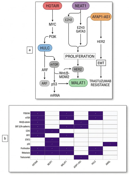
According to *GENCODE 24 data, 21,000 lncRNAs have been identified to date. Recent studies have highlighted 51 lncRNAs as crucial for breast cancer. Our current study identified six lncRNAs that we predict are associated with TNBC and play regulatory roles in intracellular and intercellular signaling during cancer development. The common cellular signaling pathways and effects of the lncRNAs investigated in this study are presented in Figure 1.
Figure 1.
(a) Effects of lncRNAs investigated in the current study on common cellular signaling pathways, (b) Molecular targets and locations at common pathway intersections of lncRNAs were investigated in the current study.
The primary hypothesis of our study was to investigate the potential use of lncRNAs as biomarkers in sporadic TNBC. The paucity of molecular markers in TNBC complicates early diagnosis and prognosis, limiting treatment strategies. Because lncRNAs transported via exosomes are known to play an active role in tumor progression and metastasis, examining serum levels of these molecules could reveal the biological heterogeneity of TNBC through a non-invasive approach. In particular, downregulation of HOTAIR, HULC, and ANRIL, and upregulation of NEAT1 and MALAT1, reflect the TNBC-specific molecular signature and may guide clinical practice in identifying diagnostic, prognostic, and therapeutic targets. Furthermore, we demonstrate that the HOTAIR, HULC, and ANRIL lncRNAs investigated in this study are present at consistent levels in both tissue- and serum-derived exosomal RNAs. This finding reveals that these genes are strong biomarker candidates and can be evaluated through serum samples without the need for invasive biopsy.
2. Results
Of the 582 patients initially diagnosed with breast cancer, 21 of 62 TNBC patients with sporadic cases were included in the study according to exclusion criteria. A germline cancer NGS panel was performed on all 62 patients diagnosed with TNBC, and 21 (33.87%) were diagnosed with sporadic breast cancer after no germline pathogenic or likely pathogenic variants were detected. Seven (33.33%) of our patients developed metastasis within the first three years. Additionally, two cases with metastasis were reported to have uterine polyps, one case with a renal cyst, and one case with a thyroid nodule. After receiving their initial diagnosis, the patients in our patient group began combined chemotherapy and adjunctive treatments based on their clinical condition. Since the initiation of our study, we have also determined the recurrence and survival rates of the patients.
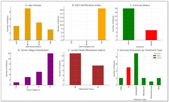
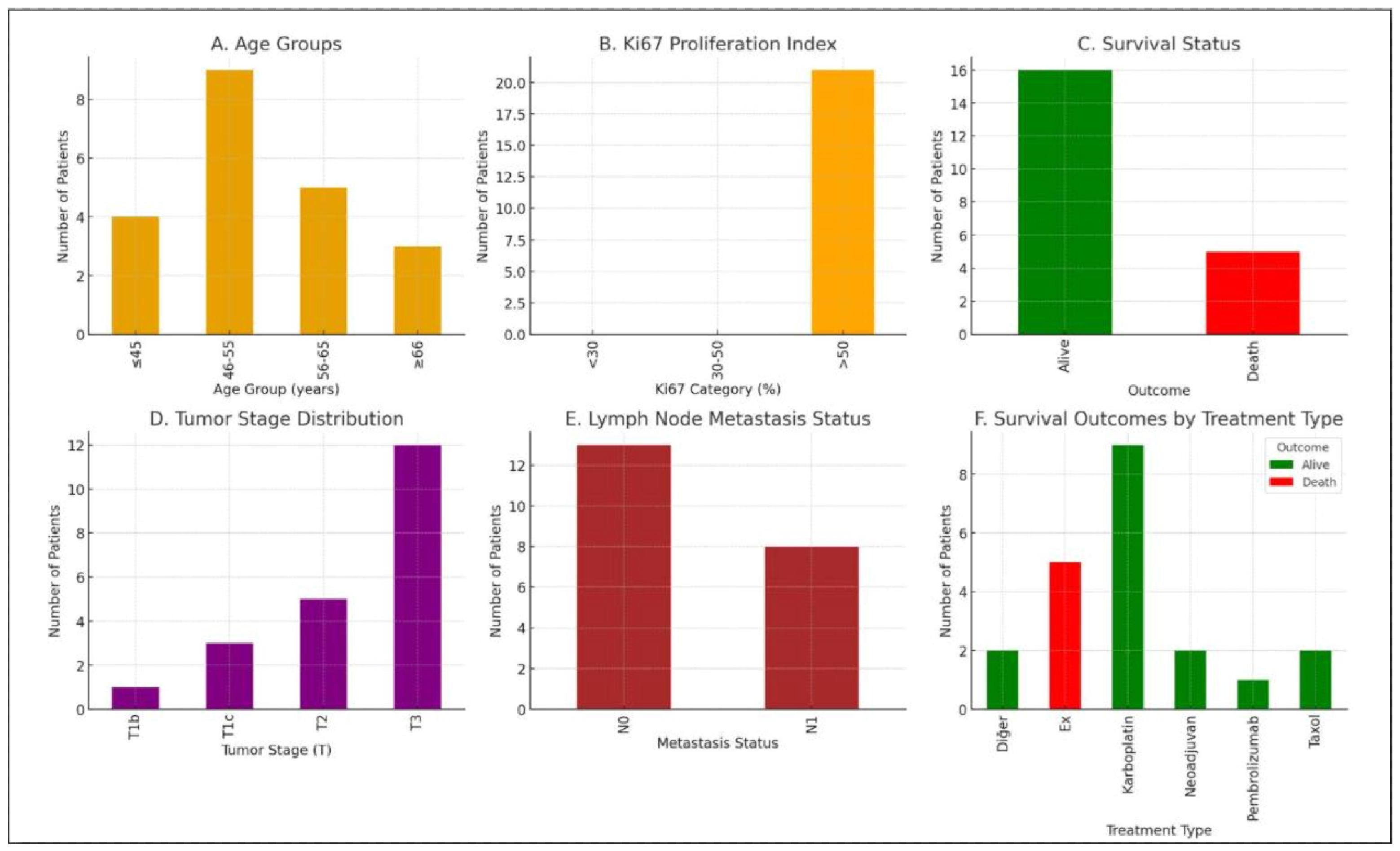
The patients included in the patient group were aged between 46 and 55 (mean age: 50.5). This suggests that TNBC is more common in our patient population, particularly in middle-aged women. The majority of patients had a Ki67 proliferation index above 30%. This supports the conclusion that TNBC patients, most of whom were in stage T3, exhibit aggressive biological behavior. This distribution supports the conclusion that most cases were in advanced stages at diagnosis. Nodal metastases (N0) were detected in two-thirds of the cohort, while nodal metastases (N1) were detected in one-third. These patients had poorer survival compared to N0 patients. All patients who received neoadjuvant therapy, carboplatin- and taxol-based regimens, and pembrolizumab are currently under follow-up and alive. The higher survival rate in carboplatin-containing protocols is noteworthy.
The four-year survival rate from diagnosis and treatment initiation to the present day was calculated using the Kaplan–Meier method. Five of the 21 patients included in the study died during this period. Since 16 patients were alive at the end of four years, the survival rate was 16/21 ≈ 0.76 (76.20%). Because TNBC is a more aggressive subtype of breast cancer than other breast cancer subtypes, the risk of recurrence is higher in individuals who respond positively after the first course of treatment. According to our data over the four years, 15 of the 21 patients (71.42%) experienced recurrence, with 12 occurring within the first two years.
Our criteria for comparing lncRNA expression levels between groups in sporadic TNBC patients, which constitute the hypothesis of our study, are as follows: patient group tissue–control group tissue data, patient group serum–control group serum data, patient group tissue–patient group serum data, control group tissue–control group serum data, patient group metastasis status–lncRNA expression levels, patient group cancer markers in blood–lncRNA expression levels.
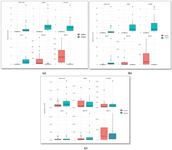
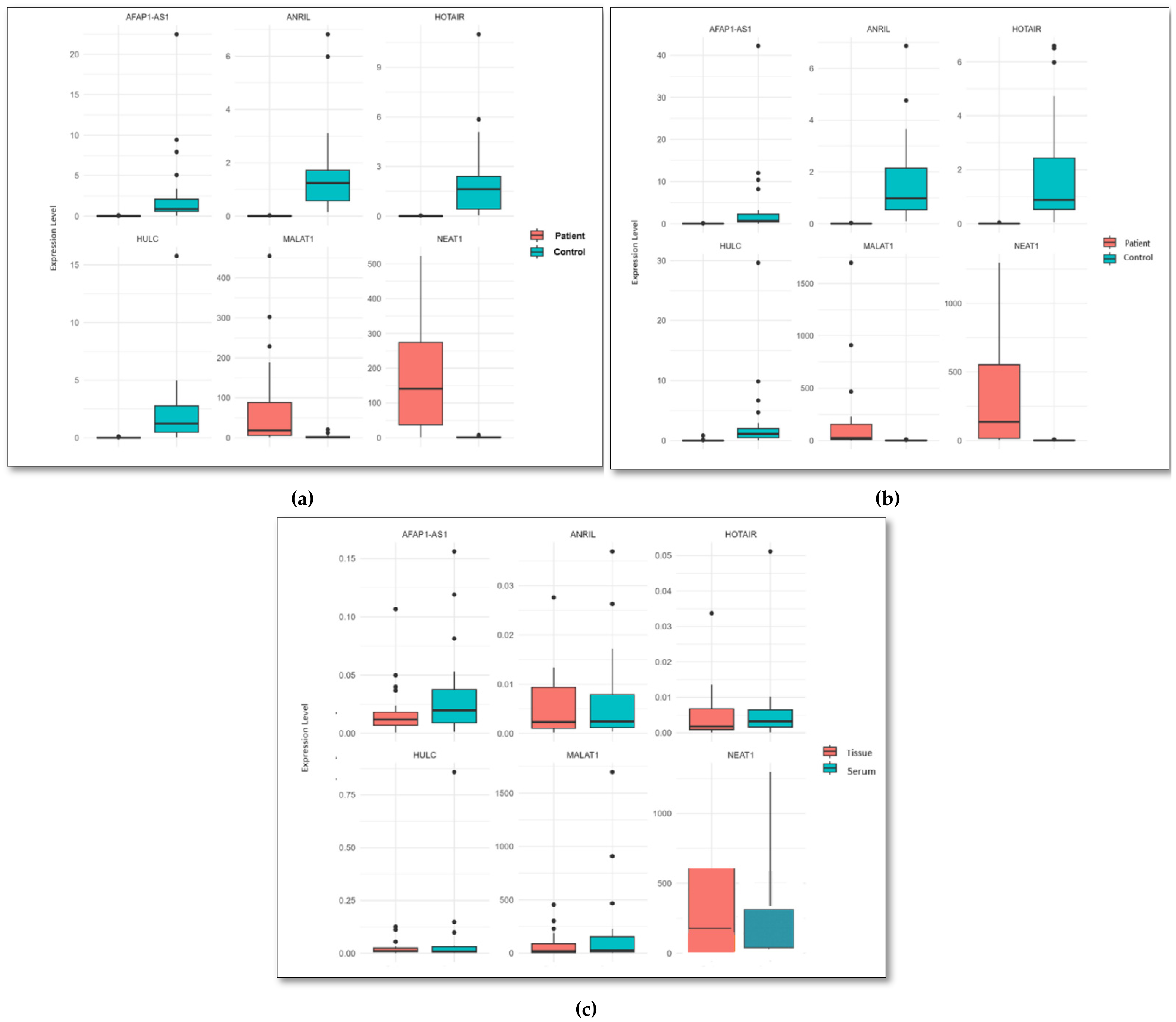
When the tissue data from the patient and control groups were statistically compared, it was found that HOTAIR, HULC, ANRIL, and AFAP1_AS1 were downregulated in the patient group, while NEAT1 and MALAT1 were upregulated (Table 1, Figure 2).When the serum data from the patient and control groups were statistically compared, it was found that HOTAIR, HULC, ANRIL, and AFAP1_AS1 were downregulated in the patient group, while NEAT1 and MALAT1 were upregulated (Table 2 and Figure 2). When the tissue and serum samples from the patient group were statistically compared, the expression levels of exosomal HOTAIR, HULC, and ANRIL genes were found to be consistent between the two. Based on this result, we predict that the expression levels of exosomal HOTAIR, HULC, and ANRIL in serum can serve as biomarkers without the need for invasive procedures such as tissue biopsy (Table 3 and Figure 2). The consistency between tissue and serum expression patterns was further evaluated based on the direction of expression changes. HOTAIR, HULC, and ANRIL showed concordant expression patterns in both tissue and serum samples, with consistent downregulation across sample types. In addition, statistical analyses performed in the patient group (Table 3) demonstrated significant differences in expression levels, further supporting the consistency between tissue and serum-derived exosomal lncRNA profiles.
Table 1.
Comparison of gene expression values of long non-coding RNAs between patient and control group tissue samples.
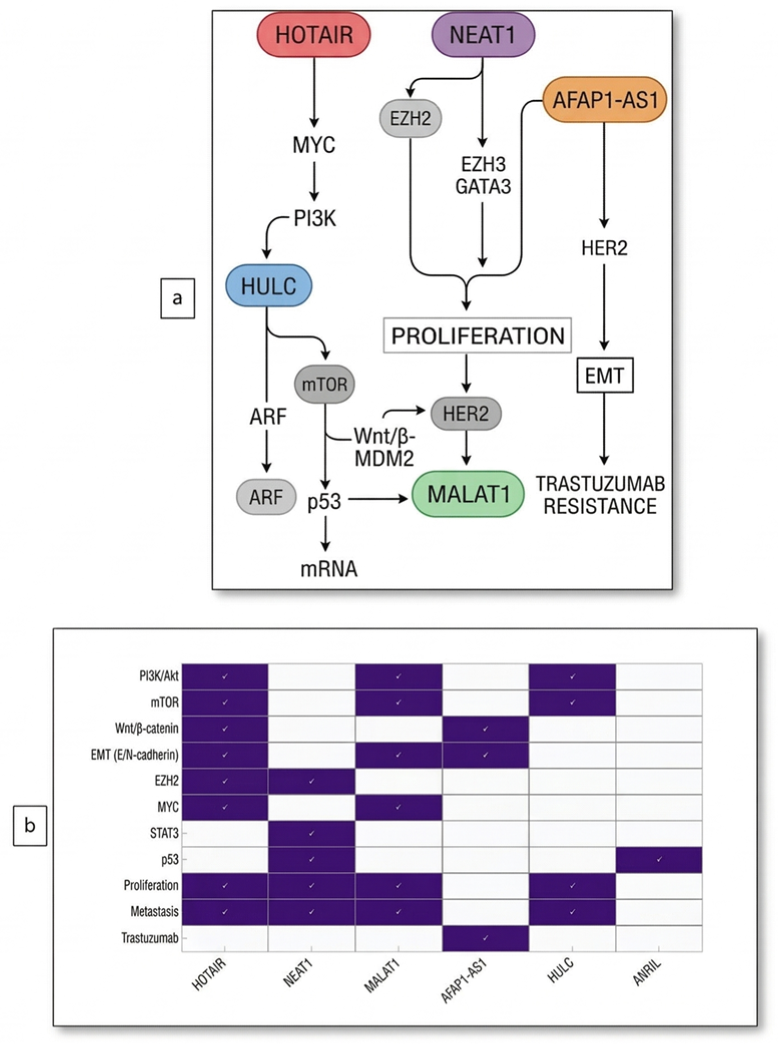
Figure 2.
(a) Comparison of gene expression of long non-coding RNAs between patient and control group tissue samples, (b) Comparison of gene expression of long non-coding RNAs between patient and control group serum samples, (c) Comparison of gene expression of long non-coding RNAs between patient group tissue and serum sample.
Table 2.
Comparison of gene expression values of long non-coding RNAs between patient and control group serum samples.
Table 3.
Comparison of gene expression values of long non-coding RNAs between tissue and serum samples of the patient group.
The NEAT1 gene was more highly expressed in tissue than in serum. The AFAP1-AS1 and MALAT1 genes were less expressed in tissue but more so in serum. The relationship between metastasis and the expression levels of related genes in the patient group was evaluated using the T-test and Mann–Whitney U test, and the NEAT1 gene was found to be highly expressed in non-metastatic patients (p < 0.0042). No significant differences were found for other genes. Biochemical cancer markers (CEA, CEA 15-3) in the blood were also statistically evaluated using the T-test and Mann–Whitney U test. No significant results were found for any of the genes in the T-test results. However, an abnormal decrease in NEAT1 gene expression was observed using the Mann–Whitney U test. When control group tissue and control group serum data were compared, no statistically significant difference was detected.
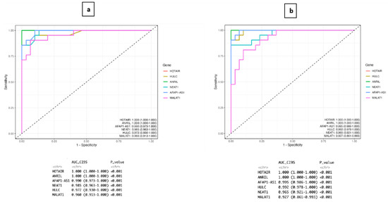
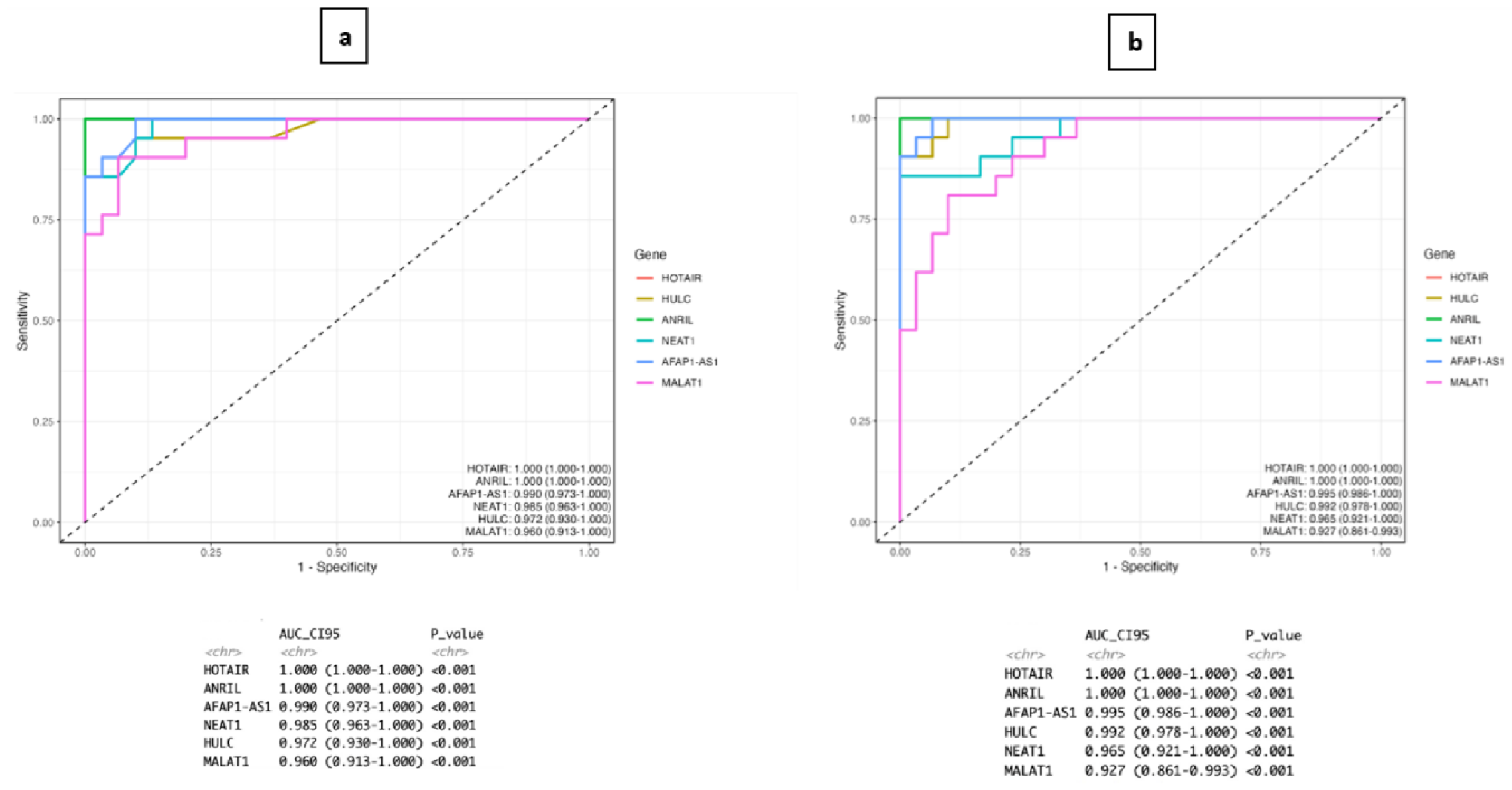
ROC analysis demonstrated that serum exosomal lncRNAs exhibited strong diagnostic performance in distinguishing sporadic TNBC patients from healthy controls. HOTAIR and ANRIL showed excellent discriminatory power (AUC = 1.000), while AFAP1-AS1, NEAT1, HULC, and MALAT1 demonstrated high diagnostic accuracy with AUC values ranging from 0.960 to 0.990 (p < 0.001 for all genes). These findings suggest that circulating exosomal lncRNAs may be promising, non-invasive biomarkers for TNBC detection. However, the number of patients included in this study was relatively limited due to technical constraints associated with exosome isolation and subsequent molecular analysis (Figure 3). Furthermore, the analyzed lncRNAs showed strong diagnostic performance in distinguishing TNBC patients from healthy controls. However, these findings should be interpreted with caution due to the relatively small cohort size, and further validation in larger, independent cohorts is required. Overall, our results highlight the potential clinical utility of exosomal lncRNAs as promising biomarkers for TNBC diagnosis and monitoring.
Figure 3.
Receiver operating characteristic (ROC) curve analysis of exosomal lncRNAs for distinguishing TNBC patients from healthy controls, (a) ROC curves based on serum-derived exosomal lncRNA expression levels, (b) ROC curves based on tissue-derived exosomal lncRNA expression levels.
3. Discussion
TNBC is characterized by profound biological heterogeneity, which complicates efforts to identify meaningful molecular markers. Our study addresses this gap by comparing exosomal lncRNA expression profiles in tumor tissue and serum from patients with sporadic TNBC. In this context, our study suggests that exosomal lncRNAs, particularly HOTAIR, HULC, and ANRIL, may be promising biomarker candidates. These three lncRNAs showed consistent downregulation in both tumor tissue and serum-derived exosomes, suggesting that circulating exosomal RNA may reflect tumor biology without requiring invasive tissue sampling. On the other hand, increased expression of NEAT1 and MALAT1 further underscores their potential contribution to tumor aggressiveness and may aid in identifying subgroups within sporadic TNBC.
Research shows that exosome secretion is higher in individuals with cancer than in healthy individuals. The increased exosome secretion in individuals with cancer may be explained by the activation of the TSAP6 (metalloreductase) gene, which regulates the exosomal secretion pathway within the p53 pathway [10]. A meta-analysis of international data reported that highly expressed lncRNAs are positively correlated with lymph node metastasis and distant metastasis. Our study identified the lncRNAs HOTAIR, NEAT1, AFAP1-AS1, ANRIL, MALAT1, and HULC, which are linked to metastasis. These lncRNAs are prominent not only in metastasis but also in various mechanisms during the cancerization process of TNBC cells.
To summarize the results of our study, the downregulated lncRNAs were HOTAIR, HULC, ANRIL, and AFAP1_AS1, while the upregulated lncRNAs were NEAT1 and MALAT1. Studies reported in the literature generally only examined expression levels in cell lines, tumor tissue, or serum/plasma samples. Studies that combined or compared all parameters are almost nonexistent. Therefore, further studies are needed to enrich the existing data. In our study, the data obtained for HOTAIR, ANRIL, and AFAP1_AS1 differ from those reported in the literature. In our study, we specifically investigated lncRNA expression levels in primary tumor tissues and serum samples from sporadic TNBC patients before chemotherapy or radiotherapy.
HOTAIR, whose elevated expression has been associated with metastasis in TNBC studies, may also act as a tumor suppressor gene, depending on the pathway in which it functions [11]. Expression levels vary in various subgroups of breast cancer, particularly in tumor tissues. It is known for its increased expression levels in serum samples [12]. In our study, a statistically significant difference in HOTAIR expression levels was observed between tissue and serum samples from the patient and control groups. HOTAIR expression levels were lower in both tissue and serum samples in the patient group than in the control group. HOTAIR expression levels in tissue and serum samples from the patient group were parallel. A study including 2192 individuals with TNBC emphasized that HOTAIR was highly expressed in tumor tissue, which may be associated with poor prognosis and metastasis [13]. The number of studies evaluating HOTAIR expression in serum samples from TNBC patients is quite limited, and, generally, increased expression has been reported only in breast cancer patients. Li et al. reported that expression levels in tumor tissue may vary with cancer stage [14]. The study emphasized that HOTAIR expression levels in stage I breast cancer were lower than in stages II, III, and IV [15]. Another study conducted with a breast cancer cell line reported that HOTAIR regulates resistance to radiation and chemotherapy in breast cancer [16]. Another study conducted in 2019 demonstrated the effects of HOTAIR expression on treatment resistance in TNBC cells using a recombinant plasmid vector containing HOTAIR. This study, conducted in a cell line, reported that resistance to the treatment agent decreased in cell groups with lower HOTAIR expression [17]. The results of our study regarding HOTAIR expression levels differ from those reported in the literature.
Unlike other lncRNAs, NEAT1 forms a unique nuclear body (paraspeckles) that facilitates the assembly of various protein components, including RNA-binding proteins. Its association with metastasis is highlighted by its increased expression levels in other breast cancer subgroups, including TNBC. NEAT1 is regulated by p53 to suppress tumor transformation. NEAT1 affects aerobic glucose and lactic acid fermentation for energy production in cancer cells. This is also related to the Warburg effect.
NEAT1 is known for its elevated expression in studies of breast cancer and TNBC. In 2019, Shin et al. reported that NEAT1 expression levels in blood samples from TNBC patients were higher than in healthy individuals [18]. In another study of 164 TNBC individuals, increased expression levels in tumor tissue were associated with lymph node metastasis, highlighting its utility as a biomarker for patient follow-up and cancer management [19]. Furthermore, in another study involving treatment-resistant cancer stem cells, it was concluded that when NEAT1 was completely knocked out, chemotherapy-resistant cells became sensitive to the drug, thereby promoting drug uptake [20]. In our study, consistent with the literature, NEAT1 expression levels were higher in tissue and serum samples from the patient group than in those from the control group. We also found that NEAT1 expression was higher in tissue samples from the patient group compared to serum samples.
AFAP1-AS1, which plays a role in various types of cancer, acts as an oncogene in TNBC cells, increasing metastasis and tumor invasion, and many studies on breast cancer have reported increased expression levels in tissue and serum. In our research, we found that AFAP1_AS1 was less expressed in the patient group than in the control group. When the patient group was evaluated within the patient group, its expression level in tissue was lower than in serum samples. A study of the TNBC MDA-MB-231 cell line reported high AFAP1_AS1 expression. It has also been reported that this upregulation may increase the proliferation and invasiveness of TNBC cells [21]. Studies investigating drug resistance mechanisms have reported that certain lncRNAs are upregulated in trastuzumab-resistant breast cancer cells and can be transferred via exosomes, contributing to the dissemination of resistance between cells. For instance, AFAP1-AS1 has been associated with trastuzumab resistance and may be involved in exosome-mediated intercellular communication, although further validation is required [22]. Our data for AFAP1_AS1, which has been frequently reported to show increased expression in previous studies, differ from those studies.
ANRIL is involved in the mechanisms of apoptosis, metastasis, invasion, and proliferation in cancer cells via the p53 pathway. It is known that expression levels are higher in TNBC cell lines than in healthy cells. In our study, contrary to the literature, ANRIL expression levels were lower in tissue and serum samples from the patient group than in those from the control group. Studies on TNBC in the literature generally include cell lines and plasma samples obtained from blood. Among studies conducted to date, only one addresses TNBC status in tumorigenic patient tissue. This 2017 study included 25 TNBC and 35 healthy individuals. It was reported that ANRIL expression levels were higher in TNBC patients compared to the healthy group, while expression levels in tumor tissue were lower than in plasma [23].
In TNBC cells, MALAT1 regulates cell growth and metabolism, primarily through the PI3K/Akt pathway, via the KDM5B (Lysine demethylase 5b) protein [24]. Dysregulation of post-transcriptional cell signaling affects mechanisms such as metastasis and response to therapy [25]. MALAT1 is critical for TNBC because it participates in the MAPK/ERK, PI3K/Akt, and Wnt/β-catenin pathways. These pathways are directly linked to the regulation of the cell signaling network in metastasis, proliferation, and cancer development. A recently published study highlighted increased MALAT1 expression in TNBC cell lines and suggested that it may be effective in treatment via the JAK/STAT pathway [26]. In a separate study of 88 patients who underwent mastectomy in the early stages of TNBC, high MALAT1 expression was found in tissue samples, and it was reported that this may be associated with lymph node metastasis [27]. In our study, consistent with the literature, MALAT1 expression levels in tissue and serum samples were higher in the patient group than in the control group. When the patient group was evaluated within itself, MALAT1 was found to be less expressed in tissue samples than in serum samples.
First discovered for its role in hepatocellular carcinoma cells, HULC is known for its decreased expression levels in TNBC and other breast cancer subtypes. Studies have reported that HULC acts primarily through the PI3K/AKT/mTOR pathway in TNBC cells [28]. It participates in the PI3K/AKT/mTOR pathway network through MMP-2 and MMP-9 (Matrix metalloproteinase 2/Matrix metalloproteinase 9), which are calcium- and zinc-dependent proteolytic enzymes. In the literature, low HULC expression has been reported in studies conducted on breast cancer cell lines, plasma, and tumor tissue. In 2022, a study on an experimental animal model of cisplatin resistance reported low HULC expression in the metastatic TNBC group [29]. In our study, HULC expression, unlike other lncRNAs, was found to be associated with patient survival. Additionally, in our study, HULC expression was lower in the patient group than in the control group. When tissue and serum samples from the patient group were compared, expression levels were quite similar. Based on this result, HULC expression levels in serum samples may serve as a biomarker in TNBC.
Considering the studies conducted over the last seven years, it is interesting that the selected patient groups are not clearly defined with respect to TNBC. The patients in our study were sporadic TNBC patients. The literature lacks a clear definition of TNBC subgroups in the studies conducted. Few studies specifically address mutations in genes that influence breast cancer. The results of our study support the concept of three lncRNAs as biomarkers. When patient group tissue and patient group serum data are compared statistically, the values of the exosomal HOTAIR, HULC, and ANRIL genes obtained from tissue and serum appear consistent. This suggests that these three genes have biomarker value and can be studied using exosomal RNA isolated from patient serum, without invasive procedures.
Our findings, demonstrating concordant expression patterns between tissue- and serum-derived exosomal lncRNAs, are consistent with previous studies showing that circulating RNA molecules can reflect tumor tissue biology. Several studies have shown that exosomal lncRNAs in serum or plasma exhibit expression profiles comparable to those observed in tumor tissues, supporting their potential as non-invasive biomarkers in cancer diagnosis and monitoring [30,31]. These observations strengthen the reliability of serum-derived exosomal lncRNAs as surrogate markers of tumor gene expression.
The similar expression patterns of HOTAIR, HULC, and ANRIL observed in both tumor tissue and serum-derived exosomal samples suggest that circulating exosomal lncRNAs may partially reflect tumor-associated molecular changes. Given that exosomes carry molecular cargo derived from their cells of origin, this concordance may indicate that certain tumor-specific expression patterns are preserved in circulation.
Although these findings support the potential use of serum-derived exosomal lncRNAs as non-invasive biomarkers, they should be interpreted with caution. The consistency observed in this study provides preliminary evidence for their possible clinical relevance, particularly in TNBC, where minimally invasive approaches are of interest. However, further studies with larger cohorts are needed to validate these observations and better understand their applicability in clinical practice. The differential expression patterns observed in this study, including the upregulation of NEAT1 and MALAT1 and the downregulation of HOTAIR, HULC, ANRIL, and AFAP1-AS1, further support the biological heterogeneity of TNBC. These distinct expression profiles may help identify molecular subgroups and suggest the potential to develop subtype-oriented biomarker panels.
We believe that differences in some of the results obtained in our study from the literature are related to heterogeneous cohort definitions in other studies, differences in TNBC subtype distribution (luminal androgen receptor [LAR] vs. mesenchymal ratios), tissue/cell composition, and technical/standardization differences. The preferred heterogeneous cohort feature in the literature, namely the inclusion of BRCA1/2 carriers and/or patients who have received neoadjuvant treatment, is a significant factor that increases lncRNA expression levels. In particular, the heterogeneity of tumor origins and the lack of clarity in TNBC subtype distributions in other studies directly affect the results. The prevalence of LAR tumor types in sporadic TNBC patients is low, and this low prevalence may be responsible for decreased lncRNA expression levels.
In the present study, limited associations were observed between lncRNA expression levels and clinicopathological features. Among the analyzed genes, only NEAT1 showed a significant relationship with metastasis status, being more highly expressed in non-metastatic patients. No significant correlations were identified for the other lncRNAs or serum tumor markers.
The ROC analysis results from this study demonstrate that both tissue- and serum-derived exosomal lncRNAs have high diagnostic accuracy in distinguishing sporadic TNBC patients from healthy individuals. The near-perfect discriminatory power observed, particularly for HOTAIR and ANRIL, suggests that these molecules could be potential biomarkers for TNBC diagnosis. Similarly, the high AUC values obtained for AFAP1-AS1, NEAT1, HULC, and MALAT1 also support the diagnostic performance of these lncRNAs. These consistent results in tissue and serum samples indicate that exosomal lncRNAs can carry tumor-derived molecular signals into circulation. They may therefore play an important role in non-invasive diagnostic approaches. However, due to the limited number of patients in our study and the analyses performed at a single center, the clinical potential of these biomarkers needs to be confirmed in larger, multicenter studies.
Although the ROC analysis demonstrated high diagnostic performance across all evaluated lncRNAs, these findings should be interpreted with caution due to the relatively small sample size. Therefore, the results should be considered preliminary and require validation in larger, independent cohorts. Nevertheless, these results highlight the potential of exosomal lncRNAs as promising non-invasive biomarkers in sporadic TNBC. The high diagnostic accuracy observed for certain exosomal lncRNAs, particularly HOTAIR and ANRIL, suggests their potential utility in distinguishing TNBC patients from healthy individuals. However, these findings should be interpreted with caution due to the relatively small sample size and require validation in larger cohorts before clinical application.
Additionally, the tumor microenvironment also influences lncRNA expression levels. If stromal/immune cells are abundant in the tumor microenvironment, lncRNA expression levels directly increase. Epigenetic factors, such as promoter methylation and histone modifications, can reduce gene expression in specific cell types. It is known that post-transcriptional m6A-mediated degradation mechanisms can reduce lncRNA expression, particularly in the sporadic TNBC subtype [32].
In this study, sporadic TNBC was defined as the absence of pathogenic or likely pathogenic variants identified through germline genetic testing with a hereditary breast cancer NGS panel, as well as of copy number variations (deletions/duplications) evaluated by MLPA analysis. Therefore, the classification of sporadic cases reflects the genetic testing strategy applied within this cohort. It should be noted that this definition is limited to the scope of the applied genetic testing methods and may not completely exclude all hereditary predispositions.
Therefore, it is crucial to conduct studies using subtype-stratified and composition-adjusted models, orthogonally validate selected targets with RT-qPCR, and align sub-cohorts across open datasets. Our findings reflect the biological specificity of sporadic TNBC and provide a narrower but more consistent reference for clinical subclassification.
The heterogeneous expression patterns of lncRNAs observed in this study further support the concept that TNBC represents a biologically diverse group of tumors rather than a single entity. This molecular heterogeneity may have important implications for personalized treatment strategies, as distinct lncRNA expression profiles could aid in patient stratification and the identification of clinically relevant subgroups. In this context, serum-derived exosomal lncRNAs may provide a minimally invasive approach to capture tumor heterogeneity and support more tailored therapeutic decision-making. However, further validation is required before clinical implementation.
4. Materials and Methods
4.1. Study Group
Among the patients who presented to the Departments of Medical Genetics, Department of General Surgery, Department of Radiology, and Department of Medical Oncology at Trakya University, Faculty of Medicine, with a clinical diagnosis of breast cancer between 2021 and 2025, 62 patients diagnosed with TNBC were initially screened for the hereditary breast cancer NGS panel (Qiagen Cancer Panel Kit/Hilden, Germany). Of the 62 TNBC patients, 21 (33.87%) patients with no pathogenic or likely pathogenic variants were evaluated and included in the study as having sporadic breast cancer. Tumor tissue and peripheral blood samples were collected from patients diagnosed with TNBC in two different ways. Tumor tissue samples were obtained by mastectomy by the general surgeon and by core biopsy by the radiologist (Table 4 and Figure 4).
Table 4.
Detailed sample information of the patient group.
Figure 4.
Clinical, pathological, and treatment response characteristics of the TNBC patient cohort included in the current study.
The patient, who presented to the General Surgery Department for tissue sampling, initially had a tumor diagnosed using mammography/ultrasound techniques. After confirming the presence of the cancer, a core biopsy was performed in the Radiology Department. After histochemical analysis of the biopsy samples in the Pathology Department confirmed the presence of TNBC, a true-cut biopsy was performed using the biopsy marker for further testing.
Peripheral blood samples were collected from the patients before neoadjuvant chemotherapy treatment. Patients who underwent mammoplasty for breast reduction, etc., by a plastic, reconstructive, and esthetic surgeon were included in the healthy control group. The admission criteria for the patients and controls are listed in Table 5. When preparing patient sample data, metastasis (TNM) staging information was added to the existing information. TNM staging prioritizes primary tumor grading/breast cancer staging (T) and metastasis/lymph node involvement. The tissue and serum samples in the control group consisted of 30 healthy individuals who underwent breast reduction surgery in the Plastic Surgery Department.
Table 5.
Criteria required for patient and control group members in the current study.
4.2. RNA Isolation
Tissue samples from the patient and control groups were stored in RNA preservation solution at −20 °C until RNA isolation began. Blood samples from both groups were centrifuged at 3000 rpm, as was done, to separate the serum, which was stored at −80 °C. For RNA isolation from tissue samples, the Thermo-RNAqueous™ Total RNA Isolation Kit (Waltham, MA, USA) was used. To check the concentration and purity of the RNAs obtained after isolation, values in the 230–260 spectrum range were examined on the NanoDrop™ 2000/2000c device (Thermo Fisher Scientific, Waltham, MA, USA). After isolation, samples were stored at −80 °C.
Exosome isolation was performed using the Total Exosome Isolation Kit (Thermo Fisher Scientific, Waltham, MA, USA) according to the manufacturer’s protocol, which uses a polymer-based precipitation method to enrich extracellular vesicles. Following isolation, exosomal RNA and protein were extracted using the Total Exosome RNA and Protein Isolation Kit (Thermo Fisher Scientific, USA). To support the presence of exosome-enriched vesicles, commonly reported exosomal-associated proteins, including CD9, CD63, and TSG101, were evaluated within the applied workflow. Total protein concentration of isolated exosomes was quantified using the Qubit™ Protein HS Assay Kit (Thermo Fisher Scientific, USA), confirming the presence of vesicle-associated proteins. The applied methodology represents an enrichment-based approach for extracellular vesicle isolation and was performed consistently across all samples. The workflow was designed in accordance with commonly used experimental approaches in extracellular vesicle research. In addition, RNA isolated from exosomal preparations was assessed for concentration and purity using spectrophotometric methods and was successfully amplified by RT-qPCR, supporting the integrity and biological origin of the vesicular RNA. In addition, guidance was sought from researchers familiar with the MISEV2018 recommendations during the study design phase. All procedures were performed in accordance with the minimal experimental considerations for extracellular vesicle studies as outlined in the MISEV2018 guidelines [33]. After isolation, samples were stored at −80 °C.
4.3. Conversion of Isolated RNA Samples into Complementary DNA
Patient and control RNAs were converted to complementary DNA (cDNA) by Polymerase Chain Reaction using the Thermo Fisher-High-Capacity cDNA Reverse Transcription Kit (Waltham, MA, USA). After the reaction, the samples were stored at −20 °C.
4.4. Real-Time PCR Study
Gene expression of lncRNAs was studied using the Thermo Fisher Scientific TaqMan™ Non-coding RNA Assay (Waltham, MA, USA) on the Applied Biosystems StepOnePlus Real-Time PCR System. The process was performed in triplicate for each patient in a 96-well plate using specific lncRNA assays. The housekeeping gene GAPDH (Glyceraldehyde-3-phosphate dehydrogenase) was selected as the endogenous control based on its widespread use and reported stability in similar experimental settings [34]. The expression stability of GAPDH was evaluated across all analyzed samples, and Ct values showed minimal variation between groups, supporting its suitability for normalization in this study. The amplification conditions consisted of an initial denaturation step followed by 40 amplification cycles according to the manufacturer’s recommendations. Melt curve analysis was performed at the end of each run to confirm amplification specificity. Relative expression levels were calculated using the 2−ΔΔCt method after normalization with the reference gene. Samples with Ct values greater than 35 were excluded from the analysis. Technical replicates with ΔCt > 1 were considered outliers and excluded from further evaluation. PCR efficiency and standard curve parameters were evaluated to ensure alignment with the Minimum Information for Publication of Quantitative Real-Time PCR Experiments (MIQE) guidelines [35].
4.5. Statistical Evaluation of Data
All data obtained from the study were evaluated using Mann–Whitney U and Wilcoxon W tests (Bonferroni correction/p < 0.001) in the *SPSS 20.0 (license: 10240642) program. The numerical data obtained from the evaluation were visualized using the *R analysis program (version 4.2.1). To determine the diagnostic performance of exome lncRNAs, ROC curve analysis was performed using IBM SPSS Statistics (SPSS 20.0; license 10240642; IBM Corp., Armonk, NY, USA). Area under the curve (AUC), 95% confidence intervals (CI), and p-values were calculated to assess the discriminatory ability between TNBC patients and a healthy control group. To evaluate the consistency between tissue and serum expression patterns, concordance was assessed based on the direction of expression changes (upregulation or downregulation) observed in both sample types.
5. Conclusions
Our study results demonstrate the biomarker potential of exosomal lncRNAs in sporadic TNBC, thus contributing significantly to the limited literature in this field. The similar expression of HOTAIR, HULC, and ANRIL in both tumor tissue and serum-derived exosomal lncRNAs supports the conclusion that these lncRNAs can be considered non-invasive biomarkers, particularly in sporadic TNBC patients. This has the potential to reduce the reliance on invasive procedures in the diagnosis and prognosis monitoring of sporadic TNBC.
Furthermore, the upregulation of NEAT1 and MALAT1, along with the distinct expression profiles of AFAP1-AS1, reflects the biological heterogeneity of TNBC, contributing to a better understanding of its subtypes at the molecular level. Our study offers a unique approach by performing a comparative analysis based on both tissue and serum, unlike studies in the literature that typically focus on a single sample type (tissue or serum). In conclusion, the obtained data provide a new perspective on biomarker-based clinical management of TNBC and provide guidance for future, more comprehensive, prospective studies. We anticipate that exosomal lncRNAs may be promising biomarkers for early diagnosis, prognosis, and the development of personalized treatment strategies, particularly in subtypes such as TNBC, where targeted treatment options are limited.
Author Contributions
Conceptualization, H.S.G., Y.A.S., S.T., E.T., D.D., S.D. and N.T.; Methodology, H.S.G., Y.A.S., S.T., E.T., D.D., S.D. and N.T.; Software, H.S.G. and S.K.; Validation, H.S.G., S.K. and E.A.; Formal analysis, H.S.G. and S.K.; Investigation, H.S.G., H.G., Y.A.S., S.T., E.T., D.D., E.A., S.D. and N.T.; Resources, H.S.G., Y.A.S., S.T., E.T., D.D., E.A. and N.T.; Data curation, H.S.G., H.G., S.Y., S.K., E.A. and S.D.; Writing—original draft, H.S.G., H.G. and S.Y.; Writing—review & editing, H.S.G. and S.Y.; Visualization, H.S.G.; Supervision, H.G. and S.Y.; Project administration, H.G.; Funding acquisition, H.G. All authors have read and agreed to the published version of the manuscript.
Funding
This work was financially supported by the Trakya University Scientific Research Coordination Unit (Project No: TUBAP-2022-/48).
Institutional Review Board Statement
The study was conducted in accordance with the Declaration of Helsinki and approved by the Scientific Research Ethics Committee of Trakya University Faculty of Medicine (protocol code: TÜTF-BAEK 2021/272, approval date: 14 June 2021, decision number: 13/06).
Informed Consent Statement
In studies using human participants, written informed consent to participate in the study was obtained from the participants (or their parents/legal guardians/next of kin).
Data Availability Statement
The raw data supporting the conclusions of this article will be made available by the authors on request.
Conflicts of Interest
The authors have no conflicts of interest to declare.
References
- Jain, A.; Barg, A.; Parris, C.N. Combination strategies with PARP inhibitors in BRCA-mutated triple-negative breast cancer: Overcoming resistance mechanisms. Oncogene 2025, 44, 193–207, Erratum in Oncogene 2025, 44, 1063. https://doi.org/10.1038/s41388-025-03364-6. [Google Scholar] [CrossRef] [Scilit]
- Perou, C.M.; Sørlie, T.; Eisen, M.B.; van de Rijn, M.; Jeffrey, S.S.; Rees, C.A.; Pollack, J.R.; Ross, D.T.; Johnsen, H.; Akslen, L.A.; et al. Molecular portraits of human breast tumours. Nature 2000, 406, 747–752. [Google Scholar] [CrossRef] [Scilit]
- Yu, C.; Zheng, H.; Liu, X.; Xie, G. The Analysis of E-Cadherin, N-Cadherin, Vimentin, HER-2, CEA, CA15-3 and SF Expression in the Diagnosis of Canine Mammary Tumors. Animals 2022, 12, 3050. [Google Scholar] [CrossRef] [Scilit] [PubMed]
- Costa-Silva, B.; Aiello, N.M.; Ocean, A.J.; Singh, S.; Zhang, H.; Thakur, B.K.; Becker, A.; Hoshino, A.; Mark, M.T.; Molina, H.; et al. Pancreatic cancer exosomes initiate pre-metastatic niche formation in the liver. Nat. Cell Biol. 2015, 17, 816–826. [Google Scholar] [CrossRef] [Scilit]
- Xu, J.; Wu, K.J.; Jia, Q.J.; Ding, X.F. Roles of miRNA and lncRNA in triple-negative breast cancer. Life Sci. Biotechnol. 2020, 21, 673–689. [Google Scholar]
- Klinge, C.M. Non-Coding RNAs in Breast Cancer: Intracellular and Intercellular Communication. Non-Coding RNA 2018, 4, 40. [Google Scholar] [CrossRef] [Scilit]
- Zhang, P.; Zhou, H.; Lu, K.; Lu, Y.; Wang, Y.; Feng, T. Exosome-mediated delivery of MALAT1 induces cell proliferation in breast Cancer. OncoTargets Ther. 2018, 9, 291–299. [Google Scholar] [CrossRef] [Scilit]
- Wang, J.; Yue, B.L.; Huang, Y.Z.; Lan, X.Y.; Liu, W.J.; Chen, H. Exosomal RNAs: Novel Potential Biomarkers for Diseases—A Review. Int. J. Mol. Sci. 2022, 23, 2461. [Google Scholar] [CrossRef] [Scilit] [PubMed]
- Yu, X.; Harris, S.L.; Levine, A.J. The regulation of exosome secretion: A novel function of the p53 protein. Cancer Res. 2006, 66, 4795–4801. [Google Scholar] [CrossRef] [Scilit]
- Nazari, M.; Babakhanzadeh, E.; Mollazadeh, A.; Ahmadzade, M.; Mohammadi Soleimani, E.; Hajimaqsoudi, E. HOTAIR in cancer: Diagnostic, prognostic, and therapeutic perspectives. Cancer Cell Int. 2024, 24, 415. [Google Scholar] [CrossRef] [Scilit] [PubMed]
- Shi, S.H.; Jiang, J.; Sun, L.; Zhang, W.; Zhuang, Z.G. Dynamic regulative biomarker: Long noncoding RNA (lncRNA) in metastatic breast cancer. Clin. Lab. 2020, 66, 1701–1707. [Google Scholar] [CrossRef] [Scilit]
- Tuluhong, D.; Dunzhu, W.; Wang, J.; Chen, T.; Li, H.; Li, Q.; Wang, S. Prognostic Value of Differentially Expressed LncRNAs in Triple-Negative Breast Cancer: A Systematic Review and Meta-Analysis. Crit. Rev. Eukaryot. Gene Expr. 2020, 30, 447–456. [Google Scholar] [CrossRef] [Scilit]
- Raju, G.S.R.; Pavitra, E.; Bandaru, S.S.; Varaprasad, G.L.; Nagaraju, G.P.; Malla, R.R.; Huh, Y.S.; Han, Y.K. HOTAIR: A potential metastatic, drug-resistant and prognostic regulator of breast cancer. Mol. Cancer 2023, 22, 65. [Google Scholar] [CrossRef] [Scilit]
- Li, J.; Gao, C.; Liu, C.; Zhou, C.; Ma, X.; Li, H.; Li, J.; Wang, X.; Qi, L.; Yao, Y.; et al. Four lncRNAs associated with breast cancer prognosis identified by coexpression network analysis. J. Cell. Physiol. 2019, 234, 14019–14030. [Google Scholar] [CrossRef] [Scilit]
- Zhou, Y.; Wang, C.; Liu, X.; Wu, C.; Yin, H. Long non-coding RNA HOTAIR enhances radioresistance in MDA-MB231 breast cancer cells. Oncol. Lett. 2017, 13, 1143–1148. [Google Scholar] [CrossRef] [Scilit]
- Chen, T.; Liu, Z.; Zeng, W.; Huang, T. Down-regulation of long non-coding RNA HOTAIR sensitizes breast cancer to trastuzumab. Sci. Rep. 2019, 9, 19881. [Google Scholar] [CrossRef] [Scilit] [PubMed]
- Shin, V.Y.; Chen, J.; Cheuk, I.W.; Siu, M.T.; Ho, C.W.; Wang, X.; Jin, H.; Kwong, A. Long non-coding RNA NEAT1 confers oncogenic role in triple-negative breast cancer through modulating chemoresistance and cancer stemness. Cell Death Dis. 2019, 10, 270. [Google Scholar] [CrossRef] [Scilit] [PubMed]
- Sørensen, K.P.; Thomassen, M.; Tan, Q.; Bak, M.; Cold, S.; Burton, M.; Larsen, M.J.; Kruse, T.A. Long non-codingRNA expression profiles predict metastasis in lymph node-negative breast cancer independently of traditional prognostic markers. Breast Cancer Res. 2015, 17, 55. [Google Scholar] [CrossRef] [Scilit] [PubMed]
- Li, X.; Wang, S.; Li, Z.; Long, X.; Guo, Z.; Zhang, G.; Zu, J.; Chen, Y.; Wen, L. The lncRNA NEAT1 facilitates cell growth and invasion via the miR-211/HMGA2 axis in breast cancer. Int. J. Biol. Macromol. 2017, 105, 346–353. [Google Scholar] [CrossRef] [Scilit]
- Cai, B.; Wang, X.; Bu, Q.; Li, P.; Xue, Q.; Zhang, J.; Ding, P.; Sun, D. LncRNA AFAP1-AS1 Knockdown Represses Cell Proliferation, Migration, and Induced Apoptosis in Breast Cancer by Downregulating SEPT2 Via Sponging miR-497-5p. Cancer Biother. Radiopharm. 2020, 37, 662–672. [Google Scholar] [CrossRef] [Scilit]
- Li, Z.X.; Zhu, Q.N.; Zhang, H.B.; Hu, Y.; Wang, G.; Zhu, Y.S. MALAT1: A potential biomarker in Cancer. Cancer Manag. Res. 2018, 10, 6757–6768. [Google Scholar] [CrossRef] [Scilit]
- Liu, C.; Lu, C.; Yixi, L.; Hong, J.; Dong, F.; Ruan, S.; Hu, T.; Zhao, X. Exosomal Linc00969 induces trastuzumab resistance in breast cancer by increasing HER-2 protein expression and mRNA stability by binding to HUR. Breast Cancer Res. 2023, 25, 124. [Google Scholar] [CrossRef] [Scilit]
- Dong, Y.; Liang, G.; Yuan, B.; Yang, C.; Gao, R.; Zhou, X. MALAT1 promotes the proliferation and metastasis of osteosarcoma cells by activating the PI3K/Akt pathway. Tumour Biol. 2015, 36, 1477–1486. [Google Scholar] [CrossRef] [Scilit] [PubMed]
- Goh, C.Y.; Wyse, C.; Ho, M.; O’Beirne, E.; Howard, J.; Lindsay, S.; Kelly, P.; Higgins, M.; McCann, A. Exosomes in triple negative breast cancer: Garbage disposals or Trojan horses? Cancer Lett. 2020, 473, 90–97. [Google Scholar] [CrossRef] [Scilit]
- Youness, R.A.; Khater, N.; El-Khouly, A.; Nafea, H.; Manie, T.; Habashy, D.; Gad, M.Z. Direct and indirect modulation of STAT3/CSE/H2S axis in triple negative breast cancer by non-coding RNAs: MALAT-1 lncRNA, miR-486-5p and miR-30a-5p. Pathol. Res. Pract. 2025, 265, 155729. [Google Scholar] [CrossRef] [Scilit]
- Elbasateeny, S.S.; Yassin, M.A.; Mokhtar, M.M.; Ismail, A.M.; Ebian, H.F.; Hussein, S.; Shazly, S.A.; Abdelwabab, M.M. Prognostic Implications of MALAT1 and BACH1 Expression and Their Correlation with CTCs and Mo-MDSCs in Triple Negative Breast Cancer and Surgical Management Options. Int. J. Breast Cancer 2022. advance online publication. [Google Scholar] [CrossRef] [Scilit] [PubMed]
- Navaei, Z.N.; Khalili-Tanha, G.; Zangouei, A.S.; Abbaszadegan, M.R.; Moghbeli, M. PI3K/AKT signaling pathway as a critical regulator of Cisplatin response in tumor cells. Oncol. Res. 2021, 29, 235–250. [Google Scholar] [CrossRef] [Scilit]
- Zhou, L.; Li, H.; Sun, T.; Wen, X.; Niu, C.; Li, M.; Li, W.; Hoffman, A.R.; Hu, J.F.; Cui, J. HULC targets the IGF1R-PI3K-AKT axis in trans to promote breast cancer metastasis and cisplatin resistance. Cancer Lett. 2022. advance online publication. [Google Scholar] [CrossRef] [Scilit]
- Porman, A.M.; Roberts, J.T.; Duncan, E.D.; Chrupcala, M.L.; Levine, A.A.; Kennedy, M.A.; Williams, M.M.; Richer, J.K.; Johnson, A.M. A single N6-methyladenosine site regulates lncRNA HOTAIR function in breast cancer cells. PLoS Biol. 2022, 20, e3001885. [Google Scholar] [CrossRef] [Scilit]
- Liu, T.; Zhang, X.; Gao, S.; Jing, F.; Yang, Y.; Du, L.; Zheng, G.; Li, P.; Li, C.; Wang, C. Exosomal long noncoding RNA CRNDE-h as a novel serum-based biomarker for diagnosis and prognosis of colorectal cancer. Oncotarget 2016, 20, 85551–85563. [Google Scholar] [CrossRef] [Scilit] [PubMed]
- Mezher, M.; Abdallah, S.; Ashekyan, O.; Shoukari, A.A.; Choubassy, H.; Kurdi, A.; Temraz, S.; Nasr, R. Insights on the Biomarker Potential of Exosomal Non-Coding RNAs in Colorectal Cancer: An In Silico Characterization of Related Exosomal lncRNA/circRNA-miRNA-Target Axis. Cells 2023, 4, 1081. [Google Scholar] [CrossRef] [Scilit] [PubMed]
- Chang, J.; Zhang, L.; Li, Z.; Qian, C.; Du, J. Exosomal non-coding RNAs (ncRNAs) as potential biomarkers in tumor early diagnosis. Biochim. Biophys. Acta Rev. Cancer 2024, 1879, 189188. [Google Scholar] [CrossRef] [Scilit]
- Théry, C.; Witwer, K.W.; Aikawa, E.; Alcaraz, M.J.; Anderson, J.D.; Andriantsitohaina, R.; Antoniou, A.; Arab, T.; Archer, F.; Atkin-Smith, G.K.; et al. Minimal information for studies of extracellular vesicles 2018 (MISEV2018): A position statement of the International Society for Extracellular Vesicles and update of the MISEV2014 guidelines. J. Extracell. Vesicles 2018, 7, 1535750. [Google Scholar] [CrossRef] [Scilit]
- Gorji-Bahri, G.; Moradtabrizi, N.; Vakhshiteh, F.; Hashemi, A. Validation of common reference genes stability in exosomal mRNA-isolated from liver and breast cancer cell lines. Cell Biol. Int. 2021, 45, 1098–1110. [Google Scholar] [CrossRef] [Scilit] [PubMed]
- Bustin, S.A.; Ruijter, J.M.; van den Hoff, M.J.B.; Kubista, M.; Pfaffl, M.W.; Shipley, G.L.; Tran, N.; Rödiger, S.; Untergasser, A.; Mueller, R.; et al. MIQE 2.0: Revision of the Minimum Information for Publication of Quantitative Real-Time PCR Experiments Guidelines. Clin. Chem. 2025, 71, 634–651. [Google Scholar] [CrossRef] [Scilit] [PubMed]

Disclaimer/Publisher’s Note: The statements, opinions and data contained in all publications are solely those of the individual author(s) and contributor(s) and not of MDPI and/or the editor(s). MDPI and/or the editor(s) disclaim responsibility for any injury to people or property resulting from any ideas, methods, instructions or products referred to in the content. |
© 2026 by the authors. Licensee MDPI, Basel, Switzerland. This article is an open access article distributed under the terms and conditions of the Creative Commons Attribution (CC BY) license.