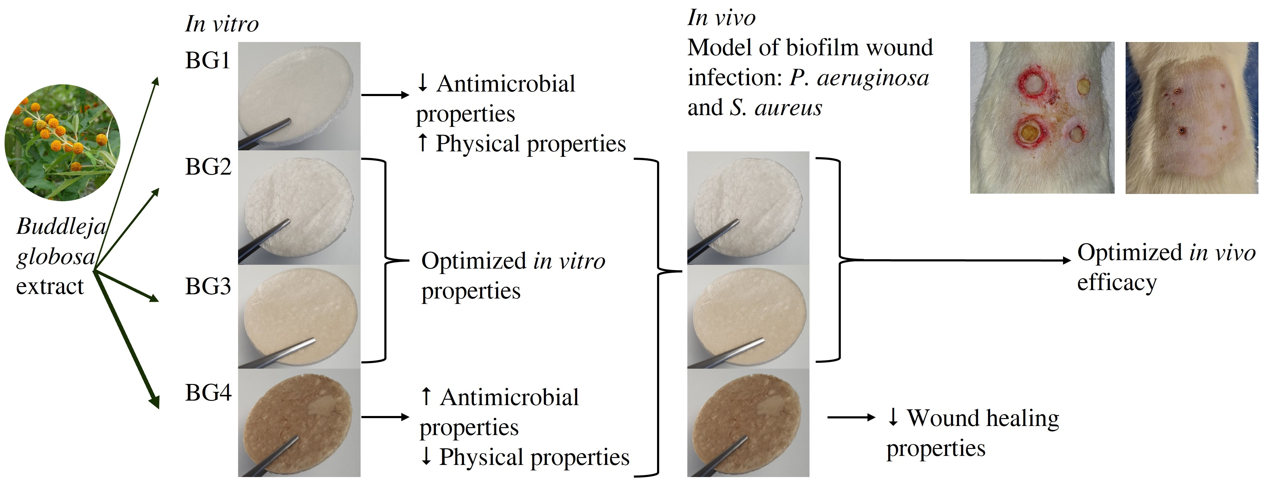
Optimization of Buddleja globosa-Loaded Polymeric Scaffolds for the Treatment of Biofilm-Infected Wounds

 ,
,  , , , and
Abstract
1. Introduction
2. Results
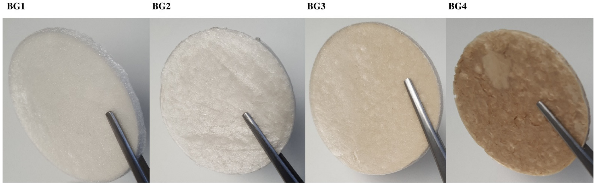
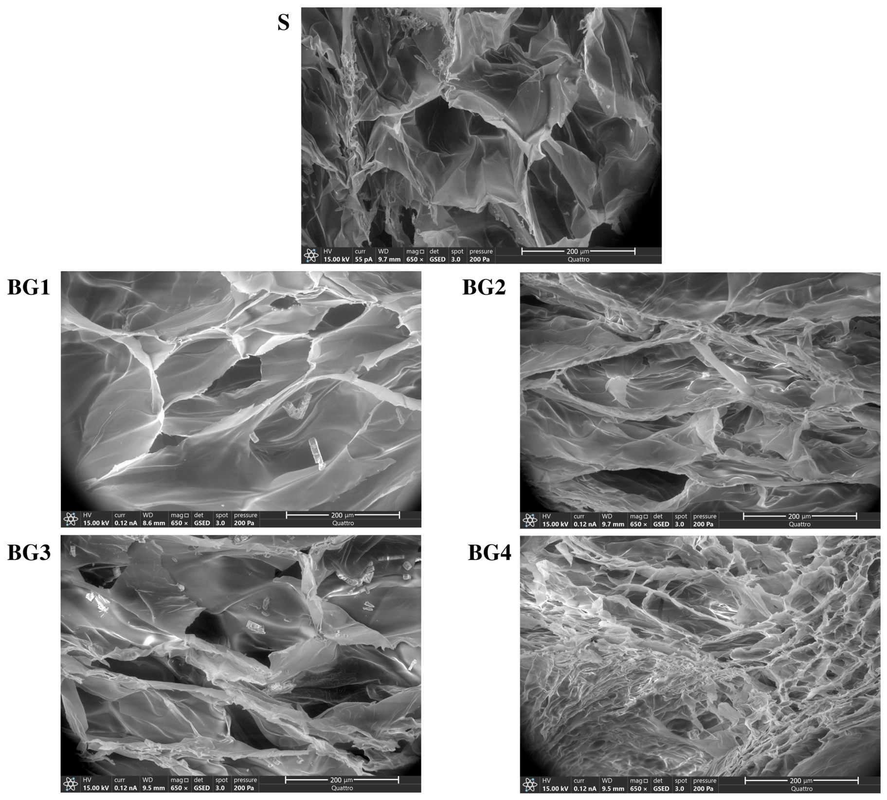
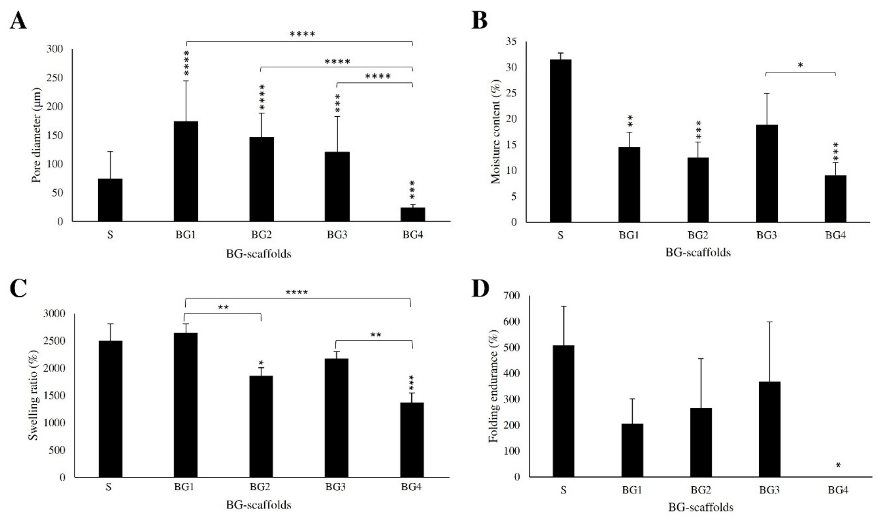
2.1. Physicochemical Characterization of the Scaffolds
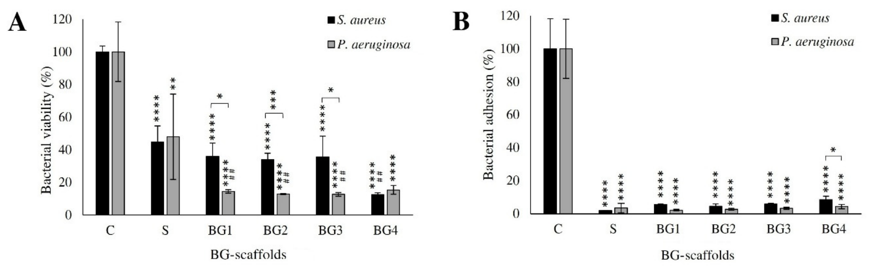
2.2. In Vitro Antimicrobial Properties and Cytocompatibility
2.3. In Vitro Antimicrobial Properties Against Dual-Species Biofilm (DSB)
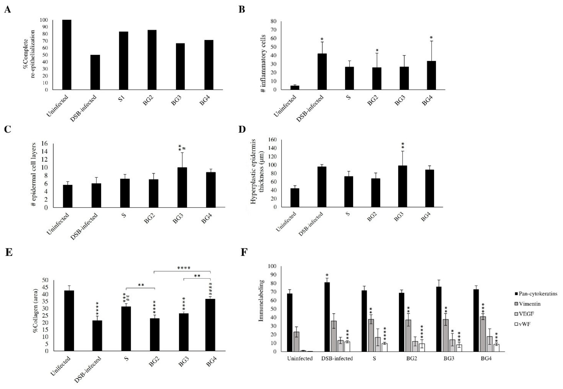
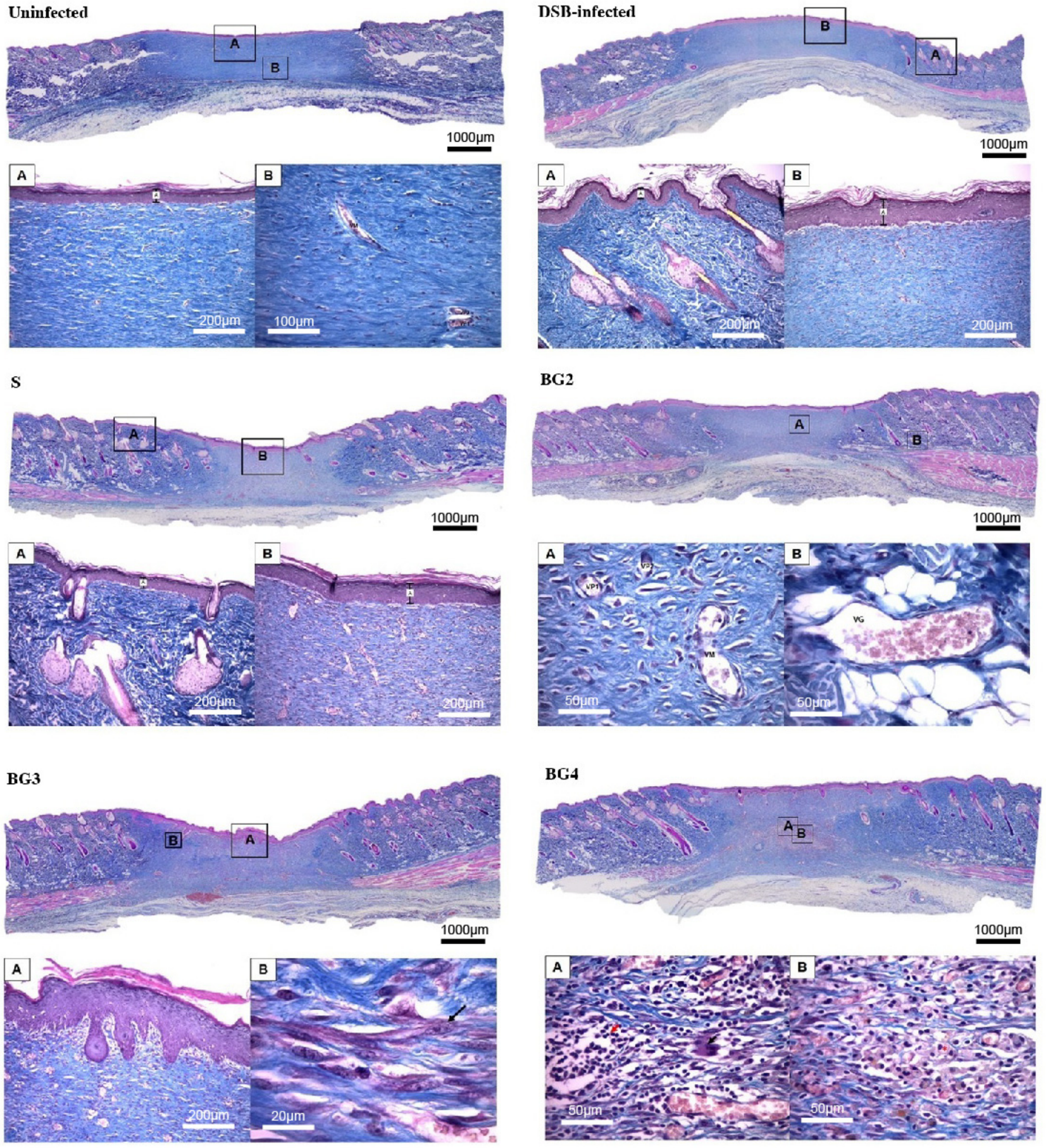
2.4. In Vivo Efficacy of the BG Scaffolds on a Murine Model of Infected Chronic Wound
3. Discussion
3.1. Optimal Performance of BG2 and BG3 Scaffolds
3.2. Impact of Dual-Species Biofilm Infection on Wound Healing
3.3. Influence of Physicochemical Properties and Extract–Polymer Interactions
4. Materials and Methods
4.1. Materials
4.2. Scaffold Development
4.3. Physical Properties of the BG Scaffolds
4.4. Antimicrobial Properties and Cytocompatibility In Vitro
4.5. Scaffold Efficacy in In Vitro Infected Wound Model
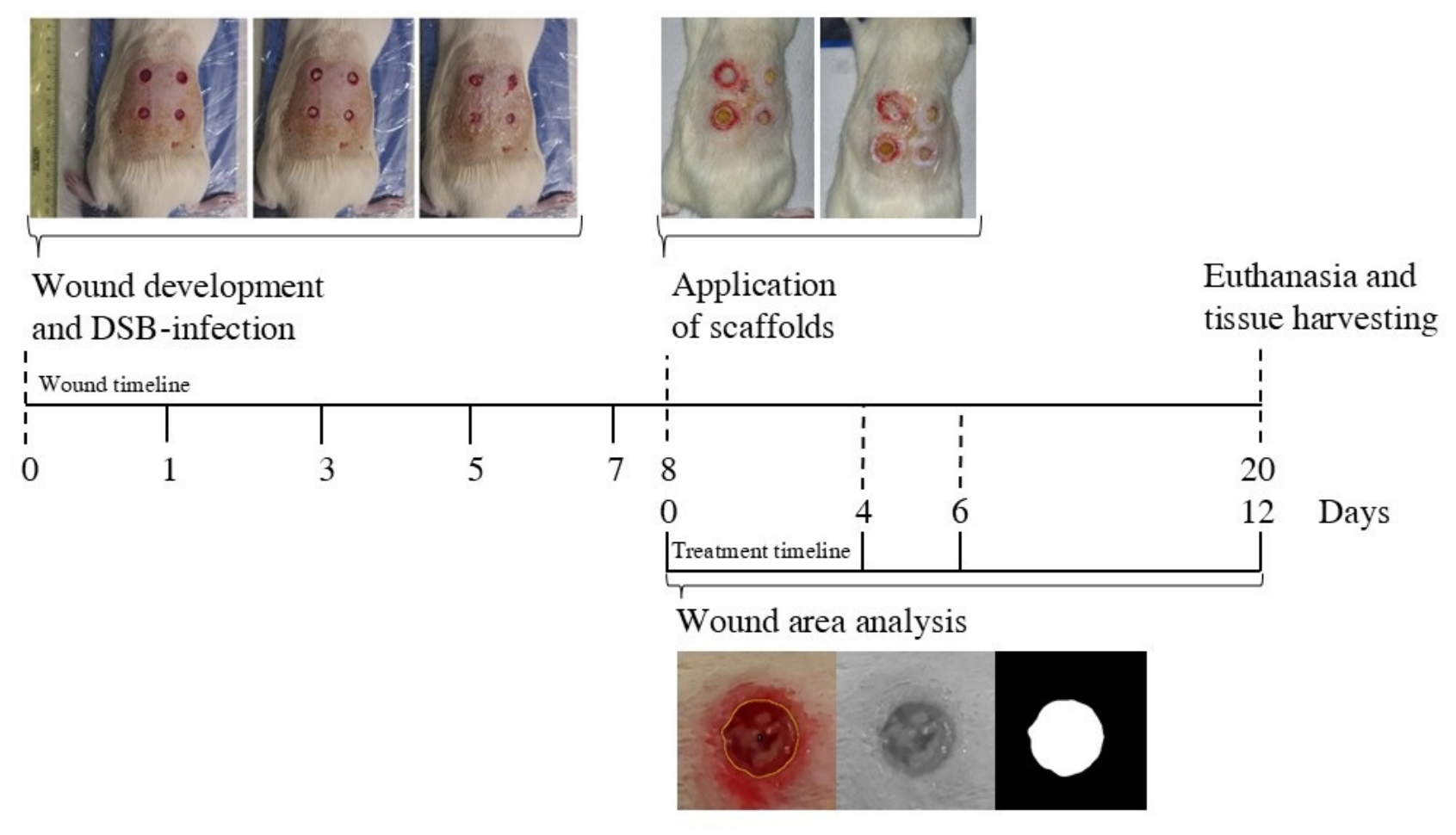
4.6. Scaffold Efficacy in an In Vivo Infected Wound Model
4.7. Wound Closure Analysis
4.8. Microbial Enumeration and Histological Analysis
4.9. Histological Analysis
4.10. Statistical Analysis
5. Conclusions
Supplementary Materials
Author Contributions
Funding
Institutional Review Board Statement
Data Availability Statement
Acknowledgments
Conflicts of Interest
Abbreviations
| BG | Buddleja globosa |
| BG1 to BG4 | Scaffolds containing increasing concentrations of BG extract |
| CFU | Colony-forming units |
| DAB | Diaminobenzidine |
| DMEM | Dulbecco’s Modified Eagle Medium |
| DSB | Dual-species biofilm |
| EDC HCl | N-(3-dimethylaminopropyl)-N′-ethylcarbodiimide hydrochloride |
| FDA | Fluorescein diacetate |
| FESEM | Field-Emission Scanning Electron Microscopy |
| LB | Luria–Bertani |
| MES | 2-(N-morpholino) ethanesulfonic acid |
| NHS | N-hydroxysuccinimide |
| PBS | Phosphate saline buffer |
| S | Reference scaffold |
| TSA | Tryptic soy agar |
| TWS | Trainable Weka Segmentation |
| VEGF | Vascular endothelial growth factor |
| vWF | Von Willebrand Factor |
References
- Falanga, V.; Isseroff, R.R.; Soulika, A.M.; Romanelli, M.; Margolis, D.; Kapp, S.; Granick, M.; Harding, K. Chronic wounds. Nat. Rev. Dis. Prim. 2022, 8, 50. [Google Scholar] [CrossRef] [Scilit] [PubMed]
- James, G.A.; Swogger, E.; Wolcott, R.; deLancey Pulcini, E.; Secor, P.; Sestrich, J.; Costerton, J.W.; Stewart, P.S. Biofilms in chronic wounds. Wound Repair Regen. 2008, 16, 37–44. [Google Scholar] [CrossRef] [Scilit]
- Vyas, K.S.; Wong, L.K. Detection of biofilm in wounds as an early indicator for risk for tissue infection and wound chronicity. Ann. Plast. Surg. 2016, 76, 127–131. [Google Scholar] [CrossRef] [Scilit]
- Malone, M.; Bjarnsholt, T.; McBain, A.J.; James, G.A.; Stoodley, P.; Leaper, D.; Tachi, M.; Schultz, G.; Swanson, T.; Wolcott, R.D. The prevalence of biofilms in chronic wounds: A systematic review and meta-analysis of published data. J. Wound Care 2017, 26, 20–25. [Google Scholar] [CrossRef] [Scilit]
- Zhao, G.; Usui, M.L.; Lippman, S.I.; James, G.A.; Stewart, P.S.; Fleckman, P.; Olerud, J.E. Biofilms and inflammation in chronic wounds. Adv. Wound Care 2013, 2, 389–399. [Google Scholar] [CrossRef] [Scilit]
- Mori, Y.; Nakagami, G.; Kitamura, A.; Minematsu, T.; Kinoshita, M.; Suga, H.; Kurita, M.; Hayashi, C.; Kawasaki, A.; Sanada, H. Effectiveness of Biofilm-based Wound Care System on Wound Healing in Chronic Wounds. Wound Repair Regen. 2019, 27, 540–547. [Google Scholar] [CrossRef] [Scilit]
- Wolcott, R.D.; Rhoads, D.D. A study of biofilm-based wound management in subjects with critical limb ischaemia. J. Wound Care 2008, 17, 145–155. [Google Scholar] [CrossRef] [Scilit] [PubMed]
- Fonder, M.A.; Lazarus, G.S.; Cowan, D.A.; Aronson-Cook, B.; Kohli, A.R.; Mamelak, A.J. Treating the chronic wound: A practical approach to the care of nonhealing wounds and wound care dressings. J. Am. Acad. Dermatol. 2008, 58, 185–206. [Google Scholar] [CrossRef] [Scilit]
- Sen, C.K. Wound healing essentials: Let there be oxygen. Wound Repair Regen. 2009, 17, 1–18. [Google Scholar] [CrossRef] [Scilit]
- Boateng, J.S.; Matthews, K.H.; Stevens, H.N.E.; Eccleston, G.M. Wound healing dressings and drug delivery systems: A review. J. Pharm. Sci. 2008, 97, 2892–2923. [Google Scholar] [CrossRef] [Scilit] [PubMed]
- Qin, J.; Chen, F.; Wu, P.; Sun, G. Recent advances in bioengineered scaffolds for cutaneous wound healing. Front. Bioeng. Biotechnol. 2022, 10, 841583. [Google Scholar] [CrossRef] [Scilit] [PubMed]
- Teepe, R.G.C.; Koebrugge, E.J.; Löwik, C.W.G.M.; Petit, P.L.C.; Bosboom, R.W.; Twiss, I.M.; Boxma, H.; Vermeer, B.J.; Ponec, M. Cytotoxic effects of topical antimicrobial and antiseptic agents on human keratinocytes in vitro. J. Trauma. Acute Care Surg. 1993, 35, 8–19. [Google Scholar] [CrossRef] [Scilit] [PubMed]
- Burd, A.; Kwok, C.H.; Hung, S.C.; Chan, H.S.; Gu, H.; Lam, W.K.; Huang, L. A comparative study of the cytotoxicity of silver-based dressings in monolayer cell, tissue explant, and animal models. Wound Repair Regen. 2007, 15, 94–104. [Google Scholar] [CrossRef] [Scilit] [PubMed]
- Vater, C.; Adamovic, A.; Ruttensteiner, L.; Steiner, K.; Tajpara, P.; Klang, V.; Elbe-Bürger, A.; Wirth, M.; Valenta, C. Cytotoxicity of lecithin-based nanoemulsions on human skin cells and ex vivo skin permeation: Comparison to conventional surfactant types. Int. J. Pharm. 2019, 566, 383–390. [Google Scholar] [CrossRef] [Scilit]
- Calderón-Montaño, J.M.; Jiménez-Alonso, J.J.; Guillén-Mancina, E.; Burgos-Morón, E.; López-Lázaro, M. A 30-s exposure to ethanol 20% is cytotoxic to human keratinocytes: Possible mechanistic link between alcohol-containing mouthwashes and oral cancer. Clin. Oral. Investig. 2018, 22, 2943–2946. [Google Scholar] [CrossRef] [Scilit]
- Houghton, P. Ethnopharmacology of some Buddleja species. J. Ethnopharmacol. 1984, 11, 293–308. [Google Scholar] [CrossRef] [Scilit]
- Suwalsky, M.; Duguet, J.; Speisky, H. An In Vitro Study of the Antioxidant and Antihemolytic Properties of Buddleja globosa (Matico). J. Membr. Biol. 2017, 250, 239–248. [Google Scholar] [CrossRef] [Scilit]
- Backhouse, N.; Rosales, L.; Apablaza, C.; Goïty, L.; Erazo, S.; Negrete, R.; Theodoluz, C.; Rodríguez, J.; Delporte, C. Analgesic, anti-inflammatory and antioxidant properties of Buddleja globosa, Buddlejaceae. J. Ethnopharmacol. 2008, 116, 263–269. [Google Scholar] [CrossRef] [Scilit]
- Mensah, A.Y.; Sampson, J.; Houghton, P.J.; Hylands, P.J.; Westbrook, J.; Dunn, M.; Hughes, M.A.; Cherry, G.W. Effects of Buddleja globosa leaf and its constituents relevant to wound healing. J. Ethnopharmacol. 2001, 77, 219–226. [Google Scholar] [CrossRef] [Scilit]
- Pardo, F.; Perich, F.; Villarroel, L.; Torres, R. Isolation of verbascoside, an antimicrobial constituent of Buddleja globosa leaves. J. Ethnopharmacol. 1993, 39, 221–222. [Google Scholar] [CrossRef] [Scilit]
- Mensah, A.Y.; Houghton, P.J.; Bloomfield, S.; Vlietinck, A.; Vanden Berghe, D. Known and Novel Terpenes from Buddleja globosa Displaying Selective Antifungal Activity Against Dermatophytes. J. Nat. Prod. 2000, 63, 1210–1213. [Google Scholar] [CrossRef] [Scilit]
- Marčetić, M.; Bufan, B.; Drobac, M.; Stanković, J.A.; Ranin, N.A.; Milenković, M.T.; Božić, D.D. Multifaceted Biological Properties of Verbascoside/Acteoside: Antimicrobial, Cytotoxic, Anti-Inflammatory, and Immunomodulatory Effects. Antibiotics 2025, 14, 697. [Google Scholar] [CrossRef] [Scilit]
- Fazli, M.; Bjarnsholt, T.; Kirketerp-Møller, K.; Jørgensen, B.; Andersen, A.S.; Krogfelt, K.A.; Givskov, M.; Tolker-Nielsen, T. Nonrandom distribution of Pseudomonas aeruginosa and Staphylococcus aureus in chronic wounds. J. Clin. Microbiol. 2009, 47, 4084–4089. [Google Scholar] [CrossRef] [Scilit]
- Kirketerp-Møller, K.; Jensen, P.Ø.; Fazli, M.; Madsen, K.G.; Pedersen, J.; Moser, C.; Tolker-Nielsen, T.; Høiby, N.; Givskov, M.; Bjarnsholt, T. Distribution, Organization, and Ecology of Bacteria in Chronic Wounds. J. Clin. Microbiol. 2008, 46, 2717–2722. [Google Scholar] [CrossRef] [Scilit]
- Zhao, G.; Hochwalt, P.C.; Usui, M.L.; Underwood, R.A.; Singh, P.K.; James, G.A.; Stewart, P.S.; Fleckman, P.; Olerud, J.E. Delayed wound healing in diabetic (db/db) mice with Pseudomonas aeruginosa biofilm challenge: A model for the study of chronic wounds. Wound Repair Regen. 2010, 18, 467–477. [Google Scholar] [CrossRef] [Scilit] [PubMed]
- Watters, C.; DeLeon, K.; Trivedi, U.; Griswold, J.A.; Lyte, M.; Hampel, K.J.; Wargo, M.J.; Rumbaugh, K.P. Pseudomonas aeruginosa biofilms perturb wound resolution and antibiotic tolerance in diabetic mice. Med. Microbiol. Immunol. 2013, 202, 131–141. [Google Scholar] [CrossRef] [Scilit]
- Chen, S.; Li, R.; Cheng, C.; Xu, J.; Jin, C.; Gao, F.; Wang, J.; Zhang, J.; Zhang, J.; Wang, H.; et al. Pseudomonas aeruginosa infection alters the macrophage phenotype switching process during wound healing in diabetic mice. Cell Biol. Int. 2018, 42, 877–889. [Google Scholar] [CrossRef] [Scilit] [PubMed]
- Serra, R.; Grande, R.; Butrico, L.; Rossi, A.; Settimio, U.F.; Caroleo, B.; Amato, B.; Gallelli, L.; de Franciscis, S. Chronic wound infections: The role of Pseudomonas aeruginosa and Staphylococcus aureus. Expert. Rev. Anti Infect. Ther. 2015, 13, 605–613. [Google Scholar] [CrossRef] [Scilit] [PubMed]
- DeLeon, S.; Clinton, A.; Fowler, H.; Everett, J.; Horswill, A.R.; Rumbaugh, K.P. Synergistic interactions of Pseudomonas aeruginosa and Staphylococcus aureus in an in vitro wound model. Infect. Immun. 2014, 82, 4718–4728. [Google Scholar] [CrossRef] [Scilit]
- Landa, G.; Clarhaut, J.; Buyck, J.; Mendoza, G.; Arruebo, M.; Tewes, F. Impact of mixed Staphylococcus aureus-Pseudomonas aeruginosa biofilm on susceptibility to antimicrobial treatments in a 3D in vitro model. Sci. Rep. 2024, 14, 27877. [Google Scholar] [CrossRef] [Scilit]
- Ceriani, R.; Cherif-Pino, D.A.; Pérez-Basáez, P.; Escobar, M.; Leyton, P.; Weinstein-Oppenheimer, C.R.; Moraga-Espinoza, D.F.; Bahamondez-Canas, T.F. Tailoring Polymeric Scaffolds with Buddleja globosa Extract for Dual Antimicrobial and Biocompatible Wound Healing Applications. Molecules 2025, 30, 2428. [Google Scholar] [CrossRef] [Scilit]
- Enrione, J.; Díaz-Calderón, P.; Weinstein-Oppenheimer, C.R.; Sánchez, E.; Fuentes, M.A.; Brown, D.I.; Herrera, H.; Acevedo, C.A. Designing a gelatin/chitosan/hyaluronic acid biopolymer using a thermophysical approach for use in tissue engineering. Bioprocess. Biosyst. Eng. 2013, 36, 1947–1956. [Google Scholar] [CrossRef] [Scilit]
- Coelho-Rato, L.S.; Parvanian, S.; Modi, M.K.; Eriksson, J.E. Vimentin at the core of wound healing. Trends Cell Biol. 2024, 34, 239–254. [Google Scholar] [CrossRef] [Scilit]
- Monika, P.; Chandraprabha, M.N.; Murthy, K.N.C.; Rangarajan, A.; Waiker, P.V.; Sathish, M. Human primary chronic wound derived fibroblasts demonstrate differential pattern in expression of fibroblast specific markers, cell cycle arrest and reduced proliferation. Exp. Mol. Pathol. 2022, 127, 104803. [Google Scholar] [CrossRef] [Scilit] [PubMed]
- Letelier, M.E.; Jones, R.; López, C.; Palma, K.; Aracena, P.; Razmilic, I.; Polanco, X.; Vogel, H. Safety profile and wound healing properties of a Standardized Buddleja globosa Hope (Matico) Extract In Sprague-Dawley Rats. Rev. Farmacol. Chile 2012, 5, 13–19. [Google Scholar]
- Hubrecht, R.C.; Carter, E. The 3Rs and Humane Experimental Technique: Implementing Change. Animals 2019, 9, 754. [Google Scholar] [CrossRef] [Scilit] [PubMed]
- Dalton, T.; Dowd, S.E.; Wolcott, R.D.; Sun, Y.; Watters, C.; Griswold, J.A.; Rumbaugh, K.P. An in vivo polymicrobial biofilm wound infection model to study interspecies interactions. PLoS ONE 2011, 6, e27317. [Google Scholar] [CrossRef] [Scilit]
- Trøstrup, H.; Thomsen, K.; Calum, H.; Høiby, N.; Moser, C. Animal models of chronic wound care: The application of biofilms in clinical research. Chronic Wound Care Manag. Res. 2016, 3, 123–132. [Google Scholar] [CrossRef] [Scilit]
- Versey, Z.; Nizer, W.S.d.C.; Russell, E.; Zigic, S.; DeZeeuw, K.G.; Marek, J.E.; Overhage, J.; Cassol, E. Biofilm-Innate Immune Interface: Contribution to Chronic Wound Formation. Front. Immunol. 2021, 12, 648554. [Google Scholar] [CrossRef] [Scilit]
- Thaarup, I.C.; Iversen, A.K.S.; Lichtenberg, M.; Bjarnsholt, T.; Jakobsen, T.H. Biofilm Survival Strategies in Chronic Wounds. Microorganisms 2022, 10, 775. [Google Scholar] [CrossRef] [Scilit]
- Arts, M.J.T.J.; Haenen, G.R.M.M.; Wilms, L.C.; Beetstra, S.A.J.N.; Heijnen, C.G.M.; Voss, H.-P.; Bast, A. Interactions between Flavonoids and Proteins: Effect on the Total Antioxidant Capacity. J. Agric. Food Chem. 2002, 50, 1184–1187. [Google Scholar] [CrossRef] [Scilit]
- Zhang, X.; Do, M.D.; Casey, P.; Sulistio, A.; Qiao, G.G.; Lundin, L.; Lillford, P.; Kosaraju, S. Chemical Cross-Linking Gelatin with Natural Phenolic Compounds as Studied by High-Resolution NMR Spectroscopy. Biomacromolecules 2010, 11, 1125–1132. [Google Scholar] [CrossRef] [Scilit]
- Choi, D.J.; Park, S.J.; Gu, B.K.; Kim, Y.-J.; Chung, S.; Kim, C.-H. Effect of the pore size in a 3D bioprinted gelatin scaffold on fibroblast proliferation. J. Ind. Eng. Chem. 2018, 67, 388–395. [Google Scholar] [CrossRef] [Scilit]
- Araya, N.; Leiva-Soto, M.A.; Bruna, M.V.; Castro-Munoz, A.; Behrend-Keim, B.; Moraga-Espinoza, D.; Bahamondez-Canas, T.F. Formulation of water-soluble Buddleja globosa Hope extracts and characterization of their antimicrobial properties against Pseudomonas aeruginosa. Front. Pharmacol. 2022, 13, 921511. [Google Scholar] [CrossRef] [Scilit]
- Schindelin, J.; Arganda-Carreras, I.; Frise, E.; Kaynig, V.; Longair, M.; Pietzsch, T.; Preibisch, S.; Rueden, C.; Saalfeld, S.; Schmid, B.; et al. Fiji: An open-source platform for biological-image analysis. Nat. Methods 2012, 9, 676–682. [Google Scholar] [CrossRef] [Scilit] [PubMed]
- Peeters, E.; Nelis, H.J.; Coenye, T. Comparison of multiple methods for quantification of microbial biofilms grown in microtiter plates. J. Microbiol. Methods 2008, 72, 157–165. [Google Scholar] [CrossRef] [Scilit] [PubMed]
- Gong, X.; Liang, Z.; Yang, Y.; Liu, H.; Ji, J.; Fan, Y. A resazurin-based, nondestructive assay for monitoring cell proliferation during a scaffold-based 3D culture process. Regen. Biomater. 2020, 7, 271–281. [Google Scholar] [CrossRef] [Scilit] [PubMed]
- Sun, Y.; Dowd, S.E.; Smith, E.; Rhoads, D.D.; Wolcott, R.D. In vitro multispecies Lubbock chronic wound biofilm model. Wound Repair Regen. 2008, 16, 805–813. [Google Scholar] [CrossRef] [Scilit] [PubMed]
- Kucera, J.; Sojka, M.; Pavlik, V.; Szuszkiewicz, K.; Velebny, V.; Klein, P. Multispecies biofilm in an artificial wound bed—A novel model for in vitro assessment of solid antimicrobial dressings. J. Microbiol. Methods 2014, 103, 18–24. [Google Scholar] [CrossRef] [Scilit]
- Naghili, H.; Tajik, H.; Mardani, K.; Razavi Rouhani, S.M.; Ehsani, A.; Zare, P. Validation of drop plate technique for bacterial enumeration by parametric and nonparametric tests. Vet. Res. Forum 2013, 4, 179–183. [Google Scholar]
- Acevedo, C.A.; Brown, D.I.; Young, M.E.; Reyes, J.G. Senescent cultures of human dermal fibroblasts modified phenotype when immobilized in fibrin polymer. J. Biomater. Sci. Polym. Ed. 2009, 20, 1929–1942. [Google Scholar] [CrossRef] [Scilit] [PubMed]
- Sharun, K.; Banu, S.A.; Mamachan, M.; Subash, A.; Mathesh, K.; Kumar, R.; Vinodhkumar, O.R.; Dhama, K.; Abualigah, L.; Pawde, A.M.; et al. Comparative evaluation of masson’s trichrome and picrosirius red staining for digital collagen quantification using ImageJ in rabbit wound healing research. J. Exp. Biol. Agric. Sci. 2023, 11, 822–833. [Google Scholar] [CrossRef] [Scilit]





| Scaffold | Mass (mg) | Density (g/mL) | BG Content Expressed as Verbascoside (mg/g) |
|---|---|---|---|
| S | 241.1 ± 1.6 | 0.017 ± 0.001 | 0 |
| BG1 | 305.2 ± 1.2 | 0.024 ± 0.001 | 0.06 |
| BG2 | 305.3 ± 2.7 | 0.024 ± 0.001 | 0.7 |
| BG3 | 319.0 ± 0.7 | 0.026 ± 0.001 | 6.4 |
| BG4 | 517.3 ± 7.6 | 0.040 ± 0.001 | 50 |
| Scaffold | C (mL) | HA (mL) | G (mL) | BG Content Expressed as Verbascoside (µg/mL) |
|---|---|---|---|---|
| S | 3.8 | 1.9 | 13.3 | 0 |
| BG1 | 5.7 | 0.95 | 13.3 | 1 |
| BG2 | 5.7 | 0.95 | 13.3 | 10 |
| BG3 | 5.7 | 0.95 | 13.3 | 100 |
| BG4 | 5.7 | 0.95 | 13.3 | 1000 |
Disclaimer/Publisher’s Note: The statements, opinions and data contained in all publications are solely those of the individual author(s) and contributor(s) and not of MDPI and/or the editor(s). MDPI and/or the editor(s) disclaim responsibility for any injury to people or property resulting from any ideas, methods, instructions or products referred to in the content. |
© 2026 by the authors. Licensee MDPI, Basel, Switzerland. This article is an open access article distributed under the terms and conditions of the Creative Commons Attribution (CC BY) license.
Share and Cite
Bahamondez-Canas, T.F.; García-Collao, I.; Perez-Basaez, P.; Delgado-Gazzoni, C.V.; Garrido-Vera, H.E.; Ceriani, R.; Weinstein-Oppenheimer, C.R.; Moraga-Espinoza, D.F. Optimization of Buddleja globosa-Loaded Polymeric Scaffolds for the Treatment of Biofilm-Infected Wounds. Int. J. Mol. Sci. 2026, 27, 4240. https://doi.org/10.3390/ijms27104240
Bahamondez-Canas TF, García-Collao I, Perez-Basaez P, Delgado-Gazzoni CV, Garrido-Vera HE, Ceriani R, Weinstein-Oppenheimer CR, Moraga-Espinoza DF. Optimization of Buddleja globosa-Loaded Polymeric Scaffolds for the Treatment of Biofilm-Infected Wounds. International Journal of Molecular Sciences. 2026; 27(10):4240. https://doi.org/10.3390/ijms27104240
Chicago/Turabian StyleBahamondez-Canas, Tania F., Ivan García-Collao, Pamela Perez-Basaez, Carolina V. Delgado-Gazzoni, Henry E. Garrido-Vera, Ricardo Ceriani, Caroline R. Weinstein-Oppenheimer, and Daniel F. Moraga-Espinoza. 2026. "Optimization of Buddleja globosa-Loaded Polymeric Scaffolds for the Treatment of Biofilm-Infected Wounds" International Journal of Molecular Sciences 27, no. 10: 4240. https://doi.org/10.3390/ijms27104240
APA StyleBahamondez-Canas, T. F., García-Collao, I., Perez-Basaez, P., Delgado-Gazzoni, C. V., Garrido-Vera, H. E., Ceriani, R., Weinstein-Oppenheimer, C. R., & Moraga-Espinoza, D. F. (2026). Optimization of Buddleja globosa-Loaded Polymeric Scaffolds for the Treatment of Biofilm-Infected Wounds. International Journal of Molecular Sciences, 27(10), 4240. https://doi.org/10.3390/ijms27104240