1. Introduction
Hair follicles (HFs) are highly specialized, dynamic mini-organs characterized by life-long cyclic self-renewal. Their distinct microanatomical niches harbor diverse stem cell populations that drive transitions through phases of growth (anagen), regression (catagen), and rest (telogen) [
1]. During the anagen phase, bulge stem cells migrate to the follicular bulb [
1,
2,
3], differentiating into outer root sheath (ORS) and inner root sheath (IRS) cells [
4,
5].
Following matrix cell exhaustion, the lower cycling region undergoes apoptosis, eventually leading to a quiescent telogen state before a new cycle initiates [
1,
3]. This intrinsic regenerative capacity positions the HF as an accessible and powerful model for clinical tissue repair [
6,
7,
8,
9].
The regenerative potential of the HF relies on a complex interplay between somatic cells and the multiple stem cell compartments. Keratinocytes populating the ORS maintain a progenitor-like state and actively participate in epithelial repair [
10]. These cells also demonstrate remarkably high reprogramming efficiencies for generating induced pluripotent stem cells (iPSCs) due to their endogenous expression of epithelial transcription factors [
11,
12]. Concurrently, the dermal papilla houses mesenchymal stem cell (MSC)-like populations exhibiting classical multilineage plasticity and potent immunomodulatory effects [
13,
14]. The follicular bulge also acts as a niche for neural crest-derived stem cells (NCSCs) capable of broad ectodermal and mesodermal differentiation [
15], alongside pluripotent-like (HAP) cells isolated from the upper ORS [
7,
16].
The therapeutic utility of HF-derived cells extends beyond direct tissue integration, relying heavily on paracrine mechanisms mediated by the cellular secretome [
17,
18]. This localized mixture of growth factors, cytokines, and extracellular vesicles is naturally adapted to the cutaneous environment [
19]. Preclinical evidence indicates that secretomes from HF compartments, particularly their exosomal fractions, can stimulate angiogenesis, promote re-epithelialization, and enhance wound closure in dermal injury models [
14,
20]. Translating these cell-free systems into clinical applications offers a standardized approach that mitigates the safety risks associated with whole-cell therapies [
21,
22].
Despite the robust regenerative profile of HF derivatives, both intrinsic and extrinsic aging heavily compromise therapeutic efficacy. Extended in vitro expansion induces replicative senescence, triggering DNA damage and secretory skew that significantly diminish paracrine function and tissue repair capacity [
23,
24]. While some primary tissue secretomes exhibit resilience to chronological age [
25], cumulative oxidative stress and cellular aging inevitably degrade the differentiation potential of adult stem cells [
26,
27]. Consequently, securing unpassaged, youthful cells through early-stage cryopreservation is critical to circumvent replicative decay and maximize the biological fidelity of autologous cellular therapies [
28].
In this study, we investigate the regenerative capabilities of human hair follicles, focusing on both cellular and cell-free constituents. We present original experimental data detailing the isolation, expansion, and directed multilineage differentiation of HF-derived keratinocytes and MSCs, alongside their efficient reprogramming into iPSCs. We also characterize the complex protein and vesicular profile of the autologous hair follicle secretome (aHFS) and assess its functional capacity to accelerate wound healing in vitro. By integrating these findings, we aim to validate the hair follicle as an optimal, accessible cell source for personalized regenerative medicine and emphasize the utility of follicular biobanking.
3. Discussion
The present study underscores the multidimensional regenerative capacity of the human hair follicle, which acts as a highly accessible reservoir for both somatic and mesenchymal stem cells. A persistent challenge in the clinical translation of autologous cell therapies remains the reliance on invasive tissue harvesting, which is frequently coupled with harsh enzymatic dissociation protocols [
29]. These conventional methods can induce cellular stress and alter baseline cellular phenotypes [
23]. In contrast, the present study establishes a streamlined, enzyme-free explant methodology utilizing non-invasively plucked hair follicles. We demonstrate that this approach successfully isolates and expands both HF-derived keratinocytes and MSCs while preserving their native progenitor characteristics, thus mitigating culture-induced artifacts.
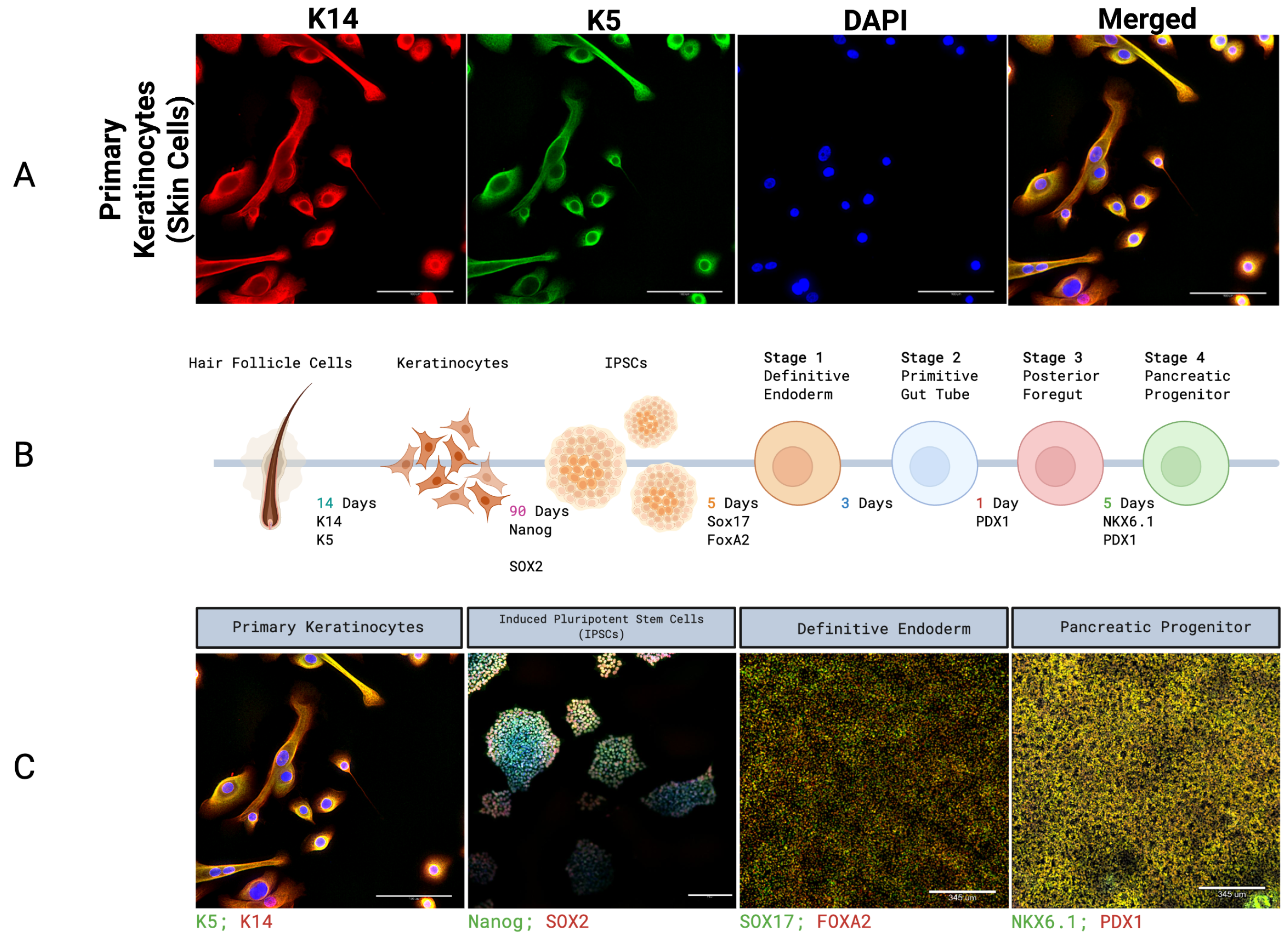
Importantly, our explant methodology yielded a robust population of outer root sheath keratinocytes, which serve as a highly efficient source for cellular reprogramming. As established in the literature, keratinocytes exhibit superior reprogramming efficiency compared to dermal fibroblasts, a distinct advantage driven by their endogenous expression of key epithelial transcription factors [
29,
30]. Leveraging this baseline advantage, we successfully reprogrammed these HF-derived keratinocytes into iPSCs using an integration-free episomal approach. We subsequently demonstrated their capacity to differentiate across germ layers into definitive endoderm and pancreatic progenitor lineages (PDX1+/NKX6.1+). While the comprehensive functional characterization of these keratinocyte-derived pancreatic progenitors has been extensively detailed in our previous work [
12], their generation here confirms that non-invasively plucked hair follicles reliably provide high-quality keratinocytes for potential use in regenerative therapies.
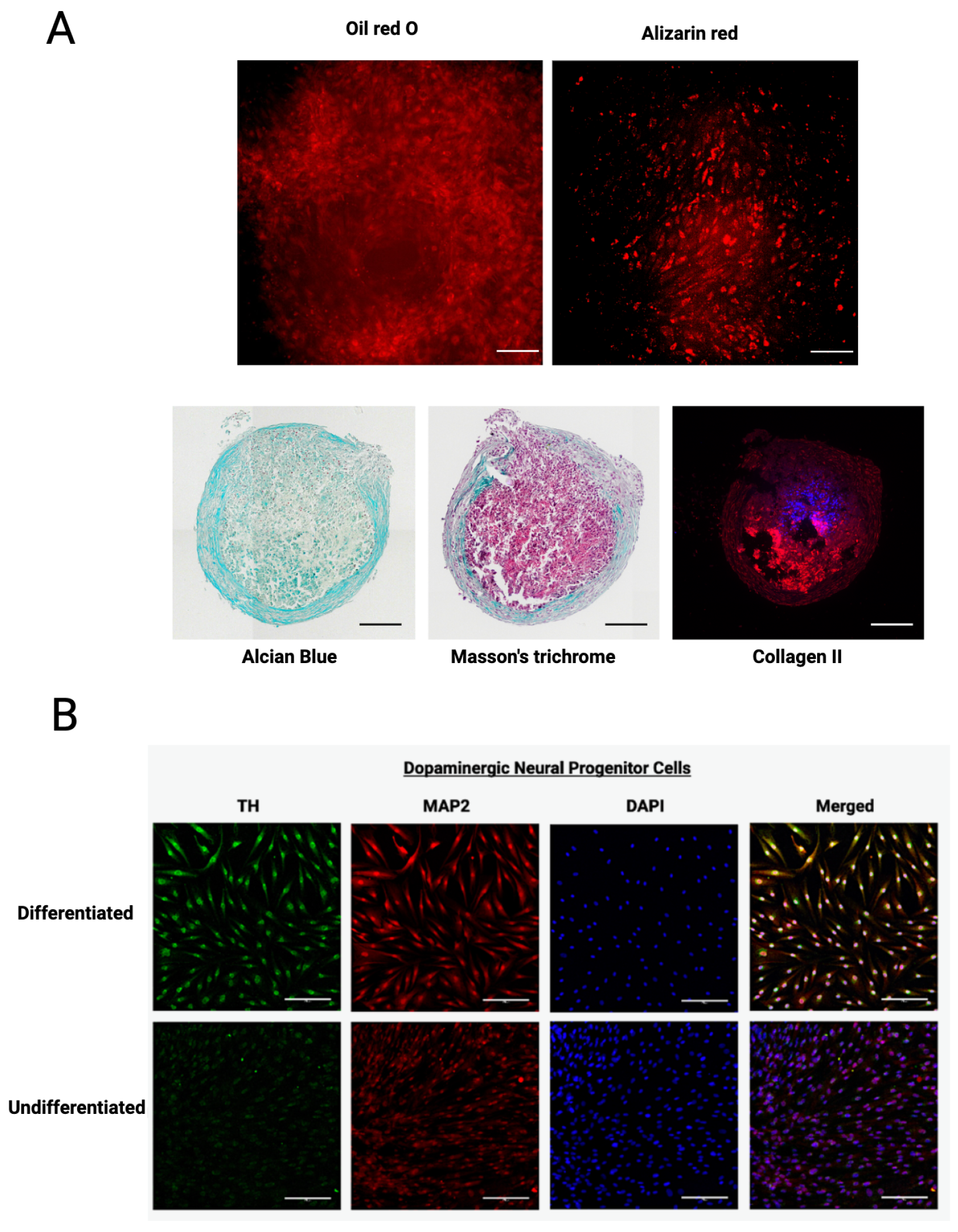
In parallel, the mesenchymal stem cells (HF-MSCs) isolated via our non-invasive explant method exhibited profound developmental versatility. Beyond classical mesodermal differentiation, HF-MSCs demonstrated the capacity for neurogenic-like induction, adopting dopaminergic phenotypes (TH+/MAP2+). This intrinsic multilineage capacity, achieved without genetic manipulation, underscores the plucked follicle as a highly competitive alternative to traditional sources of bone marrow or stem cells derived from adipose tissue. Furthermore, recent advances in expanding HF-MSCs within fully defined serum-free media enhance their clinical translatability for tissue engineering and immune modulation [
31].
Beyond direct cellular integration, the therapeutic efficacy of HF-derived cells is highly mediated by their potent paracrine activity [
17,
18]. Our characterization of the autologous hair follicle secretome (aHFS) revealed a robust concentration of regenerative cytokines, growth factors, and extracellular vesicles. A major innovation of this work lies in the scale and rigor of our cell-free analysis. While previous reports have described the regenerative properties of HF-derived conditioned media [
32] and its associated extracellular vesicles [
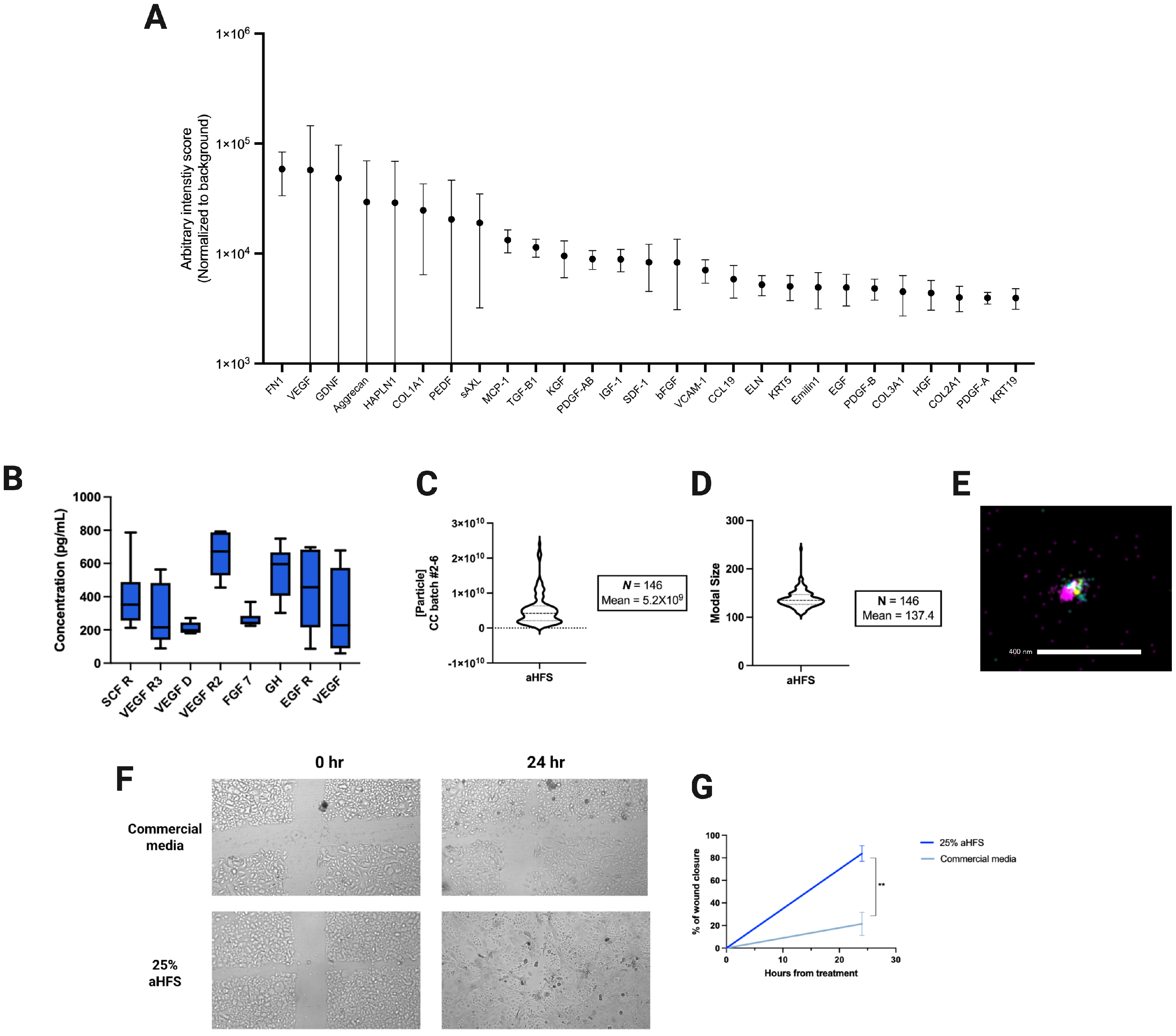
33], this study provides a uniquely comprehensive functional and vesicular characterization of the autologous hair follicle secretome (aHFS). Importantly, the secretome was generated within a chemically defined, serum-free environment. This methodological rigor ensures that the potent wound-closure capabilities and specific cytokine enrichments (e.g., VEGF-A, FGF-7) observed in our assays are driven exclusively by autologous paracrine factors and extracellular vesicles, free from the confounding influence of exogenous animal proteins. The pronounced wound-closure capabilities demonstrated in our in vitro keratinocyte scratch assays firmly confirm this regenerative capacity.
Cellular aging remains a fundamental barrier to scalable cell-based therapies. Extended in vitro expansion and chronological aging drive variability in gene expression, upregulate senescence markers, and ultimately diminish differentiation potential [
23]. Although iPSC reprogramming can partially reset age-associated molecular phenotypes, it remains insufficient to completely erase accumulated DNA damage or epigenetic scarring [
27,
34]. Consequently, the functional utility of autologous cellular therapies is most robust when sourced from a biologically young tissue resource.
To address this biological limitation, the cryopreservation of intact, plucked hair follicles offers a highly practical strategy for personalized regenerative medicine. By circumventing the need for invasive biopsies, early-stage biobanking of these follicles arrests cellular aging at the point of collection, preserving native telomere length and mitochondrial integrity [
28]. This approach secures a single, non-invasively acquired tissue source that can simultaneously yield scalable populations for patient-specific therapies, offering an accessible, multi-modal framework for the future of autologous interventions.
Despite the robust regenerative potential demonstrated, this study has two primary limitations. First, while our profiling identified key regenerative cytokines and confirmed the presence of exosomes, the autologous secretome is an inherently complex conditioned medium. This biochemical complexity, combined with the natural biological variability observed across human donors, makes it challenging to pinpoint the exact contributions of specific soluble versus vesicular fractions. Second, although our initial protein analysis provides valuable foundational data, the cohort size for this specific profiling was limited. More detailed demographic stratification in larger studies is required to comprehensively understand how variables such as donor sex, genetics, and underlying health conditions might influence individual secretome profiles.
4. Materials and Methods
4.1. Hair Follicle Collection
Hair follicles were collected from the occipital region of the scalp of 28 participants. Participants consented to the collection and subsequent use of their follicles. The follicles were collected using a plucking motion and transported in Acorn’s Cell Transportation MediumTM (Gibco, Emeryville, CA, USA) for further analysis.
4.2. Primary Cell Culture Using the Explant Culture Method
The samples were transferred into 35 mm sterile Petri dishes (Thermo Fisher Scientific, Waltham, MA, USA; Cat. #153066) containing CTS-DPBS without Mg2+ and Ca2+ (Gibco, Grand Island, NY, USA; Cat. #A1285601) and then washed for 3 min in the solution. The dishes were transferred into the ISO-7 cleanroom for further processing. For each participant, 10 follicles were trimmed and placed in the middle of a 48-well tissue culture plate (Nunc, Roskilde, Denmark; Cat. #150687) coated with Matrigel (Corning, New York, NY, USA; Cat. #354230). Only follicles containing an intact ORS were selected. To mitigate against the follicles drying, a drop of KSR medium—comprising 20% Knockout Serum Replacement (Gibco, Grand Island, NY, USA; Cat. #10828028) in DMEM/F12 (Gibco; Cat. #11320033), supplemented with 1× MEM Non-Essential Amino Acids (Gibco; Cat. #11140050), 0.1 mM -Mercaptoethanol (Gibco; Cat. #21985023), and 1× Antibiotic-Antimycotic (Gibco; Cat. #15240062)—was added to each well prior to plating. Once all the follicles were plated, the media were removed, and a fresh drop of media (50 L) containing 10 ng/L bFGF (Thermo Fisher Scientific, Waltham, MA, USA; Cat. #PHG0024) was added on top of each follicle. The plates were placed in a 37 °C incubator at 5% CO2 overnight. The next morning, each well was flooded with 150 L of media. A complete media change was conducted every other day.
4.3. Keratinocyte Isolation and Expansion
At 75% confluency, cells were enzymatically dissociated using 200 L of TrypLE Express (Gibco, Grand Island, NY, USA; Cat. #12604013) per well. The suspension of cells was pooled for each participant and centrifuged at 300× g for 5 min. The resulting supernatant was discarded, and the pellets were resuspended in 1 mL of DermaCult Keratinocyte Expansion Medium (STEMCELL Technologies, Vancouver, BC, Canada; Cat. #05420) supplemented with Hydrocortisone (STEMCELL Technologies; Cat. #07925). The solution was added to a 12-well tissue culture plate (Greiner Bio-One, Frickenhausen, Germany; Cat. #665180) coated with Rat Tail Collagen Type I (Gibco, Grand Island, NY, USA; Cat. #A1048301). The cells were incubated in a 37 °C incubator at 5% CO2, and the media were changed every other day. The cells were expanded until 80% confluency and then passaged onto 6-well tissue culture plates (Greiner Bio-One; Cat. #657160) at a density of /cm2. At each passage, cells were counted using a hemocytometer, and keratinocyte expansion rates were extrapolated. On passage 3, the cells were taken for reprogramming.
4.4. iPSC Reprogramming and Directed Differentiation
Reprogramming of HF-derived keratinocytes into induced pluripotent stem cells (iPSCs) was performed using an integration-free episomal approach, as previously described. The generated iPSCs were maintained and expanded in Essential 8 medium (Thermo Fisher Scientific, Waltham, MA, USA; Cat. #A2656101). Successful reprogramming was confirmed prior to differentiation via the expression of core pluripotency markers Nanog and SOX2. To initiate directed differentiation, iPSCs were dissociated and seeded onto 24-well plates coated with Geltrex (Thermo Fisher Scientific, Waltham, MA, USA; Cat. #A1413301) at lineage-appropriate densities. Stage-specific differentiation was driven using the STEMdiff Trilineage Differentiation Kit (STEMCELL Technologies, Vancouver, BC, Canada; Cat. #05230) according to the manufacturer’s guidelines, successfully inducing the sequential expression of definitive endoderm markers (SOX17, FOXA2) and subsequent pancreatic progenitor markers (NKX6.1, PDX1).
4.5. MSC Isolation and Characterization
Mesenchymal stem cells (MSCs) were isolated by plating plucked hair follicles directly onto culture plates. Prior to plating, the anagen phase of the follicles was confirmed visually using a Leica DMi1 inverted phase-contrast microscope (Leica Microsystems, Wetzlar, Germany). Adherent, spindle-shaped cells were allowed to migrate from the dermal papilla region. These cells were subsequently harvested and expanded in DMEM/F12 basal medium (Thermo Fisher Scientific, Waltham, MA, USA; Cat. #11320033) supplemented with 10% Fetal Bovine Serum (FBS; Thermo Fisher Scientific; Cat. #A5256801) under standard incubation conditions (37 °C, 5% CO2). Cells were utilized between passages 2 and 5.
4.6. Flow Cytometry
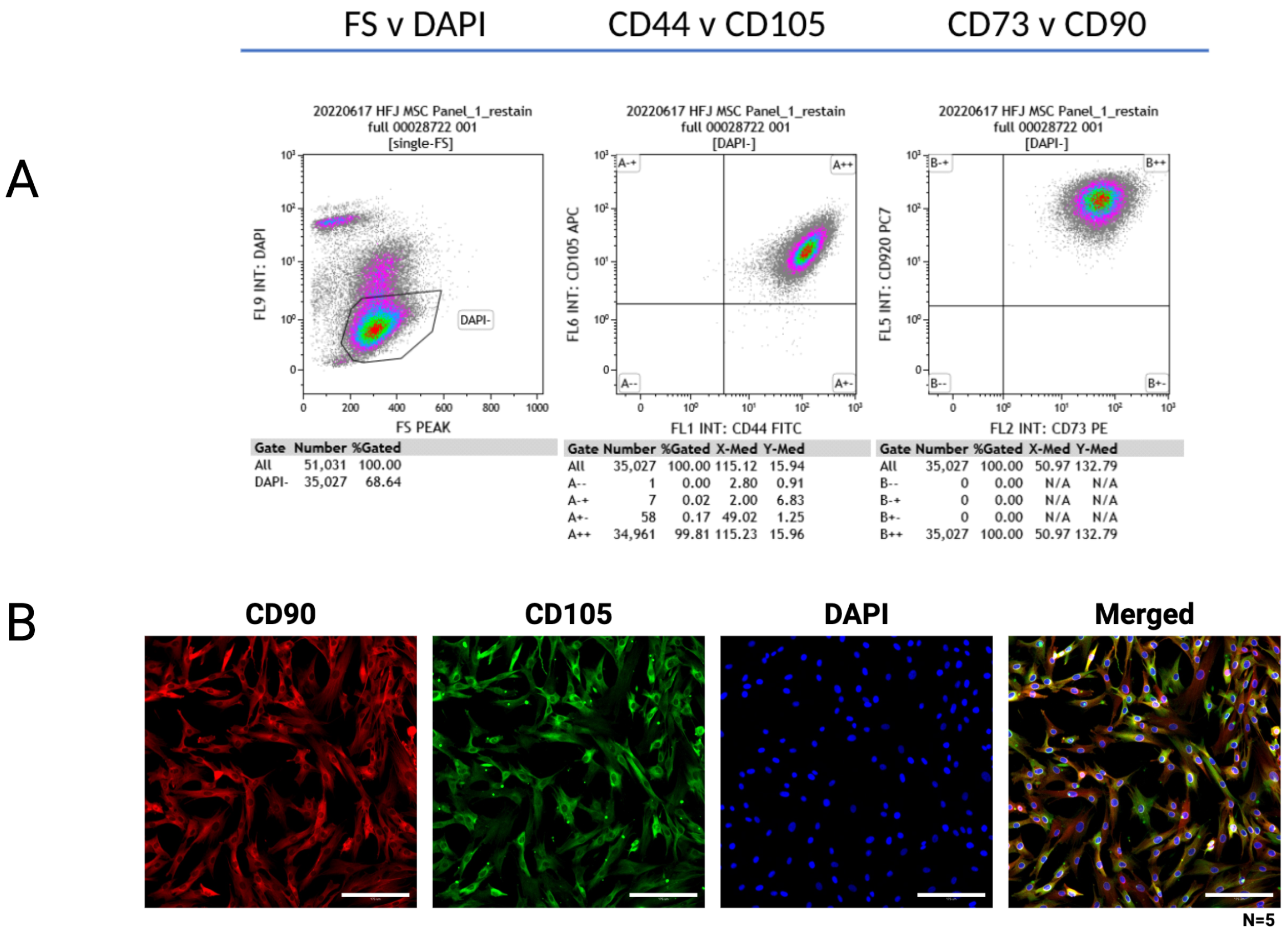
HF-MSCs were harvested using TrypLE Express (Gibco, Grand Island, NY, USA; Cat. #12604013) and centrifuged (300× g, 5 min). Pellets were resuspended in 2% FBS/PBS and blocked with Human TruStain FcX (BioLegend, San Diego, CA, USA; Cat. #422301) for 10 min at room temperature. Cells were incubated with fluorophore-conjugated antibodies against CD44 (Invitrogen, Thermo Fisher Scientific, Waltham, MA, USA; Cat. #11-0441-82), CD73 (BioLegend, San Diego, CA, USA; Cat. #344004), CD90 (Invitrogen; Cat. #25-0909-41), and CD105 (BioLegend; Cat. #323208) for 20 min on ice in the dark. After washing twice, cells were stained with DAPI (Thermo Fisher Scientific, Waltham, MA, USA; Cat. #D1306) for 5 min. Data were acquired on a BD FACSCelesta flow cytometer (BD Biosciences, San Jose, CA, USA) and analyzed using FlowJo software (v10, BD Biosciences).
4.7. Multilineage Differentiation
To evaluate multilineage mesodermal plasticity, HF-MSCs underwent lineage-specific induction. Cells were cultured in respective adipogenic (MesenCultTM Adipogenic Differentiation Kit, STEMCELL Technologies, Vancouver, BC, Canada; Cat. #05412), osteogenic (MesenCultTM Osteogenic Differentiation Kit, STEMCELL Technologies; Cat. #05465), and chondrogenic (MesenCultTM-ACF Chondrogenic Differentiation Kit, STEMCELL Technologies; Cat. #05455) induction media. Differentiation was confirmed using Oil Red O (Thermo Fisher Scientific, Waltham, MA, USA; Cat. #189400250), Alizarin Red S (Thermo Fisher Scientific; Cat. #AA42746AE), and Alcian Blue staining (performed at The Centre for Phenogenomics Pathology Core, Hospital for Sick Children, Toronto, ON, Canada). To achieve the high contrast required for detecting faint micro-lipid droplets and early-stage mineral deposits, the intrinsic fluorescent properties of Oil Red O and Alizarin Red S were utilized for imaging. Chondrogenesis was further confirmed using Masson’s Trichrome and Collagen II staining (The Centre for Phenogenomics, Toronto, ON, Canada).
Neurogenic-like differentiation was induced under specific conditions as previously described [
35]. Briefly, HF-MSCs (passages 3–5) at 70–80% confluency were dissociated using TrypLE Express (Gibco, Grand Island, NY, USA; Cat. #12604013) and seeded into 12-well plates in standard 10% FBS media in DMEM/F12 (Gibco; Cat. #11320033). Following overnight adhesion at 37 °C, the growth medium was replaced with a neural induction medium consisting of Neurobasal Medium (Gibco; Cat. #21103049) supplemented with B-27 (0.25×, Thermo Fisher Scientific, Waltham, MA, USA; Cat. #17504044), 250 ng/mL SHH (Thermo Fisher Scientific; Cat. #PHC7014), 100 ng/mL FGF8 (Thermo Fisher Scientific; Cat. #PHG0271), and 50 ng/mL bFGF (Thermo Fisher Scientific; Cat. #13256029). The cells were incubated at 37 °C without media changes for 9 days. On day 9, the culture was supplemented with 50 ng/mL BDNF (Thermo Fisher Scientific; Cat. #RP-8642) and incubated for an additional 3 days (12 days total). To characterize the neurogenic potential, cells were fixed in 4% PFA and stained with primary antibodies against Tyrosine Hydroxylase (TH; Abcam, Cambridge, UK; Cat. #ab112) and MAP2 (Abcam; Cat. #ab32454) prior to visualization.
4.8. Immunofluorescence
For immunofluorescence analysis, cells were seeded into 12-well cell culture plates (Nunc, Roskilde, Denmark; Cat. #150628) and fixed in 3.7% paraformaldehyde (Thermo Fisher Scientific, Waltham, MA, USA; Cat. #A11313.22) for 20 min at room temperature. Following three washes with PBS, cells were permeabilized using 0.1% Triton X-100 (Thermo Fisher Scientific; Cat. #A16046.AE) for 10 min and subsequently blocked with 1% BSA (Thermo Fisher Scientific; Cat. #37525) for 1 h at room temperature. Cells were then incubated overnight at 4 °C with primary antibodies targeting specific lineage markers: K5 (Invitrogen, Thermo Fisher Scientific, Waltham, MA, USA; Cat. #PA5-32465) and K14 (Invitrogen; Cat. #MA5-11599) for keratinocyte maintenance; CD90 (Invitrogen; Cat. #MA1-35307) and CD105 (Invitrogen; Cat. #MA5-11854) for MSCs; Nanog (Invitrogen; Cat. #PA1-097), SOX2 (Invitrogen; Cat. #MA1-014), SOX17 (Invitrogen; Cat. #703465), FOXA2 (Invitrogen; Cat. #MA5-31362), NKX6.1 (Invitrogen; Cat. #PA5-106121), and PDX1 (Invitrogen; Cat. #PA5-85093) for differentiation; and TH (Invitrogen; Cat. #OPA1-04050) and MAP2 (Invitrogen; Cat. #MA5-12826). After 24 h, cells were incubated with species-specific secondary antibodies for 1 h. Nuclei were counterstained with DAPI (Thermo Fisher Scientific; Cat. #D1306) for 5 min. Plates were imaged using the IXM Confocal System (Molecular Devices, San Jose, CA, USA).
4.9. Secretome Collection and Protein Arrays
Ten anagen hair follicles (confirmed visually as described in
Section 4.5) for each participant were trimmed and plated in duplicate in 48-well plates. Cells were cultured in a serum-free, xeno-free, chemically defined medium (DMEM/F12 supplemented with a proprietary blend of factors; Acorn Biolabs, Toronto, ON, Canada). After 48 h of cell culture, the secretome was collected. Throughout the culture period, cells were evaluated daily via phase-contrast microscopy to confirm monolayer integrity and the absence of cellular detachment. The conditioned medium was centrifuged at 300×
g for 5 min to remove cellular debris, filtered using 0.2
m filters (Amicon, Merck Millipore, Burlington, MA, USA; Cat. #UFC500324), and then lyophilized. A semi-quantitative Quantibody
® glass slide-based cytokine array (RayBiotech, Peachtree Corners, GA, USA; Cat. #QAM-CAA-4000) was utilized to profile growth factors and cytokines. Signal intensities were normalized to background media. Specific analyte concentrations (VEGF-A, GH, FGF-7) were further quantified via ELISA assays (RayBiotech; Cat. #ELH-VEGF-1).
4.10. EV Characterization
The vesicular fraction was obtained from the aHFS using the Total Exosome Isolation Kit (from cell culture media) (Invitrogen, Thermo Fisher Scientific, Waltham, MA, USA; Cat. #44783598). The vesicular content was characterized using Nanoparticle Tracking Analysis (NTA) on a NanoSight NS300 instrument (Malvern Panalytical, Malvern, UK) to determine modal particle size and concentration (). Super-resolution microscopy was performed using the ONI platform (ONI, Oxford, UK). Lyophilized secretome preparations were triple-labeled for the canonical exosome tetraspanins using antibodies against CD81 (Invitrogen; Cat. #MA5-13548), CD63 (Invitrogen; Cat. #MA5-14717), and CD9 (Invitrogen; Cat. #MA5-14712).
4.11. Scratch Assay
An in vitro scratch wound assay was performed using primary human keratinocytes to evaluate directed cellular migration. Cells were seeded in 12-well plates (Greiner Bio-One, Frickenhausen, Germany; Cat. #665180) and grown to 90% confluence in KGM (Keratinocyte Growth Medium) (Lonza, Basel, Switzerland; Cat. #CC-3111) to form a uniform monolayer. A standardized linear scratch was created in the center of each well using a sterile 200 L pipette tip held at a constant angle and pressure to ensure a uniform wound width. To guarantee that the exact same field of view was analyzed over time, reference lines were drawn on the underside of the plate, and images were captured at the intersection of the scratch and the reference marker. Following scratching, cells were gently washed with PBS to remove detached debris and treated with 25% aHFS diluted in basal medium. KGM-Gold CaFree BulletKit (Lonza, Basel, Switzerland; Cat. #00195130) served as the standard control, ensuring that baseline measurements were not confounded by undefined serum proteins. The wound closure was monitored, and phase-contrast images were captured at 0 and 24 h using a Leica DMi1 microscope (Leica Microsystems, Wetzlar, Germany) to quantify percentage area closure.
4.12. Statistical Analysis
Quantitative data are presented as the mean ± standard error of the mean (SEM) from at least three independent experiments (). Statistical significance between two groups was determined using an unpaired Student’s t-test. For comparisons involving more than two groups, a one-way analysis of variance (ANOVA) followed by Tukey’s post hoc test for multiple comparisons was performed. All statistical analyses were conducted using GraphPad Prism software (v8.0; GraphPad Software, San Diego, CA, USA). A p-value was considered statistically significant (* , ** ).





 ,
,