
Updated Meta-Analysis on Vitamin Supplementation for Chronic Pruritus: Expanding Evidence Beyond Vitamin D


Abstract

1. Introduction
2. Results
2.1. Study Search and Characteristics of Included Patients
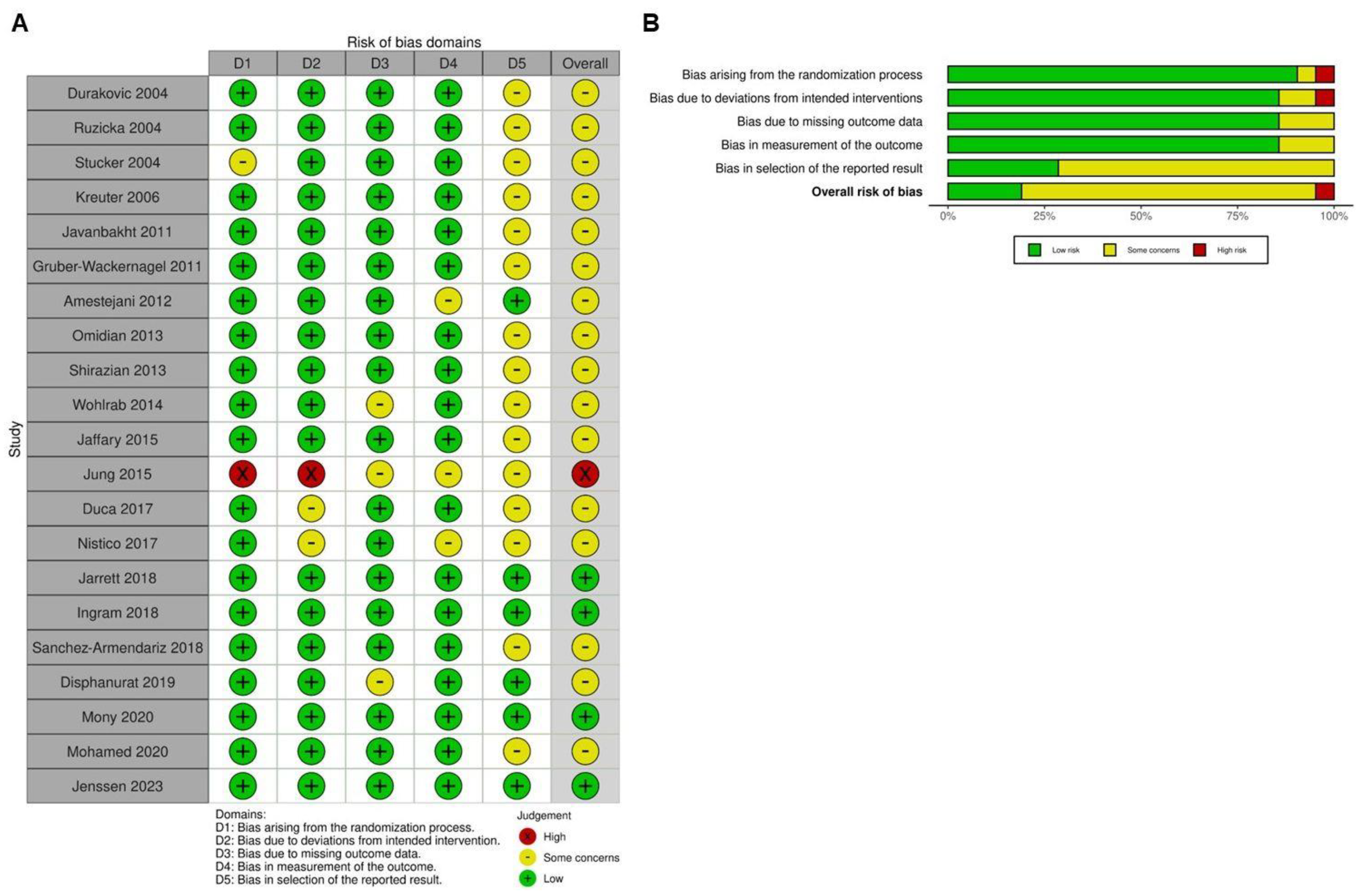
2.2. Quality Assessment
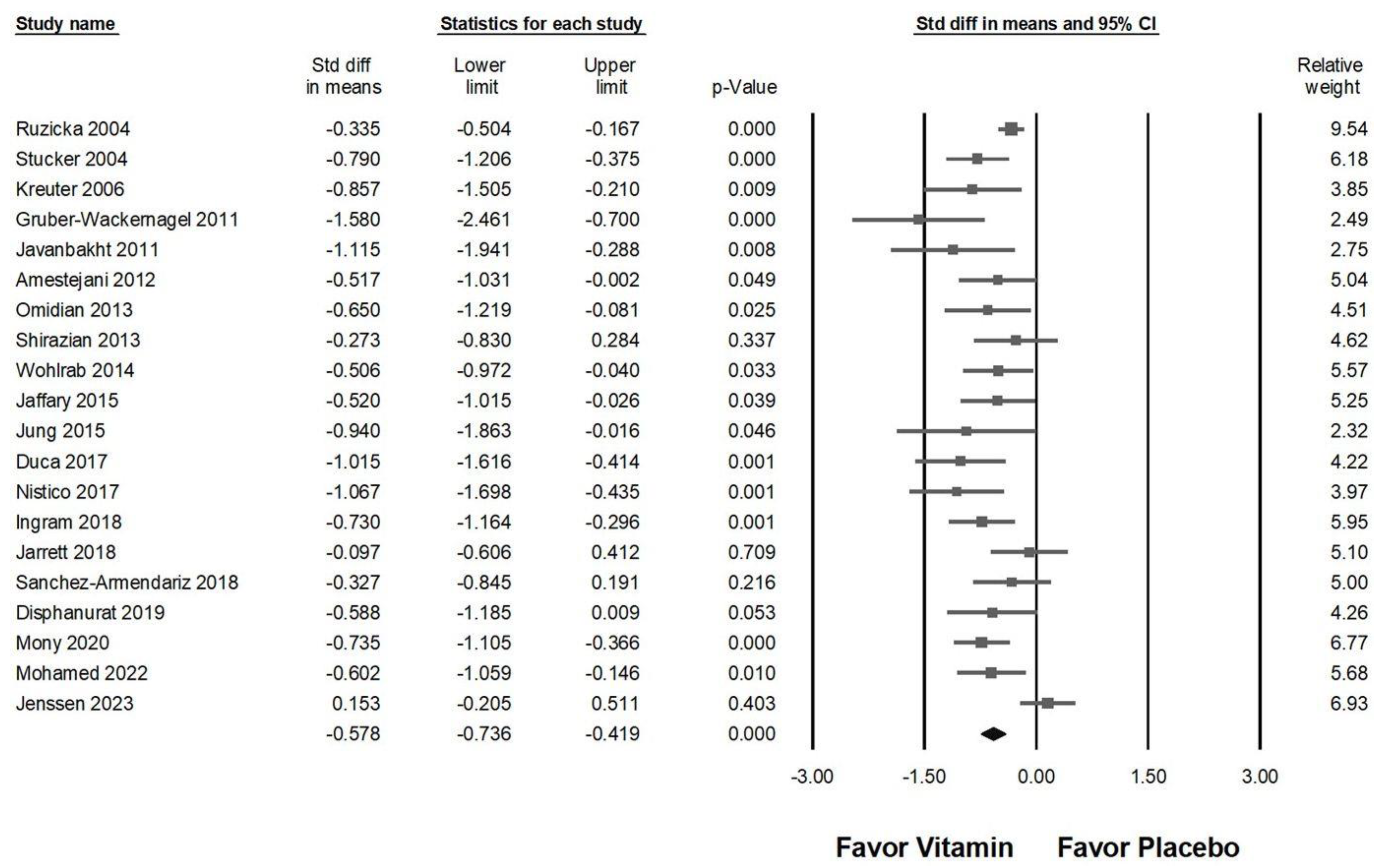
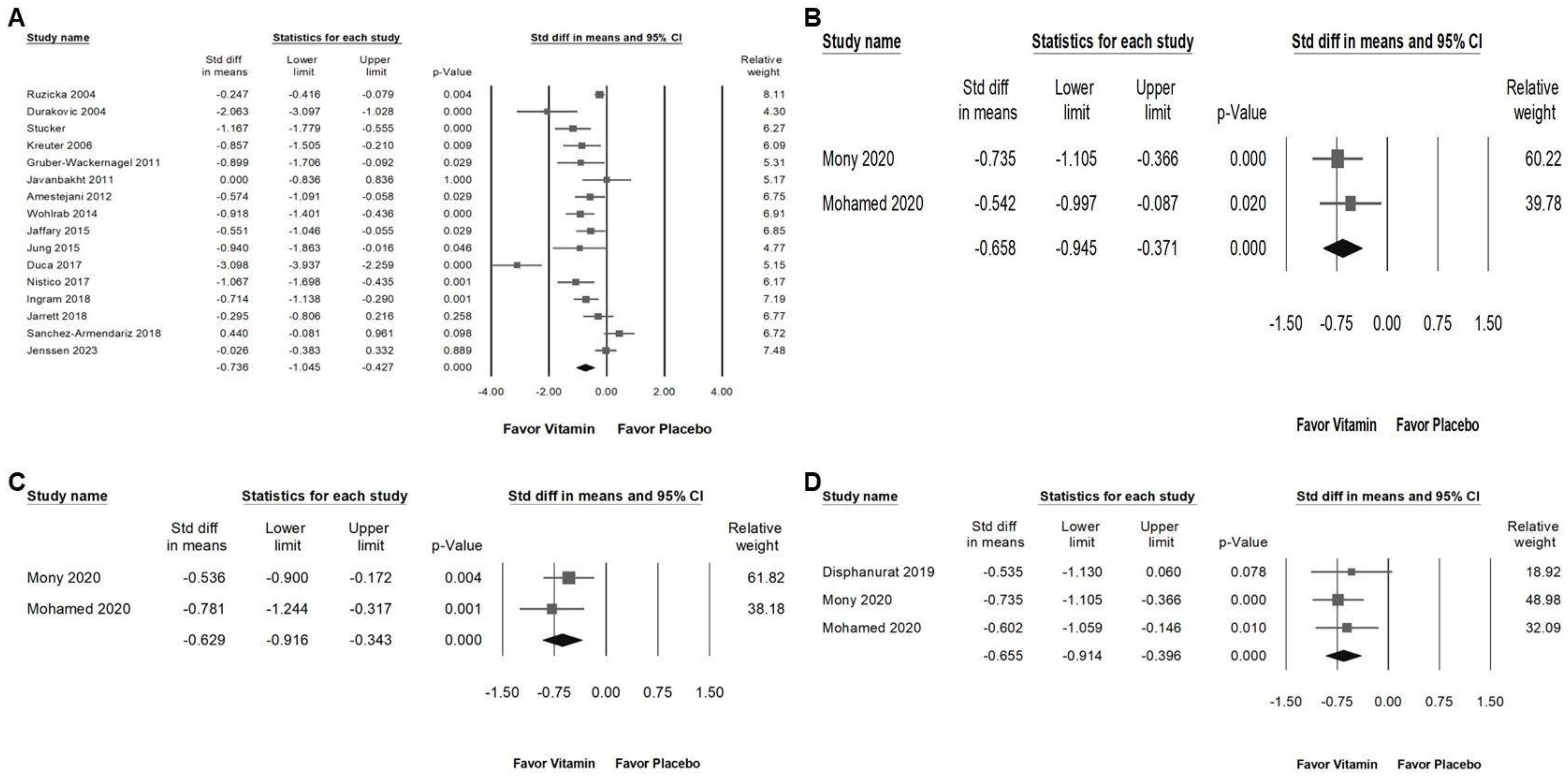
2.3. Impact of Vitamin Supplementation on Chronic Pruritus
2.4. Subgroup Analysis of Vitamin Supplementation Effects on Chronic Pruritus
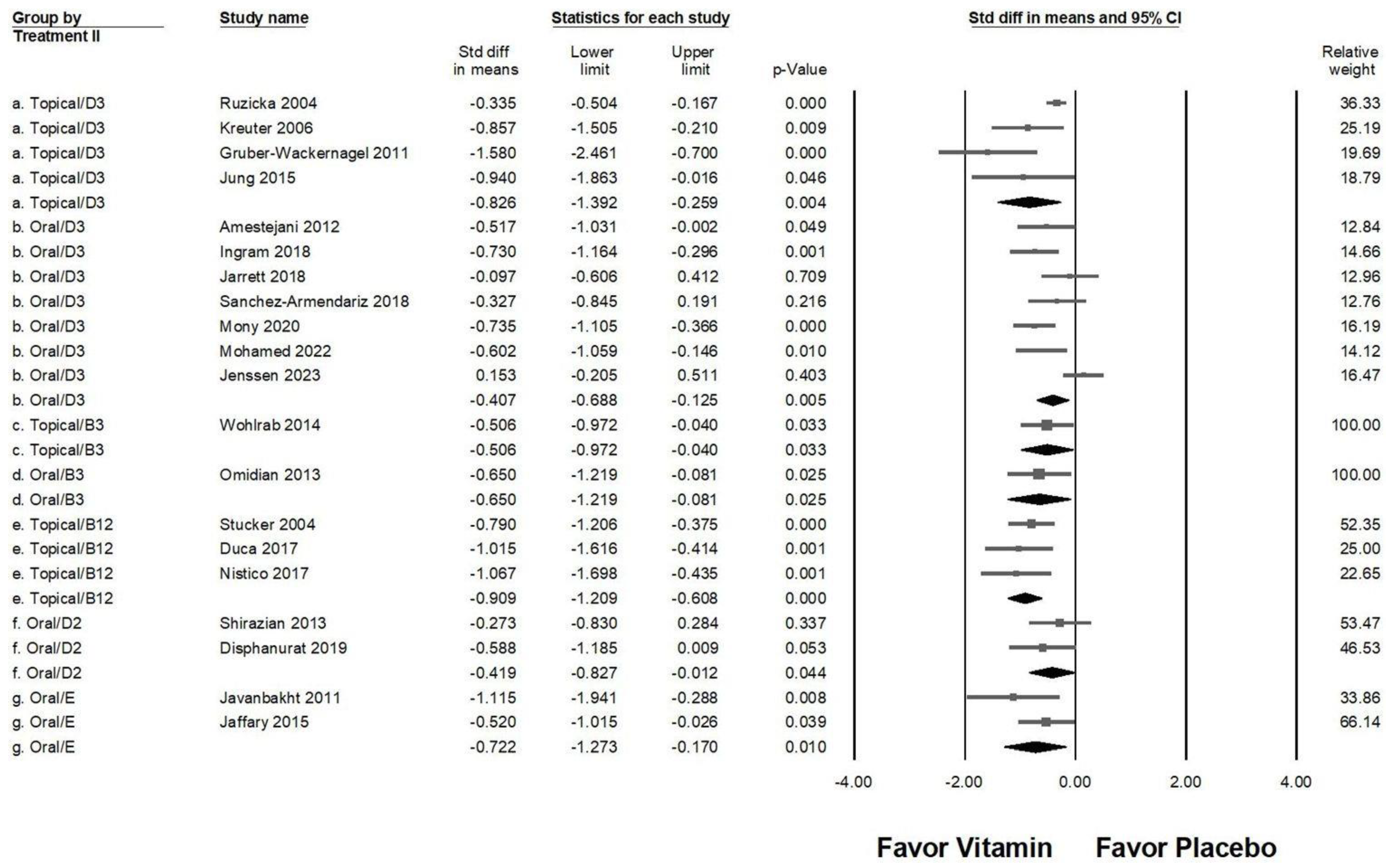
2.5. Effectiveness of Different Vitamin Types and Administration Routes in Pruritus Management
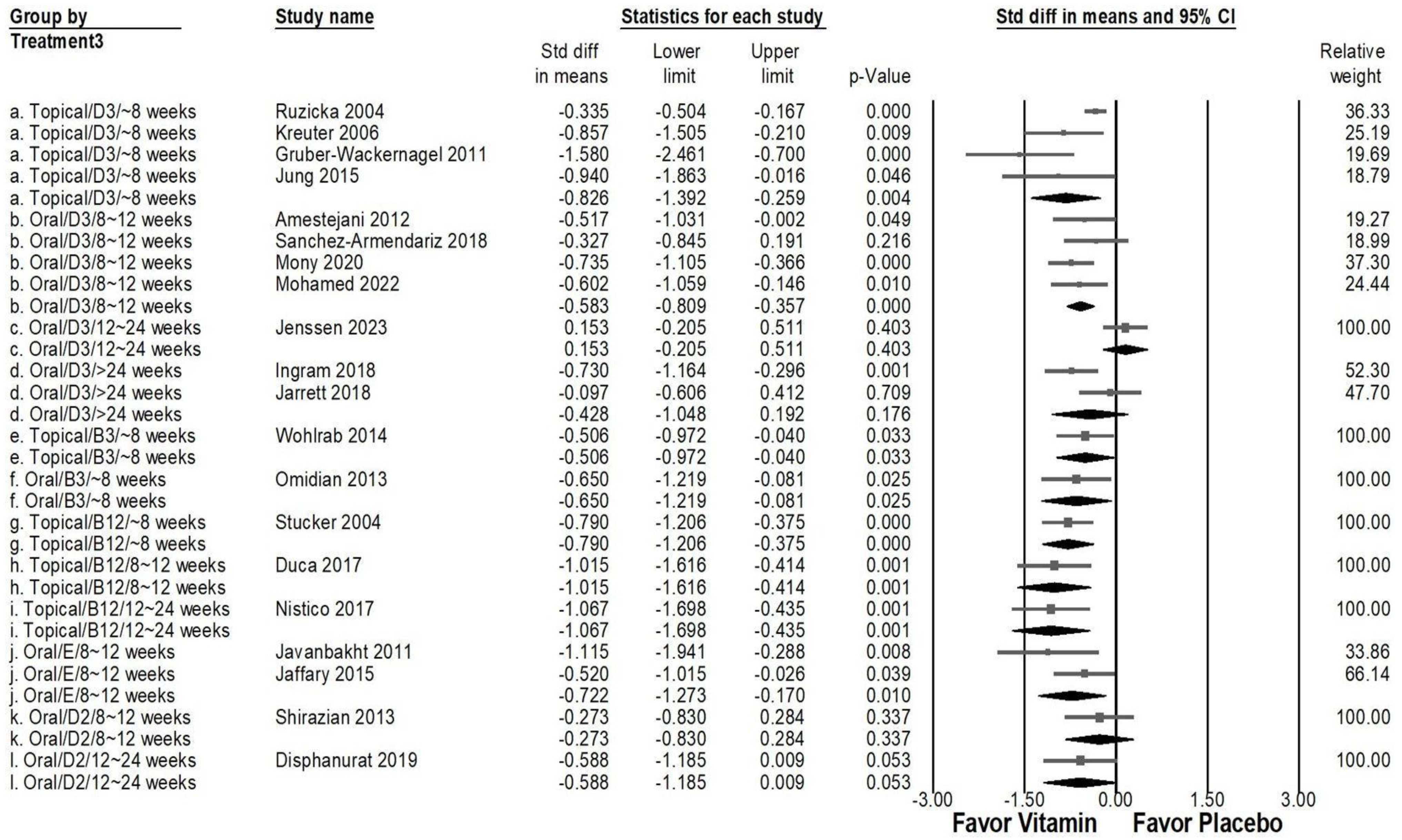
2.6. Comparative Efficacy of Vitamin Types, Administration Methods, and Treatment Duration in Pruritus Management
2.7. Impact of Vitamin Supplementation on Skin Lesion Reduction and Inflammatory Cytokine Suppression
2.8. Sensitivity Analysis
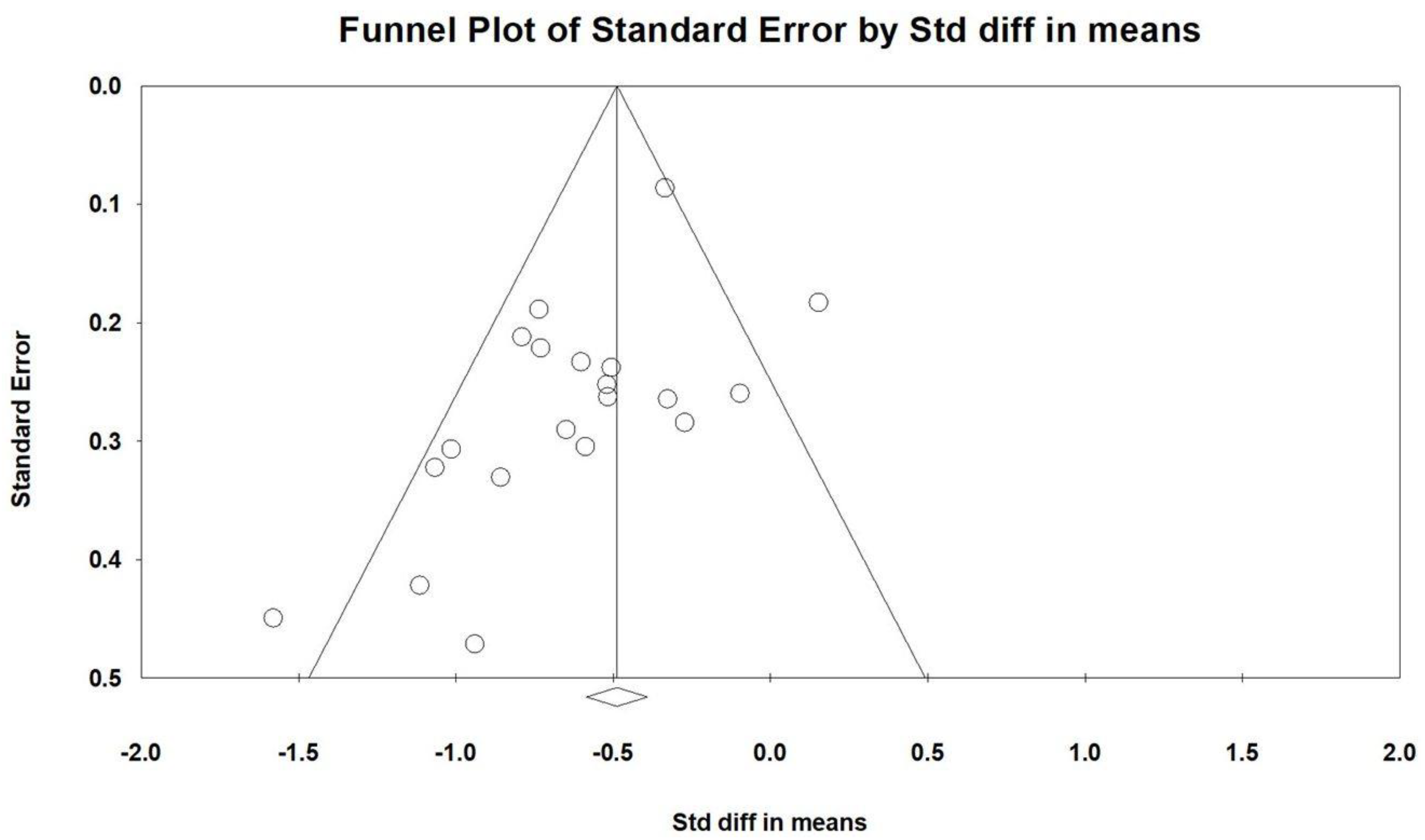
2.9. Publishing Bias
3. Discussion
4. Methods and Materials
4.1. Data Sources and Selection Criteria
4.2. Selection of Studies
4.3. Data Extraction
4.4. Outcomes
4.5. Assessment of Methodological Quality
4.6. Statistical Analyses
5. Conclusions
Author Contributions
Funding
Institutional Review Board Statement
Informed Consent Statement
Data Availability Statement
Acknowledgments
Conflicts of Interest
References
- Metz, M.; Stander, S. Chronic pruritus—Pathogenesis, clinical aspects and treatment. J. Eur. Acad. Dermatol. Venereol. 2010, 24, 1249–1260. [Google Scholar] [CrossRef] [PubMed]
- Chang, A.-S.; Chen, S.C.; Osterberg, L.; Brandt, S.; von Grote, E.C.; Meckfessel, M.H. A daily skincare regimen with a unique ceramide and filaggrin formulation rapidly improves chronic xerosis, pruritus, and quality of life in older adults. Geriatr. Nurs. 2018, 39, 24–28. [Google Scholar] [CrossRef] [PubMed]
- Etter, L.; Myers, S.A. Pruritus in systemic disease: Mechanisms and management. Dermatol. Clin. 2002, 20, 459–472. [Google Scholar] [CrossRef]
- Butler, D.C.; Berger, T.; Elmariah, S.; Kim, B.; Chisolm, S.; Kwatra, S.G.; Mollanazar, N.; Yosipovitch, G. Chronic Pruritus: A Review. JAMA 2024, 331, 2114–2124. [Google Scholar] [CrossRef]
- Mettang, T.; Kremer, A.E. Uremic pruritus. Kidney Int. 2015, 87, 685–691. [Google Scholar] [CrossRef]
- Bains, P.; Kaur, M.; Kaur, J.; Sharma, S. Nicotinamide: Mechanism of action and indications in dermatology. Indian J. Dermatol. Venereol. Leprol. 2018, 84, 234–237. [Google Scholar] [CrossRef] [PubMed]
- Stücker, M.; Pieck, C.; Stoerb, C.; Niedner, R.; Hartung, J.; Altmeyer, P. Topical vitamin B12—A new therapeutic approach in atopic dermatitis-evaluation of efficacy and tolerability in a randomized placebo-controlled multicentre clinical trial. Br. J. Dermatol. 2004, 150, 977–983. [Google Scholar] [CrossRef]
- Gruber-Wackernagel, A.; Bambach, I.; Legat, F.J.; Hofer, A.; Byrne, S.N.; Quehenberger, F.; Wolf, P. Randomized double-blinded placebo-controlled intra-individual trial on topical treatment with a 1,25-dihydroxyvitamin D3 analogue in polymorphic lighteruption. Br. J. Dermatol. 2011, 165, 152–163. [Google Scholar] [CrossRef]
- Jung, K.E.; Woo, Y.R.; Lee, J.S.; Shin, J.H.; Jeong, J.U.; Koo, D.W.; Bang, K.T. Effect of topical vitamin D on chronic kidney disease-associated pruritus: An open-label pilot study. J. Dermatol. 2015, 42, 800–803. [Google Scholar] [CrossRef]
- Javanbakht, M.H.; Keshavarz, S.A.; Djalali, M.; Siassi, F.; Eshraghian, M.R.; Firooz, A.; Seirafi, H.; Ehsani, A.H.; Chamari, M.; Mirshafiey, A. Randomized controlled trial using vitamins E and D supplementation in atopic dermatitis. J. Dermatol. Treat. 2011, 22, 144–150. [Google Scholar] [CrossRef]
- Del Duca, E.; Farnetani, F.; De Carvalho, N.; Bottoni, U.; Pellacani, G.; Nistico, S.P. Superiority of a vitamin B12-containing emollient compared to a standard emollient in the maintenance treatment of mild-to-moderate plaque psoriasis. Int. J. Immunopathol. Pharmacol. 2017, 30, 439–444. [Google Scholar] [CrossRef]
- Wohlrab, J.; Bangemann, N.; Kleine-Tebbe, A.; Thill, M.; Kummel, S.; Grischke, E.M.; Richter, R.; Seite, S.; Luftner, D. Barrier protective use of skin care to prevent chemotherapy-induced cutaneous symptoms and to maintain quality of life in patients with breast cancer. Breast Cancer Targets Ther. 2014, 6, 115–122. [Google Scholar] [CrossRef]
- Bhan, I.; Dobens, D.; Tamez, H.; Deferio, J.J.; Li, Y.C.; Warren, H.S.; Ankers, E.; Wenger, J.; Tucker, J.K.; Trottier, C.; et al. Nutritional vitamin D supplementation in dialysis: A randomized trial. Clin. J. Am. Soc. Nephrol. 2015, 10, 611–619. [Google Scholar] [CrossRef] [PubMed]
- Li, C.P.; Huang, S.C.; Hsiao, Y.; Tsai, R.Y. Evaluating the Role of Vitamin D in Alleviating Chronic Pruritus: A Meta-Analysis. Int. J. Mol. Sci. 2024, 25, 9983. [Google Scholar] [CrossRef] [PubMed]
- Nachbar, F.; Korting, H.C. The role of vitamin E in normal and damaged skin. J. Mol. Med. 1995, 73, 7–17. [Google Scholar] [CrossRef]
- Brescoll, J.; Daveluy, S. A review of vitamin B12 in dermatology. Am. J. Clin. Dermatol. 2015, 16, 27–33. [Google Scholar] [CrossRef]
- Wohlrab, J.; Kreft, D. Niacinamide—Mechanisms of action and its topical use in dermatology. Ski. Pharmacol. Physiol. 2014, 27, 311–315. [Google Scholar] [CrossRef] [PubMed]
- Alora-Palli, M.B.; Perkins, A.C.; Van Cott, A.; Kimball, A.B. Efficacy and tolerability of a cosmetically acceptable coal tar solution in the treatment of moderate plaque psoriasis: A controlled comparison with calcipotriene (calcipotriol) cream. Am. J. Clin. Dermatol. 2010, 11, 275–283. [Google Scholar] [CrossRef]
- Cassano, N.; Miracapillo, A.; Coviello, C.; Loconsole, F.; Bellino, M.; Vena, G.A. Treatment of psoriasis vulgaris with the two-compound product calcipotriol/betamethasone dipropionate followed by different formulations of calcipotriol. Clin. Drug Investig. 2006, 26, 227–233. [Google Scholar] [CrossRef]
- Guenther, L.C.; Poulin, Y.P.; Pariser, D.M. A comparison of tazarotene 0.1% gel once daily plus mometasone furoate 0.1% cream once daily versus calcipotriene 0.005% ointment twice daily in the treatment of plaque psoriasis. Clin. Ther. 2000, 22, 1225–1238. [Google Scholar] [CrossRef]
- Rorie, A.; Goldner, W.S.; Lyden, E.; Poole, J.A. Beneficial role for supplemental vitamin D3 treatment in chronic urticaria: A randomized study. Ann. Allergy Asthma Immunol. 2014, 112, 376–382. [Google Scholar] [CrossRef] [PubMed]
- Veien, N.K.; Bjerke, J.R.; Rossmann-Ringdahl, I.; Jakobsen, H.B. Once daily treatment of psoriasis with tacalcitol compared with twice daily treatment with calcipotriol. A double-blind trial. Br. J. Dermatol. 1997, 137, 581–586. [Google Scholar] [CrossRef]
- Takahashi, H.; Tsuji, H.; Ishida-Yamamoto, A.; Iizuka, H. Comparison of clinical effects of psoriasis treatment regimens among calcipotriol alone, narrowband ultraviolet B phototherapy alone, combination of calcipotriol and narrowband ultraviolet B phototherapy once a week, and combination of calcipotriol and narrowband ultraviolet B phototherapy more than twice a week. J. Dermatol. 2013, 40, 424–427. [Google Scholar] [CrossRef]
- Juntongjin, P.; Pongprasert, R. Calcipotriol ointment shows comparable efficacy to topical steroids in chronic hand eczema. Dermatol. Ther. 2019, 32, e12956. [Google Scholar] [CrossRef]
- Ozkan, I.; Kose, O.; Ozmen, I.; Arca, E. Efficacy and safety of non-laser, targeted UVB phototherapy alone and in combination with psoralen gel or calcipotriol ointment in the treatment of localized, chronic, plaque-type psoriasis. Int. J. Dermatol. 2012, 51, 609–613. [Google Scholar] [CrossRef] [PubMed]
- Theng, C.T.; Tan, S.H.; Goh, C.L.; Suresh, S.; Wong, H.B.; Machin, D.; Singapore Lichen Planus Study, G. A randomized controlled trial to compare calcipotriol with betamethasone valerate for the treatment of cutaneous lichen planus. J. Dermatolog. Treat. 2004, 15, 141–145. [Google Scholar] [CrossRef]
- Stucker, M.; Memmel, U.; Hoffmann, M.; Hartung, J.; Altmeyer, P. Vitamin B12 cream containing avocado oil in the therapy of plaque psoriasis. Dermatology 2001, 203, 141–147. [Google Scholar] [CrossRef]
- Thaci, D.; Daiber, W.; Boehncke, W.H.; Kaufmann, R. Calcipotriol solution for the treatment of scalp psoriasis: Evaluation of efficacy, safety and acceptance in 3396 patients. Dermatology 2001, 203, 153–156. [Google Scholar] [CrossRef] [PubMed]
- Guttmann-Gruber, C.; Pinon Hofbauer, J.; Tockner, B.; Reichl, V.; Klausegger, A.; Hofbauer, P.; Wolkersdorfer, M.; Tham, K.C.; Lim, S.S.; Common, J.E.; et al. Impact of low-dose calcipotriol ointment on wound healing, pruritus and pain in patients with dystrophic epidermolysis bullosa: A randomized, double-blind, placebo-controlled trial. Orphanet J. Rare Dis. 2021, 16, 473. [Google Scholar] [CrossRef]
- Byremo, G.; Rod, G.; Carlsen, K.H. Effect of climatic change in children with atopic eczema. Allergy 2006, 61, 1403–1410. [Google Scholar] [CrossRef]
- Gooderham, M.; Debarre, J.M.; Keddy-Grant, J.; Xu, Z.; Kurvits, M.; Goodfield, M. Safety and efficacy of calcipotriol plus betamethasone dipropionate gel in the treatment of scalp psoriasis in adolescents 12–17 years of age. Br. J. Dermatol. 2014, 171, 1470–1477. [Google Scholar] [CrossRef] [PubMed]
- Camargo, C.A., Jr.; Ganmaa, D.; Sidbury, R.; Erdenedelger, K.; Radnaakhand, N.; Khandsuren, B. Randomized trial of vitamin D supplementation for winter-related atopic dermatitis in children. J. Allergy Clin. Immunol. 2014, 134, 831–835. [Google Scholar] [CrossRef]
- Sidbury, R.; Sullivan, A.F.; Thadhani, R.I.; Camargo, C.A., Jr. Randomized controlled trial of vitamin D supplementation for winter-related atopic dermatitis in Boston: A pilot study. Br. J. Dermatol. 2008, 159, 245–247. [Google Scholar] [CrossRef] [PubMed]
- Di Filippo, P.; Scaparrotta, A.; Rapino, D.; Cingolani, A.; Attanasi, M.; Petrosino, M.I.; Chuang, K.; Di Pillo, S.; Chiarelli, F. Vitamin D supplementation modulates the immune system and improves atopic dermatitis in children. Int. Arch. Allergy Immunol. 2015, 166, 91–96. [Google Scholar] [CrossRef]
- Lebwohl, M.; Menter, A.; Weiss, J.; Clark, S.D.; Flores, J.; Powers, J.; Balin, A.K.; Kempers, S.; Glinert, R.J.; Fleming, T.; et al. Calcitriol 3 microg/g ointment in the management of mild to moderate plaque type psoriasis: Results from 2 placebo-controlled, multicenter, randomized double-blind, clinical studies. J. Drugs Dermatol. 2007, 6, 428–435. [Google Scholar] [PubMed]
- Kircik, L.H.; Schlesinger, T.E.; Tanghetti, E. Efficacy and Safety of Calcipotriene 0.005%/Betamethasone Dipropionate 0.064% Foam with Apremilast for Moderate Plaque Psoriasis. J. Drugs Dermatol. 2020, 19, 874–880. [Google Scholar] [CrossRef]
- Pinter, A.; Reich, A.; Arenberger, P.; Gold, L.S.; Armstrong, A.; Iversen, L.; Praestegaard, M.; Augustin, M. Randomized Phase 3 trial demonstrating high efficacy, favourable safety and convenience of a novel calcipotriol and betamethasone dipropionate cream for the treatment of psoriasis. J. Eur. Acad. Dermatol. Venereol. 2023, 37, 2327–2335. [Google Scholar] [CrossRef]
- Jalili, A.; Lebwohl, M.; Stein Gold, L.; Andersen, S.B.; Jensen, K.L.; Pink, A.E.; Segaert, S.; Berg, P.; Calzavara-Pinton, P.G.; de la Cueva Dobao, P.; et al. Itch relief in patients with psoriasis: Effectiveness of calcipotriol plus betamethasone dipropionate foam. J. Eur. Acad. Dermatol. Venereol. 2019, 33, 709–717. [Google Scholar] [CrossRef]
- Akizawa, T.; Ohashi, Y.; Akiba, T.; Suzuki, M.; Nishizawa, Y.; Ogata, E.; Slatopolsky, E.; Kurokawa, K. Dose-response study of 22-oxacalcitriol in patients with secondary hyperparathyroidism. Ther. Apher. Dial. 2004, 8, 480–491. [Google Scholar] [CrossRef]
- Hata, T.R.; Audish, D.; Kotol, P.; Coda, A.; Kabigting, F.; Miller, J.; Alexandrescu, D.; Boguniewicz, M.; Taylor, P.; Aertker, L.; et al. A randomized controlled double-blind investigation of the effects of vitamin D dietary supplementation in subjects with atopic dermatitis. J. Eur. Acad. Dermatol. Venereol. 2014, 28, 781–789. [Google Scholar] [CrossRef]
- Durakovic, C.; Ray, S.; Holick, M.F. Topical paricalcitol (19-nor-1 alpha,25-dihydroxyvitamin D2) is a novel, safe and effective treatment for plaque psoriasis: A pilot study. Br. J. Dermatol. 2004, 151, 190–195. [Google Scholar] [CrossRef] [PubMed]
- Kreuter, A.; Sommer, A.; Hyun, J.; Brautigam, M.; Brockmeyer, N.H.; Altmeyer, P.; Gambichler, T. 1% pimecrolimus, 0.005% calcipotriol, and 0.1% betamethasone in the treatment of intertriginous psoriasis: A double-blind, randomized controlled study. Arch. Dermatol. 2006, 142, 1138–1143. [Google Scholar] [CrossRef] [PubMed]
- Ruzicka, T.; Trompke, C. Treatment of scalp psoriasis. An effective and safe tacalcitol emulsion. Hautarzt 2004, 55, 165–170. [Google Scholar] [CrossRef]
- Omidian, M.; Khazanee, A.; Yaghoobi, R.; Ghorbani, A.R.; Pazyar, N.; Beladimousavi, S.S.; Ghadimi, M.; Mohebbipour, A.; Feily, A. Therapeutic effect of oral nicotinamide on refractory uremic pruritus: A randomized, double-blind study. Saudi J. Kidney Dis. Transpl. 2013, 24, 995–999. [Google Scholar] [CrossRef]
- Shirazian, S.; Schanler, M.; Shastry, S.; Dwivedi, S.; Kumar, M.; Rice, K.; Miyawaki, N.; Ghosh, S.; Fishbane, S. The effect of ergocalciferol on uremic pruritus severity: A randomized controlled trial. J. Ren. Nutr. 2013, 23, 308–314. [Google Scholar] [CrossRef] [PubMed]
- Amestejani, M.; Salehi, B.S.; Vasigh, M.; Sobhkhiz, A.; Karami, M.; Alinia, H.; Kamrava, S.K.; Shamspour, N.; Ghalehbaghi, B.; Behzadi, A.H. Vitamin D supplementation in the treatment of atopic dermatitis: A clinical trial study. J. Drugs Dermatol. 2012, 11, 327–330. [Google Scholar]
- Jaffary, F.; Faghihi, G.; Mokhtarian, A.; Hosseini, S.M. Effects of oral vitamin E on treatment of atopic dermatitis: A randomized controlled trial. J. Res. Med. Sci. 2015, 20, 1053–1057. [Google Scholar] [CrossRef]
- Sanchez-Armendariz, K.; Garcia-Gil, A.; Romero, C.A.; Contreras-Ruiz, J.; Karam-Orante, M.; Balcazar-Antonio, D.; Dominguez-Cherit, J. Oral vitamin D3 5000 IU/day as an adjuvant in the treatment of atopic dermatitis: A randomized control trial. Int. J. Dermatol. 2018, 57, 1516–1520. [Google Scholar] [CrossRef]
- Ingram, M.A.; Jones, M.B.; Stonehouse, W.; Jarrett, P.; Scragg, R.; Mugridge, O.; von Hurst, P.R. Oral vitamin D3 supplementation for chronic plaque psoriasis: A randomized, double-blind, placebo-controlled trial. J. Dermatolog. Treat. 2018, 29, 648–657. [Google Scholar] [CrossRef]
- Mohamed, A.A.; Hussein, M.S.; Salah, E.M.; Eldemery, A.; Darwish, M.M.; Ghaith, D.M.; Attala, R.A.; El Borolossy, R. Efficacy and safety of active vitamin D supplementation in chronic spontaneous urticaria patients. J. Dermatolog. Treat. 2022, 33, 427–432. [Google Scholar] [CrossRef]
- Mony, A.; Chandrashekar, L.; Rajappa, M.; Munisamy, M.; Sahoo, J.P.; Selvarajan, S. Effect of vitamin D supplementation on clinical outcome and biochemical profile in South Indian population with vitamin D-deficient chronic urticarial—A randomized double-blind placebo controlled trial. Clin. Chim. Acta 2020, 504, 1–6. [Google Scholar] [CrossRef]
- Nistico, S.P.; Del Duca, E.; Tamburi, F.; Pignataro, E.; De Carvalho, N.; Farnetani, F.; Pellacani, G. Superiority of a vitamin B12-barrier cream compared with standard glycerol-petrolatum-based emollient cream in the treatment of atopic dermatitis: A randomized, left-to-right comparative trial. Dermatol. Ther. 2017, 30, e12523. [Google Scholar] [CrossRef]
- Disphanurat, W.; Viarasilpa, W.; Chakkavittumrong, P.; Pongcharoen, P. The Clinical Effect of Oral Vitamin D2 Supplementation on Psoriasis: A Double-Blind, Randomized, Placebo-Controlled Study. Dermatol. Res. Pract. 2019, 2019, 5237642. [Google Scholar] [CrossRef]
- Jenssen, M.; Furberg, A.S.; Jorde, R.; Wilsgaard, T.; Danielsen, K. Effect of Vitamin D Supplementation on Psoriasis Severity in Patients with Lower-Range Serum 25-Hydroxyvitamin D Levels: A Randomized Clinical Trial. JAMA Dermatol. 2023, 159, 518–525. [Google Scholar] [CrossRef] [PubMed]
- Jarrett, P.; Camargo, C.A., Jr.; Coomarasamy, C.; Scragg, R. A randomized, double-blind, placebo-controlled trial of the effect of monthly vitamin D supplementation in mild psoriasis. J. Dermatolog. Treat. 2018, 29, 324–328. [Google Scholar] [CrossRef] [PubMed]
- Politis, A.; Olgiati, P.; Malitas, P.; Albani, D.; Signorini, A.; Polito, L.; De Mauro, S.; Zisaki, A.; Piperi, C.; Stamouli, E.; et al. Vitamin B12 levels in Alzheimer’s disease: Association with clinical features and cytokine production. J. Alzheimers Dis. 2010, 19, 481–488. [Google Scholar] [CrossRef]
- Granerus, G.; Lonnqvist, B.; Nystrand, J.; Roupe, G. Serum tryptase measured with B12 and G5 antibody-based immunoassays in mastocytosis patients and its relation to histamine turnover. Br. J. Dermatol. 1998, 139, 858–861. [Google Scholar] [CrossRef] [PubMed]
- Keen, M.A.; Hassan, I. Vitamin E in dermatology. Indian Dermatol. Online J. 2016, 7, 311–315. [Google Scholar] [CrossRef]
- Thiele, J.J.; Hsieh, S.N.; Ekanayake-Mudiyanselage, S. Vitamin E: Critical review of its current use in cosmetic and clinical dermatology. Dermatol. Surg. 2005, 31, 805–813. [Google Scholar] [CrossRef]
- Michalak, M.; Pierzak, M.; Krecisz, B.; Suliga, E. Bioactive Compounds for Skin Health: A Review. Nutrients 2021, 13, 203. [Google Scholar] [CrossRef]
- Bikle, D.D. Vitamin D metabolism, mechanism of action, and clinical applications. Chem. Biol. 2014, 21, 319–329. [Google Scholar] [CrossRef] [PubMed]
- Slominski, A.T.; Brozyna, A.A.; Zmijewski, M.A.; Jozwicki, W.; Jetten, A.M.; Mason, R.S.; Tuckey, R.C.; Elmets, C.A. Vitamin D signaling and melanoma: Role of vitamin D and its receptors in melanoma progression and management. Lab. Investig. 2017, 97, 706–724. [Google Scholar] [CrossRef] [PubMed]
- Benson, A.A.; Toh, J.A.; Vernon, N.; Jariwala, S.P. The role of vitamin D in the immunopathogenesis of allergic skin diseases. Allergy 2012, 67, 296–301. [Google Scholar] [CrossRef] [PubMed]
- Zhang, P.; Xu, Q.; Zhu, R. Vitamin D and allergic diseases. Front. Immunol. 2024, 15, 1420883. [Google Scholar] [CrossRef]
- Page, M.J.; McKenzie, J.E.; Bossuyt, P.M.; Boutron, I.; Hoffmann, T.C.; Mulrow, C.D.; Shamseer, L.; Tetzlaff, J.M.; Akl, E.A.; Brennan, S.E.; et al. The PRISMA 2020 statement: An updated guideline for reporting systematic reviews. BMJ 2021, 372, n71. [Google Scholar] [CrossRef]
- Higgins, J.-T. Cochrane Collaboration. Cochrane Handbook for Systematic Reviews of Interventions, 2nd ed.; Wiley-Blackwell: Hoboken, NJ, USA, 2019; p xxviii; 694p. [Google Scholar]


| Author (Year)/Country | Inclusion Criteria | Exclusion Criteria | Sample Size (% of Male)/Age | Study Design | Placebo Using | Intervention/Vitamin Type/Duration | Main Results | Secondary Results |
|---|---|---|---|---|---|---|---|---|
| ~8 weeks | ||||||||
| Ruzicka (2004) [43]/Germany | Patients with scalp psoriasis. | 1. Elevated calcium or phosphate levels. 2. Patients who had used topical retinoids, vitamin D3 analogs. 3. Patients with severe comorbidities, known hypersensitivity to vitamin D3 analogs, or conditions that could interfere with the assessment of the study medication’s efficacy, safety, or tolerability. 4. Women of childbearing potential who were not using effective contraception. | P: 273/not explicitly detailed I: 273 (51.2)/average age 45 years | RCT/Double-Blind/Placebo | Placebo emulsion | Tacalcitol emulsion (4 µg/g) applied once daily to the scalp/8 weeks | 1. The Tacalcitol group showed a significant improvement in scalp psoriasis compared to the placebo group. The median sum score decreased by 53% in the Tacalcitol group versus 30% in the placebo group (p < 0.0001). 2. Tacalcitol was significantly superior to placebo in reducing erythema, scaling, and infiltration. | 1. Tacalcitol also showed significant improvement in patient-reported outcomes, including itching and scaling. 2. Treatment was well-tolerated, with similar incidences of local side effects in both the Tacalcitol and placebo groups. No serious adverse events were reported, and calcium homeostasis remained unaffected. |
| Stücker (2004) [7]/Germany | 1. Diagnosed with atopic dermatitis for at least 2 years. 2. Pruritus, typical morphology/distribution, chronic recurrent course, personal or family history of atopic disease. 3. Chronic but not acute inflammatory atopic dermatitis without superinfection. | 1. Recent use of topical corticosteroids (last 4 weeks). 2. Recent systemic corticosteroids, ciclosporin, or photopheresis treatment (last 4 weeks). 3. Ultraviolet therapy (last 2 weeks). 4. Participation in another drug trial (last 3 months). 5. Known allergy to vitamin B12 or excipients. 6. Pregnancy or lactation. 7. Strong psychosomatic conditions or poor compliance. 8. Requirement for another topical treatment that could not be discontinued during the trial. | 49 participants (30 females, 19 males) Mean age: 33.6 ± 14.1 years | RCT/Double-Blind/Placebo | Placebo cream | Vitamin B12 (cyanocobalamin) topical cream (0.07%) Applied twice daily/8 weeks | Significant reduction in atopic dermatitis severity in the vitamin B12-treated side compared to placebo Modified SASSAD score: Vitamin B12 cream: 55.34 ± 5.74. Placebo: 28.87 ± 4.86. p < 0.001 (statistically significant) | Well-tolerated treatment, with no serious adverse events Some mild cutaneous adverse effects (e.g., itching, redness, burning) reported in a few patients. |
| Kreuter (2006) [42]/Germany | 1. Adult patients, at least 18 years old. 2. Continuous intertriginous psoriasis for a minimum of 6 months. 3. Otherwise healthy. | 1. Use of systemic corticosteroids, immunosuppressants, UV light therapy (e.g., UV-A, UV-B, psoralen–UV-A) in the previous 4 weeks. 2. Topical treatment of target lesions in the previous 2 weeks. 3. Acute guttate or pustular psoriasis, pregnancy or lactation, severe concurrent infectious diseases, diseases associated with immunosuppression or malignancy, drug dependency, mental dysfunction, or other factors limiting compliance. | P: 20 (60)/53.8 ± 17.1 I: (1% Pimecrolimus): 20 (60)/53.2 ± 14.5 I: (0.005% Calcipotriol): 20 (75)/52.1 ± 13.3 I: (0.1% Betamethaone Valerate): 20 (50)/50.4 ± 11.9 | RCT/Double-Blind/Placebo | Placebo cream | 1% pimecrolimus, 0.005% calcipotriol, 0.1% betamethasone valerate, or vehicle cream/4 weeks | VAS for pruritus: decreased by 78% for 0.1% betamethasone, 57% for 0.005% calcipotriol, 35% for 1% pimecrolimus, and 43% for vehicle. | 1. Mean reduction in MPAS after 28 days: 86.4% for 0.1% betamethasone, 62.4% for 0.005% calcipotriol, 39.7% for 1% pimecrolimus, and 21.1% for vehicle. 2. Betamethasone was significantly more effective than pimecrolimus and vehicle (p < 0.05). |
| Gruber-Wackernagel (2011) [8]/Austria | 1. Age above 18 years. 2. Confirmed diagnosis of PLE either by typical patient history and/or histology of skin lesions and/or positive photoprovocation results. | 1. Presence of or history of malignant skin tumors. 2. Dysplastic nevus syndrome. 3. Photosensitive diseases such as porphyria, chronic actinic dermatitis, xeroderma pigmentosum or basal cell nevus syndrome. 4. Autoimmune disorders such as lupus erythematosus or dermatomyositis. 5. Psychiatric disorders. 6. Immune deficiency or systemic treatment with steroids and/or other immunosuppressive drugs within 6 months before the study. 7. Pregnancy or lactation. 8. UV exposure in the test fields within 8 weeks before the study. 9. General poor health status. 10. Severe liver or renal disease. 11. Disorders of calcium metabolism or therapy for such disorders with vitamin D-containing drugs. | 1. 13 patients (3 men, 10 women; 23% male). 2. Mean age: 37.4 years | RCT/Double-Blind/Placebo/Intraindividual half-body trial | Placebo cream | Calcipotriol cream applied twice daily for 7 days before the start of photoprovocation testing with solar-simulated UV radiation. | Calcipotriol pretreatment significantly reduced PLE symptoms by an average of 32% compared with placebo throughout the observation period from 48 to 144 h after the first photoprovocation exposure (p = 0.0022) | Calcipotriol pretreatment resulted in a significantly lower PLE test score in 58% (48 h), 75% (72 h), and 83% (144 h) of the cases compared with placebo Reduced erythema and increased pigmentation were observed with calcipotriol pretreatment |
| Omidian (2013) [44]/Not mentioned | 1. Chronic kidney disease (CKD) patients with refractory uremic pruritus (UP). 2. Must have discontinued all anti-pruritic agents at least two weeks before the study. | Not explicitly mentioned. | Gender distribution and age range not specified in the excerpt. | RCT/Double-Blind/Placebo | Placebo tablets | Oral nicotinamide (vitamin B3) 500 mg twice a day vitamin Type/4 weeks | No significant difference between the reductions in pruritus between groups. However, the interaction effect between drug and time was significant (p < 0.026), indicating that the effect of nicotinamide may become more pronounced over time. | All patients completed the study period. The study suggests that a longer intervention period (>4 weeks) might be needed to observe a more significant effect of nicotinamide in reducing uremic pruritus. |
| Wohlrab (2014) [12]/Germany | 1. Female patients aged 18 years or older. 2. Breast cancer. 3. Indicated for adjuvant or neoadjuvant chemotherapy with anthracyclines or taxanes. 4. Combination with trastuzumab was allowed. | 1. With pre-existing clinical signs of a barrier dysfunction. 2. History of atopic or psoriatic disposition. 3. Usage of pharmaceutical or over-the-counter products with vasoactive, anti-inflammatory, or diuretic effects. 4. Use of medications affecting lipid metabolism. | 95 patients. Age range: 25–77 years | RCT/Not specified as double-blind/reference-controlled crossover study | Usual skincare | Test preparation: a lipophilic cream containing 4% niacinamide (vitamin B3), shea butter, and thermal spring water/6 weeks | 1. No significant difference in total DLQI score. 2. Significant improvement in the “Symptoms and Feelings” subscale favoring (p < 0.05). 3. VAS scores for pruritus, dryness, and irritability showed significant improvements (p < 0.05). | No significant side effects related to the test preparation Patients tolerated the skincare regimen well, with improved quality of life in terms of skin-related symptoms. |
| Jung (2015) [9]/Korea | 1. Patients with chronic kidney disease-associated pruritus (CKD-aP). 2. Patients undergoing hemodialysis. | Not explicitly mentioned in the provided text. | P: 10 (30) I: 10 (30) Mean age: 63.3 years | RCT/Double-Blind/Placebo/Single-center, open-label pilot study | Vehicle solution | Topical vitamin D (calcipotriol) or vehicle solution applied twice daily/4 weeks | 1. Both MPAS and VAS scores significantly decreased after 2 and 4 weeks of topical vitamin D treatment compared with the vehicle (p < 0.05). 2. Dermoscopic evaluation showed significant improvement in skin dryness for the vitamin D-treated group compared to the vehicle group. | 1. No significant side effects were observed. 2. One patient reported a temporary sensation of “feeling heavy” after applying topical vitamin D but continued the treatment without further discomfort. |
| 8~12 weeks | ||||||||
| Durakovic (2004) [41]/United States | 1. With moderate plaque psoriasis involving at least 5% of body surface area. 2. Two target lesions of at least 5 cm in diameter. 3. Plaque elevation, scaling, and erythema with at least moderate severity (score of 2 on a scale of 0–4). | 1. History of hepatic failure, renal failure, nephrocalcinosis, hypercalcemia, hypercalciuria, or hyperphosphatemia. 2. Women of childbearing age who were pregnant, lactating, or unwilling to use effective contraception. 3. Patients using calcium supplements or drugs influencing calcium metabolism. | Paricalcitol-treated Group: 11 (88), mean age 46.5 years (range 29–65). | RCT/Double-Blind/placebo/self-controlled study | Placebo ointment | 15 µg/g paricalcitol ointment (19-nor-1α,25-dihydroxyvitamin D2) once daily/12 weeks | Paricalcitol-treated lesions showed a significant decrease in scaling (74%), erythema (69%), and plaque elevation (71%) compared to placebo-treated lesions, which showed reductions of 32%, 22%, and 8%, respectively. | 1. Serum calcium and phosphorus levels, as well as the 24-h urinary calcium/creatinine ratio, remained within normal ranges throughout the study. 2. Immunohistochemical analysis showed that paricalcitol treatment markedly reduced the immunoreactivity of transglutaminase K in psoriatic lesions, bringing it closer to the pattern observed in non-lesional skin. |
| Javanbakht (2011) [10]/Iran | 1. Patients diagnosed with AD based on Hanifin and Rajka’s criteria. 2. Objective SCORA between 10 and 70. 3. Normal hepatic and renal function. | 1. Use of vitamins, minerals, fatty acid supplements, oral contraceptive pills, steroid hormones (oral or parenteral), anti-epileptic agents, and anticoagulant drugs. 2. Pregnant or nursing. 3. Undergoing phototherapy or taking systemic corticosteroids/immunosuppressive drugs. | P: 11 (10)/26.1 ± 2.8 I: (vitamin D): 12 (33)/21.2 ± 14.6 I: (vitamin E): 11 (37.5)/29.0 ± 2.09 I: (vitamin D+E): 11 (33)/27.5 ± 2.3 | RCT/Double-Blind/Placebo | Placebo tablets | Group D: 1600 IU vitamin D3 + vitamin E placebo Group E: 600 IU synthetic all-rac-α-tocopherol (vitamin E) + vitamin D placebo Group DE: 1600 IU vitamin D3 + 600 IU vitamin E/60 days | 1. SCORAD scores were significantly reduced in Groups D, E, and DE compared to baseline. 2. Significant reduction in objective SCORAD, lichenification, and pruritus. | 1. Plasma α-tocopherol (vitamin E levels) significantly increased in Groups E and DE. 2. No significant adverse effects were reported. |
| Amestejani (2012) [46]/Not mentioned | 1. Diagnosed with AD. 2. Severity of AD evaluated using SCORAD and TIS. | Not explicitly mentioned in the excerpt (potential additional details may be in the full study text) | I: 30 P: 30 Age: Not specified Gender: Not specified | RCT/Double-Blind/Placebo | Placebo tablets | Cholecalciferol (vitamin D3) 1600 IU daily/60 days | 1. SCORAD and TIS scores significantly improved in the vitamin D group (p < 0.05) 2. No significant improvement in the placebo group (p > 0.05) 3. Patients with mild, moderate, and severe AD showed significant improvements with vitamin D supplementation | No adverse effects were reported related to vitamin D supplementation |
| Shirazian (2013) [45]/United States | 1. Adult patients undergoing maintenance HD. 2. Described excessive pruritus. 3. On HD for more than 3 months. | 1. PTH level less than 70 pg/mL or greater than 1000 pg/mL. 2. Serum phosphorus greater than 7.0 mg/dL. 3. Serum calcium greater than 11 mg/dL. 4. Active malignancy. 5. Current ergocalciferol treatment. | P: 25 (56)/66.2 ± 13.7 I: 25 (60)/66.1 ± 14.7 | RCT/Double-Blind/Placebo/Single-center | Placebo pill | Ergocalciferol 50,000 international units (IU) or placebo once weekly for 12 weeks | Both groups experenced a decrease in pruritus scores, with a reduction of 38.9% in the treatment group and 47.5% in the placebo group. No significant difference in pruritus severity between groups (p = 0.34). Adjusted analysis also found no significant treatment effect. | 1. No significant differences in calcium, phosphorus, and PTH levels between groups. 2. Significant increase in 25-hydroxy vitamin D levels in the treatment group compared to placebo (19 ng/mL vs. 1.4 ng/mL, p < 0.01). |
| Jaffary (2015) [47]/Iran | Diagnosed with mild-to-moderate AD based on Hanifin and Rajka diagnostic criteria | 1. Severe AD with SCORAD > 50 requiring hospitalization. 2. Pregnant or nursing mothers. 3. Coagulopathies or patients using anticoagulant medications 4. Systemic corticosteroid or immunosuppressant users 5. History of allergy to vitamin E | P: 27 I: 28 | RCT/Double-Blind/Placebo | Placebo contained no active ingredient and was identical in appearance to vitamin E capsules. | Vitamin E (α-tocopherol), 400 IU daily/16 weeks | 1. Significant Itching reduction (p < 0.05). 2. Extent of skin lesions reduction (p < 0.05). 3. SCORAD index reduction (p < 0.05). | 1. No adverse effects related to vitamin E supplementation were reported. 2. Women had a greater reduction in pruritus and lesion extent. 3. Men showed a greater decrease in total SCORAD index. |
| Duca (2017) [11]/Italy | 1. Patients with mild-to-moderate plaque psoriasis. 2. Diagnosed based on PASI. 3. Symmetric plaque psoriasis. | 1. Other psoriasis forms (palmoplantar, inverse psoriasis). 2. Use of systemic/biological treatments. 3. Allergy to study products. 4. Serious comorbidities affecting study results. 5. Pregnant, breastfeeding, or planning pregnancy. 6. Poor treatment adherence. | 1. Total: 24 patients 2. Age: 48.2 ± 15.4 years 3. Gender distribution: 13 males (54.2%), 11 females (45.8%) | RCT/Single-Blind/Intra-patient left vs. right comparison study | The control group used a glycerol-petrolatum-based emollient cream (Cetaphil®, Mavena® B12 ointment, Milan, Italy) | 1. Topical vitamin B12 (cyanocobalamin) ointment (Mavena® B12) 2. Concentration: 0.07% cyanocobalamin + 20% avocado oil/12 weeks + 4 week washout | 1. Significant PASI reduction (p < 0.001). 2. Pruritus (itching) and erythema were significantly reduced. | 1. After the 4-week washout period, PASI scores slightly increased in both groups, but the vitamin B12-treated side maintained significantly lower PASI scores than the control side. 2. No major adverse effects reported. |
| Sánchez-Armendáriz (2018) [48]/Mexico | 1. Diagnosed with moderate-to-severe AD based on Hanifin–Rajka criteria and SCORAD severity scale. 2. No prior vitamin D supplementation. | 1. Primary immunodeficiency disorders. 2. Renal tubular acidosis. 3. Pregnancy. 4. Use of other supplements. 5. Failed to complete the 12-week follow-up. | P: 29/12.2 ± 12.9 I: 29/12.9 ± 10.6 | RCT/Double-Blind/Placebo | Received cellulose capsules identical in size and color to the vitamin D3 capsules | Oral vitamin D3 (Cholecalciferol) 5000 IU daily/12 weeks | 1. 80% of patients with serum levels < 20 ng/mL still had moderate-to-severe AD, despite treatment. 2. No significant differences in pruritus scores between groups. | No cases of hypercalcemia or other safety concerns were reported. |
| Mony (2020) [51]/India | 1. Adults (20–50 years) with chronic urticaria (CU) for more than 6 weeks. 2. Vitamin D deficiency (serum vitamin D < 20 ng/mL). | 1. Acute urticaria, physical urticaria, urticarial vasculitis, hereditary or acquired angioedema. 2. Symptoms of vitamin D deficiency (musculoskeletal pain, fractures). 3. Hepatic or renal dysfunction, malignancies, infections, inflammatory cutaneous disorders. 4. Pregnant and lactating women. 5. Patients who have taken vitamin D supplementation in the past 6 months. | P: 60 (21)/36.71 ± 11.01 I: 60 (20)/38.80 ± 12.54 | RCT/Double-Blind/Placebo | Matched placebo | Experimental group: 60,000 IU of vitamin D3 (cholecalciferol) fortnightly for 12 weeks Control group: similar-looking placebo fortnightly for 12 weeks Both groups received standard treatment with levocetirizine | 1. Significant reduction in UAS7 scores and medication dosage in the vitamin D treated group compared to the placebo group (p < 0.0001). 2. Significant reduction in inflammatory cytokines (IL-6, IL-17, TGF-β, hs-CRP) in the vitamin D treated group compared to the placebo group. | 1. Significant increase in 25-OH vitamin D and vitamin D binding protein (VDBP) in the vitamin D treated group compared to the placebo group. 2. No significant change in cytokine concentrations or vitamin D levels in the placebo group. |
| Mohamed (2022) [50]/Egypt | Adults >18 years of age, having urticaria episodes at least 2 days per week for 6 weeks or longer with/without angioedema | 1. With only physical urticaria, urticarial vasculitis, hereditary or acquired angioedema. 2. With dyslipidemia, diabetes, hypertension, pre-existing cardiovascular disease, cerebrovascular accidents, hypothyroidism, smokers, and other systemic or cutaneous disorders including atopic dermatitis, psoriasis, etc. 3. With hypercalcemia (>11 mg/dL), diabetes, renal insufficiency, hepatic disorders, hyperparathyroidism, sarcoidosis, other granulomatous disorders, malignancy. 4. Pregnant and lactating women and patients who have taken vitamin D supplementation in the past 6 months. | P: 67 (50.7)/39.34 ± 7.23 I: 77 (40.2)/36.50 ± 5.12 | RCT/Single-Blind/Placebo | Matched placebo | Group 1 (Study group): 38 patients received 0.25 µg alfacalcidol once daily for 12 weeks in addition to the standard therapy (Hydroxyzine 25 mg/day) Group 2 (Placebo group): 39 patients received an oral placebo taken with the same regimen for 12 weeks in addition to the standard therapy (Hydroxyzine 25 mg/day) | The UAS7 total score significantly lower in the study group after active vitamin D administration compared to the placebo group (p < 0.01) | 1. No significant change in UAS7 total score or number of patients in each severity level in the placebo group compared to their baseline results. 2. Significant increase in mean serum 25(OH) D levels in the study group compared to placebo group and their baseline results (p < 0.001). 3. Significant decrease in mean serum IL6, hsCRP, and TNFα levels in the study group compared to placebo group and their baseline results (p < 0.01). 4. Significant negative correlation (r = −0.67, p < 0.05) between serum 25(OH) D levels and total UAS7 scores, indicating disease severity. |
| 12~24 weeks | ||||||||
| Nistico (2017) [52]/Italy | 1. With a confirmed clinical diagnosis of mild atopic dermatitis (AD). 2. SCORAD index < 25 points. | 1. With severe AD or other dermatological conditions. 2. Systemic treatments that could interfere with study results. 3. History of hypersensitivity to the study compounds. | 22 Caucasian patients (65) | RCT/single-blind, intra-patient, left-to-right comparative trial. | Comparison was between MB12 cream vs. glycerol-petrolatum-based emollient cream. | Topical vitamin B12 barrier cream vs. glycerol-petrolatum-based emollient cream. 2–3 times per day/12 weeks | 1. Both treatments reduced SCORAD scores, but B12-treated sites showed a significantly greater reduction p < 0.001. 2. Pruritus severity (VAS scale) reduced from 8.7 to 1.7 in B12-treated sites. | Both treatments were well tolerated. |
| Disphanurat (2019) [53]/Thailand | 1. Diagnosed with chronic plaque-type psoriasis. 2. Mild psoriasis (PASI score < 10) | 1. Recent or current use of systemic therapy or phototherapy (within 30 days before enrollment). 2. Hepatic or renal impairment, cancer. 3. Use of immunosuppressive medications, chemotherapy, vitamin D supplements, calcium supplements, bisphosphonates, antiepileptic agents, or anticoagulants. 4. History of hypercalcemia, nephrolithiasis, or parathyroid disease. 5. Pregnant or breastfeeding women. | P: 22 (50)/49.41 ± 15.92 I: 23 (43.5)/52.39 ± 14.19 | RCT/Double-Blind/Placebo | Identical-looking placebo pills | 1. Vitamin D2 (Calciferol capsules, 20,000 IU per capsule). 2. Three capsules (60,000 IU total) every 2 weeks/6 months. | 1. PASI score improvement was significantly greater in the vitamin D group compared to the placebo group at 3 months (p = 0.034). 2. PASI percentage improvement (p = 0.039). | Vitamin D deficiency significantly reduced in the vitamin D group by the end of the study. |
| Jenssen (2023) [54]/Norway | 1. Active plaque psoriasis (PASI > 0). 2. Baseline serum 25-hydroxyvitamin D [25(OH)D] levels < 24 ng/mL | 1. Nut allergy. 2. Primary hyperparathyroidism, granulomatous diseases. 3. Systolic blood pressure > 174 mmHg, diastolic BP > 104 mmHg. 4. Creatinine > 130 μmol/L (men) or >120 μmol/L (women). 5. HbA1c > 9.0%. 6. Pregnancy, history of kidney stones in the last 5 years. 7. Diagnosed or treated for organ cancer or malignant melanoma in the last 12 months. 8. Severe physical or mental illness preventing participation. 9. Use of phototherapy, heliotherapy, or vitamin D supplementation (>800 IU/day). 10. Recent initiation or increase in systemic psoriasis treatment. | P: 61 (63.9)/54.0 ± 9.1 I: 59 (62.7)/53.3 ± 10.9 | RCT/Double-Blind/placebo-controlled trial with two parallel groups | Identical-looking placebo capsules | Loading dose: 100,000 IU Followed by: 20,000 IU per week/4 months | 1. No significant difference in PASI score changes between the vitamin D and placebo groups. 2. Adjusted difference in PASI: 0.11 (p = 0.52). 3. No significant difference in PGA score. 4. No significant difference in self-administered PASI or DLQI scores. 5. No adverse effects reported. | 1. Post-intervention 25(OH) D levels increased in the vitamin D group (mean: 29.7 ng/mL) compared to the placebo group (mean: 12.0 ng/mL). 2. Only 41.1% of participants in the vitamin D group achieved 25(OH)D levels > 30 ng/mL. |
| >24 weeks | ||||||||
| Ingram (2018) [49]/New Zealand | 1. Adults aged 18 years and older with chronic plaque psoriasis. 2. Patients with stable psoriasis not requiring systemic treatment and with no history of using more than 999 IU/day of vitamin D supplements in the last two months. | 1. Chronic kidney or liver disease, smoking, pregnancy, lactation, or planned pregnancy. 2. Use of phototherapy, systemic steroids, or other psoriasis treatments within the last three months. | P: 34 (50)/46.7 ± 13.7 I: 67 (58)/50.7 ± 13.43 | RCT/Double-Blind/Placebo | Placebo capsules identical in appearance to vitamin D3 capsules, taken once monthly for 11 months. | Monthly oral doses of 100,000 IU of vitamin D2. | 1. No significant difference in PASI scores between the vitamin D and placebo groups over the 12-month period. 2. Both groups showed a mild improvement in PASI scores from baseline, but the improvement was not significantly different between the groups. | Serum 24(OH)D concentrations significantly increased in the treatment group and unexpectedly also increased in the placebo group, possibly due to increased sun exposure or other factors. |
| Jarrett (2018) [55]/New Zealand | 1. Diagnosed with mild psoriasis, confirmed through self-reporting and physician evaluation. 2. Individuals willing to participate in a 12-month follow-up. | 1. Current use of vitamin D supplements exceeding 600 IU/day (ages 50–70) or 800 IU/day (ages 71–84). 2. History of psychiatric disorders affecting compliance. 3. Conditions such as hypercalcemia, nephrolithiasis, sarcoidosis, parathyroid disease, or gastric bypass surgery. 4. Participation in another study that could interfere. 5. Baseline serum calcium > 2.50 mmol/L. | P: 42 (65)/64.7 ± 7.4 I: 23 (35)/68.4 ± 8.7 | RCT/Double-Blind/Placebo | Sunflower lecithin placebo capsules. | Vitamin D3 (Cholecalciferol) 200,000 IU oral capsule at baseline. 100,000 IU/month/12 months. | No significant difference between vitamin D and placebo groups in psoriasis severity reduction. | Results do not support the use of monthly high-dose vitamin D supplementation for treating mild psoriasis. |
Disclaimer/Publisher’s Note: The statements, opinions and data contained in all publications are solely those of the individual author(s) and contributor(s) and not of MDPI and/or the editor(s). MDPI and/or the editor(s) disclaim responsibility for any injury to people or property resulting from any ideas, methods, instructions or products referred to in the content. |
© 2025 by the authors. Licensee MDPI, Basel, Switzerland. This article is an open access article distributed under the terms and conditions of the Creative Commons Attribution (CC BY) license (https://creativecommons.org/licenses/by/4.0/).
Share and Cite
Kuo, W.-H.; Chang, K.-S.; Chang, M.-H.; Hsiao, Y.; Tsai, R.-Y. Updated Meta-Analysis on Vitamin Supplementation for Chronic Pruritus: Expanding Evidence Beyond Vitamin D. Int. J. Mol. Sci. 2025, 26, 3840. https://doi.org/10.3390/ijms26083840
Kuo W-H, Chang K-S, Chang M-H, Hsiao Y, Tsai R-Y. Updated Meta-Analysis on Vitamin Supplementation for Chronic Pruritus: Expanding Evidence Beyond Vitamin D. International Journal of Molecular Sciences. 2025; 26(8):3840. https://doi.org/10.3390/ijms26083840
Chicago/Turabian StyleKuo, Wu-Hsien, Ko-Shih Chang, Mu-Hsin Chang, Yao Hsiao, and Ru-Yin Tsai. 2025. "Updated Meta-Analysis on Vitamin Supplementation for Chronic Pruritus: Expanding Evidence Beyond Vitamin D" International Journal of Molecular Sciences 26, no. 8: 3840. https://doi.org/10.3390/ijms26083840
APA StyleKuo, W.-H., Chang, K.-S., Chang, M.-H., Hsiao, Y., & Tsai, R.-Y. (2025). Updated Meta-Analysis on Vitamin Supplementation for Chronic Pruritus: Expanding Evidence Beyond Vitamin D. International Journal of Molecular Sciences, 26(8), 3840. https://doi.org/10.3390/ijms26083840