Article

Identification of CDK1 as a Biomarker for the Treatment of Liver Fibrosis and Hepatocellular Carcinoma Through Bioinformatics Analysis

The Key Laboratory of Study and Discovery of Small Targeted Molecules of Hunan Province, School of Pharmaceutic Science, Health Science Center, Hunan Normal University, Changsha 410013, China
*
Author to whom correspondence should be addressed.
Int. J. Mol. Sci. 2025, 26(8), 3816; https://doi.org/10.3390/ijms26083816
Submission received: 17 March 2025 / Revised: 4 April 2025 / Accepted: 6 April 2025 / Published: 17 April 2025
(This article belongs to the Special Issue Advancements in Cancer Biomarkers)

Abstract

Cyclin-dependent kinase 1 (CDK1) has emerged as a critical regulator of cell cycle progression, yet its role in liver fibrosis-associated hepatocellular carcinoma (LF-HCC) remains underexplored. This study aimed to systematically evaluate CDK1’s prognostic significance, immune regulatory functions, and therapeutic potential in LF-HCC pathogenesis. Integrated bioinformatics approaches were applied to multi-omics datasets from GEO, TCGA, and TIMER databases. Differentially expressed genes were identified through enrichment analysis and protein–protein interaction networks. Survival outcomes were assessed via Kaplan–Meier analysis, while immune cell infiltration patterns were quantified using CIBERSORT. Molecular docking simulations evaluated CDK1’s binding affinity with pharmacologically active compounds (alvocidib, seliciclib, alsterpaullone) using AutoDock Vina. CDK1 demonstrated significant overexpression in LF-HCC tissues compared to normal controls (p < 0.001). Elevated CDK1 expression correlated with reduced overall survival (HR = 2.41, 95% CI:1.78–3.26, p = 0.003) and advanced tumor staging (p = 0.007). Immune profiling revealed strong associations between CDK1 levels and immunosuppressive cell infiltration, particularly regulatory T cells (r = 0.63, p = 0.001) and myeloid-derived suppressor cells (r = 0.58, p = 0.004). Molecular docking confirmed high-affinity binding of CDK1 to kinase inhibitors through conserved hydrogen-bond interactions (binding energy ≤ −8.5 kcal/mol), with alvocidib showing optimal binding stability. This multimodal analysis establishes CDK1 as both a prognostic biomarker and immunomodulatory regulator in LF-HCC pathogenesis. The enzyme’s dual role in driving tumor progression and reshaping the immune microenvironment positions it as a promising therapeutic target. Computational validation of CDK1 inhibitors provides a rational basis for developing precision therapies against LF-HCC, bridging translational gaps between biomarker discovery and clinical application.

1. Introduction

Recently, liver cancer has become the second leading lethal factor in the world [1,2]. Hepatocellular carcinoma is the largest category of all primary HCC and is one of the leading causes of HCC-related deaths [3]. The onset of this disease is usually by sequential [4] liver injury, hepatitis, liver fibrosis, cirrhosis, and hepatocellular carcinoma. Hepatocellular carcinogenesis is 90% [5] due to cirrhosis, whereas the usual cause of cirrhosis [6] is the poor treatment of liver fibrosis. Therefore, we go back to the source, take liver fibrosis as the research direction, explore the effective mechanism of treating liver fibrosis, and achieve the purpose of preventing hepatocellular carcinoma from hepatocellular carcinoma.
Liver fibrosis is the first stage of progression to cirrhosis in many chronic liver diseases, which is characterized by collagen fiber deposition [7] caused by the imbalance of liver extracellular matrix (ECM) secretion and degradation. At present, the pathogenesis of liver fibrosis is mainly manifested as abnormal repair of liver tissue damage, proliferation and activation of hepatic stellate cells, and accumulation of extracellular matrix. However, the pathogenesis of the disease has not been defined by [7,8], and no anti-fibrotic drugs for liver fibrosis have been put into clinical use. The ideal route for drug therapy is to achieve the prevention or reversal of fibrosis [9] in the liver by eliminating several underlying etiologies already manifested in the patient. However, the reversal of liver fibrosis is obviously impractical. Previous studies have also found some new methods for the treatment of mild liver fibrosis, such as targeted collagen homeostasis therapy, nanoparticle therapy, and others [10,11]. However, due to their high production costs, low universality, and difficulty of mass production, they cannot be put into clinical use in a short time and are not widely adopted. Therefore, it is urgent to explore the universal action targets for patients with liver fibrosis and to clarify the relevant mechanism of the target action [12].
We used microarray comparison to analyze the gene expression profiles of human growth and aging-activated HSCs and found that CDK1 gene expression was positively correlated with the occurrence of liver fibrosis. CDK1 is the most likely critical target for the treatment of liver fibrosis. CDK1, as a member of the serine/threonine-specific protein kinase family, is a cyclin-dependent kinase with potential effects as a classical target for the treatment of a wide variety of tumors [13]. The occurrence and development of HCC are closely related to abnormal cell cycle regulation. As a key regulator of the G2/M phase turnover of the cell cycle, CDK1 plays an important role in HCC cell proliferation. Inhibition of CDK1 activity blocks the proliferation of HCC cells and induces apoptosis [14]. In addition, the combined effects of CDK1 and other cell cycle-related proteins, such as aurora kinase A (AURKA), are also considered as an important target for the treatment of liver cancer [15]. Therefore, we speculate that the inhibition of CDK1 expression is useful in treating hepatocellular carcinoma. Many inhibitory drugs targeting CDK1 have entered the clinical research stage [16]. However, although we clarified that CDK1 plays a role in the treatment of liver fibrosis, the location of action, pathways, and mechanisms of CDK1 in the treatment of liver fibrosis are not clear [17]. The key of this study is to identify new targets for liver cancer and accurately predict the pathways and mechanisms of the targets.
At present, the conventional experimental method is long, the scientific research cost is high, and the fault tolerance rate is low. This has led to most existing treatments for liver fibrosis being studied only around existing targets, without new breakthroughs. At the same time, the current target prediction is often based on the existing literature and the independent research of researchers, which has strong subjectivity. As a rapidly developing technology, bioinformatics has generated a relatively complete research system, with a more objective prediction of new targets, more diversified research directions, richer research content, and more diverse research results, and is the first choice for target prediction and role research [18].
As a key regulator of the cell cycle, CDK1 plays an important role in the development of liver fibrosis and liver cancer. Therefore, designing anti-liver fibrosis and liver cancer drugs targeting CDK1 has broad application prospects [13]. Through an in-depth study of the specific mechanism of CDK1 in liver fibrosis and liver cancer, it can provide a more accurate treatment plan for patients, improve the treatment effect, and reduce adverse reactions. In the study of CDK1 inhibitors, the synergistic effect of TCM components (e.g., taine) has been widely observed [14]. This helps to promote the modernization of TCM and provides a scientific basis for the application of TCM in the treatment of liver fibrosis and liver cancer. By inhibiting the activity of CDK1, it can slow down the progression of liver fibrosis and inhibit the growth of liver cancer, which can prolong the survival time of patients and improve the quality of life [19]. This is of great significance for improving the patient prognosis and reducing the social burden.
This study aimed to predict novel targets for the treatment of liver fibrosis. Later, the action pathway and mechanism of the new target were studied, and clinical studies, such as immune infiltration and prognostic response, were conducted to provide a new direction for subsequent research on the treatment of liver fibrosis.

2. Results

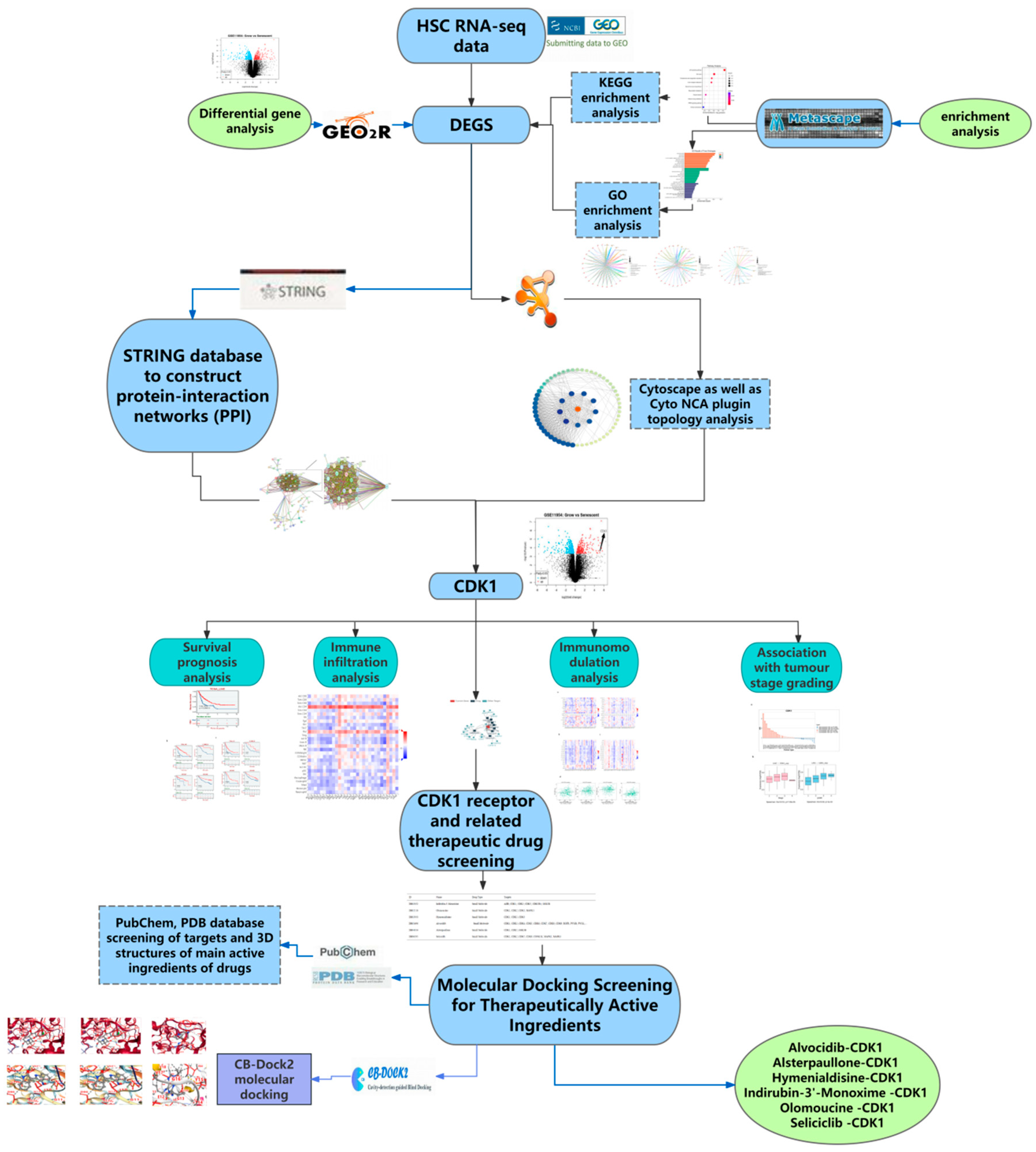
2.1. Acquisition of DEGs and the GEO2R Analysis

The GEO database is a public high-throughput gene expression database, and genomics studies based on this database comply with ethical relevant regulations and avoid many problems. The GSE11954 dataset, provided by Krizhanovsky V and Lowe SW, analyzed the effects of growing and aging human-activated hepatic stellate cells (HSCs) on liver fibrosis and identified 54,676 genes involved in the suppression of liver fibrosis by aging human-activated hepatic stellate cells. By GEO2R analysis, 100 significantly divergent genes were selected as DEGs for further analysis. All the results are shown in Table 1 and Figure 1.

2.2. Results of the GO KEGG Enrichment Analysis

To further analyze how DGEs function on liver fibrosis in humans, we performed GO and KEGG enrichment analysis on the total 100 DEGs obtained from the DAVID database. Through GO enrichment analysis (Figure 2a), we found that DGEs were significantly enriched in biological processes, such as mitosis, nuclear division, organelle division, and mitosis, involved in microtubule cytoskeleton organization (Figure 2b). In addition, we found that DEGs were mainly involved in the spindle and the midbody (Figure 2c). In addition, these genes were mainly related to the aldehyde alcohol NADP + 1-oxidoreductase activity and alcohol dehydrogenase (NADP +) activity (Figure 2d). In KEGG enrichment analysis, DEGs mainly acted on the four pathways of p53 signaling (hsa04115), cell cycle (hsa04110), complement and coagulation cascade (hsa04610), and the interaction between ECM and receptor (hsa04512) (Figure 2e). All of the analysis results are presented in Figure 2.

2.3. Construction of PPI Networks and Screening of Key Hub Genes

The DEGs were analyzed using the STRING database to construct a protein interaction network. Table 2 shows that the PPI network maps 85 nodes and 447 edges with reciprocal relationships, with a local clustering coefficient of 0.609 and a PPI enrichment p-value < 1.0 × 10¹⁶. Meanwhile, the hub differential genes were screened from the PPI network using the MCC algorithm and cytoNCA plugin with “Degree value” and “Betweenness value” as ranking conditions. As shown in Figure 3a,b, the first ranked DEG in two categories of different screening value conditions was CDK1. Therefore, based on the logical reliability analysis, we identified CDK1 as a new key target for the treatment of liver fibrosis.

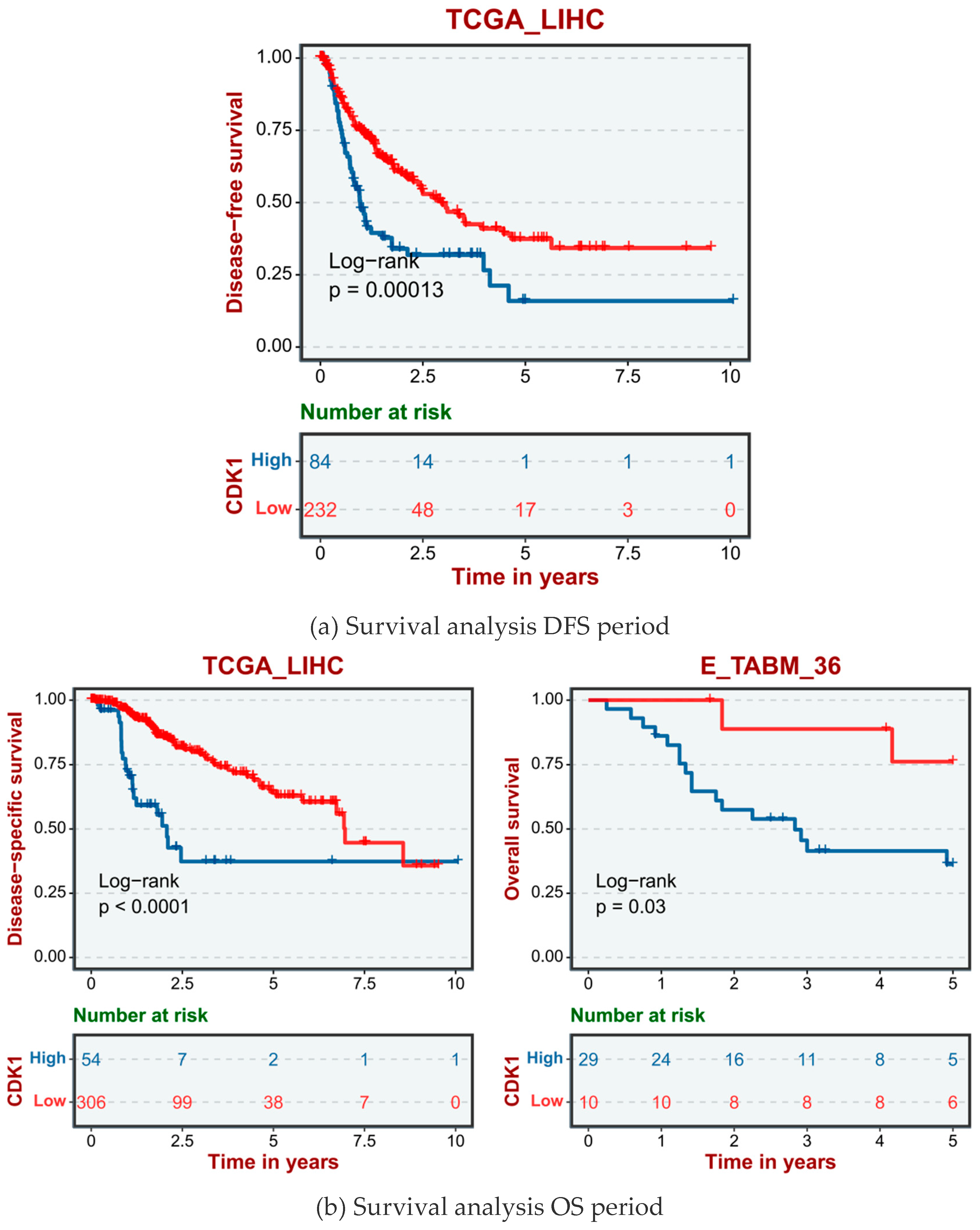
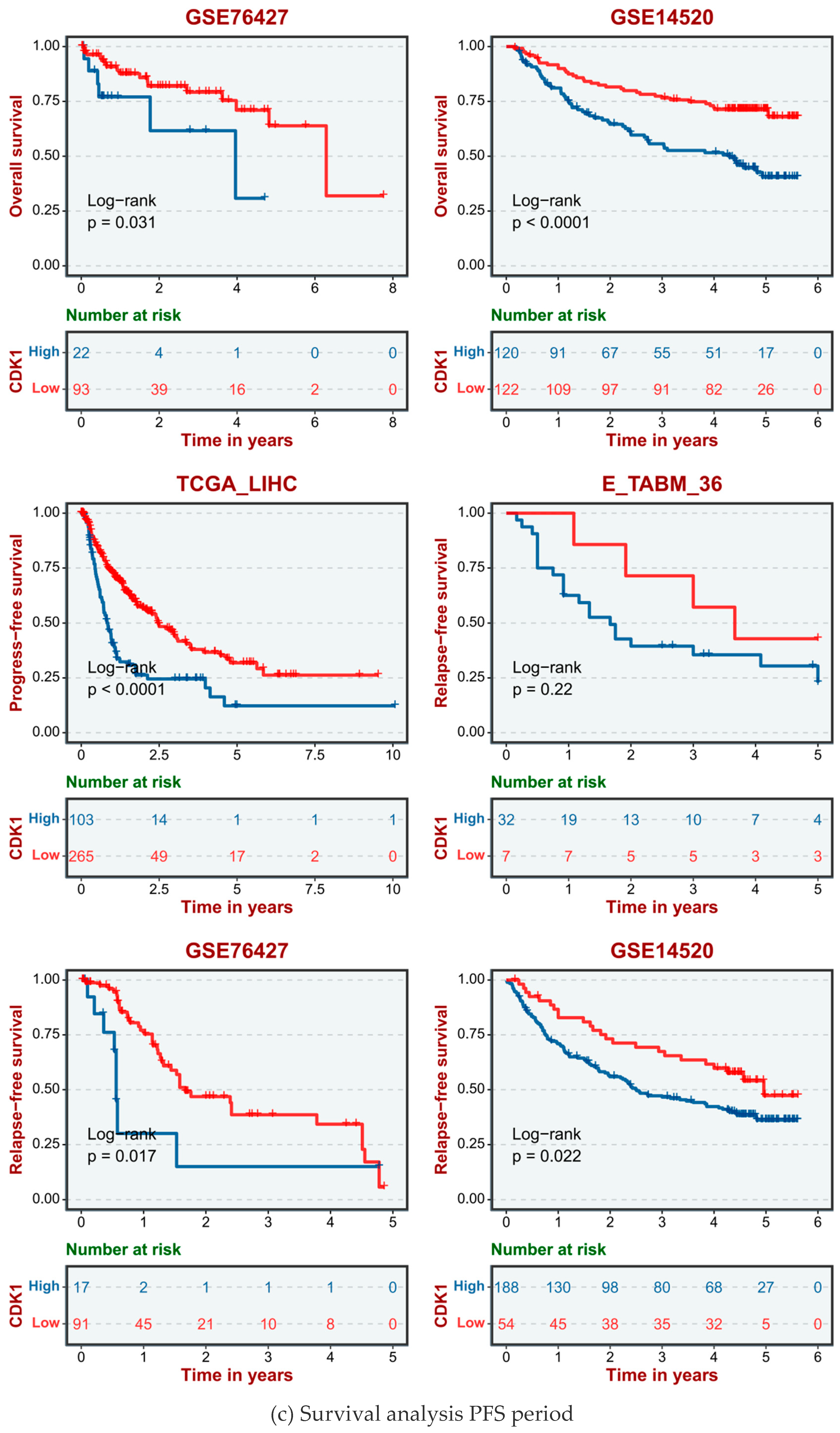
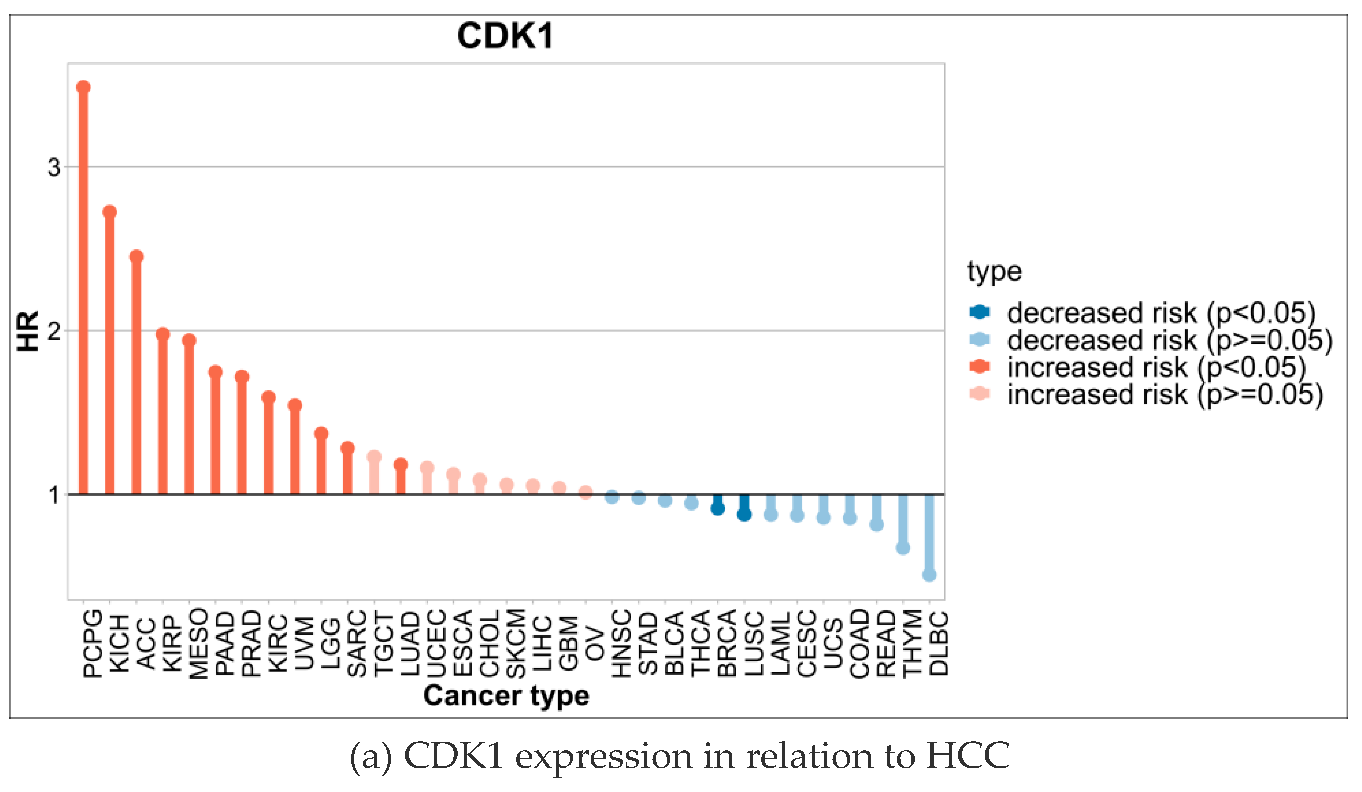
2.4. High Expression of CDK1 in HCC Cells Significantly Reduced the Survival Rate of Hepatocellular Carcinoma Patients

The human data sets and GSE database from TCGA data included TCGA_LIHC, E-TAMB_36, GSE14520, and GSE76427. According to the different expression levels of the CDK1 gene in human HCC cells, hepatocellular carcinoma cases were divided into a high CDK1 expression group and a low CDK1 expression group. The disease-free survival (DFS, Figure 4a), overall survival (OS, Figure 4b), and progression-free survival (PFS, Figure 4c) showed that the survival cycle of normal hepatocytes with high CDK1 gene expression was lower than that of normal hepatocytes with low CDK1 expression for a long time.

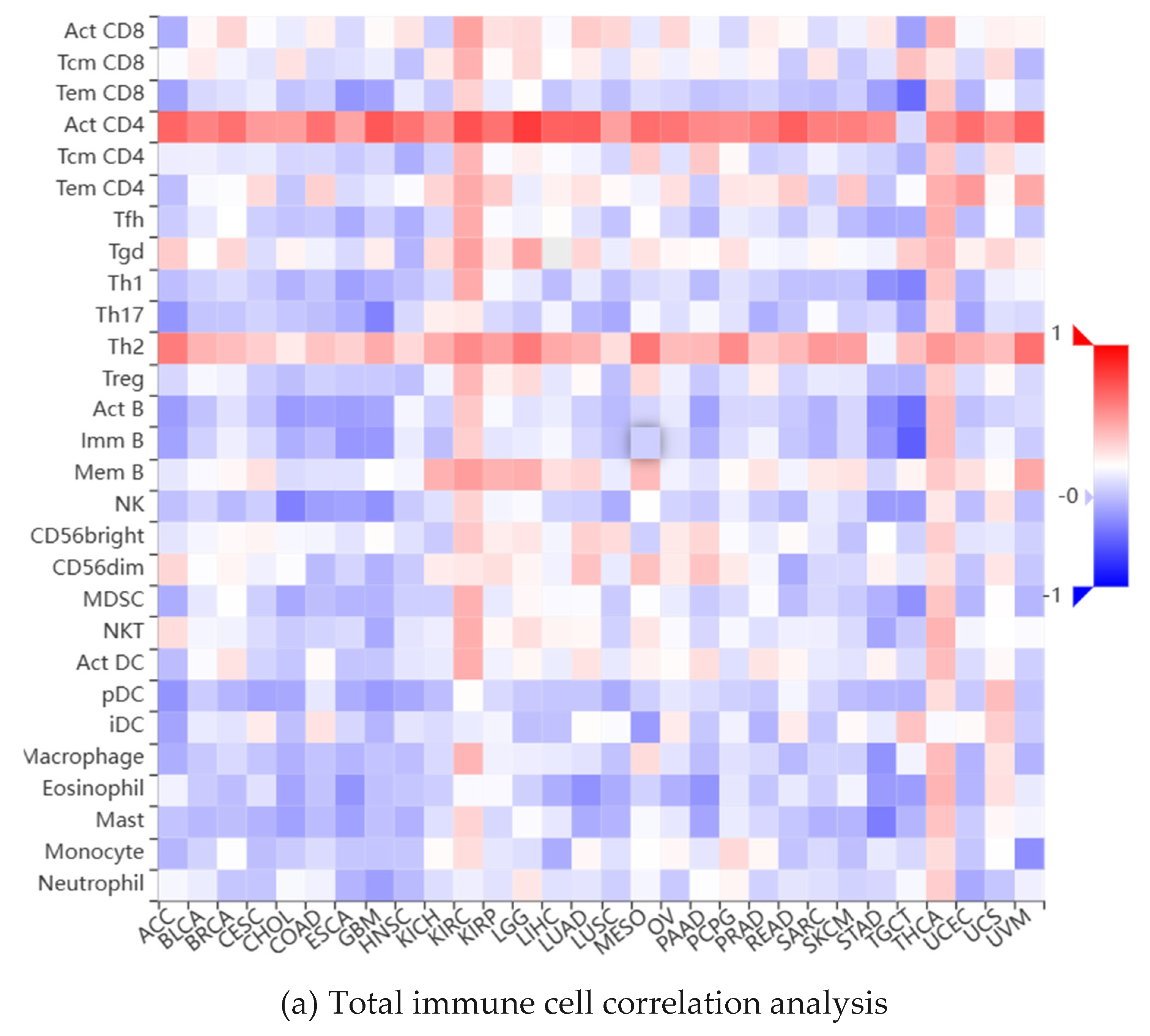
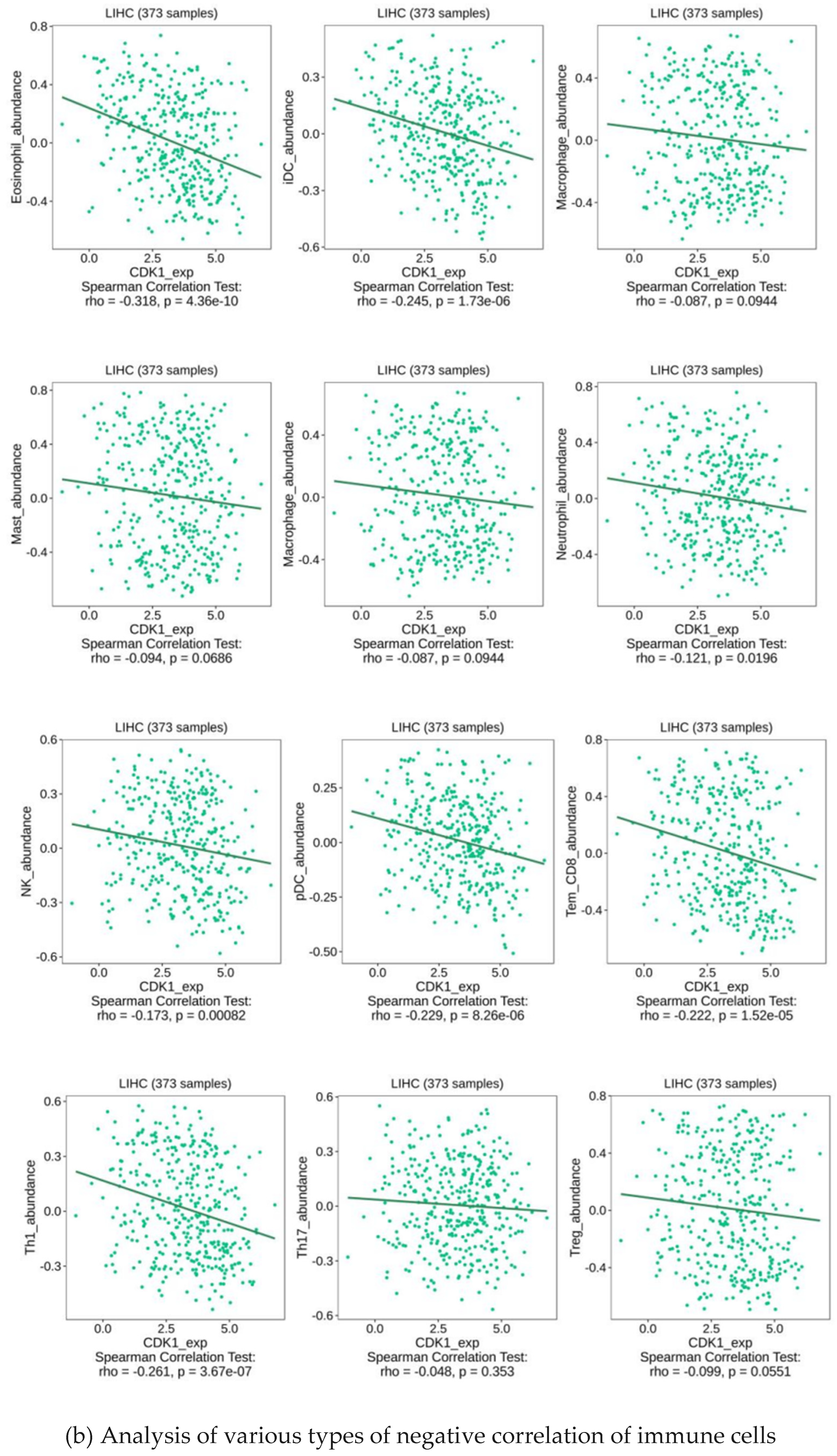
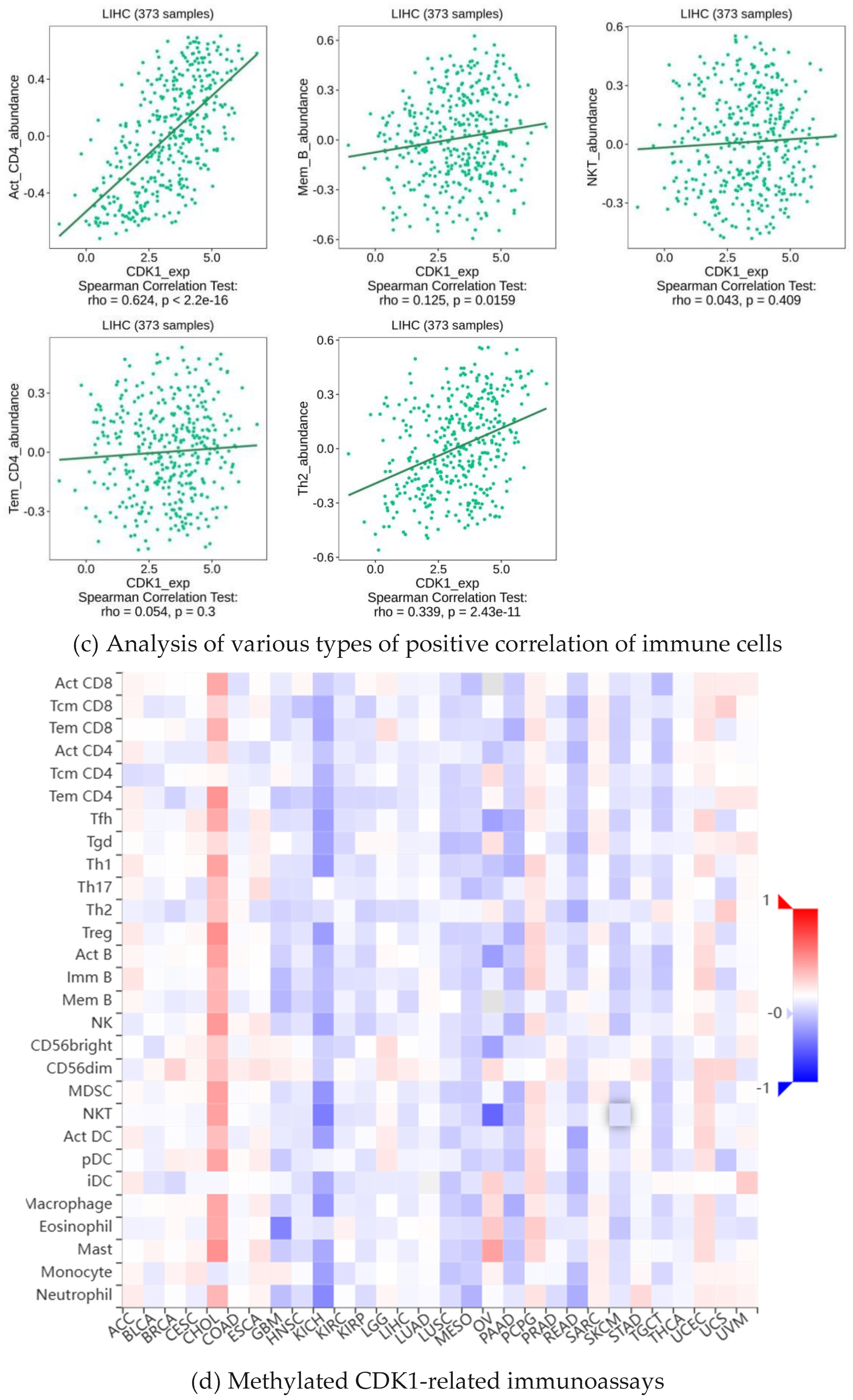
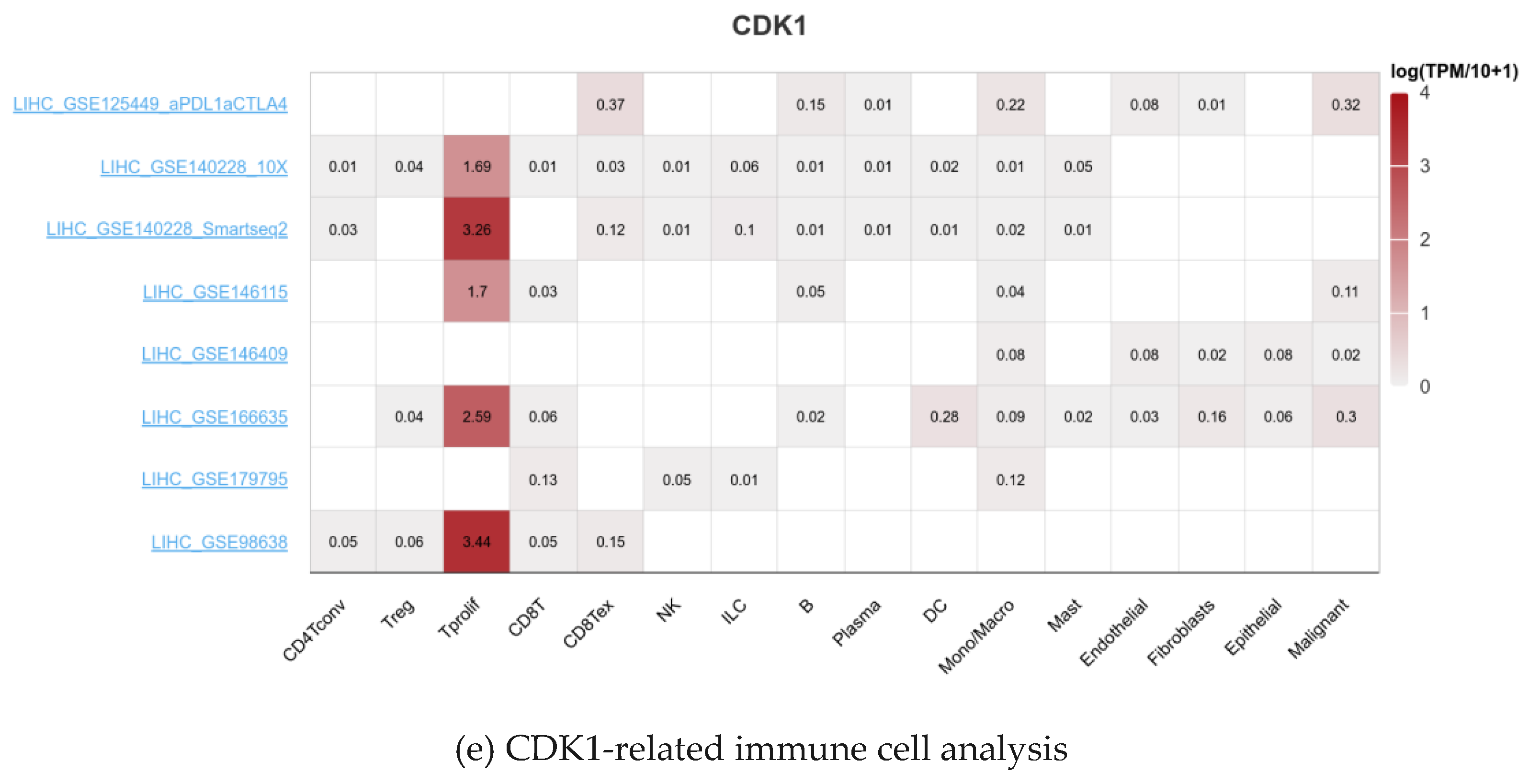
2.5. Analysis of Immune Infiltration of CDK1

As a key factor in cell cycle regulation, CDK1 has a regulatory effect on the tumor immune microenvironment, and its abnormal expression may affect the proliferation and apoptosis of HCC cells. Using multiple algorithms, including CIBERSORT, CIBERSORT_ABS, EPIC, ESTIMATE, MCPcounter, Quantiseq, TIMER, and xCell, we investigated the relationship between the CDK1 gene and a variety of immune cells (Figure 5a). Through analysis, we found that the expression between the CDK1 gene and a variety of activated T cells, activated dendritic cells, neutrophils, macrophages, activated B cells, monocytes, GMP, and other immune-related cells has a strong negative correlation (Figure 5b) and the expression between the CDK1 gene and a variety of immune cells, including Tem CD4 cells, CD4 + T cells, memory B cells, NKT cells, and Th 2 cells, has a positive correlation (Figure 5c). By analyzing the immune cell infiltration of the methylated CDK1 gene, we found that the expression of the methylated CDK1 gene had a weak correlation with multiple immune cells. In other words, inhibitors of the CDK1 gene could reduce the inhibition of the gene on the immune effect (Figure 5d). Using the GEO database, we obtained the hepatocellular carcinoma-related data sets, including GSE125449 aPDL1aCTLA4, GSE140228 10X, GsE140228 Smartseq2, GSE146115, GSE146409, GSE166635, GSE179795, and GSE98638. By constructing visual heat maps, we found that the CDK1 gene was highly expressed in Tprolif cells (Figure 5e). Tprolif cells are a class of lymphocytes with immunomodulatory functions, which have high value-added ability, can divide and multiply rapidly, and play an important role in the immune system. Such cells can produce and release a variety of immune regulators, which can participate in the body’s immune response and participate in the maintenance of immune balance. So, we speculated that CDK1 inhibitors can reduce the expression of the CDK1 gene in Tprolif cells, promote the proliferation of Tprolif cells, and strengthen the immunosuppressive effect of immune cells on hepatocellular carcinoma.

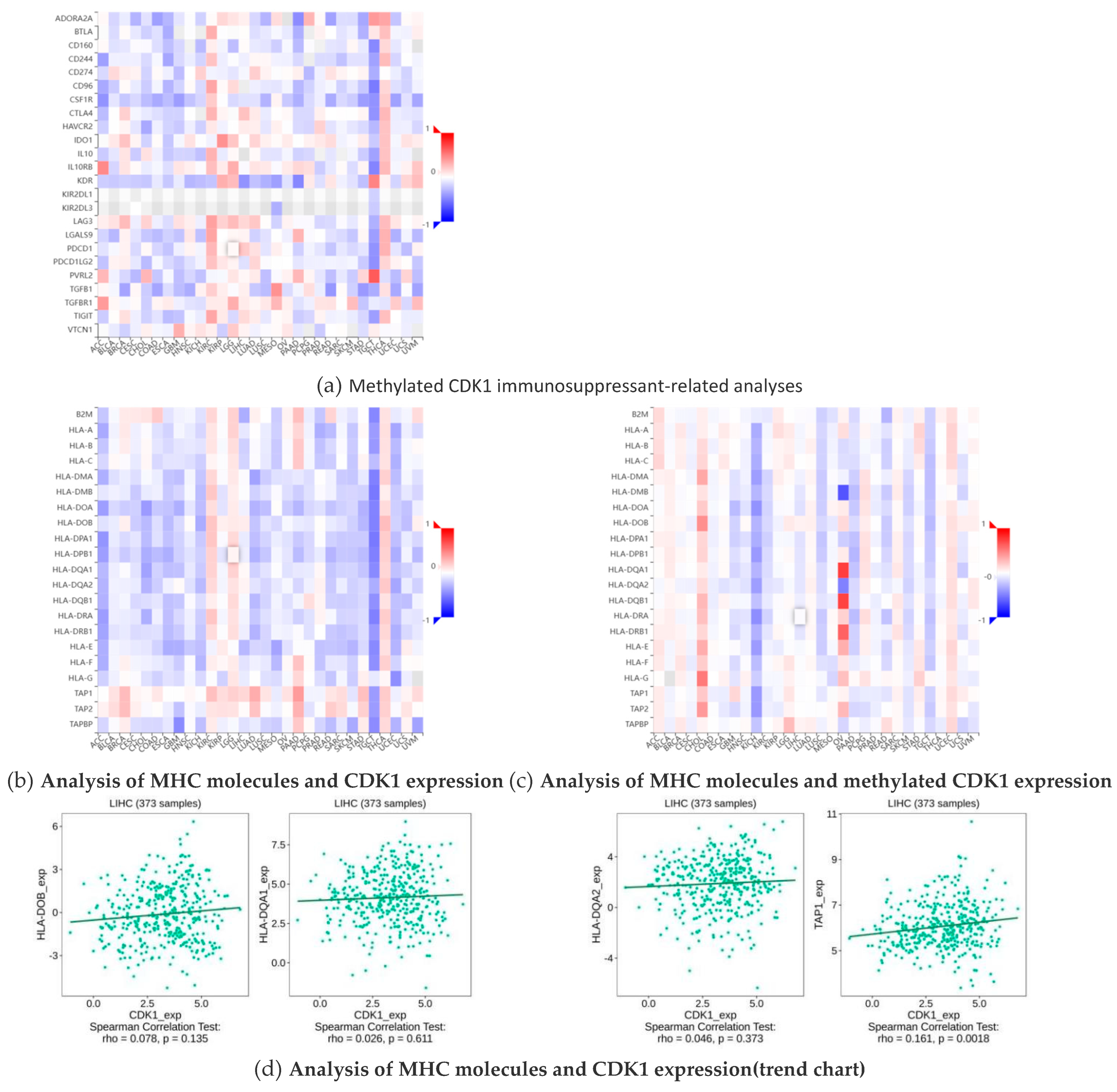
2.6. Methylation of CDK1 Disables Immunosuppressants in Hepatocellular Carcinoma

Immunosuppressants have an inhibitory effect on the immune response of the body, which reduces the autoimmune response by inhibiting the proliferation and function of cells associated with the immune response. MHC (major histocompatibility complex) plays a crucial role in the immune system, not only determining the host tissue compatibility, but also is closely related to the host immune response and immune regulation. Through bioinformatics and related statistical methods, we visually demonstrate the different effects of immunosuppressants and immunostimulants on CDK1 and its methylated forms in hepatocellular carcinoma (HCC). By comparing the data on hepatocellular carcinoma obtained from public databases, we found that the expression of CDK1 contributed to the effect of multiple immunosuppressants, while methylated CDK1 showed the opposite trend, namely, the effect of immunosuppressants. This difference is clearly reflected in the cohesion heatmap (Figure 6a). In addition, we also observed that the expression changes in the MHC molecules TAP 1, HLA-DOB, HLA-DQA 1, and HLA-DQA 2 showed some positive correlation with CDK1 expression (Figure 6b,d) and negatively correlated with methylated CDK1 expression (Figure 6c). This further suggests a synergistic role of these molecules in immune regulation and tumorigenesis and development.

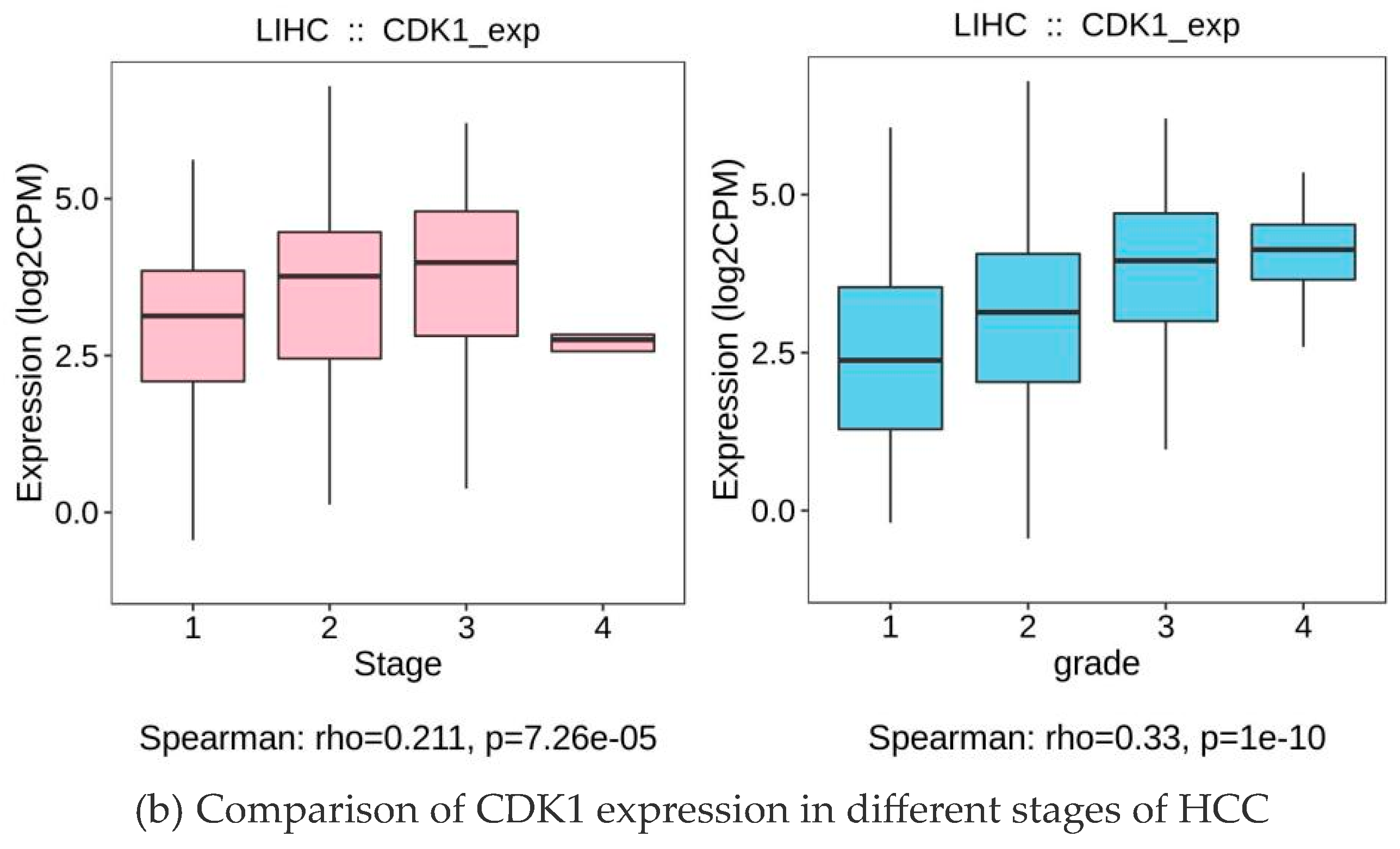
2.7. CDK1 Expression Was Positively Correlated with the Grade and Staging of the Tumors

Through the analysis, the expression level of CDK1 was closely related to HCC (Figure 7a). Specifically, in early-stage HCC patients, CDK1 was higher than in normal liver tissue but was lower compared to middle and late-stage patients. With tumor progression to the metaphase, the expression level of CDK1 increased significantly, suggesting that CDK1 may be involved in the malignant progression of HCC. CDCC 1 expression reached its highest levels in advanced HCC patients and was significantly associated with poor prognosis in patients (Figure 7b). Furthermore, according to the Barcelona staging (BCLC) and TNM staging systems, we found that high CDK1 expression was significantly associated with more advanced stages (e.g., stage BCLC C or TNM III/IV) (p < 0.05).

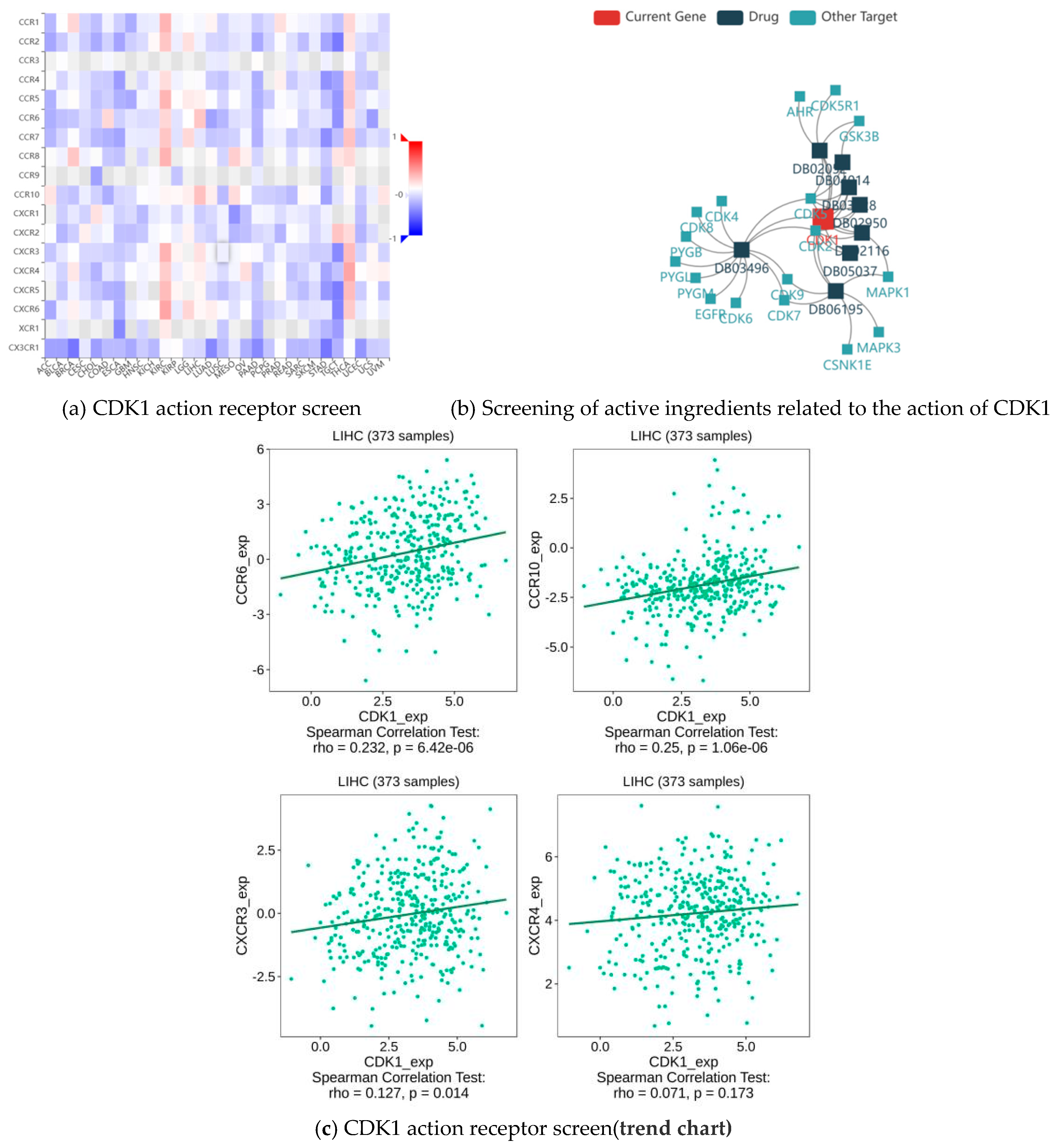
2.8. CDK1 Action Receptor and Active Component of HCC Treatment

The analysis shows that the top four receptors for CDK1 were CCR 6, CCR 10, CXCR 3, and CXCR 4 (Figure 8a,c). CCR 6, CCR 10, CXCR 3, and CXCR 4 are important members of the chemokine receptor family, which play important roles in cell migration, immune response, inflammation, as well as tumor growth and metastasis. They do not directly act as receptors for CDK1, but they bind to chemokines through different signaling pathways. Through drug screening, we identified the active components of indirubin-3′-monoxime, olomoucine, hymenialdisine, alvocidib, alsterpaullone, and seliciclib 6 (Figure 8b, Table 3), all of which are small molecule drugs and closely related to CDK1.

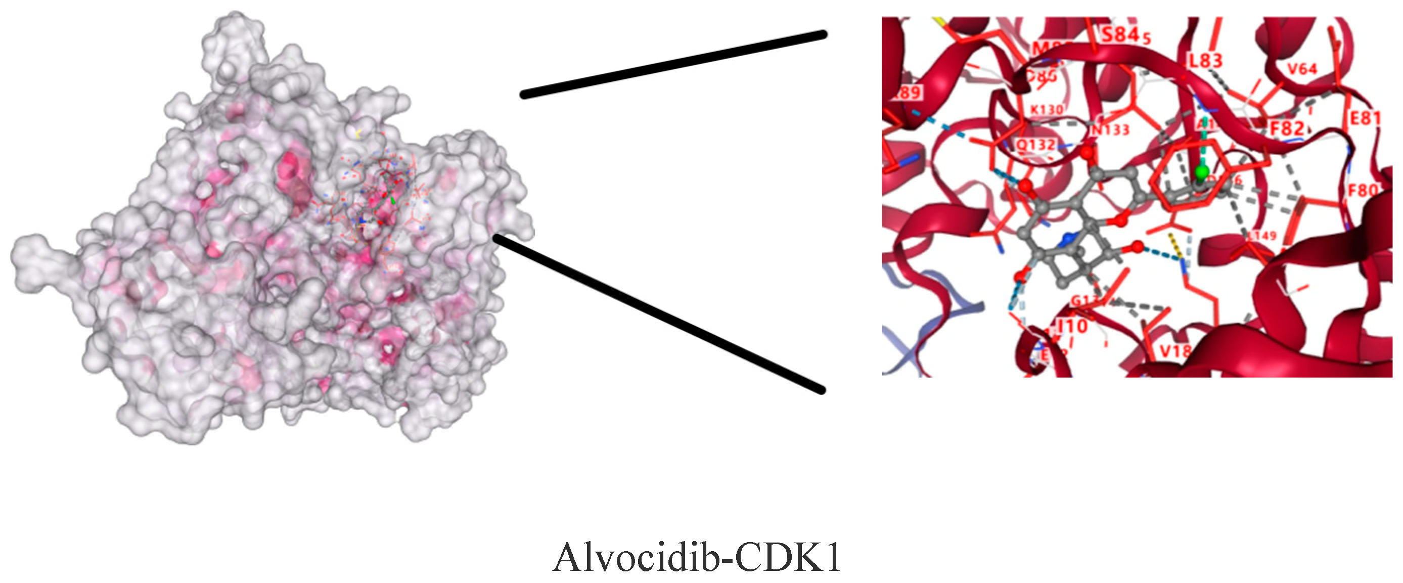
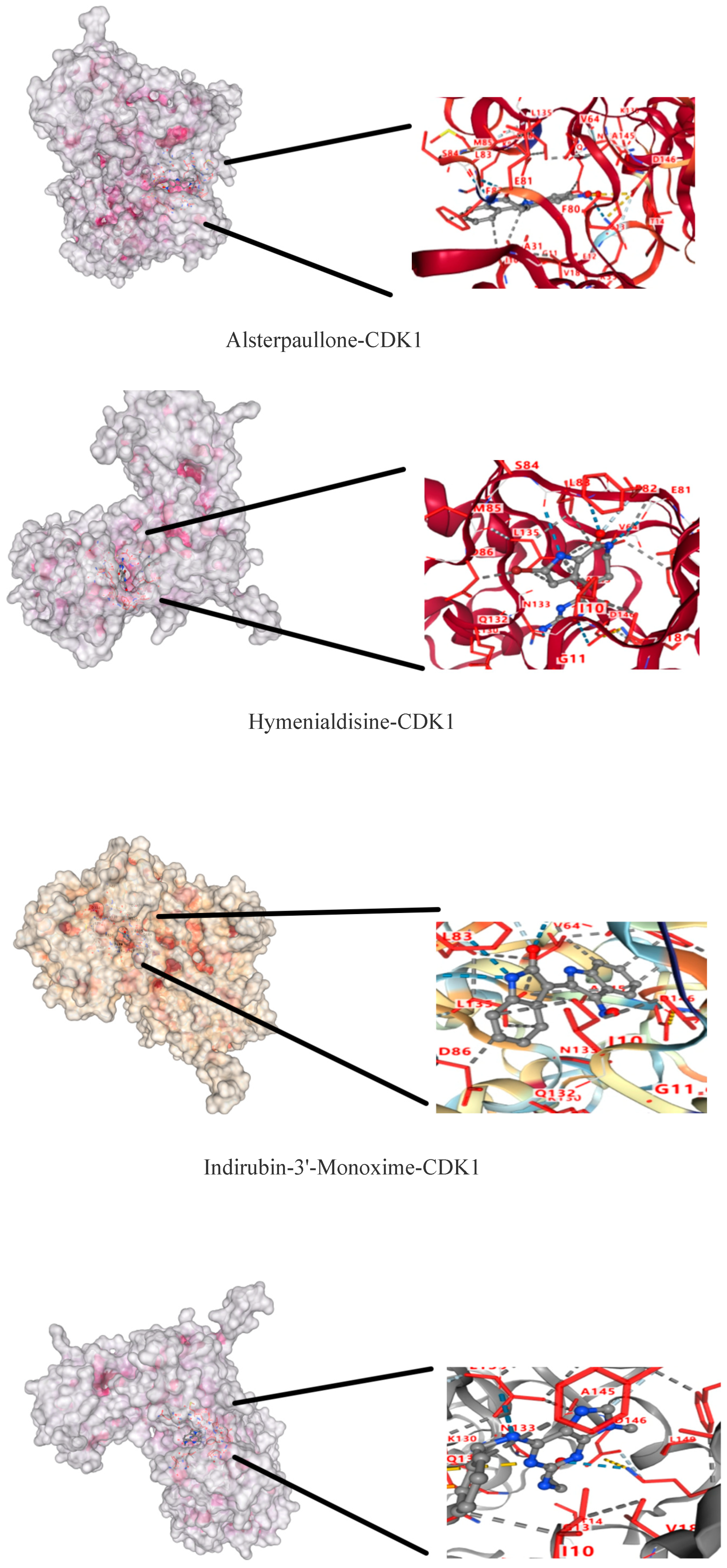
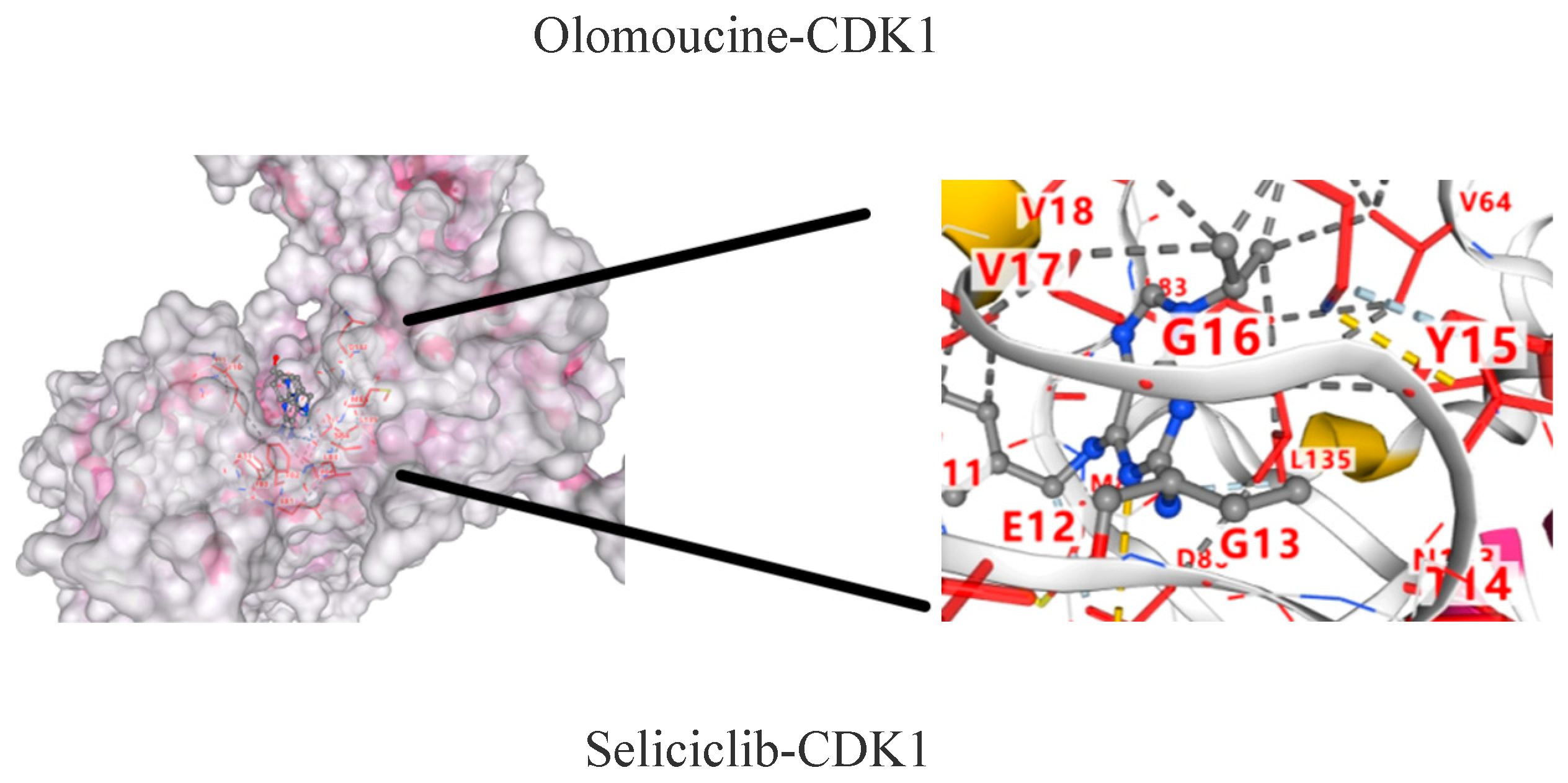
2.9. Molecular Docking Results of CDK1 and Active Components

The molecular dockings of indirubin-3′-monoxime, olomoucine, hymenialdisine, alvocidib, alsterpaullone, seliciclib and CDK1 are shown in Figure 9. The main active components showed different affinities with the core targets, and the top three were alvocidib-CDK1, seliciclib-CDK1, and alsterpaullone-CDK1 with binding energies of −8.7, −8.0, and −7.8kJ/mol, respectively (Table 4). It can be seen in the 3D diagram that the small molecule active components are embedded in the single molecule target protein, and the small molecule target CDK1 and the active components of the macromolecule protein bind closely through hydrogen bonding in the form of sidechain acceptor or backbone acceptor, showing good affinity.

3. Discussion

In this study, we deeply explored the biological role of CDK1 in liver fibrosis and liver CC, revealing its central position in cell cycle regulation and its potential value as a therapeutic target. Through comprehensive analysis of differential gene expression data, pathway enrichment analysis, gene survival analysis, and immune infiltration data, we found that the expression level of CDK1 is closely related to the progression of liver fibrosis; this finding not only provides a new perspective for understanding the pathogenesis of liver cancer, but also opened up a new way for the development of targeted treatment strategies [20].
The high expression of CDK1, a key regulator of cell cycle G2/M turnover, in liver fibrosis HCC suggests its important role in promoting tumor cell proliferation and inhibiting apoptosis [16]. Our data analysis showed that high expression of CDK1 was significantly associated with shorter survival of HCC patients, and this result was validated in multiple independent datasets, enhancing its reliability as a prognostic marker [21,22]. Using immune infiltration analysis, we observed a significant association of CDK1 expression with the infiltration pattern of immune cells in the tumor microenvironment [23], suggesting that CDK1 may affect tumor growth and spread [24] by regulating the immune response. Further bioinformatics analysis revealed multiple key signaling pathways regulated by CDK1, including cell cycle regulation, p53 signaling pathway, and ECM protein and receptor interaction, and the abnormal activation of these pathways was closely related to the development of HCC. Our results not only confirm the multiple biological functions of CDK1 in liver fibrosis and liver CC, but also provide a rationale for CDK1-based targeted therapy. Small-molecule inhibitors against CDK1 have shown antitumor activity in multiple cancer models, providing new hope for the treatment of HCC [25]. When exploring the clinical application of CDK1 as a biomarker for liver fibrosis, we must first recognize its central role in cell cycle regulation, which is not only widely recognized in basic biological studies, but also gradually shows its importance in clinical pathology [26]. The finding that CDK1 expression levels are closely associated with the prognosis of HCC patients has been validated in multiple independent studies, including gene expression profiling [27] in over 500 HCC samples, showing a significant correlation of higher CDK1 expression with worse survival [19]. Further, the expression pattern of CDK1 not only provides potential biomarkers for the early diagnosis of HCC, but also provides new perspectives on the development of personalized treatment strategies. Monitoring of CDK1 mRNA levels by real-time quantitative PCR technology can provide clinicians with real-time information about tumor progression and treatment response [28], thus achieving more precise treatment regimen adjustments. Development and clinical trials of CDK1 inhibitors have also shown promising results [29], especially when used in combination with traditional chemotherapeutic agents, that significantly improve therapeutic efficacy and reduce side effects [30,31]. In the field of immunotherapy, CDK1 expression is closely associated with immune cell infiltration patterns in the tumor microenvironment [32], and this finding provides a rationale for the development of novel therapeutic strategies based on immune checkpoint inhibitors. By analyzing the relationship between CDK1 expression and tumor-infiltrating lymphocytes (TILs) [33], researchers can more accurately predict patients’ responses to immunotherapy, thus achieving more accurate individualized immunotherapy. Despite the great potential of CDK1 as a biomarker, its clinical translation still faces many challenges [34,35,36]. How to standardize the detection method of CDK1 to ensure its consistency and reliability in different laboratories and clinical settings is an urgent problem. The dynamic changes in CDK1 expression and its relationship with tumor heterogeneity also require further studies to clarify the tumor heterogeneity.
CDK1 plays an important role in the activation and proliferation of hepatic stellate cells (HSCs) by regulating liver fibrosis [37]. It was shown that inhibition of CDK1 activity can downregulate the proliferation of HSCs and reduce ECM synthesis, thereby inhibiting the progression of liver fibrosis [7,32,38]. Although the direct application of CDK1 inhibitors in the treatment of liver fibrosis is still under research, some indirect evidence shows that TCM liver recovery can be significantly inhibited by regulating the CDK1-related signaling pathway (such as MAPKAP-1 signaling pathway), which may be related to the indirect regulation of CDK1 [39].
The direct application of CDK1 inhibitors in the treatment of cancer is being gradually explored. Several studies have shown that CDK1 inhibitors were able to significantly inhibit the growth and metastasis of HCC cells and improve the survival rate of patients [29]. However, due to the complexity and heterogeneity of HCC, the therapeutic efficacy of a single CDK1 inhibitor is limited, and it usually needs to be combined with other therapies (for example, immunotherapy, targeted therapy, etc.) to achieve optimal efficacy [40]. Different studies have some differences in the mechanism of CDK1 treatment for liver fibrosis and liver cancer. Some studies have focused on the direct inhibition of CDK1 on HSCs or HCC cell proliferation, while others focus on the regulation of CDK1-related signaling pathways. Moreover, different studies also showed differences in the choice and dosage of CDK1 inhibitors, which may lead to differences in the results of efficacy assessment. Although CDK1 has demonstrated some potential in treating liver fibrosis and liver cancer, some limitations of the current study remain. The complexity of CDK1 in cell cycle regulation makes its single inhibitory effect limited. Moreover, the specificity and safety of CDK1 inhibitors still needs further validation. Meanwhile, the conversion efficiency between preclinical studies and clinical trials needs to be improved to accelerate the clinical application of CDK1 inhibitors in the treatment of liver fibrosis and liver cancer.
For the bioinformatics, survival analysis, and immune infiltration analysis methods used in this study, we first focused on the complexity of bioinformatics and the analysis and its profound impact on the results. Bioinformatics tools, such as the Gene Expression Comprehensive Analysis (GEO) database and Gene Ontology (GO) analysis, provide us with a macro-to-micro perspective, revealing the key role of CDK1 in liver fibrosis and liver cancer. Survival analysis, especially the application of the Kaplan survival curves, provided us with a direct link between CDK1 expression and patient survival [41]. The intuition and statistical rigor of bioinformatics technology are able to provide strong support for clinical decision-making [42]. Immune infiltration analysis, through algorithms, such as CIBERSORT and TIMER, allowed us to gain insight into the role of CDK1 in the tumor microenvironment, especially the influence on immune cell infiltration related to liver fibrosis [43]. This approach is able to provide detailed information about the tumor immune microenvironment for the development of personalized treatment strategies [44,45]. This study mainly relied on gene expression data in public databases, which may not fully reflect the complexity of clinical reality [46]. The immune cell infiltration situation in the tumor microenvironment may be influenced by multiple factors, and our analysis may fail to fully consider the interaction of these factors. Therefore, future studies should combine more clinical data and experimental validation to provide a more comprehensive understanding of the mechanism of CDK1 action in liver fibrosis and HCC.
The potential of CDK1 as a therapeutic biomarker for liver fibrosis and HCC should be further exploited and validated in future studies [47]. The interdisciplinary research approach will combine molecular biology, cell biology, bioinformatics, modern biological technology, and clinical medicine, to deeply analyze the specific mechanism of action of CDK1 in the process of liver fibrosis. For example, by precise editing of the CDK1 gene [48] by CRISPR-Cas 9 technology, researchers can observe direct changes in cell cycle regulation to more accurately assess CDK1 function; combined with high-throughput sequencing technology [49], allowing a comprehensive analysis of other key genes and proteins in the CDK1 regulatory network. These new modern biotechnologies will be even more informative for developing new therapeutic strategies.
In terms of clinical application, the expression level of CDK1 can be used as a basis for personalized treatment of patients with liver fibrosis. Through techniques such as real-time PCR [50], physicians can evaluate the patients, CDK1 expression status before treatment, and then choose the most appropriate drugs and treatment options. For patients with high CDK1 expression, the use of drugs that inhibit cell cycle progression, such as CDK1 inhibitors, may need to be considered to achieve better therapeutic effects. Meanwhile, long-term clinical follow-up studies will help to evaluate the accuracy and reliability of CDK1 as a prognostic marker, which is important for improving the survival rate and quality of life of patients. Research combined with immunotherapy will also be an important direction in the future. CDK1 not only plays a role in cell cycle regulation, but may also influence immune cell infiltration and activity in the tumor microenvironment.
The use of bioinformatics to study CDK1 as a therapeutic target for liver fibrosis and hepatocellular carcinoma has important potential, but there are many limitations. The histological data of hepatocellular carcinoma and liver fibrosis in public databases (e.g., TCGA, GEO) may come from different populations, different pathological stages, or different assay platforms, leading to batch effects or confounding factors interfering with the results of the analyses. And some datasets lack detailed clinicopathological parameters (e.g., treatment history, cirrhosis background, and viral hepatitis infection status), which limits the precise resolution of the CDK1 mechanism of action. During the multi-stage process of liver fibrosis to hepatocellular carcinoma transformation, the expression and function of CDK1 may change dynamically, and it is difficult for static histological data to reflect such temporal regulation. In addition, CDK1 not only regulates the cell cycle (G2/M phase), but also may cross-talk with other pathways (e.g., p53, Wnt/β-catenin, and EMT) through phosphorylation, and it is difficult for bioinformatics tools (e.g., STRING and KEGG) to comprehensively analyze its dynamic regulatory network. Meanwhile, CDK1 may be involved in non-cell cycle-related functions, such as DNA damage repair and metabolic reprogramming, and traditional enrichment analyses, that may miss these potential mechanisms. In molecular docking, CDK family members (e.g., CDK2/4/6) are highly similar in structure, and small molecules targeting CDK1 may lead to off-target toxicity due to insufficient selectivity, whereas computational simulations (e.g., molecular docking) are difficult to fully predict the actual binding specificity. In liver fibrosis, CDK1 may promote fibrosis through the activation of hepatic stellate cells (HSCs), whereas in hepatocellular carcinoma, it may drive tumor cell proliferation, but existing analyses often ignore cell-type specificity (e.g., without integrating single-cell sequencing data). In terms of animal experiments, bioinformatics-screened CDK1-related targets or inhibitors (e.g., dinaciclib) need to be validated by in vitro/in vivo experiments, but some of the studies are only at the computational prediction stage and lack functional (e.g., knockout, organoid models) or pharmacodynamic validation. And the histological data based on mouse models may have species differences with human diseases, affecting the reliability of target validation.
CDK1, as a key enzyme in cell cycle regulation, shows promising applications in the treatment of liver fibrosis and hepatocellular carcinoma. However, many challenges need to be overcome to realize its clinical applications. Future studies should further develop highly efficient and low-toxicity CDK1 inhibitors and strengthen the interface between preclinical studies and clinical trials to promote the widespread application of CDK1 inhibitors in the treatment of liver fibrosis and hepatocellular carcinoma. It also needs to be combined with multi-omics dynamic analysis. Variable modulator design and precise delivery methods, and the interdisciplinary collaboration of computational biology-experimental oncology-clinical medicine to promote the development of CDK1 targets from mechanism research to clinical translation.

4. Materials and Method

In this study, we integrated relevant methods, including differential gene analysis, protein interaction analysis, enrichment analysis, survival analysis, immunohistology-related analysis, and molecular docking simulation, combined with multiple public databases and RNA-seq data, to predict new targets for the treatment of hepatic fibrosis, as well as to study the relevant mechanism of action of the new targets, predict the relevant active ingredients for the treatment, and validate the results by combining them with molecular docking simulations (Figure 10).

4.1. Acquisition of RNA-Seq Data of Activated Hepatic Stellate Cells (HSCs)

To investigate differential gene expression in growing and senescent-activated hepatic stellate cells (HSCs), GSE11954 gene expression profiles were downloaded from the Gene Expression Omnibus (GEO, https://www.ncbi.nlm.nih.gov/geo, accessed on 10 August 2024) database.

4.2. The Differential Gene Expression Analysis of the Microarray Data

GEO2R (https://www.ncbi.nlm.nih.gov/geo/geo2r/, accessed on 10 August 2024) is a tool for further differential analysis of microarray data in the GEO database, which we can use to compare two or more groups of samples from the GEO dataset to obtain differentially expressed genes. Analysis of differentially expressed genes (DEGs) of human-activated liver stellate cells (HSCs) using GEO2R was performed. The targets obtained from the analysis were standardized, and the adjusted p-values < 0.05, |log2FC|> 2 were selected for DEGs.

4.3. Protein Interaction (PPI) Networks of DEGs

The STRING database (https://string-db.org/, accessed on 11 August 2024) is a database that searches for possible potential interactions between encoded proteins and builds protein interaction networks. DEGs were submitted to the STRING database, the species was set as “Homo sapiens”, the minimum interaction threshold was set to “highest confidence > 0.4”, and the disconnected free target site was cleared to obtain the PPI protein interaction network. Then, the maximum group centrality (MCC) algorithm of Cytoscape3.8.0 and Cyto NCA (2024.8.11) plug-in can simultaneously set “Degree value” and “Betweenness value” as benchmark parameters to conduct topological analysis of the PPI network and screen key DEGs. Finally, we selected the first genes with Degree and Betweenness values in the PPI network and identified them as the key genes of aging human-activated hepatic stellate cells (HSCs) to inhibit liver fibrosis, that is, in the treatment of liver fibrosis disease.

4.4. Enrichment Analysis and Functional Annotation of the 2.4 DEGs

The obtained DEGs were imported into the DAVID (https://davidbioinformatics.nih.gov/, accessed on 12 August 2024) database to obtain relevant biological information. The gene ontology (GO) enrichment analysis and the Kyoto Gene-Genome Encyclopedia (KEGG) enrichment analysis were performed after the screening of the obtained biological information. Through bioinformatics analysis, we obtained information about the biological process of DEGs (biological process, BP), the cellular location of DEGs products (cellular component, CC), the activity of DEGs products at the molecular level (molecular function, MF) and the action pathway of DEGs, and visualized the analysis results.

4.5. Analysis of Survival Prognosis Related to CDK1

In order to study the prognostic relationship between the CDK1 gene and liver fibrosis, we used the TCGA database (https://portal.gdc.cancer.gov, accessed on 13 August 2024) and GEO database (https://www.ncbi.nlm.nih.gov/geo/, accessed on 13 August 2024), combined with Kaplan–Meier plotter (http://kmplot.com/analysis/, accessed on 14 August 2024), to analyze the key DEG data and draw the survival curve.

4.6. Analysis of Immune Cell Infiltration

The relationship between CDK1 gene expression in Homa cells and each immune cell was determined by the Web server from the TIMER database (https://cistrome.shinyapps.io/timer/, accessed on 16 August 2024). A variety of cells associated with tumor immunity were evaluated for immune infiltration, were analyzed for their immunomodulatory effects, and results were visualized. The GEO database was used to obtain various data sets on hepatocellular carcinoma and make a visual heat map to analyze the expression levels of target genes in different cell types in different data sets in hepatocellular carcinoma.

4.7. Analysis of the Interaction of CDK1 with Immunosuppressants and MHC Molecules

Sequence, expression profiles, and interaction data of CDK1 and its potential chemokines and receptors were downloaded from public databases, including NCBI (National Center for Biotechnology Information), Ensembl, and UniProt, to analyze the expression of CDK1 chemokines and receptors. The raw data were cleaned and analyzed by the t-test and negative binomial distribution analysis (DESeq2, edgeR) to study the application of CDK1 and immune regulators (immunosuppressants, MHC molecules) in human cancer, and the results were visualized as cluster heatmaps.

4.8. Association Between CDK1 Expression and Stage Grade of Hepatocellular Carcinoma in Human

Gene expression data from patients with hepatocellular carcinoma (HCC) were obtained from the TCGA database, while other relevant RNA high-throughput sequencing data were downloaded from the GEO database to validate the discovery of the TCGA data. The expression differences in CDK1 gene in different pathological stages and tumor grades were analyzed using the UALCAN platform, and the results were visualized.

4.9. Selection of Receptors and Drugs for CDK1 Action in Human Hepatocellular Carcinoma

The rapid search of the CDK1 gene from the TISIDB database yielded the key receptors for CDK1 action on HCC and the active components for the treatment of HCC.

4.10. Molecular Docking and Kinetic Simulation for the Validation of the Effective Components for the Treatment of Hepatocellular Carcinoma

In the PubChem database (pubchem.ncbi.nlm.nih.gov, accessed on 22 September 2024) and the PDB database (www.rcsb.org, accessed on 22 September 2024), the 3D structure of all the active components and the protein crystal structure of CDK1 were obtained, and the above components and targets were matched one-to-one by molecular docking analysis on the CB-Dock 2 platform (http://cadd.labshare.cn, accessed on 22 September 2024).

5. Conclusions

In conclusion, in this study, we deeply explored the use of the expression pattern, biological function, and its possibility as a potential biomarker of CDK1 in liver fibrosis and liver CC. The combination of bioinformatics analysis, survival analysis and immune infiltration analysis has revealed the key role of CDK1 in liver fibrosis and liver cancer. The expression level of CDK1 may not only serve as an independent predictor of HCC prognosis, but may also provide new ideas for molecular typing and personalized treatment of HCC. This finding not only enriches our understanding of the molecular mechanisms of HCC, but also provides new perspectives for future clinical applications.
Further, this study not only deepens our understanding of the role of CDK1 in liver fibrosis HCC at the theoretical level, but also provides new tools and strategies for the precise treatment and prognosis evaluation of HCC at the practical level. The transformation and application of these research results are expected to improve the treatment effect and quality of life of liver cancer patients in the future and have important scientific value and clinical significance.

Author Contributions

J.Q.: designed the study, performed bioinformatics analyses, developed computational pipelines, validated results, and drafted manuscript. Z.L.: conceptualization (lead), writing–review and editing (lead). All authors have read and agreed to the published version of the manuscript.

Funding

This study was supported by grant 82170607 from the National Natural Science Foundation of China, grant 2022JJ10037 from Hunan Provincial Natural Science Foundation of China, grant KF2022001, 2022XKQ0205 and 2023JC101 from Hunan Normal University.

Institutional Review Board Statement

This study involved purely bioinformatics-based analyses and computational validation, utilizing publicly available datasets and/or computational tools. No human or animal subjects, tissues, or clinical data were used; therefore, ethical approval was not required.

Informed Consent Statement

Not applicable.

Data Availability Statement

All datasets, analytical scripts, and computational workflows generated or analyzed during this study are publicly available in the GEO, TIMER, TISIDB and TCGA databases. Detailed descriptions of tools and parameters ensure full reproducibility of the results.

Acknowledgments

The author gratefully acknowledges Li Zhuan (Hunan Normal University) for his valuable insights during methodological discussions. Technical support from the Small Molecule Targeted Drug Research and Development Laboratory and the Medical Faculty of Hunan Normal University is also sincerely appreciated.

Conflicts of Interest

The author declares no competing interests, financial or otherwise, that could influence the objectivity of this work.

References

  1. El-Serag, H.B. Epidemiology of viral hepatitis and hepatocellular carcinoma. Gastroenterology 2012, 142, 1264–1273.e1. [Google Scholar] [CrossRef] [PubMed] [PubMed Central]
  2. Tang, A.; Hallouch, O.; Chernyak, V.; Kamaya, A.; Sirlin, C.B. Epidemiology of hepatocellular carcinoma: Target population for surveillance and diagnosis. Abdom. Radiol. 2018, 43, 13–25. [Google Scholar] [CrossRef] [PubMed]
  3. Forner, A.; Reig, M.; Bruix, J. Hepatocellular carcinoma. Lancet 2018, 391, 1301–1314. [Google Scholar] [CrossRef] [PubMed]
  4. Chidambaranathan-Reghupaty, S.; Fisher, P.B.; Sarkar, D. Hepatocellular carcinoma (HCC): Epidemiology, etiology and molecular classification. Adv. Cancer Res. 2021, 149, 1–61. [Google Scholar] [CrossRef] [PubMed] [PubMed Central]
  5. Kim, E.; Viatour, P. Hepatocellular carcinoma: Old friends and new tricks. Exp. Mol. Med. 2020, 52, 1898–1907. [Google Scholar] [CrossRef] [PubMed] [PubMed Central]
  6. Wu, M.; Liu, Z.; Zhang, A.; Li, N. Identification of key genes and pathways in hepatocellular carcinoma: A preliminary bioinformatics analysis. Medicine 2019, 98, e14287. [Google Scholar] [CrossRef] [PubMed] [PubMed Central]
  7. Aydın, M.M.; Akçalı, K.C. Liver fibrosis. Turk. J. Gastroenterol. 2018, 29, 14–21. [Google Scholar] [CrossRef] [PubMed] [PubMed Central]
  8. Bataller, R.; Brenner, D.A. Liver fibrosis. J. Clin. Investig. 2005, 115, 209–218, Erratum in: J. Clin. Investig. 2005, 115, 1100. [Google Scholar] [CrossRef] [PubMed] [PubMed Central]
  9. Xu, Y.; Zhou, X.; Wang, X.; Jin, Y.; Zhou, L.; Ye, J. Progress of mesenchymal stem cells (MSCs) & MSC-Exosomes combined with drugs intervention in liver fibrosis. Biomed. Pharmacother. 2024, 176, 116848. [Google Scholar] [CrossRef] [PubMed]
  10. Luangmonkong, T.; Parichatikanond, W.; Olinga, P. Targeting collagen homeostasis for the treatment of liver fibrosis: Opportunities and challenges. Biochem. Pharmacol. 2023, 215, 115740. [Google Scholar] [CrossRef] [PubMed]
  11. Poilil Surendran, S.; George Thomas, R.; Moon, M.J.; Jeong, Y.Y. Nanoparticles for the treatment of liver fibrosis. Int. J. Nanomed. 2017, 12, 6997–7006. [Google Scholar] [CrossRef] [PubMed] [PubMed Central]
  12. Wang, Y.; Zhou, J.; Han, J. Analysis of the clinicopathological characteristics of patients with autoimmune hepatitis and drug-induced liver injury. J. Pract. Liver Dis. 2023, 26, 218–221. [Google Scholar]
  13. Yang, Y.; Liu, Q.; Guo, X.; Yuan, Q.; Nian, S.; Kang, P.; Xu, Z.; Li, L.; Ye, Y. Systematic Pan-Cancer Analysis Identifies CDK1 as an Immunological and Prognostic Biomarker. J. Oncol. 2022, 2022, 8115474. [Google Scholar] [CrossRef] [PubMed] [PubMed Central]
  14. Yi, L.; Xu, Q.; Huang, J. Effect of Uine on proliferation and apoptosis of human thyroid cancer cells. Anhui Med. 2021, 12. [Google Scholar]
  15. He, Y.; Xie, J.; Zheng, L.; Guo, C.; Ma, Y. Expression and significance of CDK 1 and AURKA in the serum of patients with HBV-associated hepatocellular carcinoma. J. Clin. Hepatobiliary Dis. 2024, 40, 1390–1396. [Google Scholar]
  16. Ren, L.; Yang, Y.; Li, W.; Zheng, X.; Liu, J.; Li, S.; Yang, H.; Zhang, Y.; Ge, B.; Zhang, S.; et al. CDK1 serves as a therapeutic target of adrenocortical carcinoma via regulating epithelial-mesenchymal transition, G2/M phase transition, and PANoptosis. J. Transl. Med. 2022, 20, 444. [Google Scholar] [CrossRef] [PubMed] [PubMed Central]
  17. Roehlen, N.; Crouchet, E.; Baumert, T.F. Liver Fibrosis: Mechanistic Concepts and Therapeutic Perspectives. Cells 2020, 9, 875. [Google Scholar] [CrossRef] [PubMed] [PubMed Central]
  18. Zhou, Z.; Li, Y.; Hao, H.; Wang, Y.; Zhou, Z.; Wang, Z.; Chu, X. Screening Hub Genes as Prognostic Biomarkers of Hepatocellular Carcinoma by Bioinformatics Analysis. Cell Transplant. 2019, 28 (Suppl. 1), 76S–86S. [Google Scholar] [CrossRef] [PubMed] [PubMed Central]
  19. Wang, L.; Zhang, J. Prognosis of breast cancer based on cox proportional hazards regression model, lasso and survival tree. Stat. Appl. 2018, 7, 99–110. [Google Scholar]
  20. Dhar, D.; Baglieri, J.; Kisseleva, T.; Brenner, D.A. Mechanisms of liver fibrosis and its role in liver cancer. Exp. Biol. Med. 2020, 245, 96–108. [Google Scholar] [CrossRef] [PubMed] [PubMed Central]
  21. Hou, S.; Li, Y.; Wei, Z. Identification of HCC molecular biomarkers based on differential co-expression. Genom. Appl. Biol. 2021, 40, 429–434. [Google Scholar]
  22. Lan, L.; Li, J.; Ren, Q. Evaluation of the value of the novel markers AFP-L3 and PIVKAII in HCC diagnosis. J. Kunming Med. Univ. Kunming Yike Daxue Xuebao 2023, 44, 136–141. [Google Scholar]
  23. Zou, Y.; Ruan, S.; Jin, L.; Chen, Z.; Han, H.; Zhang, Y.; Jian, Z.; Lin, Y.; Shi, N.; Jin, H. CDK1, CCNB1, and CCNB2 are Prognostic Biomarkers and Correlated with Immune Infiltration in Hepatocellular Carcinoma. Med. Sci. Monit. 2020, 26, e925289. [Google Scholar] [CrossRef] [PubMed] [PubMed Central]
  24. Wang, Q.; Bode, A.M.; Zhang, T. Targeting CDK1 in cancer: Mechanisms and implications. NPJ Precis. Oncol. 2023, 7, 58. [Google Scholar] [CrossRef] [PubMed] [PubMed Central]
  25. Sunada, S.; Saito, H.; Zhang, D.; Xu, Z.; Miki, Y. CDK1 inhibitor controls G2/M phase transition and reverses DNA damage sensitivity. Biochem. Biophys. Res. Commun. 2021, 550, 56–61. [Google Scholar] [CrossRef] [PubMed]
  26. Parola, M.; Pinzani, M. Liver fibrosis: Pathophysiology, pathogenetic targets and clinical issues. Mol. Asp. Med. 2019, 65, 37–55. [Google Scholar] [CrossRef] [PubMed]
  27. Wu, L.; Li, W.; Liu, J. Screening and bioinformatics analysis of potential biological gene markers for virus-associated liver cancer based on GEO data. J. Mod. Lab. Med. 2021, 36, 106–110. [Google Scholar]
  28. Ginzinger, D.G. Gene quantification using real-time quantitative PCR: An emerging technology hits the mainstream. Exp. Hematol. 2002, 30, 503–512. [Google Scholar] [CrossRef] [PubMed]
  29. Ye, K.; Zheng, W.; Ye, H.; Yang, L. CDK 1 participates in the development of hepatocellular carcinoma and its value of inhibitor application. Clin. Pharmacol. Ther. China 2021, 26, 1086–1094. [Google Scholar]
  30. Li, J. Kkilling of Paclitaxel and CDK 1 Inhibitor and Molecular Mechanism. Ph.D. Thesis, Guangzhou University of Traditional Chinese Medicine, Guangzhou, China, 2012. [Google Scholar]
  31. Zhou, X.; Gu, D.; Tong, C.; Zeng, J.; Wu, H.; Zhao, X. The therapeutic mechanism of the P on CCl 4 induced liver fibrosis in mice. Shaanxi Tradit. Chin. Med. 2022, 43, 151–156. [Google Scholar]
  32. Jones, M.C.; Askari, J.A.; Humphries, J.D.; Humphries, M.J. Cell adhesion is regulated by CDK1 during the cell cycle. J. Cell Biol. 2018, 217, 3203–3218. [Google Scholar] [CrossRef] [PubMed] [PubMed Central]
  33. Li, C.; Li, J.; Chen, R. Progress in liver fibrosis and the immune microenvironment of liver cancer. J. Fudan Univ. Med. Ed. 2023, 50, 889–896. [Google Scholar]
  34. Dai, J.; Jiang, J. New progress in the diagnosis of tumor markers in hepatocellular carcinoma. Diagn. Theory Pract. 2023, 22, 486–493. [Google Scholar]
  35. Zhang, Y. Hsa_circ_0006091 as a Molecular Marker in Hepatocellular Carcinoma. Ph.D. Thesis, Anhui Medical University, Hefei, China, 2021. [Google Scholar]
  36. Mi, N.; Bai, M.; Gao, L.; Ma, H.; Fu, W.; Lin, Y.; Meng, W. Screening and validation of HCC prognostic markers based on bioinformatics methods. Life Sci. Res. 2022, 26, 538–548. [Google Scholar]
  37. Liao, H.; Ji, F.; Ying, S. CDK1: Beyond cell cycle regulation. Aging 2017, 9, 2465–2466. [Google Scholar] [CrossRef] [PubMed] [PubMed Central]
  38. Li, H.; Yu, S.; Chen, L.; Lai, Z.; Liu, H.; Liu, H.; Shen, C. Effect of mesenchymal stem cells combined with immunosuppressants on immune rejection in a liver transplant rat model. J. Clin. Hepatol. Linchuang Gandanbing Zazhi 2024, 40, 1209–1214. [Google Scholar]
  39. Jia, Y.; Zhao, F.; Zhang, C.; Jiang, M.; Kong, L.; Li, D.; Li, C. Ininhibitory effect of Chinese medicine liver rehabilitation on TNF-α-mediated inflammatory liver injury and liver fibrosis in rats. Chin. J. Microecol. 2016, 28, 146–150. [Google Scholar] [CrossRef]
  40. Du, M.; Jiang, Z.; Zhang, L. Progress in liver injury caused by commonly used immunosuppressive agents. Drug Eval. Study 2022, 45, 982–988. [Google Scholar]
  41. Li, W.; Ding, Z.; Wang, D.; Pan, Y.; Liu, Y.; Zhang, S.; Li, J.; Yan, M. Bioinformatics analysis based on osteosarcoma core driver gene screening and construction of a predicted gene model for patient survival. J. Jilin Univ. Med. Ed. 2021, 47, 1570–1580. [Google Scholar]
  42. Huang, Y.; Xia, L.; Lei, Q.; Zhang, C.; Liu, H.; Li, L. The protective effect and mechanism of buside D on immune liver fibrosis in rats. J. Army Med. Univ. 2022, 44, 1410–1420. [Google Scholar]
  43. Huang, W.; Feng, C. Association study between LBH and progression of liver fibrosis and distribution of immune cells in the liver. Chin. J. General. Surg. 2023, 32, 1053–1060. [Google Scholar]
  44. Du, Y.; Zhu, S.; Zeng, H.; Wang, Z.; Huang, Y.; Zhou, Y.; Zhang, W.; Zhu, J.; Yang, C. Research Progress on the Effect of Autophagy and Exosomes on Liver Fibrosis. Curr. Stem Cell Res. Ther. 2024, 19, 785–797. [Google Scholar] [CrossRef] [PubMed]
  45. Xu, J.; Jin, W.; Li, X. A new perspective on liver fibrosis therapy: Targeting macrophage metabolism. J. Clin. Hepatol. Linchuang Gandanbing Zazhi 2023, 39, 922–928. [Google Scholar]
  46. Ke, S.; Wang, W.; Liao, H.; Peng, Z.; Qiu, X.; Tang, H.; Li, Q.; Xia, X. Bioinformatics methods to screen the core genes in glioblastoma. Bioinformatics 2020, 18, 56–64. [Google Scholar]
  47. Halligan, S.; Mallett, S. The current state of imaging biomarker development and evaluation. Br. J. Radiol. 2025, tqaf027, epub ahead of print. [Google Scholar] [CrossRef] [PubMed]
  48. Fang, M.; Song, Y.; Jin, L.; Li, H.; Song, Y.; Liu, Z. Progress in safety of CRISPR/Cas 9 gene editing technology. Chin. J. Pathog. Biol. 2024, 19, 976–979. [Google Scholar] [CrossRef]
  49. Rose, S.A.; Wroblewska, A.; Dhainaut, M.; Yoshida, H.; Shaffer, J.M.; Bektesevic, A.; Ben-Zvi, B.; Rhoads, A.; Kim, E.Y.; Yu, B.; et al. A microRNA expression and regulatory element activity atlas of the mouse immune system. Nat. Immunol. 2021, 22, 914–927. [Google Scholar] [CrossRef]
  50. Hu, J.; Wei, L.; Teng, X.; Du, Y. The value of digital PCR technique in the differential diagnosis of benign and malignant thyroid nodules. Lab. Med. Clin. 2024, 17, 2537–2541. [Google Scholar]
Figure 1. Volcano plot of the differential gene analysis.
Ijms 26 03816 g001
Figure 2. Results of the differential gene enrichment analysis.
Ijms 26 03816 g002aIjms 26 03816 g002bIjms 26 03816 g002c
Figure 3. Protein interaction and screening of key differential genes.
Ijms 26 03816 g003aIjms 26 03816 g003b
Figure 4. Survival analysis of CDK1 gene expression in normal hepatocytes.
Ijms 26 03816 g004aIjms 26 03816 g004b
Figure 5. Immune infiltration of the CDK1 gene.
Ijms 26 03816 g005aIjms 26 03816 g005bIjms 26 03816 g005cIjms 26 03816 g005d
Figure 6. Interactions of CDK1 and immunosuppressants, MHC molecules.
Ijms 26 03816 g006
Figure 7. CDK1 expression and tumor grade and staging.
Ijms 26 03816 g007aIjms 26 03816 g007b
Figure 8. CDK1 action receptors and associated effective therapeutic components.
Ijms 26 03816 g008
Figure 9. CDK1—active component molecular docking.
Ijms 26 03816 g009aIjms 26 03816 g009bIjms 26 03816 g009c
Figure 10. Research on methods and materials.
Ijms 26 03816 g010
Table 1. Top100 significantly differential genes.
Gene Symboladj.p.Valp ValuetBlogFC
TOP2A0.127890.005357.3−1.898196.1906911
CALD10.055370.00055915.80.852166.10536365
RRM20.388280.04323.38−4.558255.6877799
CDK10.069210.0010812.70.087815.681935
ANLN0.041920.00022921.31.810795.6470256
DLGAP50.10930.003668.33−1.41815.58844865
NEK20.032180.00011127.22.500465.4713496
CDKN30.065420.00087213.60.338515.4477173
PRC10.004337.92 × 10−83074.61635.438704
HMMR0.119770.004667.66−1.721925.4364495
TOP2A0.101020.002879.06−1.110795.34630875
APOBEC3B0.19720.01355.26−3.078825.2771654
ASPM0.024380.000027943.23.508725.25215545
SHROOM30.071340.0012312.1−0.070625.1339738
CDK10.04690.00036418.31.327915.11074285
MIR503///MIR424///MIR503HG0.264290.02314.31−3.764464.9491692
CDKN30.028260.000052934.93.09134.9007493
CDK10.041220.00022221.61.844744.88621195
MYH100.102630.0038.92−1.166864.8265022
DEPDC10.023480.000013754.83.866174.82202725
NUF20.067140.0009613.20.225034.80625185
CDC200.021410.0000064970.44.137844.7577602
SHCBP10.413790.0483.24−4.691014.746537
NUSAP10.049440.00041217.51.191524.6542621
MEGF60.143610.007026.64−2.244084.6460274
KIAA01010.195790.01335.29−3.059214.59005025
CCNB20.063240.00074714.30.519134.54231145
TK10.070430.0011612.40.004814.53425635
ST6GALNAC50.046960.00036718.21.319034.4934988
TRIP130.219950.01664.87−3.343734.42963645
KIF110.040770.00021721.71.865884.3848553
ANLN0.072840.0013411.8−0.171914.37412475
UBE2C0.093330.00249.63−0.889674.129496
BIRC50.023430.0000095761.84.008974.04899235
GALNT150.1070.003488.48−1.354624.0395688
PBK0.109060.003648.35−1.408964.0233828
CCNB10.187060.01215.46−2.944243.98987825
SYNPO20.175460.01075.71−2.784453.9871827
TK10.080960.0017510.7−0.495983.942686
ID40.192440.01285.36−3.011873.8204956
CEP550.023480.000011358.53.946893.8085747
CENPH0.038120.00018722.82.013523.78016685
KNL10.025380.000034940.13.372283.74212255
COL4A20.139320.006626.78−2.169193.7290946
ITGA110.200150.0145.19−3.123183.72201025
PTN0.080790.0017310.8−0.48723.6569635
SORBS10.173350.01055.76−2.753163.60330895
OXTR0.036260.00015724.22.180183.58367625
KIF20A0.120460.004737.62−1.741083.54996585
NREP0.1860.0125.48−2.928713.5390412
GPAT30.143740.00703−6.63−2.24599−4.0068785
LRRN30.023910.0000265−43.93.53655−4.0428889
GDF150.037920.000179−23.22.05611−4.1874975
LOC101930400///AKR1C2///AKR1C10.263560.023−4.32−3.75854−4.2147407
H2BFS0.236720.0191−4.63−3.52323−4.2734285
HIST1H4H0.07280.00132−11.8−0.1598−4.2883313
KCNJ20.128260.00549−7.23−1.93174−4.3042694
F2RL10.049370.000407−17.61.20609−4.3446080
MMP100.13830.00651−6.82−2.1471−4.3788025
CD240.162180.00915−6.05−2.58109−4.4126261
DUSP60.069210.00107−12.70.09775−4.4189292
MDM20.036160.000155−24.32.19471−4.4415299
CNIH30.179980.0113−5.61−2.84847−4.4545942
HIST1H2BC///HIST1H2BI///HIST1H2BE///HIST1H2BF///HIST1H2BG0.030570.0000729−31.32.84933−4.5961153
HIST1H1C0.304620.0292−3.94−4.06465−4.7696097
CORO2B0.199160.0138−5.21−3.11184−4.7864721
HIST1H2BC0.067140.000945−13.20.24434−4.7936413
HIST1H4H0.175790.0108−5.7−2.79027−4.8343656
MIR146A0.032180.000101−28.12.58075−4.8751408
PDK40.071330.00121−12.2−0.05298−4.8824006
CTSS0.236020.019−4.64−3.51716−4.8909467
PLAU0.080980.00175−10.7−0.49883−4.9086587
RRAD0.063240.000748−14.30.51759−4.9342201
GK0.046560.00035−18.51.37074−4.9782953
BTBD110.110790.0039−8.15−1.49722−5.0217412
CLU0.049270.000402−17.71.22063−5.1144443
AKR1B100.054460.00053−16.10.91266−5.1255345
CD240.099740.0028−9.14−1.08137−5.2352129
DCBLD20.061860.000702−14.60.59178−5.3608140
LINC009730.023910.0000267−43.93.53382−5.4841241
MMP10.137720.00646−6.84−2.13708−5.5256527
AADAC0.023910.0000221−46.73.63589−5.5888679
SERPIND10.009660.00000035−1864.56539−5.6156946
KYNU0.067730.001−130.1743−5.6242355
ST3GAL60.032180.000107−27.62.5333−5.7433365
PLAU0.319480.0315−3.83−4.16154−5.7927380
SLC16A60.038120.000186−22.92.01797−5.8349129
CD240.072270.00127−12−0.11069−5.9268551
THBD0.121690.00481−7.58−1.76274−5.9403651
CD240.072420.00128−11.9−0.12211−6.0572334
IL13RA20.042140.000237−21.11.77762−6.3166654
CXCL50.030570.0000727−31.32.85071−6.428763
SPP10.096650.0026−9.37−0.99003−6.4387306
HSD11B10.045780.000335−18.81.41684−6.5452208
NEFL0.021410.00000638−70.84.14284−6.9522091
MMP30.276050.025−4.18−3.86739−7.1158428
THBD0.026660.0000419−37.73.25337−7.5474292
IL240.053730.000515−16.20.94497−7.646795
RAB27B0.030570.0000737−31.22.84013−7.6470385
EHF0.023430.00000969−61.54.00437−7.7647490
Table 2. Related parameters of the PPI network.
ParameterNumerical Value
number of nodes:85
number of edges:447
average node degree:10.5
expected number of edges:82
PPI enrichment p-value:<1.0 × 10⁻¹⁶
Table 3. Active components for treating HCC.
IDNameDrug TypeTargets
DB02052Indirubin-3′-MonoximeSmall MoleculeAHR, CDK1, CDK2, CDK5, CDK5R1, GSK3B
DB02116OlomoucineSmall MoleculeCDK1, CDK2, CDK5, MAPK1
DB02950HymenialdisineSmall MoleculeCDK1, CDK2, CDK5
DB03496AlvocidibSmall MoleculeCDK1, CDK2, CDK4, CDK5, CDK6, CDK7, CDK8, CDK9, EGFR, PYGB, PYGL,…
DB04014AlsterpaulloneSmall MoleculeCDK1, CDK5, GSK3B
DB06195SeliciclibSmall MoleculeCDK1, CDK2, CDK7, CDK9, CSNK1E, MAPK1, MAPK3
Table 4. Docking and binding energy of CDK1-active component molecules.
Active IngredientsBinding Afinity (eV)
CDK1
Indirubin-3′-Monoxime−2.6
Olomoucine−6.7
Hymenialdisine−6.3
Alvocidib−8.7
Alsterpaullone−7.8
Seliciclib−8.0
Disclaimer/Publisher’s Note: The statements, opinions and data contained in all publications are solely those of the individual author(s) and contributor(s) and not of MDPI and/or the editor(s). MDPI and/or the editor(s) disclaim responsibility for any injury to people or property resulting from any ideas, methods, instructions or products referred to in the content.

