Article

Vulnerable Nucleotide Pools and Genomic Instability in Yeast Strains with Deletion of the ADE12 Gene Encoding for Adenylosuccinate Synthetase

by
Elena R. Tarakhovskaya
1,2,
Yulia V. Andreychuk
1,3,
Tatiana E. Bilova
2,
Claudia Wiesner
4,
Youri I. Pavlov
5,6 and
Elena I. Stepchenkova
1,3,7,*
1
Vavilov Institute of General Genetics, Saint Petersburg Branch, Russian Academy of Sciences, 199034 Saint Petersburg, Russia
2
Department of Plant Physiology and Biochemistry, Faculty of Biology, Saint Petersburg State University, 199034 Saint Petersburg, Russia
3
Laboratory of Amyloid Biology, Saint Petersburg State University, 199034 Saint Petersburg, Russia
4
Faculty of Chemistry and Mineralogy, Leipzig University, 04103 Leipzig, Germany
5
Eppley Institute for Research in Cancer, Fred and Pamela Buffett Cancer Center, University of Nebraska Medical Center, Omaha, NE 68198, USA
6
Department of Biochemistry and Molecular Biology, Microbiology and Pathology, Genetics Cell Biology and Anatomy, University of Nebraska Medical Center, Omaha, NE 68198, USA
7
Department of Genetics and Biotechnology, Saint Petersburg State University, 199034 Saint Petersburg, Russia
*
Author to whom correspondence should be addressed.
Int. J. Mol. Sci. 2025, 26(8), 3458; https://doi.org/10.3390/ijms26083458
Submission received: 7 March 2025 / Revised: 31 March 2025 / Accepted: 1 April 2025 / Published: 8 April 2025
(This article belongs to the Special Issue Yeast: Molecular and Cell Biology: 2nd Edition)

Abstract

:
Adenylosuccinate synthetase (AdSS), encoded by the ADE12 gene in yeast Saccharomyces cerevisiae, plays a critical role in purine biosynthesis, catalyzing the conversion of inosine 5′-monophosphate (IMP) and aspartic acid to adenylosuccinate, a substrate for the following adenosine 5′-monophosphate (AMP) synthesis step. Mutants lacking AdSS activity exhibit a range of pleiotropic phenotypes: slow growth, poor spore germination, accumulation, and secretion of inosine and hypoxanthine. We report new phenotypes of ade12 mutants and explain their molecular mechanisms. A GC-MS analysis showed that ade12 mutants have highly altered metabolite profiles: the accumulation of IMP leads to an impaired cellular energy metabolism, resulting in a dysregulation of key processes—the metabolism of nucleotides, carbohydrates, and amino acids. These metabolic perturbations explain the cell division arrest observed in ade12 yeast strains. A slowed replication in ade12 mutants, because of the insufficient availability of energy, nucleotides, and proteins, leads to the error-prone DNA polymerase ζ-dependent elevation of spontaneous mutagenesis, connecting multiple roles of AdSS in metabolism with genome stability control.

1. Introduction

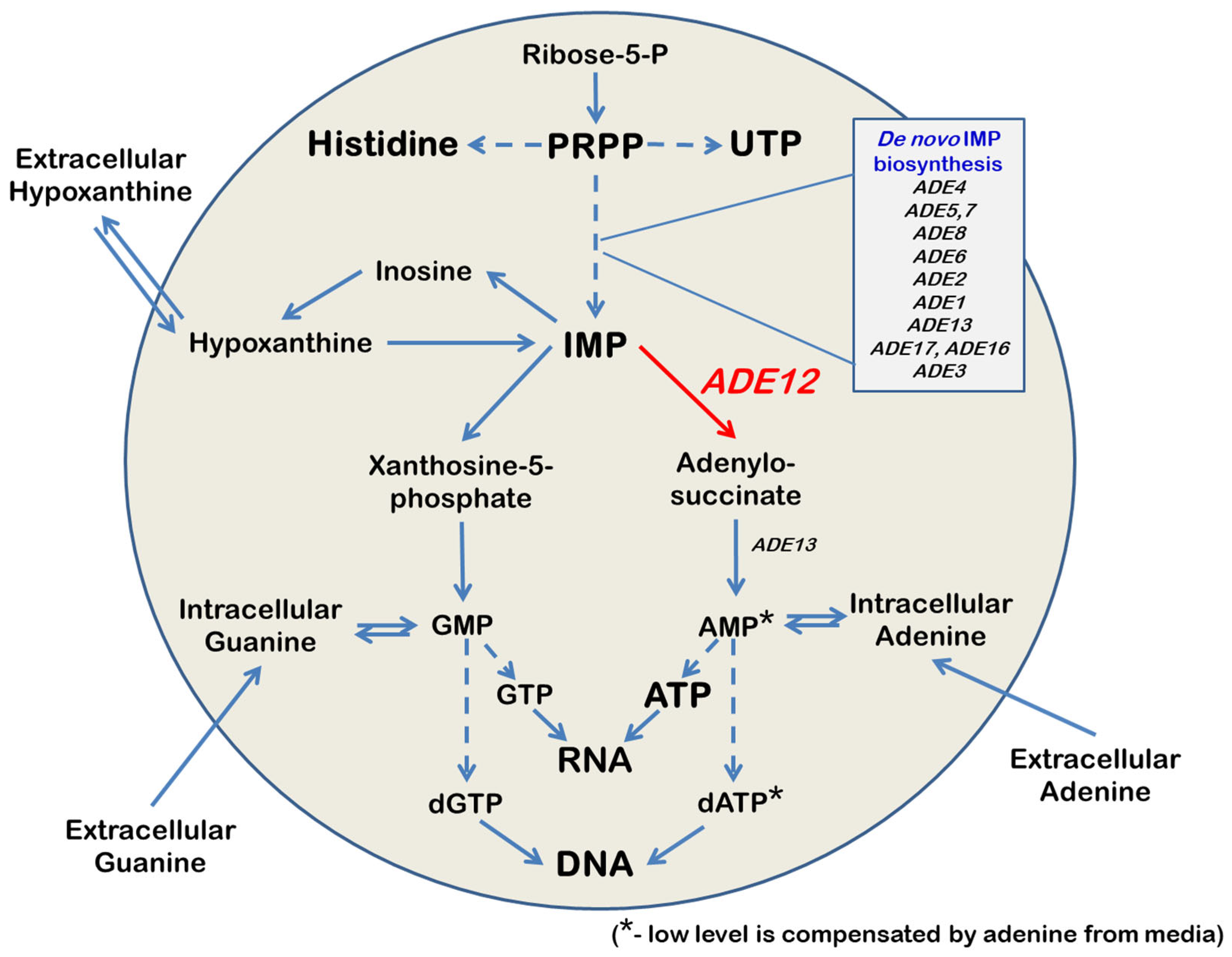
Adenylosuccinate synthetase (AdSS) is critical in cellular metabolism (Figure 1). AdSS catalyzes the first committed step in the synthesis of adenosine 5′-monophosphate (AMP) [1,2,3], a precursor of nucleic acids, and signaling and energy-transmitting molecules. AdSS utilizes inosine 5′-monophosphate (IMP) as a substrate to synthesize adenylosuccinate. In the second step, adenylosuccinate lyase cleaves adenylosuccinate to produce AMP [1]. In addition to the de novo IMP biosynthesis [4,5], IMP can also be derived from extracellular purines through a branched salvage pathway [6,7]. Thus, IMP is a unique link between the salvage and de novo purine biosynthesis pathways (Figure 1).
The purine biosynthesis pathway is highly conserved in all living organisms, and AdSS was found in bacteria, fungi, plants, and animals [2,3,8,9,10]. While most organisms possess both de novo purine nucleotide biosynthesis and purine salvage pathways, certain pathogenic organisms, e.g., Helicobacter pylori [11] and Leishmania gonovani [12], rely exclusively on the purine salvage pathway for purine nucleotide synthesis. The development of AdSS inhibitors is considered to be a promising therapeutic strategy in the fight against antibiotic-resistant strains of these parasites [13,14]. In humans, mutations in the ADSSL1 gene, which encodes a muscle-specific isoform of AdSS, are associated with an ultra-rare myopathy. The ADSSL1 myopathy was first described by Park and colleagues in 2016 in Korean patients suffering from distal muscle weakness [15]. Symptoms of the disease typically manifest during childhood and progress gradually with age. The affected patients experience distal muscle dysfunction and fatigue after mild physical exercise, which is later accompanied by facial muscle weakness. Cardiomyopathy and respiratory deficiency are also common in advanced disease stages, with death frequently caused by respiratory failure [16,17,18]. There have been 124 genetically diagnosed cases by 2024, mainly in Asian populations, but it is estimated that there could be about 4500 undiagnosed ADSSL1 myopathy patients worldwide [19].
Because of IMP’s central position in the purine metabolism pathway, it could be predicted that mutations in the genes encoding enzymes that control IMP utilization or biosynthesis will have pleotropic effects. This idea is undoubtedly true in the case of the ADE12 gene encoding AdSS in yeast Saccharomyces cerevisiae. Like most mutants in the de novo purine biosynthesis pathway, yeast ade12 mutants are auxotrophic for adenine [1,20]. All auxotrophic ade mutants, except ade12, can grow on a medium containing hypoxanthine as the sole purine source and utilize it via the purine salvage pathway. Yeast cells can utilize external hypoxanthine and convert it to IMP; however, AMP biosynthesis requires AdSS activity. Therefore, adenine, but not hypoxanthine, can compensate for the growth defect of the ade12 mutants on minimal media [1,20]. Additionally, yeast ade12 mutants exhibit slow growth even on complete media, and spores harboring the ade12 mutation demonstrate poor germination rates. It is possible that ade12 deletion blocks the production of AMP and, subsequently, ATP, leading to energy depletion and the slowdown of all energy-consuming processes required for cell growth, including DNA synthesis. Mutations affecting the upstream steps of purine metabolism rescue these defects [1,20]. It is also known that the ade12 mutants accumulate hypoxanthine and inosine and excrete them into the medium [1]. Interestingly, in the food industry, IMP and GMP, combined with monosodium glutamate, increase the umami flavor. The performance of industrial strains of filamentous fungi Ashbya gossypii, producing IMP and GMP for umami flavor, is improved by AdSS inhibition, which increases the excretion of IMP and GMP into the medium [21]. In our previous experiments, we have found that ade12 mutants are sensitive to mutagenic analogues of adenine (6-N-hydroxylaminopurine—HAP and 2-amino-6-hydroxylaminopurine—AHA) [22,23]. We propose that the toxic effect of ADE12 deletion and sensitivity to HAP and AHA are caused by the elevated levels of IMP, which can presumably be converted into ribo- and deoxyribonucleosidetriphosphates. The excess ITP and dITP can saturate Ham1p—pyrophosphatase, an essential enzyme for the destruction of the nucleoside triphosphates of hypoxanthine [24,25], and HAP in nucleotide pools, leading to the frequent incorporation of mutagenic dHAPTP and dAHATP into DNA during replication [26,27].
The main goal of this study is to elucidate the mechanisms by which an AdSS deficiency leads to a decrease in the growth rate and an increase in mutation rates in S. cerevisiae, thereby contributing to a deeper understanding of purine biosynthesis and its broader biological consequences.

2. Results

2.1. Refined Phenotypes of the ade12 Null Mutant

We generated ade12 mutants by replacing the ADE12 gene with TRP1 (see Materials and Methods, Table S1). The phenotypic characteristics of the newly derived ade12 strains exhibited several differences from previous studies [1,20]. In particular, our ade12 strains were unable to grow on the YPD medium and had growth defects on the synthetic (SD) dropout medium containing a low concentration of adenine (5 µg/mL) (Figure 2). Thus, the ade12 null mutation is conditionally lethal. Previous studies have shown that hypoxanthine does not compensate ade12, and growth defects can be partially alleviated by the addition of supplemental adenine to the medium or through genetic modifications at earlier stages of IMP biosynthesis [1,20]. Double mutants carrying both ade12 and ade5,7 had significantly improved growth on the YPD medium compared to single ade12 mutants (Figure 2), consistent with the literature [20]. To elucidate the difference between the YPD medium and synthetic dropout medium in the purine concentration, we measured the purine content in the yeast extract and peptone components used in preparing the YPD medium in our laboratory. The YPD medium contained 7 µg/mL adenine and 2 µg/mL hypoxanthine (Table S2). The concentration of adenine and hypoxanthine in the YPD medium alone cannot account for the significant differences in growth rates between YPD and SD medium (Figure 2). It is possible that other components in the YPD medium beyond the purine availability or slight differences in pH negatively impact the viability of ade12 strains.
The ade12 null strain unable to grow on the YPD produces YPD+ revertants with a relatively high frequency of 1.2 × 10⁻⁵. We picked up 567 independent YPD+ revertants and determined if they acquired additional mutations in purine metabolism genes using a genetic complementation test. We crossed YPD+ revertants to tester strains carrying mutations in one of the ADE genes (ADE4, ADE5,7, ADE8, ADE6, ADE2, ADE1, or ADE3); then, hybrids were examined for adenine auxotrophy (Table 1). Most of the YPD+ revertants exhibited an additional mutation in one of the ADE genes, and the frequency of these additional mutations correlated (r = 0.88, p = 0.02) with the gene length (excluding the ADE3 gene) (Table 1). Around 20% of YPD+ revertants had suppressor mutations in another gene/other genes (Table 1). Interestingly, the ade12 deletion strain clone in our laboratory, which was obtained from the Yeast Knockout (YKO) collection developed by the Saccharomyces Genome Deletion Project [28,29], carried an ade4 mutation in addition to the ade12. This is consistent with our data (Table 1, Figure 2) and with the literature that additional mutations in ADE genes reduce the toxic effect of ade12 [1,20]. It is likely that the strains considered single ade12 mutants but growing on the YPD carry additional suppressor mutations.

2.2. Yeast ade12 Null Mutation Leads to a Moderate Increase in Pol ζ-Dependent Mutagenesis, Which Is Suppressed by a High Level of Adenine in the Medium

The relatively high frequency of the YPD+ revertants in the ade12 strain led us to suspect an elevated mutability. We measured the frequency of direct mutations in the reporter CAN1 gene in WT and ade12 strains grown in a synthetic medium that contained 7 µg/mL adenine and 2 µg/mL hypoxanthine. The ade12 mutant grew significantly worse than the WT strain (the relative survival of the ade12 strain compared to WT was about 10%) and exhibited around five times the elevated mutant frequency (Table 2).
We measured the spontaneous mutation frequency in the double ade5,7 ade12 mutant grown in the standard YPD medium and YPD supplemented with additional adenine (100 µg/mL) (YPDA). We have shown that, even in the presence of the ade5,7 mutation, which suppresses the ade12 lethality on YPD or SD with low concentrations of adenine, the ade12 mutation increased the frequency of mutagenesis in the YPD medium without additional adenine (Table 3).
Additional adenine in the medium suppresses elevated mutagenesis in ade5,7 ade12 strains (Table 3). In yeast, replication disturbance caused by mutations in the replicative DNA polymerase genes or by the addition of hydroxyurea, an inhibitor of ribonucleotide reductase, leads to a moderate increase in mutagenesis [30,31]. The increase is attributed to the recruitment of specialized DNA polymerase ζ (Pol ζ), whose catalytic subunit is encoded by the REV3 gene. We hypothesized that, in ade12 mutants, the increase in mutagenesis frequency is also associated with Pol ζ’s activity. We compared the mutagenesis frequency in ade5,7 ade12 and the ade5,7 ade12 rev3 strains to test this. The inactivation of the Pol ζ led to a reduction in mutagenesis to the level of the ade5,7 strain, suggesting that mutations arising in the ade12 background are introduced by Pol ζ. Thus, the genomic DNA becomes more accessible for the Pol ζ in ade12 cells.

2.3. Overexpression of the ADE12 Is Not Mutagenic and Suppresses the Mutagenic Effect of HAP

We investigated the effect of the ADE12 overexpression on spontaneous mutagenesis. We compared the frequency of the Canr mutations in the ade5,7 strains with either a multicopy plasmid (pRS425-ADE12) carrying the ADE12 gene with its native promoter or the control vector pRS425. The spontaneous mutation frequency in the CAN1 reporter gene did not differ significantly (Table 4). Thus, the overexpression of the ADE12 is not mutagenic. Additionally, we assessed the influence of the ADE12 overexpression on the sensitivity of the yeast strain to the mutagenic base analogue, HAP. Our findings revealed that the ADE12 overexpression significantly reduced HAP-induced mutagenesis by nearly seven-fold (Table 4). These results are consistent with the previously obtained data indicating that AdSS is involved in the detoxification of HAP, as the deletion of ADE12 leads to an increase in HAP-induced mutagenesis [22,23], while the overproduction of the protein ensures its reduction compared to the wild-type strain (Table 4). Thus, AdSS plays an active role in protecting the cell from the accumulation of the mutagenic deoxynucleoside triphosphate of HAP. It is probable that the cooperative action of several enzymes in the purine salvage pathway—specifically adenine deaminase (Aah1), guanine hypoxanthine phosphoribosyltransferase (Hpt1), and AdSS [22,23]—facilitates the rapid conversion of HAP to AMP within the cell. For instance, HAP is first converted to hypoxanthine by Aah1, after which Hpt1 catalyzes the synthesis of IMP by attaching a phosphoribose moiety to hypoxanthine. Subsequently, AdSS utilizes IMP as a substrate to synthesize AMP. Previous studies have demonstrated that E. coli AdSS can synthesize HAPMP from IMP and hydroxylamine (used instead of aspartate) and is capable of performing both the forward and reverse reactions [32]. Therefore, it is plausible that AdSS may also use HAPMP as a substrate, converting it to IMP, which is no longer mutagenic in yeast.

2.4. The ade12 Mutants Accumulate IMP Derivatives and Storage Carbohydrates and Lack Pyrimidine Nucleotide Precursors, Amino Acids, and Sugar-Phosphates

To investigate the potential metabolic pathways underlying the toxic effect of ade12 mutation in S. cerevisiae, we characterized and compared the biochemical composition of the WT and mutant yeast cells incubated in the YPD and YPDA medium. The GC-MS-based metabolite profiling of S. cerevisiae revealed 360 individual analytes, of which 191 compounds were identified based on the coelution with authentic standards, retention index (RI) values, and spectral similarity, or structurally annotated to specific chemical classes by RI and spectral similarity to the MS-library data. The metabolite profiles were represented by 54 carbohydrates (sugars, polyols, and sugar acids), 45 amino acids and their derivatives, including 20 proteinogenic amino acids and 25 non-proteinogenic ones (sarcosine, 2-aminobutyric acid, pipecolic acid, β-alanine, ornithine, cysteinesulfinic acid, etc.), 27 organic acids (di- and tricarboxylic acids of TCA cycle, glyceric acid, benzoic acid, etc.), 19 fatty acids and their derivatives, 11 sterols (ergosterol, lanosterol, etc.), 10 nitrogenous bases, nucleosides, and their derivatives (adenine, uracil, hypoxanthine, adenosine, inosine, etc.), 7 amines (putrescine, spermidine, ethanolamine, etc.), 7 phenolic compounds (4-hydroxyphenylacetic acid, phenyllactic acid, phloroglucinol, etc.), and a miscellaneous group of metabolites including pantothenic acid, nicotinamide, phosphates, etc. (Table S3). The most abundant compounds in the metabolite profiles of the WT cells were glycerol, myo-inositol, trehalose, and several amino acids (glutamic acid, lysine, aspartic acid, and threonine). In the cells of ade12 mutants, inosine was also one of the dominant analytes.
Regarding the detection of nucleosides in the GC-MS-based metabolite profiles, it is important to emphasize that these analytes may at least be partially derived from the corresponding nucleotides. Due to the hot flash evaporation of the liquid sample required for the GC-MS analysis, thermolabile nucleotide molecules may lose their phosphate moieties; those may also hydrolyze already during the sample preparation (in particular, derivatization) before chromatographic separation. Thus, changes in the nucleoside content may indicate changes in the amount of the corresponding nucleotides in the yeast cells. In particular, a high content of inosine in the samples of the ade12 strain most probably suggests an accumulation of IMP in the mutant cells. The effect of the ade12 mutation on the nucleotide pools was additionally measured using another approach (see Section 2.5).
Differences between the metabolite profiles of the WT and mutant S. cerevisiae cells are clearly revealed by a principal component analysis (PCA), where the first two PCs explain 80% of the variance (Figure 3A). PC1 divides all the samples into two distinct groups, separating the WT cells and ade12 mutants.
The contribution of specific metabolites to the ade12-dependent changes in the metabolite profiles of yeast was illustrated by the Volcano plot (Figure S1). Table 5 lists 63 identified or structurally annotated metabolites (excluding 51 unknowns) with significantly different abundances.
The ade12 mutation dramatically changed the yeast metabolism (Table 5, Figure S1). The most affected groups of metabolites (fold change up to 7500) were nucleosides (in particular, inosine and its methylated derivative) and amino acids involved in the metabolism of pyrimidine nucleosides and nucleotides (4,5-dihydroorotic and orotic acids). Most of the metabolites showed a decreased abundance in the ade12 mutants compared to the WT cells. The most prominent feature was a general drop in the content of amino acids (both proteinogenic and non-proteinogenic). The other downregulated groups of compounds were fatty acids, sugar phosphates, organic acids not involved in the TCA cycle, and several secondary metabolites (Table 5). The upregulated metabolites were presented by nucleoside inosine (presumably derived from IMP and, thus, reflecting IMP accumulation) and metabolically linked compounds (1-methylinosine, and hypoxanthine), organic acids (mainly TCA cycle intermediates), different carbohydrates (including the dominant compounds, trehalose and glycerol), three amino acids, ergosterol, and polyamine putrescine (Table 5).
At the next step of our study, we examined how the factors mitigating the toxic effect of ade12 mutation (extra adenine supplementation or additional ade5,7 mutation) might have influenced the metabolite profiles of S. cerevisiae cells. The addition of extra adenine (100 µg/mL) to the growth medium did not affect the metabolite profiles of the WT cells (Figure S2) but erased the ade12-dependent changes in the metabolism of yeast (Figure 3B, Table 6). In this case, samples of mutant cells had a much higher inter-group variance, and the list of compounds with significantly different abundances between the two compared groups included only five identified metabolites and one unknown analyte (Figure S3, Table 6). Most of the affected metabolites were the same as in the variant without adenine addition (Table 5), though with much lower fold change values. Notably, the analytes contributing the most to the difference between the metabolite profiles of WT and ade12 without an adenine addition (inosine and 4,5-dihydroorotic acid) did not show significantly different abundances in the presence of extra adenine. We suggest that a higher concentration (compared to 100 µg/mL) of extracellular adenine would completely negate the changes in metabolite profiles of the yeast cells induced by the ade12 mutation.
The metabolite profiles of the yeast cells bearing the ade5,7 mutation differed considerably from the profiles of the WT cells (Figure S4). Moreover, PCA also showed a great difference between the ade5,7 and ade5,7 ade12 strains (Figure 3C). A deeper analysis of the specific compounds contributing to the ade12-dependent shift in the biochemical composition of the yeast cells on the background of the ade5,7 mutation revealed 83 identified or structurally annotated metabolites (excluding unknowns) with significantly different abundances (p < 0.05, fold change ≥ 2) (Figure S5, Table S4). About 26% of affected metabolites were the same as those contributing to the ade12-dependent changes in the cells not bearing the ade5,7 mutation: orotic acid, 2-aminobutyric acid, threonine, cysteine, β-alanine, glycerol-3-phosphate, ribose-5-phosphate, and lanosterol (downregulated); and glycerol, arabitol, mannitol, glyceric acid, α-ketoglutaric acid, putrescine, and hypoxanthine (upregulated) (Table 5 and Table S4). Notably, when accompanied by the ade5,7 mutation, the ade12 mutation did not result in the considerable accumulation of inosine and 1-methylinosine in the yeast cells. The most affected metabolites were uridine and orotic acid (downregulated, with fold changes of 125 and 46, respectively). Among the compounds accumulated in the ade5,7 ade12 cells, different nitrogen-containing metabolites were conspicuous: polyamines and metabolically linked compounds (ornithine and arginine), nitrogen-rich amino acids (glutamine and histidine), and nitrogenous bases (hypoxanthine and uracil) (Table S4).

2.5. Inactivation of the ADE12 Gene Leads to a Dramatic Decrease in the NTP and dNTP Pools, Which Is Suppressed by the Adenine-Rich Media or by Blocking IMP Biosynthesis De Novo

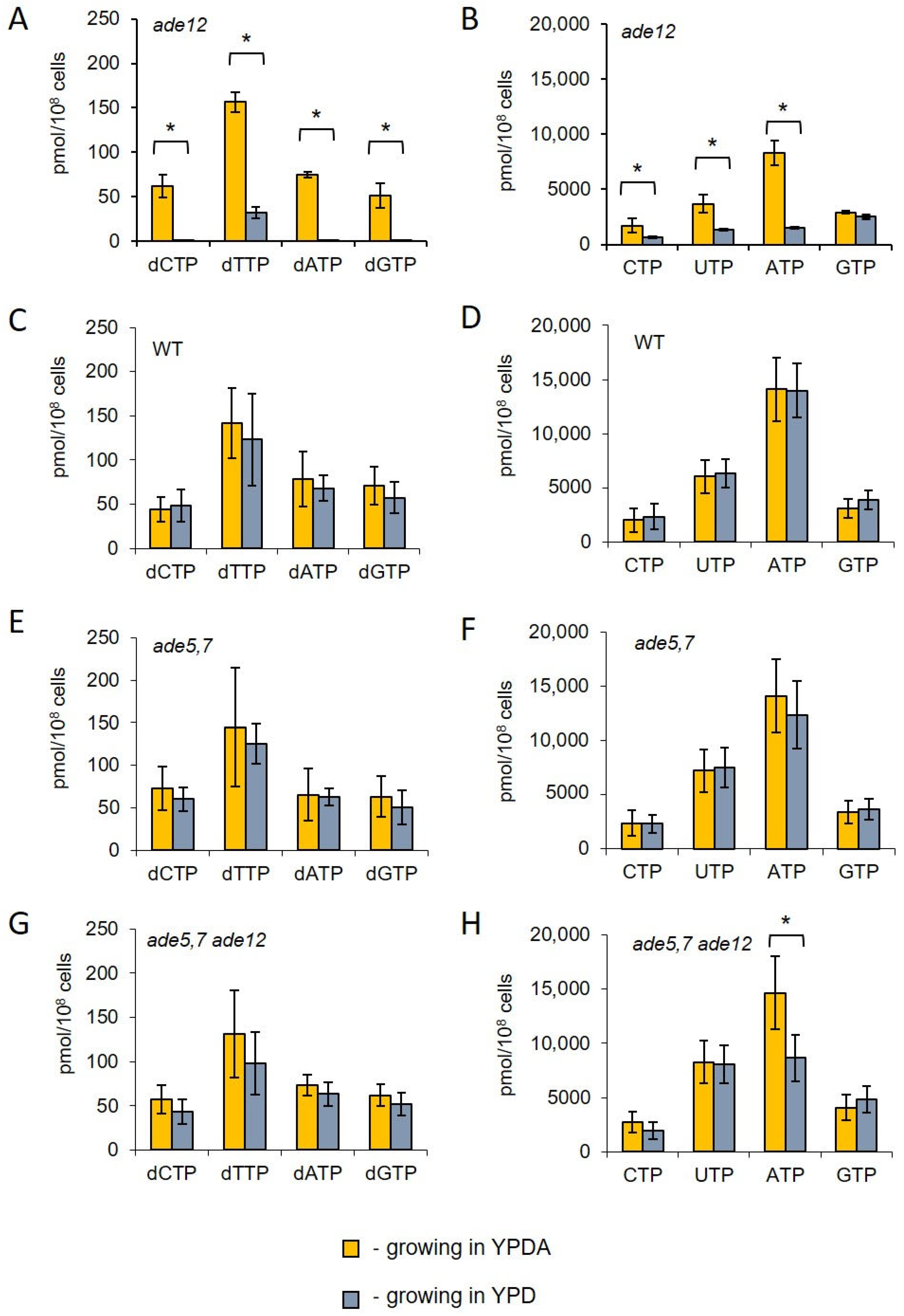
The results obtained from the metabolomic analysis suggested that ade12 mutants might have disturbed nucleotide pools. Specifically, there was a notable accumulation of inosine and its derivatives and a considerable decrease in the content of metabolites related to pyrimidine nucleotide synthesis. Therefore, we investigated the levels of NTPs and dNTPs in the cells of the WT, ade5,7, ade12, and ade12 ade5,7 strains after 6 h of incubation in the YPD or YPDA medium. For this purpose, three cultures for each strain were first grown in the YPDA medium, and then cells were transferred to fresh YPD or YPDA medium and incubated further for up to 6 h. Aliquots of each culture were collected to evaluate the NTP and dNTP pools (Figure 4) and the cell cycle progression (Figure 5). We found that the ade12 lethality in the YPD medium correlates with a severe decrease in intracellular nucleotide pools (Figure 4). After 6 h of incubation in YPD media, the dNTP pools in the ade12 mutants decreased to almost undetectable levels of dCTP, dATP, and dGTP (Figure 4A). The content of CTP and UTP decreased three-fold, with the most dramatic decline (five-fold) for ATP (Figure 4B). These changes in the nucleotide pools make DNA synthesis impossible due to the absence of both building blocks (dNTP) and energy (ATP).
Unlike the ade12 mutants, the WT (Figure 4C,D) and ade5,7 (Figure 4E,F) strains did not demonstrate changes in the dNTP and NTP pools while growing in the YPDA and YPD media. Moreover, there was no statistically significant difference between the NTP pools of the double mutants ade5,7 ade12 incubated in the presence of extra adenine (Figure 4 H) and the WT (Figure 4D) or ade5,7 strains (Figure 4F). At the same time, the content of ATP in the ade12 ade5,7 mutant grown in the YPD medium decreased by 30% (Figure 4H). The amounts of other NTPs (CTP, UTP, and GTP) and dNTPs in the ade12 ade5,7 were unaffected (Figure 4G,H).
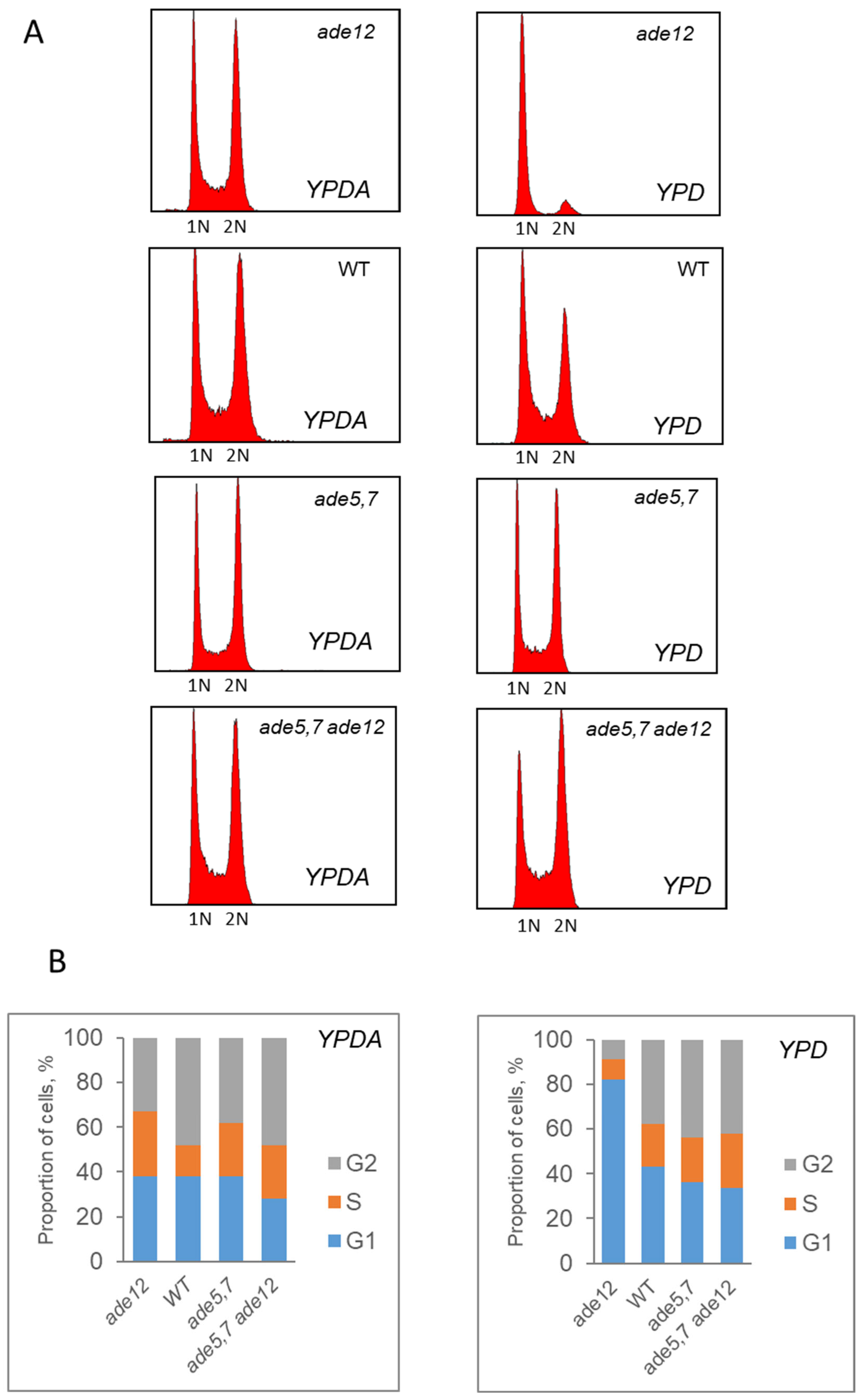
According to flow cytometry, ade12 mutants exhibited a slowed cell cycle progression when incubated in the YPD medium (Figure 5). In the presence of extra adenine, the mutant strain did not differ from the WT, and both cultures contained the cells passing through the G1, S, and G2 phases of the cell cycle. However, after incubation in the YPD medium, the cultures of the ade12 strain had an increased number of cells in the G1 phase, while the number of cells passing through the S and G2 phases decreased dramatically (Figure 5). This result is consistent with the main feature of the ade12 phenotype: the inability to grow in the YPD medium lacking extra adenine.

3. Discussion

This study demonstrated that ade12 mutants lacking AdSS cannot grow in a standard YPD medium and possess a mutator phenotype. Both manifestations are suppressed by the addition of adenine to the growth medium or by the genetic blocking of de novo IMP biosynthesis. To elucidate the molecular mechanisms underlying the phenotypes of the ade12 mutation, we conducted genetic and biochemical analyses with a special focus on the nucleotide pools.
Flow cytometry data (Figure 5), in agreement with the results of the culture growth experiments (Figure 2), confirm that yeast cells bearing the ade12 mutation cannot divide normally and do not progress through the cell cycle, stopping at the G1 phase. Similar phenotypes were also observed in the yeast strains with mutations in the other genes involved in AMP biosynthesis. For example, the ade2-1 mutant (strain W303-1A) growing in a YPD medium without additional adenine supplementation had reduced cell division and budding rates, and a larger cell size [33]. Similarly, the ade8 mutants grown in the medium lacking adenine (SD-ade) demonstrated a two-fold decrease in the budding rate compared to the control (SD+ade), an increase in cell size, and a predominance of cells with 1n DNA content indicative of G1-phase arrest [34].
We hypothesized that the most probable biochemical cause of cell division failure in the ade12 strain is a deficiency of the key molecules maintaining this process. These molecules are nucleic acid constituents (nucleotides, nucleosides, and their derivatives), proteins, and energy-transferring compounds, such as ATP and NADH. The ADE12-encoded AdSS catalyzes one of the critical steps of purine nucleotide biosynthesis (Figure 1). Therefore, it is logical to assume that the mutant cells may encounter an overall reduction and imbalance of the nucleotide pools. Our findings indicate that, in ade12 cells, NTP levels significantly decline when adenine is limited in the medium, and it is accompanied by an even more dramatic decrease in dNTP levels (Figure 4). The activity of the ribonucleotide reductase responsible for producing dNTPs is regulated by the NTP/dNTP ratio [35]. Consequently, as NTP levels decrease, dNTP production is also diminished, rendering DNA replication impossible.
The metabolomics data provide further insights into the ade12-dependent changes in cell biochemistry. Our results showed that ade12 mutation leads not only to an imbalance of the nucleotide pools, but also to a general impairment of nitrogen exchange in the yeast cells. A decrease in the content of many amino acids (including both proteinogenic ones and those involved in nucleotide metabolism) and the accumulation of their precursors (such as α-ketoglutaric acid) imply a general inhibition of amino acid and, consequently, protein biosynthesis. Most of the nitrogen released due to this inhibition in the mutant cells is invested into forming IMP constituents and derivatives (inosine, methylinosine, and hypoxanthine) (Table 5). The accumulation of inosine and hypoxanthine in the yeast cells bearing the ade12 mutation was seen earlier [1]. Notably, the author, similar to us, believed that this inosine was derived from IMP and, thus, was a manifest of IMP accumulation. In WT cells, the excessive accumulation of IMP is prevented by the feedback inhibition of purine biosynthesis by ATP and ADP [20,36]. However, this regulation fails in the ade12 mutants lacking extracellular adenine [20].
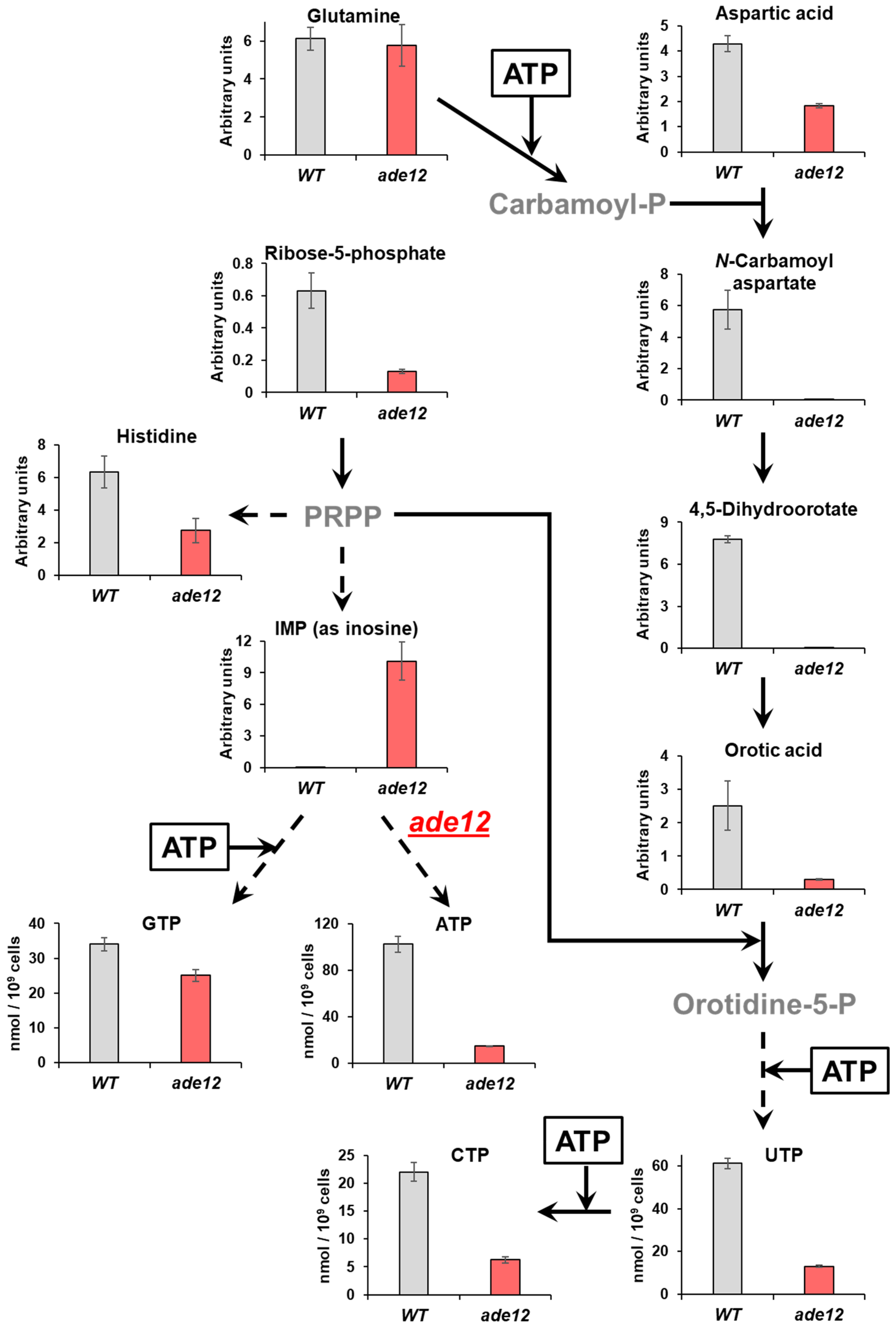
Both the metabolite profiling and analysis of the nucleotide pools showed that the cells bearing a mutation in the purine biosynthesis pathway also exhibited considerable impairment in pyrimidine nucleotide biosynthesis. Thus, one of the most striking features of the metabolite profiles of ade12 cells, along with inosine accumulation, was the three-orders-of-magnitude decrease in the content of 4,5-dihydroorotic acid, compared to the WT strain (Table 5, Figure 6). The purine and pyrimidine biosynthesis pathways are metabolically and energetically interconnected (Figure 6) [36], and we suggest that the main cause of the pyrimidine synthesis impairment in the ade12 strain may be energy limitation. Indeed, the dramatic decrease in ATP content and a general decline in the amounts of sugar phosphates and nicotinamide (a NADH constituent) indicate that the ade12 cells encounter severe energy deprivation (Figure 4 and Figure 6, Table 5). A similar drop in ATP and ADP content was shown earlier in the ade8 mutants growing under adenine limitation [34]. We can see a decrease in the contents of the key intermediates of de novo pyrimidine biosynthesis at the next step after the first ATP-dependent reaction (formation of N-carbamoyl aspartate from glutamine via carbamoyl phosphate) (Figure 6). We could not detect carbamoyl phosphate in our samples, but its formation is likely suppressed in the ade12 cells by an ATP shortage. This effect may be one of the reasons why glutamine is among the few amino acids whose content did not decline in the mutant cells. One more reason for the UTP and CTP decrease in the ade12 strain may be the deficit of 5-phosphoribosyl pyrophosphate (PRPP), an intermediate enabling the metabolic crosstalk between the purine, pyrimidine, and histidine biosynthesis pathways (Figure 1) [36]. In the mutant cells, the main flux of PRPP is shifted towards the formation of IMP on account of the inhibition of the other pathway branches, supporting pyrimidine and histidine synthesis (Figure 6).
The other conspicuous feature of the metabolite profiles of the ade12 cells is an enhanced accumulation of storage carbohydrates (trehalose, glycerol, and mannitol) compared to the WT strain (Table 5). Such an increase in carbon investment into metabolically relatively inert sugars and polyols is typical for non-dividing cells [37]. It may be explained by the impairment of nitrogen exchange and the inhibition of the biosynthesis of N-containing compounds (nucleic acids, proteins, and their monomers) in the ade12 mutants with cell division arrest. Similar effects were seen for yeast cells under various stress conditions. Trehalose, the major soluble carbohydrate in yeast cells, not only serves as a storage compound but also plays a protective role under stress conditions [37]. Trehalose helps to maintain cellular integrity under heat, cold, desiccation, dehydration, and oxidation stresses primarily by preventing protein denaturation. Numerous studies, initiated by the pioneering work of Singer and Lindquist [38], have demonstrated that trehalose interacts with proteins and functions as a chemical chaperone, recovering protein aggregation and maintaining proteins in a partially folded state to facilitate their refolding by chaperones. Additionally, trehalose has been shown to prevent the amyloid aggregation of huntingtin [39] and amyloid beta [40]. Glycerol also plays a protective role in yeast: cells of S. cerevisiae rapidly accumulate glycerol when exposed to a highly osmolar medium to counteract dehydration [41,42]. We may conclude that the adenine limitation in ade12 mutant leads to the activation of the protective mechanisms that typically serve to safeguard cells from adverse environmental conditions. According to the literature, mutations in the other ADE genes (ade2 and ade8) also induce trehalose accumulation in the yeast cells, which is accompanied by a decrease in protein and nucleic acid content [33,34]. A similar phenotype has also been documented in yeast cells under the limitations of other compounds, such as uracil and leucine [43]. Our data on the changes in the carbohydrate composition in ade12 cells are consistent with the findings from other studies indicating that the majority of yeast genes involved in purine biosynthesis (ADE13, ADE1, ADE2, ADE4, ADE5,7, ADE6, ADE8, and ADE16) are involved in the metabolism of carbon sources [44,45]. It has been shown that ade13 mutants cannot grow in a complete medium containing glucose, but can grow in medium containing glycerol or ethanol as the sole carbon source. Additional mutations (ade1, ade2, ade4, and ade5,7) occurring at earlier stages of purine biosynthesis alleviate the conditional lethality associated with the ade13 mutation [44]. Mutations in the ADE2 gene result in the inability of yeast cells to utilize hypoxanthine as a purine source in a glycerol medium. Mutations in ade4, ade5, ade8, ade6, and ade7 suppress this phenotype of the ade2 mutation [45]. It has been shown that the expression of the ADE16 gene, but not ADE17, depends on the type of carbon source in the medium. Specifically, the amount of Ade16p protein is higher in cells grown on non-fermentable carbon sources than in cells grown in the glucose medium [46]. Thus, the ADE genes, including ADE12, either directly regulate glycolysis or are involved in shaping cellular responses to the type of carbon source in the medium. The effect is explained by the need to coordinate the cell division with the nucleotide synthesis rates required for DNA replication and other cellular processes, depending on the quality of the carbon sources present in the environment [44]. The pivotal role of AdSS in coordinating purine and carbohydrate metabolism was supported by findings from a metabolite screening of endogenous factors that mediate glucose-induced insulin secretion in the rat insulinoma cell line 832/13. Glucose stimulation resulted in insulin secretion, decreased IMP levels, and increased adenylosuccinate, a product of AdSS [47].
Biochemical studies of the ade12 cells grown in the presence of extra adenine or with the additional ade5,7 mutation enabling the block of de novo IMP biosynthesis showed that both these factors effectively mitigate the most conspicuous ade12-dependent metabolic disorders, such as an IMP accumulation and 4,5-dihydroorotate decrease, as well as nucleotide pool imbalance (Table 6 and Table S4, Figure 4). Though the double ade5,7 ade12 mutants still have a reduced ATP content when grown without the adenine addition, the difference is much smaller than that for the single ade12 mutants (Figure 4). The mitigating effect of adenine supplementation may be explained by the restoration of the cell AMP pool due to the salvage pathway consuming extracellular adenine (Figure 1) [48,49,50]. The mechanism of the ameliorating influence of the ade5,7 mutation is more complex, as this mutation itself considerably affects yeast cell metabolism (Figure S4). The metabolomic data imply that the main target of the ade12-dependent changes occurring on the background of the ade5,7 mutation is again the nitrogen exchange, as double mutants specifically accumulate multiple nitrogen-rich metabolites, such as polyamines, arginine, and histidine (Table S4). However, this branch of our research goes beyond the scope of this paper and deserves a separate, more in-depth study, which is an ongoing plan in our laboratory.
Another question we addressed in the current research is the mechanism of the mutator phenotype of the ade12 mutants. It is important to note that, in this study, we have shown, for the first time, that spontaneous mutagenesis is moderately increased in the strains bearing the ade12 mutation; meanwhile, earlier, the ade12 strain was also reported to have an increased mutation frequency induced by the mutagenic hypoxanthine analogues HAP and AHA [22,23]. Below, we discuss three non-contradictory explanations for the spontaneous mutator phenotype in the ade12 strains.
First is the direct role of ADE12 in maintaining the accuracy of genome replication. S. cerevisiae AdSS specifically binds to ss-DNA containing autonomous replication sequences (ARSs) [51]. This binding inhibits AdSS enzymatic activity and occurs only if AdSS is phosphorylated. However, the activity is specific for AdSS from S. cerevisiae, while AdSS from E. coli and Dictiostelium discoideum showed only 1% of the activity found in the yeast homologue. Thus, AdSS may regulate genome replication. If this is actually the case, then, in the absence of AdSS, the accuracy of replication and repair closely related to the stages of the cell cycle may decrease and lead to an increased level of spontaneous mutagenesis. There are several arguments against this hypothesis. Thus, ss-DNA binding activity was proven only in vitro, and it is unclear how the results correspond to AdSS properties in living cells. AdSS is located in the cytoplasm [52], thus making its DNA-binding activity irrelevant. Our findings do not support this mechanism. We have shown that elevated mutagenesis is suppressed in the ade12 mutant in YPDA. Moreover, the overexpression of the ADE12 gene does not affect the spontaneous mutation frequency. These results suggest that the level of spontaneous mutagenesis does not depend on the physical presence of AdSS in the origins of replication, but rather on AdSS’s role in purine metabolism.
A second assumption would be that the elevation of IMP in the cytoplasm of ade12 mutants may promote the synthesis of dITP, which can then be incorporated into DNA, potentially leading to increased mutagenesis. However, the experimental data do not support this hypothesis, as the levels of inosine incorporation into DNA were reported to remain unchanged in the AdSS-deficient strains of both yeast and bacteria [53]. Moreover, it was shown in E. coli that dITP incorporation into DNA is non-mutagenic due to inosine’s strong pairing preference for cytosine [54]. This may be the case for yeast as well, as S. cerevisiae lacks inosine glycosylase due to the absence of a requirement to protect DNA from inosine incorporation.
A third idea is the indirect role of the missing AdSS in the increased mutation rate in the ade12 strain. We have shown that the mutagenic effect observed in the ade12 strain entirely depends on Pol ζ’s activity. Therefore, it appears that elevated IMP levels do not directly contribute to increased mutagenesis. In the ade12 mutants grown in the YPD medium, dNTP pools and the ATP content are insufficient to maintain replication, resulting in cell cycle arrest. Under conditions of replication stalling, specialized DNA polymerase is more likely to be recruited for replication, similar to what occurs when yeast cells are incubated in a medium with hydroxyurea or during defective-replisome-induced mutagenesis (DRIM) [31]. This notion is supported by evidence that Pol ζ, unlike replicative polymerases, is active at low dNTP levels [55]. Interestingly, it was shown before that adenine starvation elevates the mutation rate in the yeast strains lacking the proofreading activity of DNA polymerase δ (pol3-01) [56]. Thus, through various mechanisms involving different DNA polymerases, adenine starvation in yeast can lead to an increased frequency of mutagenesis. The connections we have identified between nucleotide biosynthesis and elevated Pol ζ-dependent mutagenesis open up prospects for the discovery and development of nucleotide-analogue anticancer drugs [57].
We conclude that the low viability of ade12 mutants in the standard YPD or synthetic medium low in adenine results from the impaired cellular energy metabolism, leading to a cascading dysregulation of key metabolic processes—the metabolism of nucleotides, carbohydrates, and amino acids. These metabolic perturbations may account for the cell division arrest observed in the ade12 yeast strains. The mutagenic effect observed in ade12 mutants appears to be a consequence of the slowed replication due to the insufficient energy, nucleotides, and proteins. Under these conditions, genomic DNA becomes more accessible to error-prone Pol ζ, increasing the likelihood of mutations. ADSSL1 myopathy in humans presents a similar challenge regarding energy metabolism, where ATP deficiency is a key issue. The parallels between the metabolic dysregulation observed in ade12 yeast mutants and human myopathies highlight the importance of understanding connections between energy metabolism and biosynthetic pathways in both yeast and higher organisms for the potential identification of therapeutic targets.
4. Materials and Methods

4.1. Yeast S. cerevisiae Strains, Plasmids, and Incubation Conditions

We used a series of yeast S. cerevisiae strains isogenic to the strain LAN201-ura3-Δ (MATa ade5-1 lys2-Tn5-13 trp1-289 his7-2 leu2-3,112 ura3-Δ) [30]. First, we obtained T1 strain by replacing ade5-1 mutation (C1158A) with WT allele of the ADE5,7 gene. For that, we amplified 802 b.p. fragment of the ADE5,7 gene using chromosomal DNA of the BY4742 strain as a template and two primers: ADE5,7-890 GTTAGAATATAATGTCAGATTCGG and ADE5,7-1668R GAATGTCAAGAGCACCAGTGGC. Then, we transformed LAN201-ura3-Δ strain with the PCR fragment and selected transformants on medium without adenine. The T1 strain was used to introduce disruptions of the ADE5,7, ADE12, and REV3 genes. To make ade5,7::kanMX disruption, we transformed the T1 strain with the PCR fragment, obtained with the use of two primers ADE5,7-F GTGAAAGATGCCACCATACG and ADE5,7-R AAAGTGTCTTGCACCATACC and chromosomal DNA of ade5,7 strain from Yeast Knockout collection [28,29]. To introduce ADE12::TRP1 disruption, we first obtained PCR product using two primers AdeDISTRPL ATGGTTAACGTTGTATTGGGCTCCCAATGGGGTGATGAGGGTAAAGCAGAGCAGATTGTA and AdeDISTRPR GGTACCAACCCATTCAACAGGAACGCCAACAAAATCTTCAATATATTCGCATCTGTGCGG and pRS304 plasmid [58] as a template. Then, we transformed yeast cells with the PCR product and selected transformants on SD medium without tryptophan containing high amount (100 µg/mL) of adenine. To generate the rev3::LEU2 disruption, we transformed yeast cells with the integrative pAM56 plasmid (constructed by A. Morrison), which was linearized with XbaI. All disruptions were validated by PCR and phenotypic analysis. To map suppressor mutations using a genetic complementation test, we employed a panel of tester strains carrying mutations in one of the ADE genes (ADE4, ADE5,7, ADE8, ADE6, ADE2, ADE1, or ADE3), obtained from the Peterhof genetic collection of yeast lines (Table S1).
To test effect of the ADE12 overexpression, we cloned WT allele of the ADE12 gene into pRS425 vector by BamHI and NotI restriction sites. For amplification of the WT copy of the ADE12, we used two primers ADE12_BAMH1_F TCGGGGATCCATGTCCCACTTACCTGTTCC and ADE12_NOT1_R GTACGCGGCCGCGGTGTTCGTTACATTGATTACTG, as well as chromosomal DNA of the BY4742 strain.

4.2. Media and Growth Conditions

Yeast cells were grown in standard YPD medium (0.5% BactoTM yeast extract (Becton, Dickenson and Company, Franklin Lakes, USA), 2% BactoTM peptone (Becton, Dickenson, and Company, Franklin Lakes, USA), 2% glucose), or in YPD medium with the addition of adenine (100 µg/mL) (YPDA), or in standard minimal synthetic medium (SD) (0.67% Yeast Nitrogen Base w/o amino acids (Becton, Dickenson and Company, Franklin Lakes, USA), 2% glucose, and amino acids, adenine, and uracil, required for growth of auxotrophic strains in standard concentrations) [51]. We also used YPD or SD medium supplemented with adenine or hypoxanthine at different concentrations (5, 20, or 100 µg/mL). Agar 2% was used to prepare the solid YPD, YPDA, or SD complete and dropout medium. SD medium containing 40 µg/mL of L-canavanine and all the supplements that are required for growth of auxotrophic strains was used for the selection of Canr mutants. Frequencies of spontaneous Canr mutations were determined as described [59].

4.3. Complementation Test

To verify whether the YPD+ revertants carry additional mutations in de novo adenine biosynthesis genes, we used a set of alpha mating-type tester strains, each carrying a mutation in one of the ADE genes (ADE4, ADE5,7, ADE8, ADE6, ADE2, ADE1, or ADE3) and the wild-type allele of the ADE12 (Table S1). Independent clones of the ade12 strain grown on the YPD medium were crossed with each tester strain. Then, we examined the ability of the resulting hybrids to grow on a medium lacking adenine. If the hybrid was prototrophic for adenine, it indicated that the tested YPD+ clone did not carry additional mutations in the ADE genes. If the hybrid of a YPD+ revertant was auxotrophic for adenine, the tested revertant carried a mutation allelic to the ADE mutation in the tester strain. After testing all YPD+ clones, we evaluated the frequency of mutations in each ADE gene in the ade12 strains grown on the YPD medium.

4.4. GC-MS Analysis

Three to six independent cultures of each yeast strain were used for GC-MS analysis. Cultures of WT and ade12 strains were first grown for two days in YPDA liquid medium. Then, two 1.5 mL aliquots were taken from each culture and washed three times with sterile distilled water. Cells from each aliquot were then diluted with 6 mL fresh YPD or YPDA and incubated for 4 h at 30 °C on a shaker. In case of ade5,7 and ade5,7 ade12 strains, overnight cultures in YPD medium at 30 °C were used. On completion of incubation, cells were harvested and washed three times with distilled water. After removal of all liquid, the cells were weighed and mixed with glass beads (200 µL) and 10 volumes of cold (−25 °C) methanol. The cells were then disrupted using a vortex mixer. After overnight extraction (4 °C), samples were centrifuged and aliquots of 100 µL of methanol extracts were transferred to clean 1.5 mL polypropylene Eppendorf tubes (VWR, Dresden, Germany) and vacuum-dried in the CentriVap vacuum concentrator system (Labconco, Kansas City, MO, USA) for subsequent analysis.
GC-MS analysis was carried out according to [60]. Briefly, vacuum-dried methanolic extracts were incubated by shaking in methoxyamine hydrochloride (Sigma-Aldrich Chemie GmbH, Taufkirchen, Germany) solution in pyridine and N,O-bis(trimethylsilyl)-trifluoroacetamide (Macherey-Nagel GmbH and Co KG, Düren, Germany). After derivatization, samples were transferred to glass vial micro-inserts and subjected to GC-MS analysis on a Trace GC Ultra gas chromatograph equipped with an A200S autosampler (CTC Analytics, Zwingen, Switzerland) and coupled with a MAT95 XP double-focusing sector field mass spectrometer (Thermo Electron, Bremen, Germany) with standard electron impact ionization (70 eV). Separation was accomplished on a DB-5MS Ultra Inert column (Agilent, Waldbronn, Germany; 30 m × 0.25 mm ID and 0.25 µm film) at 0.9 mL/min carrier gas flow (He 5.0 Alphagaz, Air Liquide, Düsseldorf, Germany) after splitless injection at 250 °C. Within each sequence, a mixture of alkanes (C10-C32) in hexane was measured for the calculation of Kovats retention indices. A mix of authentic standards containing 20 amino acids, 20 sugars and polyols, and 19 organic acids was co-spiked to confirm the identity of expected compounds.
Peak deconvolution was accomplished using AMDIS 2.66. The retention indices were automatically calculated using an AMDIS calibration file containing the batch retention times of each alkane. GMD (Golm metabolome database, GMD_20100614_VAR5_ALK, 24 September 2010), [61] and NIST14 (National Institute of Standards and Technology, Gaithersburg, MD, USA) were used for identification of the peaks based on spectra comparison. Experiments were carried out with 5 to 20 biological replicates (taken from different individuals). Quantitation of metabolites was performed by peak integration of the corresponding extracted ion chromatograms (m/z ± 0.5) for representative intense signals at specific retention times using Xcalibur 3.0.
MetaboAnalyst 6.0 web application (http://www.metaboanalyst.ca, accessed on 12 December 2024) was used for data processing and normalization procedures and creation of figures. The metabolomic data processing included peak area normalization to the median of all areas within the corresponding chromatogram, generalized logarithm transformation, and range data scaling (mean-centered and divided by the range of each variable). Normalized metabolomic data were subjected to a principal component analysis with Volcano plot representation. The results of univariate statistics were presented as mean ± standard deviation of 3–6 biological replicates. Fold change (FC) values in the abundance of individual metabolites and significance of differences (p-values) were estimated by Student’s t-test. The threshold values for FC and p-value presented in the tables and figures were 2 and 0.05, respectively. To assess the reliability of the FC in the relative levels of individual metabolites, false discovery rates (FDRs) at p ≤ 0.05 were estimated for all comparisons using the Benjamini–Hochberg method [62].

4.5. NTP and dNTP Pools Measurement

dNTP and NTP pools were measured as described in [63]. The cells were grown overnight in YPDA. The cultures were diluted to the OD = 0.2 in fresh liquid YPD media and incubated up to 6 h. The cells were collected and harvested by filtration through 25 mm White AAWP nitrocellulose filters (0.8 mm, Millipore AB, Solna, Sweden) at a density of 0.4 × 10⁷ to 0.5 × 10⁷ cells/mL, and NTPs and dNTPs were extracted in trichloroacetic acid and MgCl2 followed by extraction with a Freon–trioctylamine mix. dNTPs were separated from NTPs using boronate columns (Affigel 601, BioRad, Berkley, USA) and analyzed by HPLC on a LaChrom Elite UV detector (Hitachi, Tokyo, Japan) using a Partisphere SAX column (Hichrom, UK). For each strain, three independent clones were analyzed. Data are presented as mean ± standard deviation of 3 biological replicates.

4.6. Flow Cytometry

Cells from the growing cultures were analyzed for cell cycle progression and ploidy by staining of DNA with SYBR Green (Molecular Probes) as described [64] in a Becton Dickinson FC500.

Supplementary Materials

The following supporting information can be downloaded at: https://www.mdpi.com/article/10.3390/ijms26083458/s1.

Author Contributions

Conceptualization, E.I.S. and E.R.T.; methodology, E.I.S., C.W. and E.R.T.; validation, E.R.T. and E.I.S.; formal analysis, E.R.T., E.I.S. and Y.V.A.; investigation, E.R.T., E.I.S., Y.V.A. and T.E.B.; resources, E.I.S., Y.I.P. and C.W.; data curation, E.R.T. and E.I.S.; writing—original draft preparation, E.R.T., E.I.S., Y.I.P. and Y.V.A.; writing—review and editing, all authors; visualization, E.R.T., E.I.S. and Y.V.A.; supervision, E.I.S.; project administration, E.I.S.; funding acquisition, E.I.S. All authors have read and agreed to the published version of the manuscript.

Funding

This research was funded by Saint Petersburg State University, project number 125021902561-6.

Institutional Review Board Statement

Not applicable.

Informed Consent Statement

Not applicable.

Data Availability Statement

Data are contained within the article and Supplementary Materials.

Acknowledgments

The authors acknowledge the support from the Resource Center “Bio-Bank Center” and the Resource Center for Molecular and Cell Technologies (Research Park, Saint Petersburg State University). The mass spectrometry core facility at the Faculty of Chemistry and Mineralogy at Leipzig University, MS-UL, is acknowledged for providing technical support during the GC–MS analyses. We are thankful to Alexandr Zekhnov and Vladimir Alenin for productive discussions during the initial stages of the research. We are also very grateful to Andrei Chabes (Department of Medical Biochemistry and Biophysics, Umeå University) who kindly provided the opportunity to measure the dNTP and NTP pools.

Conflicts of Interest

The authors declare no conflicts of interest.

Abbreviations

The following abbreviations are used in this manuscript:
ADPAdenosine 5′-diphosphate
AdSSAdenylosuccinate synthetase
AHA2-amino-6-N-hydroxylaminopurine
AMPAdenosine 5′-monophosphate
ARSAutonomous replication sequences
ATPAdenosine 5′-triphosphate
b.p.Base pairs
CTP2′-Cytidine 5′-triphosphate
dATP2′-Deoxyadenosine 5′-triphosphate
dCTP2′-Deoxycytidine 5′-triphosphate
dGTP2′-Deoxyguanosine 5′-triphosphate
dHAPTP6-N-hydroxylaminopurine 2′-deoxynucleoside 5′-triphosphate
dITP2′-Deoxyinosine 5′-triphosphate
dNTP2′-Deoxynucleoside 5′-triphosphate
dTTP2′-Deoxythymidine 5′-triphosphate
FDRFalse discovery rate
GMPGuanosine 5′-monophosphate
GS-MSGas chromatography–mass spectrometry
GTPGuanosine 5′-triphosphate
HAP6-N-hydroxylaminopurine
HAPMP6-N-hydroxylaminopurine nucleoside 5′-monophosphate
HAPTP6-N-hydroxylaminopurine nucleoside 5′-triphosphate
IMPInosine 5′-monophosphate
ITPInosine 5′-triphosphate
NADHNicotinamide-adenine dinucleotide
NTPNucleoside 5′-triphosphate
PCAPrincipal component analysis
Pol ζDNA polymerase ζ
PRPP5-phosphoribosyl pyrophosphate
RIRetention index
Ribose-5-PD-Ribose 5-phosphate
SDSynthetic defined (yeast medium)
TCATricarboxylic acid cycle
UTPUridine 5′-triphosphate
YKOYeast Knockout
YPDYeast extract, peptone, dextrose (yeast complete medium)
YPDAYeast extract, peptone, dextrose, adenine (yeast YPD medium, supplemented with 100 µg/mL of adenine)

References

  1. Dorfman, B.Z. The isolation of adenylosuccinate synthetase mutants in yeast by selection for constitutive behavior in pigmented strains. Genetics 1969, 61, 377–389. [Google Scholar] [CrossRef] [PubMed]
  2. Lipps, G.; Krauss, G. Adenylosuccinate synthase from Saccharomyces cerevisiae: Homologous overexpression, purification and characterization of the recombinant protein. Biochem. J. 1999, 341 Pt 3, 537–543. [Google Scholar] [CrossRef] [PubMed]
  3. Ryzhova, T.A.; Andreichuk, Y.V.; Domkin, V.D. Adenylosuccinate synthetase of the yeast Saccharomyces cerevisiae: purification and properties. Biochemistry 1998, 63, 650–656. [Google Scholar] [PubMed]
  4. Zalkin, H.; Dixon, J.E. De novo purine nucleotide biosynthesis. Prog. Nucleic Acid. Res. Mol. Biol. 1992, 42, 259–287. [Google Scholar] [CrossRef]
  5. Pareek, V.; Pedley, A.M.; Benkovic, S.J. Human de novo purine biosynthesis. Crit. Rev. Biochem. Mol. Biol. 2021, 56, 1–16. [Google Scholar] [CrossRef]
  6. Gaber, A.; Elbakry, A.M.; Aljarari, R.M.; Jaber, F.A.; Khadrawy, Y.A.; Sabry, D.; Abo, E.R.E.; Ahmed, O.M. Bone marrow-derived mesenchymal stem cells and gamma-secretase inhibitor treatments suppress amyloid-beta25-35-induced cognitive impairment in rat dams and cortical degeneration in offspring. Stem Cells Int. 2023, 2023, 2690949. [Google Scholar] [CrossRef]
  7. Lecoq, K.; Konrad, M.; Daignan-Fornier, B. Yeast GMP kinase mutants constitutively express AMP biosynthesis genes by phenocopying a hypoxanthine-guanine phosphoribosyltransferase defect. Genetics 2000, 156, 953–961. [Google Scholar] [CrossRef]
  8. Bass, M.B.; Fromm, H.J.; Stayton, M.M. Overproduction, purification, and characterization of adenylosuccinate synthetase from Escherichia coli. Arch. Biochem. Biophys. 1987, 256, 335–342. [Google Scholar] [CrossRef]
  9. Hatch, M.D. Adenylosuccinate synthetase and adenylosuccinate lyase from plant tissues. Biochem. J. 1966, 98, 198–203. [Google Scholar] [CrossRef]
  10. Iancu, C.V.; Borza, T.; Choe, J.Y.; Fromm, H.J.; Honzatko, R.B. Recombinant mouse muscle adenylosuccinate synthetase: Overexpression, kinetics, and crystal structure. J. Biol. Chem. 2001, 276, 42146–42152. [Google Scholar] [CrossRef]
  11. Liechti, G.; Goldberg, J.B. Helicobacter pylori relies primarily on the purine salvage pathway for purine nucleotide biosynthesis. J. Bacteriol. 2012, 194, 839–854. [Google Scholar] [CrossRef] [PubMed]
  12. Marr, J.J.; Berens, R.L.; Nelson, D.J. Purine metabolism in Leishmania donovani and Leishmania braziliensis. Biochim. Biophys. Acta 1978, 544, 360–371. [Google Scholar] [CrossRef] [PubMed]
  13. Bubic, A.; Narczyk, M.; Petek, A.; Wojtys, M.I.; Maksymiuk, W.; Wielgus-Kutrowska, B.; Winiewska-Szajewska, M.; Pavkov-Keller, T.; Bertosa, B.; Stefanic, Z.; et al. The pursuit of new alternative ways to eradicate Helicobacter pylori continues: Detailed characterization of interactions in the adenylosuccinate synthetase active site. Int. J. Biol. Macromol. 2023, 226, 37–50. [Google Scholar] [CrossRef] [PubMed]
  14. Soni, M.; Pratap, J.V. Development of novel anti-leishmanials: The case for structure-based approaches. Pathogens 2022, 11, 950. [Google Scholar] [CrossRef]
  15. Park, H.J.; Hong, Y.B.; Choi, Y.C.; Lee, J.; Kim, E.J.; Lee, J.S.; Mo, W.M.; Ki, S.M.; Kim, H.I.; Kim, H.J.; et al. ADSSL1 mutation relevant to autosomal recessive adolescent onset distal myopathy. Ann. Neurol. 2016, 79, 231–243. [Google Scholar] [CrossRef]
  16. Park, H.J.; Lee, J.E.; Choi, G.S.; Koo, H.; Han, S.J.; Yoo, J.H.; Choi, Y.C.; Park, K.D. Electron microscopy pathology of ADSSL1 myopathy. J. Clin. Neurol. 2017, 13, 105–106. [Google Scholar] [CrossRef]
  17. Mroczek, M.; Durmus, H.; Bijarnia-Mahay, S.; Topf, A.; Ghaoui, R.; Bryen, S.; Duff, J.; England, E.; Cooper, S.T.; MacArthur, D.G.; et al. Expanding the disease phenotype of ADSSL1-associated myopathy in non-Korean patients. Neuromuscul. Disord. 2020, 30, 310–314. [Google Scholar] [CrossRef]
  18. Baskar, D.; Polavarapu, K.; Preethish-Kumar, V.; Vengalil, S.; Nashi, S.; Topf, A.; Thomas, A.; Sanka, S.B.; Menon, D.; Srivastava, K.; et al. Childhood-onset myopathy with preserved ambulation caused by a recurrent ADSSL1 missense variant. Neurol. Genet. 2024, 10, e200122. [Google Scholar] [CrossRef]
  19. Rybalka, E.; Park, H.J.; Nalini, A.; Baskar, D.; Polavarapu, K.; Durmus, H.; Xia, Y.; Wan, L.; Shieh, P.B.; Moghadaszadeh, B.; et al. Current insights in ultra-rare adenylosuccinate synthetase 1 myopathy—meeting report on the First Clinical and Scientific Conference. 3 June 2024, National Centre for Advancing Translational Science, Rockville, Maryland, the United States of America. Orphanet J. Rare Dis. 2024, 19, 438. [Google Scholar] [CrossRef]
  20. Guetsova, M.L.; Lecoq, K.; Daignan-Fornier, B. The isolation and characterization of Saccharomyces cerevisiae mutants that constitutively express purine biosynthetic genes. Genetics 1997, 147, 383–397. [Google Scholar] [CrossRef]
  21. Ledesma-Amaro, R.; Buey, R.M.; Revuelta, J.L. The filamentous fungus Ashbya gossypii as a competitive industrial inosine producer. Biotechnol. Bioeng. 2016, 113, 2060–2063. [Google Scholar] [CrossRef] [PubMed]
  22. Stepchenkova, E.I.; Kozmin, S.G.; Alenin, V.V.; Pavlov, Y.I. Genome-wide screening for genes whose deletions confer sensitivity to mutagenic purine base analogs in yeast. BMC Genet. 2005, 6, 31. [Google Scholar] [CrossRef]
  23. Stepchenkova, E.I.; Koz’min, S.G.; Alenin, V.V.; Pavlov Iu, I. [Genetic control of metabolism of mutagenic purine base analogs 6-hydroxylaminopurine and 2-amino-6-hydroxylaminopurine in yeast Saccharomyces cerevisiae]. Genetika 2009, 45, 471–477. [Google Scholar] [PubMed]
  24. Simone, P.D.; Pavlov, Y.I.; Borgstahl, G.E.O. ITPA (inosine triphosphate pyrophosphatase): From surveillance of nucleotide pools to human disease and pharmacogenetics. Mutat. Res. 2013, 753, 131–146. [Google Scholar] [CrossRef]
  25. Burgis, N.E.; VanWormer, K.; Robbins, D.; Smith, J. An ITPA enzyme with improved substrate selectivity. Protein J. 2024, 43, 62–71. [Google Scholar] [CrossRef]
  26. Noskov, V.N.; Staak, K.; Shcherbakova, P.V.; Kozmin, S.G.; Negishi, K.; Ono, B.C.; Hayatsu, H.; Pavlov, Y.I. HAM1, the gene controlling 6-N-hydroxylaminopurine sensitivity and mutagenesis in the yeast Saccharomyces cerevisiae. Yeast 1996, 12, 17–29. [Google Scholar] [CrossRef]
  27. Kozmin, S.G.; Leroy, P.; Pavlov, Y.I. Overexpression of the yeast HAM1 gene prevents 6-N-hydroxylaminopurine mutagenesis in Escherichia coli. Acta Biochim. Pol. 1998, 45, 645–652. [Google Scholar]
  28. Wach, A.; Brachat, A.; Pohlmann, R.; Philippsen, P. New heterologous modules for classical or PCR-based gene disruptions in Saccharomyces cerevisiae. Yeast 1994, 10, 1793–1808. [Google Scholar] [CrossRef]
  29. Winzeler, E.A.; Shoemaker, D.D.; Astromoff, A.; Liang, H.; Anderson, K.; Andre, B.; Bangham, R.; Benito, R.; Boeke, J.D.; Bussey, H.; et al. Functional characterization of the S. cerevisiae genome by gene deletion and parallel analysis. Science 1999, 285, 901–906. [Google Scholar] [CrossRef]
  30. Stepchenkova, E.I.; Tarakhovskaya, E.R.; Siebler, H.M.; Pavlov, Y.I. Defect of Fe-S cluster binding by DNA polymerase delta in yeast suppresses UV-induced mutagenesis, but enhances DNA polymerase zeta-dependent spontaneous mutagenesis. DNA Repair. (Amst) 2017, 49, 60–69. [Google Scholar] [CrossRef]
  31. Northam, M.R.; Robinson, H.A.; Kochenova, O.V.; Shcherbakova, P.V. Participation of DNA polymerase zeta in replication of undamaged DNA in Saccharomyces cerevisiae. Genetics 2010, 184, 27–42. [Google Scholar] [CrossRef] [PubMed]
  32. Lieberman, I. Enzymatic synthesis of adenosine-5′-phosphate from inosine-5′-phosphate. J. Biol. Chem. 1956, 223, 327–339. [Google Scholar] [PubMed]
  33. Kokina, A.; Kibilds, J.; Liepins, J. Adenine auxotrophy—Be aware: Some effects of adenine auxotrophy in Saccharomyces cerevisiae strain W303-1A. FEMS Yeast Res. 2014, 14, 697–707. [Google Scholar] [CrossRef] [PubMed]
  34. Kokina, A.; Tanilas, K.; Ozolina, Z.; Pleiko, K.; Shvirksts, K.; Vamza, I.; Liepins, J. Purine auxotrophic starvation evokes phenotype similar to stationary phase cells in budding yeast. J. Fungi 2021, 8, 29. [Google Scholar] [CrossRef]
  35. Chabes, A.; Thelander, L. DNA building blocks at the foundation of better survival. Cell Cycle 2003, 2, 171–173. [Google Scholar] [CrossRef]
  36. Ljungdahl, P.O.; Daignan-Fornier, B. Regulation of amino acid, nucleotide, and phosphate metabolism in Saccharomyces cerevisiae. Genetics 2012, 190, 885–929. [Google Scholar] [CrossRef]
  37. Rubio-Texeira, M.; Van Zeebroeck, G.; Thevelein, J.M. 10 Trehalose metabolism: Enzymatic pathways and physiological functions. In Biochemistry and Molecular Biology; Hoffmeister, D., Ed.; Springer International Publishing: Cham, Switzerland, 2016; pp. 191–277. [Google Scholar]
  38. Singer, M.A.; Lindquist, S. Multiple effects of trehalose on protein folding in vitro and in vivo. Mol. Cell 1998, 1, 639–648. [Google Scholar] [CrossRef]
  39. Tanaka, M.; Machida, Y.; Niu, S.; Ikeda, T.; Jana, N.R.; Doi, H.; Kurosawa, M.; Nekooki, M.; Nukina, N. Trehalose alleviates polyglutamine-mediated pathology in a mouse model of Huntington disease. Nat. Med. 2004, 10, 148–154. [Google Scholar] [CrossRef]
  40. Liu, R.; Barkhordarian, H.; Emadi, S.; Park, C.B.; Sierks, M.R. Trehalose differentially inhibits aggregation and neurotoxicity of beta-amyloid 40 and 42. Neurobiol. Dis. 2005, 20, 74–81. [Google Scholar] [CrossRef]
  41. Nevoigt, E.; Stahl, U. Osmoregulation and glycerol metabolism in the yeast Saccharomyces cerevisiae. FEMS Microbiol. Rev. 1997, 21, 231–241. [Google Scholar] [CrossRef]
  42. Mager, W.H.; Siderius, M. Novel insights into the osmotic stress response of yeast. FEMS Yeast Res. 2002, 2, 251–257. [Google Scholar] [CrossRef] [PubMed]
  43. Saldanha, A.J.; Brauer, M.J.; Botstein, D. Nutritional homeostasis in batch and steady-state culture of yeast. Mol. Biol. Cell 2004, 15, 4089–4104. [Google Scholar] [CrossRef] [PubMed]
  44. Zekhnov, A.M.; Domkin, V.D.; Dembereliin, O.; Shubochkina, E.A.; Smirnov, M.N. [Mutation of ade13-1 of the yeast Saccharomyces cerevisiae leads to the absence of growth on a complete medium with glucose and epistatically interacts with mutations in other genes for purine biosynthesis]. Genetika 1995, 31, 15–23. [Google Scholar]
  45. Zekhnov, A.M.; Andreichuk Iu, V.; Domkin, V.D. [New phenotypic manifestation of the ad2 mutation in Saccharomyces cerevisiae yeast—The inability to grow on a synthetic medium with glycerol and hypoxanthine]. Genetika 1998, 34, 190–197. [Google Scholar]
  46. Tibbetts, A.S.; Appling, D.R. Characterization of two 5-aminoimidazole-4-carboxamide ribonucleotide transformylase/inosine monophosphate cyclohydrolase isozymes from Saccharomyces cerevisiae. J. Biol. Chem. 2000, 275, 20920–20927. [Google Scholar] [CrossRef]
  47. Gooding, J.R.; Jensen, M.V.; Dai, X.; Wenner, B.R.; Lu, D.; Arumugam, R.; Ferdaoussi, M.; MacDonald, P.E.; Newgard, C.B. Adenylosuccinate is an insulin secretagogue derived from glucose-induced purine metabolism. Cell Rep. 2015, 13, 157–167. [Google Scholar] [CrossRef]
  48. Deeley, M.C. Adenine deaminase and adenine utilization in Saccharomyces cerevisiae. J. Bacteriol. 1992, 174, 3102–3110. [Google Scholar] [CrossRef]
  49. Woods, R.A.; Roberts, D.G.; Stein, D.S.; Filpula, D. Adenine phosphoribosyltransferase mutants in Saccharomyces cerevisiae. J. Gen. Microbiol. 1984, 130, 2629–2637. [Google Scholar] [CrossRef]
  50. Woods, R.A.; Roberts, D.G.; Friedman, T.; Jolly, D.; Filpula, D. Hypoxanthine: Guanine phosphoribosyltransferase mutants in Saccharomyces cerevisiae. Mol. Gen. Genet. 1983, 191, 407–412. [Google Scholar] [CrossRef]
  51. Zeidler, R.; Hobert, O.; Johannes, L.; Faulhammer, H.; Krauss, G. Characterization of two novel single-stranded DNA-specific autonomously replicating sequence-binding proteins from Saccharomyces cerevisiae, one of which is adenylosuccinate synthetase. J. Biol. Chem. 1993, 268, 20191–20197. [Google Scholar]
  52. Huh, W.-K.; Falvo, J.V.; Gerke, L.C.; Carroll, A.S.; Howson, R.W.; Weissman, J.S.; O’Shea, E.K. Global analysis of protein localization in budding yeast. Nature 2003, 425, 686–691. [Google Scholar] [CrossRef] [PubMed]
  53. Pang, B.; McFaline, J.L.; Burgis, N.E.; Dong, M.; Taghizadeh, K.; Sullivan, M.R.; Elmquist, C.E.; Cunningham, R.P.; Dedon, P.C. Defects in purine nucleotide metabolism lead to substantial incorporation of xanthine and hypoxanthine into DNA and RNA. Proc. Natl. Acad. Sci. USA 2012, 109, 2319–2324. [Google Scholar] [CrossRef] [PubMed]
  54. Budke, B.; Kuzminov, A. Hypoxanthine incorporation is nonmutagenic in Escherichia coli. J. Bacteriol. 2006, 188, 6553–6560. [Google Scholar] [CrossRef] [PubMed]
  55. Kochenova, O.V.; Bezalel-Buch, R.; Tran, P.; Makarova, A.V.; Chabes, A.; Burgers, P.M.; Shcherbakova, P.V. Yeast DNA polymerase zeta maintains consistent activity and mutagenicity across a wide range of physiological dNTP concentrations. Nucleic Acids Res. 2017, 45, 1200–1218. [Google Scholar] [CrossRef]
  56. Achilli, A.; Matmati, N.; Casalone, E.; Morpurgo, G.; Lucaccioni, A.; Pavlov, Y.I.; Babudri, N. The exceptionally high rate of spontaneous mutations in the polymerase delta proofreading exonuclease-deficient Saccharomyces cerevisiae strain starved for adenine. BMC Genet. 2004, 5, 34. [Google Scholar] [CrossRef]
  57. Lin, J.C.; Oludare, A.; Jung, H. Connecting dots between nucleotide biosynthesis and DNA lesion repair/bypass in cancer. Biosci. Rep. 2024, 44, BSR20231382. [Google Scholar] [CrossRef]
  58. Sikorski, R.S.; Hieter, P. A system of shuttle vectors and yeast host strains designed for efficient manipulation of DNA in Saccharomyces cerevisiae. Genetics 1989, 122, 19–27. [Google Scholar] [CrossRef]
  59. Shcherbakova, P.V.; Kunkel, T.A. Mutator phenotypes conferred by MLH1 overexpression and by heterozygosity for mlh1 mutations. Mol. Cell Biol. 1999, 19, 3177–3183. [Google Scholar] [CrossRef]
  60. Hutschenreuther, A.; Kiontke, A.; Birkenmeier, G.; Birkemeyer, C. Comparison of extraction conditions and normalization approaches for cellular metabolomics of adherent growing cells with GC-MS. Anal. Methods 2012, 4, 1953–1963. [Google Scholar] [CrossRef]
  61. Kopka, J.; Schauer, N.; Krueger, S.; Birkemeyer, C.; Usadel, B.; Bergmuller, E.; Dormann, P.; Weckwerth, W.; Gibon, Y.; Stitt, M.; et al. GMD@CSB.DB: The Golm Metabolome Database. Bioinformatics 2005, 21, 1635–1638. [Google Scholar] [CrossRef]
  62. Benjamini, Y.; Hochberg, Y. Controlling the false discovery rate: A practical and powerful approach to multiple testing. J. R. Stat. Soc. Ser. B (Methodol.) 1995, 57, 289–300. [Google Scholar] [CrossRef]
  63. Kumar, D.; Viberg, J.; Nilsson, A.K.; Chabes, A. Highly mutagenic and severely imbalanced dNTP pools can escape detection by the S-phase checkpoint. Nucleic Acids Res. 2010, 38, 3975–3983. [Google Scholar] [CrossRef] [PubMed]
  64. Sabouri, N.; Viberg, J.; Goyal, D.K.; Johansson, E.; Chabes, A. Evidence for lesion bypass by yeast replicative DNA polymerases during DNA damage. Nucleic Acids Res. 2008, 36, 5660–5667. [Google Scholar] [CrossRef] [PubMed]
Figure 1. Scheme of the purine metabolism and routes of the extracellular nitrogenous base consumption. Direct reactions are presented as straight lines, and reactions involving several steps are presented as dashed lines. AMP—Adenosine 5′-monophosphate, ATP—Adenosine 5′-triphosphate, dATP—2′-Deoxyadenosine 5′-triphosphate, dGTP—2′-Deoxyguanosine 5′-triphosphate, GMP—Guanosine 5′-monophosphate, GTP—Guanosine 5′-triphosphate, IMP—inosine 5′-phosphate, PRPP—5-phosphoribosyl pyrophosphate, Ribose-5-P—D-Ribose 5-phosphate, and UTP—Uridine 5′-triphosphate.
Ijms 26 03458 g001
Figure 2. Deletion of the ADE12 gene in yeast causes a growth defect that can be suppressed on medium containing extra adenine but not hypoxanthine. Ten-fold serial dilutions of the saturated yeast overnight cultures of WT, ade5,7, ade12, and ade5,7 ade12 strains in YPDA were plated on YPD (upper panel) or SD (lower panel) containing adenine or hypoxanthine in different concentrations (0, 5, 20, or 100 µg/mL). Images were taken after 2 days of incubation at 30 °C.
Ijms 26 03458 g002
Figure 3. Sample scores for the first two principal components derived from PCA of the low-molecular-weight metabolite profiles of S. cerevisiae cells cultivated on pure YPD (A,C) or YPDA medium (B).
Ijms 26 03458 g003
Figure 4. dNTP (A,C,E,G) and NTP (B,D,F,H) pools (pmol/108 cells) in ade12 (A,B), WT (C,D), ade5,7 (E,F), and ade5,7 ade12 (G,H) strains after 6 h of incubation in YPD. The strains were grown overnight in YPDA. The overnight cultures were diluted in the fresh YPD or YPDA medium until the optical density was 0.3–0.5. The samples were taken after 6 h of incubation in YPD or YPDA. * Statistically significant difference (p-value < 0.05), t-test.
Ijms 26 03458 g004
Figure 5. Analysis of the cell cycle progression by flow cytometry in the ade12 mutants and control strains after 6 h of incubation in YPDA or YPD medium. DNA content in cells of ade12, WT, ade5,7, and ade5,7 ade12 strains after incubation in YPDA or YPD medium (A). Proportion of cells in various stages of the cell cycle of ade12, WT, ade5,7, and ade5,7 ade12 strains after incubation in YPDA or YPD medium (B).
Ijms 26 03458 g005
Figure 6. The ade12-dependent changes in the proportions of key intermediates in the pathways of de novo purine and pyrimidine nucleotide biosynthesis. Direct reactions are presented as straight lines, and reactions involving several steps are presented as dashed lines. Metabolites which were not determined are labeled in grey. IMP—inosine 5′-phosphate, and PRPP—5-phosphoribosyl pyrophosphate. IMP was measured as its derivative, inosine (see Section 2.4 for explanations). Bars represent the means ± standard deviation. Arbitrary units are normalized peak areas of extracted ion chromatograms.
Ijms 26 03458 g006
Table 1. Proportion of suppressor mutations among YPD+ revertants of the ade12 strain.
GeneProportion of Revertants, %Gene Length, b.p.
ADE411.11533
ADE5,722.22408
ADE81.5645
ADE626.65077
ADE215.31716
ADE13.0921
ADE30.52841
Genes outside the panel of tester strains19.7n/a
Table 2. Elevated frequency of Canr mutants in ade12 strains.
StrainMedian of Mutant Frequency × 10−795% Confidence Interval for Median
WT9.66.9–16.0
ade1250.2 *32.0–102.5
* Statistically significant difference from WT strains (p-value < 0.001), Wilcoxon–Mann–Whitney test.
Table 3. The rev3 mutation and additional adenine (YPDA) in YPD medium suppress mutability in ade5,7 ade12 strain.
StrainMediumMedian of Canr Mutant Frequency × 10−795% Confidence Interval for Median
ade5,7YPD20.216.4–32.0
YPDA8.85.7–10.9
ade5,7 rev3YPD7.45.4–16.5
YPDA3.52.0–5.6
ade5,7 ade12YPD84.9 *59.1–141.4
YPDA9.67.4–19.5
ade5,7 ade12 rev3YPD12.08.9–14.4
YPDA5.72.4–12.7
* Statistically significant difference from all other strains and conditions (p-value < 0.001), Wilcoxon–Mann–Whitney test.
Table 4. ADE12 overexpression does not affect spontaneous and lowers HAP-induced mutagenesis.
VectorHAP, µg/mLMedian of Canr Mutant Frequency × 10−795% Confidence Interval for Median
pRS425016.39.1–25.8
53169.0 *2761.2–3675.5
pRS425-ADE12018.99.5–29.3
5478.9 *286.6–560.6
* Statistically significant difference from all other strains and conditions (p-value < 0.001), Wilcoxon–Mann–Whitney test.
Table 5. The metabolites contributing to the ade12-dependent changes in the metabolite profiles of S. cerevisiae. The table includes identified or structurally annotated compounds having significantly (p < 0.05, FDR-adjusted) different abundances in the tested yeast strains (WT vs. ade12) with fold changes ≥ 2. The metabolites are grouped based on their chemical classes. The statistical analysis was performed using MetaboAnalyst 6.0 (http://www.metaboanalyst.ca, accessed on 27 January 2025).
MetaboliteFold ChangeAdjusted p-Value
Downregulated Metabolites
Amino acids:
4,5-Dihydroorotic acid2883.30.001
Thiazolidine-4-carboxylic acid11.40.002
2-Aminobutyric acid9.3<0.001
Orotic acid8.40.005
Tryptophan5.50.008
Phenylalanine4.9<0.001
β-Alanine4.10.024
Ornithine3.90.011
Leucine3.50.001
Isoleucine3.40.001
Homoserine3.40.009
Serine3.20.001
Arginine derivative3.00.014
α-Aminoadipic acid3.00.002
Glycine2.90.037
Methionine sulfoxide2.80.011
Cysteinesulfinic acid2.70.010
Tyrosine2.60.005
Aspartic acid2.30.001
Histidine2.30.031
Alanine2.30.003
Threonine2.20.040
Valine2.20.003
Cysteine2.00.013
Organic acids:
Mevalonic acid9.2<0.001
2-Hydroxyisobutyric acid6.50.005
2-Hydroxybutyric acid2.60.004
Fatty acids and their derivatives:
Lauric acid4.90.001
Myristic acid4.10.005
Pentadecanoic acid2.70.036
1-Myristoylglycerol2.60.007
Palmitelaidic acid2.30.004
Sugar phosphates:
Ribose-5-phosphate4.80.002
Pentose phosphate RI20844.00.001
Glycerol-3-phosphate3.9<0.001
Miscellaneous:
Adenosine4.20.005
Nicotinamide3.90.008
Lanosterol2.80.005
2-Phenylethanol2.20.018
Upregulated Metabolites
Nucleosides and nitrogenous bases:
Inosine7505.1<0.001
1-Methylinosine42.00.004
5-Methylthioadenosine4.00.010
Hypoxanthine3.1<0.001
Organic acids:
α-Ketoglutaric acid10.50.005
2-Hydroxyglutaric acid8.00.001
Malic acid4.20.001
Isocitric acid3.20.010
Glyceric acid2.60.002
Sugars and polyols:
Glycerol6.8<0.001
Trisaccharide RI35643.40.007
Trehalose3.20.001
Arabitol2.50.002
Sugar RI33632.40.003
Trisaccharide RI34632.40.004
Disaccharide RI29762.20.013
Maltotriose2.10.047
Disaccharide RI29942.10.016
Mannitol2.10.034
Amino acids:
Pipecolic acid5.9<0.001
Glutamic acid3.10.010
Sarcosine2.30.008
Miscellaneous:
Ergosterol10.00.005
Putrescine3.60.037
Table 6. The metabolites contributing to the ade12-dependent changes in the metabolite profiles of S. cerevisiae grown in the YPDA medium. The table includes identified compounds having significantly (p < 0.05, FDR-adjusted) different abundances in the tested yeast strains (WT vs. ade12) with fold changes ≥ 2. The statistical analysis was performed using MetaboAnalyst 6.0 (http://www.metaboanalyst.ca, accessed on 27 January 2025).
MetaboliteFold ChangeAdjusted p-Value
Downregulated metabolites
Glycerol-3-phosphate3.20.005
Fumaric acid2.90.005
2-Aminobutyric acid2.90.010
Upregulated metabolites
1-Methylinosine18.10.005
Hypoxanthine2.00.004
Disclaimer/Publisher’s Note: The statements, opinions and data contained in all publications are solely those of the individual author(s) and contributor(s) and not of MDPI and/or the editor(s). MDPI and/or the editor(s) disclaim responsibility for any injury to people or property resulting from any ideas, methods, instructions or products referred to in the content.