Review

Unraveling the Complexities of Myeloid-Derived Suppressor Cells in Inflammatory Bowel Disease

by
Yangzhuangzhuang Zhu
and
Siyan Cao
*
Division of Gastroenterology, Department of Medicine, Washington University School of Medicine, St. Louis, MO 63110, USA
*
Author to whom correspondence should be addressed.
Int. J. Mol. Sci. 2025, 26(7), 3291; https://doi.org/10.3390/ijms26073291
Submission received: 3 March 2025 / Revised: 25 March 2025 / Accepted: 28 March 2025 / Published: 2 April 2025
(This article belongs to the Special Issue New Advances in Autoimmune Diseases)

Abstract

Myeloid-derived suppressor cells (MDSCs) regulate immune responses in many pathological conditions, one of which is inflammatory bowel disease (IBD), an incurable chronic disorder of the digestive tract and beyond. The pathophysiology of IBD remains unclear, likely involving aberrant innate and adaptive immunity. Studies have reported altered population of MDSCs in patients with IBD. However, their distribution varies among patients and different preclinical models of IBD. The expansion and activation of MDSCs are likely driven by various stimuli during intestinal inflammation, but the in-depth mechanisms remain poorly understood. The role of MDSCs in the pathogenesis of IBD appears to be paradoxical. In addition to intestinal inflammation, suppressive MDSCs may promote colitis-to-colon cancer transition. In this Review, we summarize recent progresses on the features, activation, and roles of MDSCs in the development of IBD and IBD-associated colon cancer.

1. Introduction

The term myeloid-derived suppressor cells (MDSCs) was first proposed in 2007 to describe a heterogeneous group of immature myeloid cells with potent immunosuppressive ability [1]. MDSCs develop from a common myeloid progenitor in response to persistent stimulation from conditions including infection, inflammation, and cancers. MDSCs are divided into two major groups, monocytic MDSCs (M-MDSCs) and granulocytic/polymorphonuclear MDSCs (G-MDSCs/PMN-MDSCs). They share a similar phenotype and morphology with mature monocytes/neutrophils but possess distinct functional characteristics. The main role of MDSCs is believed to terminate persistent immune responses and avoid tissue damage. The initial study of MDSCs was carried out in cancers, where this activity of MDSCs was amplified by cancer cells for them to escape from immune surveillance and evade immunotherapy [2]. In recent studies, the accumulation of MDSCs was also observed in a variety of autoimmune and inflammatory diseases, such as rheumatoid arthritis, systemic lupus erythematosus, psoriasis, and inflammatory bowel disease (IBD). However, the role of MDSCs in those diseases remains poorly understood.
IBD, consisting of ulcerative colitis (UC) and Crohn’s disease (CD), is a chronic inflammatory disorder characterized by relapsing inflammation of the gastrointestinal tract and extraintestinal organs [3]. IBD affects broad populations regardless of age, sex, ethnicity, and region. The latest Global Burden of Disease Study reported approximately 4.9 million prevalent cases of IBD, with 40,998 deaths and 1.62 million disability-adjusted life-years (DALYs). China and the United States have the highest number of cases [4]. Despite its relatively low mortality, the chronic disease course and resulting healthcare needs have imposed a substantial economic burden on IBD patients and global healthcare systems [5]. Moreover, patients with IBD are at higher risk of developing colorectal cancer (CRC), which is responsible for 10% to 15% of deaths in IBD [6]. The pathogenesis of IBD remains unclear. Some evidence suggests that it results from a dysregulated and persistent inflammatory response in a susceptible host to commensal microbes and other environmental stimuli [7]. The dysfunction in innate and adaptive immune responses has been highlighted in IBD pathogenesis [8]. Recent studies offered insights into MDSCs as an important component in maintaining intestinal homeostasis. In this Review, we provide an overview of the recent studies on MDSCs in IBD and a summary of the features, expansion, activation, and roles of MDSCs in IBD and IBD-associated CRC.

2. Defining Features of MDSCs

M-MDSCs and G-MDSCs share a similar phenotype and morphology with mature monocytes and neutrophils, respectively. However, M-MDSCs and G-MDSCs exhibit their distinct transcriptional profile and functional characteristics.

2.1. Markers of Mouse MDSCs

MDSCs were originally described in tumor-bearing mice. Numerous phenotype markers were identified to define MDSCs and their subsets [9]. The typical phenotype of mouse MDSCs is CD11b+Gr-1+, which is almost universally discriminant in experimental models [10,11]. Given that CD11b+Gr-1+ cells also include monocytic, granulocytic, and immature myeloid cells, other markers such as CD80, CD115, and CD124 were proposed to describe suppressive MDSCs in some cancer models [12,13,14]. It is important to notice that those functional surface markers may not be discriminant in M-MDSCs and G-MDSCs in some experimental models [11]. The subpopulations G-MDSCs and M-MDSCs are generally identified based on the expression of two epitopes of Gr-1, Ly6G and Ly6C. The CD11b+Ly6Clo/intLy6Ghigh cells with high side scatter (SSC) are identified as G-MDSCs, whereas CD11b+Ly6ChighLy6G cells with low SSC are identified as M-MDSCs. Functionally, M-MDSCs were thought to be more immune suppressive than G-MDSCs. To better distinguish the two subpopulations, CD49d has been suggested as an alternative marker for Gr-1 in tumor-bearing mice and an IBD mouse model. CD11b+CD49d+ cells expressing monocytic markers (e.g., F4/80) and Ly6GLy6Chigh displayed a similar functional characteristic with M-MDSCs [15]. Future studies should characterize the subtypes of pathologically expanded myeloid cells in different preclinical IBD models.
In addition, morphologic analysis showed a multi-lobed or donut-shaped nucleus in those granulocytes, which may help distinguish G-MDSCs from neutrophils in mice [16]. In chronic colitis induced in immunodeficient Rag−/− mice after transfer of naïve CD4+CD45RBhigh T cells, approximately 75% of Gr-1+ cells in the spleen exhibited a donut-shaped nucleus characteristic of “bands” that represented newly released, partially matured neutrophils including G-MDSCs, whereas Gr-1+ cells isolated from colonic lamina propria (LP) were mostly mature “segmented” neutrophils [17].

2.2. Markers of Human MDSCs

Gr-1 is not expressed in human leukocytes, which makes identifying human MDSCs more complicated. Some studies identified human MDSCs as HLA-DRCD11b+CD33+ cells, which are insufficient to characterize all MDSCs in humans. For MDSC subtypes, M-MDSCs are characterized by monocytic markers CD14+CD15, but differ from monocytes by HLA-DR−/lo expression. G-MDSCs resemble granulocytes and are characterized as CD14CD15+ and CD66b+. Myeloid cell marker CD33 can be used to replace CD11b. M-MDSCs express CD33, while PMN-MDSCs are CD33dim [18]. However, none of those markers is specific and they are unable to distinguish G-MDSCs and M-MDSCs from neutrophils and monocytes. It was reported that G-MDSCs had higher forward scatter (FSC) and similar SSC compared to neutrophils, suggesting that G-MDSCs are larger than neutrophils while maintaining similar granularity [19]. It is also possible to use gradient centrifugation (1.077 g/mL density for standard mononuclear cell isolation) to physically separate G-MDSCs from neutrophils, with G-MDSCs enriched in the low density. However, this approach is limited by neutrophil contamination in the low-density fraction. Furthermore, the expression of HLA-DR is not sufficient to differentiate M-MDSCs from monocytes in some circumstances when M-MDSCs differentiate into an antigen presenting cell-like phenotype [20]. Therefore, finding reliable characteristic markers of human MDSCs remains one of the most pressing issues in this field.

3. Inflammation-Driven MDSC Expansion and Activation in IBD

MDSCs likely arise from persistent stimulation of the myeloid cell compartment in the settings of chronic inflammation. An overlapped two-signal model was proposed to describe the expansion and activation of MDSCs [21]. The first signal for MDSC expansion is induced by myeloid growth factors and inflammatory stimuli, including granulocyte-macrophage colony-stimulating factor (GM-CSF), M-CSF, G-CSF, interleukin-6 (IL-6), etc., which activate the signal transducer and activator of transcription 3 (STAT3) and STAT5 in MDSCs. The second signal that manifests in promoting MDSCs’ immune suppressive activity is provided by toll-like receptor (TLR) ligands as well as proinflammatory cytokines including interferon-γ (IFN-γ), IL-1β, IL-13, etc., through nuclear factor kappa-B (NF-κB), STAT1, STAT6, and cyclo-oxygenase-2 (COX-2)/prostaglandin E2 (PGE2). Though a “one-signal” model implies that one factor is sufficient to promote the differentiation and activation of MDSCs, the “two-signal” model, in which the accumulation of suppressive MDSCs requires two strong signals, is more favored by current evidence [21]. This “two-signal” model initially explained how immature myeloid cells become immune-suppressive MDSCs in response to environmental clues.

3.1. MDSC Distribution in IBD Patients and Mouse Models of IBD

Substantial variability in the ratio of G-MDSCs/M-MDSCs has been reported in human diseases including IBD. M-MDSCs are considered the main subtype of MDSCs in IBD based on their increased numbers in the peripheral blood of patients with UC or CD [22,23,24]. Notably, the changes in MDSC frequencies were inconsistent in different studies. One study reported increased HLA-DR−/loCD14CD33+CD15+ cells, which can be classified as G-MDSCs, in patients with active CD or UC. However, no elevation of HLA-DR−/loCD14CD33+CD15+ MDSCs was found in patients with quiescent CD or UC. These increased G-MDSCs were shown to promote T cell proliferation in vitro [25]. Other studies identified increased suppressive CD14+HLA-DR−/lo MDSCs (M-MDSCs) in the peripheral blood of patients with active CD and UC, with no change in G-MDSCs in the blood samples [22,23,24]. Moreover, the frequency of M-MDSCs in patients with active CD was higher than those with CD in remission, which remained higher than that in healthy individuals; the frequency of M-MDSCs in patients with active UC was also elevated than that in patients with quiescent UC [23]. Those studies suggest that the expansion of M-MDSCs and G-MDSCs may be related to the disease activity of IBD. The changing inflammatory environment during the progression of IBD and treatments may also alter the subtypes, function, and distribution of MDSCs. Further studies are needed to provide more detailed information on the distribution of both MDSC subtypes in the peripheral blood and intestinal tissues of IBD patients with different disease activity and therapeutic interventions.
Increased MDSCs were reported in the spleen, peripheral blood, bone marrow, mesenteric lymph nodes, Peyer’s patches, and colonic LP in some murine models of IBD (Table 1). Some researchers proposed that MDSCs expanded only in T cell-dependent colitis [22]. One study reported elevated MDSCs in 2,4,6-trinitrobenzene sulfonic acid (TNBS)-induced colitis, whereas no significant change was found in the spleen or mesenteric lymph nodes of dextran sulfate sodium (DSS)-induced acute colitis that does not require T or B lymphocytes [26]. However, more studies suggested that the accumulation of MDSCs is not unique to T cell-dependent colitis (Table 1). One of these studies showed increased MDSCs in the spleen and LP in TNBS-induced colitis, whereas only LP MDSCs were upregulated in DSS-induced acute colitis [27]. This was interpreted as the local inflamed colonic LP being the primary site where MDSCs are recruited. Experiments using colitis induced in immunodeficient mice may help address the requirement for T cells in regulating the numbers and activities of MDSCs. Mice’s genetic backgrounds, microbiome, and dosage/timing of DSS may also account for these different results in the literature.

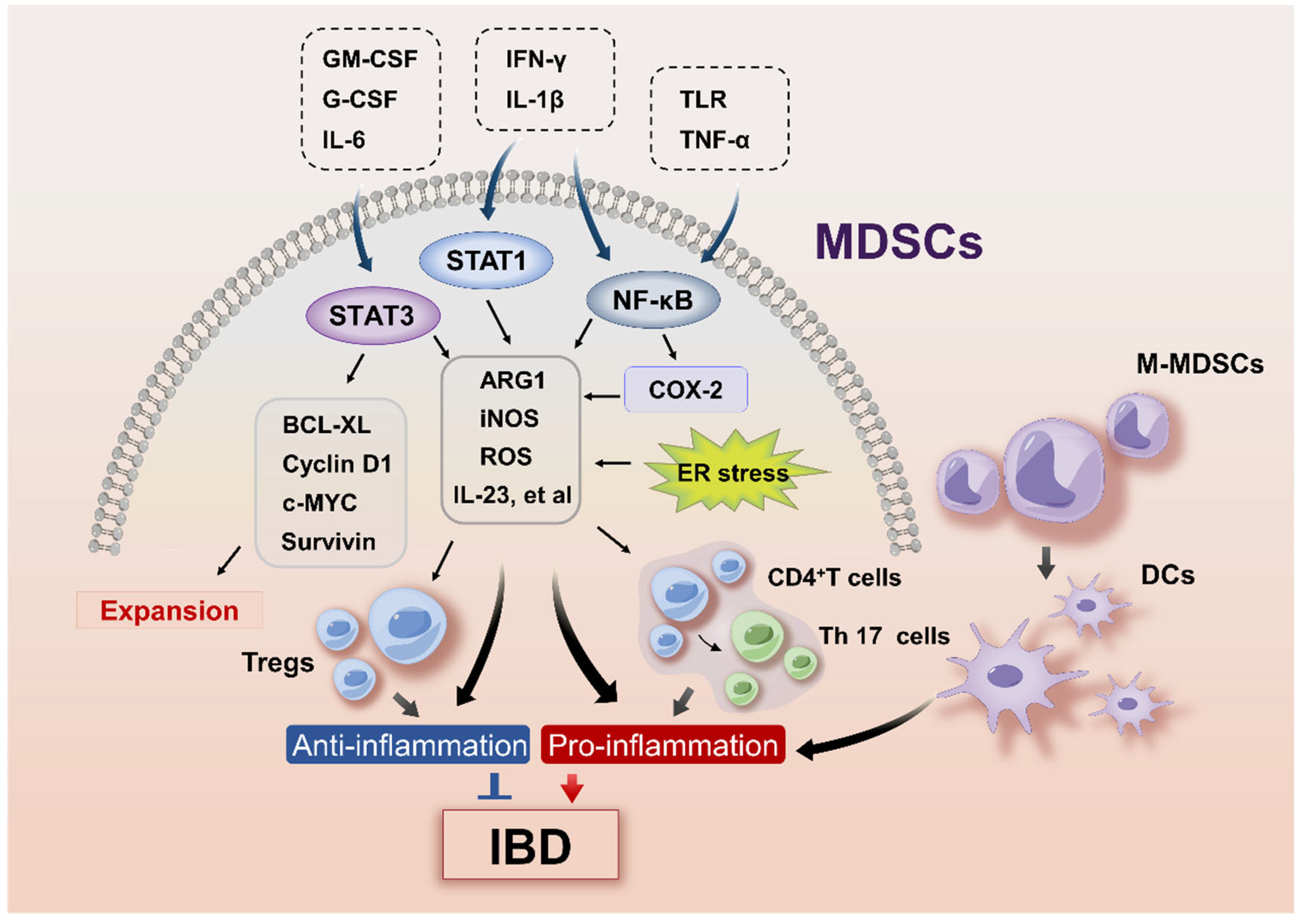
3.2. Signaling Pathways Regulating MDSC Expansion in IBD

The intracellular pathways reported to orchestrate MDSCs in IBD include STAT3 and GM-CSF (Figure 1). The upregulation of STAT3 is a hallmark of MDSCs and is directly involved in their expansion and activation [13]. The abnormal and persistent activation of STAT3 in MDSCs upregulated the expression of B-cell lymphoma XL (BCL-XL), cyclin D1, c-MYC, survivin protein, and S100 calcium-binding proteins A8/9 (S100A8/9) that prevented their apoptosis and promoted proliferation [13]. In recent years, studies have reported Stat3-activated suppressive MDSCs in IBD mouse models [31,32]. In a DSS-induced colitis model, the hyperactivation of Stat3 in myeloid cells in gp130757F/F mice ameliorated intestinal inflammation, which was attributed to the expansion and activation of MDSCs [36]. DSS-treated LysMcre/Stat3flox mice with myeloid-specific Stat3 deficiency showed diminished G-MDSCs and M-MDSCs in the colon compared to gp130757F/F mice [36]. In addition, the STAT signals may control MDSC generation in mice. It was shown that the inhibition of enhancer of zest homolog 2 (Ezh2), a major histone methyltransferase, promoted the differentiation of hematopoietic progenitor cells (HPCs) into MDSCs by activating the Janus kinase (JAK)-STAT and TNF signaling pathways in DSS-induced colitis [33].
STAT3 activation in MDSCs may require growth factors including GM-CSF, G-CSF, and IL-6 [37]. GM-CSF and IL-6 are the major myeloid growth factors responsible for MDSC expansion, which was confirmed by a study showing that human MDSCs with potent suppressive capacity were best generated by GM-CSF plus IL-6, whereas GM-CSF alone had weaker effects [38]. The expansion of MDSCs in the colon of mice with colitis was accompanied by increased GM-CSF and IL-6 in different IBD mouse models [22,24,35], suggesting that GM-CSF and IL-6 may be involved in MDSC expansion in IBD. GM-CSF is mainly secreted by CD4+ T cells, group 3 innate lymphoid cells, and Paneth cells in the intestinal mucosa [39]. Notably, GM-CSF is also one of the signature cytokines for the IL-23 pathway that precipitates the onset and progression of IBD [40]. In addition to STATs, NOD-like receptor protein 3 (NLRP3) is implicated in the recruitment of MDSCs to the tumor site [41,42]. NLRP3 mediates the conversion of pro-caspase-1 to its mature form to generate IL-1β and IL-18 [43], while caspase-1-mediated inflammasome pathways control TNBS-induced colitis in mice [28]. MDSCs were significantly upregulated in the bone marrow, spleen, Peyer’s patches, and LP of the TNBS-treated WT mice but not in TNBS-treated caspase-1 knockout mice, suggesting that caspase-1-mediated inflammasome pathways promote the recruitment of MDSCs.

3.3. Signaling Pathways Involved in MDSC Activation in IBD

The STAT3 and NF-κB pathways appear to be the major regulators of MDSC activation in IBD (Figure 1). STAT3 and NF-κB upregulated the expression of arginase 1 (ARG1) and inducible nitric oxide synthase (iNOS), the main suppressors of MDSCs in murine colitis induced by dinitrobenzene sulfonic acid (DNB) or DSS [31,32]. NF-κB is positively related to the severity of intestinal inflammation [44] and is activated by various proinflammatory mediators, such as TLR ligands, IFN-γ, IL-1β, and TNF-α, all of which have been shown to increase the suppressive activity of MDSCs [21,45,46]. Indeed, increased concentrations of IL-1β, TNF-α, and IFN-γ in DSS colitis was accompanied by the expansion of functional MDSCs [47]. IFN-γ produced by T cells upregulated ARG1 and iNOS in MDSCs in a STAT1-dependent manner; while the blockade of IFN-γ abolished MDSC-mediated T cell suppression [48]. The enhanced IFN-γ response was also accompanied by upregulation of iNOS in MDSCs in Il-17a−/− mice in Il10-deficiency-induced IBD [35]. In addition, the canonical activation of NF-κB leads to the production of COX-2 [49]. COX-2 and COX-2-derived PGE2 were shown to stimulate the expression of ARG1 and iNOS to activate suppressive MDSCs in cancers [50,51,52], although the findings have not yet been confirmed in IBD. In recent years, endoplasmic reticulum (ER) stress response has also emerged as an important mechanism regulating MDSC activation. Three major sensors of ER stress exist in mammalian cells: protein kinase RNA-like ER kinase (PERK), inositol-requiring enzyme 1 (IRE1), and activating transcription factor 6 (ATF6). CCAAT-enhancer-binding protein homologous protein (CHOP) and spliced X-box binding protein-1 (XBP1s) are vital effectors of the PERK and IRE1 pathways, respectively [53]. ER stress promotes the suppressive activity of MDSCs by inducing the expression of ARG1, iNOS, and NADPH oxidase 2 (NOX2) [54,55]. ER stress and the unfolded protein response (UPR) have been functionally and genetically linked to human IBD [56,57]. It remains elusive how ER stress in IBD regulates the expansion and activation of MDSCs.

3.4. Hormone-Driven MDSC Activity in IBD

In a DNB-induced IBD mouse model, estrogen steroid 17β-estradiol was reported to mitigate IBD by augmenting the suppressive activity of MDSCs [31]. In DSS colitis, the activation of estrogen receptors increased MDSC-derived ARG1 and consequently alleviated colitis [32]. The cases, deaths, and DALYs of IBD were higher among females than males [4]. Several studies have reported sex differences in patients with IBD. A pooled analysis of population-based studies from the Asia–Pacific region showed that CD and UC patients were predominantly male in most age groups from adolescence to middle/late-middle age [58]. However, a pooled analysis of population-based studies from Western countries reported that female patients had a lower risk of CD during childhood but a higher risk of CD thereafter, whereas men had a higher incidence of UC than women after the age of 45 [59]. A cross-sectional cohort study uncovered that over half of the women with IBD reported worsening symptoms during menses, whereas symptom worsening during pregnancy was more commonly seen in UC than CD. Moreover, oral contraceptive use has been associated with an increased risk of CD in female patients [60]. Sex differences in immune response are likely related to the differential organization of chromosomes, reproductive organs, and levels of sex steroids [61]. The distribution of MDSCs and subsets has been reported to be sex-specific in glioblastoma [62], systemic lupus erythematosus [63], obesity [55], arthritis [64], and breast milk for newborns [65]. The sex-dependent variations affect tumorigenesis and immunotherapy response [66]. Bayik, et al. reported that males had enhanced M-MDSCs accumulation in the tumor microenvironment, while females had elevated G-MDSCs in the peripheral circulation. This distinctive abundance of MDSC subsets may result in sex differences in anti-tumor immunity. It was further identified that G-MDSCs targeted with an anti-IL-1β antibody provided survival benefits to female tumor-bearing mice, while M-MDSCs targeted with fludarabine extended survival in male tumor-bearing mice [67]. These studies indicate that the unique characteristics of MDSC subsets driven by sex may determine their response to different therapies. Further investigations are needed to explore how sex is a factor in regulating MDSCs in IBD.

4. MDSCs in IBD Pathogenesis

MDSCs are known for their immunosuppressive activity on T cells, B cells, and natural killer (NK) cells. M-MDSCs and G-MDSCs utilize different mechanisms to interfere with the proliferation, trafficking, and viability of lymphocytes. G-MDSCs express high levels of ARG1 and NOX2, leading to increased reactive oxygen species (ROS) and decreased nitric oxide (NO) [13]. ROS is short-lived and unstable, which requires direct MDSC–T cell contact that renders T cells unresponsive to specific antigens. M-MDSCs appear to be more suppressive than G-MDSCs when assessed on a per-cell basis [68]. M-MDSCs expressed high levels of ARG1, transforming growth factor (TGF)-β, IL-10, and iNOS, which resulted in high NO and low ROS production [10,68,69,70]. NO has a longer half-life than ROS and allows cell–cell interaction via cellular proximity without direct cell contact. This enables M-MDSCs to suppress T cell responses both in antigen-specific and non-specific manners.

4.1. The Protective Role of MDSCs in IBD

The excessive and prolonged activation of adaptive immunity drives tissue damage in IBD. Adaptive responses are orchestrated by a combination of resident and recruited cell populations, including a variety of T cells including T-helper (Th)1, Th2, Th17, and regulatory T cells (Tregs) [7]. Most studies of MDSCs in IBD have concentrated on their interaction with T cells. MDSCs were shown to inhibit the proliferation of CD4+ T cells and promote the differentiation of Tregs and Th17 cells [71,72]. Touw, et al. reported that glatiramer acetate promoted MDSC-dependent Treg conversion while inhibiting the secretion of proinflammatory cytokines in DSS colitis in mice [73]. Lee, et al. showed that Tregs and MDSCs established a positive feedback loop during murine colitis, in which Treg enhanced the formation of M-MDSCs but not G-MDSCs, and boosted their inhibitory functions by secreting TGF-β [74].
The protective role of MDSCs was first uncovered by cell transfer and depletion experiments. Most studies confirmed the beneficial effects of MDSC transfer in mouse IBD models. The adoptive transfer of splenic MDSCs isolated from mice with colitis attenuated both acute and chronic intestinal inflammation [22,75]. The depletion of myeloid cells using an anti-Gr-1 antibody exacerbated intestinal inflammation in mice [76,77]. ARG1, iNOS, and ROS appear to be the major factors that mediate the suppressive activity of MDSCs on T cells in IBD. MDSCs inhibited T cell proliferation through competition for L-arginine, which serves as a substrate for iNOS and arginase [78,79]. A similar expression of ARG1 was found in CD14+CD19HLA-DR−/lo M-MDSCs from healthy controls and IBD patients. ARG-1-expressing M-MDSCs suppressed the proliferation of patients’ PBMCs in a dose-dependent manner [22]. In DSS colitis, Arg-1 deletion in myeloid cells attenuated the protective role of MDSCs during colitis by upregulating IL-17A and IL-17F, while the adoptive transfer of suppressive MDSCs alleviated colitis in ArgmyeKO mice [32].
ROS has cytotoxic effects in many conditions but is intrinsically involved in the transcriptional and metabolic reprogramming of MDSCs, thus influencing their differentiation and immunosuppressive activity. MDSC-derived ROS modified TCR and CD8 molecules and caused CD8+ T cells to lose their ability to bind phosphorylated MHC, leading to antigen-specific tolerance [80]. iNOS interplayed with ARG1 and NOX-derived ROS to produce peroxynitrite (ONOO-) that inhibits the proliferation, migration, and antigen-specific non-responsiveness of T cells [81,82,83]. IFN-γ produced by activated T cells triggered iNOS expression in suppressive MDSCs [84]. In a Rag1−/− IBD model, expanded M-MDSCs suppressed T cell proliferation in an iNOS-, IFN-γ-, COX-1/2-, and cell contact-dependent manner, without inducing their apoptosis [34]. M-MDSCs suppressed the production of Th1- and Th2-type cytokines and enhanced the generation of Th17 and foxp3+ T cells [34]. In addition, the neuroendocrine pathways also participate in the regulation of MDSC activity. Zheng, et al. reported an IL-10-dependent inhibitory effect of M-MDSCs on DSS-induced colitis. Acetylcholine promoted IL-10 secretion by M-MDSCs and suppressed inflammation by activating the nicotinic acetylcholine receptor (nAChR)/extracellular signal-regulated kinase (ERK) pathways, whereas the expression of iNOS and ARG1 remained unchanged [85]. Pituitary hormone α-MSH was found to act on bone marrow progenitors to boost myelopoiesis, MDSC accumulation, and immunosuppression through its melanocortin receptor MC5R [86]. The finding that MC5R displayed cytoplasmic staining at the mononuclear and polymorphonuclear inflammatory infiltrate levels in IBD patients suggests a potential role of melanocortin system in regulating MDSCs in IBD [87].

4.2. The Proinflammatory Function of MDSCs in IBD

Although previous findings suggested a protective role of MDSCs in IBD, several studies showed a proinflammatory role of MDSCs in intestinal inflammation (Figure 1). CD33+CD15+HLA-DR−/loCD14 G-MDSCs isolated from peripheral blood of patients with active IBD was found to stimulate CD4+CD25 T cell proliferation [25]. Furthermore, the adoptive transfer of bone marrow-derived Gr1+CD11b+ MDSCs to TNBS-treated mice worsened the colitis, suggesting that this inflammatory environment switched off the suppressive capacity of MDSCs. The proinflammatory role of MDSCs has been reported in multiple autoimmune diseases, such as psoriasis, systemic lupus erythematosus, and rheumatoid arthritis [88,89,90]. The excessive proliferation of MDSCs led to augmented Th17 cell infiltration through the secretion of inflammatory cytokines including IL-23, IL-1β, and IL-6 [91,92].
The underlying mechanisms for the paradoxical effects of MDSCs in IBD remain unclear, one explanation being related to iNOS and ROS. iNOS enhances production of NO, which plays a complex role in the pathogenesis of IBD. NO compromises the integrity of intestinal epithelial barrier, which allows the translocation of luminal bacteria, toxins, and antigens into the mucosa, initiating intestinal inflammation [93]. Increased NO produced by MDSCs was shown to exacerbate colitis and promote death of Il-10−/− mice with colitis, though those MDSCs remained suppressive to CD4+ T cells [35]. In addition, Zigmond, et al. reported proinflammatory CX3CR1-GFPintLy6Chi monocytes, with a similar phenotype as M-MDSCs, highly expressed Trem, Inos, Il-6, and Il-23 in mice with DSS colitis. The formation of those proinflammatory monocytes required TLR2 and nucleotide-binding oligomerization domain 2 (NOD2) [20]. ROS may also contribute to the pathogenic function of MDSCs in IBD by causing progressive cellular damage and tissue destruction [80].

4.3. Plasticity of M-MDSCs in IBD

In recent years, more evidence points out the functional plasticity of heterogeneous MDSCs in experimental models of IBD. These characteristics are mainly associated with M-MDSCs, whereas G-MDSCs are non-proliferating cells with a short half-life [94]. Several studies uncovered the possible differentiation fates of Ly6Chi monocytes and identified them as a driver for colonel inflammation in IBD [20,95,96]. The Ly6Chi monocytes have a similar phenotype to M-MDSCs in IBD. Chen, et al. reported that CD11b+CD14+CX3CR1+ dendritic cells (DCs) in the intestinal LP were derived from Ly6Chi monocytes in a GM-CSF-dependent manner. These Ly6Chi monocytes-derived DCs exacerbated DSS-induced colitis by secreting TNF-α [95]. Aymeric, et al. further elaborated the distinctive patterns of M-MDSCs in the steady state and inflammatory settings. Ly6Chi monocytes in the blood were identified as the precursors of F4/80hiCX3CR1hiCD11c+ macrophages in noninflammatory environment, whereas Ly6Chi monocytes were recruited to the colonic LP and differentiated into proinflammatory CD103CX3CR1intCD11b+ DCs in a T cell transfer model of colitis. Those proinflammatory DCs produced high levels of IL-12, IL-23, iNOS, and TNF-α and drove the differentiation of IFN-γ–producing T cells [96]. Zigmond, et al. further uncovered the differentiation of Ly6Chi monocytes with time in murine IBD: grafted Ly6Chi monocytes differentiated into CX3CR1-GFPintLy6ChiF4/80loCD11c cells 1 day after adoptive transfer into DSS-challenged mice, then, by 3 days after, they became antigen-presenting CX3CR1-GFPintLy6CloF4/80lo cells with high expression of MHCII and CD11c [20].

5. Effect of MDSCs During the Development of IBD-Associated Colorectal Cancer

Long-standing inflammation in IBD poses an increased risk of colorectal cancer [95]. MDSCs may contribute to IBD-initiated carcinogenesis by inhibiting cytotoxic T lymphocytes (CTLs), driving STAT3-dependent proliferation signals in intestinal epithelial cells (IECs), and inducing DNA damage by producing ROS [96]. Recent studies have highlighted the role of MDSCs in the immune microenvironment of IBD (Figure 2; discussion above). Poh, et al. showed that the chronic circulation of MUC1loCD11b+Gr1+ cells in the peripheral blood of IBD patients activated pro-tumorigenic pathways for colorectal cancer [97]. Mucin 1 (MUC1) is a tumor-associated antigen and a susceptibility gene for CD [98]. Low MUC1 expression on MDSCs promoted the expansion and suppressive capacity of MDSCs. This was accompanied by significantly reduced inflammatory lesions in the colon of mice with colitis-associated cancer (CAC) induced by azoxymethane (AOM)/DSS, indicating that the recruitment and sustained activation of MDSCs contribute to CAC [97].

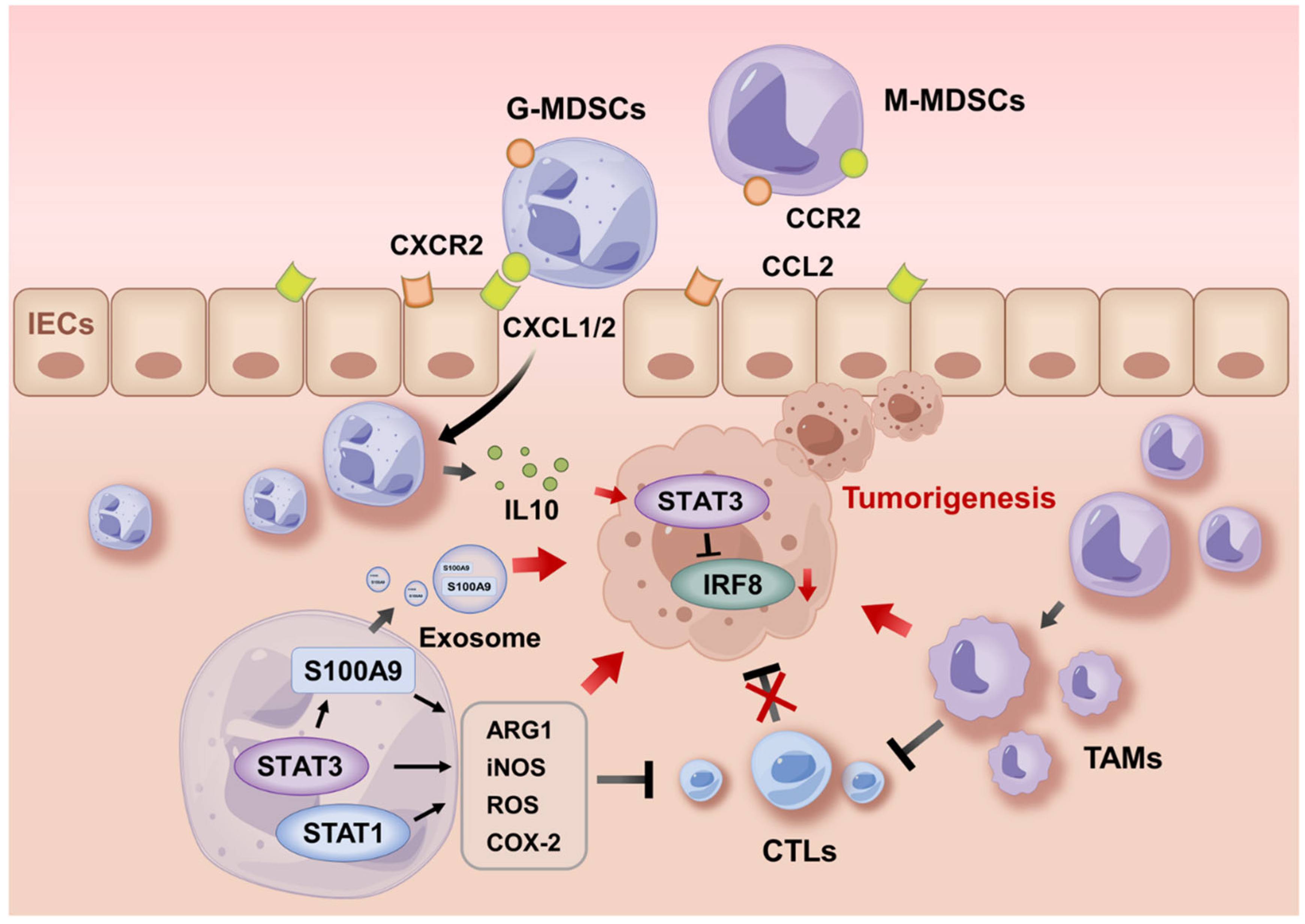
5.1. MDSC Recruitment During Tumorigenesis

How are MDSCs recruited from the peripheral blood into inflamed colonic mucosa and tumors? Katoh, et al. showed that the pro-tumorigenic chemokine receptor CXCR2 was required for the recruitment of suppressive MDSCs, which was promoted by the PGE2-induced upregulation of CXCR2 ligands in colonic mucosa and tumors in an AOM/DSS-induced CAC model [97]. This was verified by the findings that downregulated CXCL2 reduced the infiltration of MDSCs and tumor growth, while early-life microbiota restricted MDSC-driven carcinogenesis by dampening the production of CXCL2 [98,99]. MDSC recruitment may also be mediated by other chemokines, cytokines, and growth factors. In AOM/DSS-induced and T-bet−/−, Rag2−/− CAC models, IL-6, CCL2, G-CSF, and GM-CSF (encoded by Csf2) fostered MDSC accumulation in evolving colonic tumors and boosted T cell suppression by MDSCs in a STAT-dependent manner [100,101,102]. In AOM/DSS-induced CAC, the number of MDSCs decreased significantly in Csf2−/− mice with thwarted cancer, whereas the adoptive transfer of MDSCs from tumor-bearing mice into Csf2−/− counterparts led to cancer recurrence [103]. Plasmacytoid dendritic cells (pDCs) impeded MDSC recruitment during AOM/DSS-induced CAC by producing type I IFN that suppresses the release of CCL2 and CXCL1 [104]. STAT1 is also required for G-MDSC accumulation in both the spleen and blood via IL-17 during the initial phase of CAC. The blockade of IL-17 in Stat1−/− mice diminished G-MDSCs in murine CAC [105], indicating the functional role of STAT1 and STAT3 in the recruitment and activation of MDSCs.
Additionally, Chen, et al. reported that Olfm4 deficiency in myeloid cells abrogated the recruitment of G-MDSCs, leading to delayed progression of AOM/DSS-induced CAC [106]. Olfm4 deletion in myeloid cells blunted tumorigenesis but aggravated DSS-induced acute colitis in mice, which may be due to decreased frequency and immunosuppressive activity of G-MDSCs [106]. Those studies highlight a complex role of MDSCs in IBD and CAC: on one hand, suppressive MDSCs may alleviate intestinal inflammation, which is supported by data from adoptive transfer of suppressive MDSCs into IBD mice; on the other hand, expanded suppressive MDSCs in the peripheral blood may help initiate CAC by hampering anti-tumor immunity.

5.2. Activities of MDSCs During Tumor Progression

Recruited MDSCs mediate the immune escape of tumor cells through both T cell-dependent and -independent effects. Wang, et al. reported that increased commensal fungus in Card−/− mice induced MDSC accumulation, upregulated S100a9 and Arg-1 expression, and inhibited effector T cells, leading to accelerated AOM/DSS CAC in mice [107]. Wu, et al. further reported that the inhibition of S100A9 and STAT3 lowered ROS and ARG1 in MDSCs, restoring T cell responses in murine CAC [108]. In terms of non-T cell-dependent effects, Wan, et al. reported that S100A9 in G-MDSC-derived exosomes promoted the stemness of colon cancer cells, fostering their susceptibility to both AOM/DSS CAC and CT26 cell-induced cancer independent of T cells [109]. MDSC-derived IL-10 activated STAT3 in colonic epithelial cells that directly bond to the Dnmt1 and Dnmt3b promoters to stimulate their expression, which then silenced tumor suppressor Irf8 and advanced AOM/DSS CAC [110]. Moreover, the plasticity of M-MDSCs allowed them to differentiate into M2-like tumor-associated macrophages (TAMs) to drive tumor progression. Xun, et al. reported that natural product Dioscin halted AOM/DSS CAC by inducing the differentiation of MDSCs into the M1- but not M2-like phenotype [111]. G-MDSCs were reported to foster M-MDSC differentiation by producing exosomes. Starting from the inflammation stage of the AOM/DSS model, G-MDSCs accelerated the transition from colitis to cancer by secreting exosomes that drive the accumulation of M2 macrophages in colorectal tissue. G-MDSC-derived exosomes contributed to the differentiation of CD11b+Ly6GLy6C+ M-MDSCs into CD11b+F4/80+CD206+Ly6Clo M2 macrophages through miR-93–5p cargo-mediated STAT3 inhibition [112].
Several other mechanisms in the activation of suppressive MDSCs may also regulate tumorigenesis. An elevated level of COX-2 was reported in AOM/DSS-induced murine CAC [113,114]. COX-2 produced by MDSCs may induce chromosomal instability and epithelial cell transformation leading to carcinogenesis [115,116]. Of note, COX-2 inhibitor Celebrex is approved by the U.S. Food and Drug Administration for the chemoprevention of familial adenomatous polyposis, an inherited cancer syndrome [117,118]. Excessive ROS production is associated with epithelial cell damage in IBD, and elevated ROS was observed in many types of cancers and likely involved in metastasis [119]. During IBD, MDSC-derived ROS may contribute to the development of dysplastic lesions [120,121]. In AOM/DSS-induced CAC, MDSCs displayed immunosuppressive activity by promoting STAT3-mediated ROS production [122]. Increased ROS generated by MDSCs may also induce ER stress, which plays a critical role in maintaining the suppressive function of MDSCs (as above). Suppressive MDSCs from cancer patients and from mice bearing EL4 and EG7 thymomas, LLC lung carcinoma, CT26 colon carcinoma, or 4T1 mammary carcinoma exhibited higher ER stress response compared with healthy controls. Upregulated XBP1 and CHOP may increase the expression of death receptor 5 (DR5), which shortened MDSCs’ lifespan to stimulate the proliferation of their precursors, thus supporting their expansion and accumulation [123]. The endogenous production of ROS or peroxynitrite in MDSCs has been shown to activate the expression of CHOP [124]. In tumor-bearing mice, Chop deficiency reduced the expansion and immunosuppressive activity of MDSCs [124]. Despite those findings, whether ER stress promotes colitis-associated tumorigenic process by inducing immunosuppressive MDSCs remains unknown.

6. Discussion

MDSCs expand systemically and locally as a part of the immunosuppressive response to limit tissue injury in IBD. MDSC subsets exhibit a broad distribution and expansion patterns across the disease stages of IBD (Table 1). The immunosuppressive function of MDSCs failed to dampen acute intestinal inflammation in multiple preclinical models. Moreover, persistent activation driven by recurrent epithelial injury or microbial translocation in IBD may reshape them into more heterogeneous phenotypes. Key unanswered questions include how MDSCs interact with gut microbiota, epithelial cells, and other immune cells to exhibit disease stage- and tissue-specific behaviors in IBD. Exploring the distribution and corresponding functions of MDSCs and their subsets in active disease and the remission of IBD will help better understand their clinical significance. In addition, the importance of physical conditions and functionality should not be underestimated in the management of IBD. E.g., physical activities were reported to be beneficial in quiescent or mild IBD and associated with a lower risk of active disease [125]. Recent studies introduced physical exercise as a promising strategy in cancers and inflammatory diseases due to its effect on immune cells, including inhibiting MDSC infiltration [126,127,128,129]. Whether physical exercise benefits IBD through regulating MDSCs remains unclear.
M-MDSCs were reported to be the major subset in peripheral blood in some IBD models [30,32,36,85]. There is substantial variability in M-MDSC/G-MDSC ratios and functions in the colon and peripheral lymphoid organs in mice. The mice’s genetic backgrounds, microbiome, and experimental design may count for these discrepancies. Though MDSCs were identified in the colon of almost all IBD mouse models and the peripheral blood of IBD patients, the reporting of colonic MDSCs in IBD patients has been limited. The discrepancy may be partly due to the lack of characteristic markers in humans, tissue-specific distribution of MDSCs, and stages of the disease. Previous reports have linked MDSC expansion to tumorigenesis. The immunostaining of UC colon tissues showed a low expression of the G-MDSCs marker CD15, which increased during the colitis-to-cancer transition [130]. The migration of MDSCs to tumors likely creates fertile ground for carcinogenesis and metastasis. The spleen has been reported to be the reservoir of CD11b+ Ly-6Chi monocytic and CD11b+ Ly-6Ghi granulocytes in mice [131]. After migrating to the tumor site, the inflammatory and hypoxic tumor microenvironment upregulated the expression of suppressive factors in MDSCs and mediated the differentiation of M-MDSCs into TAM. Consistently, tumor MDSCs possessed a more potent suppressive activity than splenic MDSCs [132]. This may be partly explained by different G-MDSC/M-MDSC ratios in peripheral lymphoid organs and tumors. In the peripheral lymphoid organs of most cancer patients, G-MDSCs appear to be the major subset, whereas the proportion of M-MDSCs was higher in the tumor itself [133]. Further understanding of the mechanisms that drive the distribution of MDSCs and the shift between proinflammatory and anti-inflammatory phenotypes may help develop new immunotherapeutic interventions.
The therapeutic strategies to target MDSCs include chemotherapy, natural products, small molecule inhibitors and agonists, and antibodies, as well as the transfer of MDSCs or MDSC-derived exosomes. In cancers, chemotherapy drugs, antibodies, and inhibitors were tested to impede the recruitment and activity of MDSCs [134,135]. In IBD, targeted therapies including biologics and small molecules have revolutionized its management. TNF antibodies, IL-12/IL-23p40 antibody (ustekinumab), selective IL-23p19 antibodies (e.g., risankizumab, guselkumab, mirikizumab), and JAK inhibitors (e.g., tofacitinib, upadacitinib), which have demonstrated efficacy in IBD, may also impact myeloid cells [136]. For instance, tofacitinib was shown to induce significantly higher numbers of MDSCs and PMN-MDSCs in mice, accompanied by decreased Th17 cells and ILC1s [137,138]. Myeloid cells expressing Fc γ receptor 1 (or CD64) have been identified as a key source of IL-23 in inflamed IBD tissues. Guselkumab with a native Fc domain binds to CD64 on IL-23–producing inflammatory monocytes while capturing secreted IL-23 molecules [139], although the impact of guselkumab on MDSCs remains unknown. More preclinical and clinical studies are needed to define the role of MDSCs in response to those targeted treatments. Although the expansion of proinflammatory MDSCs was reported in patients and mice with IBD [20,25,35], clinical benefits of modulating MDSCs have not been reported. This may be explained by findings that the depletion of myeloid cells using anti-Gr-1 antibodies exacerbated intestinal inflammation. In addition, considering the critical role of suppressive MDSCs in carcinogenesis, the safety of MDSC activators or transfer of MDSCs needs to be carefully evaluated.

7. Conclusions

The expansion of MDSCs has been widely reported in IBD patients and mouse models of IBD. There is substantial variability in the activity and distribution of MDSCs in different models. The chronic inflammatory environment and sex hormones likely drive the expansion and activation of MDSCs. The functional and clinical significance of MDSCs in IBD have been clouded by both pro-homeostatic and proinflammatory roles. Moreover, prolonged suppressive G-MDSCs in the peripheral blood may promote the colitis-to-cancer transition in the colon. More studies are needed to explore the complex immune regulation of MDSCs in IBD and CAC in animal models and patients.

Author Contributions

Writing—original draft preparation, Y.Z.; writing—review and editing, S.C. All authors have read and agreed to the published version of the manuscript.

Funding

The work was funded by an NIH/NIDDK K08 Clinical Investigator Award (1K08DK140612-01, Siyan Cao). Siyan Cao was also supported by a Crohn’s & Colitis Foundation Career Development Award (CCF1062472), American Gastroenterological Association Fellowship-to-Faculty Transition Award (AGA2023-32-03), and Washington University Clinical and Translational Research Funding Program (CTRFP1714).

Institutional Review Board Statement

Not applicable.

Informed Consent Statement

Not applicable.

Data Availability Statement

No new data were created or analyzed in this study. Data sharing is not applicable to this article.

Conflicts of Interest

The authors declare no conflicts of interest.

Abbreviations

AOM, azoxymethane; ARG1, Arginase 1; ATF6, activating transcription factor 6; BCL-XL, B-cell lymphoma XL; CAC, colitis-associated cancer; CCL2, C-C motif chemokine ligand 2; CD, Crohn’s disease; CHOP, CCAAT-enhancer-binding protein homologous protein; COX-2, cyclo-oxygenase-2; CTLs, cytotoxic T lymphocytes; CRC, colorectal cancer; CXCR2, CXC chemokine receptor 2; DALYs, disability-adjusted life-years; DCs, dendritic cells; DR5, death receptor 5; DSS, dextran sulfate sodium; ER, endoplasmic reticulum; ERK, extracellular regulated protein kinases; Ezh2, zest homolog 2; FSC, forward scatter; GM-CSF, granulocyte-macrophage colony-stimulating factor; G-MDSC/PMN-MDSCs, granulocytic/polymorphonuclear MDSCs; HPCs, hematopoietic progenitor cells; IBD, inflammatory bowel disease; IECs, intestinal epithelial cells; IFN-γ, interferon-γ; IL-6, interleukin-6; IRE1, inositol-requiring enzyme 1; IRF8, interferon regulatory factor 8; iNOS, inducible nitric oxide synthase; JAK, Janus kinase; MDSCs, myeloid-derived suppressor cells; MUC1, mucin 1; M-MDSCs, monocytic MDSCs; NF-κB, nuclear factor kappa-B; NK cells, natural killer cells; NLRP3, NOD-like receptor protein 3; NO, nitric oxide; NOD2, nucleotide-binding oligomerization domain 2; NOX2, NADPH oxidase 2; ONOO-, peroxynitrite; PERK, protein kinase RNA-like ER kinase; PGE2, prostaglandin E2; ROS, reactive oxygen species; SSC, side scatter; ROS, reactive oxygen species; STAT3, signal transducer and activator of transcription 3; S100A8/9, S100 calcium-binding proteins A8/9; TAMs, tumor-associated macrophages; TGF-β, transforming growth factor-β; TLRs, toll-like receptors; TNBS, 2,4,6-trinitrobenzene sulfonic acid; Th1, T-helper 1; Tregs, regulatory T cells; UC, ulcerative colitis; UPR, unfolded protein response; XBP1, X-box binding protein-1; nAChRs, nicotinic acetylcholine receptors.

References

  1. Gabrilovich, D.I.; Bronte, V.; Chen, S.-H.; Colombo, M.P.; Ochoa, A.; Ostrand-Rosenberg, S.; Schreiber, H. The terminology issue for myeloid-derived suppressor cells. Cancer Res. 2007, 67, 425. [Google Scholar] [CrossRef]
  2. Tesi, R.J. MDSC; the Most Important Cell You Have Never Heard Of. Trends Pharmacol. Sci. 2019, 40, 4–7. [Google Scholar]
  3. Buie, M.J.; Quan, J.; Windsor, J.W.; Coward, S.; Hansen, T.M.; King, J.A.; Kotze, P.G.; Gearry, R.B.; Ng, S.C.; Mak, J.W.Y.; et al. Global Hospitalization Trends for Crohn’s Disease and Ulcerative Colitis in the 21st Century: A Systematic Review With Temporal Analyses. Clin. Gastroenterol. Hepatol. 2023, 21, 2211–2221. [Google Scholar]
  4. Wang, R.; Li, Z.; Liu, S.; Zhang, D. Global, regional and national burden of inflammatory bowel disease in 204 countries and territories from 1990 to 2019: A systematic analysis based on the Global Burden of Disease Study 2019. BMJ Open 2023, 13, e065186. [Google Scholar]
  5. Barreiro-de Acosta, M.; Molero, A.; Artime, E.; Díaz-Cerezo, S.; Lizán, L.; de Paz, H.D.; Martín-Arranz, M.D. Epidemiological, Clinical, Patient-Reported and Economic Burden of Inflammatory Bowel Disease (Ulcerative colitis and Crohn’s disease) in Spain: A Systematic Review. Adv. Ther. 2023, 40, 1975–2014. [Google Scholar]
  6. Fornaro, R.; Caratto, M.; Caratto, E.; Caristo, G.; Fornaro, F.; Giovinazzo, D.; Sticchi, C.; Casaccia, M.; Andorno, E. Colorectal Cancer in Patients With Inflammatory Bowel Disease: The Need for a Real Surveillance Program. Clin. Color. Cancer 2016, 15, 204–212. [Google Scholar]
  7. Xavier, R.J.; Podolsky, D.K. Unravelling the pathogenesis of inflammatory bowel disease. Nature 2007, 448, 427–434. [Google Scholar] [CrossRef]
  8. Saez, A.; Herrero-Fernandez, B.; Gomez-Bris, R.; Sánchez-Martinez, H.; Gonzalez-Granado, J.M. Pathophysiology of Inflammatory Bowel Disease: Innate Immune System. Int. J. Mol. Sci. 2023, 24, 1526. [Google Scholar] [CrossRef]
  9. Talmadge, J.E.; Gabrilovich, D.I. History of myeloid-derived suppressor cells. Nat. Rev. Cancer 2013, 13, 739–752. [Google Scholar] [CrossRef]
  10. Bronte, V.; Brandau, S.; Chen, S.-H.; Colombo, M.P.; Frey, A.B.; Greten, T.F.; Mandruzzato, S.; Murray, P.J.; Ochoa, A.; Ostrand-Rosenberg, S.; et al. Recommendations for myeloid-derived suppressor cell nomenclature and characterization standards. Nat. Commun. 2016, 7, 12150. [Google Scholar]
  11. Damuzzo, V.; Pinton, L.; Desantis, G.; Solito, S.; Marigo, I.; Bronte, V.; Mandruzzato, S. Complexity and challenges in defining myeloid-derived suppressor cells. Cytom. B Clin. Cytom. 2015, 88, 77–91. [Google Scholar] [CrossRef]
  12. Yang, R.; Cai, Z.; Zhang, Y.; Yutzy, W.H.; Roby, K.F.; Roden, R.B.S. CD80 in immune suppression by mouse ovarian carcinoma-associated Gr-1+CD11b+ myeloid cells. Cancer Res. 2006, 66, 6807–6815. [Google Scholar] [CrossRef]
  13. Gabrilovich, D.I.; Nagaraj, S. Myeloid-derived suppressor cells as regulators of the immune system. Nat. Rev. Immunol. 2009, 9, 162–174. [Google Scholar] [CrossRef] [PubMed]
  14. Zou, Y.; Kamada, N.; Seong, S.-Y.; Seo, S.-U. CD115- monocytic myeloid-derived suppressor cells are precursors of OLFM4high polymorphonuclear myeloid-derived suppressor cells. Commun. Biol. 2023, 6, 272. [Google Scholar] [CrossRef]
  15. Haile, L.A.; Gamrekelashvili, J.; Manns, M.P.; Korangy, F.; Greten, T.F. CD49d is a new marker for distinct myeloid-derived suppressor cell subpopulations in mice. J. Immunol. 2010, 185, 203–210. [Google Scholar] [CrossRef] [PubMed]
  16. Biermann, H.; Pietz, B.; Dreier, R.; Schmid, K.W.; Sorg, C.; Sunderkötter, C. Murine leukocytes with ring-shaped nuclei include granulocytes, monocytes, and their precursors. J. Leukoc. Biol. 1999, 65, 217–231. [Google Scholar] [CrossRef] [PubMed]
  17. Ostanin, D.V.; Kurmaeva, E.; Furr, K.; Bao, R.; Hoffman, J.; Berney, S.; Grisham, M.B. Acquisition of antigen-presenting functions by neutrophils isolated from mice with chronic colitis. J. Immunol. 2012, 188, 1491–1502. [Google Scholar] [CrossRef]
  18. Cassetta, L.; Baekkevold, E.S.; Brandau, S.; Bujko, A.; Cassatella, M.A.; Dorhoi, A.; Krieg, C.; Lin, A.; Loré, K.; Marini, O.; et al. Deciphering myeloid-derived suppressor cells: Isolation and markers in humans, mice and non-human primates. Cancer Immunol. Immunother. 2019, 68, 687–697. [Google Scholar] [CrossRef]
  19. Sagiv, J.Y.; Michaeli, J.; Assi, S.; Mishalian, I.; Kisos, H.; Levy, L.; Damti, P.; Lumbroso, D.; Polyansky, L.; Sionov, R.V.; et al. Phenotypic diversity and plasticity in circulating neutrophil subpopulations in cancer. Cell Rep. 2015, 10, 562–573. [Google Scholar] [CrossRef]
  20. Zigmond, E.; Varol, C.; Farache, J.; Elmaliah, E.; Satpathy, A.T.; Friedlander, G.; Mack, M.; Shpigel, N.; Boneca, I.G.; Murphy, K.M.; et al. Ly6C hi monocytes in the inflamed colon give rise to proinflammatory effector cells and migratory antigen-presenting cells. Immunity 2012, 37, 1076–1090. [Google Scholar] [CrossRef]
  21. Condamine, T.; Gabrilovich, D.I. Molecular mechanisms regulating myeloid-derived suppressor cell differentiation and function. Trends Immunol. 2011, 32, 19–25. [Google Scholar] [PubMed]
  22. Haile, L.A.; von Wasielewski, R.; Gamrekelashvili, J.; Krüger, C.; Bachmann, O.; Westendorf, A.M.; Buer, J.; Liblau, R.; Manns, M.P.; Korangy, F.; et al. Myeloid-derived suppressor cells in inflammatory bowel disease: A new immunoregulatory pathway. Gastroenterology 2008, 135, 871–881.e5. [Google Scholar]
  23. Xi, Q.; Li, Y.; Dai, J.; Chen, W. High frequency of mononuclear myeloid-derived suppressor cells is associated with exacerbation of inflammatory bowel disease. Immunol. Investig. 2015, 44, 279–287. [Google Scholar]
  24. Xue, G.; Hua, L.; Liu, D.; Zhong, M.; Chen, Y.; Zhou, B.; Xie, Y.; Li, J. Tim-4 expressing monocytes as a novel indicator to assess disease activity and severity of ulcerative colitis. Life Sci. 2021, 269, 119077. [Google Scholar]
  25. Kontaki, E.; Boumpas, D.T.; Tzardi, M.; Mouzas, I.A.; Papadakis, K.A.; Verginis, P. Aberrant function of myeloid-derived suppressor cells (MDSCs) in experimental colitis and in inflammatory bowel disease (IBD) immune responses. Autoimmunity 2017, 50, 170–181. [Google Scholar] [PubMed]
  26. Agollah, G.D.; Wu, G.; Peng, H.-L.; Kwon, S. Dextran sulfate sodium-induced acute colitis impairs dermal lymphatic function in mice. World J. Gastroenterol. 2015, 21, 12767–12777. [Google Scholar] [CrossRef]
  27. Guan, Q.; Moreno, S.; Qing, G.; Weiss, C.R.; Lu, L.; Bernstein, C.N.; Warrington, R.J.; Ma, Y.; Peng, Z. The role and potential therapeutic application of myeloid-derived suppressor cells in TNBS-induced colitis. J. Leukoc. Biol. 2013, 94, 803–811. [Google Scholar] [CrossRef]
  28. Oh, S.Y.; Cho, K.-A.; Kang, J.L.; Kim, K.H.; Woo, S.-Y. Comparison of experimental mouse models of inflammatory bowel disease. Int. J. Mol. Med. 2014, 33, 333–340. [Google Scholar] [CrossRef]
  29. Xiao, T.; Zhang, P.; Feng, T.; Lu, K.; Wang, X.; Zhou, S.; Qiang, Y. Butyrate functions in concert with myeloid-derived suppressor cells recruited by CCR9 to alleviate DSS-induced murine colitis. Int. Immunopharmacol. 2021, 99, 108034. [Google Scholar]
  30. De Cicco, P.; Sanders, T.; Cirino, G.; Maloy, K.J.; Ianaro, A. Hydrogen Sulfide Reduces Myeloid-Derived Suppressor Cell-Mediated Inflammatory Response in a Model of Helicobacter hepaticus-Induced Colitis. Front. Immunol. 2018, 9, 499. [Google Scholar]
  31. Li, P.; Chen, Y.; Xiang, Y.; Guo, R.; Li, X.; Liu, J.; Zhou, Y.; Fu, X. 17β-estradiol promotes myeloid-derived suppressor cells functions and alleviates inflammatory bowel disease by activation of Stat3 and NF-κB signalings. J. Steroid Biochem. Mol. Biol. 2024, 242, 106540. [Google Scholar]
  32. Ma, Z.; Zhen, Y.; Hu, C.; Yi, H. Myeloid-Derived Suppressor Cell-Derived Arginase-1 Oppositely Modulates IL-17A and IL-17F Through the ESR/STAT3 Pathway During Colitis in Mice. Front. Immunol. 2020, 11, 687. [Google Scholar] [CrossRef]
  33. Zhou, J.; Huang, S.; Wang, Z.; Huang, J.; Xu, L.; Tang, X.; Wan, Y.Y.; Li, Q.-J.; Symonds, A.L.J.; Long, H.; et al. Targeting EZH2 histone methyltransferase activity alleviates experimental intestinal inflammation. Nat. Commun. 2019, 10, 2427. [Google Scholar] [CrossRef]
  34. Kurmaeva, E.; Bhattacharya, D.; Goodman, W.; Omenetti, S.; Merendino, A.; Berney, S.; Pizarro, T.; Ostanin, D.V. Immunosuppressive monocytes: Possible homeostatic mechanism to restrain chronic intestinal inflammation. J. Leukoc. Biol. 2014, 96, 377–389. [Google Scholar] [CrossRef] [PubMed]
  35. Tachibana, M.; Watanabe, N.; Koda, Y.; Oya, Y.; Kaminuma, O.; Katayama, K.; Fan, Z.; Sakurai, F.; Kawabata, K.; Hiroi, T.; et al. Ablation of IL-17A leads to severe colitis in IL-10-deficient mice: Implications of myeloid-derived suppressor cells and NO production. Int. Immunol. 2020, 32, 187–201. [Google Scholar] [CrossRef]
  36. Däbritz, J.; Judd, L.M.; Chalinor, H.V.; Menheniott, T.R.; Giraud, A.S. Altered gp130 signalling ameliorates experimental colitis via myeloid cell-specific STAT3 activation and myeloid-derived suppressor cells. Sci. Rep. 2016, 6, 20584. [Google Scholar] [CrossRef] [PubMed]
  37. Condamine, T.; Mastio, J.; Gabrilovich, D.I. Transcriptional regulation of myeloid-derived suppressor cells. J. Leukoc. Biol. 2015, 98, 913–922. [Google Scholar] [CrossRef]
  38. Lechner, M.G.; Liebertz, D.J.; Epstein, A.L. Characterization of cytokine-induced myeloid-derived suppressor cells from normal human peripheral blood mononuclear cells. J. Immunol. 2010, 185, 2273–2284. [Google Scholar] [CrossRef]
  39. Fukuzawa, H.; Sawada, M.; Kayahara, T.; Morita-Fujisawa, Y.; Suzuki, K.; Seno, H.; Takaishi, S.; Chiba, T. Identification of GM-CSF in Paneth cells using single-cell RT-PCR. Biochem. Biophys. Res. Commun. 2003, 312, 897–902. [Google Scholar] [CrossRef]
  40. Geremia, A.; Arancibia-Cárcamo, C.V. Innate Lymphoid Cells in Intestinal Inflammation. Front. Immunol. 2017, 8, 1296. [Google Scholar]
  41. van Deventer, H.W.; Burgents, J.E.; Wu, Q.P.; Woodford, R.-M.T.; Brickey, W.J.; Allen, I.C.; McElvania-Tekippe, E.; Serody, J.S.; Ting, J.P.Y. The inflammasome component NLRP3 impairs antitumor vaccine by enhancing the accumulation of tumor-associated myeloid-derived suppressor cells. Cancer Res. 2010, 70, 10161–10169. [Google Scholar] [PubMed]
  42. Chow, M.T.; Sceneay, J.; Paget, C.; Wong, C.S.F.; Duret, H.; Tschopp, J.; Möller, A.; Smyth, M.J. NLRP3 suppresses NK cell-mediated responses to carcinogen-induced tumors and metastases. Cancer Res. 2012, 72, 5721–5732. [Google Scholar]
  43. Zhang, W.-J.; Chen, S.-J.; Zhou, S.-C.; Wu, S.-Z.; Wang, H. Inflammasomes and Fibrosis. Front. Immunol. 2021, 12, 643149. [Google Scholar]
  44. Mukherjee, T.; Kumar, N.; Chawla, M.; Philpott, D.J.; Basak, S. The NF-κB signaling system in the immunopathogenesis of inflammatory bowel disease. Sci. Signal 2024, 17, eadh1641. [Google Scholar]
  45. Barnabei, L.; Laplantine, E.; Mbongo, W.; Rieux-Laucat, F.; Weil, R. NF-κB: At the Borders of Autoimmunity and Inflammation. Front. Immunol. 2021, 12, 716469. [Google Scholar]
  46. Bunt, S.K.; Clements, V.K.; Hanson, E.M.; Sinha, P.; Ostrand-Rosenberg, S. Inflammation enhances myeloid-derived suppressor cell cross-talk by signaling through Toll-like receptor 4. J. Leukoc. Biol. 2009, 85, 996–1004. [Google Scholar]
  47. Guo, Y.; Xiong, J.; Wang, J.; Wen, J.; Zhi, F. Inhibition of Rac family protein impairs colitis and colitis-associated cancer in mice. Am. J. Cancer Res. 2018, 8, 70–80. [Google Scholar]
  48. Kusmartsev, S.; Gabrilovich, D.I. STAT1 signaling regulates tumor-associated macrophage-mediated T cell deletion. J. Immunol. 2005, 174, 4880–4891. [Google Scholar]
  49. McDaniel, D.K.; Eden, K.; Ringel, V.M.; Allen, I.C. Emerging Roles for Noncanonical NF-κB Signaling in the Modulation of Inflammatory Bowel Disease Pathobiology. Inflamm. Bowel Dis. 2016, 22, 2265–2279. [Google Scholar]
  50. Rodriguez, P.C.; Hernandez, C.P.; Quiceno, D.; Dubinett, S.M.; Zabaleta, J.; Ochoa, J.B.; Gilbert, J.; Ochoa, A.C. Arginase I in myeloid suppressor cells is induced by COX-2 in lung carcinoma. J. Exp. Med. 2005, 202, 931–939. [Google Scholar]
  51. Sinha, P.; Clements, V.K.; Fulton, A.M.; Ostrand-Rosenberg, S. Prostaglandin E2 promotes tumor progression by inducing myeloid-derived suppressor cells. Cancer Res. 2007, 67, 4507–4513. [Google Scholar] [PubMed]
  52. Talmadge, J.E. Pathways mediating the expansion and immunosuppressive activity of myeloid-derived suppressor cells and their relevance to cancer therapy. Clin. Cancer Res. 2007, 13 Pt 1, 5243–5248. [Google Scholar]
  53. Chen, X.; Shi, C.; He, M.; Xiong, S.; Xia, X. Endoplasmic reticulum stress: Molecular mechanism and therapeutic targets. Signal Transduct. Target. Ther. 2023, 8, 352. [Google Scholar]
  54. Mandula, J.K.; Rodriguez, P.C. Tumor-related stress regulates functional plasticity of MDSCs. Cell Immunol. 2021, 363, 104312. [Google Scholar]
  55. Brummer, C.; Singer, K.; Brand, A.; Bruss, C.; Renner, K.; Herr, W.; Pukrop, T.; Dorn, C.; Hellerbrand, C.; Matos, C.; et al. Sex-Dependent T Cell Dysregulation in Mice with Diet-Induced Obesity. Int. J. Mol. Sci. 2024, 25, 8234. [Google Scholar] [CrossRef]
  56. Hetz, C.; Zhang, K.; Kaufman, R.J. Mechanisms, regulation and functions of the unfolded protein response. Nat. Rev. Mol. Cell Biol. 2020, 21, 421–438. [Google Scholar] [PubMed]
  57. Kaser, A.; Zeissig, S.; Blumberg, R.S. Genes and environment: How will our concepts on the pathophysiology of IBD develop in the future? Dig. Dis. 2010, 28, 395–405. [Google Scholar]
  58. Shah, S.C.; Khalili, H.; Chen, C.-Y.; Ahn, H.S.; Ng, S.C.; Burisch, J.; Colombel, J.-F. Sex-based differences in the incidence of inflammatory bowel diseases-pooled analysis of population-based studies from the Asia-Pacific region. Aliment. Pharmacol. Ther. 2019, 49, 904–911. [Google Scholar] [PubMed]
  59. Shah, S.C.; Khalili, H.; Gower-Rousseau, C.; Olen, O.; Benchimol, E.I.; Lynge, E.; Nielsen, K.R.; Brassard, P.; Vutcovici, M.; Bitton, A.; et al. Sex-Based Differences in Incidence of Inflammatory Bowel Diseases-Pooled Analysis of Population-Based Studies From Western Countries. Gastroenterology 2018, 155, 1079–1089. [Google Scholar]
  60. Khalili, H.; Higuchi, L.M.; Ananthakrishnan, A.N.; Richter, J.M.; Feskanich, D.; Fuchs, C.S.; Chan, A.T. Oral contraceptives, reproductive factors and risk of inflammatory bowel disease. Gut 2013, 62, 1153–1159. [Google Scholar]
  61. Klein, S.L.; Flanagan, K.L. Sex differences in immune responses. Nat. Rev. Immunol. 2016, 16, 626–638. [Google Scholar] [CrossRef]
  62. Hiller-Vallina, S.; Mondejar-Ruescas, L.; Caamaño-Moreno, M.; Cómitre-Mariano, B.; Alcivar-López, D.; Sepulveda, J.M.; Hernández-Laín, A.; Pérez-Núñez, Á.; Segura-Collar, B.; Gargini, R. Sexual-biased necroinflammation is revealed as a predictor of bevacizumab benefit in glioblastoma. Neuro Oncol. 2024, 26, 1213–1227. [Google Scholar] [CrossRef]
  63. Davison, L.M.; Alberto, A.A.; Dand, H.A.; Keller, E.J.; Patt, M.; Khan, A.; Dvorina, N.; White, A.; Sakurai, N.; Liegl, L.N.; et al. S100a9 Protects Male Lupus-Prone NZBWF1 Mice From Disease Development. Front. Immunol. 2021, 12, 681503. [Google Scholar] [CrossRef] [PubMed]
  64. Behrens, M.; Luckey, D.; Luthra, H.; David, C.; Taneja, V. B cells influence sex specificity of arthritis via myeloid suppressors and chemokines in humanized mice. Clin. Immunol. 2017, 178, 10–19. [Google Scholar] [CrossRef]
  65. Köstlin-Gille, N.; Flaig, L.-A.; Ginzel, M.; Arand, J.; Poets, C.F.; Gille, C. Granulocytic Myeloid-Derived Suppressor Cells in Breast Milk (BM-MDSC) Correlate with Gestational Age and Postnatal Age and Are Influenced by Infant’s Sex. Nutrients 2020, 12, 2571. [Google Scholar] [CrossRef] [PubMed]
  66. Conforti, F.; Pala, L.; Bagnardi, V.; De Pas, T.; Martinetti, M.; Viale, G.; Gelber, R.D.; Goldhirsch, A. Cancer immunotherapy efficacy and patients’ sex: A systematic review and meta-analysis. Lancet Oncol. 2018, 19, 737–746. [Google Scholar] [CrossRef] [PubMed]
  67. Bayik, D.; Zhou, Y.; Park, C.; Hong, C.; Vail, D.; Silver, D.J.; Lauko, A.; Roversi, G.; Watson, D.C.; Lo, A.; et al. Myeloid-Derived Suppressor Cell Subsets Drive Glioblastoma Growth in a Sex-Specific Manner. Cancer Discov. 2020, 10, 1210–1225. [Google Scholar] [CrossRef]
  68. Movahedi, K.; Guilliams, M.; Van den Bossche, J.; Van den Bergh, R.; Gysemans, C.; Beschin, A.; De Baetselier, P.; Van Ginderachter, J.A. Identification of discrete tumor-induced myeloid-derived suppressor cell subpopulations with distinct T cell-suppressive activity. Blood 2008, 111, 4233–4244. [Google Scholar] [CrossRef]
  69. Dolcetti, L.; Peranzoni, E.; Ugel, S.; Marigo, I.; Fernandez Gomez, A.; Mesa, C.; Geilich, M.; Winkels, G.; Traggiai, E.; Casati, A.; et al. Hierarchy of immunosuppressive strength among myeloid-derived suppressor cell subsets is determined by GM-CSF. Eur. J. Immunol. 2010, 40, 22–35. [Google Scholar] [CrossRef]
  70. Zhao, Y.; Wu, T.; Shao, S.; Shi, B.; Zhao, Y. Phenotype, development, and biological function of myeloid-derived suppressor cells. Oncoimmunology 2016, 5, e1004983. [Google Scholar] [CrossRef]
  71. Ferrer, G.; Jung, B.; Chiu, P.Y.; Aslam, R.; Palacios, F.; Mazzarello, A.N.; Vergani, S.; Bagnara, D.; Chen, S.-S.; Yancopoulos, S.; et al. Myeloid-derived suppressor cell subtypes differentially influence T-cell function, T-helper subset differentiation, and clinical course in CLL. Leukemia 2021, 35, 3163–3175. [Google Scholar] [CrossRef] [PubMed]
  72. Jayakumar, A.; Bothwell, A.L.M. Functional Diversity of Myeloid-Derived Suppressor Cells: The Multitasking Hydra of Cancer. J. Immunol. 2019, 203, 1095–1103. [Google Scholar] [CrossRef] [PubMed]
  73. van der Touw, W.; Kang, K.; Luan, Y.; Ma, G.; Mai, S.; Qin, L.; Bian, G.; Zhang, R.; Mungamuri, S.K.; Hu, H.-M.; et al. Glatiramer Acetate Enhances Myeloid-Derived Suppressor Cell Function via Recognition of Paired Ig-like Receptor, B. J. Immunol. 2018, 201, 1727–1734. [Google Scholar] [CrossRef] [PubMed]
  74. Lee, C.-R.; Kwak, Y.; Yang, T.; Han, J.H.; Park, S.-H.; Ye, M.B.; Lee, W.; Sim, K.-Y.; Kang, J.-A.; Kim, Y.-C.; et al. Myeloid-Derived Suppressor Cells Are Controlled by Regulatory T Cells via TGF-β during Murine Colitis. Cell Rep. 2016, 17, 3219–3232. [Google Scholar] [CrossRef]
  75. Zhang, R.; Ito, S.; Nishio, N.; Cheng, Z.; Suzuki, H.; Isobe, K.I. Dextran sulphate sodium increases splenic Gr1+CD11b+ cells which accelerate recovery from colitis following intravenous transplantation. Clin. Exp. Immunol. 2011, 164, 417–427. [Google Scholar] [CrossRef]
  76. Nemoto, Y.; Kanai, T.; Tohda, S.; Totsuka, T.; Okamoto, R.; Tsuchiya, K.; Nakamura, T.; Sakamoto, N.; Fukuda, T.; Miura, O.; et al. Negative feedback regulation of colitogenic CD4+ T cells by increased granulopoiesis. Inflamm. Bowel Dis. 2008, 14, 1491–1503. [Google Scholar] [CrossRef]
  77. Kühl, A.A.; Kakirman, H.; Janotta, M.; Dreher, S.; Cremer, P.; Pawlowski, N.N.; Loddenkemper, C.; Heimesaat, M.M.; Grollich, K.; Zeitz, M.; et al. Aggravation of different types of experimental colitis by depletion or adhesion blockade of neutrophils. Gastroenterology 2007, 133, 1882–1892. [Google Scholar] [CrossRef]
  78. Rodriguez, P.C.; Zea, A.H.; Culotta, K.S.; Zabaleta, J.; Ochoa, J.B.; Ochoa, A.C. Regulation of T cell receptor CD3zeta chain expression by L-arginine. J. Biol. Chem. 2002, 277, 21123–21129. [Google Scholar] [CrossRef]
  79. Rodriguez, P.C.; Quiceno, D.G.; Ochoa, A.C. L-arginine availability regulates T-lymphocyte cell-cycle progression. Blood 2007, 109, 1568–1573. [Google Scholar] [CrossRef]
  80. Ohl, K.; Tenbrock, K. Reactive Oxygen Species as Regulators of MDSC-Mediated Immune Suppression. Front. Immunol. 2018, 9, 2499. [Google Scholar] [CrossRef]
  81. Bronte, V.; Zanovello, P. Regulation of immune responses by L-arginine metabolism. Nat. Rev. Immunol. 2005, 5, 641–654. [Google Scholar] [CrossRef] [PubMed]
  82. Gabrilovich, D.I.; Ostrand-Rosenberg, S.; Bronte, V. Coordinated regulation of myeloid cells by tumours. Nat. Rev. Immunol. 2012, 12, 253–268. [Google Scholar] [PubMed]
  83. Nagaraj, S.; Gupta, K.; Pisarev, V.; Kinarsky, L.; Sherman, S.; Kang, L.; Herber, D.L.; Schneck, J.; Gabrilovich, D.I. Altered recognition of antigen is a mechanism of CD8+ T cell tolerance in cancer. Nat. Med. 2007, 13, 828–835. [Google Scholar]
  84. Gallina, G.; Dolcetti, L.; Serafini, P.; De Santo, C.; Marigo, I.; Colombo, M.P.; Basso, G.; Brombacher, F.; Borrello, I.; Zanovello, P.; et al. Tumors induce a subset of inflammatory monocytes with immunosuppressive activity on CD8+ T cells. J. Clin. Investig. 2006, 116, 2777–2790. [Google Scholar]
  85. Zheng, W.; Song, H.; Luo, Z.; Wu, H.; Chen, L.; Wang, Y.; Cui, H.; Zhang, Y.; Wang, B.; Li, W.; et al. Acetylcholine ameliorates colitis by promoting IL-10 secretion of monocytic myeloid-derived suppressor cells through the nAChR/ERK pathway. Proc. Natl. Acad. Sci. USA 2021, 118, e2017762118. [Google Scholar] [PubMed]
  86. Xu, Y.; Yan, J.; Tao, Y.; Qian, X.; Zhang, C.; Yin, L.; Gu, P.; Liu, Y.; Pan, Y.; Tang, R.; et al. Pituitary hormone α-MSH promotes tumor-induced myelopoiesis and immunosuppression. Science 2022, 377, 1085–1091. [Google Scholar]
  87. Gravina, A.G.; Panarese, I.; Trotta, M.C.; D’Amico, M.; Pellegrino, R.; Ferraraccio, F.; Galdiero, M.; Alfano, R.; Grieco, P.; Federico, A. Melanocortin 3,5 receptors immunohistochemical expression in colonic mucosa of inflammatory bowel disease patients: A matter of disease activity? World J. Gastroenterol. 2024, 30, 1132–1142. [Google Scholar]
  88. Cao, L.Y.; Chung, J.-S.; Teshima, T.; Feigenbaum, L.; Cruz, P.D.; Jacobe, H.T.; Chong, B.F.; Ariizumi, K. Myeloid-Derived Suppressor Cells in Psoriasis Are an Expanded Population Exhibiting Diverse T-Cell-Suppressor Mechanisms. J. Investig. Dermatol. 2016, 136, 1801–1810. [Google Scholar]
  89. Guo, C.; Hu, F.; Yi, H.; Feng, Z.; Li, C.; Shi, L.; Li, Y.; Liu, H.; Yu, X.; Wang, H.; et al. Myeloid-derived suppressor cells have a proinflammatory role in the pathogenesis of autoimmune arthritis. Ann. Rheum. Dis. 2016, 75, 278–285. [Google Scholar]
  90. Wu, H.; Zhen, Y.; Ma, Z.; Li, H.; Yu, J.; Xu, Z.-G.; Wang, X.-Y.; Yi, H.; Yang, Y.-G. Arginase-1-dependent promotion of TH17 differentiation and disease progression by MDSCs in systemic lupus erythematosus. Sci. Transl. Med. 2016, 8, 331ra40. [Google Scholar]
  91. Ilkovitch, D.; Ferris, L.K. Myeloid-derived suppressor cells are elevated in patients with psoriasis and produce various molecules. Mol. Med. Rep. 2016, 14, 3935–3940. [Google Scholar] [PubMed]
  92. Soler, D.C.; McCormick, T.S. Expanding the List of Dysregulated Immunosuppressive Cells in Psoriasis. J. Investig. Dermatol. 2016, 136, 1749–1751. [Google Scholar] [PubMed]
  93. Kolios, G.; Valatas, V.; Ward, S.G. Nitric oxide in inflammatory bowel disease: A universal messenger in an unsolved puzzle. Immunology 2004, 113, 427–437. [Google Scholar] [PubMed]
  94. Youn, J.-I.; Kumar, V.; Collazo, M.; Nefedova, Y.; Condamine, T.; Cheng, P.; Villagra, A.; Antonia, S.; McCaffrey, J.C.; Fishman, M.; et al. Epigenetic silencing of retinoblastoma gene regulates pathologic differentiation of myeloid cells in cancer. Nat. Immunol. 2013, 14, 211–220. [Google Scholar]
  95. Varol, C.; Vallon-Eberhard, A.; Elinav, E.; Aychek, T.; Shapira, Y.; Luche, H.; Fehling, H.J.; Hardt, W.-D.; Shakhar, G.; Jung, S. Intestinal lamina propria dendritic cell subsets have different origin and functions. Immunity 2009, 31, 502–512. [Google Scholar]
  96. Rivollier, A.; He, J.; Kole, A.; Valatas, V.; Kelsall, B.L. Inflammation switches the differentiation program of Ly6Chi monocytes from antiinflammatory macrophages to inflammatory dendritic cells in the colon. J. Exp. Med. 2012, 209, 139–155. [Google Scholar]
  97. Katoh, H.; Wang, D.; Daikoku, T.; Sun, H.; Dey, S.K.; Dubois, R.N. CXCR2-expressing myeloid-derived suppressor cells are essential to promote colitis-associated tumorigenesis. Cancer Cell 2013, 24, 631–644. [Google Scholar]
  98. Sun, H.; Tang, C.; Chung, S.-H.; Ye, X.-Q.; Makusheva, Y.; Han, W.; Kubo, M.; Shichino, S.; Ueha, S.; Matsushima, K.; et al. Blocking DCIR mitigates colitis and prevents colorectal tumors by enhancing the GM-CSF-STAT5 pathway. Cell Rep. 2022, 40, 111158. [Google Scholar]
  99. Harusato, A.; Viennois, E.; Etienne-Mesmin, L.; Matsuyama, S.; Abo, H.; Osuka, S.; Lukacs, N.W.; Naito, Y.; Itoh, Y.; Li, J.-D.; et al. Early-Life Microbiota Exposure Restricts Myeloid-Derived Suppressor Cell-Driven Colonic Tumorigenesis. Cancer Immunol. Res. 2019, 7, 544–551. [Google Scholar]
  100. Chun, E.; Lavoie, S.; Michaud, M.; Gallini, C.A.; Kim, J.; Soucy, G.; Odze, R.; Glickman, J.N.; Garrett, W.S. CCL2 Promotes Colorectal Carcinogenesis by Enhancing Polymorphonuclear Myeloid-Derived Suppressor Cell Population and Function. Cell Rep. 2015, 12, 244–257. [Google Scholar]
  101. Li, W.; Zhang, X.; Chen, Y.; Xie, Y.; Liu, J.; Feng, Q.; Wang, Y.; Yuan, W.; Ma, J. G-CSF is a key modulator of MDSC and could be a potential therapeutic target in colitis-associated colorectal cancers. Protein Cell 2016, 7, 130–140. [Google Scholar] [CrossRef]
  102. Li, Z.-W.; Sun, B.; Gong, T.; Guo, S.; Zhang, J.; Wang, J.; Sugawara, A.; Jiang, M.; Yan, J.; Gurary, A.; et al. GNAI1 and GNAI3 Reduce Colitis-Associated Tumorigenesis in Mice by Blocking IL6 Signaling and Down-regulating Expression of GNAI2. Gastroenterology 2019, 156, 2297–2312. [Google Scholar] [CrossRef]
  103. Ma, N.; Liu, Q.; Hou, L.; Wang, Y.; Liu, Z. MDSCs are involved in the protumorigenic potentials of GM-CSF in colitis-associated cancer. Int. J. Immunopathol. Pharmacol. 2017, 30, 152–162. [Google Scholar] [CrossRef] [PubMed]
  104. Hong, E.-H.; Cho, J.; Ahn, J.-H.; Kwon, B.-E.; Kweon, M.-N.; Seo, S.-U.; Yoon, B.-I.; Chang, S.-Y.; Ko, H.-J. Plasmacytoid dendritic cells regulate colitis-associated tumorigenesis by controlling myeloid-derived suppressor cell infiltration. Cancer Lett. 2020, 493, 102–112. [Google Scholar] [CrossRef]
  105. Delgado-Ramirez, Y.; Baltazar-Perez, I.; Martinez, Y.; Callejas, B.E.; Medina-Andrade, I.; Olguín, J.E.; Delgado-Buenrostro, N.L.; Chirino, Y.I.; Terrazas, L.I.; Leon-Cabrera, S. STAT1 Is Required for Decreasing Accumulation of Granulocytic Cells via IL-17 during Initial Steps of Colitis-Associated Cancer. Int. J. Mol. Sci. 2021, 22, 7695. [Google Scholar] [CrossRef] [PubMed]
  106. Chen, Z.; Zhang, X.; Xing, Z.; Lv, S.; Huang, L.; Liu, J.; Ye, S.; Li, X.; Chen, M.; Zuo, S.; et al. OLFM4 deficiency delays the progression of colitis to colorectal cancer by abrogating PMN-MDSCs recruitment. Oncogene 2022, 41, 3131–3150. [Google Scholar] [CrossRef]
  107. Wang, T.; Fan, C.; Yao, A.; Xu, X.; Zheng, G.; You, Y.; Jiang, C.; Zhao, X.; Hou, Y.; Hung, M.-C.; et al. The Adaptor Protein CARD9 Protects against Colon Cancer by Restricting Mycobiota-Mediated Expansion of Myeloid-Derived Suppressor Cells. Immunity 2018, 49, 504–514.e4. [Google Scholar] [CrossRef] [PubMed]
  108. Wu, T.; Wang, C.; Wang, W.; Hui, Y.; Zhang, R.; Qiao, L.; Dai, Y. Embelin impairs the accumulation and activation of MDSCs in colitis-associated tumorigenesis. Oncoimmunology 2018, 7, e1498437. [Google Scholar] [CrossRef]
  109. Wang, Y.; Yin, K.; Tian, J.; Xia, X.; Ma, J.; Tang, X.; Xu, H.; Wang, S. Granulocytic Myeloid-Derived Suppressor Cells Promote the Stemness of Colorectal Cancer Cells through Exosomal S100A9. Adv. Sci. 2019, 6, 1901278. [Google Scholar] [CrossRef]
  110. Ibrahim, M.L.; Klement, J.D.; Lu, C.; Redd, P.S.; Xiao, W.; Yang, D.; Browning, D.D.; Savage, N.M.; Buckhaults, P.J.; Morse, H.C.; et al. Myeloid-Derived Suppressor Cells Produce IL-10 to Elicit DNMT3b-Dependent IRF8 Silencing to Promote Colitis-Associated Colon Tumorigenesis. Cell Rep. 2018, 25, 3036–3046.e6. [Google Scholar] [CrossRef]
  111. Xun, J.; Zhou, S.; Lv, Z.; Wang, B.; Luo, H.; Zhang, L.; Yang, L.; Zhang, A.; Wu, X.; Wang, Z.; et al. Dioscin modulates macrophages polarization and MDSCs differentiation to inhibit tumorigenesis of colitis-associated colorectal cancer. Int. Immunopharmacol. 2023, 117, 109839. [Google Scholar] [CrossRef] [PubMed]
  112. Wang, Y.; Liu, H.; Zhang, Z.; Bian, D.; Shao, K.; Wang, S.; Ding, Y. G-MDSC-derived exosomes mediate the differentiation of M-MDSC into M2 macrophages promoting colitis-to-cancer transition. J. Immunother. Cancer 2023, 11, e006166. [Google Scholar] [CrossRef] [PubMed]
  113. Hidalgo-García, L.; Ruiz-Malagon, A.J.; Huertas, F.; Rodríguez-Sojo, M.J.; Molina-Tijeras, J.A.; Diez-Echave, P.; Becerra, P.; Mirón, B.; Morón, R.; Rodríguez-Nogales, A.; et al. Administration of intestinal mesenchymal stromal cells reduces colitis-associated cancer in C57BL/6J mice modulating the immune response and gut dysbiosis. Pharmacol. Res. 2023, 195, 106891. [Google Scholar] [CrossRef] [PubMed]
  114. Takahashi, M.; Wakabayashi, K. Gene mutations and altered gene expression in azoxymethane-induced colon carcinogenesis in rodents. Cancer Sci. 2004, 95, 475–480. [Google Scholar] [CrossRef]
  115. Wang, X.; Yang, Y.; Huycke, M.M. Commensal bacteria drive endogenous transformation and tumour stem cell marker expression through a bystander effect. Gut 2015, 64, 459–468. [Google Scholar] [CrossRef] [PubMed]
  116. Terzić, J.; Grivennikov, S.; Karin, E.; Karin, M. Inflammation and colon cancer. Gastroenterology 2010, 138, 2101–2114. [Google Scholar] [CrossRef]
  117. Gwyn, K.; Sinicrope, F.A. Chemoprevention of colorectal cancer. Am. J. Gastroenterol. 2002, 97, 13–21. [Google Scholar] [CrossRef]
  118. Bertagnolli, M.M.; Eagle, C.J.; Zauber, A.G.; Redston, M.; Solomon, S.D.; Kim, K.; Tang, J.; Rosenstein, R.B.; Wittes, J.; Corle, D.; et al. Celecoxib for the prevention of sporadic colorectal adenomas. N. Engl. J. Med. 2006, 355, 873–884. [Google Scholar] [CrossRef] [PubMed]
  119. Chen, Y.; Zhang, H.; Zhou, H.J.; Ji, W.; Min, W. Mitochondrial Redox Signaling and Tumor Progression. Cancers 2016, 8, 40. [Google Scholar] [CrossRef]
  120. Rogler, G. Chronic ulcerative colitis and colorectal cancer. Cancer Lett. 2014, 345, 235–241. [Google Scholar] [CrossRef]
  121. Shah, S.C.; Itzkowitz, S.H. Colorectal Cancer in Inflammatory Bowel Disease: Mechanisms and Management. Gastroenterology 2022, 162, 715–730.e3. [Google Scholar] [PubMed]
  122. Ke, Z.; Wang, C.; Wu, T.; Wang, W.; Yang, Y.; Dai, Y. PAR2 deficiency enhances myeloid cell-mediated immunosuppression and promotes colitis-associated tumorigenesis. Cancer Lett. 2020, 469, 437–446. [Google Scholar] [PubMed]
  123. Condamine, T.; Kumar, V.; Ramachandran, I.R.; Youn, J.-I.; Celis, E.; Finnberg, N.; El-Deiry, W.S.; Winograd, R.; Vonderheide, R.H.; English, N.R.; et al. ER stress regulates myeloid-derived suppressor cell fate through TRAIL-R-mediated apoptosis. J. Clin. Investig. 2014, 124, 2626–2639. [Google Scholar] [CrossRef] [PubMed]
  124. Thevenot, P.T.; Sierra, R.A.; Raber, P.L.; Al-Khami, A.A.; Trillo-Tinoco, J.; Zarreii, P.; Ochoa, A.C.; Cui, Y.; Del Valle, L.; Rodriguez, P.C. The stress-response sensor chop regulates the function and accumulation of myeloid-derived suppressor cells in tumors. Immunity 2014, 41, 389–401. [Google Scholar] [CrossRef]
  125. Colombel, J.-F.; Shin, A.; Gibson, P.R. AGA Clinical Practice Update on Functional Gastrointestinal Symptoms in Patients With Inflammatory Bowel Disease: Expert Review. Clin. Gastroenterol. Hepatol. 2019, 17, 380–390.e1. [Google Scholar]
  126. He, A.; Pu, Y.; Jia, C.; Wu, M.; He, H.; Xia, Y. The Influence of Exercise on Cancer Risk, the Tumor Microenvironment and the Treatment of Cancer. Sports Med. 2024, 54, 1371–1397. [Google Scholar] [CrossRef]
  127. Buzaglo, G.B.B.; Telles, G.D.; Araújo, R.B.; Junior, G.D.S.; Ruberti, O.M.; Ferreira, M.L.V.; Derchain, S.F.M.; Vechin, F.C.; Conceição, M.S. The Therapeutic Potential of Physical Exercise in Cancer: The Role of Chemokines. Int. J. Mol. Sci. 2024, 25, 13740. [Google Scholar] [CrossRef]
  128. Gholamnezhad, Z.; Safarian, B.; Esparham, A.; Mirzaei, M.; Esmaeilzadeh, M.; Boskabady, M.H. The modulatory effects of exercise on lipopolysaccharide-induced lung inflammation and injury: A systemic review. Life Sci. 2022, 293, 120306. [Google Scholar] [CrossRef]
  129. Cui, B.; Guo, X.; Zhou, W.; Zhang, X.; He, K.; Bai, T.; Lin, D.; Wei-Zhang, S.; Zhao, Y.; Liu, S.; et al. Exercise alleviates neovascular age-related macular degeneration by inhibiting AIM2 inflammasome in myeloid cells. Metabolism 2023, 144, 155584. [Google Scholar] [CrossRef]
  130. Poh, T.W.; Madsen, C.S.; Gorman, J.E.; Marler, R.J.; Leighton, J.A.; Cohen, P.A.; Gendler, S.J. Downregulation of hematopoietic MUC1 during experimental colitis increases tumor-promoting myeloid-derived suppressor cells. Clin. Cancer Res. 2013, 19, 5039–5052. [Google Scholar] [CrossRef]
  131. Cortez-Retamozo, V.; Etzrodt, M.; Newton, A.; Rauch, P.J.; Chudnovskiy, A.; Berger, C.; Ryan, R.J.H.; Iwamoto, Y.; Marinelli, B.; Gorbatov, R.; et al. Origins of tumor-associated macrophages and neutrophils. Proc. Natl. Acad. Sci. USA 2012, 109, 2491–2496. [Google Scholar] [CrossRef] [PubMed]
  132. Haverkamp, J.M.; Crist, S.A.; Elzey, B.D.; Cimen, C.; Ratliff, T.L. In vivo suppressive function of myeloid-derived suppressor cells is limited to the inflammatory site. Eur. J. Immunol. 2011, 41, 749–759. [Google Scholar] [CrossRef]
  133. De Cicco, P.; Ercolano, G.; Ianaro, A. The New Era of Cancer Immunotherapy: Targeting Myeloid-Derived Suppressor Cells to Overcome Immune Evasion. Front. Immunol. 2020, 11, 1680. [Google Scholar] [CrossRef]
  134. Wang, Y.; Ding, Y.; Deng, Y.; Zheng, Y.; Wang, S. Role of myeloid-derived suppressor cells in the promotion and immunotherapy of colitis-associated cancer. J. Immunother. Cancer 2020, 8, e000609. [Google Scholar] [CrossRef]
  135. Zhao, F.; Gong, W.; Song, J.; Shen, Z.; Cui, D. The paradoxical role of MDSCs in inflammatory bowel diseases: From bench to bedside. Front. Immunol. 2022, 13, 1021634. [Google Scholar] [CrossRef] [PubMed]
  136. Kobayashi, T.; Hibi, T. Improving IBD outcomes in the era of many treatment options. Nat. Rev. Gastroenterol. Hepatol. 2023, 20, 79–80. [Google Scholar] [CrossRef] [PubMed]
  137. Nishimura, K.; Saegusa, J.; Matsuki, F.; Akashi, K.; Kageyama, G.; Morinobu, A. Tofacitinib facilitates the expansion of myeloid-derived suppressor cells and ameliorates arthritis in SKG mice. Arthritis Rheumatol. 2015, 67, 893–902. [Google Scholar] [CrossRef]
  138. Sendo, S.; Saegusa, J.; Yamada, H.; Nishimura, K.; Morinobu, A. Tofacitinib facilitates the expansion of myeloid-derived suppressor cells and ameliorates interstitial lung disease in SKG mice. Arthritis Res. Ther. 2019, 21, 184. [Google Scholar] [CrossRef]
  139. Sachen, K.L.; Hammaker, D.; Sarabia, I.; Stoveken, B.; Hartman, J.; Leppard, K.L.; Manieri, N.A.; Bao, P.; Greving, C.; Lacy, E.R.; et al. Guselkumab binding to CD64+ IL-23–producing myeloid cells enhances potency for neutralizing IL-23 signaling. Front. Immunol. 2025, 16, 1532852. [Google Scholar] [CrossRef]
Figure 1. The paradoxical role of MDSCs in the pathogenesis of IBD. The expansion of MDSCs is driven by myeloid growth factors and inflammatory signals including GM-CSF, G-CSF, IL-6, etc. that activate STAT3. Activation of STAT3 in MDSCs upregulates their expression of BCL-XL, cyclin D1, c-MYC, and survivin protein to prevent apoptosis and promote proliferation of MDSCs. Inflammatory mediators including TLRs, IFN-γ, IL-1β, and TNF-α promote MDSCs activities by activating STAT3, STAT1, and NF-κB signaling pathways, leading to upregulation of arginase 1 (ARG1), inducible nitric oxide synthase (iNOS), and NADPH oxidase 2 (NOX2) that produces reactive oxygen species (ROS). These suppressive factors may alleviate IBD by suppressing effector T cell responses and promoting Treg activity. On the contrary, Th17 cells induced by MDSCs-derived IL-23 and DCs differentiated from M-MDSCs may exacerbate IBD.
Ijms 26 03291 g001
Figure 2. MDSCs in IBD-associated cancer. The upregulated expression of CXC chemokine receptor 2 (CXCR2) ligands and C-C motif chemokine ligand 2 (CCL2) in inflamed colonic mucosa and tumors recruited MDSCs via the ligand–receptor combination. MDSCs mediated the immune escape of tumor cells through both T cell-dependent and T cell-independent effects. IL-6, CCL2, G-CSF, and GM-CSF fostered MDSC accumulation and enhanced the suppression activities of MDSCs in a STAT3- and STAT1-dependent manner. ROS, ARG1, iNOS, and COX-2 in MDSCs promoted tumorigenesis by inhibiting cytotoxic T lymphocytes (CTLs). S100A9 in G-MDSC-derived exosomes increased the stemness of colon cancer cells, enhancing their susceptibility to CAC. IL-10 secreted by MDSCs activated STAT3 in colon epithelial cells, resulting in the silencing of tumor suppressor interferon regulatory factor 8 (IRF8). In addition, the plasticity of M-MDSCs may allow them to differentiate into M2-like tumor-associated macrophages (TAMs) that accelerate tumor progression.
Ijms 26 03291 g002
Table 1. Distribution and frequencies of MDSCs in IBD mouse models.
MiceSexColitis ModelColitis StageDistribution and FrequencyReferences
PBSPMLNLPPPBMBaMInt.Col.IleumIELs
Balb/cFIntrarectal injection of 2–3% TNBSAcute-Inc.Inc.--------[25]
C57BL/6F4% DSS in drinking water for 7 daysAcute-Inc.-Inc.Inc.-Dec.----[28]
2% DSS in drinking water for 5 days, drinking water for 5 days, 3 cyclesChronic-Inc.-nsInc.-ns----
Intrarectal injection of 2% TNBSAcute-Inc. Inc.Inc.-Inc.----
Balb/c-DSS (5%, 7%, 7%) to the drinking water on days 0, 15, and 27, respectively, for 5 days--nsns--------[22]
Adoptive transfer of CD8+Tcells isolated from CL4-TCR mice into VILLIN-HA miceAcute-Inc.-----Inc.---
SCID mice transferred with WT CD4+CD45RBhigh T cellsChronic-Inc.---------
Balb/cM5% DSS induced in drinking water for 22 daysChronicInc.Inc.---Inc.-----[23]
Balb/cFIntrarectally administered TNBS (1.5 mg and 1.8 mg) at a 1-week interval.Acute-Inc.-Inc.-------[27]
C57BL/64% DSS induced in drinking water for 5 daysAcute-ns-Inc.-------
C57BL/6M2% DSS in drinking water for 7 daysAcute-Inc.---Inc.--Inc.--[29]
--Rag2−/− mice infected with Helicobacter hepaticusChronic-Inc.-Inc.-------[30]
C57BL/6F4% 2,4-Dinitrobenzenesulfonic acid (DNB) enema for 5 daysChronicInc.Inc.Inc.Inc.-Inc.-----[31]
C57BL/6F3.5% DSS in drinking water for 9 daysAcuteInc.ns---------[32]
C57BL/6F2.5% DSS in drinking water for 7 daysAcuteInc.--Inc.-Inc.-----[33]
C57BL/6-Rag2−/− mice transferred with WT CD4+CD45RBhigh T cellsChronicInc.-Inc.Inc.-------[34]
-3%DSS drinking in water for 7 daysAcute-Inc.------Inc.--
F, MTnfΔARE miceChronic-Inc.-------Inc.-
C57BL/6-Il10−/− miceChronicnsnsInc.Inc.-ns-----[35]
C57BL/6-3% DSS in drinking water for 7 daysAcute-nsnsInc.----Inc.-Inc.[36]
Notes: F, female; M, male; Inc., increased; Dec., decreased; ns, not significant; PB, peripheral blood; SP, spleen; MLN, mesenteric lymph nodes; LP, lamina propria; PP, Peyer’s patch; BM, bone marrow; BaM, basal membrane; Int., small intestine; Col., colon; IELs, intraepithelial lymphocytes.
Disclaimer/Publisher’s Note: The statements, opinions and data contained in all publications are solely those of the individual author(s) and contributor(s) and not of MDPI and/or the editor(s). MDPI and/or the editor(s) disclaim responsibility for any injury to people or property resulting from any ideas, methods, instructions or products referred to in the content.

