
Synthesis and Biological Evaluation of Quercetagetin Derivatives as the Inhibitors of Mcl-1 and Bcl-2 Against Leukemia

Abstract

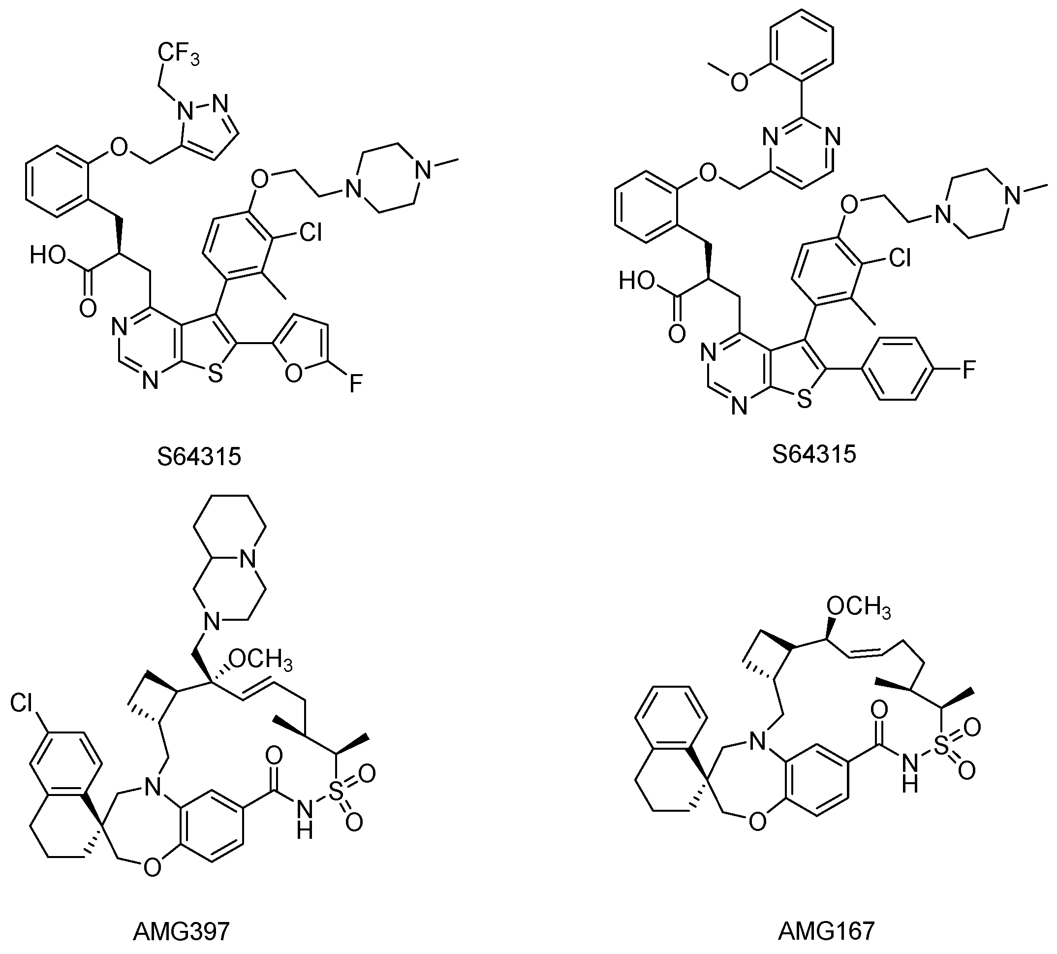
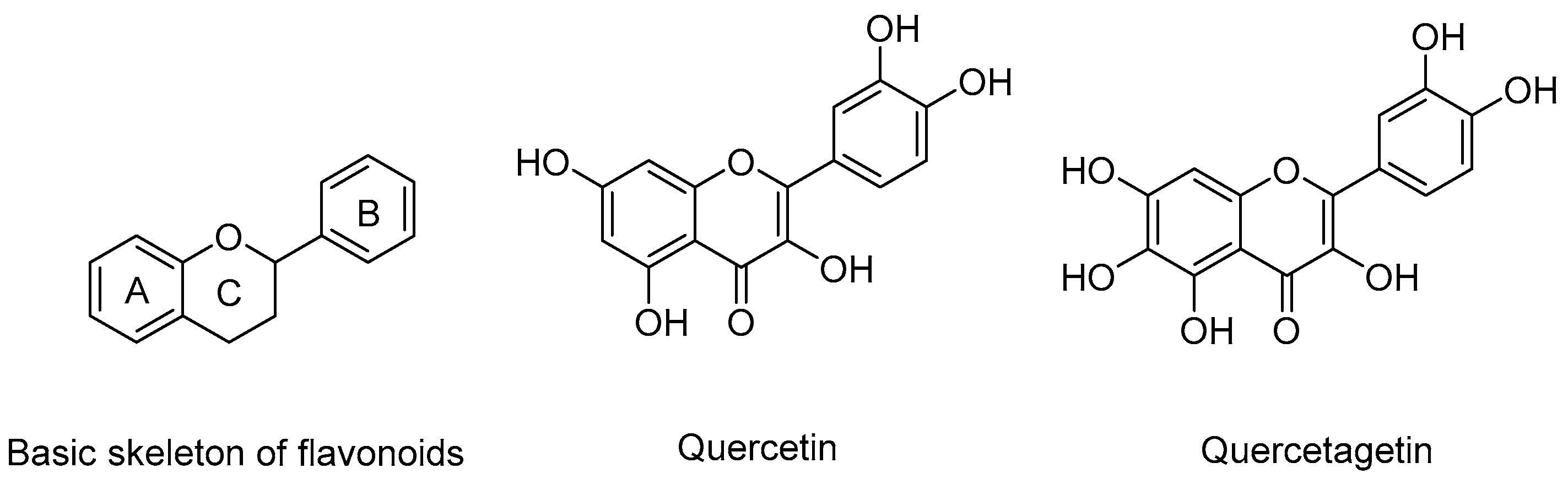
1. Introduction
2. Results
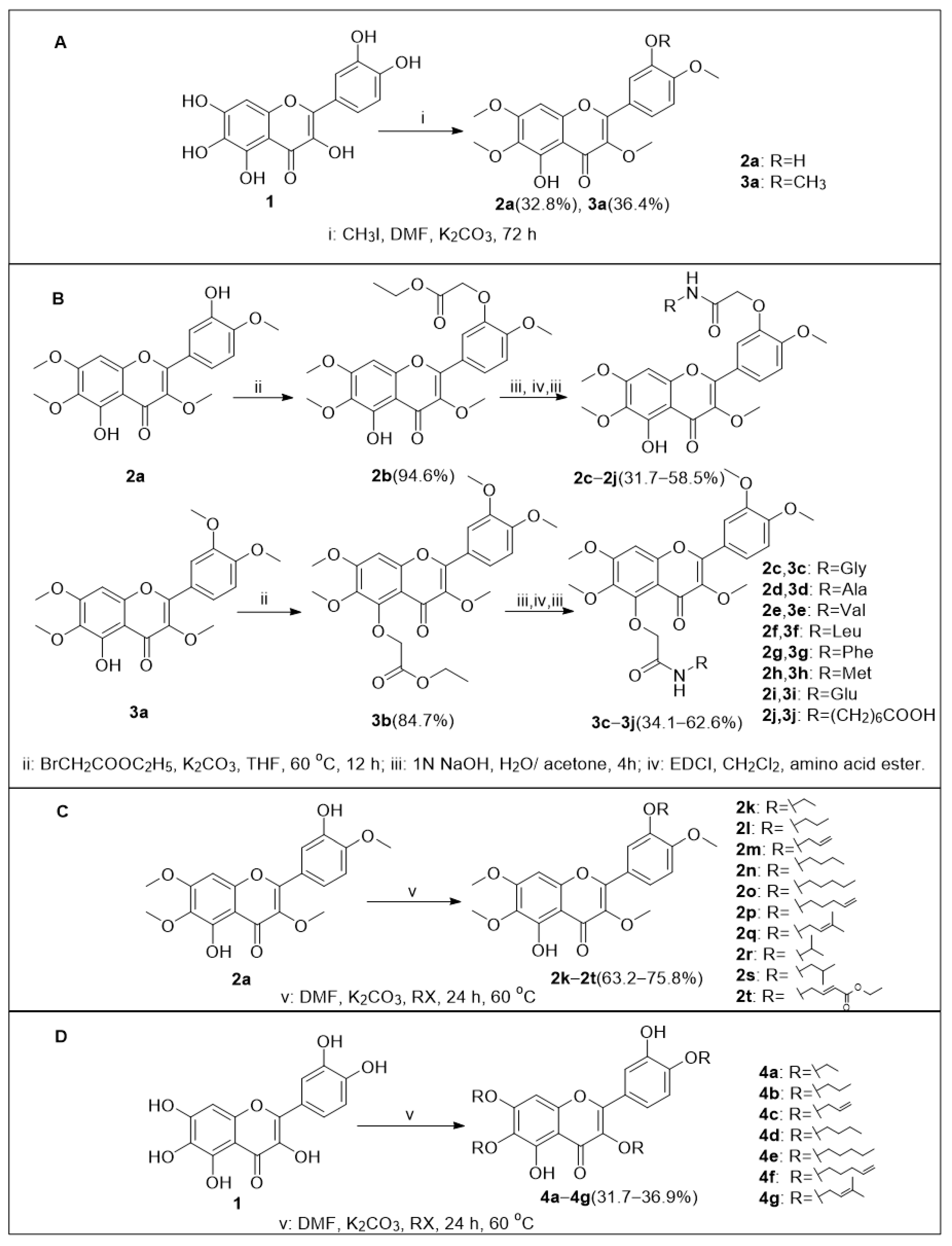
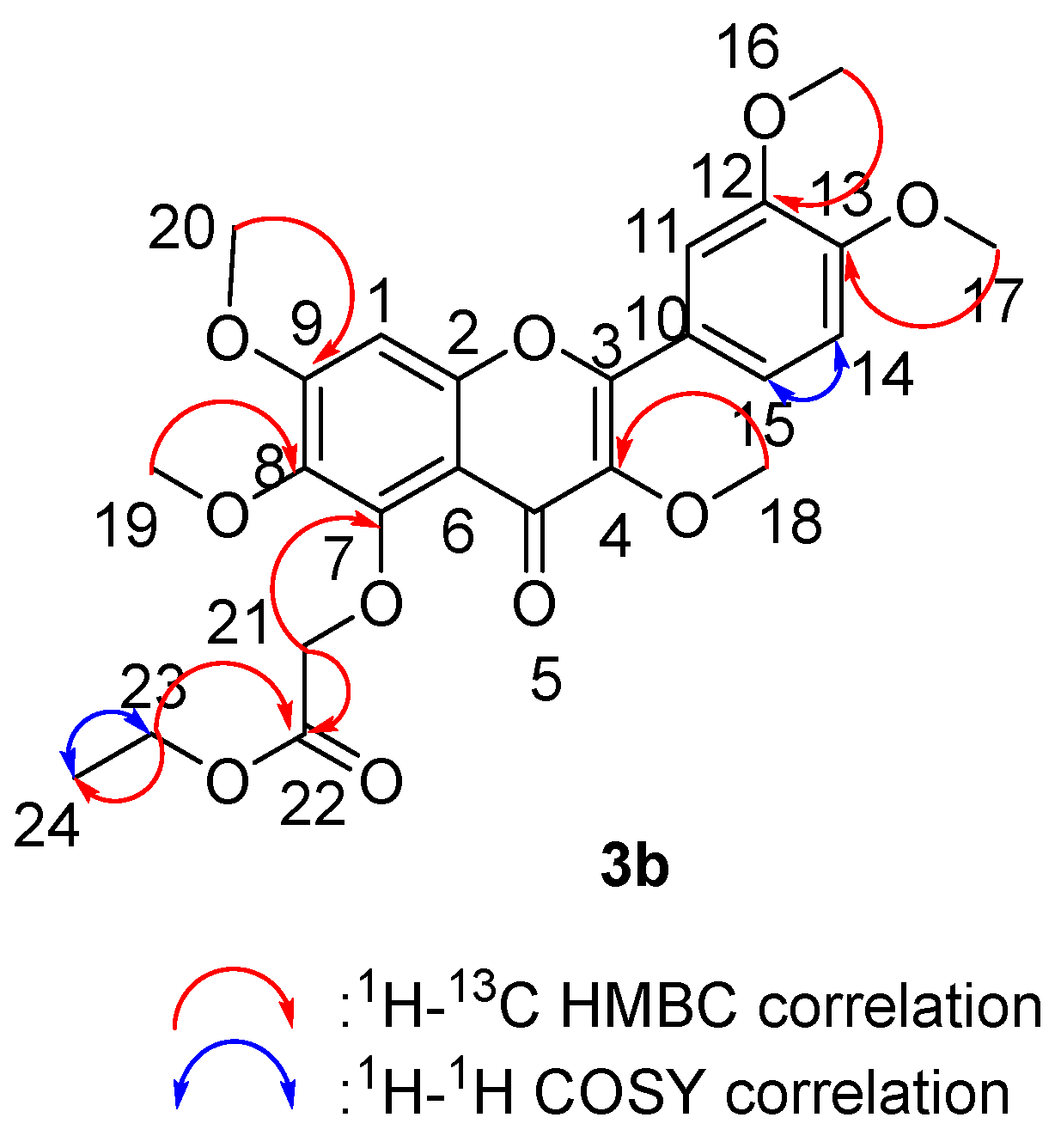
2.1. Chemistry
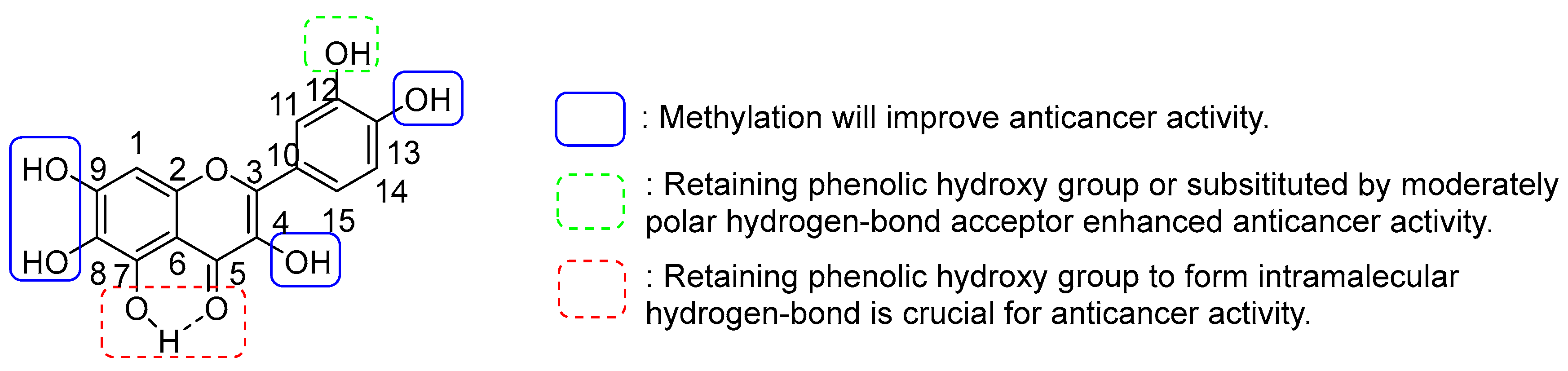
2.2. Anticancer Activity Assay and Structure–Activity Relationship (SAR) Analyses
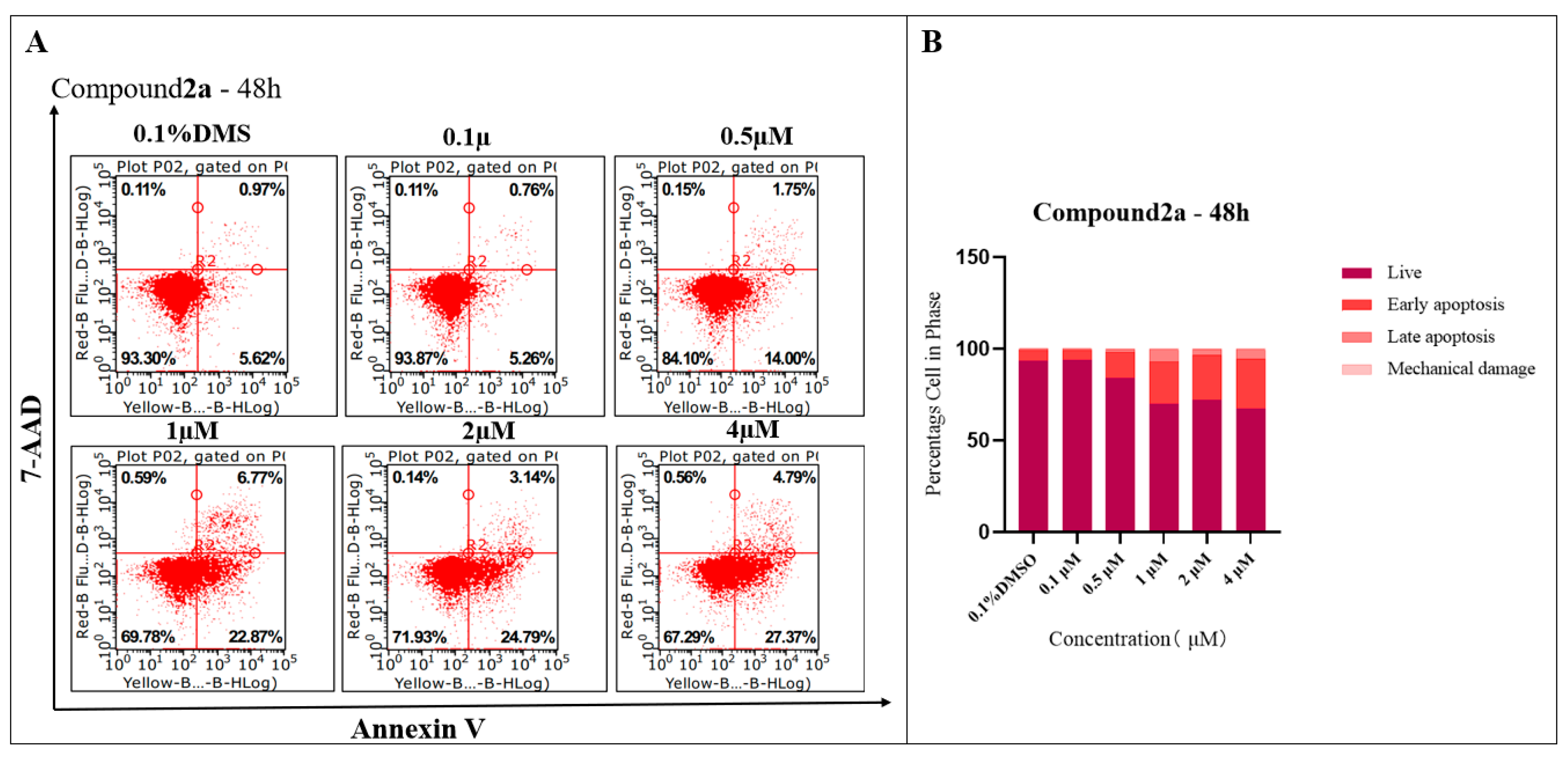
2.3. Compound 2a-Induced Apoptosis in K562 Cells
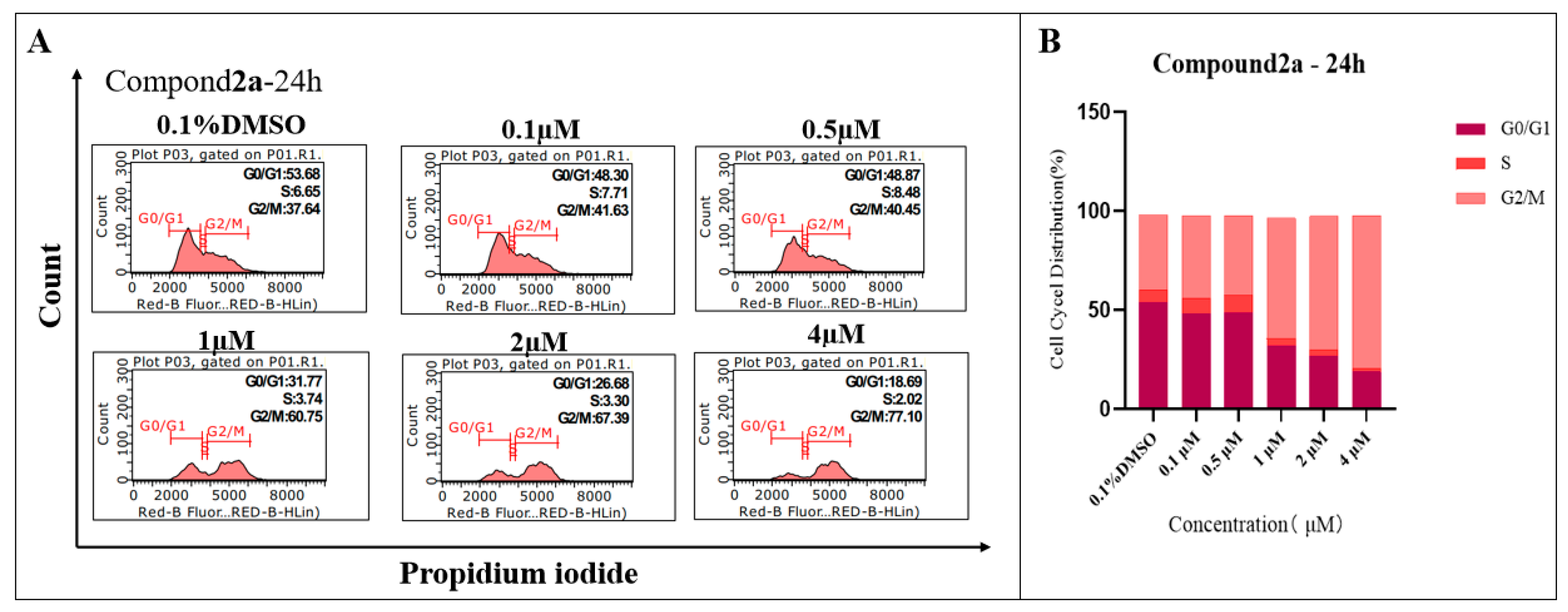
2.4. Compound 2a Effectively Arrested the G2 Phase of K562 Cells
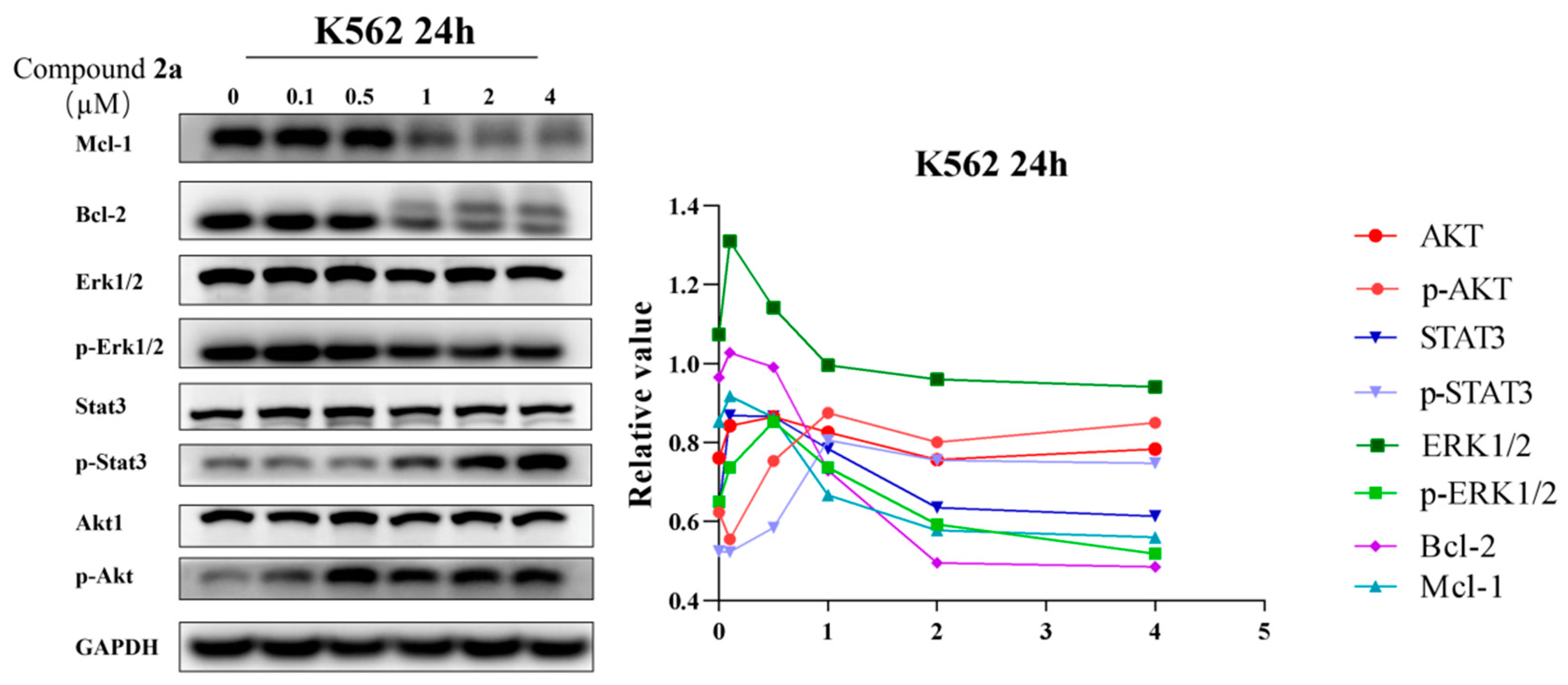
2.5. Compound 2a Suppresses the Expression of Oncoproteins
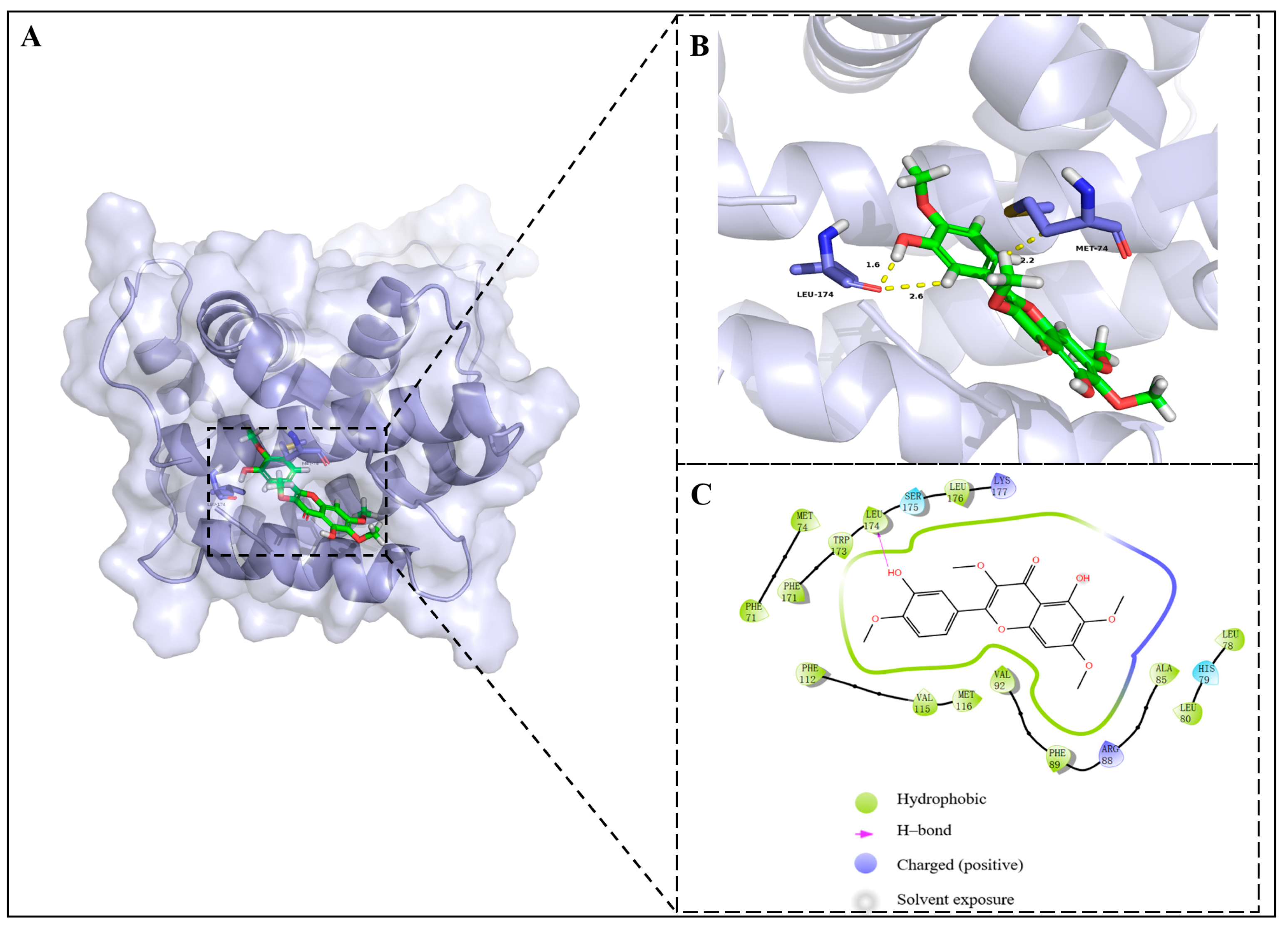
2.6. Molecular Docking Analysis
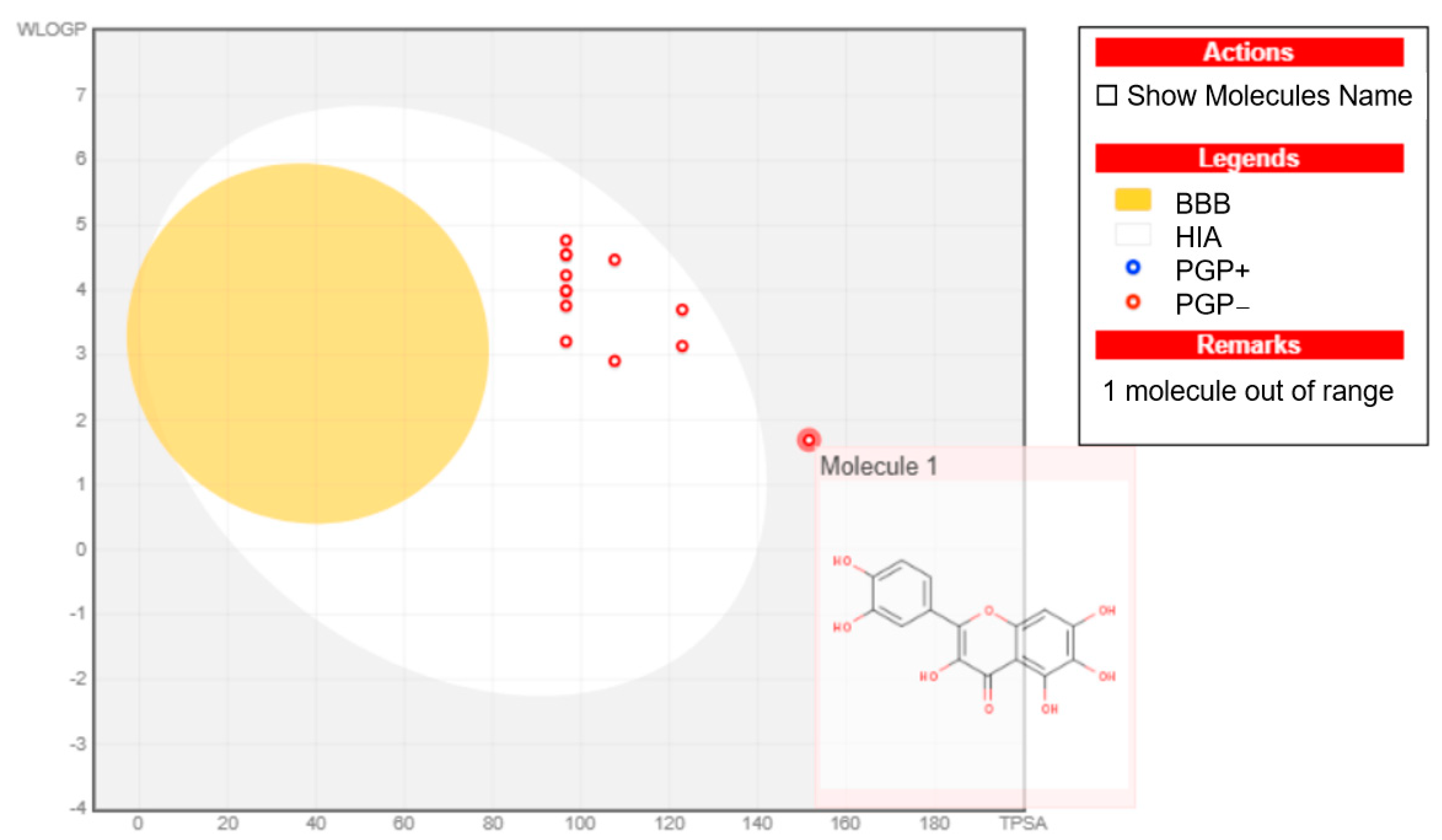
2.7. ’BOILED-Egg Model’ Analyses and RO5 Evaluation
3. Discussion
4. Materials and Methods
4.1. Reactants and Equipment
4.2. NMR and HRMS Spectral Analysis
4.3. Biological Evaluation
4.3.1. In Vitro Cytotoxicity Assay
4.3.2. Apoptosis Analysis
4.3.3. Cell Cycle Analysis
4.3.4. Western Blot Analysis
4.4. In Silico Evaluation
4.4.1. Molecular Docking Studies
4.4.2. ADME Studies and ‘BOILED-Egg’ Model Studies
5. Conclusions
Supplementary Materials
Author Contributions
Funding
Institutional Review Board Statement
Informed Consent Statement
Data Availability Statement
Conflicts of Interest
References
- Hao, T.; Li-Talley, M.; Buck, A.; Chen, W.Y. An emerging trend of rapid increase of leukemia but not all cancers in the aging population in the United States. Sci. Rep. 2019, 9, 1–13. [Google Scholar] [CrossRef] [PubMed]
- Bray, F.; Ferlay, J.; Soerjomataram, I.; Rebecca, L.S.; Lindsey, A.T.; Ahmedin, J. Global cancer statistics 2018: GLOBOCAN estimates of incidence and mortality worldwide for 36 cancers in 185 countries. CA Cancer J. Clin. 2018, 68, 394–424. [Google Scholar] [CrossRef] [PubMed]
- Nørgaard, J.M.; Olesen, L.H.; Hokland, P. Changing picture of cellular drug resistance in human leukemia. Crit. Rev. Oncol.-Hematol. 2004, 50, 39–49. [Google Scholar] [CrossRef]
- Ross, D.D. Novel mechanisms of drug resistance in leukemia. Leukemia 2000, 14, 467–473. [Google Scholar] [CrossRef]
- Czabotar, P.E.; Lessene, G.; Strasser, A.; Adams, J.M. Control of apoptosis by the BCL- 2 protein family: Implications for physiology and therapy. Nat. Rev. Mol. Cell Biol. 2014, 15, 49–63. [Google Scholar] [CrossRef]
- Tsujimoto, Y.; Cossman, J.; Jaffe, E.; Croce, C.M. Involvement of the bcl-2 gene in human follicular lymphoma. Science 1985, 228, 1440–1443. [Google Scholar] [CrossRef]
- Tsujimoto, Y.; Yunis, J.; Onorato-Showe, L.; Erikson, J.; Nowell, P.S.; Croce, C.M. Molecular cloning of the chromosomal breakpoint of B-cell lymphomas and leukemias with the t(11;14) chromosome translocation. Science 1984, 224, 1403–1406. [Google Scholar] [CrossRef] [PubMed]
- Cleary, M.L.; Smith, S.D.; Sklar, J. Cloning and structural analysis of cDNAs for bcl-2 and a hybrid bcl-2/immunoglobulin transcript resulting from the t(14;18) translocation. Cell 1986, 47, 19–28. [Google Scholar] [CrossRef]
- Pepper, C.; Hoy, T.; Bentley, D.P. Bcl-2/Bax ratios in chronic lymphocytic leukaemia and their correlation with in vitro apoptosis and clinical resistance. Brit. J. Cancer 1997, 76, 935–938. [Google Scholar] [CrossRef]
- Hanada, M.; Delia, D.; Aiello, A.; Stadtmauer, E.; Reed, J.C. Bcl-2 gene hypomethylation and high-level expression in B-cell chronic lymphocytic leukemia. Blood 1993, 82, 1820–1828. [Google Scholar] [CrossRef]
- Kitada, S.; Andersen, J.; Akar, S.; Zapata, J.M.; Reed, J.C. Expression of apoptosis-regulating proteins in chronic lymphocytic leukemia: Correlations with in vitro and in vivo chemoresponses. Blood 1998, 91, 3379–3389. [Google Scholar] [CrossRef] [PubMed]
- Krajewski, S.; Krajewska, M.; Shabaik, A.; Wang, H.G.; Irie, S.; Fong, L.; Reed, J.C. Immunohistochemical analysis of in vivo patterns of Bcl-X expression. Cancer Res. 1994, 54, 5501–5507. [Google Scholar] [PubMed]
- Horita, M.; Andreu, E.J.; Benito, A.; Arbona, C.; Sanz, C.; Benet, I.; Prosper, F.; Fernandez-Luna, J.L. Blockade of the Bcr-Abl kinase activity induces apoptosis of chronic myelogenous leukemia cells by suppressing signal transducer and activator of transcription 5-dependent expression of Bcl-xL. J. Exp. Med. 2000, 191, 977–984. [Google Scholar] [CrossRef] [PubMed]
- Wuilleme-Toumi, S.; Robillard, N.; Gomez, P.; Moreau, P.; Gouill, S.L.; Avet-Loiseau, H.; Harousseau, J.L.; Amiot, M.; Bataille, R. Mcl-1 is overexpressed in multiple myeloma and associated with relapse and shorter survival. Leukemia 2005, 19, 1248–1252. [Google Scholar] [CrossRef]
- Le Gouill, S.; Podar, K.; Amiot, M.; Hideshima, T.; Anderson, K.C. VEGF induces Mcl-1 up-regulation and protects multiple myeloma cells against apoptosis. Blood 2004, 104, 2886–2892. [Google Scholar] [CrossRef]
- Xiang, Z.; Luo, H.; Payton, J.E.; Cain, J.; Ley, T.J.; Opferman, J.T.; Tomasson, M.H.; Xiang, Z.F.; Luo, H. Mcl1 haploinsufficiency protects mice from Myc-induced acute myeloid leukemia. J. Clin. Invest. 2010, 120, 2109–2118. [Google Scholar] [CrossRef] [PubMed]
- Kotschy, A.; Szlavi, Z.; Murray, J.; Davidson, J.; Maragno, A.; Toumelin-Braizat, G.L.; Chanrion, M.; Kelly, J.; Gong, J.N.; Moujalled, D.M.; et al. The MCL1 inhibitor S63845 is tolerable and effective in diverse cancer models. Nature 2016, 538, 477–481. [Google Scholar] [CrossRef]
- Szlavik, Z.; Csekei, M.; Pacza l, A.; Szabo, Z.B.; Kotschy, A. Discovery of S64315, a Potent and Selective Mcl-1 Inhibitor. J. Med. Chem. 2020, 63, 13762–13795. [Google Scholar] [CrossRef]
- Caenepeel, S.; Brown, S.P.; Belmontes, B.; Moody, G.; Keegan, K.S.; Chui, D.; Whittington, D.A.; Huang, X.; Poppe, L.; Cheng, A.C.; et al. AMG 176, a Selective MCL1 Inhibitor, Is Effective in Hematologic Cancer Models Alone and in Combination with Established Therapies. Cancer Discov. 2018, 8, 1582–1597. [Google Scholar] [CrossRef]
- Shen, N.; Wang, T.F.; Gan, Q.; Liu, S.; Wang, L.; Jin, B. Plant flavonoids: Classification, distribution, biosynthesis, and antioxidant activity. Food Chem. 2022, 383, 132531. [Google Scholar] [CrossRef]
- Kruger, M.J.; Davies, N.; Myburgh, K.H.; Lecour, S. Proanthocyanidins, anthocyanins and cardiovascular diseases. Food Res. Int. 2014, 59, 41–52. [Google Scholar] [CrossRef]
- Wang, X.; Ouyang, Y.Y.; Liu, J.; Zhao, G. Flavonoid intake and risk of CVD: A systematic review and meta-analysis of prospective cohort studies. Br. J. Nutr. 2014, 111, 1–11. [Google Scholar] [CrossRef] [PubMed]
- Alqurashi, R.M.; Galante, L.A.; Rowland, I.R.; Spencer Jeremy, P.E.; Commane, D.M. Consumption of a flavonoid-rich açai meal is associated with acute improvements in vascular function and a reduction in total oxidative status in healthy overweight men. Am. J. Clin. Nutr. 2016, 104, 1227–1235. [Google Scholar] [CrossRef]
- Peluso, I.; Raguzzini, A.; Serafini, M. Effect of flavonoids on circulating levels of TNF-α and IL-6 in humans: A systematic review and meta-analysis. Mol. Nutr. Food Res. 2013, 57, 784–801. [Google Scholar] [CrossRef] [PubMed]
- Serafini, M.; Peluso, I.; Raguzzini, A. 3rd international immunonutrition workshop session 1: Antioxidants and the immune system flavonoids as antiinflammatory agents. Proc. Nutr. Soc. 2010, 69, 273–278. [Google Scholar] [CrossRef]
- Marzocchella, L.; Fantini, M.; Benvenuto, M.; Masuelli, L.; Benvenuto, M.; Fantini, M.; Marzocchella, L. Dietary flavonoids: Molecular mechanisms of action as anti-inflammatory agents. Recent Pat. Inflamm. Allergy Drug Discov. 2011, 5, 200–220. [Google Scholar] [CrossRef]
- Spagnuolo, C.; Cerella, C.; Russo, M.; Chateauvieux, S.; Diederich, M.; Russo, G.L. Quercetin downregulates Mcl-1 by acting on mRNA stability and proteindegradation. Br. J. Cancer 2011, 105, 221–230. [Google Scholar] [CrossRef]
- Yuan, J.; Wong, I.L.K.; Jiang, T.; Wang, S.W.; Liu, T.; Wen, B.J.; Chow, M.C.; Sheng, B.W. Synthesis of methylated quercetin derivatives and their reversal activities on P-gp- and BCRP-mediated multidrug resistance tumour cells. Eur. J. Med. Chem. 2012, 54, 413–422. [Google Scholar] [CrossRef]
- Sassi, N.; Mattarei, A.; Espina, V.; Liotta, L.; Zorrati, M.; Paradisi, C.; Biasutto, L. Potential anti-cancer activity of 7-O-pentyl quercetin: Efficient, membrane-targeted kinase inhibition and pro-oxidant effect. Pharmacol. Res. 2017, 124, 9–19. [Google Scholar] [CrossRef]
- Duan, N.; Hu, X.H.; Zhou, R.; Li, Y.R.; Wu, W.H.; Liu, N. A Review on Dietary Flavonoids as Modulators of the Tumor Microenvironment. Mol. Nutr. Food Res. 2023, 67, e22004345. [Google Scholar] [CrossRef]
- Rathi, A.; Chaudhury, A.; Anjum, F.; Ahamd, F.; Haider, S.; Khan, Z.F.; Taiyab, A.; Chakrabarty, A.; Islam, A.; Hassan, M.I.; et al. Targeting prostate cancer via therapeutic targeting of PIM-1 kinase by Naringenin and Quercetin. Int. J. Biol. Macromol. 2024, 267, 133882. [Google Scholar] [CrossRef] [PubMed]
- Aherne, S.A.; O’Brien, N.M. Lack of effect of the flavonoids, myricetin, quercetin, and rutin, on repair of H2O2-induced DNA single-strand breaks in Caco-2, Hep G2, and V79 cells. Nutr. Cancer 2000, 38, 106–115. [Google Scholar] [CrossRef]
- Aalinkeel, R.; Bindukumar, B.; Reynolds, J.L.; Sykes, D.E.; Mahajan, S.D.; Chadha, K.C.; Schwartz, S.A. The dietary bioflavonoid, quercetin, selectively induces apoptosis of prostate cancer cells by down-regulating the expression of heat shock protein 90. Prostate 2008, 68, 1773–1789. [Google Scholar] [CrossRef] [PubMed]
- Bach, A.; Bender-Sigel, J.; Schrenk, D.; Flügel, D.; Kietzmann, T. The antioxidant quercetin inhibits cellular proliferation via HIF-1-dependent induction of p21WAF. Antioxid Redox Sign. 2010, 13, 437–448. [Google Scholar] [CrossRef]
- Shi, Y.; Su, X.; Cui, H.; Yv, L.; Du, H.; Han, Y. Combination of quercetin and adriamycin effectively suppresses the growth of refractory acute leukemia. Oncol. Lett. 2019, 18, 153–160. [Google Scholar] [CrossRef] [PubMed]
- Zhang, H.W.; Hu, J.J.; Fu, R.Q.; Xin, L.; Zhang, Y.H.; Jing, L.; Lei, L.; Li, Y.N.; Qin, D.; Luo, Q.S. Flavonoids inhibit cell proliferation and induce apoptosis and autophagy through downregulation of PI3Kγ mediated PI3K/AKT/mTOR/p70S6K/ULK signaling pathway in human breast cancer cells. Sci. Rep. 2018, 8, 11255. [Google Scholar] [CrossRef]
- Shi, Z.H.; Li, N.G.; Tang, Y.P.; Shi, Q.P.; Zhang, W.; Zhang, P.X.; Dong, Z.X.; Li, W.; Zhang, X.; Fu, H.A. Synthesis, biological evaluation and SAR analysis of O-alkylated analogs of quercetin for anticancer. Bioorg. Med. Chem. Lett. 2014, 24, 4424–4427. [Google Scholar] [CrossRef]
- Sana, R.; Shazia, A.; Muhammad, M.A.; Ashraf, M.; Hussain, S.; Hamid, S. Quercetagetin 3,7 dimethyl ether polymorphs as multi-targeted anti-amyloid agents: Target to cognitive impairment in Alzheimer’s disease. J. Mol. Struc. 2025, 1321, 139792. [Google Scholar]
- Sohee, B.; Nam, J.K.; Grzegorz, M.; Arciniega, M.; Jung, S.K.; Byun, S.; Song, N.R.; Heo, Y.S.; Kin, B.Y.; Lee, H.J.; et al. Structural and Functional Analysis of the Natural JNK1 Inhibitor Quercetagetin. J. Mol. Biol. 2013, 425, 411–423. [Google Scholar]
- Yang, Y.J.; Yu, Q.C.; Hu, L.A.; Dai, B.T.; Qi, R.X.; Chang, Y.; Zhang, Q.W.; Zhang, Z.; Li, Y.J.; Zhang, X.M. Enantioselective semisynthesis of novel cephalotaxine esters with potent antineoplastic activities against leukemia. Eur. J. Med. Chem. 2022, 244, 114731. [Google Scholar] [CrossRef]
- Chen, Z.P.; Yu, Q.C.; Chen, J.; Yu, X.W.; Cao, J.Q.; Zhai, Y.J.; Tan, Y.; Zhao, Z.C.; Li, W.; Zou, X.Y. Bufadienolide-fatty acid conjugates from the fertilized eggs of toad bufo gargarizans: Isolation, characterization, toxicity, and antiproliferative evaluation. J. Agric. Food Chem. 2024, 72, 17377–17391. [Google Scholar] [CrossRef] [PubMed]
- Sun, Y.L.; Chen, Z.W.; Liu, G.B.; Chen, X.A.; Shi, Z.H.; Feng, H.X.; Yu, L.; Li, G.D.; Ding, K.; Huang, H.; et al. Discovery of a potent and selective covalent threonine tyrosine kinase (TTK) inhibitor. Bioorg. Chem. 2024, 143, 107053. [Google Scholar] [CrossRef] [PubMed]
- Ulusoy, N.G.; Emirdaǧ, S.; Söze, E.; Radwan, M.O.; Çiftçi, H.; Aksel, M.; Bölükbaşı, S.Ş.; Özmen, A.; Yaylı, N.; Karayıldırım, T.; et al. Design, semi-synthesis and examination of new gypsogenin derivatives against leukemia via Abl tyrosine kinase inhibition and apoptosis induction. Int. J. Biol. Macromol. 2022, 222, 1487–1499. [Google Scholar] [CrossRef] [PubMed]


| Compounds | IC50 (µmol/L) | |||
|---|---|---|---|---|
| U937 | K562 | K562-IR | KG-1 | |
| 1 | 4.619 ± 0.218 | 0.980 ± 0.172 | 0.509 ± 0.128 | >10 |
| 2a | 0.276 ± 0.047 | 0.159 ± 0.032 | 0.312 ± 0.025 | 0.271 ± 0.261 |
| 2b | 0.310 ± 0.063 | 0.366 ± 0.041 | 0.304 ± 0.059 | 0.441 ± 0.084 |
| 2c | >10 | >10 | >10 | >10 |
| 2d | >10 | >10 | >10 | >10 |
| 2e | >10 | >10 | >10 | >10 |
| 2f | >10 | >10 | >10 | >10 |
| 2g | >10 | >10 | >10 | >10 |
| 2h | >10 | >10 | >10 | >10 |
| 2i | >10 | >10 | >10 | >10 |
| 2j | >10 | >10 | >10 | >10 |
| 2k | >10 | >10 | >10 | >10 |
| 2l | >10 | 7.51 ± 0.094 | >10 | 4.093 ± 0.045 |
| 2m | >10 | >10 | 2.402 ± 0.374 | >10 |
| 2n | >10 | >10 | >10 | >10 |
| 2o | >10 | >10 | 9.946 ± 0.642 | >10 |
| 2p | >10 | >10 | 9.187 ± 0.106 | >10 |
| 2q | >10 | 9.299 ± 1.031 | 6.361 ± 0.843 | 6.555 ± 0.497 |
| 2r | >10 | >10 | 3.188 ± 0.295 | 6.25 ± 0.265 |
| 2s | >10 | >10 | >10 | 7.607 ± 0.604 |
| 2t | >10 | >10 | 5.466 ± 0.067 | 7.688 ± 0.828 |
| 3a | 0.332 ± 0.051 | 0.893 ± 0.073 | 0.619 ± 0.029 | 4.400 ± 0.256 |
| 3b | >10 | >10 | >10 | >10 |
| 3c | >10 | >10 | >10 | >10 |
| 3d | >10 | >10 | >10 | >10 |
| 3e | >10 | >10 | >10 | >10 |
| 3f | >10 | >10 | >10 | >10 |
| 3g | >10 | >10 | >10 | >10 |
| 3h | >10 | >10 | >10 | >10 |
| 3i | >10 | >10 | >10 | >10 |
| 3j | >10 | >10 | >10 | >10 |
| 4a | 8.401 ± 1.274 | 9.408 ± 0.782 | 4.758 ± 0.453 | 3.559 ± 0.301 |
| 4b | >10 | >10 | >10 | >10 |
| 4c | >10 | >10 | >10 | >10 |
| 4d | >10 | >10 | >10 | >10 |
| 4e | >10 | >10 | >10 | 8.592 ± 0.568 |
| 4f | >10 | >10 | >10 | >10 |
| 4g | >10 | >10 | >10 | >10 |
| Imatinib | ND | 0.089 ± 0.016 | 6.050 ± 0.275 | ND |
| Taxol | 0.002 ± 0.001 | 0.006 ± 0.001 | 0.005 ± 0.002 | 0.005 ± 0.001 |
| Compound | mol_MW | QPlogPo/w | donorHB | accptHB | RotabB | GI Absorption | Rule of Five Violations |
|---|---|---|---|---|---|---|---|
| 1 | 318.24 | 0.92 | 6 | 8 | 1 | Low | 1 |
| 2a | 374.34 | 2.51 | 2 | 8 | 5 | High | 0 |
| 3a | 388.37 | 2.82 | 1 | 8 | 6 | High | 0 |
| 2c | 460.43 | 3.00 | 1 | 10 | 10 | High | 2 |
| 4a | 430.45 | 3.81 | 2 | 8 | 9 | High | 0 |
| 4e | 598.77 | 7.88 | 2 | 8 | 21 | Low | 3 |
| 2l | 416.42 | 3.50 | 1 | 8 | 8 | High | 0 |
| 2m | 414.41 | 3.36 | 1 | 8 | 8 | High | 0 |
| 2o | 444.47 | 4.19 | 1 | 8 | 10 | High | 1 |
| 2p | 442.46 | 4.01 | 1 | 8 | 10 | High | 1 |
| 2q | 442.46 | 3.97 | 1 | 8 | 8 | High | 0 |
| 2r | 416.42 | 3.42 | 1 | 8 | 7 | High | 0 |
| 2s | 430.45 | 3.78 | 1 | 8 | 8 | High | 0 |
| 2t | 486.47 | 3.37 | 1 | 10 | 11 | High | 2 |
Disclaimer/Publisher’s Note: The statements, opinions and data contained in all publications are solely those of the individual author(s) and contributor(s) and not of MDPI and/or the editor(s). MDPI and/or the editor(s) disclaim responsibility for any injury to people or property resulting from any ideas, methods, instructions or products referred to in the content. |
© 2025 by the authors. Licensee MDPI, Basel, Switzerland. This article is an open access article distributed under the terms and conditions of the Creative Commons Attribution (CC BY) license (https://creativecommons.org/licenses/by/4.0/).
Share and Cite
Li, K.; Ge, X.; Liu, W.; Huang, L.; Lv, X.; Tang, Y.; He, Z.; Yang, Y.; Chen, M.; Zeng, J.; et al. Synthesis and Biological Evaluation of Quercetagetin Derivatives as the Inhibitors of Mcl-1 and Bcl-2 Against Leukemia. Int. J. Mol. Sci. 2025, 26, 2727. https://doi.org/10.3390/ijms26062727
Li K, Ge X, Liu W, Huang L, Lv X, Tang Y, He Z, Yang Y, Chen M, Zeng J, et al. Synthesis and Biological Evaluation of Quercetagetin Derivatives as the Inhibitors of Mcl-1 and Bcl-2 Against Leukemia. International Journal of Molecular Sciences. 2025; 26(6):2727. https://doi.org/10.3390/ijms26062727
Chicago/Turabian StyleLi, Kang, Xiaomei Ge, Wei Liu, Lei Huang, Xinye Lv, Yuhui Tang, Zhehao He, Yingxue Yang, Miaofen Chen, Jianguo Zeng, and et al. 2025. "Synthesis and Biological Evaluation of Quercetagetin Derivatives as the Inhibitors of Mcl-1 and Bcl-2 Against Leukemia" International Journal of Molecular Sciences 26, no. 6: 2727. https://doi.org/10.3390/ijms26062727
APA StyleLi, K., Ge, X., Liu, W., Huang, L., Lv, X., Tang, Y., He, Z., Yang, Y., Chen, M., Zeng, J., & Cheng, P. (2025). Synthesis and Biological Evaluation of Quercetagetin Derivatives as the Inhibitors of Mcl-1 and Bcl-2 Against Leukemia. International Journal of Molecular Sciences, 26(6), 2727. https://doi.org/10.3390/ijms26062727