Article

In Vitro Analysis of Probiotic Properties Related to the Adaptation of Levilactobacillus brevis to Intestinal Microenvironment and Involvement of S-Layer Proteins

1
Department of Biochemical Engineering, Faculty of Food Technology and Biotechnology, University of Zagreb, Pierottijeva 6, 10000 Zagreb, Croatia
2
Center for Research and Knowledge Transfer in Biotechnology, University of Zagreb, Rockefellerova 10, 10000 Zagreb, Croatia
*
Author to whom correspondence should be addressed.
Int. J. Mol. Sci. 2025, 26(6), 2425; https://doi.org/10.3390/ijms26062425
Submission received: 4 February 2025 / Revised: 4 March 2025 / Accepted: 5 March 2025 / Published: 7 March 2025
(This article belongs to the Special Issue Probiotics in Health and Disease)

Abstract

:
Although rare, the ability to produce surface S-layer proteins is beneficially associated with particular Lactobacillus strains being investigated as probiotics. Therefore, this work aimed to study specific probiotic functionalities of selected Levilactobacillus brevis strains MB1, MB2, MB13 and MB20, isolated from human milk microbiota, and to assess the contribution of S-proteins. Firstly, Rapid Annotation using Subsystem Technology revealed that cell wall-related genes were abundant in analysed L. brevis genomes. Furthermore, the results demonstrated that S-proteins mediate aggregation capacity and competitive exclusion of selected pathogens by L. brevis strains. The improvement of Caco-2 epithelial monolayer barrier function was demonstrated by the increase in JAM-A and occludin expressions when L. brevis strains or S-proteins were added, with the effect being most pronounced after treatment with MB2 and S-proteins of MB1. L. brevis strains, especially MB20, exerted the potential to adhere to recombinant human ZG16. Strain MB2 and MB20-S-proteins improved the barrier function of HT29 epithelial monolayer, as evidenced by increased ZG16 expression. Analysed L. brevis strains and S-proteins differentially affected the protein expression of IL-1β, IL-6 and IL-8, and IL-10 cytokines. The most prominent effect was observed by S-proteins of MB20, since IL-1β production was decreased while IL-10 production was significantly increased.

1. Introduction

Probiotic strains, including those belonging to the lactic acid bacteria (LAB), have been described as having diverse functions, such as the exclusion of pathogens through competition for nutrients and receptors for their binding, production of antimicrobial substances that inhibit their growth, promotion of epithelial barrier function by increasing the production of mucins and the expression of tight junction (TJ) proteins, preventing the translocation of pathogens from the gut into the blood, and even by regulating the neuro-immune axis [1,2]. The in vivo efficacy of probiotics is determined by their ability to adapt to the specific conditions of the digestive tract, but also by their interactions with resident bacteria and host cells. Specific mechanisms and clinical outcomes of probiotic strains are mediated by probiotic cell components or cell debris and, more recently, there is also evidence that non-living cells can also act as health promoters with enhanced bioactivity, which has contributed to the development of the concept of postbiotics [3]. The term postbiotics refers to soluble factors, such as metabolites or by-products, that are secreted by live bacteria or released after bacterial lysis and can have a beneficial effect on the consumer. These ingredients have production advantages over probiotics as cell viability is not required, which simplifies the manufacturing process and application [4]. Most in vitro and in vivo studies have focused on postbiotics derived from various LAB strains [5,6]. Lactobacillus S-layer proteins are hypothesised to have postbiotic characteristics and can be considered as probiotic effector molecules. S-layer proteins are the first to interact with the microenvironment and contribute to the probiotic effect of the strain that produces them [7]. Their protective role in stressful conditions, in mediating interactions between mucus and epithelial cells, as well as their immunomodulatory effect have been demonstrated [8,9,10,11].
Our research group has previously isolated Lactobacillus strains, producing potentially therapeutic biomolecules, including the S-proteins [12,13]. The role of S-proteins for Levilactobacillus brevis strains MB1, MB2, MB13 and MB20, isolated from mother’s milk, in terms of better survival under simulated gastrointestinal tract (GIT) conditions and better adhesion to Caco-2 cells was confirmed [14]. The aim of this work was to investigate the role of S-proteins and Lactobacillus strains that produce them, by performing in vitro analysis of specific properties related to the adaptation and effect of probiotics in the intestinal milieu, more specifically by targeted characterization of adhesion capacities, ability to impact intestinal barrier function, and assessment of stimulation of specific cytokines production.

2. Results and Discussion

2.1. Genome Features of L. brevis Strains

Accurate taxonomic identification of probiotic strains is essential since their safety and health effects are strain dependent. Therefore, the genomes of S-protein-producing L. brevis strains MB1, MB2, MB13 and MB20, isolated from human milk microbiota, were sequenced and deposited in the NCBI database (BioProject PRJNA388578) under accession numbers SAMN22155538, SAMN22155539, SAMN22155540 and SAMN221555341, respectively [13]. In addition, the annotation, distribution and categorisation of the genes of the analysed strains was performed using the RAST server, on which all data on the sequenced genomes of the L. brevis strains MB1, MB2, MB13 and MB20 are stored and available (Figure S1). Genome annotation using the RAST server revealed that genes related to the cell wall and capsule represent one of the most abundant gene groups of L. brevis strains MB1, MB2, MB13 and MB20, which is in line with expectations, as S-proteins account for about 10–20% of the total cellular proteins of the producer bacteria.
Comparison of the genome data of L. brevis strains MB1, MB2, MB13 and MB20 (Table S1), also obtained using the RAST server, shows that there are no large jumps in the highlighted values between the individual L. brevis strains MB1, MB2, MB13 and MB20 and that the genome features obtained are consistent with previously published genome sequences of L. brevis strains isolated from different microhabitats [8,10,15]. The results obtained show a high degree of overlap between the studied genomes, which was to be expected as they belong to the same species and produce S-proteins.

2.2. Role of S-proteins in the Properties Important for Intestinal Adaptation of L. brevis Strains

Čuljak et al. [14] previously demonstrated the role of S-proteins of L. brevis strains with regard to better survival under simulated GIT conditions and improved adhesion to Caco-2 cells. In this paper, following previous experiments, autoaggregation and coaggregation assays, as well as competitive exclusion of potentially pathogenic bacteria E. coli 3014 and S. Typhimurium FP1 were performed. Autoaggregation, i.e., the cluster of bacteria of the same strain, is thought to be the first step in adhesion to epithelial cells and mucosal surfaces, allowing bacteria to form a barrier by colonising the GIT and thus preventing the adhesion of undesirable bacteria [16]. Similarly, coaggregation, i.e., the aggregation of two different bacterial strains, of probiotic bacteria with pathogens can also prevent pathogens from colonising the GIT [17].
The percentage of autoaggregation was lower in all L. brevis strains after removal of the S-layer, with S-proteins of L. brevis strains MB1 and MB20 being the most pronounced (p < 0.001). Our results confirm that S-layer proteins, as the outermost barrier surrounding the cell surface, play an important role in the autoaggregation process. These results are in agreement with those of Alp et al. [18], who also found a role of S-proteins in the autoaggregation of L. casei DA4, L. coryniformis DA263, L. plantarum DA100 and DA140, as evidenced by a reduced aggregation ability after removal of the S-layer by LiCl treatment.
The results related to the characterisation of the coaggregation of L. brevis strains indicated a role of S-proteins when coaggregating with S. Typhimurium FP1, since the percentage of coaggregation was significantly lower after S-layer removal, with the greatest difference observed for strain MB13 (p < 0.001) (Table 1). Regarding coaggregation with E. coli 3014, only strains MB1 and MB2 showed a statistically significant increase in coaggregation ability when S-proteins were present compared to GHCl-treated strains (p < 0.05), while there was no difference in strains MB13 and MB20 (Table 2). These results indicate that S-proteins do not play a crucial role in aggregation mechanisms in all strains expressing them, but that other cell wall components may play a more important role. Our results are consistent with the findings of Alp et al. [18], who demonstrated a role of S-proteins of strains L. casei DA4, L. coryniformis DA263 and L. plantarum DA140 in coaggregation with Salmonella enterica serovar Enteritidis, with the exception of L. plantarum DA100.
Competitive exclusion is defined as a process in which one bacterial species competes with another bacterial species for adhesion to receptors in the GIT, as manifested by the ability to bind to the surface of the host’s mucosa, secretion of various metabolites and competition for available nutrients [19]. Probiotics with the ability to adhere to the intestinal epithelium are prone to preventing colonisation with intestinal pathogens. Lactobacillus rhamnosus GG has been shown to be effective against infection with vancomycin-resistant enterococci (VRE) as it excludes Enterococus faecium, which can easily acquire new resistance to antibiotics and is a successful coloniser of the human intestinal tract [20]. Ayala et al. [21] also demonstrated that certain LAB strains have the ability to competitively exclude Salmonella Montevideo, Escherichia coli O157:H7 and Listeria monocytogenes N1-002. Zhang et al. [22] point the role of S-layer protein extracts in preventing the adhesion of pathogens and thus intestinal colonisation. The competitive exclusion of E. coli 3014 and S. Typhimurium FP1 and the involvement of S-layer were analysed on Caco-2 cells with the ability to spontaneously differentiate into a monolayer having similar properties as small intestine enterocytes [22]. The results showed that S-proteins of L. brevis strains MB13 and MB20 contributed to the competitive exclusion of E. coli 3014 (Figure 1A), as a statistically significant decrease in log(CFU/mL) values was observed when S-proteins were present on the cell surface (p < 0.0001). The analysed strains successfully excluded the adhesion of S. Typhimurium FP1 as a significant increase in log(CFU/mL) values was observed after removal of the S-layer compared to the results achieved followed by S-protein extraction (Figure 1B). A particularly pronounced effect in reducing the ability of the competitive exclusion was observed in L. brevis strains MB1 and MB20 as there was a highly statistically significant difference between the intact and S-layer-depleted strain (p < 0.001), suggesting a mediating role of S-proteins. This suggests that L. brevis S-proteins have a potential role in the competitive exclusion of pathogenic bacteria and possibly affinity for the Caco-2 cell line. The results support the hypothesis that S-proteins mediate the interaction between producer cells and intestinal epithelium [14]. Noteworthy, the results demonstrated that four L. brevis strains, with or without S-layer, reduced the cell adhesion of E. coli 3014 and S. Typhimurium FP1 to Caco-2 cells, as evident from the significantly higher cell counts of E. coli 3014 and S. Typhimurium FP1, which reached levels of 8.45 ± 0.18 and 6.43 ± 0.25 log(CFU/mL) in control experiments, respectively.

2.3. Capacity of L. brevis Strains and Their S-proteins to Improve Epithelial Barrier Function In Vitro

The intestinal epithelium maintains a balance between the selective active and passive transport of molecules, forming a physical barrier that protects against the invasion of various pathogens [23,24]. The intercellular TJ proteins regulate the passive transport of luminal fluid and molecules between epithelial cells. TJs are the most apical intercellular junctions of epithelial cells formed by the transmembrane proteins occludins, claudins and junctional adhesion molecules (JAMs), as well as peripheral membrane proteins such as zonula occludens (ZO) [25]. The altered expression can lead to a loss of intestinal barrier integrity, which allows uncontrolled transfer of the intestinal microbiota and intestinal content submucosally, following the activation of the inflammatory process [26].
The secretory intestinal proteins like mucin MUC2 also represent an important physical component of the intestinal protective barrier by covering the intestinal epithelial cells and forming an intestinal mucosal layer that improves food absorption, provides attachment sites for symbiotic intestinal bacteria and limits pathogen colonisation [27]. Another important component of the extracellular mucus layer is the protein ZG16 present throughout intestine [28], which crosslinks a polymer network of mucins and other secreted mucus glycoproteins. Primary ZG16 is responsible for intestinal mucus stratification and the agglomeration of Gram-positive microbiota in the distal mucus layer, preventing them from reaching the intestinal epithelial cells [29]. It shows an anti-inflammatory and anti-tumour activity, preventing bacteria from binding to the colonic epithelium [28]. Research has shown that probiotics increase the expression of genes encoding the aforementioned TJ proteins and regulate the expression of mucin, thereby influencing the formation of the mucus layer and maintaining the integrity of the intestinal epithelial barrier [27,30,31]. In addition to the intact LAB cells, some of their metabolites also show an effect on the maintenance of the intestinal barrier [27].
Therefore, the role of L. brevis strains MB1, MB2, MB13 and MB20 and their S-proteins in improving the epithelial monolayer barrier function of human cell cultures was investigated by monitoring the expression of JAM-A, occludin and ZO-1, and ZG16 by fluorescence microscopy (Figure S2). In an experimental setup, the expression of individual TJ proteins after treatment with L. brevis strains and derived S-proteins was monitored under four different conditions, simulating some different possible scenarios in the intestine. These included conditioning with a medium that represents the homeostatic specific pathogen-free (SPF) state in the gut (as in Figure 2, Figure 3, Figure 4 and Figure 5); a medium preconditioned with LPS mimicking the presence of Gram-negative pathogens such as E. coli (Bs in Figure 2, Figure 3, Figure 4 and Figure 5); a medium pretreated with TNF-representing inflammatory conditions (Cs in Figure 2, Figure 3, Figure 4 and Figure 5); and lastly, a medium exposed to TNF and LPS which should simulate the combination of inflammatory conditions and the presence of pathogenic E. coli (Ds in the Figure 2, Figure 3, Figure 4 and Figure 5). According to the results, a statistically significant increase in JAM-A expression was observed in cells grown in the medium (Figure 2A) after exposure to MB2 (p < 0.01), MB13 (p < 0.01) and MB20 (p < 0.05) strains and S-proteins of MB1 (p < 0.05), MB2 (p < 0.0001), MB13 (p < 0.01) and MB20 (p < 0.0001) strains. When the cells were treated with LPS (Figure 2B), there was a statistically significant increase in JAM-A expression after exposure to all L. brevis strains tested (p < 0.05 for MB1, MB2 and MB13, p < 0.01 for MB20) and S-proteins of strains MB1 (p < 0.01), MB2 (p < 0.01) and MB13 (p < 0.05). Furthermore, since the addition of TNF and LPS to the medium emulates a proinflammatory environment in the intestine, we also studied whether treatment of analysed L. brevis strains and derived S-proteins in such conditions would exert a protective activity in preserving the Caco-2 cells’ monolayer. Simultaneous addition of S-proteins or L. brevis strains together with TNF-α (Figure 2C) resulted in a statistically significant increase in JAM-A expression, with strain MB2 and its S-proteins being the most pronounced (p < 0.0001). Similarly, the simultaneous addition of S-proteins or intact producer cells together with TNF-α and LPS (Figure 2D) resulted in a statistically significant increase in JAM-A expression, with strains MB2 and MB20 and their S-proteins standing out the most (p < 0.0001). The results of increased expression of JAM-A suggest a role of L. brevis strains and their S-proteins in maintaining the TJ integrity in homeostasis and inflammation. These results are consistent with the findings of Yin et al. [32] on L. plantarum CGMCC 1258, which expresses surface layer and which have shown that its integral domain micro integral membrane protein (MIMP) can restore damaged TJ function by increasing the expression of JAM-1, occludin and claudin-1.
Further analysis demonstrated that the addition of L. brevis/S-proteins had no significant effect on the expression of occludin in the medium compared to the control (Figure 3A). However, when the cells were treated with LPS (Figure 3B), the addition of S-proteins of strains MB1, MB2 and MB13 significantly reduced occludin expression (p < 0.05). On the contrary, the addition of all L. brevis/S-proteins tested significantly increased the expression of occludin in the cells after the simultaneous addition of TNF-α (Figure 3C), with MB2 strain and S-proteins of MB13 strain being the most pronounced (p < 0.0001). Simultaneous treatment of cells with L. brevis/S-proteins together with TNF-α and LPS (Figure 3D) resulted in a statistically significant increase in occludin expression after the addition of MB2 (p < 0.05) and MB13 (p < 0.001) strains and S-proteins of MB1 (p < 0.01) and MB13 (p < 0.01) strains. The results of increased expression of occludin suggest a role of S-proteins and L. brevis in maintaining the integrity of TJ, which is consistent with the results of Bendinelli et al. [33] who demonstrated that S-layer proteins of lactobacilli positively modulate the expression of occludin.
Figure 3. Occludin expression in Caco-2 cells after establishing a monolayer and cultivation in (A) medium, (B) medium supplemented with LPS, (C) medium supplemented with TNF-α and (D) medium supplemented with LPS and TNF-α, after exposure to Levilactobacillus brevis MB1, MB2, MB13 and MB20 strains or their S-proteins. MFI—mean fluorescence intensity; control—protein expression when Caco-2 cells were not exposed to L. brevis/S-proteins. The error bars represent the standard deviations of the mean values of the results of three replicates. * Statistically significantly different (* p < 0.05, ** p < 0.01, *** p < 0.001, **** p < 0.0001) according to Tukey’s honestly significant difference (HSD) test.
Ijms 26 02425 g003
Analysis of the results of the expression of ZO-1 in Caco-2 cells indicates a statistically significant decrease in the expression of this TJ protein after the addition of strain MB13 (p < 0.05) and its S-proteins (p < 0.01) (Figure 4A). Similarly, the addition of S-proteins of MB13 strain (p < 0.05) significantly reduced ZO-1 expression after LPS treatment (Figure 4B). The addition of L. brevis/S-proteins did not significantly alter the expression of ZO-1 after simultaneous treatment of cells with L. brevis/S-proteins and TNF-α (Figure 4C). On the other hand, simultaneous treatment of the cells with L. brevis/S-proteins together with TNF-α and LPS (Figure 4D) significantly reduced the expression of ZO-1 after treatment with MB2 strain (p < 0.001). Increased expression of ZO-1 is important for maintaining the intestinal barrier integrity [34]. However, in our study, ZO-1 expression shows a variable outcome depending on the treatment and exposure to the selected probiotic strain, which may be transient, and the balance could eventually be restored through different pathways.
Figure 4. ZO-1 expression in Caco-2 cells after establishing a monolayer and cultivation in (A) medium, (B) medium supplemented with LPS, (C) medium supplemented with TNF-α and (D) medium supplemented with LPS and TNF-α, after exposure to Levilactobacillus brevis MB1, MB2, MB13 and MB20 strains or their S-proteins. MFI—mean fluorescence intensity; control—protein expression when Caco-2 cells were not exposed to L. brevis/S-proteins. The error bars represent the standard deviations of the mean values of the results of three replicates. * Statistically significantly different (* p < 0.05, ** p < 0.01, *** p < 0.001), according to Tukey’s honestly significant difference (HSD) test.
Ijms 26 02425 g004
Besides the densely O-glycosylated MUC2, proteomic analysis of colonic mucus identified a small lectin-like protein, ZG16, as a highly abundant protein [29]. Therefore, we further studied the ability of intact L. brevis cells or their S-proteins to influence ZG16 expression on the model of HT29 cells. Since ZG16 protein is a protein secreted by colon goblet cells, HT29 cells were used for the analysis of ZG16 expression as they show capability to differentiate into goblet-like cells when exposed to 10 µM MTX, up to 20%. During exposure of HT29 cells to MB2 strain (p < 0.01) and S-proteins expressed by MB20 strain (p < 0.001), statistically significant increased ZG16 expression was observed, while moderate expression was observed for the other analysed strains and their S-proteins (Figure 5A). Treatment of the cells with TNF-α was not found to result in a statistically significant change in ZG16 expression (Figure 5B). The results of increased expression of ZG16 under physiological conditions by MB2 and MB20 strains may be associated as having a role in maintaining intestinal homeostasis. It has been shown that decreased expression of ZG16 is associated with ulcerative colitis and colon cancer [35,36,37], both diseases characterised by chronic cytokine secretion and persistent intestinal inflammation. In our experimental setup, short-term treatment of HT29 cells with TNF-α alone increased ZG16 expression compared to untreated cells, similar to MUC2 upregulation in HM3 colon adenocarcinoma cells [38]. L. brevis/S-protein cotreatment with TNF-α showed no additive effect on ZG16 expression. However, TNF-α has been shown to play a pathological role in dextran sulphate sodium (DSS)-induced colitis where anti-TNF-α antibody treatment ameliorated disease. In the acute phase upon DSS onset, TNF-α upregulates MUC2 expression, whereas later in the disease course the loss of goblet cells and decreased MUC2 production are evident [39]. It would be worthwhile to investigate whether prolonged TNF-α treatment could influence ZG16 production in HT29 cells and whether L. brevis/S-proteins from the tested probiotic strains can modulate this pathological process.
Figure 5. ZG16 expression in HT29MTX+5FU cells after establishing a monolayer and cultivation in (A) medium and in (B) medium supplemented with TNF-α, and subsequent exposure of LAB Levilactobacillus brevis MB1, MB2, MB13 and MB20 strains or their S-proteins. MFI—mean fluorescence intensity; control—protein expression when HT29MTX+5FU cells were not exposed to L. brevis/S-proteins. The error bars represent the standard deviations of the mean values of the results of three replicates. * Statistically significantly different (** p < 0.01, *** p < 0.001) according to Tukey’s honestly significant difference (HSD) test.
Ijms 26 02425 g005
The results indicate that L. brevis MB1, MB2, MB13 and MB20 or their S-proteins have a different effect on the expression of individual TJ proteins depending on the conditions under which Caco-2 cells were incubated. Among the strains tested, L. brevis MB2 stood out, although in the case of ZO-1 expression it caused a statistically significant decrease in Caco-2 cells incubated simultaneously with TNF-α and LPS. As for the S-proteins, those of strain MB20 caused the highest increase in the expression of JAM-A and occludin. Clearly, these data warrant further research to investigate the mechanism by which the S-layer bearing Lactobacillus strains exerts its effects on TJ proteins in the model of Caco-2 cells. The ability of the Lactobacillus strains analysed, particularly MB2, to improve Caco-2 barrier function, which may consequently prevent or even reverse the increase in epithelial permeability and thus cause pathogen exclusion, prompts further investigation that could, among others, also include further characterisation of polarized epithelial cell monolayers by measuring TEER to quantify the integrity of TJ between intestinal epithelial cells.

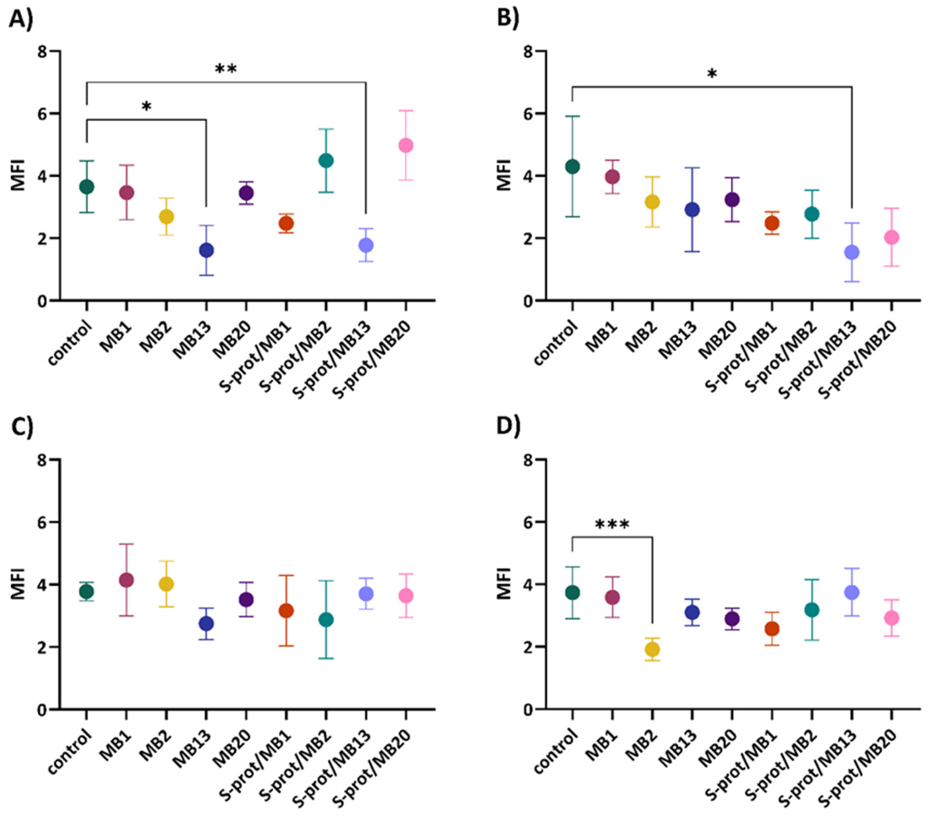
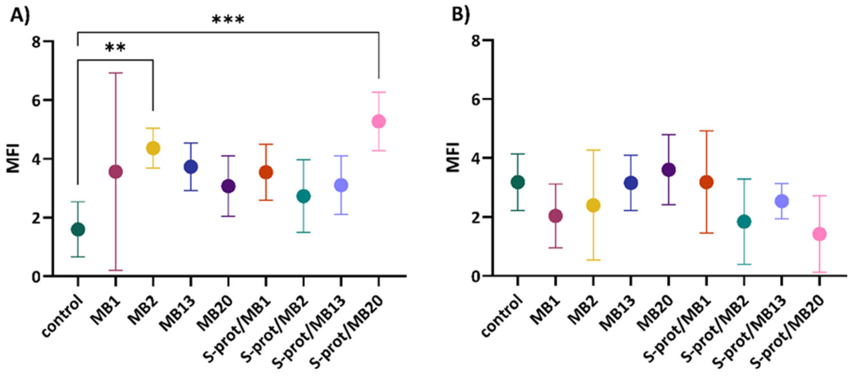
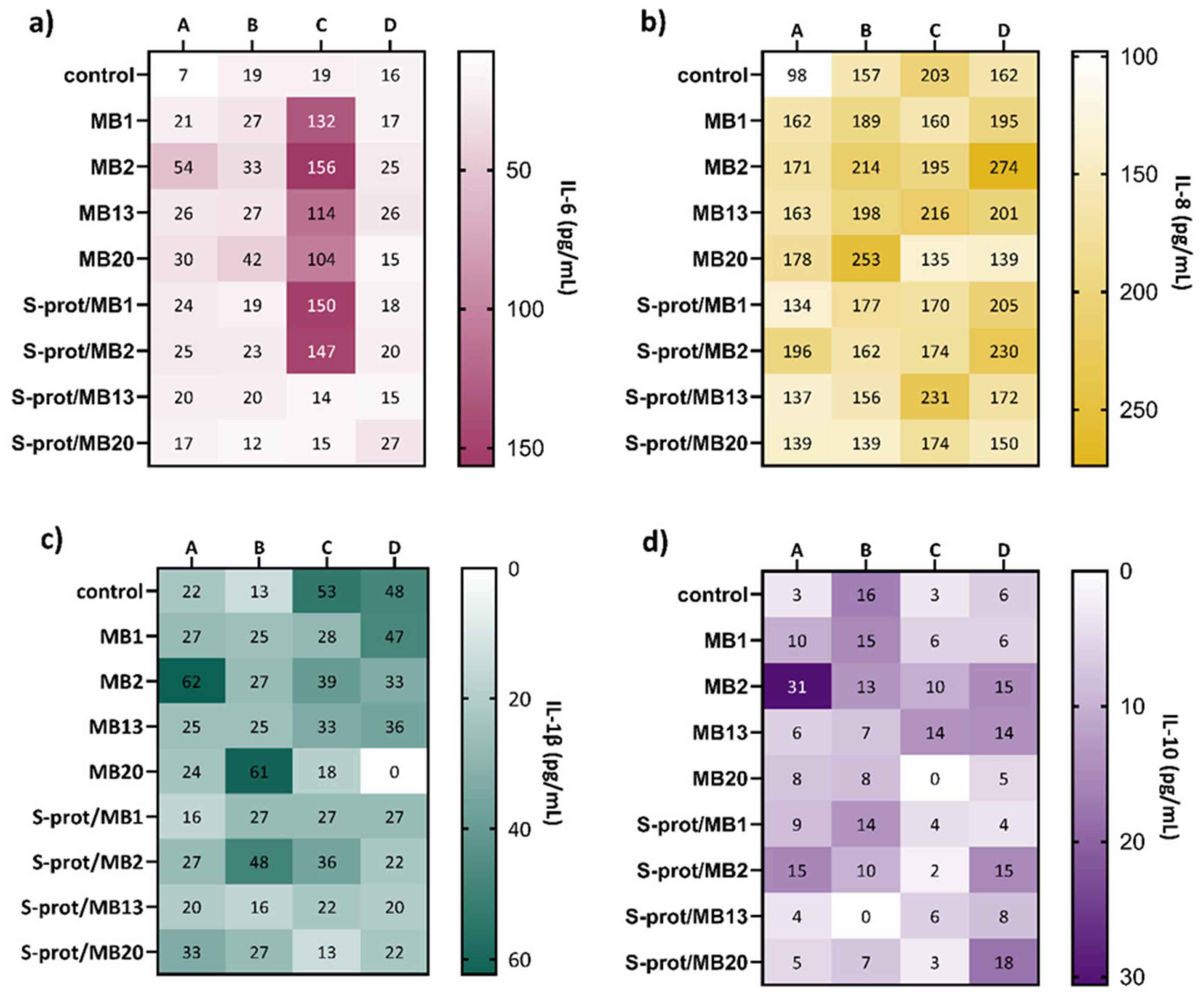
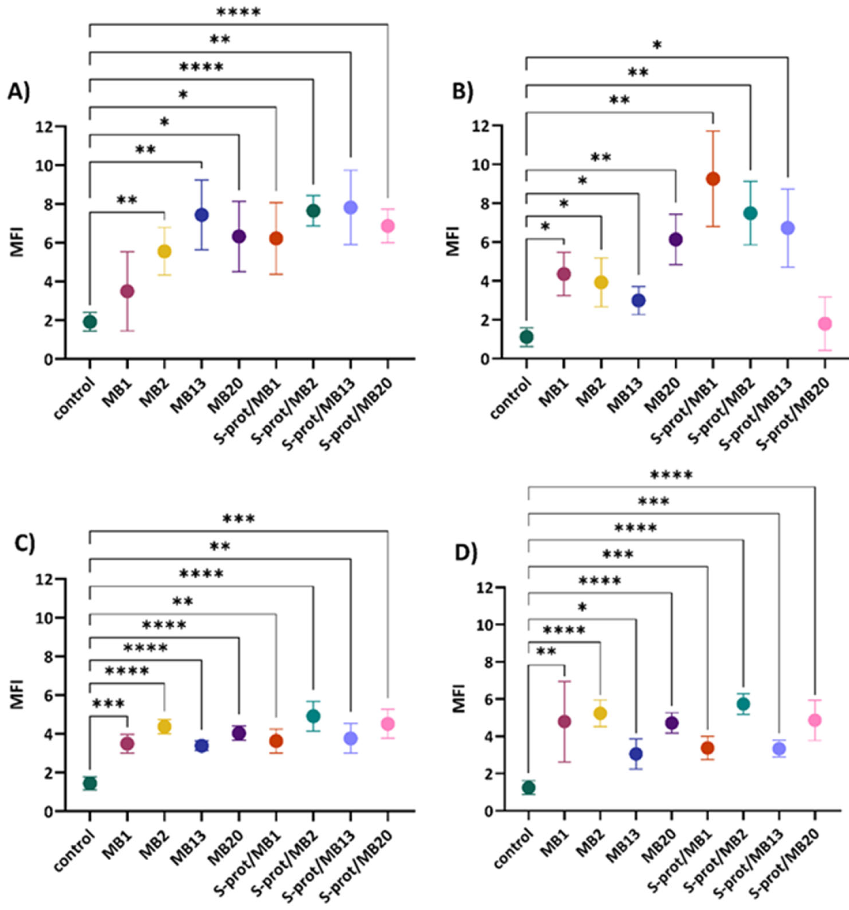
2.4. The Immunomodulatory Effect of L. brevis Strains and S-proteins In Vitro

Inflammatory bowel disease is associated with a dysfunction of the intestinal epithelial barrier causing a disruption of the selective permeability that allows the passage of intestinal contents, including the microbiota. This dysfunction is characterised by a reduced expression of proteins required for the functional assembly of the TJ. As a result, contact of subepithelial cells with bacteria leads to the initiation of inflammation associated with increased leukocyte infiltration and the production of pro-inflammatory cytokines IFN-γ, TNF-α and IL-1β [26]. Certain LAB strains have been shown to modulate the expression of pro-inflammatory cytokines, but the mechanisms involved are not yet fully understood [40]. Interestingly, Lactobacillus strains that produce S-proteins, which contribute to the survival of the producing cells under unfavourable environmental conditions, also display efficient adherence to the intestinal epithelial cells [14]. Along with adhesion, modulation of mucosal and systemic immune responses is an important mechanism in the action of probiotics. Therefore, investigating the specificity of immunomodulation induced by probiotic bacteria is of particular importance for their functionality in GIT. Previously, Johnson et al. [41] found that the S-layer proteins of L. acidophilus NCFM interact with DC-SIGN of dendritic cells, thereby triggering the differentiation of naïve T cells towards the Treg or Th2. Additionally, probiotic lactobacilli can induce pro-inflammatory cytokines such as interleukin ILβ-1, IL-6 and IL-8, as well as the anti-inflammatory cytokine IL-10 through interactions with intestinal epithelial cells as part of the host’s innate immune response recognised as protective immunity. These findings led us to investigate the potential of our S-layer-carrying L. brevis strains to stimulate cytokine secretion by Caco-2 cell line. In this context, the immunomodulatory effect of L. brevis/S-proteins was tested after mimicking inflammation by adding TNF-α and LPS (Figure 6). The levels of IL-6 increased after the addition of almost all L. brevis/S-proteins after incubation of Caco-2 cells only in the medium, with the largest increase with the MB2 strain (Figure 6a). When the Caco-2 cells were cultured in the medium with the addition of LPS, no significant change in IL-6 concentration was observed after the addition of L. brevis/S-proteins compared to the control. On the other hand, the simultaneous addition of L. brevis/S-proteins to TNF-α-treated Caco-2 cells increased IL-6 production after addition of all tested L. brevis strains and S-proteins of MB1 and MB2 strains. The IL-6 concentration in the control and after addition of L. brevis/S-proteins, after simultaneous incubation of Caco-2 cells in medium with TNF-α and LPS, is similar.
IL-8 production increased following the addition of almost all L. brevis/S-proteins after incubation of Caco-2 cells in medium only, with S-proteins of MB2 strain being the most prominent (Figure 6b). The production of IL-8 after addition of LPS did not change significantly after addition of L. brevis/S-proteins, except for strains MB2 and MB20, where the concentration increased. The simultaneous addition of almost all L. brevis/S-proteins tested and TNF-α resulted in a slight decrease in IL-8 production, except for MB13 strain and its S-proteins. After simultaneous treatment of cells with TNF-α and LPS, the IL-8 production increased after addition of MB2 strain and its S-proteins.
The production of IL-1β increased only after addition of the MB2 strain compared to the control when the Caco-2 cells were incubated in the medium (Figure 6c). The culture with L. brevis/S-proteins and LPS increased the production of IL-1β, especially with MB20 strain, whereas addition of TNF-α decreased IL-1β production compared to the control, with the largest decrease when MB20 strain and its S-proteins were added. A complete block of IL-1β production was observed following culturing the cells with MB20 strain and with TNF-α and LPS.
Figure 6. Effect of Levilactobacillus brevis MB1, MB2, MB13 and MB20 strains and their S-proteins on the production of IL-6 (a), IL-8 (b), IL-1β (c) and IL-10 (d) by Caco-2 cells (pg/mL) after the establishment of a monolayer and cultivation in (A) medium, (B) medium supplemented with LPS, (C) medium supplemented with TNF-α and (D) medium supplemented with LPS and TNF-α. control—expression when Caco-2 cells were not exposed to L. brevis/S-proteins.
Ijms 26 02425 g006
The results represent the mean values of duplicate samples pooled from five wells.
As can be observed from Figure 6d, the overall IL-10 production in different treatments is very low. The addition of L. brevis/S-proteins increased IL-10 production only after the treatment by MB2 strain. The culture with L. brevis/S-proteins and LPS did not alter the production of IL-10, with the exception of the S-proteins of MB13, which completely blocked IL-10 production. When TNF-α was added to Caco-2 cultured in the medium, MB13 increased IL-10 production compared to the control. IL-10 production increased after the addition of MB2 and MB13, as well as S-proteins of MB2 and MB20, when the cells were cultured in medium supplemented with LPS and TNF-α.
These experiments demonstrated the immunomodulatory effect of L. brevis strains and their S-proteins, mainly by modulating the production of pro-inflammatory cytokines. In a similar study, the application of L. fermentum CQPC04 significantly inhibited the production of pro-inflammatory cytokines IFN-γ, IL-1β, TNF-α, IL-6 and IL-12 and increased the release of the anti-inflammatory cytokine IL-10 in serum, which reduced colon damage [42]. The results obtained suggest the potential probiotic properties of these strains and their biomolecules, with S-proteins of strain L. brevis MB20, which reduced the expression of the IL-1β and increased the expression of the IL-10 standing out in particular.

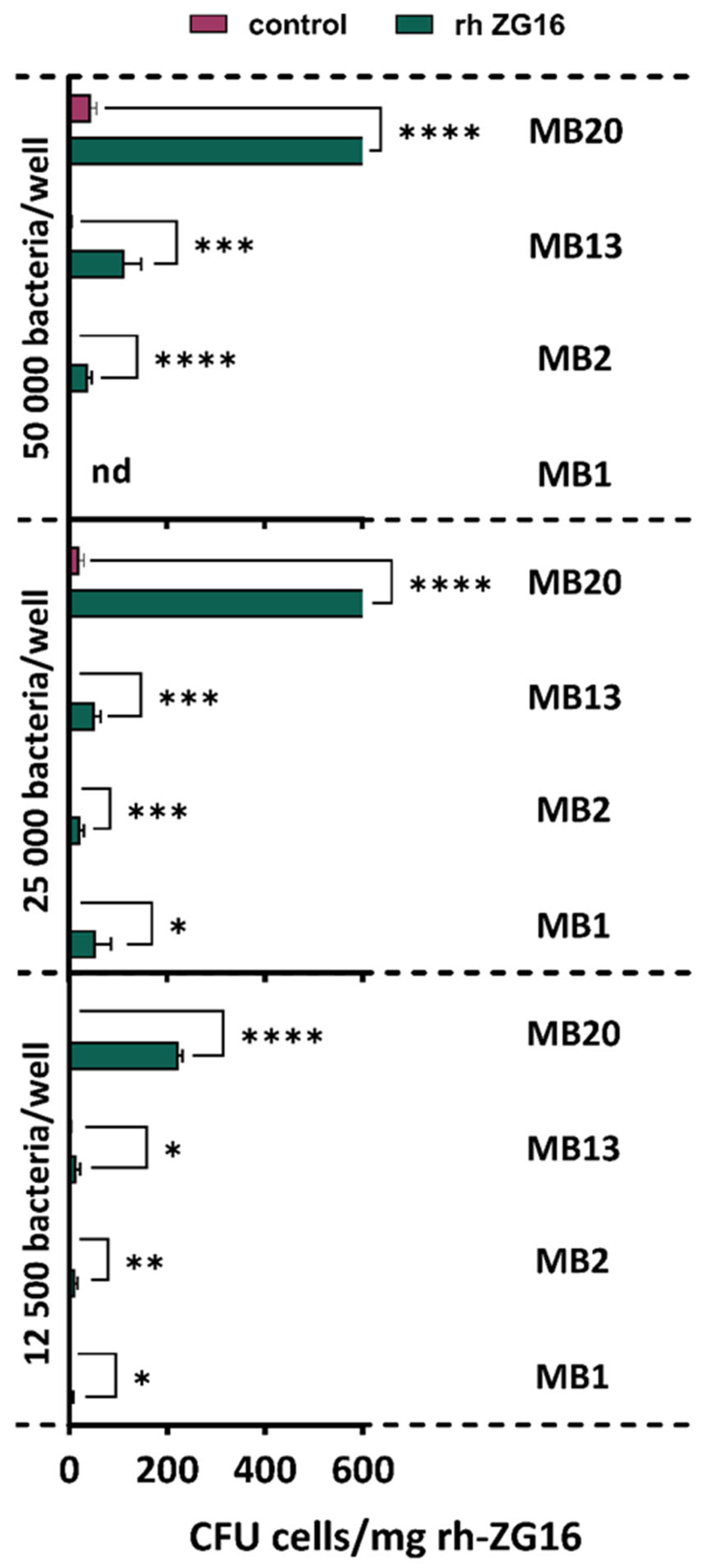
2.5. Adhesion to ZG16 Protein

The intestinal epithelial cells are covered with a layer of mucus that prevents direct contact with the intestinal microbiota and also serves as a binding site for probiotic species [43]. One of the components of the mucus layer is the ZG16 protein, whose function is not yet fully understood but which is known to prevent bacterial invasion [36]. According to Bergström et al. [29], Lactobacillus jensenii peptidoglycan has been shown to bind ZG16. It is suggested that ZG16 aggregates bacteria and thereby cooperates with the inner colon mucus layer and limits bacterial translocation into the host [29,44].
The results of analysis of adhesion ability show that all L. brevis strains tested bind the hr ZG16 protein to some degree, with MB20 adhering most successfully, whereas MB1 has the weakest level of adhesion (Figure 7). Therefore, the possible role of S-proteins in the interaction with mucus components, but obviously adhesion properties are strain-specific. Phùng et al. [45] also investigated the ability of various probiotic strains to adhere to the mucus, of which Lactiplantibacillus plantarum and Bifidobacterium longum subsp. infantis showed a greater ability.

3. Materials and Methods

3.1. Bacterial Strains and Cell Lines

S-layer-producing L. brevis MB1, MB2, MB13 and MB20 were stored at −80 °C in de Man Rogosa Sharpe (MRS; Difco, Detroit, MI, USA) broth, while E. coli 3014 and S. enterica serovar Typhimurium FP1 were maintained in Brain Heart Infusion (BHI; Biolife, Milan, Italy) broth supplemented with 15% v/v glycerol (Alkaloid, Skopje, Macedonia). All strains used in this study are part of the Culture Collection of the Laboratory for Antibiotic, Enzyme, Probiotic and Starter Cultures Technology, University of Zagreb Faculty of Food Technology and Biotechnology. Prior to each experiment, all strains were subcultured twice MRS broth and incubated at 37 °C in microaerophilic conditions.
Caco-2 cells (American Type Culture Collection, ATCC HTB-37, Manassas, VA, USA) were stored in DMEM Ham’s F-12 medium containing L-glutamine (Capricorn Scientific, Ebsdorfergrund, Germany) with 10% FBS (Gibco, London, UK) and 10% dimethyl sulphoxide (DMSO; Kemika, Zagreb, Croatia) in liquid nitrogen. Caco-2 cells were routinely grown in 24-well plates until 80–90% confluent monolayers were reached and then gently rinsed three times with PBS (pH = 7.4). The HT-29 cell line was stored and prepared similarly to the Caco-2 cell line. Research activities were conducted in accordance with all regulatory quality and safety SOP requirements.

3.2. Functional Annotation of L. brevis Genomes

Previously isolated, sequenced and in NCBI deposited genomes of L. brevis MB1, MB2, MB13 and MB20 [13] were annotated using the RAST (Rapid Annotations using Subsystems Technology; http://rast.nmpdr.org/rast.cgi, accessed on 1 July 2024) server, by identifying protein-coding, rRNA and tRNA genes, assigning them a function, predicting which subsystems are present in the genome, and using the information obtained to construct the metabolic network of an individual organism [46].

3.3. Extraction of S-Layer Proteins from the Surface of L. brevis Strains

Following the protocol described in Čuljak et al. [14], L. brevis cells MB1, MB2, MB13 and MB20 were harvested after overnight incubation, washed in saline and suspended in 3 M guanidine hydrochloride (GHCl; Sigma-Aldrich, St. Louis, MO, USA) and incubated for 1 h at room temperature with shaking to obtain S-layer-depleted L. brevis strains (S−), or suspended in 5 M GHCl and incubated on ice for 2 h to obtain S-protein extracts. The S-protein extracts were separated from the cells by centrifugation at 4200 rpm for 10 min at 4 °C, and the cells were washed twice with saline to remove residual GHCl. Dialysis of S-protein extracts was performed using a 12–14 kDa Spectra/Por membrane (Spectrum Laboratories, Rancho Dominguez, CA, USA) in phosphate-buffered saline (PBS; pH = 7.4; Gibco, London, UK) at 4 °C on a magnetic stirrer. After dialysis, the samples were lyophilised in Christ Alpha 1–2 LDplus (Martin Christ, Osterode am Harz, Germany).
Untreated L. brevis cells (S+) were prepared for the experiments in the same way, except that distilled water was used instead of GHCl. To confirm the removal of the S-layer from the treated bacterial cells, the protein extracts were subjected to sodium dodecyl sulphate-polyacrylamide gel electrophoresis (SDS-PAGE) according to the protocol of Barukčić Jurina et al. [47]. The electrophoretic separation of the proteins was performed using a PowerPac HC™ (Biorad, Hercules, CA, USA) at a constant voltage of 150 V, along with the ProSieve QuadColor Protein Marker 4.6–315 kDa (Lonza, Morristown, NJ, USA).

3.4. Autoaggregation and Coaggregation Assay

Autoaggregation assay was performed as described in Leboš Pavunc et al. [48]. Previously prepared S-layer depleted (S−) and non-depleted (S+) L. brevis strains were washed twice with PBS (pH = 7.4). Washed bacterial cells were suspended in PBS to achieve an OD620 = 1 and autoaggregation was determined over a period of 5 h using the INFINITE F PLEX microplate reader (TECAN, Männedorf, Switzerland). The autoaggregation percentage is expressed as follows:
A u t o a g g r e g a t i o n % = 1 A t A 0 × 100
where At represents the absorbance at 5 h and A0 the absorbance at 0 h.
For the coaggregation assay, E. coli 3014 and S. Typhiumurium FP1 were prepared in the same way as L. brevis strains in autoaggregation assay. The percentage of coaggregation was calculated using the following equation:
C o a g g r e g a t i o n % = A x + A y 2 A x + y A x + A y 2 × 100
where x and y represent each of the two bacterial strains in the experiment and x + y their mixture.

3.5. Competitive Exclusion Assay

The role of S-proteins of L. brevis strains MB1, MB2, MB13 and MB20 in the competitive exclusion of potentially pathogenic E. coli 3014 and S. Typhimurium FP1 was determined as described in Butorac et al. [10], with modifications. Untreated (S+) and GHCl-treated (S−) L. brevis strains were incubated at 37 °C for 1 h with a Caco-2 monolayer. After incubation, Caco-2 cells were washed with PBS (pH = 7.4) and 1 mL of suspension of potentially pathogenic bacteria was added to the wells and incubation was continued at 37 °C for 1 h. After incubation, the wells were washed 3 times with 1 mL PBS (pH = 7.4) to remove non-adherent bacterial cells and incubated for 10 min in 0.05% v/v Triton X-100 (AppliChem, Darmstadt, Germany) solution. The contents of each well were centrifuged at 13,000 rpm for 5 min. Finally, the cell precipitate was suspended in 1 mL PBS (pH = 7.4), and the total number of viable adherent E. coli 3014 and S. Typhimurium FP1 cells was determined using the spot-plate method on Rapid (Biorad, Hercules, CA, USA) and XLD (Biolife, Milan, Italy) agar plates, respectively.

3.6. The Effect of L. brevis and Their S-proteins on the Improvement of Epithelial Barrier Function In Vitro

3.6.1. Cell Treatments

Caco-2 cell cultures at 80–90% confluent were washed three times with PBS and then incubated with either L. brevis strains (multiplicity of infection of 2) or their respective S-proteins (50 µg/mL) for 20 h. Caco-2 cells without any treatment were used as control. Experiments were also performed with simultaneous incubation of Caco-2 cells with either lipopolysaccharide (LPS) from E. coli (1 µg/mL) or TNF-α (10 ng/mL), or by their combined application, followed by the addition of L. brevis strains or S-proteins.
To analyse the capacity of L. brevis strains and their S-proteins to affect the expression of zymogen granule membrane protein 16 (ZG16), HT29 cells conditioned with methotrexate (MTX) and 5-fluorouracil (5FU) were also used. The HT29 cell sub clone was prepared by gradual adaption to the increasing concentrations of MTX and 5-FU (1 µM, 2 µM, 5 µM, and 10 µM) over 25–30 days. After conditioning, the cells were passaged and transferred into T25 flasks with medium without chemotherapeutics (DMEM/F12 + 10%FCS + L-Glu + Pen/Strep) and maintained in that medium. Next, HT29 cells were treated with L. brevis strains or S-proteins for 5 days. The effect was also monitored when the cells were simultaneously exposed to TNF-α (5 ng/mL) followed by treatment with L. brevis strains or S-proteins for 5 days.

3.6.2. Immunofluorescent Microscopic Analysis of TJ Protein Expression

For immunolabelling of occludin (#CL488-27260) and ZO-1 (#CL488-21773), rabbit polyclonal antibodies conjugated to CoraLite® Plus 488 (Proteintech, Rosemont, IL, USA) were used, while for JAM-A, a rabbit monoclonal antibody (clone EPR23244-12, Abcam, Cambridge, UK) and a secondary antibody conjugated with AlexaFluor 488 fluorochrome (Proteintech, Rosemont, IL, USA) were used. For immunolabelling of ZG16, a rabbit polyclonal antibody (#AB185483, Abcam, Cambridge, UK) and a secondary antibody conjugated with DyLight 488 fluorochrome (Abcam, Cambridge, UK) were used. The cell nuclei were stained with 4′,6-diamidino-2-phenylindole (DAPI; Sigma-Aldrich, St. Louis, MO, USA). For each antibody, a corresponding isotype control antibody was used in parallel.
After treatments, the cells were fixed by adding 70% v/v cold ethanol and permeabilised in a 0.1% solution of Triton-X 100 in PBS (pH = 7.4). After washing and blocking non-specific binding with 4% bovine serum albumin (BSA; Sigma-Aldrich, St. Louis, MO, USA), primary antibodies were added to the cells. If unlabelled antibodies were used, an additional step of incubating the cells with labelled secondary antibodies followed. Finally, the cells were spiked with 150 nM DAPI solution and then analysed using the EVOS FLc Cell Imager (Thermo Fisher Scientific, Waltham, MA, USA). For each sample, 6 images of cells at different positions in the well were captured and processed in ImageJ (https://imagej.nih.gov/ij/, accessed on 26 August 2024). The intensity of protein expression is expressed as the coefficient of mean fluorescence intensity (MFI) representing the ratio of the intensity of specific fluorescence and the isotypic control antibodies.

3.7. Flow Cytometric Measurement of Cytokines

The expression of IL-1β, IL-6, IL-8 and IL-10 cytokines was analysed after incubation of Caco-2 cells with L. brevis MB1, MB2, MB13 and MB20, or their S-proteins, under different treatments described in the previous chapter. Composite samples of media supernatant from individual wells (5 wells for each condition), stored at −80 °C, were used for analysis in duplicates after thawing. Samples were analysed using the cytometric bead array (CBA) method on an LSRII flow cytometer (Becton Dickinson, Franklin Lakes, NJ, USA). Frozen cell media were thawed and prepared for flow cytometer analysis according to the CBA kit LEGENDplex (Biolegend, San Diego, CA, USA) manufacturer’s instructions for selected cytokines. The obtained fluorescence data were analysed using GainData® (arigo’s ELISA Calculator, available at the link https://www.arigobio.com/ELISA-calculator, accessed on 9 September 2024).

3.8. Ability of L. brevis Strains to Adhere to Recombinant Human ZG16 Protein

In order to test the ability of selected strains to adhere to the ZG16 protein, the ZG16 protein (#ab276544, Abcam, Cambridge, UK) or the I-Block reagent (control; Thermo Fisher Scientific, Waltham, MA, USA), both at a concentration of 8 µg/mL, were added to a 96-well microtitre plate and incubated for 4 h. After incubation, the contents of the wells were removed and 1% BSA solution was added, followed by overnight incubation at 4 °C. To achieve the desired number of bacteria per well (50,000, 25,000 and 12,500 bacteria), the corresponding volumes of bacterial suspension and medium were added to the wells, followed by overnight incubation (16 h) at 37 °C and 5% CO2. After incubation, the wells were washed three times with PBS preheated to 37 °C followed with 10 mM dithiothreitol (Thermo Fisher Scientific, Waltham, MA, USA) in PBS treatment for 15 min to detach adhered bacteria. The contents of the wells were transferred to sterile tubes and the wells were additionally washed with PBS or 1% BSA in PBS. Pooled bacterial suspensions were centrifuged at 3220× g for 15 min. The bacteria in the pellet were suspended in 50 μL of PBS and inoculated in duplicate of 25 μL on previously prepared MRS agar plates. After 48 h of anaerobic incubation at 37 °C, the grown colonies were counted.

3.9. Statistical Analysis

The results are expressed as the mean of three independent values ± standard deviation. Statistical significance was determined using one-way analysis of variance (ANOVA). Pairwise differences between group means were determined using Tukey’s honestly significant difference test for pairwise comparisons after analysis of variance (https://www.statskingdom.com/index.html, accessed on 2 September 2024) [49]. Differences between samples were considered significant at p < 0.05. Images were created using GraphPad Prism v.9.4.1 (GraphPad Software, La Jolla, CA, USA).

4. Conclusions

The role of S-proteins in the autoaggregation, coaggregation, and competitive exclusion of S. Typhimurium FP1 of all examined strains as well as in the coaggregation with E. coli 3014 of strains MB1 and MB2 and competitive exclusion against E. coli 3014 of strains MB13 and MB20 were demonstrated. In vitro experiments on human cell cultures demonstrated the beneficial effect on intestinal cell barrier function as evident from the capacity of increase in the expression of JAM-A, occludin and ZG16 proteins, which are required for the maintenance of intestinal integrity. Furthermore, the immunomodulatory effect of the L. brevis strains and their S-proteins was detected as a decrease in the production of the pro-inflammatory cytokines IL-6, IL-8 and IL-1β and an increase in the production of the anti-inflammatory cytokine IL-10. In addition, all tested L. brevis strains were found to bind to the ZG16 protein, with strain MB20 exhibiting the highest affinity and adhesion capacity for the human ZG16 protein. Finally, to conclude, the study supports in vitro evidence for various aspects of the beneficial activities of L. brevis strains such as aggregation, competitive exclusion, effect on tight junction proteins and cytokine expression, in the context of the involvement of S-layer proteins in these processes. This contributes to the characterisation of the probiotic mechanisms of action of S-layer producing L. brevis strains and stimulates further research important to their adaptation in the intestinal microenvironment and thus gut functionality.

Supplementary Materials

The following supporting information can be downloaded at: www.mdpi.com/article/10.3390/ijms26062425/s1.

Author Contributions

Methodology, Formal analysis, Investigation, Visualisation, Writing—Original Draft, N.Č.; Conceptualization, Validation, Writing—Review & Editing, K.B. (Krešo Bendelja); Project administration, Supervision, Writing—Review & Editing, A.L.P.; Data Curation, Investigation, K.B. (Katarina Butorac); Data Curation, Investigation, M.B.; Methodology, Formal analysis, A.S.M.; Methodology, Formal analysis, Ž.C.; Investigation, J.H.; Supervision, Formal analysis, Validation, Writing—Review & Editing, J.N.; Supervision, Writing—Review & Editing, J.Š.; Conceptualization, Resources, Funding acquisition, Supervision, Writing—Review & Editing, B.K. All authors have read and agreed to the published version of the manuscript.

Funding

This research was funded by the Croatian Science Foundation through the projects IP-2019-04-2237 and IP-2024-05-6548. The authors also acknowledge the financial support from the University of Zagreb, Croatia. The authors are grateful for the equipment provided with financial support from the project “Bioprospecting of the Adriatic Sea” (KK.01.1.1.01.0002) granted to the Scientific Centre of Excellence for Marine Bioprospecting (BioProCro) at the Ruđer Bošković Institute, Zagreb, Croatia.

Institutional Review Board Statement

Not applicable.

Informed Consent Statement

Not applicable.

Data Availability Statement

The original contributions presented in this study are included in the article and Supplementary Materials. Further inquiries can be directed to the corresponding author.

Acknowledgments

The authors acknowledge Ksenija Durgo (University of Zagreb Faculty of Food Technology and Biotechnology, Laboratory for Biology and Microbial Genetics) who provided Caco-2 cells for competitive exclusion experiment.

Conflicts of Interest

The authors declare no conflicts of interest.

References

  1. Suez, J.; Zmora, N.; Segal, E.; Elinav, E. The pros, cons, and many unknowns of probiotics. Nat. Med. 2019, 25, 716–729. [Google Scholar] [CrossRef]
  2. Singh, R.P.; Shadan, A.; Ma, Y. Biotechnological applications of probiotics: A multifarious weapon to disease and metabolic abnormality. Probiotics Antimicrob. Proteins 2022, 14, 1184–1210. [Google Scholar] [CrossRef]
  3. Barros, C.P.; Guimaraes, J.T.; Esmerino, E.A.; Duarte, M.C.K.; Silva, M.C.; Silva, R.; Ferreira, B.M.; Sant’Ana, A.S.; Freitas, M.Q.; Cruz, A.G. Paraprobiotics and postbiotics: Concepts and potential applications in dairy products. Curr. Opin. Food Sci. 2020, 32, 1–8. [Google Scholar] [CrossRef]
  4. Monteiro, S.S.; Schnorr, C.E.; Pasquali, M.A.D.B. Paraprobiotics and Postbiotics-Current State of Scientific Research and Future Trends toward the Development of Functional Foods. Foods 2023, 12, 2394. [Google Scholar] [CrossRef] [PubMed]
  5. Cuevas-González, P.F.; Liceaga, A.M.; Aguilar-Toalá, J.E. Postbiotics and Paraprobiotics: From concepts to applications. Food Res. Int. 2020, 136, 109502. [Google Scholar] [CrossRef] [PubMed]
  6. Banić, M.; Butorac, K.; Čuljak, N.; Butorac, A.; Novak, J.; Pavunc, A.L.; Rušanac, A.; Stanečić, Ž.; Lovrić, M.; Šušković, J.; et al. An Integrated Comprehensive Peptidomics and In Silico Analysis of Bioactive Peptide-Rich Milk Fermented by Three Autochthonous Cocci Strains. Int. J. Mol. Sci. 2024, 25, 2431. [Google Scholar] [CrossRef]
  7. Assandri, M.H.; Malamud, M.; Trejo, F.M.; de los A Serradell, M. S-layer proteins as immune players: Tales from pathogenic and non-pathogenic bacteria. Curr. Res. Microb. Sci. 2023, 4, 100187. [Google Scholar] [CrossRef]
  8. Banić, M.; Uroić, K.; Pavunc, A.L.; Novak, J.; Zorić, K.; Durgo, K.; Petković, H.; Jamnik, P.; Kazazić, S.; Kazazić, S.; et al. Characterization of S-layer proteins of potential probiotic starter culture Lactobacillus brevis SF9B isolated from sauerkraut. LWT Food Sci. Technol. 2018, 93, 257–267. [Google Scholar] [CrossRef]
  9. Angelin, J.; Kavitha, M. Exopolysaccharides from probiotic bacteria and their health potential. Int. J. Biol. Macromol. 2020, 162, 853–865. [Google Scholar] [CrossRef]
  10. Butorac, K.; Banić, M.; Novak, J.; Pavunc, A.L.; Uroić, K.; Durgo, K.; Oršolić, N.; Kukolj, M.; Radović, S.; Scalabrin, S.; et al. The functional capacity of plantaricin-producing Lactobacillus plantarum SF9C and S-layer-carrying Lactobacillus brevis SF9B to withstand gastrointestinal transit. Microb. Cell Factories 2020, 19, 106. [Google Scholar] [CrossRef]
  11. Čuljak, N.; Bellich, B.; Pedron, A.; Butorac, K.; Pavunc, A.L.; Novak, J.; Banić, M.; Šušković, J.; Cescutti, P.; Kos, B. Limosilactobacillus fermentum strains MC1 and D12: Functional properties and exopolysaccharides characterization. J. Biol. Macromol. 2024, 273, 133215. [Google Scholar] [CrossRef] [PubMed]
  12. Butorac, K.; Novak, J.; Bellich, B.; Terán, L.C.; Banić, M.; Pavunc, A.L.; Zjalić, S.; Cescutti, P.; Šušković, J.; Kos, B. Lyophilized alginate-based microspheres containing Lactobacillus fermentum D12, an exopolysaccharides producer, contribute to the strain’s functionality in vitro. Microb. Cell Factories 2021, 20, 85. [Google Scholar] [CrossRef]
  13. Banić, M.; Butorac, K.; Čuljak, N.; Pavunc, A.L.; Novak, J.; Bellich, B.; Kazazić, S.; Kazazić, S.; Cescutti, P.; Šušković, J.; et al. The Human Milk Microbiota Produces Potential Therapeutic Biomolecules and Shapes the Intestinal Microbiota of Infants. Int. J. Mol. Sci. 2022, 23, 14382. [Google Scholar] [CrossRef]
  14. Čuljak, N.; Pavunc, A.L.; Dučkić, K.; Butorac, K.; Banić, M.; Novak, J.; Šušković, J.; Kos, B. Functionality and protective effect of S-layer proteins in microencapsulated freeze-dried probiotic Levilactobacillus brevis strains. Mljekarstvo 2024, 74, 208–218. [Google Scholar] [CrossRef]
  15. Kant, R.; Uroić, K.; Hynonen, U.; Kos, B.; Šušković, J.; Palva, A. Genome Sequence of Lactobacillus brevis Strain D6, Isolated from Smoked Fresh Cheese. Genome Announc. 2016, 4, e00264-16. [Google Scholar] [CrossRef]
  16. Krausova, G.; Hyrslova, I.; Hynstova, I. In vitro evaluation of adhesion capacity, hydrophobicity, and auto-aggregation of newly isolated potential probiotic strains. Fermentation 2019, 5, 100. [Google Scholar] [CrossRef]
  17. Dlamini, Z.C.; Langa, R.L.S.; Aiyegoro, O.A.; Okoh, A.I. Safety Evaluation and Colonisation Abilities of Four Lactic Acid Bacteria as Future Probiotics. Probiotics Antimicrob. Proteins 2019, 11, 397–402. [Google Scholar] [CrossRef] [PubMed]
  18. Alp, D.; Kuleaşan, H.; Altıntaş, A.K. The importance of the S-layer on the adhesion and aggregation ability of lactic acid bacteria. Mol. Biol. Rep. 2020, 47, 3449–3457. [Google Scholar] [CrossRef]
  19. van Zyl, W.F.; Deane, S.M.; Dicks, L.M.T. Molecular insights into probiotic mechanisms of action employed against intestinal pathogenic bacteria. Gut Microbes 2020, 12, e1831339. [Google Scholar] [CrossRef]
  20. Tytgat, H.L.; Douillard, F.P.; Reunanen, J.; Rasinkangas, P.; Hendrickx, A.P.; Laine, P.K.; Paulin, L.; Satokari, R.; de Vos, W.M. Lactobacillus rhamnosus GG outcompetes Enterococcus faecium via mucus-binding pili: Evidence for a novel and heterospecific probiotic mechanism. Appl. Environ. Microbiol. 2016, 82, 5756–5762. [Google Scholar] [CrossRef]
  21. Ayala, D.I.; Cook, P.W.; Franco, J.G.; Bugarel, M.; Kottapalli, K.R.; Loneragan, G.H.; Brashears, M.M.; Nightingale, K.K. A Systematic Approach to Identify and Characterize the Effectiveness and Safety of Novel Probiotic Strains to Control Foodborne Pathogens. Front. Microbiol. 2019, 10, 1108. [Google Scholar] [CrossRef] [PubMed]
  22. Zhang, J.; Gao, J.; Guo, Y.; Wu, Z.; Pan, D. Extraction of Lactobacillus acidophilus CICC 6074 S-layer proteins and their ability to inhibit enteropathogenic Escherichia coli. Curr. Microbiol. 2017, 74, 1123–1129. [Google Scholar] [CrossRef] [PubMed]
  23. Buckley, A.; Turner, J.R. Cell biology of tight junction barrier regulation and mucosal disease. CHS Perspect. Biol. 2018, 10, a029314. [Google Scholar] [CrossRef]
  24. Suárez, L.J.; Arboleda, S.; Angelov, N.; Arce, R.M. Oral Versus Gastrointestinal Mucosal Immune Niches in Homeostasis and Allostasis. Front. Immunol. 2021, 12, 705206. [Google Scholar] [CrossRef]
  25. Otani, T.; Furuse, M. Tight junction structure and function revisited. Trends Cell Biol. 2020, 30, 805–817. [Google Scholar] [CrossRef] [PubMed]
  26. Landy, J.; Ronde, E.; English, N.; Clark, S.K.; Hart, A.L.; Knight, S.C.; Ciclitira, P.J.; Al-Hassi, H.O. Tight junctions in inflammatory bowel diseases and inflammatory bowel disease associated colorectal cancer. World J. Gastroenterol. 2016, 22, 3117–3126. [Google Scholar] [CrossRef]
  27. Gou, H.Z.; Zhang, Y.L.; Ren, L.F.; Li, Z.J.; Zhabg, L. How do intestinal probiotics restore the intestinal barrier? Front. Micriobiol. 2022, 13, 929346. [Google Scholar] [CrossRef]
  28. Meng, H.; Li, W.; Boardman, L.A.; Wang, L. Loss of ZG16 is associated with molecular and clinicopathological phenotypes of colorectal cancer. BMC Cancer 2018, 18, 433. [Google Scholar] [CrossRef]
  29. Bergström, J.H.; Birchenough, G.M.; Katona, G.; Schroeder, B.O.; Schütte, A.; Ermund, A.; Johansson, M.E.; Hansson, G.C. Gram-positive bacteria are held at a distance in the colon mucus by the lectin-like protein ZG16. Proc. Natl. Acad. Sci. USA 2016, 113, 13833–13838. [Google Scholar] [CrossRef]
  30. Fu, G.; Zhao, K.; Chen, H.; Wang, Y.; Nie, L.; Wei, H.; Wan, C. Effect of 3 lactobacilli on immunoregulation and intestinal microbiota in a β-lactoglobulin–induced allergic mouse model. J. Dairy Sci. 2019, 102, 1943–1958. [Google Scholar] [CrossRef]
  31. Di Tommaso, N.; Gasbarrini, A.; Ponziani, F.R. Intestinal Barrier in Human Health and Disease. Int. J. Environ. Res. Public Health 2021, 18, 12836. [Google Scholar] [CrossRef] [PubMed]
  32. Yin, M.; Yan, X.; Weng, W.; Yang, Y.; Gao, R.; Liu, M.; Pan, C.; Zhu, Q.; Li, H.; Wei, Q.; et al. Micro integral membrane protein (MIMP), a newly discovered anti-inflammatory protein of Lactobacillus plantarum, enhances the gut barrier and modulates microbiota and inflammatory cytokines. Cell. Physiol. Biochem. 2018, 45, 474–490. [Google Scholar] [CrossRef]
  33. Bendinelli, P.; De Noni, I.; Cattaneo, S.; Silvetti, T.; Brasca, M.; Piazzalunga, F.; Donetti, E.; Ferraretto, A. Surface layer proteins from Lactobacillus helveticus ATCC® 15009™ affect the gut barrier morphology and function. Tissue Barriers 2023, 12, 2289838. [Google Scholar] [CrossRef]
  34. Suzuki, T. Regulation of the intestinal barrier by nutrients: The role of tight junctions. Anim. Sci. J. 2020, 91, e13357. [Google Scholar] [CrossRef]
  35. van der Post, S.; Jabbar, K.S.; Birchenough, G.; Arike, L.; Akhtar, N.; Sjovall, H.; Johansson, M.E.V.; Hansson, G.C. Structural weakening of the colonic mucus barrier is an early event in ulcerative colitis pathogenesis. Gut 2019, 68, 2142–2151. [Google Scholar] [CrossRef]
  36. Javitt, G.; Kinzel, A.; Reznik, N.; Fass, D. Conformational switches and redox properties of the colon cancer-associated human lectin ZG16. FEBS J. 2021, 288, 6465–6475. [Google Scholar] [CrossRef] [PubMed]
  37. Chen, X.; Chen, Y.; Zhang, Y.; Zhang, Y.; Wang, Y.; Li, Y.; Sun, Y.; Meng, G.; Yang, G.; Li, H. ZG16 impacts gut microbiota-associated intestinal inflammation and pulmonary mucosal function through bacterial metabolites. Int. Immunopharmacol. 2024, 141, 112995. [Google Scholar] [CrossRef]
  38. Ahn, D.H.; Crawley, S.C.; Hokari, R.; Kato, S.; Yang, S.C.; Li, J.D.; Kim, Y.S. TNF-alpha activates MUC2 transcription via NF-kappaB but inhibits via JNK activation. Cell Physiol. Biochem. 2005, 15, 29–40. [Google Scholar] [CrossRef]
  39. Dharmani, P.; Leung, P.; Chadee, K. Tumor necrosis factor-α and Muc2 mucin play major roles in disease onset and progression in dextran sodium sulphate-induced colitis. PLoS ONE 2011, 6, e25058. [Google Scholar] [CrossRef] [PubMed]
  40. Acosta, M.P.; Goyette-Desjardins, G.; Scheffel, J.; Dudeck, A.; Ruland, J.; Lepenies, B. S-Layer from Lactobacillus brevis modulates antigen-presenting cell functions via the Mincle-Syk-Card9 Axis. Front. Immunol. 2021, 12, 511. [Google Scholar] [CrossRef]
  41. Johnson, B.; Selle, K.; O’Flaherty, S.; Goh, Y.J.; Klaenhammer, T. Identification of extracellular surface-layer associated proteins in Lactobacillus acidophilus NCFM. Microbiology 2013, 159, 2269–2282. [Google Scholar] [CrossRef] [PubMed]
  42. Zhou, X.; Liu, H.; Zhang, J.; Mu, J.; Zalan, Z.; Hegyi, F.; Takács, K.; Zhao, X.; Du, M. Protective effect of Lactobacillus fermentum CQPC04 on dextran sulfate sodium–induced colitis in mice is associated with modulation of the nuclear factor-κB signaling pathway. J. Dairy Sci. 2019, 102, 9570–9585. [Google Scholar] [CrossRef] [PubMed]
  43. Ayivi, R.D.; Gyawali, R.; Krastanov, A.; Aljaloud, S.O.; Worku, M.; Tahergorabi, R.; da Silva, R.C.; Ibrahim, S.A. Lactic acid bacteria: Food safety and human health applications. Dairy 2020, 1, 202–232. [Google Scholar] [CrossRef]
  44. Song, C.; Chai, Z.; Chen, S.; Zhang, H.; Zhang, X.; Zhou, Y. Intestinal mucus components and secretion mechanisms: What we do and do not know. Exp. Mol. Med. 2023, 55, 681–691. [Google Scholar] [CrossRef]
  45. Phùng, T.T.T.; Dupont, S.; Beney, L.; Moundanga, S.; Denimal, E.; Hồ, P.H.; Karbowiak, T. Ex-vivo investigation of probiotic bacterial adhesion to the intestinal mucus. Heliyon 2024, 10, e36339. [Google Scholar] [CrossRef]
  46. Aziz, R.K.; Bartels, D.; Best, A.A.; DeJongh, M.; Disz, T.; Edwards, R.A.; Formsma, K.; Gerdes, S.; Glass, E.M.; Kubal, M.; et al. The RAST Server: Rapid Annotations using Subsystems Technology. BMC Genom. 2008, 9, 75. [Google Scholar] [CrossRef]
  47. Jurina, I.B.; Blažević, I.; Jurina, T.; Pavunc, A.L.; Božanić, R.; Jakopović, K.L. Evaluation of heat-induced changes and 5-hydroxymethylfurfural formation in mare milk—A comparative study to cow milk. Mljekarstvo 2024, 74, 89–105. [Google Scholar] [CrossRef]
  48. Pavunc, A.L.; Penava, L.; Ranilović, J.; Novak, J.; Banić, M.; Butorac, K.; Petrović, E.; Mihaljević-Herman, V.; Bendelja, K.; Savić-Mlakar, A.; et al. Influence of Dehydrated Wheat/Rice Cereal Matrices on Probiotic Activity of Bifidobacterium animalis ssp. lactis BB-12®. Food Technol. Biotechnol. 2019, 57, 147. [Google Scholar] [CrossRef]
  49. Statistics Kingdom, One-Way ANOVA Calculator and Tukey HSD, Statistics Kingdom: Website for Statistics Calculators. 2024. Available online: http://www.statskingdom.com (accessed on 2 September 2024).
Figure 1. Competitive exclusion of potentially pathogenic bacteria (A) Escherichia coli 3014 and (B) S. serovar Typhimurium FP1 after pre-incubation of Caco-2 cells with Levilactobacillus brevis MB1, MB2, MB13 and M20 strains, with (S+) or without (S−) S-layer. The error bars represent the standard deviations of the mean values of the results of three replicates. * Statistically significantly different (* p < 0.05, ** p < 0.01, *** p < 0.001, **** p < 0.0001), according to Tukey’s honestly significant difference (HSD) test.
Ijms 26 02425 g001
Figure 2. JAM-A expression in Caco-2 cells after establishing a monolayer and cultivation in (A) medium, (B) medium supplemented with LPS, (C) medium supplemented with TNF-α and (D) medium supplemented with LPS and TNF-α, after exposure to L. brevis MB1, MB2, MB13 and MB20 strains or their S-proteins. MFI—mean fluorescence intensity; control—protein expression when Caco-2 cells were not exposed to L. brevis/S-proteins. The error bars represent the standard deviations of the mean values of the results of three replicates. * Statistically significantly different (* p < 0.05, ** p < 0.01, *** p < 0.001, **** p < 0.0001) according to Tukey’s honestly significant difference (HSD) test.
Ijms 26 02425 g002
Figure 7. Adhesion of L. brevis strains MB1, MB2, MB13 and MB20 to recombinant human ZG16 protein (rh ZG16) and I-Block (control) when 50,000, 25,000 and 12,500 bacterial cells were added to the wells. nd—not done (concentration of the cells was too high in the stock solution). The error bars represent the standard deviations of the mean values of the results of three replicates. * Statistically significantly different (* p < 0.05, ** p < 0.01, *** p < 0.001, **** p < 0.0001).
Ijms 26 02425 g007
Table 1. Autoaggregation ability (%) of untreated (S+) and GHCl-treated (S−) Levilactobactobacillus brevis MB1, MB2, MB13 and MB20 strains.
Autoaggregation %
L. brevisUntreated (S+)GHCl-Treated (S−)
MB173.57 ± 5.91 a26.44 ± 4.57 b
MB266.04 ± 8.33 a42.53 ± 7.18 b
MB1377.15 ± 4.89 a41.51 ± 12.57 b
MB2069.65 ± 5.27 a21.92 ± 3.26 b
Results are reported as mean values ± standard deviation. Different letters in superscript indicate a statistically significant difference (p < 0.05) between S+ and S− of the same strain.
Table 2. Coaggregation ability (%) of untreated (S+) and GHCl-treated (S−) Levilactobacillus brevis MB1, MB2, MB13 and MB20 strains with Escherichia coli 3014 and Salmonella enterica serovar Typhimurium FP1.
L. brevisCoaggregation %
S. Typhimurium FP1E. coli 3014
Untreated (S+)GHCl-Treated (S−)Untreated (S+)GHCl-Treated (S−)
MB147.07 ± 8.05 a19.62 ± 2.71 b41.54 ± 2.96 a28.02 ± 3.74 b
MB246.73 ± 8.04 a16.97 ± 0.96 b32.41 ± 4.35 a18.59 ± 2.69 b
MB1326.36 ± 1.14 a16.35 ± 0.94 b26.34 ± 4.07 a36.72 ± 5.06 a
MB2034.63 ± 3.17 a22.20 ± 1.64 b35.12 ± 8.42 a23.55 ± 4.43 a
Results are reported as mean values ± standard deviation. Different letters in superscript indicate a statistically significant difference (p < 0.05) between S+ and S− of the same strain.
Disclaimer/Publisher’s Note: The statements, opinions and data contained in all publications are solely those of the individual author(s) and contributor(s) and not of MDPI and/or the editor(s). MDPI and/or the editor(s) disclaim responsibility for any injury to people or property resulting from any ideas, methods, instructions or products referred to in the content.

