Review

CSE/H2S Signaling Pathways in Enhancing Muscle Function and Insulin Sensitivity During Exercise

1
School of Physical Education and Health, Guangzhou University of Chinese Medicine, Guangzhou 510006, China
2
South China Research Center for Acupuncture and Moxibustion, Medical College of Acu-Moxi and Rehabilitation, Guangzhou University of Chinese Medicine, Guangzhou 510006, China
3
College of Sports and Health, Guangzhou Sport University, Guangzhou 510500, China
*
Authors to whom correspondence should be addressed.
Int. J. Mol. Sci. 2025, 26(4), 1741; https://doi.org/10.3390/ijms26041741
Submission received: 20 January 2025 / Revised: 14 February 2025 / Accepted: 17 February 2025 / Published: 18 February 2025
(This article belongs to the Special Issue Molecular and Physiological Mechanisms of Exercise)

Abstract

Exercise plays a crucial role in maintaining metabolic health, enhancing muscle function, and improving insulin sensitivity, thereby preventing metabolic diseases such as type 2 diabetes. Emerging evidence highlights the significance of the cystathionine γ-lyase (CSE)/hydrogen sulfide (H2S) signaling pathway as a pivotal regulator in the molecular and physiological adaptations induced by exercise. This review comprehensively examines the biosynthesis and metabolism of H2S, its distribution in different muscle tissues, and the mechanisms by which CSE/H2S influences muscle contraction, repair, and protein synthesis. Additionally, it explores how CSE/H2S modulates insulin signaling pathways, glucose uptake, and lipid metabolism, thereby enhancing insulin sensitivity. The potential of H2S donors as exercise supplements is also discussed, highlighting their ability to improve exercise performance and metabolic health. Current research advancements, including the application of multi-omics approaches, are reviewed to provide a deeper understanding of the complex molecular networks involved. Furthermore, the challenges and future directions in CSE/H2S research are addressed, emphasizing the need for further mechanistic studies and clinical applications. This review underscores the therapeutic potential of targeting the CSE/H2S pathway to optimize the benefits of exercise and improve metabolic health.

1. Introduction

Exercise plays a pivotal role in maintaining and enhancing overall health, particularly concerning metabolic health, muscle function, and insulin sensitivity [1]. Regular physical activity has been shown to reduce the risk of chronic diseases such as type 2 diabetes, cardiovascular diseases, and obesity [2]. Through various mechanisms, exercise enhances metabolic flexibility, allowing the body to efficiently switch between fuel sources like carbohydrates and fats [3]. Additionally, engaging in consistent physical activity promotes muscle hypertrophy and strength, which are essential for daily functional activities and overall quality of life [4]. Furthermore, exercise improves insulin sensitivity, facilitating better glucose uptake by cells and thereby regulating blood glucose levels [5]. These multifaceted benefits underscore the critical importance of incorporating exercise into daily routines for long-term health maintenance and disease prevention.
The beneficial effects of exercise are underpinned by a complex array of molecular and physiological adaptations [6]. Understanding these adaptations requires an exploration of the intricate signaling pathways that are activated in response to physical activity [7,8,9,10]. For instance, exercise induces the activation of AMP-activated protein kinase (AMPK), which plays a key role in energy homeostasis by enhancing glucose uptake and fatty acid oxidation [10]. Similarly, the mitogen-activated protein kinase (MAPK) pathways are involved in regulating muscle growth and repair [9]. These signaling cascades not only facilitate immediate responses to exercise but also drive long-term adaptations such as increased mitochondrial biogenesis and improved oxidative capacity [7,8]. By elucidating these underlying mechanisms, we can better comprehend how exercise confers its health benefits and potentially identify novel therapeutic targets for metabolic and muscular disorders.
Cystathionine γ-lyase (CSE) is a critical enzyme in the biosynthesis of hydrogen sulfide (H2S), a gaseous signaling molecule [11]. CSE catalyzes the conversion of cystathionine to cysteine, producing H2S as a byproduct [12]. This enzyme is predominantly expressed in the cardiovascular system, liver, and skeletal muscles, highlighting its significant role in various physiological processes [13,14,15]. Beyond its enzymatic activity, CSE is implicated in regulating cellular redox status and modulating inflammatory responses [16,17]. The regulation of CSE expression and activity is influenced by several factors, including oxidative stress and hormonal signals, which in turn affect H2S production [18]. Understanding the regulation of CSE is essential for comprehending how H2S levels are maintained and modulated in different tissues.
H2S has emerged as a vital gasotransmitter, alongside nitric oxide (NO) and carbon monoxide (CO), playing diverse roles in cellular signaling [19]. H2S is involved in numerous physiological processes, including vasodilation, neurotransmission, and cytoprotection [20]. In the context of exercise, H2S has been shown to enhance muscle blood flow, thereby improving oxygen and nutrient delivery to active tissues [21]. Additionally, H2S modulates mitochondrial function, promoting efficient energy production and reducing oxidative stress [22]. Its anti-inflammatory properties contribute to the attenuation of exercise-induced muscle damage and facilitate recovery [23]. Moreover, H2S interacts with insulin signaling pathways, enhancing insulin sensitivity and glucose uptake in skeletal muscles [11,24]. The multifaceted roles of H2S underscore its significance in mediating the beneficial effects of exercise on muscle function and metabolic health.
This narrative review aims to clarify the role of CSE/H2S signaling in enhancing muscle function and insulin sensitivity during exercise. By synthesizing current evidence, we will examine how H2S mediates adaptive responses to physical activity through its influence on key molecular pathways, interactions with gasotransmitters, and regulation of processes such as mitochondrial biogenesis, antioxidant defense, and inflammation. Additionally, we will explore the therapeutic potential of targeting the CSE/H2S axis to maximize exercise benefits and address metabolic and muscular disorders.

2. Methods

To ensure transparency and replicability, a comprehensive literature search was conducted across the PubMed, Scopus, and Web of Science databases, focusing on publications from 2015 to 2025 and limited to English-language articles. The search involved a combination of keywords, including “Cystathionine γ-Lyase”, “Hydrogen Sulfide”, “Muscle Function”, “Insulin Sensitivity”, and “Exercise Adaptation”, optimized with Boolean operators. In addition to the database searches, relevant articles and reference lists from existing reviews were manually checked to enhance the thoroughness of the review.
The inclusion criteria were broadened to studies examining the role of cystathionine γ-lyase and hydrogen sulfide in muscle function, insulin sensitivity, and exercise adaptation, including both human and animal studies, while non-peer-reviewed articles, conference abstracts, and theoretical papers were excluded. A two-stage screening process was employed to assess study eligibility: an initial screening of titles and abstracts followed by a full-text review conducted by two independent reviewers. After completing this rigorous selection process, 159 relevant articles were included in this narrative review.

3. Biological Basis of CSE/H2S Signaling

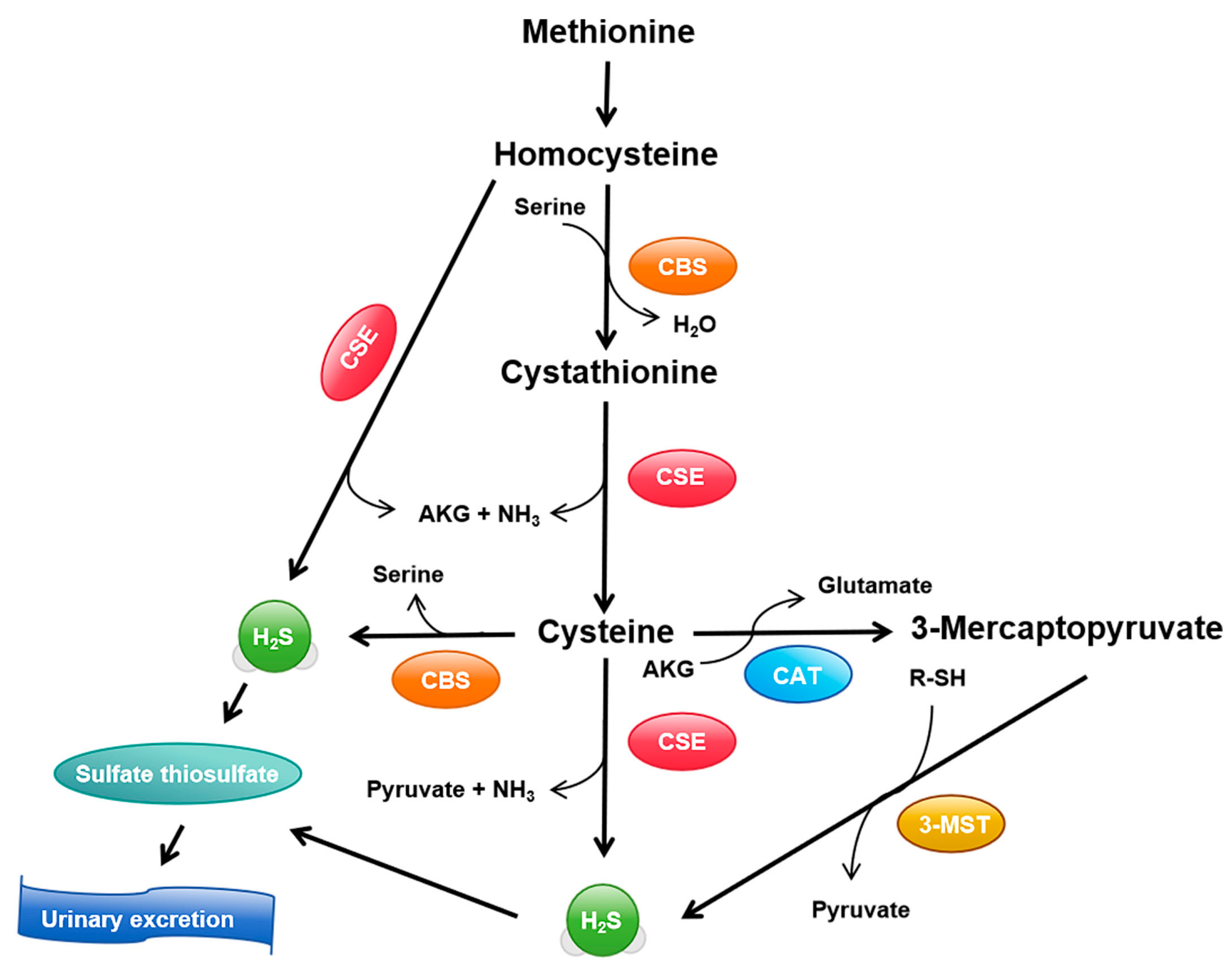
3.1. Synthesis and Metabolism of H2S

H2S production primarily relies on three key enzymes: cystathionine γ-lyase (CSE), cystathionine β-synthase (CBS), and 3-mercaptopyruvate sulfurtransferase (3-MST) [25]. These enzymes collaborate to produce H2S from various metabolic precursors. CSE catalyzes the conversion of cystathionine to cysteine and H2S in the CSE/CBS pathway, serving as one of the main routes for H2S generation [26]. CBS, predominantly expressed in the central nervous system, generates H2S from serine and cysteine [27]. 3-MST operates in the cytoplasm and mitochondria, contributing to H2S production via reactions with 3-mercaptopyruvate [28] (Figure 1). The synergistic action of these three enzymes ensures proper generation of H2S in various tissues and cell types, including skeletal muscles [29]. Additionally, non-enzymatic pathways, such as thiosulfate reduction, contribute to small amounts of H2S production [30].
Exercise has been shown to influence the activity of these enzymes, affecting H2S synthesis. Acute exercise increases CSE expression in skeletal muscles, promoting H2S production in response to the immediate metabolic demands of physical activity [31]. In contrast, chronic exercise induces long-term adaptations by upregulating CSE activity, enhancing the muscle’s ability to generate H2S during sustained or repeated exercise bouts [24]. This modulation is essential for supporting muscle function and improving metabolic efficiency during exercise.
H2S synthesis and turnover are tightly regulated by substrate availability, oxidative stress, and hormonal signals. For example, insulin and glucagon modulate H2S synthesis by influencing CSE and CBS activity, with insulin typically promoting H2S production in skeletal muscle during exercise [32,33]. Oxidative stress, a common result of intense or prolonged exercise, also affects the regulation of H2S synthesis, with high levels of reactive oxygen species (ROS) potentially enhancing CSE expression in response to cellular damage [34,35]. The homeostasis of H2S is critical, as excessive accumulation could lead to toxic effects, including mitochondrial dysfunction and impaired cellular signaling [36]. While H2S exerts beneficial effects at physiological levels, its accumulation at supra-physiological levels may interfere with normal cellular processes, leading to potential toxicity [37]. Therefore, feedback mechanisms are essential to control the rate of H2S synthesis and degradation, preventing detrimental effects on muscle cells and overall metabolic health.
The degradation of H2S primarily occurs through reactions with biological molecules, such as binding to metal ions and proteins or mitochondrial oxidation [38]. These processes ensure that H2S levels remain within a physiologically optimal range, supporting its role in exercise adaptations without causing toxicity.

3.2. Distribution and Expression in Muscle Tissues

The expression and functional roles of CSE, CBS, and 3-MST vary across different muscle tissues, influencing muscle function and metabolism. In skeletal muscle, CSE expression is particularly high in fast-twitch fibers, which is crucial for regulating muscle contraction and energy metabolism during high-intensity activities [39]. CBS, on the other hand, is predominantly expressed in cardiac muscle, where it plays a vital role in regulating blood flow and protecting myocardial cells during cardiac stress [40]. 3-MST is found in both skeletal and cardiac muscles, but its precise mechanisms of action remain less understood and require further exploration [41].
Exercise significantly modulates the expression of these enzymes, adapting them to different physiological demands. Endurance training typically increases CSE expression in skeletal muscle, enhancing H2S production, which improves blood flow and oxygen delivery to active tissues [42]. High-intensity interval training (HIIT), known for its metabolic demands, has been shown to activate CBS and 3-MST, boosting H2S production to support cellular energy demands during intense bouts of exercise [43]. Furthermore, long-term regular exercise can lead to upregulation of CSE gene expression, thereby enhancing the capacity for H2S synthesis and promoting muscle adaptation to sustained physical activity [24].
Different types of exercise, including resistance training, may also influence the expression of these enzymes, though the specific mechanisms and outcomes vary between exercise modalities. For instance, resistance training might alter the expression of CSE and CBS differently than endurance exercise due to the distinct metabolic and mechanical stresses involved [44]. Furthermore, it is essential to note that much of the research on these signaling pathways has been conducted in rodent models, and species differences should be considered when translating these findings to human physiology.

3.3. H2S as a Signaling Molecule

H2S exerts its effects on cellular targets through various mechanisms, including post-translational modifications such as sulfhydration, and interactions with other signaling molecules like NO and ROS [45,46,47]. Sulfhydration, the process by which H2S forms sulfide bonds with protein cysteine residues, alters protein function and activity, thus modulating cellular signaling pathways [45,46,47]. This modification plays a critical role in regulating several physiological processes, including mitochondrial function and stress responses during exercise.
In terms of vasodilation, H2S enhances the effects of NO by regulating its synthesis and bioavailability, contributing to increased blood flow during physical activity, which improves oxygen and nutrient delivery to muscle tissues [21]. H2S also acts as a potent scavenger of ROS, helping to mitigate oxidative stress and prevent cellular damage induced by exercise [48]. This antioxidant role is crucial for maintaining cellular integrity during intense physical exertion, supporting recovery and minimizing muscle damage.
H2S influences several key cellular pathways involved in exercise physiology, including antioxidant defenses, anti-inflammatory responses, and metabolic regulation. By activating the nuclear factor erythroid 2-related factor 2 (Nrf2) signaling pathway, H2S promotes the expression of antioxidant enzymes such as glutathione peroxidase (GPx) and superoxide dismutase (SOD), thereby reducing oxidative stress during and after exercise [21]. Furthermore, H2S plays a significant role in regulating inflammation by inhibiting the production of pro-inflammatory cytokines like TNF-α and IL-6, which are elevated during intense exercise and contribute to muscle soreness and injury [49].
Metabolically, H2S modulates cellular energy homeostasis by activating the AMPK and peroxisome proliferator-activated receptor gamma coactivator 1-alpha (PGC-1α) signaling pathways, promoting mitochondrial biogenesis and enhancing fatty acid oxidation [48]. These adaptations are vital for improving endurance performance and muscle recovery. Additionally, H2S enhances insulin signaling, improving glucose uptake and utilization and ultimately increasing insulin sensitivity in skeletal muscle [50,51].
While the beneficial roles of H2S in exercise physiology are clear, it is also important to acknowledge the potential negative effects of excessive H2S signaling. High levels of H2S may interfere with ROS signaling, disrupt apoptosis regulation, and lead to cellular dysfunction [52]. Thus, it is crucial to maintain a balanced level of H2S to prevent negative outcomes. The modulation of H2S levels during exercise, as well as its complex interaction with other signaling molecules like NO and ROS, underscores the need for further research to better understand the full scope of its physiological effects.

4. Exercise-Induced Molecular and Physiological Adaptations

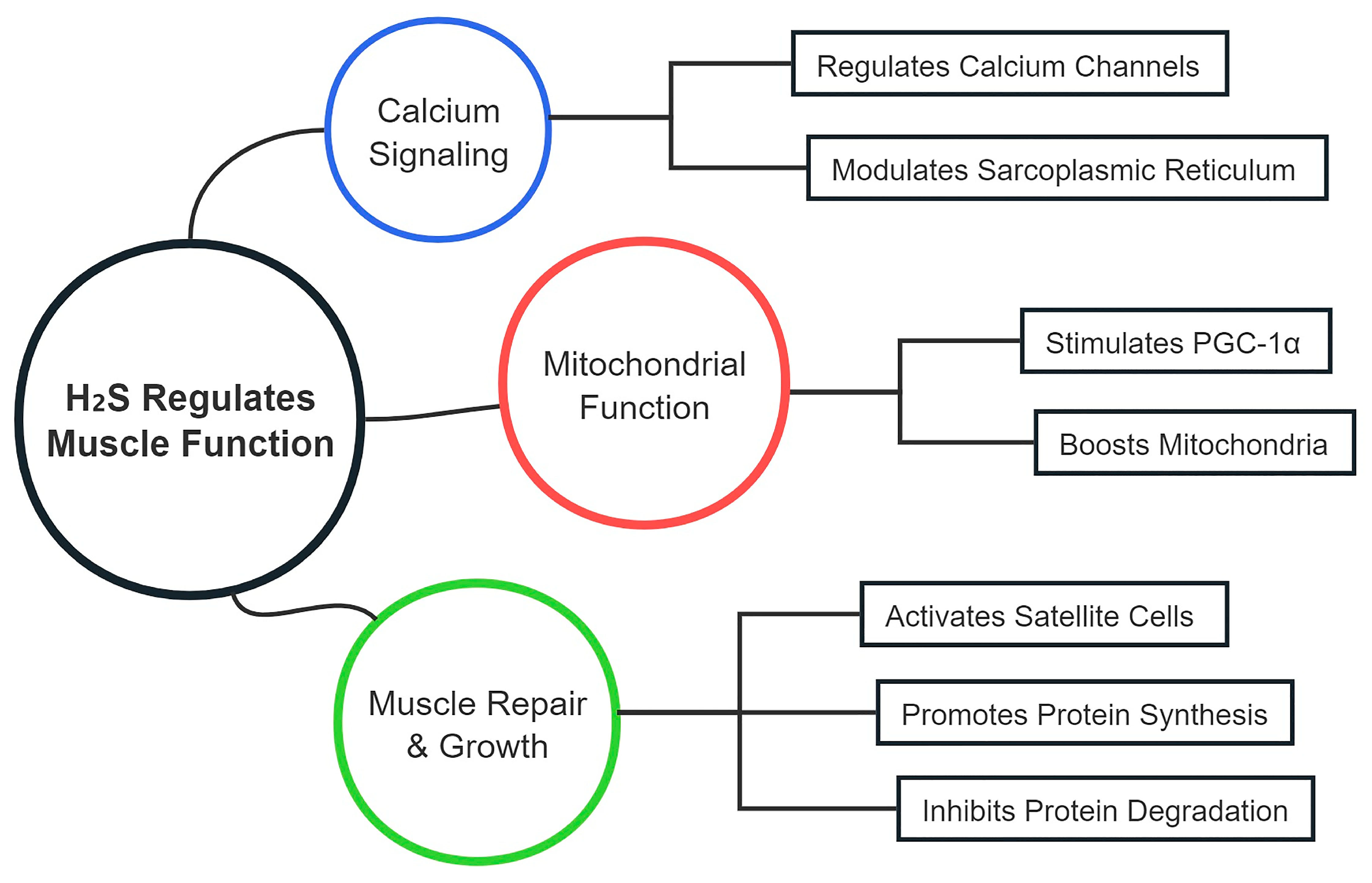
4.1. Enhancements in Muscle Function

Exercise promotes increases in muscle strength and endurance through a variety of molecular mechanisms. Strength training stimulates muscle protein synthesis and fiber hypertrophy by activating the mammalian target of rapamycin (mTOR) pathway in response to mechanical tension, metabolic stress, and muscle damage [50]. Additionally, exercise-induced mechanical stress activates satellite cells, which facilitate muscle repair and regeneration, thereby enhancing muscle strength and volume [53]. In contrast, endurance training activates the AMPK signaling pathway, which increases mitochondrial biogenesis and fatty acid oxidation capacity, leading to enhanced muscle endurance [54]. PGC-1α, a key regulator of mitochondrial biogenesis, upregulates the expression of genes linked to oxidative metabolism, thus improving the muscle’s aerobic capacity [55].
The effects of exercise vary depending on the type, intensity, and duration of the activity. Endurance training leads to a shift in muscle fiber types from glycolytic to oxidative fibers, which is driven by changes in mitochondrial density and enzyme activity within the muscle fibers [7]. Slow-twitch fibers (Type I) have higher mitochondrial content and greater oxidative enzyme activity, allowing for more efficient oxygen utilization and enhanced endurance performance [56]. Exercise-induced signaling molecules, such as Ca2+/calmodulin-dependent kinase (CaMK) and myocyte enhancer factor 2 (MEF2), regulate the conversion of muscle fibers to types with greater endurance characteristics, improving overall muscle function and performance [7,57,58].

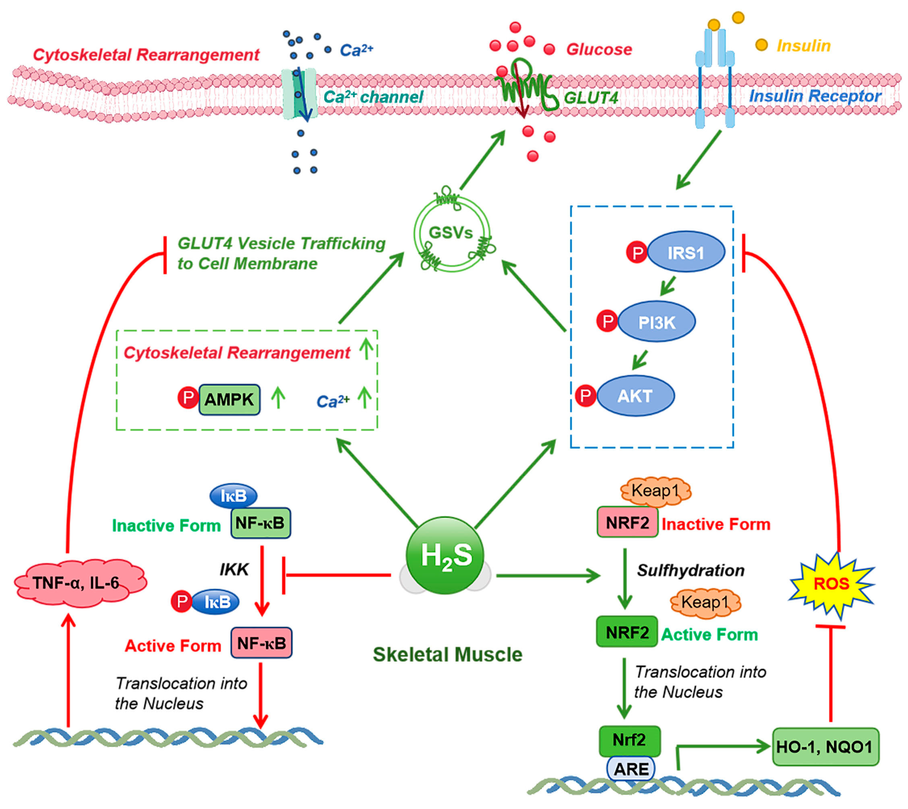
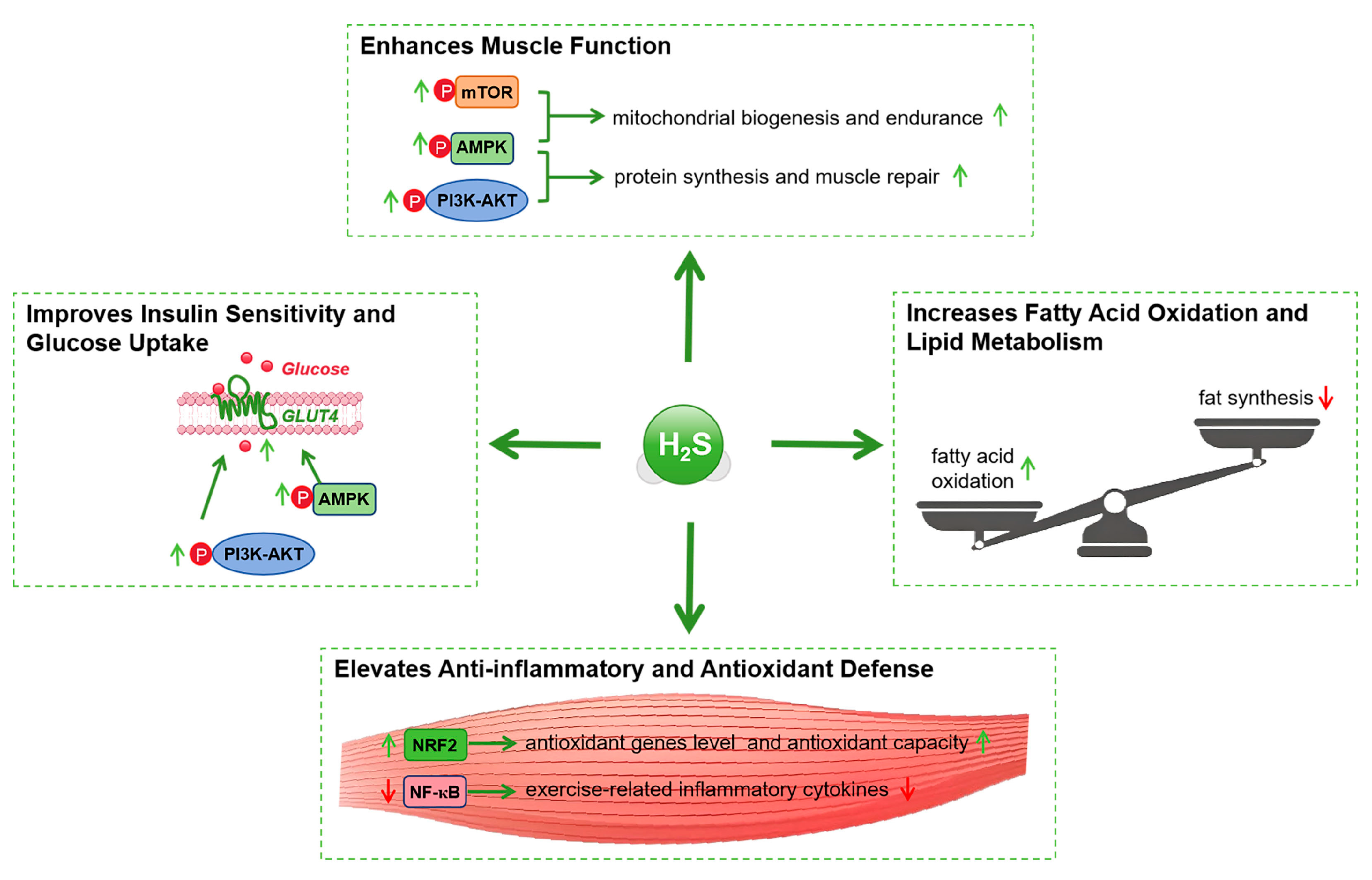
4.2. Improvements in Insulin Sensitivity

Insulin plays a critical role in glucose uptake by facilitating the translocation of GLUT4 to the muscle cell membrane, increasing intracellular glucose uptake [59]. Exercise enhances insulin sensitivity through two main pathways: first, by increasing muscle cells’ responsiveness to insulin, and second, by promoting GLUT4 translocation via insulin-independent pathways [60]. The activation of AMPK during exercise is key to GLUT4 translocation, which improves glucose uptake and helps regulate blood glucose levels even in conditions where insulin signaling is impaired [61].
Exercise influences multiple signaling pathways involved in insulin sensitivity. Exercise-induced activation of the PI3K/Akt pathway further enhances insulin sensitivity by promoting glucose uptake and metabolism [62]. Additionally, the AMPK and PGC-1α pathways promote mitochondrial biogenesis and fatty acid oxidation, improving energy metabolism efficiency and enhancing insulin sensitivity [63]. Exercise also lowers systemic inflammation by reducing inflammatory cytokine production from adipose tissue, further promoting insulin action [64]. These effects are influenced by exercise intensity, as higher-intensity exercise may provide greater improvements in insulin sensitivity by activating more robust metabolic and inflammatory responses.

4.3. Multi-Omics Approaches in Exercise Research

Multi-omics technologies, which integrate genomic, transcriptomic, proteomic, and metabolomic data, have made significant contributions to understanding exercise-induced adaptations. These approaches have revealed the complex, multi-layered molecular networks underlying exercise responses, highlighting the role of H2S and other signaling molecules in these processes. For instance, genomics, through genome-wide association studies (GWAS), has revealed genetic variations associated with individual exercise capacity [65], while transcriptomics provides insights into gene expression changes before and after exercise [66,67]. Proteomics, using mass spectrometry, uncovers dynamic changes in protein expression, post-translational modifications, and protein–protein interactions in response to exercise [68,69]. Metabolomics offers a comprehensive view of how exercise affects cellular metabolism, identifying changes in metabolites related to energy metabolism, oxidative stress, and inflammation [70,71,72] (Table 1).
In the context of CSE/H2S signaling, multi-omics data offer insights into how H2S regulates key pathways that mediate muscle function and insulin sensitivity during exercise. For example, multi-omics studies have shown that endurance training upregulates genes and proteins related to mitochondrial function and fatty acid metabolism, processes heavily influenced by H2S [73,74]. Metabolomic studies have also demonstrated that H2S supplementation enhances both energy metabolism and antioxidant capacity, contributing to better endurance and muscle recovery [75]. These findings underscore the critical role of multi-omics approaches in advancing our understanding of how H2S modulates exercise adaptations at the molecular level.
By integrating these multi-omics insights with the study of CSE/H2S signaling, we can better understand how exercise-induced molecular networks are regulated. The use of multi-omics technologies also allows us to identify biomarkers of H2S activity, which could serve as useful tools in assessing an individual’s response to exercise and tailoring personalized exercise regimens. As the field progresses, multi-omics methods will continue to refine our understanding of the molecular mechanisms that drive exercise adaptations and improve health outcomes.
Table 1. Application of multi-omics approaches in exercise and CSE/H2S research.
Omics TechnologyApplication in Exercise ResearchKey Findings and InsightsReferences
GenomicsGenome-wide association studies (GWAS) to identify genetic variations related to exercise capacityDetected genetic variations that influence exercise performance and adaptive responses, helping predict individual exercise capacity[76,77]
TranscriptomicsRNA sequencing to analyze gene expression changes before and after exerciseRevealed key genes regulating muscle adaptation, mitochondrial function, and metabolism[66,67,78]
ProteomicsMass spectrometry to identify protein expression and post-translational modificationsIdentified proteins involved in muscle repair, protein synthesis, and mitochondrial biogenesis, highlighting adaptive responses to exercise[68,69,78]
MetabolomicsHigh-throughput metabolite analysis to study exercise’s effects on metabolismUncovered metabolite changes related to energy metabolism, oxidative stress, and inflammation, highlighting metabolic adaptation[70,71,72,75]
Integrated Multi-OmicsCombining genomics, transcriptomics, proteomics, and metabolomics to study exercise adaptationsIntegrated data revealed key insights into mitochondrial function, fatty acid metabolism, and antioxidant pathways[79,80]

5. Mechanisms of CSE/H2S in Enhancing Muscle Function

5.1. Regulation of Muscle Contraction and Relaxation

Calcium ions (Ca2+) are crucial for muscle contraction and relaxation by regulating the interaction between actin and myosin filaments [81]. H2S modulates calcium signaling within muscle cells by altering the activity of calcium channels and transporters. For instance, H2S can enhance L-type calcium channel activity, increasing calcium influx during muscle contraction [82,83] (Figure 2, Table 2). However, the role of H2S in calcium signaling needs further investigation, particularly in different muscle fiber types (e.g., fast-twitch vs. slow-twitch fibers), as their calcium handling mechanisms may vary, affecting H2S’s impact on contraction and relaxation. Moreover, species differences in calcium handling (rodents vs. humans) should be addressed, as much of the current research is based on animal models.
Additionally, H2S modulates the function of the sarcoplasmic reticulum (SR), regulating ryanodine receptors (RyR) and sarcoplasmic/endoplasmic reticulum calcium ATPases (SERCA) to facilitate efficient calcium release and reuptake during muscle contractions [84]. This regulation ensures that muscle fibers can contract and relax efficiently, improving muscle endurance and performance [85,86]. However, a comparison of H2S’s effects with other established regulators, such as NO or AMPK, in calcium signaling could help clarify its relative importance in muscle function.
Mitochondria are critical for ATP production through oxidative phosphorylation [87], and H2S has been implicated in enhancing mitochondrial biogenesis and efficiency. Research comparing H2S with PGC-1α and AMPK (two key regulators of mitochondrial function) is necessary to avoid overgeneralization, as these pathways are well-established in mitochondrial biogenesis [52,88]. H2S stimulates key transcription factors like PGC-1α and NRF1, promoting mitochondrial formation and improving muscle energy capacity [89] (Figure 2, Table 2). While H2S enhances mitochondrial function, a more direct comparison with these well-characterized regulators could strengthen the evidence supporting H2S’s role in mitochondrial efficiency.
Furthermore, H2S also reduces ROS production and acts as an electron donor in the electron transport chain (ETC), thereby boosting ATP synthesis while minimizing oxidative damage [89,90]. The comparison with other mitochondrial regulators like AMPK, which also modulates mitochondrial function under exercise conditions, is important to understand whether H2S’s role is distinct or complementary.

5.2. Muscle Repair and Regeneration

Satellite cells play a vital role in muscle repair and regeneration post-exercise [92,93] (Figure 2, Table 2). H2S activates satellite cells by modulating key signaling pathways such as Notch, Wnt, and IGF-1/Akt, promoting their proliferation and differentiation into myoblasts [23,94]. This activation is crucial for muscle regeneration following strenuous exercise. However, the effects of H2S on different muscle fiber types during regeneration should be further explored, as satellite cells in different fiber types (e.g., fast-twitch vs. slow-twitch) might respond differently to H2S signaling, influencing the repair process [23].
Additionally, H2S influences the extracellular matrix and inflammatory cytokines, modulating the tissue repair environment and enhancing satellite cell activation [89,96]. The regulation of matrix metalloproteinases (MMPs) and TGF-β by H2S is vital for tissue remodeling [17,96]. Nevertheless, a more detailed comparison of H2S’s effects on muscle repair with other signaling molecules like NO, which also plays a role in muscle regeneration [23], would provide a broader understanding of H2S’s role in muscle repair.
Apoptosis is a significant factor in muscle integrity post-exercise, and H2S exhibits anti-apoptotic effects by inhibiting pro-apoptotic signaling pathways such as the mitochondrial and death receptor pathways [102,103]. This protection of muscle integrity during recovery is essential for maintaining muscle function and preventing muscle wasting. On the other hand, the dose-dependent effects of H2S on apoptosis [119], particularly in response to different exercise intensities, require further investigation to assess whether higher doses of H2S can inhibit excessive apoptosis during high-intensity training.

5.3. Protein Synthesis and Degradation

Anabolic signaling pathways, particularly mTOR, are central to muscle growth and hypertrophy. H2S influences mTOR signaling to promote muscle protein synthesis by enhancing the translation of mRNA into proteins [23,108]. However, exercise intensity-dependent effects on mTOR signaling should be incorporated, as different exercise modalities (e.g., endurance vs. resistance training) can have varying impacts on mTOR activation and subsequent protein synthesis [110]. The role of H2S in modulating these exercise-dependent variations in mTOR signaling should be better elucidated.
H2S also affects myogenic regulatory factors (MRFs) such as MyoD and myogenin, which are crucial for muscle differentiation and growth [101]. However, the impact of H2S on MRFs in different fiber types under various exercise conditions should be clarified, as fast-twitch and slow-twitch fibers may have different responses to H2S in terms of growth and hypertrophy.
In addition to promoting anabolic processes, H2S inhibits the catabolic pathways responsible for muscle protein degradation. H2S modulates the ubiquitin–proteasome system (UPS) and autophagy–lysosome pathway (ALP), reducing muscle protein breakdown during and after exercise [115]. Specifically, H2S suppresses the expression of E3 ubiquitin ligases, such as MuRF1 and MAFbx/atrogin-1, key components in the UPS, thereby preserving muscle mass and function [117]. The dose-dependent effects of H2S on protein degradation and its potential interactions with other metabolic regulators like AMPK and PGC-1α should be explored to gain a comprehensive understanding of how H2S modulates both anabolic and catabolic processes.
Furthermore, H2S interacts with FoxO transcription factors, which are involved in promoting catabolic processes. By inhibiting FoxO activity, H2S reduces the expression of genes involved in protein degradation, thereby maintaining muscle integrity and preventing atrophy [114]. Further investigation is needed to determine how H2S’s dual regulation of anabolic and catabolic pathways balances muscle growth and prevents muscle loss, particularly in response to different exercise intensities.

6. Mechanisms of CSE/H2S Signaling in Enhancing Insulin Sensitivity During Exercise

6.1. Regulation of Insulin Signaling

Insulin plays a vital role in regulating glucose homeostasis through the insulin receptor (IR) and downstream pathways like the PI3K/Akt cascade. H2S has been shown to modulate insulin sensitivity by influencing IR autophosphorylation and the activity of downstream signaling molecules such as IRS-1 and PI3K/Akt, which enhance glucose uptake and metabolism in muscle tissues [120,121,122,123] (Figure 3). However, H2S’s effects on insulin sensitivity extend beyond muscle tissue; it has also been observed to influence insulin signaling in adipose and hepatic tissues. For instance, in adipose tissue, H2S can reduce the expression of phosphatase and tensin homolog (PTEN), a negative regulator of the PI3K pathway, thus facilitating insulin signaling and improving systemic insulin sensitivity [124,125]. Additionally, H2S’s effect on GLUT4 translocation has been primarily studied in muscle tissues, but evidence suggests that it may also enhance GLUT4 translocation in adipocytes, supporting insulin action in both muscle and fat tissues [121,126]. This dual effect on different tissues underscores the complexity of H2S’s role in regulating insulin sensitivity. However, the mechanisms underlying this modulation in various tissues are not fully understood and warrant further investigation.

6.2. Regulation of Glucose and Lipid Metabolism

H2S regulates key metabolic pathways involved in glucose and lipid metabolism, providing crucial insights into its role in metabolic flexibility. One of its primary actions is enhancing glycolysis and gluconeogenesis, two opposing metabolic processes central to glucose homeostasis. H2S upregulates enzymes involved in glycolysis, such as hexokinase (HK) and phosphofructokinase-1 (PFK-1), to accelerate glucose utilization during exercise [127,128]. In contrast, in the liver, H2S enhances gluconeogenesis by promoting enzymes like glucose-6-phosphatase (G6Pase) and pyruvate carboxylase (PC) [127]. While the role of H2S in glycolysis and gluconeogenesis is becoming clearer, its effect on other regulators, such as the transcription factor PPARs, which are central to lipid metabolism, needs further exploration to understand its impact on fatty acid oxidation and adipose tissue accumulation [129].
Moreover, H2S has been shown to influence fatty acid oxidation by enhancing the activity of key enzymes involved in mitochondrial β-oxidation [130]. This action could contribute to improved insulin sensitivity by reducing excessive lipid accumulation in muscle and adipose tissues, a key factor in the development of insulin resistance. However, when compared to other regulators of fatty acid metabolism such as AMPK and PGC-1α, the unique contribution of H2S remains underexplored and warrants deeper scrutiny, especially in the context of varying exercise intensities and durations [129,131].

6.3. Anti-Inflammatory and Antioxidant Effects

Chronic low-grade inflammation, often driven by pro-inflammatory cytokines such as TNF-α and IL-6, is a major contributor to insulin resistance. H2S can reduce systemic inflammation by modulating the NF-κB signaling pathway, which regulates the production of these cytokines. Studies have shown that H2S inhibits the NF-κB pathway, which subsequently lowers inflammation and improves insulin sensitivity in various tissues, including muscle, liver, and adipose tissue [132,133]. Additionally, H2S activates the Nrf2 signaling pathway, which is involved in regulating the expression of antioxidant genes. This results in enhanced antioxidant enzyme activity, which counteracts oxidative stress that could otherwise impair insulin signaling [134,135] (Figure 3). Importantly, the dual anti-inflammatory and antioxidant effects of H2S support its potential therapeutic role in preventing metabolic diseases.
However, while the molecular mechanisms underlying H2S’s anti-inflammatory and antioxidant effects are becoming clearer, there is a need for more detailed investigations into dose-dependent responses. For instance, high doses of H2S may have unintended metabolic effects, such as altering ROS production in a way that could interfere with cellular redox balance or inflammation regulation. More research is needed to understand how different dosages of H2S influence both inflammation and oxidative stress, as well as how these effects vary between tissues. Furthermore, there is a need to explore how H2S interacts with other signaling molecules, such as NO and ROS, in the regulation of inflammation and insulin sensitivity [136]. The interplay between H2S and these other mediators could offer new avenues for targeted interventions to improve metabolic health and prevent insulin resistance.
In summary, while H2S’s role in enhancing insulin sensitivity and improving metabolic health during exercise is supported by increasing evidence, many aspects of its mechanisms remain unclear, particularly regarding its tissue-specific effects and dose-dependent actions. Future studies should aim to clarify how H2S interacts with other signaling pathways and assess the long-term effects of supplementation to better understand its full therapeutic potential.

7. H2S Supplementation in Exercise

7.1. Types of H2S Donors

Synthetic donors are widely used in experimental and clinical studies to provide controlled release of H2S. Common synthetic H2S donors include sodium hydrosulfide (NaHS) and GYY4137. NaHS is a fast-releasing donor that rapidly elevates in vivo H2S concentrations, making it suitable for acute studies [137]. On the other hand, GYY4137 is a slow-releasing donor, with kinetics closer to physiological conditions, which makes it ideal for long-term studies and clinical applications [138]. Other synthetic donors, such as AP39 and JK1, release H2S through different chemical mechanisms, offering diverse tools for research purposes [139] (Table 3).

7.2. Effects on Exercise Performance and Metabolic Health

Numerous studies have evaluated the effects of H2S supplementation on endurance, strength, and recovery during exercise. H2S donors, such as GYY4137, have been shown to significantly enhance oxygen consumption and running time during endurance exercises, suggesting positive effects on aerobic capacity [24,116,144]. Additionally, supplementation has demonstrated the ability to increase muscle strength and explosive power, promoting muscle fiber hypertrophy [24]. Furthermore, H2S supplementation has facilitated faster recovery from exercise by reducing inflammation and oxidative stress, thereby decreasing muscle soreness and damage [145] (Figure 4).
H2S donors have also been observed to improve insulin sensitivity, glucose metabolism, and lipid profiles. Studies indicate that H2S supplementation enhances insulin sensitivity, promoting glucose uptake and utilization, thereby lowering blood glucose levels [129]. In lipid metabolism, H2S donors have been shown to improve lipid profiles by stimulating fatty acid oxidation and inhibiting fat synthesis, which helps reduce adipose tissue accumulation [140] (Figure 4). These metabolic improvements may help prevent metabolic syndrome and type 2 diabetes, thereby promoting overall metabolic health.
However, it is important to critically assess the potential risks associated with H2S supplementation. One significant consideration is the dose-dependent toxicity of H2S, which can vary depending on the dose and duration of supplementation [37]. While acute supplementation has shown benefits, prolonged or excessive H2S exposure may lead to unintended metabolic effects, such as interference with other cellular signaling pathways [146]. Additionally, interactions between H2S and other gasotransmitters, such as NO or CO, warrant further investigation, as these molecules may modulate each other’s effects [21], potentially altering their efficacy or contributing to side effects.

7.3. Mechanistic Insights from Supplementation Studies

H2S supplementation exerts its effects during exercise through multiple molecular pathways. Firstly, H2S activates the AMPK and PI3K/Akt signaling pathways, facilitating GLUT4 translocation and glucose uptake, thereby improving insulin sensitivity [32,121]. Additionally, H2S activates the mTOR pathway, promoting protein synthesis and muscle growth [39]. In terms of anti-inflammatory and antioxidant effects, H2S activates and inhibits the Nrf2 and NF-κB signaling pathways, respectively, reducing inflammation and oxidative stress, thus protecting insulin signaling molecules [147].
In terms of exercise adaptations, H2S supplementation has been shown to enhance mitochondrial biogenesis and fatty acid oxidation, which complements the effects of endurance training [148]. During strength training, H2S works synergistically with exercise to promote muscle protein synthesis and muscle fiber hypertrophy, leading to increased muscle strength and mass [107]. This combined effect of exercise and H2S supplementation not only improves performance but also optimizes metabolic adaptations, contributing to better overall health.
Despite the promising results observed with H2S supplementation, it is important to evaluate whether these benefits are physiologically relevant, particularly when supra-physiological doses are used in animal models. The pharmacokinetics and bioavailability of synthetic H2S donors should be compared to endogenous production levels of H2S during exercise, which could provide insights into the real-world relevance of supplementation. Furthermore, dose–response relationships and the long-term safety of H2S supplementation must be better understood to determine optimal dosing strategies that maximize benefits while minimizing potential risks [149].
In conclusion, while H2S supplementation holds considerable potential for enhancing exercise performance and improving metabolic health, further research is needed to evaluate its long-term effects, optimal dosages, and potential interactions with other signaling pathways. The balance between efficacy and safety must be carefully addressed [150] in order to provide a realistic and well-rounded assessment of H2S supplementation in exercise physiology.

8. Current Research, Technological Advances, and Future Directions

8.1. Summary of Current Research

In recent years, numerous significant discoveries have been made regarding the role of CSE/H2S in enhancing muscle function and insulin sensitivity during exercise [24,151]. Studies have shown that CSE generates H2S, thereby regulating energy metabolism and signaling pathways within muscle cells, which promotes the enhancement of muscle endurance and strength [152]. Additionally, H2S activates the PI3K/Akt and AMPK signaling pathways, increasing insulin receptor sensitivity, facilitating GLUT4 translocation, and enhancing glucose uptake efficiency [32,121]. Further research has revealed that H2S possesses anti-inflammatory and antioxidant properties, reducing exercise-induced oxidative stress and inflammatory responses, thereby protecting the function of insulin signaling molecules [84,153].
Despite existing studies elucidating the multiple mechanisms by which CSE/H2S operates during exercise, several knowledge gaps and controversies remain. Firstly, the specific mechanisms of H2S action in different types of exercise are not yet fully understood, particularly in HIIT, requiring further exploration [153]. Secondly, the dynamic changes of H2S during long-term exercise adaptation and its long-term impacts on metabolic health lack systematic research [154]. Moreover, the interactions between the CSE/H2S signaling pathway and other gaseous signaling molecules (such as NO and CO) have not been sufficiently elucidated, necessitating in-depth investigation [129].

8.2. Technological and Methodological Advances

In recent years, significant advancements have been made in methods for detecting H2S levels and activity. Novel fluorescent probes and chromatographic–mass spectrometric techniques (such as GC-MS and LC-MS/MS) have enhanced the sensitivity and specificity of H2S detection, enabling real-time monitoring of H2S in biological systems [155]. Additionally, nanotechnology-based sensors have been developed for high-throughput and high-precision H2S detection, further advancing H2S research [156].
Multi-omics approaches have played a crucial role in uncovering new aspects of the CSE/H2S signaling pathway. Integrative analyses of genomics, transcriptomics, proteomics, and metabolomics have helped scientists comprehensively understand the complex mechanisms of H2S [157]. For example, combining transcriptomic and proteomic data allows for the identification of key genes and proteins regulated by H2S, revealing their specific functions in metabolism and signal transduction [158]. Moreover, the application of metabolomics has unveiled the broad impacts of H2S on energy metabolism networks, facilitating a deeper understanding of its metabolic regulatory functions [159].

8.3. Future Research Directions

Future studies should explore the following key questions:
How does chronic exercise influence H2S metabolism?
What is the dose–response relationship for H2S supplementation in improving exercise performance and metabolic health?
What biomarkers of H2S activity can be used to assess its role in exercise adaptations?
How does H2S interact with other gasotransmitters like NO in exercise?

9. Conclusions

In conclusion, the CSE/H2S signaling pathway plays a crucial role in enhancing muscle function, improving insulin sensitivity, and promoting metabolic health during exercise. By regulating important processes like mitochondrial biogenesis, glucose metabolism, and inflammation, H2S helps drive the positive effects of physical activity. However, there are still several challenges in fully understanding how this pathway can be applied clinically. For example, the exact mechanisms by which H2S supports exercise adaptations, particularly in different muscle fiber types and various exercise forms, need more research. Additionally, the potential risks of H2S supplementation, such as dose-dependent toxicity and interactions with other signaling pathways, must be carefully evaluated.
The therapeutic potential of targeting the CSE/H2S pathway is promising, especially in preventing metabolic diseases like type 2 diabetes, improving athletic performance, and aiding muscle recovery after exercise. However, more studies are needed to determine the best dosing strategies, identify reliable biomarkers for H2S activity, and assess its long-term effects in clinical settings. Furthermore, exploring how H2S interacts with other gasotransmitters, such as NO, could provide valuable insights and create new opportunities for therapeutic development.
Ultimately, advancing our understanding of CSE/H2S signaling holds great potential to improve both exercise outcomes and metabolic health. It could lead to personalized exercise programs and targeted therapies for metabolic and muscle-related disorders.

Author Contributions

Writing—original draft, M.X. and X.L.; writing—review and editing, M.X., D.H. and X.L.; methodology, M.X. and X.L.; supervision, L.L. and Z.L.; conceptualization, X.L. All authors have read and agreed to the published version of the manuscript.

Funding

This work was supported by the National Natural Science Foundation of China (no. 32300964) and Guangdong Provincial Sports Bureau 2024–2025 Science and Technology Innovation and Sports Culture Development Research Project (no. GDSS2024N010).

Institutional Review Board Statement

Informed Consent Statement

Data Availability Statement

All relevant materials are presented in this manuscript.

Conflicts of Interest

The authors declare no conflicts of interest. The funders had no role in the design of the study; in the collection, analyses, or interpretation of data; in the writing of the manuscript; or in the decision to publish the results.

References

  1. Pesta, D.; Anadol-Schmitz, E.; Sarabhai, T.; den Kamp, Y.O.; Gancheva, S.; Trinks, N.; Zaharia, O.-P.; Mastrototaro, L.; Lyu, K.; Habets, I.; et al. Determinants of Increased Muscle Insulin Sensitivity of Exercise-Trained versus Sedentary Normal Weight and Overweight Individuals. Sci. Adv. 2025, 11, eadr8849. [Google Scholar] [CrossRef] [PubMed]
  2. Lei, L.; Li, J.; Wang, W.; Yu, Y.; Pu, B.; Peng, Y.; Zhang, L.; Zhao, Z. The Associations of “Weekend Warrior” and Regularly Active Physical Activity with Abdominal and General Adiposity in US Adults. Obesity 2024, 32, 822–833. [Google Scholar] [CrossRef]
  3. Mambrini, S.P.; Grillo, A.; Colosimo, S.; Zarpellon, F.; Pozzi, G.; Furlan, D.; Amodeo, G.; Bertoli, S. Diet and Physical Exercise as Key Players to Tackle MASLD through Improvement of Insulin Resistance and Metabolic Flexibility. Front. Nutr. 2024, 11, 1426551. [Google Scholar] [CrossRef]
  4. Mcleod, J.C.; Currier, B.S.; Lowisz, C.V.; Phillips, S.M. The Influence of Resistance Exercise Training Prescription Variables on Skeletal Muscle Mass, Strength, and Physical Function in Healthy Adults: An Umbrella Review. J. Sport Health Sci. 2024, 13, 47–60. [Google Scholar] [CrossRef]
  5. Rosdiana, R.; Rasyid, D.; Djunaedi, D.; Noviar, R.A. Foot Exercises in Controlling Blood Sugar Levels in Elderly People with DM at the Baraka Community Health Center, Enrekang Regency, South Sulawesi. Int. J. Health Sci. 2024, 2, 167–178. [Google Scholar] [CrossRef]
  6. Thomas, A.C.Q.; Stead, C.A.; Burniston, J.G.; Phillips, S.M. Exercise-Specific Adaptations in Human Skeletal Muscle: Molecular Mechanisms of Making Muscles Fit and Mighty. Free Radic. Biol. Med. 2024, 223, 341–356. [Google Scholar] [CrossRef] [PubMed]
  7. Li, J.; Zhang, S.; Li, C.; Zhang, X.; Shan, Y.; Zhang, Z.; Bo, H.; Zhang, Y. Endurance Exercise-Induced Histone Methylation Modification Involved in Skeletal Muscle Fiber Type Transition and Mitochondrial Biogenesis. Sci. Rep. 2024, 14, 21154. [Google Scholar] [CrossRef] [PubMed]
  8. Xie, X.; Huang, C. Role of the Gut–Muscle Axis in Mitochondrial Function of Ageing Muscle under Different Exercise Modes. Ageing Res. Rev. 2024, 98, 102316. [Google Scholar] [CrossRef] [PubMed]
  9. Kim, K.M.; Yoo, G.D.; Heo, W.; Oh, H.T.; Park, J.; Shin, S.; Do, Y.; Jeong, M.G.; Hwang, E.S.; Hong, J.-H. TAZ Stimulates Exercise-Induced Muscle Satellite Cell Activation via Pard3–P38 MAPK–TAZ Signalling Axis. J. Cachexia Sarcopenia Muscle 2023, 14, 2733–2746. [Google Scholar] [CrossRef] [PubMed]
  10. Zhang, X.; Zhao, Y.; Guo, D.; Luo, M.; Zhang, Q.; Zhang, L.; Zhang, D. Exercise Improves Heart Function after Myocardial Infarction: The Merits of AMPK. Cardiovasc. Drugs Ther. 2024. [Google Scholar] [CrossRef]
  11. Lu, J.; Tang, Z.; Xu, M.; Lu, J.; Wang, F.; Ni, X.; Wang, C.; Yu, B. Skeletal Muscle Cystathionine γ-Lyase Deficiency Promotes Obesity and Insulin Resistance and Results in Hyperglycemia and Skeletal Muscle Injury upon HFD in Mice. Redox Rep. 2024, 29, 2347139. [Google Scholar] [CrossRef]
  12. Kaleta, K.; Janik, K.; Rydz, L.; Wróbel, M.; Jurkowska, H. Bridging the Gap in Cancer Research: Sulfur Metabolism of Leukemic Cells with a Focus on L-Cysteine Metabolism and Hydrogen Sulfide-Producing Enzymes. Biomolecules 2024, 14, 746. [Google Scholar] [CrossRef]
  13. Flori, L.; Benedetti, G.; Calderone, V.; Testai, L. Hydrogen Sulfide and Irisin, Potential Allies in Ensuring Cardiovascular Health. Antioxidants 2024, 13, 543. [Google Scholar] [CrossRef] [PubMed]
  14. Ligi, S.; Ali, A.; Yang, G. Cystathionine Gamma-Lyase Deficiency Exaggerates Diethylnitrosamine-Induced Liver Damage in Mice. Nitric Oxide 2024, 151, 1–9. [Google Scholar] [CrossRef]
  15. Yang, J.-H.; Gao, J.; E, Y.-Q.; Jiao, L.-J.; Wu, R.; Yan, Q.-Y.; Wei, Z.-Y.; Yan, G.-L.; Liang, J.-L.; Li, H.-Z. Hydrogen Sulfide Inhibits Skeletal Muscle Ageing by Up-Regulating Autophagy through Promoting Deubiquitination of Adenosine 5′-Monophosphate (AMP)-Activated Protein Kinase A1 via Ubiquitin Specific Peptidase 5. J. Cachexia Sarcopenia Muscle 2024, 15, 2118–2133. [Google Scholar] [CrossRef]
  16. Machado-Neto, J.A.; Cerqueira, A.R.A.; Veríssimo-Filho, S.; Muscará, M.N.; Costa, S.K.P.; Lopes, L.R. Hydrogen Sulfide Signaling in the Tumor Microenvironment: Implications in Cancer Progression and Therapy. Antioxid. Redox Signal. 2024, 40, 250–271. [Google Scholar] [CrossRef]
  17. Shi, X.; Li, H.; Guo, F.; Li, D.; Xu, F. Novel Ray of Hope for Diabetic Wound Healing: Hydrogen Sulfide and Its Releasing Agents. J. Adv. Res. 2024, 58, 105–115. [Google Scholar] [CrossRef] [PubMed]
  18. Rotimi, D.E.; Acho, M.A.; Falana, B.M.; Olaolu, T.D.; Mgbojikwe, I.; Ojo, O.A.; Adeyemi, O.S. Oxidative Stress-Induced Hormonal Disruption in Male Reproduction. Reprod. Sci. 2024, 31, 2943–2956. [Google Scholar] [CrossRef]
  19. Sarkar, S.; Kumar, R.; Matson, J.B. Hydrogels for Gasotransmitter Delivery: Nitric Oxide, Carbon Monoxide, and Hydrogen Sulfide. Macromol. Biosci. 2024, 24, 2300138. [Google Scholar] [CrossRef] [PubMed]
  20. Zhang, C.-J.; Wang, Y.; Jin, Y.-Q.; Zhu, Y.-W.; Zhu, S.-G.; Wang, Q.-M.; Jing, M.-R.; Zhang, Y.-X.; Cai, C.-B.; Feng, Z.-F.; et al. Recent Advances in the Role of Hydrogen Sulfide in Age-Related Diseases. Exp. Cell Res. 2024, 441, 114172. [Google Scholar] [CrossRef]
  21. Munteanu, C.; Popescu, C.; Vlădulescu-Trandafir, A.-I.; Onose, G. Signaling Paradigms of H2S-Induced Vasodilation: A Comprehensive Review. Antioxidants 2024, 13, 1158. [Google Scholar] [CrossRef] [PubMed]
  22. Slade, L.; Deane, C.S.; Szewczyk, N.J.; Etheridge, T.; Whiteman, M. Hydrogen Sulfide Supplementation as a Potential Treatment for Primary Mitochondrial Diseases. Pharmacol. Res. 2024, 203, 107180. [Google Scholar] [CrossRef] [PubMed]
  23. Liu, Y.-F.; Zhang, Y.-X.; Zhu, Y.-W.; Tang, A.-Q.; Liang, H.-B.; Yang, Y.-L.; Zhai, Y.-K.; Ji, X.-Y.; Wu, D.-D. Hydrogen Sulfide in Musculoskeletal Diseases: Molecular Mechanisms and Therapeutic Opportunities. Antioxid. Redox Signal. 2024; online ahead of print. [Google Scholar] [CrossRef]
  24. Xu, M.; Liu, X.; Bao, P.; Wang, Y.; Zhu, X.; Liu, Y.; Ni, X.; Lu, J. Skeletal Muscle CSE Deficiency Leads to Insulin Resistance in Mice. Antioxidants 2022, 11, 2216. [Google Scholar] [CrossRef]
  25. Dawoud, A.; Youness, R.A.; Nafea, H.; Manie, T.; Bourquin, C.; Szabo, C.; Abdel-Kader, R.M.; Gad, M.Z. Pan-Inhibition of the Three H2S Synthesizing Enzymes Restrains Tumor Progression and Immunosuppression in Breast Cancer. Cancer Cell Int. 2024, 24, 136. [Google Scholar] [CrossRef] [PubMed]
  26. Al-Taee, K.; Al-Helaly, L.; Al-Helaly, L.; Al-Helaly, L. Hydrogen Sulfide and Cystathionine γ–Lyase with Oxidants and Antioxidants Levels for Patients with Epilepsy Diseases. Pharmacogn. J. 2024, 16, 319–322. [Google Scholar] [CrossRef]
  27. Si, Y.; Song, N.; Ji, Y. Bioinformatics Analysis of the Clinical Value and Potential Mechanisms of CBS/H2S in Glioma Through 1027 Samples; Research Square Platform: Durham, NC, USA, 2024. [Google Scholar]
  28. Manna, S.; Agrawal, R.; Yadav, T.; Kumar, T.A.; Kumari, P.; Dalai, A.; Kanade, S.; Balasubramanian, N.; Singh, A.; Chakrapani, H. Orthogonal Persulfide Generation through Precision Tools Provides Insights into Mitochondrial Sulfane Sulfur. Angew. Chem. 2024, 136, e202411133. [Google Scholar] [CrossRef]
  29. He, K.; Tan, B.; Lu, A.; Bai, L.; Song, C.; Miao, Y.; Liu, B.; Chen, Q.; Teng, X.; Dai, J.; et al. Asynchronous Changes of Hydrogen Sulfide and Its Generating Enzymes in Most Tissues with the Aging Process. Biosci. Rep. 2024, 44, BSR20240320. [Google Scholar] [CrossRef]
  30. Derry, P.J.; Liopo, A.V.; Mouli, K.; McHugh, E.A.; Vo, A.T.T.; McKelvey, A.; Suva, L.J.; Wu, G.; Gao, Y.; Olson, K.R.; et al. Oxidation of Hydrogen Sulfide to Polysulfide and Thiosulfate by a Carbon Nanozyme: Therapeutic Implications with an Emphasis on down Syndrome. Adv. Mater. 2024, 36, 2211241. [Google Scholar] [CrossRef]
  31. Wu, D.; Lu, Z.; Li, M.; Feng, Z.; Li, Y. Treadmill Exercise Increases Cystathionine γ Lyase Expression and Decreases Inflammation in Skeletal Muscles of High Fat Diet Induced Obese Rats. Int. J. Clin. Exp. Med. 2016, 9, 23119–23126. [Google Scholar]
  32. Zhang, H.; Huang, Y.; Chen, S.; Tang, C.; Wang, G.; Du, J.; Jin, H. Hydrogen Sulfide Regulates Insulin Secretion and Insulin Resistance in Diabetes Mellitus, a New Promising Target for Diabetes Mellitus Treatment? A Review. J. Adv. Res. 2021, 27, 19–30. [Google Scholar] [CrossRef] [PubMed]
  33. Parsanathan, R.; Jain, S.K. Hydrogen Sulfide Regulates Irisin and Glucose Metabolism in Myotubes and Muscle of HFD-Fed Diabetic Mice. Antioxidants 2022, 11, 1369. [Google Scholar] [CrossRef] [PubMed]
  34. Zhao, Y.; Wang, Y.; Zheng, H.; Xu, Q.; Zhou, K.; Liu, H.; Xia, Y.; Wei, D.-H.; Jiang, M.; Tang, Z.-H.; et al. Hydrogen Sulfide Upregulates SIRT1 to Inhibit Ox-HDL-Induced Endothelial Cell Damage and Mitochondrial Dysfunction. Nitric Oxide 2024, 152, 78–89. [Google Scholar] [CrossRef]
  35. Pandey, T.; Pandey, V. Hydrogen Sulfide (H2S) Metabolism: Unraveling Cellular Regulation, Disease Implications, and Therapeutic Prospects for Precision Medicine. Nitric Oxide 2024, 144, 20–28. [Google Scholar] [CrossRef] [PubMed]
  36. Quinzii, C.M.; Lopez, L.C. Abnormalities of Hydrogen Sulfide and Glutathione Pathways in Mitochondrial Dysfunction. J. Adv. Res. 2021, 27, 79–84. [Google Scholar] [CrossRef]
  37. Ng, P.C.; Hendry-Hofer, T.B.; Witeof, A.E.; Brenner, M.; Mahon, S.B.; Boss, G.R.; Haouzi, P.; Bebarta, V.S. Hydrogen Sulfide Toxicity: Mechanism of Action, Clinical Presentation, and Countermeasure Development. J. Med. Toxicol. 2019, 15, 287–294. [Google Scholar] [CrossRef] [PubMed]
  38. Stoltzfus, A.T.; Ballot, J.G.; Vignane, T.; Li, H.; Worth, M.M.; Muller, L.; Siegler, M.A.; Kane, M.A.; Filipovic, M.R.; Goldberg, D.P.; et al. Chemoselective Proteomics, Zinc Fingers, and a Zinc(II) Model for H2S Mediated Persulfidation. Angew. Chem. Int. Ed. 2024, 63, e202401003. [Google Scholar] [CrossRef]
  39. Wang, R.; Li, K.; Wang, H.; Jiao, H.; Wang, X.; Zhao, J.; Lin, H. Endogenous CSE/Hydrogen Sulfide System Regulates the Effects of Glucocorticoids and Insulin on Muscle Protein Synthesis. Oxid. Med. Cell. Longev. 2019, 2019, 9752698. [Google Scholar] [CrossRef] [PubMed]
  40. Omorou, M.; Liu, N.; Huang, Y.; Al-Ward, H.; Gao, M.; Mu, C.; Zhang, L.; Hui, X. Cystathionine Beta-Synthase in Hypoxia and Ischemia/Reperfusion: A Current Overview. Arch. Biochem. Biophys. 2022, 718, 109149. [Google Scholar] [CrossRef]
  41. Rao, S.P.; Dobariya, P.; Bellamkonda, H.; More, S.S. Role of 3-Mercaptopyruvate Sulfurtransferase (3-MST) in Physiology and Disease. Antioxidants 2023, 12, 603. [Google Scholar] [CrossRef]
  42. Petrovic, D.; Slade, L.; Paikopoulos, Y.; D’Andrea, D.; Savic, N.; Stancic, A.; Miljkovic, J.L.; Vignane, T.; Drekolia, M.K.; Mladenovic, D.; et al. Ergothioneine Improves Healthspan of Aged Animals by Enhancing cGPDH Activity through CSE-Dependent Persulfidation. Cell Metab. 2025, 37, 542–556.e14. [Google Scholar] [CrossRef]
  43. Bekheit, M.; Kamera, B.; Colacino, L.; Dropmann, A.; Delibegovic, M.; Almadhoob, F.; Hanafy, N.; Bermano, G.; Hammad, S. Mechanisms Underpinning the Effect of Exercise on the Non-Alcoholic Fatty Liver Disease: Review. EXCLI J. 2025, 24, 238–266. [Google Scholar] [CrossRef]
  44. Veeranki, S.; Tyagi, S.C. Role of Hydrogen Sulfide in Skeletal Muscle Biology and Metabolism. Nitric Oxide 2015, 46, 66–71. [Google Scholar] [CrossRef] [PubMed]
  45. Li, K.; Uyanga, V.A.; Wang, X.; Jiao, H.; Zhao, J.; Zhou, Y.; Li, H.; Lin, H. Allicin Promotes Glucose Uptake by Activating AMPK through CSE/H2S-Induced S-Sulfhydration in a Muscle-Fiber Dependent Way in Broiler Chickens. Mol. Nutr. Food Res. 2024, 68, 2300622. [Google Scholar] [CrossRef] [PubMed]
  46. Zhang, H.; Pan, J.; Huang, S.; Chen, X.; Chang, A.C.Y.; Wang, C.; Zhang, J.; Zhang, H. Hydrogen Sulfide Protects Cardiomyocytes from Doxorubicin-Induced Ferroptosis through the SLC7A11/GSH/GPx4 Pathway by Keap1 S-Sulfhydration and Nrf2 Activation. Redox Biol. 2024, 70, 103066. [Google Scholar] [CrossRef]
  47. Pang, P.-P.; Zhang, H.-Y.; Zhang, D.-C.; Tang, J.-X.; Gong, Y.; Guo, Y.-C.; Zheng, C.-B. Investigating the Impact of Protein S-Sulfhydration Modification on Vascular Diseases: A Comprehensive Review. Eur. J. Pharmacol. 2024, 966, 176345. [Google Scholar] [CrossRef] [PubMed]
  48. Chen, T.; Bai, D.; Gong, C.; Cao, Y.; Yan, X.; Peng, R. Hydrogen Sulfide Mitigates Mitochondrial Dysfunction and Cellular Senescence in Diabetic Patients: Potential Therapeutic Applications. Biochem. Pharmacol. 2024, 230, 116556. [Google Scholar] [CrossRef]
  49. Li, R.-Y.; Guo, L. Exercise in Diabetic Nephropathy: Protective Effects and Molecular Mechanism. Int. J. Mol. Sci. 2024, 25, 3605. [Google Scholar] [CrossRef]
  50. Birulina, J.G.; Ivanov, V.V.; Buyko, E.E.; Voronkova, O.V.; Chernyshov, N.A. The role of endogenous H2S in experimental metabolic syndrome. RUDN J. Med. 2024, 28, 331–339. [Google Scholar] [CrossRef]
  51. Silva-Velasco, D.L.; Hong, E.; Beltran-Ornelas, J.H.; Sánchez-López, A.; Huerta de la Cruz, S.; Tapia-Martínez, J.A.; Gomez, C.B.; Centurión, D. Hydrogen Sulfide Ameliorates Hypertension and Vascular Dysfunction Induced by Insulin Resistance in Rats by Reducing Oxidative Stress and Activating eNOS. Eur. J. Pharmacol. 2024, 963, 176266. [Google Scholar] [CrossRef]
  52. Cirino, G.; Szabo, C.; Papapetropoulos, A. Physiological Roles of Hydrogen Sulfide in Mammalian Cells, Tissues, and Organs. Physiol. Rev. 2023, 103, 31–276. [Google Scholar] [CrossRef]
  53. Guo, Q.; Luo, Q.; Song, G. Control of Muscle Satellite Cell Function by Specific Exercise-Induced Cytokines and Their Applications in Muscle Maintenance. J. Cachexia Sarcopenia Muscle 2024, 15, 466–476. [Google Scholar] [CrossRef] [PubMed]
  54. Zhao, Y.-C.; Gao, B. Integrative Effects of Resistance Training and Endurance Training on Mitochondrial Remodeling in Skeletal Muscle. Eur. J. Appl. Physiol. 2024, 124, 2851–2865. [Google Scholar] [CrossRef]
  55. Kong, S.; Cai, B.; Nie, Q. PGC-1α Affects Skeletal Muscle and Adipose Tissue Development by Regulating Mitochondrial Biogenesis. Mol. Genet. Genomics 2022, 297, 621–633. [Google Scholar] [CrossRef]
  56. Yendrizal, Y.; Okilanda, A.; Masrun, M.; Ridwan, M.; Ahmed, M.; Crisari, S.; Tulyakul, S. Unlocking the Science of Physical Endurance: Training Techniques and Biological Factors. Retos Nuevas Tend. En Educ. Física Deporte Recreación 2024, 55, 504–512. [Google Scholar]
  57. Luo, X.; Zhang, H.; Cao, X.; Yang, D.; Yan, Y.; Lu, J.; Wang, X.; Wang, H. Endurance Exercise-Induced Fgf21 Promotes Skeletal Muscle Fiber Conversion through TGF-Β1 and P38 MAPK Signaling Pathway. Int. J. Mol. Sci. 2023, 24, 11401. [Google Scholar] [CrossRef]
  58. Zhang, K.; Xie, N.; Ye, H.; Miao, J.; Xia, B.; Yang, Y.; Peng, H.; Xu, S.; Wu, T.; Tao, C.; et al. Glucose Restriction Enhances Oxidative Fiber Formation: A Multi-Omic Signal Network Involving AMPK and CaMK2. iScience 2024, 27, 108590. [Google Scholar] [CrossRef]
  59. Richter, E.A. Is GLUT4 Translocation the Answer to Exercise-Stimulated Muscle Glucose Uptake? Am. J. Physiol.-Endocrinol. Metab. 2021, 320, E240–E243. [Google Scholar] [CrossRef]
  60. Richter, E.A.; Sylow, L.; Hargreaves, M. Interactions between Insulin and Exercise. Biochem. J. 2021, 478, 3827–3846. [Google Scholar] [CrossRef] [PubMed]
  61. McGee, S.L.; Hargreaves, M. Exercise Performance and Health: Role of GLUT4. Free Radic. Biol. Med. 2024, 224, 479–483. [Google Scholar] [CrossRef] [PubMed]
  62. Wang, H.; Wang, J.; Zhu, Y.; Yan, H.; Lu, Y. Effects of Different Intensity Exercise on Glucose Metabolism and Hepatic IRS/PI3K/AKT Pathway in SD Rats Exposed with TCDD. Int. J. Environ. Res. Public Health 2021, 18, 13141. [Google Scholar] [CrossRef]
  63. Yang, B.; Yu, Q.; Chang, B.; Guo, Q.; Xu, S.; Yi, X.; Cao, S. MOTS-c Interacts Synergistically with Exercise Intervention to Regulate PGC-1α Expression, Attenuate Insulin Resistance and Enhance Glucose Metabolism in Mice via AMPK Signaling Pathway. Biochim. Biophys. Acta BBA Mol. Basis Dis. 2021, 1867, 166126. [Google Scholar] [CrossRef]
  64. Atakan, M.M.; Koşar, Ş.N.; Güzel, Y.; Tin, H.T.; Yan, X. The Role of Exercise, Diet, and Cytokines in Preventing Obesity and Improving Adipose Tissue. Nutrients 2021, 13, 1459. [Google Scholar] [CrossRef] [PubMed]
  65. Gomes, C.; Almeida, J.A.; Franco, O.L.; Petriz, B. Chapter Three—Omics and the Molecular Exercise Physiology. In Advances in Clinical Chemistry; Makowski, G.S., Ed.; Elsevier: Amsterdam, The Netherlands, 2020; Volume 96, pp. 55–84. [Google Scholar]
  66. Kusano, T.; Sotani, Y.; Takeda, R.; Hatano, A.; Kawata, K.; Kano, R.; Matsumoto, M.; Kano, Y.; Hoshino, D. Time-Series Transcriptomics Reveals Distinctive mRNA Expression Dynamics Associated with Gene Ontology Specificity and Protein Expression in Skeletal Muscle after Electrical Stimulation-Induced Resistance Exercise. FASEB J. 2024, 38, e70153. [Google Scholar] [CrossRef]
  67. Beiter, T.; Zügel, M.; Hudemann, J.; Schild, M.; Fragasso, A.; Burgstahler, C.; Krüger, K.; Mooren, F.C.; Steinacker, J.M.; Nieß, A.M. The Acute, Short-, and Long-Term Effects of Endurance Exercise on Skeletal Muscle Transcriptome Profiles. Int. J. Mol. Sci. 2024, 25, 2881. [Google Scholar] [CrossRef] [PubMed]
  68. Cervone, D.T.; Moreno-Justicia, R.; Quesada, J.P.; Deshmukh, A.S. Mass Spectrometry-Based Proteomics Approaches to Interrogate Skeletal Muscle Adaptations to Exercise. Scand. J. Med. Sci. Sports 2024, 34, e14334. [Google Scholar] [CrossRef]
  69. Bhat, A.; Abu, R.; Jagadesan, S.; Vellichirammal, N.N.; Pendyala, V.V.; Yu, L.; Rudebush, T.L.; Guda, C.; Zucker, I.H.; Kumar, V.; et al. Quantitative Proteomics Identifies Novel Nrf2-Mediated Adaptative Signaling Pathways in Skeletal Muscle Following Exercise Training. Antioxidants 2023, 12, 151. [Google Scholar] [CrossRef]
  70. Wu, L.; Wang, J.; Cao, X.; Tian, Y.; Li, J. Effect of Acute High-Intensity Exercise on Myocardium Metabolic Profiles in Rat and Human Study via Metabolomics Approach. Sci. Rep. 2022, 12, 6791. [Google Scholar] [CrossRef]
  71. Ohmura, H.; Mukai, K.; Takahashi, Y.; Takahashi, T. Metabolomic Analysis of Skeletal Muscle before and after Strenuous Exercise to Fatigue. Sci. Rep. 2021, 11, 11261. [Google Scholar] [CrossRef] [PubMed]
  72. Zheng, J.; Luo, W.; Kong, C.; Xie, W.; Chen, X.; Qiu, J.; Wang, K.; Wei, H.; Zhou, Y. Impact of Aerobic Exercise on Brain Metabolism: Insights from Spatial Metabolomic Analysis. Behav. Brain Res. 2025, 478, 115339. [Google Scholar] [CrossRef] [PubMed]
  73. Wang, F.; Wu, W.; He, X.; Qian, P.; Chang, J.; Lu, Z.; Guo, J.; Bao, Y.; Guan, H.; Zhang, T. Effects of Moderate Intensity Exercise on Liver Metabolism in Mice Based on Multi-Omics Analysis. Sci. Rep. 2024, 14, 31072. [Google Scholar] [CrossRef] [PubMed]
  74. Tan, X.; Zhao, R.; Chen, J.; Yan, Z.; Sui, X.; Li, H.; Li, Q.; Du, X.; Liu, Y.; Yao, S.; et al. Integrative Transcriptomic, Proteomic and Metabolomic Analyses Yields Insights into Muscle Fiber Type in Cattle. Food Chem. 2025, 468, 142479. [Google Scholar] [CrossRef]
  75. Khoramipour, K.; Rajizadeh, M.A.; Akbari, Z.; Arjmand, M. The Effect of High-Intensity Interval Training on Type 2 Diabetic Muscle: A Metabolomics-Based Study. Heliyon 2024, 10, e34917. [Google Scholar] [CrossRef] [PubMed]
  76. Katz, D.H.; Lindholm, M.E.; Ashley, E.A. Charting the Molecular Terrain of Exercise: Energetics, Exerkines, and the Future of Multiomic Mapping. Physiology 2025, 40. [Google Scholar] [CrossRef] [PubMed]
  77. Vetr, N.G.; Gay, N.R.; Montgomery, S.B. The Impact of Exercise on Gene Regulation in Association with Complex Trait Genetics. Nat. Commun. 2024, 15, 3346. [Google Scholar] [CrossRef]
  78. Amar, D.; Gay, N.R.; Jean-Beltran, P.M.; Bae, D.; Dasari, S.; Dennis, C.; Evans, C.R.; Gaul, D.A.; Ilkayeva, O.; Ivanova, A.A.; et al. Temporal Dynamics of the Multi-Omic Response to Endurance Exercise Training. Nature 2024, 629, 174–183. [Google Scholar] [CrossRef]
  79. Jacques, M.; Landen, S.; Sharples, A.P.; Garnham, A.; Schittenhelm, R.; Stele, J.; Heikkinen, A.; Sillanpää, E.; Ollikainen, M.; Broatch, J.; et al. Molecular Landscape of Modality-Specific Exercise Adaptation in Human Skeletal Muscle through Large-Scale Multi-OMICs Integration. BioRxiv 2024, 2024-07. [Google Scholar]
  80. Owens, D.J.; Bennett, S. An Exercise Physiologist’s Guide to Metabolomics. Exp. Physiol. 2024, 109, 1066–1079. [Google Scholar] [CrossRef]
  81. Castro, A.A.; Perez, M.B.L.; Gonzalez, G.A.; Bonilla, N.C.; Santiago, G.G.; Singh, I. Significance of calcium: Its correlation with red and white muscle contraction, fatigue and potential. Int. J. Res. Med. Sci. 2024, 12, 1311–1316. [Google Scholar] [CrossRef]
  82. Quan, X.; Chen, W.; Qin, B.; Wang, J.; Luo, H.; Dai, F. The Excitatory Effect of Hydrogen Sulfide on Rat Colonic Muscle Contraction and the Underlying Mechanism. J. Pharmacol. Sci. 2022, 149, 100–107. [Google Scholar] [CrossRef]
  83. Dai, L.; Qian, Y.; Zhou, J.; Zhu, C.; Jin, L.; Li, S. Hydrogen Sulfide Inhibited L-Type Calcium Channels (CaV1.2) via up-Regulation of the Channel Sulfhydration in Vascular Smooth Muscle Cells. Eur. J. Pharmacol. 2019, 858, 172455. [Google Scholar] [CrossRef]
  84. Majumder, A.; Singh, M.; George, A.K.; Tyagi, S.C. Restoration of Skeletal Muscle Homeostasis by Hydrogen Sulfide during Hyperhomocysteinemia-Mediated Oxidative/ER Stress Condition. Can. J. Physiol. Pharmacol. 2019, 97, 441–456. [Google Scholar] [CrossRef] [PubMed]
  85. Cui, T.; Liu, W.; Yu, C.; Ren, J.; Li, Y.; Shi, X.; Li, Q.; Zhang, J. Protective Effects of Allicin on Acute Myocardial Infarction in Rats via Hydrogen Sulfide-Mediated Regulation of Coronary Arterial Vasomotor Function and Myocardial Calcium Transport. Front. Pharmacol. 2022, 12, 752244. [Google Scholar] [CrossRef]
  86. Asunción-Alvarez, D.; Palacios, J.; Ybañez-Julca, R.O.; Rodriguez-Silva, C.N.; Nwokocha, C.; Cifuentes, F.; Greensmith, D.J. Calcium Signaling in Endothelial and Vascular Smooth Muscle Cells: Sex Differences and the Influence of Estrogens and Androgens. Am. J. Physiol.-Heart Circ. Physiol. 2024, 326, H950–H970. [Google Scholar] [CrossRef]
  87. Donato, L.; Mordà, D.; Scimone, C.; Alibrandi, S.; D’Angelo, R.; Sidoti, A. From Powerhouse to Regulator: The Role of Mitoepigenetics in Mitochondrion-Related Cellular Functions and Human Diseases. Free Radic. Biol. Med. 2024, 218, 105–119. [Google Scholar] [CrossRef]
  88. Huang, S.; Chen, X.; Pan, J.; Zhang, H.; Ke, J.; Gao, L.; Yu Chang, A.C.; Zhang, J.; Zhang, H. Hydrogen Sulfide Alleviates Heart Failure with Preserved Ejection Fraction in Mice by Targeting Mitochondrial Abnormalities via PGC-1α. Nitric Oxide 2023, 136–137, 12–23. [Google Scholar] [CrossRef] [PubMed]
  89. Cheng, D.; Long, J.; Zhao, L.; Liu, J. Hydrogen: A Rising Star in Gas Medicine as a Mitochondria-Targeting Nutrient via Activating Keap1-Nrf2 Antioxidant System. Antioxidants 2023, 12, 2062. [Google Scholar] [CrossRef] [PubMed]
  90. Huang, D.; Jing, G.; Zhu, S. Regulation of Mitochondrial Respiration by Hydrogen Sulfide. Antioxidants 2023, 12, 1644. [Google Scholar] [CrossRef]
  91. Lu, F.; Zhang, S.; Dong, S.; Wang, M.; Pang, K.; Zhao, Y.; Huang, J.; Kang, J.; Liu, N.; Zhang, X.; et al. Exogenous Hydrogen Sulfide Enhances Myogenic Differentiation of C2C12 Myoblasts under High Palmitate Stress. Heliyon 2024, 10, e38661. [Google Scholar] [CrossRef] [PubMed]
  92. Xu, M.; Zhang, Q.; Liu, X.; Lu, L.; Li, Z. Impact of Alpha-Ketoglutarate on Skeletal Muscle Health and Exercise Performance: A Narrative Review. Nutrients 2024, 16, 3968. [Google Scholar] [CrossRef]
  93. Kaczmarek, A.; Kaczmarek, M.; Ciałowicz, M.; Clemente, F.M.; Wolański, P.; Badicu, G.; Murawska-Ciałowicz, E. The Role of Satellite Cells in Skeletal Muscle Regeneration—The Effect of Exercise and Age. Biology 2021, 10, 1056. [Google Scholar] [CrossRef]
  94. Shuang, T. The Interaction Among Hydrogen Sulfide, Estrogen and Insulin-like Growth Factor-1 in Vascular Smooth Muscle Cells. Ph.D. Thesis, Laurentian University, Sudbury, ON, Canada, 2019. [Google Scholar]
  95. Kaziród, K.; Myszka, M.; Dulak, J.; Łoboda, A. Hydrogen Sulfide as a Therapeutic Option for the Treatment of Duchenne Muscular Dystrophy and Other Muscle-Related Diseases. Cell. Mol. Life Sci. 2022, 79, 608. [Google Scholar] [CrossRef] [PubMed]
  96. Zhu, J.; Yang, G. H2S Signaling and Extracellular Matrix Remodeling in Cardiovascular Diseases: A Tale of Tense Relationship. Nitric Oxide 2021, 116, 14–26. [Google Scholar] [CrossRef]
  97. Wang, R. Physiological Implications of Hydrogen Sulfide: A Whiff Exploration That Blossomed. Physiol. Rev. 2012, 92, 791–896. [Google Scholar] [CrossRef] [PubMed]
  98. Paul, B.D.; Snyder, S.H.; Kashfi, K. Effects of Hydrogen Sulfide on Mitochondrial Function and Cellular Bioenergetics. Redox Biol. 2021, 38, 101772. [Google Scholar] [CrossRef]
  99. Qiu, Y.; Fernández-García, B.; Lehmann, H.I.; Li, G.; Kroemer, G.; López-Otín, C.; Xiao, J. Exercise Sustains the Hallmarks of Health. J. Sport Health Sci. 2023, 12, 8–35. [Google Scholar] [CrossRef] [PubMed]
  100. Soori, R.; Ghram, A.; Zare Shahneh, M.; Choobineh, S.; Costa, P.B.; Voltarelli, F.A. Effects of High Intensity Interval Training and Aging on Cardiac Muscle Apoptosis Markers in C57BL/6 Mice. Sport Sci. Health 2021, 17, 173–179. [Google Scholar] [CrossRef]
  101. Zhang, Y.; Masters, L.; Wang, Y.; Wu, L.; Pei, Y.; Guo, B.; Parissenti, A.; Lees, S.J.; Wang, R.; Yang, G. Cystathionine Gamma-Lyase/H2S Signaling Facilitates Myogenesis under Aging and Injury Condition. FASEB J. 2021, 35, e21511. [Google Scholar] [CrossRef] [PubMed]
  102. Gao, W.; Liu, Y.-F.; Zhang, Y.-X.; Wang, Y.; Jin, Y.-Q.; Yuan, H.; Liang, X.-Y.; Ji, X.-Y.; Jiang, Q.-Y.; Wu, D.-D. The Potential Role of Hydrogen Sulfide in Cancer Cell Apoptosis. Cell Death Discov. 2024, 10, 1–15. [Google Scholar] [CrossRef] [PubMed]
  103. Wu, D.; Luo, N.; Wang, L.; Zhao, Z.; Bu, H.; Xu, G.; Yan, Y.; Che, X.; Jiao, Z.; Zhao, T.; et al. Hydrogen Sulfide Ameliorates Chronic Renal Failure in Rats by Inhibiting Apoptosis and Inflammation through ROS/MAPK and NF-κB Signaling Pathways. Sci. Rep. 2017, 7, 455. [Google Scholar] [CrossRef]
  104. Tao, B.-B.; Zhu, Q.; Zhu, Y.-C. Mechanisms Underlying the Hydrogen Sulfide Actions: Target Molecules and Downstream Signaling Pathways. Antioxid. Redox Signal. 2024, 40, 86–109. [Google Scholar] [CrossRef]
  105. Majumder, A. Effects of Hydrogen Sulfide in Hyperhomocysteinemia-Mediated Skeletal Muscle Myopathy. Ph.D. Thesis, University of Louisville, Louisville, KS, USA, 2018. [Google Scholar] [CrossRef]
  106. Łoboda, A.; Dulak, J. Cardioprotective Effects of Hydrogen Sulfide and Its Potential Therapeutic Implications in the Amelioration of Duchenne Muscular Dystrophy Cardiomyopathy. Cells 2024, 13, 158. [Google Scholar] [CrossRef]
  107. Lin, J.-Y.; Ho, T.-J.; Tsai, B.C.-K.; Chiang, C.-Y.; Kao, H.-C.; Kuo, W.-W.; Chen, R.-J.; Viswanadha, V.P.; Huang, C.-W.; Huang, C.-Y. Exercise Renovates H2S and Nrf2-Related Antioxidant Pathways to Suppress Apoptosis in the Natural Ageing Process of Male Rat Cortex. Biogerontology 2021, 22, 495–506. [Google Scholar] [CrossRef] [PubMed]
  108. Xu, M.; Liu, X.; Bao, P.; Wang, Y.J.; Lu, J.; Liu, Y.J. H2S Protects against Immobilization-Induced Muscle Atrophy via Reducing Oxidative Stress and Inflammation. Front. Physiol. 2022, 13, 844539. [Google Scholar] [CrossRef]
  109. Blackwood, E.A.; Glembotski, C.C. Hydrogen Sulfide: The Gas That Fuels Longevity. J. Cardiovasc. Aging 2022, 2, 26. [Google Scholar] [CrossRef]
  110. Zheng, X.-B.; Wang, C.; Zhang, M.; Yao, B.-Q.; Wu, H.-Y.; Hou, S.-X. Exogenous H2S Targeting PI3K/AKT/mTOR Pathway Alleviates Chronic Intermittent Hypoxia-Induced Myocardial Damage through Inhibiting Oxidative Stress and Enhancing Autophagy. Sleep Breath. 2024, 29, 43. [Google Scholar] [CrossRef]
  111. Soria, M.; González-Haro, C.; Esteva, S.; Escanero, J.F.; Pina, J.R. Effect of Sulphurous Mineral Water in Haematological and Biochemical Markers of Muscle Damage after an Endurance Exercise in Well-Trained Athletes. J. Sports Sci. 2014, 32, 954–962. [Google Scholar] [CrossRef] [PubMed]
  112. Bitar, M.S.; Nader, J.; Al-Ali, W.; Al Madhoun, A.; Arefanian, H.; Al-Mulla, F. Hydrogen Sulfide Donor NaHS Improves Metabolism and Reduces Muscle Atrophy in Type 2 Diabetes: Implication for Understanding Sarcopenic Pathophysiology. Oxid. Med. Cell. Longev. 2018, 2018, 6825452. [Google Scholar] [CrossRef] [PubMed]
  113. Adel, M.; Elsayed, H.R.H.; El-Nablaway, M.; Hamed, S.; Eladl, A.; Fouad, S.; El Nashar, E.M.; Al-Otaibi, M.L.; Rabei, M.R. Targeting Hydrogen Sulfide Modulates Dexamethasone-Induced Muscle Atrophy and Microvascular Rarefaction, through Inhibition of NOX4 and Induction of MGF, M2 Macrophages and Endothelial Progenitors. Cells 2022, 11, 2500. [Google Scholar] [CrossRef]
  114. Tian, X.; Zhou, D.; Zhang, Y.; Song, Y.; Zhang, Q.; Bu, D.; Sun, Y.; Wu, L.; Long, Y.; Tang, C.; et al. Persulfidation of Transcription Factor FOXO1 at Cysteine 457: A Novel Mechanism by Which H2S Inhibits Vascular Smooth Muscle Cell Proliferation. J. Adv. Res. 2021, 27, 155–164. [Google Scholar] [CrossRef]
  115. Haberecht-Müller, S.; Krüger, E.; Fielitz, J. Out of Control: The Role of the Ubiquitin Proteasome System in Skeletal Muscle during Inflammation. Biomolecules 2021, 11, 1327. [Google Scholar] [CrossRef]
  116. Majumder, A.; Singh, M.; Behera, J.; Theilen, N.T.; George, A.K.; Tyagi, N.; Metreveli, N.; Tyagi, S.C. Hydrogen Sulfide Alleviates Hyperhomocysteinemia-Mediated Skeletal Muscle Atrophy via Mitigation of Oxidative and Endoplasmic Reticulum Stress Injury. Am. J. Physiol.-Cell Physiol. 2018, 315, C609–C622. [Google Scholar] [CrossRef]
  117. Chen, R.; Zheng, Y.; Zhou, C.; Dai, H.; Wang, Y.; Chu, Y.; Luo, J. N-Acetylcysteine Attenuates Sepsis-Induced Muscle Atrophy by Downregulating Endoplasmic Reticulum Stress. Biomedicines 2024, 12, 902. [Google Scholar] [CrossRef] [PubMed]
  118. Lv, S.; Liu, H.; Wang, H. Exogenous Hydrogen Sulfide Plays an Important Role by Regulating Autophagy in Diabetic-Related Diseases. Int. J. Mol. Sci. 2021, 22, 6715. [Google Scholar] [CrossRef] [PubMed]
  119. Strutynska, N.; Strutynskyi, R.; Mys, L.; Luchkova, A.; Korkach, Y.; Goshovska, Y.; Chorna, S.; Sagach, V. Exercise Restores Endogenous H2S Synthesis and Mitochondrial Function in the Heart of Old Rats. Eur. J. Clin. Investig. 2022, 52, e13829. [Google Scholar] [CrossRef] [PubMed]
  120. Li, Y.; Li, W.; Zhu, X.; Xu, N.; Meng, Q.; Jiang, W.; Zhang, L.; Yang, M.; Xu, F.; Li, Y. VEGFB Ameliorates Insulin Resistance in NAFLD via the PI3K/AKT Signal Pathway. J. Transl. Med. 2024, 22, 976. [Google Scholar] [CrossRef]
  121. Munteanu, C.; Rotariu, M.; Turnea, M.; Dogaru, G.; Popescu, C.; Spînu, A.; Andone, I.; Postoiu, R.; Ionescu, E.V.; Oprea, C.; et al. Recent Advances in Molecular Research on Hydrogen Sulfide (H2S) Role in Diabetes Mellitus (DM)—A Systematic Review. Int. J. Mol. Sci. 2022, 23, 6720. [Google Scholar] [CrossRef]
  122. Lin, Y.-C.; Zeng, W.-T.; Lee, D.-Y. H2S- and Redox-State-Mediated PTP1B S-Sulfhydration in Insulin Signaling. Int. J. Mol. Sci. 2023, 24, 2898. [Google Scholar] [CrossRef] [PubMed]
  123. Chen, X.; Zhao, X.; Lan, F.; Zhou, T.; Cai, H.; Sun, H.; Kong, W.; Kong, W. Hydrogen Sulphide Treatment Increases Insulin Sensitivity and Improves Oxidant Metabolism through the CaMKKbeta-AMPK Pathway in PA-Induced IR C2C12 Cells. Sci. Rep. 2017, 7, 13248. [Google Scholar] [CrossRef] [PubMed]
  124. Liu, J.; Mesfin, F.M.; Hunter, C.E.; Olson, K.R.; Shelley, W.C.; Brokaw, J.P.; Manohar, K.; Markel, T.A. Recent Development of the Molecular and Cellular Mechanisms of Hydrogen Sulfide Gasotransmitter. Antioxidants 2022, 11, 1788. [Google Scholar] [CrossRef]
  125. Youssef, N.; Noureldein, M.H.; Riachi, M.E.; Haddad, A.; Eid, A.A. Macrophage Polarization and Signaling in Diabetic Kidney Disease: A Catalyst for Disease Progression. Am. J. Physiol.-Ren. Physiol. 2024, 326, F301–F312. [Google Scholar] [CrossRef]
  126. Deng, N.-H.; Luo, W.; Gui, D.-D.; Yan, B.-J.; Zhou, K.; Tian, K.-J.; Ren, Z.; Xiong, W.-H.; Jiang, Z.-S. Hydrogen Sulfide Plays a Potential Alternative for the Treatment of Metabolic Disorders of Diabetic Cardiomyopathy. Mol. Cell. Biochem. 2022, 477, 255–265. [Google Scholar] [CrossRef] [PubMed]
  127. Mateus, I.; Prip-Buus, C. Hydrogen Sulphide in Liver Glucose/Lipid Metabolism and Non-Alcoholic Fatty Liver Disease. Eur. J. Clin. Investig. 2022, 52, e13680. [Google Scholar] [CrossRef] [PubMed]
  128. Ju, Y.; Untereiner, A.; Wu, L.; Yang, G. H2S-Induced S-Sulfhydration of Pyruvate Carboxylase Contributes to Gluconeogenesis in Liver Cells. Biochim. Biophys. Acta BBA Gen. Subj. 2015, 1850, 2293–2303. [Google Scholar] [CrossRef] [PubMed]
  129. Piragine, E.; Calderone, V. Pharmacological Modulation of the Hydrogen Sulfide (HS) System by Dietary HS-Donors: A Novel Promising Strategy in the Prevention and Treatment of Type 2 Diabetes Mellitus. Phytother. Res. 2021, 35, 1817–1846. [Google Scholar] [CrossRef] [PubMed]
  130. Mihaylov, S.R.; Castelli, L.M.; Lin, Y.-H.; Gül, A.; Soni, N.; Hastings, C.; Flynn, H.R.; Păun, O.; Dickman, M.J.; Snijders, A.P.; et al. The Master Energy Homeostasis Regulator PGC-1α Exhibits an mRNA Nuclear Export Function. Nat. Commun. 2023, 14, 5496. [Google Scholar] [CrossRef]
  131. Shahgaldi, S.; Kahmini, F.R. A Comprehensive Review of Sirtuins: With a Major Focus on Redox Homeostasis and Metabolism. Life Sci. 2021, 282, 119803. [Google Scholar] [CrossRef]
  132. Chen, L.; Chen, R.; Wang, H.; Liang, F. Mechanisms Linking Inflammation to Insulin Resistance. Int. J. Endocrinol. 2015, 2015, 508409. [Google Scholar] [CrossRef]
  133. Bourque, C.; Zhang, Y.; Fu, M.; Racine, M.; Greasley, A.; Pei, Y.; Wu, L.; Wang, R.; Yang, G. H2S Protects Lipopolysaccharide-Induced Inflammation by Blocking NFκB Transactivation in Endothelial Cells. Toxicol. Appl. Pharmacol. 2018, 338, 20–29. [Google Scholar] [CrossRef]
  134. Saha, S.; Buttari, B.; Panieri, E.; Profumo, E.; Saso, L. An Overview of Nrf2 Signaling Pathway and Its Role in Inflammation. Molecules 2020, 25, 5474. [Google Scholar] [CrossRef]
  135. Li, M.; Mao, J.; Zhu, Y. New Therapeutic Approaches Using Hydrogen Sulfide Donors in Inflammation and Immune Response. Antioxid. Redox Signal. 2021, 35, 341–356. [Google Scholar] [CrossRef]
  136. He, Z.; Zhu, Y.; Ma, H.; Shen, Q.; Chen, X.; Wang, X.; Shao, H.; Wang, Y.; Yang, S. Hydrogen Sulfide Regulates Macrophage Polarization and Necroptosis to Accelerate Diabetic Skin Wound Healing. Int. Immunopharmacol. 2024, 132, 111990. [Google Scholar] [CrossRef]
  137. Merz, T.; McCook, O.; Brucker, C.; Waller, C.; Calzia, E.; Radermacher, P.; Datzmann, T. H2S in Critical Illness—A New Horizon for Sodium Thiosulfate? Biomolecules 2022, 12, 543. [Google Scholar] [CrossRef] [PubMed]
  138. Li, L.; Whiteman, M.; Guan, Y.Y.; Neo, K.L.; Cheng, Y.; Lee, S.W.; Zhao, Y.; Baskar, R.; Tan, C.-H.; Moore, P.K. Characterization of a Novel, Water-Soluble Hydrogen Sulfide–Releasing Molecule (GYY4137). Circulation 2008, 117, 2351–2360. [Google Scholar] [CrossRef]
  139. Lu, H.; Chen, Y.; Hu, P. Current Status and Future Prospects of Hydrogen Sulfide Donor-Based Delivery Systems. Adv. Ther. 2023, 6, 2200349. [Google Scholar] [CrossRef]
  140. Comas, F.; Latorre, J.; Ortega, F.; Arnoriaga Rodríguez, M.; Kern, M.; Lluch, A.; Ricart, W.; Blüher, M.; Gotor, C.; Romero, L.C.; et al. Activation of Endogenous H2S Biosynthesis or Supplementation with Exogenous H2S Enhances Adipose Tissue Adipogenesis and Preserves Adipocyte Physiology in Humans. Antioxid. Redox Signal. 2021, 35, 319–340. [Google Scholar] [CrossRef] [PubMed]
  141. Munteanu, C.; Onose, G.; Poștaru, M.; Turnea, M.; Rotariu, M.; Galaction, A.I. Hydrogen Sulfide and Gut Microbiota: Their Synergistic Role in Modulating Sirtuin Activity and Potential Therapeutic Implications for Neurodegenerative Diseases. Pharmaceuticals 2024, 17, 1480. [Google Scholar] [CrossRef] [PubMed]
  142. Vo, T.T.T.; Huynh, T.D.; Wang, C.-S.; Lai, K.-H.; Lin, Z.-C.; Lin, W.-N.; Chen, Y.-L.; Peng, T.-Y.; Wu, H.-C.; Lee, I.-T. The Potential Implications of Hydrogen Sulfide in Aging and Age-Related Diseases through the Lens of Mitohormesis. Antioxidants 2022, 11, 1619. [Google Scholar] [CrossRef]
  143. Olson, K.R.; Derry, P.J.; Kent, T.A.; Straub, K.D. The Effects of Antioxidant Nutraceuticals on Cellular Sulfur Metabolism and Signaling. Antioxid. Redox Signal. 2023, 38, 68–94. [Google Scholar] [CrossRef]
  144. Batallé, G.; Bai, X.; Pol, O. The Interaction between Carbon Monoxide and Hydrogen Sulfide during Chronic Joint Pain in Young Female Mice. Antioxidants 2022, 11, 1271. [Google Scholar] [CrossRef] [PubMed]
  145. Zhao, L.; Liu, X.; Zhang, J.; Dong, G.; Xiao, W.; Xu, X. Hydrogen Sulfide Alleviates Skeletal Muscle Fibrosis via Attenuating Inflammation and Oxidative Stress. Front. Physiol. 2020, 11, 533690. [Google Scholar] [CrossRef] [PubMed]
  146. Olson, K.R. H2S and Polysulfide Metabolism: Conventional and Unconventional Pathways. Biochem. Pharmacol. 2018, 149, 77–90. [Google Scholar] [CrossRef] [PubMed]
  147. Zhao, H.; Liu, H.; Yang, Y.; Lan, T.; Wang, H.; Wu, D. Hydrogen Sulfide Plays an Important Role by Regulating Endoplasmic Reticulum Stress in Diabetes-Related Diseases. Int. J. Mol. Sci. 2022, 23, 7170. [Google Scholar] [CrossRef] [PubMed]
  148. Luchkova, A.; Mys, L.; Strutynska, N.; Sagach, V. Regular Exercise Increases H2S Levels in Mitochondria and Cardiac Tissue, Enhances Genes Expression of H2S-Synthesizing Enzymes and Prevents Mitochondrial Swelling. Cardiovasc. Res. 2022, 118, cvac066.041. [Google Scholar] [CrossRef]
  149. Magli, E.; Perissutti, E.; Santagada, V.; Caliendo, G.; Corvino, A.; Esposito, G.; Esposito, G.; Fiorino, F.; Migliaccio, M.; Scognamiglio, A.; et al. H2S Donors and Their Use in Medicinal Chemistry. Biomolecules 2021, 11, 1899. [Google Scholar] [CrossRef]
  150. Wallace, J.L.; Wang, R. Hydrogen Sulfide-Based Therapeutics: Exploiting a Unique but Ubiquitous Gasotransmitter. Nat. Rev. Drug Discov. 2015, 14, 329–345. [Google Scholar] [CrossRef] [PubMed]
  151. Wang, B.; Zeng, J.; Gu, Q. Exercise Restores Bioavailability of Hydrogen Sulfide and Promotes Autophagy Influx in Livers of Mice Fed with High-Fat Diet. Can. J. Physiol. Pharmacol. 2017, 95, 667–674. [Google Scholar] [CrossRef]
  152. Zhang, Y.; Wang, Y.; Read, E.; Fu, M.; Pei, Y.; Wu, L.; Wang, R.; Yang, G. Golgi Stress Response, Hydrogen Sulfide Metabolism, and Intracellular Calcium Homeostasis. Antioxid. Redox Signal. 2020, 32, 583–601. [Google Scholar] [CrossRef] [PubMed]
  153. Davoudi, M.; Habashi, A.N. The Effect of Eight Weeks of High-Intensity Interval Training with L-Cysteine Consumption on Interleukin-13 and Oxidative Stress of Heart Tissue in Young Rats with Type 2 Diabetes. J. Shahrekord Univ. Med. Sci. 2023, 25, 172–177. [Google Scholar] [CrossRef]
  154. Munteanu, C.; Munteanu, D.; Onose, G. Hydrogen Sulfide (H2S)—Therapeutic Relevance in Rehabilitation and Balneotherapy Systematic Literature Review and Meta-Analysis Based on the PRISMA Paradigm. Balneo PRM Res. J. 2021, 12, 176–195. [Google Scholar] [CrossRef]
  155. Ibrahim, H.; Serag, A.; Farag, M.A. Emerging Analytical Tools for the Detection of the Third Gasotransmitter H2S, a Comprehensive Review. J. Adv. Res. 2021, 27, 137–153. [Google Scholar] [CrossRef]
  156. Tovar-Lopez, F.J. Recent Progress in Micro- and Nanotechnology-Enabled Sensors for Biomedical and Environmental Challenges. Sensors 2023, 23, 5406. [Google Scholar] [CrossRef] [PubMed]
  157. Chun, Y.L.; Eom, W.-J.; Lee, J.H.; Nguyen, T.N.C.; Park, K.-H.; Chung, H.-J.; Seo, H.; Huh, Y.; Kim, S.H.; Yeo, S.G.; et al. Investigation of the Hydrogen Sulfide Signaling Pathway in Schwann Cells during Peripheral Nerve Degeneration: Multi-Omics Approaches. Antioxidants 2022, 11, 1606. [Google Scholar] [CrossRef] [PubMed]
  158. Wu, H.; He, P.; Jia, R. Effects of Μ-Conotoxin GIIIB on the Cellular Activity of Mouse Skeletal Musculoblast: Combined Transcriptome and Proteome Analysis. Proteome Sci. 2023, 21, 17. [Google Scholar] [CrossRef] [PubMed]
  159. Pandey, T.; Kaundal, R.S.; Pandey, V. Biophysical Characterization of Hydrogen Sulfide: A Fundamental Exploration in Understanding Significance in Cell Signaling. Biophys. Chem. 2024, 314, 107317. [Google Scholar] [CrossRef] [PubMed]
Figure 1. H2S biosynthesis and metabolic pathways. This diagram illustrates the key enzymes and metabolic steps involved in H2S production. Methionine is converted to homocysteine, which is then catalyzed by CBS to form cystathionine. CSE further converts cystathionine to cysteine, producing H2S. Cysteine can also be converted into 3-mercaptopyruvate by CAT, which is then acted upon by 3-MST. H2S contributes to the formation of sulfate and thiosulfate, which are ultimately excreted in the urine. CBS, cystathionine β-synthase; CSE, cystathionine γ-lyase; CAT, cysteine aminotransferase; 3-MST, 3-mercaptopyruvate sulfurtransferase; H2S, hydrogen sulfide; R-SH, sulfur-hydryl group; AKG, α-ketoglutarate.
Ijms 26 01741 g001
Figure 2. Mechanisms of H2S in regulating muscle function. This figure illustrates how H2S regulates muscle function by modulating calcium signaling, mitochondrial biogenesis, and protein synthesis/degradation. H2S enhances muscle contraction and relaxation by influencing calcium channels and the sarcoplasmic reticulum. Additionally, H2S promotes mitochondrial function by stimulating key regulators of mitochondrial biogenesis and reducing oxidative stress. H2S also plays a role in muscle repair and regeneration through activation of satellite cells and modulation of the microenvironment, and it regulates muscle growth by influencing protein synthesis pathways and inhibiting protein degradation.
Ijms 26 01741 g002
Figure 3. Mechanisms of H2S in enhancing insulin sensitivity in skeletal muscle. This figure illustrates the molecular mechanisms by which H2S enhances insulin sensitivity in skeletal muscle. H2S activates the AMPK and PI3K/AKT signaling pathways, facilitating GLUT4 translocation to the cell membrane and enhancing glucose uptake. It also promotes cell membrane remodeling, calcium ion influx, and further GLUT4 insertion into the membrane. Additionally, H2S exerts anti-inflammatory and antioxidant effects by activating NRF2 and inhibiting NF-κB, thereby reducing inflammatory cytokines and improving insulin signaling. The inhibitor IKK phosphorylates IκB, leading to the release and nuclear translocation of NF-κB. KEAP1 regulates NRF2 activity, and antioxidant effects are further supported by the activation of ARE and the expression of HO-1 and NQO1. AMPK, AMP-activated protein kinase; PI3K, phosphoinositide 3-kinase; AKT, protein kinase B; GLUT4, glucose transporter type 4; NRF2, nuclear factor erythroid 2-related factor 2; NF-κB, nuclear factor kappa B; IKK, IκB kinase; IκB, IκB protein; KEAP1, Kelch-like ECH-associated protein 1; ARE, antioxidant response element; HO-1, heme oxygenase 1; NQO1, NAD(P)H quinone dehydrogenase 1.
Ijms 26 01741 g003
Figure 4. Impact of H2S supplementation on exercise-induced adaptations. This figure illustrates how H2S supplementation enhances exercise adaptations. H2S improves muscle function by promoting muscle contraction, repair, mitochondrial biogenesis, and protein synthesis while inhibiting protein degradation through the AMPK, mTOR, and PI3K/AKT pathways. It also enhances insulin sensitivity by facilitating GLUT4 translocation and glucose uptake. H2S improves metabolic health by promoting fatty acid oxidation, reducing fat synthesis, and modulating lipid metabolism. Additionally, H2S exerts anti-inflammatory and antioxidant effects by activating NRF2 and inhibiting NF-κB, reducing inflammation and oxidative stress, thereby enhancing insulin signaling and overall metabolic health. AMPK, AMP-activated protein kinase; mTOR, mechanistic target of rapamycin; PI3K, phosphoinositide 3-kinase; AKT, protein kinase B; GLUT4, glucose transporter type 4; NRF2, nuclear factor erythroid 2-related factor 2; NF-κB, nuclear factor kappa B.
Ijms 26 01741 g004
Table 2. Key findings from studies on CSE/H2S and muscle function.
Research AreaRole of H2SAssociated Signaling Pathways and MechanismsPotential Impact and Clinical RelevanceReferences
Regulation of muscle contraction and relaxationModulates calcium signaling to influence muscle contraction and relaxationActivates L-type calcium channels, regulates sarcoplasmic reticulum (RyR, SERCA pumps)Enhances calcium influx during contraction, improving muscle contraction efficiency and endurance[81,82,83,84,85,86]
Mitochondrial function and energy metabolismStimulates mitochondrial biogenesis and enhances ATP production, reducing ROS generationUpregulates PGC-1α, NRF1, TFAM; modulates electron transport chain (ETC)Improves muscle energy production, reduces oxidative damage, and enhances endurance[48,87,88,89,90]
Muscle repair and regenerationPromotes satellite cell activation and proliferation for muscle repair and regenerationModulates Notch, Wnt, IGF-1/AKT pathways, promoting cell proliferation and differentiationAccelerates muscle recovery post-exercise, reducing muscle damage[91,92,93,94,95,96,97]
Anti-apoptotic effectsInhibits apoptotic pathways to protect muscle cells from damageUpregulates Bcl-2, inhibits Bax, caspases, and activates PI3K/AKT, ERK1/2 pathwaysMaintains muscle cell integrity, reduces exercise-induced cell death[98,99,100,101,102,103,104,105,106]
Protein synthesis and degradation regulationPromotes protein synthesis and reduces protein degradation to support muscle growth and strengthActivates mTOR signaling, modulates MyoD and myogenin expression, inhibits UPS and ALP pathwaysEnhances muscle hypertrophy, strength, and reduces protein degradation[107,108,109,110,111,112,113]
Metabolism and protein homeostasisRegulates FoxO transcription factors to inhibit muscle atrophyInhibits FoxO activity, reducing the expression of genes involved in protein degradationMaintains muscle mass, prevents atrophy, especially after intense physical activity[114,115,116,117,118]
Table 3. Comparison of different H2S donors and their effects on exercise performance.
H2S DonorEffects on Exercise PerformanceEffects on Metabolic HealthReferences
NaHSIncreases in vivo H2S levels rapidly, suitable for acute studiesEnhances aerobic capacity, increases muscle strength and power, improves recovery[137]
GYY4137Improves endurance performance, increases running time and oxygen consumptionEnhances insulin sensitivity, promotes fatty acid oxidation, reduces fat synthesis[138,140]
AP39Enhances mitochondrial function and ATP production, improving enduranceImproves glucose metabolism, reduces oxidative stress[139]
JK1Increases exercise performance by modulating oxidative stress and muscle functionPromotes muscle fiber hypertrophy, improves metabolic health[139]
Dietary sources Supports aerobic performance and muscle strength through increased H2S productionEnhances insulin sensitivity, improves lipid profile and glucose metabolism[141,142,143]
Endogenous H2S productionSupports exercise performance by maintaining H2S levels through endogenous pathwaysImproves metabolic health by supporting glucose uptake and reducing adiposity[142,143]
Disclaimer/Publisher’s Note: The statements, opinions and data contained in all publications are solely those of the individual author(s) and contributor(s) and not of MDPI and/or the editor(s). MDPI and/or the editor(s) disclaim responsibility for any injury to people or property resulting from any ideas, methods, instructions or products referred to in the content.

