Review

Exercise and Epigenetic Regulation in COPD: Current Evidence and Potential Mechanistic Pathways

1
School of Physical Education and Sport Sciences, Fujian Normal University, Fuzhou 350117, China
2
Fujian Key Laboratory of Developmental and Neural Biology, College of Life Sciences, Fujian Normal University, Fuzhou 350117, China
*
Authors to whom correspondence should be addressed.
Int. J. Mol. Sci. 2025, 26(23), 11392; https://doi.org/10.3390/ijms262311392
Submission received: 23 October 2025 / Revised: 20 November 2025 / Accepted: 23 November 2025 / Published: 25 November 2025
(This article belongs to the Section Molecular Genetics and Genomics)

Abstract

Chronic obstructive pulmonary disease (COPD) remains a major global health burden, characterized by persistent airflow limitation, progressive lung tissue destruction, and systemic comorbidities. Conventional pharmacological therapies alleviate symptoms but have limited efficacy in halting disease progression. Epigenetic mechanisms, including DNA methylation, histone modifications, and non-coding RNAs, are increasingly recognized as important regulators of the inflammatory and oxidative pathways involved in COPD pathogenesis. Exercise is a cornerstone of pulmonary rehabilitation and consistently improves exercise capacity, symptoms, and systemic inflammatory markers in COPD. Emerging studies suggest that exercise is associated with changes in epigenetic regulators. This review summarizes current clinical and preclinical findings on how aerobic training, resistance exercise, and high-intensity interval training are linked to alterations in DNA methylation, histone marks, and non-coding RNA expression, and discusses the biological plausibility that these changes could influence COPD-related pathways such as antioxidant responses, protease and antiprotease balance, and inflammatory signaling. Critical gaps remain regarding tissue specificity, temporal dynamics, and causal mechanisms. Future research should prioritize longitudinal and mechanistic studies to clarify whether epigenetic responses contribute to the benefits of exercise in COPD and to assess their potential as biomarkers or therapeutic targets.

1. Introduction

Chronic obstructive pulmonary disease (COPD) is a leading cause of morbidity and mortality worldwide, affecting more than 300 million individuals and contributing to over 3 million deaths annually [1]. It is characterized by persistent airflow limitation, chronic airway inflammation, parenchymal destruction, and progressive decline in pulmonary function [2,3]. Despite advances in pharmacological therapy, including bronchodilators, corticosteroids, and emerging biologics, no available treatment can reverse structural lung damage or halt disease progression. In addition, COPD is frequently accompanied by systemic manifestations such as skeletal muscle dysfunction, cardiovascular complications, and metabolic disorders, which further exacerbate its disease burden and reduce quality of life [4,5,6,7]. These challenges underscore the urgent need to explore novel therapeutic approaches that extend beyond symptomatic management.
Recent years have witnessed growing interest in the role of epigenetics in the pathogenesis of COPD. Epigenetic mechanisms including DNA methylation, histone modifications, and non-coding RNAs (ncRNAs) act as critical regulators of gene expression without altering the DNA sequence. Environmental exposures such as cigarette smoke (CS), air pollution, and oxidative stress can induce persistent epigenetic alterations, leading to dysregulated inflammatory and immune pathways. For instance, aberrant DNA methylation has been linked to impaired antioxidant defenses and enhanced pro-inflammatory signaling [8]; histone modifications contribute to chromatin remodeling that sustains inflammatory gene transcription [9,10]; and dysregulated ncRNAs further modulate processes such as apoptosis, extracellular matrix degradation, and immune cell activation [11,12,13]. Collectively, these epigenetic changes establish a molecular memory that drives chronic inflammation and irreversible tissue injury in COPD.
Parallel to these mechanistic insights, physical exercise has long been recognized as a cornerstone of non-pharmacological COPD management. Pulmonary rehabilitation programs incorporating aerobic exercise, resistance training, and breathing exercises improve exercise capacity, reduce dyspnea, and enhance quality of life. Beyond functional outcomes, emerging evidence suggests that exercise exerts systemic anti-inflammatory and antioxidant effects, modulates metabolic homeostasis, and attenuates skeletal muscle wasting [14,15,16,17]. However, the molecular basis of these protective effects remains incompletely understood. Increasingly, studies point to epigenetic modulation as a key mechanism through which exercise exerts its beneficial impact in chronic diseases [18,19,20,21]. Exercise has been shown to reprogram DNA methylation patterns, remodel histone acetylation, and regulate ncRNAs across various tissues, thereby influencing inflammation, oxidative stress, and tissue remodeling—all central processes in COPD pathogenesis.
Despite these promising observations, the epigenetic underpinnings of exercise-mediated protection in COPD remain underexplored. Few studies have systematically examined how different exercise modalities such as aerobic training, resistance training, or high-intensity interval training (HIIT) differentially influence epigenetic regulators in the context of COPD. Moreover, there is a paucity of evidence linking specific epigenetic biomarkers to clinical improvements observed in patients undergoing exercise-based interventions. Addressing these gaps is crucial for advancing precision medicine, as integrating epigenetic signatures into rehabilitation strategies could help stratify patients, monitor treatment response, and identify novel therapeutic targets.
In this review, we synthesize current evidence on the role of epigenetic mechanisms in COPD pathogenesis and highlight how exercise may exert protective effects through epigenetic regulation. We first outline the molecular mechanisms of epigenetic dysregulation in COPD, followed by a discussion of the clinical benefits of different exercise modalities. We then explore emerging evidence for exercise-induced modulation of DNA methylation, histone modifications, and ncRNAs in COPD. Finally, we discuss the translational implications of these findings and propose future directions for research aimed at harnessing epigenetic insights to optimize exercise-based interventions in COPD.

2. Epigenetic Landscape of COPD Pathogenesis

Epigenetic regulation has emerged as a pivotal mechanism linking environmental exposures to the persistent pathological features of COPD. Unlike genetic mutations, which are irreversible, epigenetic changes are dynamic and can be modified by external stimuli such as CS, pollutants, diet, and physical activity. These changes affect chromatin structure and gene transcription without altering the DNA sequence, thereby establishing a long-lasting molecular “memory” that sustains chronic inflammation, oxidative stress, and tissue remodeling. In COPD, aberrant epigenetic patterns have been observed across multiple molecular layers, including DNA methylation, histone modifications, and ncRNAs. Each of these mechanisms contributes uniquely to the initiation and progression of airway inflammation, alveolar destruction, and impaired repair processes (Figure 1). Understanding the interplay of these epigenetic regulators provides critical insights into COPD pathogenesis and may uncover novel therapeutic targets, particularly in the context of non-pharmacological interventions such as exercise.

2.1. DNA Methylation in COPD Pathogenesis

DNA methylation, the addition of a methyl group to cytosine residues within CpG dinucleotides, is one of the most extensively studied epigenetic modifications in COPD. It serves as a key regulator of gene expression by silencing promoters or altering enhancer activity, thereby shaping transcriptional programs relevant to inflammation and tissue homeostasis [22]. Aberrant DNA methylation patterns have been consistently observed in lung tissue, airway epithelial cells, and peripheral blood of COPD patients. For example, hypermethylation of antioxidant defense genes, such as NFE2L2 and HOXA5, leads to impaired cellular responses against oxidative stress [8,23], while hypomethylation of pro-inflammatory genes such as IL-8 and TNF-α promotes sustained neutrophilic inflammation [24]. CS, the most critical risk factor for COPD, induces widespread methylation alterations that persist long after smoking cessation, contributing to the “epigenetic memory” of lung injury [25,26,27,28,29,30,31,32].
Genome-wide methylation profiling has revealed extensive abnormalities across lung compartments, implicating diverse genes and pathways in COPD [33]. For example, differentially methylated regions have been identified in CHRM1, linked to PI3K–Akt signaling and airway constriction, as well as in GLT1D1 and DTX1, associated with leukotriene responses and Notch signaling [34]. In small airways, hypermethylation and downregulation of stress-response genes such as PTEN, Nrf2/ARE, and IL17F have been reported [8]. Importantly, these alterations are highly cell type–specific, with airway fibroblasts exhibiting substantially more differentially methylated regions than parenchymal fibroblasts, highlighting heterogeneity in epigenetic regulation across lung tissues [35].
Building on these findings, epigenome-wide association studies (EWAS) have identified COPD-specific methylation signatures that correlate with disease severity, lung function decline, and exacerbation risk [36,37,38]. Collectively, these findings underscore DNA methylation as a central mechanism in COPD pathogenesis, mediating both global and cell type–specific effects, while providing molecular substrates for the long-term impact of environmental insults and the heterogeneity of disease progression.

2.2. Histone Modifications in COPD Pathogenesis

Histone modifications represent another critical layer of epigenetic regulation that directly influence chromatin accessibility and transcriptional dynamics. Post-translational modifications, including acetylation, methylation, phosphorylation, and ubiquitination of histone tails, alter nucleosome architecture and determine whether specific genomic regions are transcriptionally active or repressed [39]. In COPD, dysregulation of histone acetylation has been extensively studied [40,41]. CS–induced oxidative stress reduces the activity of histone deacetylases (HDACs), particularly HDAC2, leading to hyperacetylation of pro-inflammatory gene promoters and sustained transcription of cytokines such as IL-8 and CXCL1 [42,43,44,45]. This mechanism not only drives chronic airway inflammation but also contributes to corticosteroid resistance observed in severe COPD patients.
Histone methylation further shapes COPD pathology. Altered levels of histone methyltransferases and demethylases modulate repressive (e.g., H3K27me3) and activating (e.g., H3K4me3) marks, thereby influencing genes related to extracellular matrix degradation, cellular senescence, and immune regulation [43]. Beyond histone tail modifications, ATP-dependent chromatin remodeling complexes (e.g., SWI/SNF family) have also been implicated in COPD, as they reposition nucleosomes to regulate accessibility of stress-responsive genes [46]. Together, these mechanisms converge to establish a pro-inflammatory and proteolytic milieu in the lung, amplifying tissue injury and repair failure. Importantly, because histone modifications are reversible, they represent promising therapeutic targets, with ongoing investigations into HDAC activators, bromodomain inhibitors, and other epigenetic drugs as potential treatments for COPD.

2.3. Non-Coding RNAs as Epigenetic Regulators in COPD Pathogenesis

NcRNAs, encompassing microRNAs (miRNAs), long non-coding RNAs (lncRNAs), and circular RNAs (circRNAs), have emerged as pivotal epigenetic regulators in COPD. These molecules fine-tune gene expression post-transcriptionally and participate in multiple signaling cascades that drive inflammation, oxidative stress, apoptosis, and tissue remodeling in the lung.
MicroRNAs are the most extensively studied class in COPD. Aberrant expression of miRNAs, such as the upregulation of miR-15b, miR-21, miR-133, miR-181a, miR-206, miR-223, miR-499, and miR-638, and the downregulation of miR-1, miR-218 and miR-146a, has been linked to exaggerated inflammatory responses and defective immune regulation [47,48,49,50,51,52,53,54,55]. For instance, miR-21 contributes to COPD pathogenesis by modulating apoptosis and inflammation through the PTEN/Akt/NF-κB pathway [56], while loss of miR-146a plays a pathogenetic role in the abnormal inflammatory response in COPD [50].
LncRNAs further expand the complexity of COPD epigenetics by acting as scaffolds, sponges, or guides for chromatin-modifying complexes [57]. A growing number of lncRNAs has been found in the plasma, serum, sputum, bronchial lavage fluid, and lung tissue of COPD patients, which contribute to disease progression through diverse mechanisms [58,59,60,61]. For example, LASI lncRNA is upregulated in the airways of smokers and CSE-exposed macaques, contributing to mucus hypersecretion and goblet cell proliferation [62]. lnc-IL7R downregulation is linked to airway epithelial inflammation and impaired lung function [63]. LINC00612 and HOXA-AS2 are decreased in COPD lungs and regulate endothelial proliferation via Notch1 signaling, which is essential for lung homeostasis, damage, and repair [64,65]. LncRNA Nqo1-AS1 alleviates CS–induced oxidative stress by stabilizing Nqo1 mRNA [66], while lncRNA GAS5 promotes inflammation through the miR-223-3p/NLRP3 axis [60]. LncRNA MIAT enhances airway remodeling via the miR-29c-3p/HIF3A pathway [67], lncRNA OIP5-AS1 silencing mitigates CSE-induced apoptosis and inflammation through the miR-410-3p/IL-13 axis [68], and lncRNA CASC2 overexpression protects against epithelial cell injury by targeting the miR-18a-5p/IGF1 axis [69]. These findings indicate that CS–induced dysregulation of lncRNAs disrupts the balance between inflammation, oxidative stress, and structural cell replenishment, thereby driving COPD pathogenesis. However, functional in vivo evidence remains limited and warrants further investigation.
CircRNAs, though more recently studied, also contribute to COPD pathogenesis. By acting as miRNA sponges, circRNAs regulate the availability of miRNAs and thereby control inflammatory gene networks [70,71]. Recent profiling studies revealed thousands of circRNAs differentially expressed in the lung tissue, plasma, and PBMCs of COPD patients compared to controls [72,73,74,75,76]. Functional analyses indicated that dysregulated circRNAs are involved in key pathogenic processes, including inflammation, oxidative stress, apoptosis, and pyroptosis, partly through pathways such as MAPK, NOD-like receptor, Toll-like receptor signaling, and Th1/Th2 differentiation [72,73,77,78]. Specific circRNAs, such as circ_0008833 (promoting pyroptosis via Caspase-1/IL-1β/IL-18 signaling), circ_0006892 (protective via miR-24/PHLPP2 regulation), and circ_ANKRDII (pro-inflammatory through miR-145-5p inhibition), exemplify their diverse roles in COPD development [73,77,78]. Moreover, due to their structural stability, circRNAs such as circ_0049875, circ_0042590, and others have emerged as promising circulating biomarkers for COPD diagnosis and prognosis [75,76]. These findings suggest that circRNAs not only participate in COPD pathogenesis but may also serve as potential biomarkers and therapeutic targets.
Collectively, ncRNAs function as dynamic regulators linking environmental exposures, such as CS, to epigenetic alterations in gene expression. Their stability in body fluids makes them attractive biomarkers for COPD diagnosis and progression, while therapeutic modulation of ncRNAs holds promise for restoring epigenetic balance in diseased lungs.

3. Exercise in COPD: Benefits and Mechanisms

Exercise training is a cornerstone of pulmonary rehabilitation and represents one of the most effective non-pharmacological interventions for COPD. Unlike pharmacotherapy, which mainly provides symptomatic relief and targets acute exacerbations, exercise addresses the systemic manifestations of COPD, including skeletal muscle dysfunction, reduced exercise capacity, and impaired quality of life. A growing body of evidence indicates that structured physical activity, whether aerobic, resistance-based, or interval training, not only improves functional outcomes but also exerts profound effects on molecular pathways implicated in COPD progression.
At the physiological level, exercise enhances ventilatory efficiency, increases oxygen utilization, and improves skeletal muscle endurance, thereby reducing dyspnea and fatigue [79,80,81,82]. Importantly, exercise also modulates systemic inflammation, oxidative stress, and mitochondrial dysfunction—pathological hallmarks closely intertwined with COPD severity [15,82,83,84]. Recent studies have expanded this perspective by linking exercise to epigenetic remodeling, suggesting that physical activity may restore homeostasis in COPD partly through regulation of DNA methylation, histone modifications, and ncRNA networks.
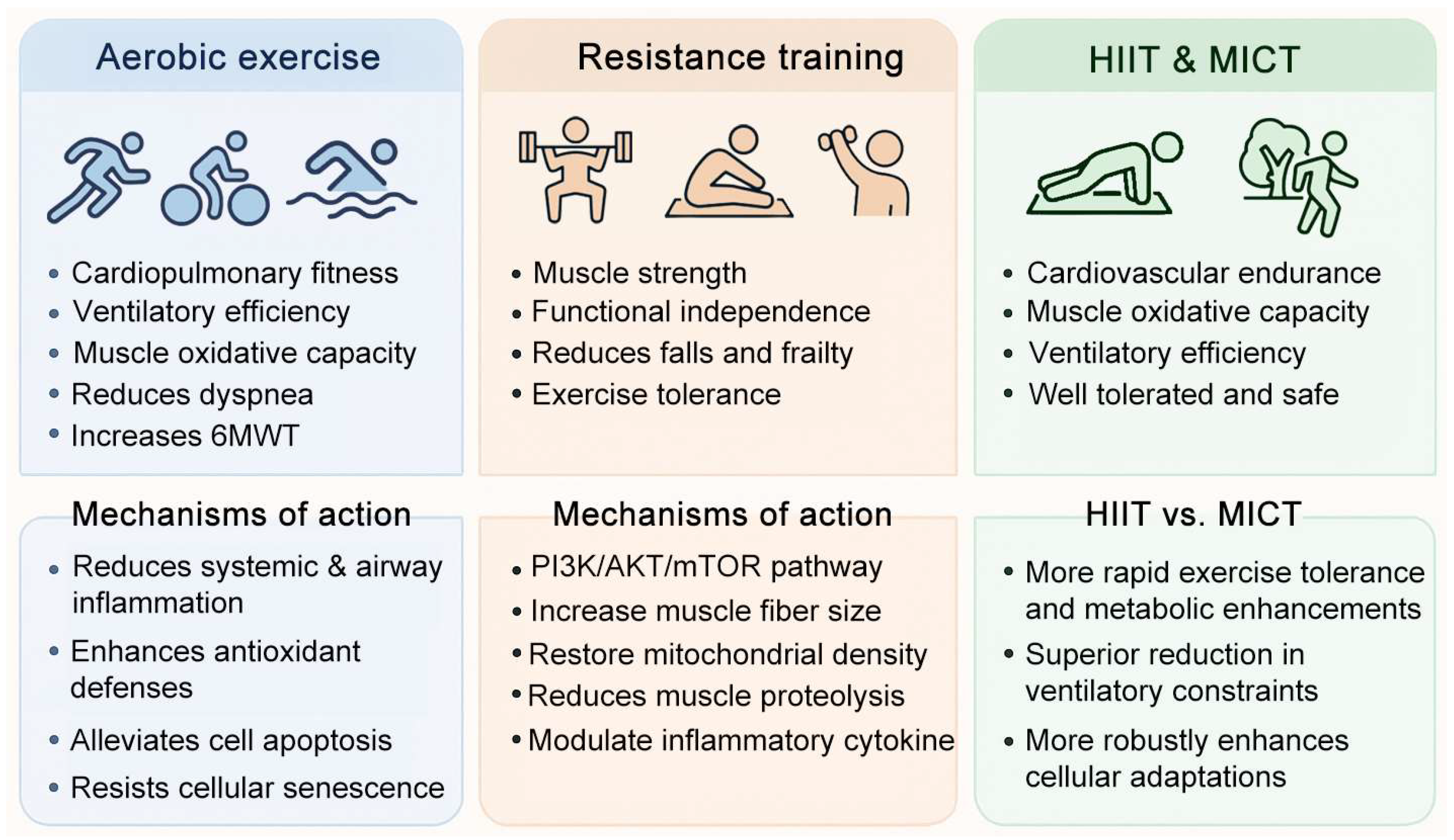
Given the heterogeneity of COPD and variability in patient response, different exercise modalities may confer distinct benefits. Aerobic exercise has been shown to improve cardiovascular fitness and anti-inflammatory capacity, resistance training to counteract muscle atrophy and weakness, and HIIT to induce rapid metabolic and respiratory adaptations. Thus, exploring the differential and overlapping effects of these exercise forms is essential for optimizing rehabilitation strategies. This section will summarize the current evidence on exercise-mediated improvements in COPD, setting the stage for mechanistic discussions on how epigenetic regulation contributes to these protective effects (Figure 2).

3.1. Aerobic Exercise–Mediated Mechanisms in COPD

Aerobic exercise, such as walking, cycling, and treadmill training, constitutes the most extensively studied form of physical activity in COPD rehabilitation. Its primary benefits are linked to improvements in cardiopulmonary fitness, ventilatory efficiency, and muscle oxidative capacity, all of which directly alleviate exertional dyspnea and enhance patients’ quality of life. Clinical trials consistently demonstrate that moderate-to-high intensity aerobic training reduces symptoms, increases six-minute walk distance, and lowers hospitalization risk in COPD patients [82,85,86,87,88,89].
Beyond functional outcomes, aerobic exercise exerts systemic effects on the underlying pathophysiology of COPD. Regular training attenuates systemic and airway inflammation, as evidenced by reductions in circulating C-reactive protein, TNF-α, and IL-6 levels [88,89,90,91]. In experimental models, aerobic exercise enhances antioxidant defenses by upregulating superoxide dismutase and catalase, thereby mitigating oxidative stress induced by CS exposure [92,93].
Evidence from animal studies provides mechanistic insight into these protective effects. In CS–exposed mouse models of COPD, treadmill running has been shown to alleviate the inflammatory response, oxidative stress injury, and cell apoptosis [94,95]. Moreover, preclinical data indicate that aerobic training preserves telomere length and reduces markers of cellular senescence in lung tissues, suggesting its role in counteracting premature aging—a hallmark of COPD pathology [96,97,98]. Importantly, animal models reveal that exercise can reprogram gene expression profiles associated with oxidative stress, inflammation, and apoptosis, reinforcing the concept that its benefits extend beyond symptom relief to disease modification [99].
Emerging evidence also highlights the potential role of aerobic exercise in modulating corticosteroid sensitivity. By restoring HDAC2 activity and reducing NF-κB–driven transcription of pro-inflammatory mediators, aerobic training may partially overcome the steroid resistance frequently observed in COPD [100]. Collectively, these findings suggest that aerobic exercise provides multifaceted protection in COPD, not only by improving pulmonary and systemic physiology but also by intervening in key molecular pathways that drive disease progression.

3.2. Mechanistic Role of Resistance Training in COPD

Resistance training, encompassing weightlifting, elastic band exercises, and bodyweight training, has emerged as a vital complement to aerobic exercise in COPD rehabilitation. Unlike aerobic training, which primarily targets cardiovascular endurance, resistance exercise directly addresses peripheral muscle weakness and atrophy—systemic manifestations strongly associated with disease severity and poor prognosis in COPD patients. Clinical studies have shown that resistance training significantly improves muscle strength, enhances functional independence, and reduces the risk of falls and frailty in patients with moderate-to-severe COPD [101,102,103,104,105,106,107,108].
At the physiological level, resistance training stimulates muscle hypertrophy by activating the PI3K/Akt/mTOR signaling pathway, thereby increasing protein synthesis and counteracting catabolic processes driven by chronic inflammation [109,110,111,112]. Patients undergoing structured resistance programs demonstrate improved quadriceps cross-sectional area and enhanced maximal voluntary contraction, both of which contribute to greater exercise tolerance [113]. Animal models further confirm the protective role of resistance-type training. Ladder-climbing or weighted-squat training protocols increase muscle fiber size, restore mitochondrial density, and reduce markers of muscle proteolysis such as atrogin-1 and MuRF1 [114,115,116,117]. Beyond musculoskeletal adaptations, resistance training may indirectly benefit pulmonary outcomes. By enhancing overall functional capacity and reducing ventilatory demand during daily activities, resistance training alleviates dyspnea and fatigue [118,119]. Moreover, preliminary data suggest that resistance exercise can modulate inflammatory cytokine profiles and improve circulating antioxidant levels, indicating systemic benefits beyond localized muscle strengthening [120,121,122].
Taken together, resistance training provides a targeted approach to addressing COPD-related muscle dysfunction and systemic weakness. When integrated with aerobic exercise, it offers synergistic improvements in physical performance, quality of life, and potentially disease-modifying pathways, making it an indispensable component of comprehensive pulmonary rehabilitation programs.

3.3. Comparative Efficacy of HIIT and MICT in COPD Rehabilitation

HIIT and MICT represent two widely investigated aerobic modalities in COPD rehabilitation. While both improve exercise capacity and quality of life, they differ in physiological demands, patient tolerance, and underlying adaptations.
MICT, typically involving prolonged cycling or treadmill walking at 50–70% of maximal workload, is a long-standing and effective method for pulmonary rehabilitation. MICT enhances cardiovascular endurance, reduces ventilatory inefficiency, and increases skeletal muscle oxidative capacity [123]. It is generally well tolerated and safe for patients across different COPD severities, making it a foundational modality in clinical practice. HIIT involves repeated short bursts of exercise at near-maximal intensity, interspersed with recovery periods. Recent studies suggest that HIIT may induce more rapid improvements in exercise tolerance and skeletal muscle metabolism compared to MICT, despite lower total exercise volume [124,125]. HIIT has been shown to reduce dynamic hyperinflation and ventilatory limitation more effectively, thereby allowing COPD patients to achieve higher training intensities with less dyspnea [126,127,128]. Moreover, HIIT enhances mitochondrial biogenesis, increases oxidative enzyme activity, and modulates systemic inflammation more robustly than MICT in certain populations [124,129,130,131]. HIIT has also been associated with greater activation of AMPK-PGC1α signaling, suggesting superior capacity to remodel energy metabolism [125,132,133,134].
Despite its advantages, HIIT may pose challenges for severely debilitated COPD patients due to higher perceived exertion and potential cardiovascular risks. Thus, patient-specific factors including baseline lung function, comorbidities, and training adherence should guide the choice between HIIT and MICT. Increasingly, hybrid approaches that combine interval and continuous elements are being explored to balance efficacy and tolerability.
In summary, both HIIT and MICT improve exercise tolerance and systemic health in COPD, but HIIT may provide more potent metabolic and anti-inflammatory benefits, whereas MICT ensures broad applicability and long-term sustainability. Tailoring exercise prescriptions to individual patient needs remains a central challenge in optimizing COPD rehabilitation outcomes.

4. Epigenetic Basis of Exercise Benefits in COPD

While the clinical and physiological benefits of exercise in COPD are well established, the molecular mechanisms that underpin these improvements remain an area of intense investigation. Among the various biological pathways, epigenetic regulation has emerged as a promising explanation for how physical activity exerts long-lasting and systemic protective effects.
In COPD, cigarette smoke, oxidative stress, and chronic inflammation disrupt the normal balance of epigenetic marks, leading to persistent activation of pro-inflammatory genes, impaired antioxidant defenses, and accelerated cellular senescence [135,136]. Exercise is increasingly recognized to counteract these maladaptive patterns by modulating epigenetic programs in lung tissue, skeletal muscle, and circulating immune cells.
Mechanical load, calcium flux, and metabolic stress generated during exercise activate upstream pathways such as AMPK–PGC-1α, CaMKII, and redox-sensitive NRF2 signaling [20,21,137,138]. These cascades converge on nuclear epigenetic regulators, influencing the activity of DNMTs (DNMT1; DNMT3A/3B), TET dioxygenases, and histone deacetylases including HDAC2 and HDAC3 [139,140]. Transcription factors such as PGC-1α, MEF2, NRF2, and NF-κB further integrate these signals by recruiting or displacing epigenetic enzymes at specific loci to fine-tune inflammatory and antioxidant gene expression [141,142]. Importantly, we also note that not all studies report robust epigenetic responses; several short-term or low-volume exercise interventions show minimal or no detectable methylation changes [138,143], underscoring heterogeneity in training protocols and tissue-specific responses.
Different exercise modalities, intensities, and durations produce heterogeneous epigenetic outcomes. Multi-week endurance training at moderate-to-high intensity consistently induces coordinated hypomethylation at genes related to mitochondrial biogenesis and oxidative metabolism in human skeletal muscle [138]. Progressive resistance training for 6–12 weeks elicits distinct and reproducible methylation remodeling at loci governing muscle growth and structural adaptation [143]. Acute bouts of exercise can trigger rapid methylation changes in isolated immune cell subsets or skeletal muscle, although studies analyzing whole PBMCs or global methylation frequently report no detectable modification after a single session, emphasizing the importance of tissue specificity and sampling time [144,145]. In COPD, small pulmonary rehabilitation cohorts show shifts in global or peripheral methylation after multi-week programs, although measurements remain blood-based and lack promoter-resolved lung data [146]. Collectively, available evidence suggests that sustained endurance or resistance training more reliably remodels epigenetic landscapes compared with single acute sessions, but inter-study heterogeneity and methodological limitations preclude firm dose–response prescriptions at present.
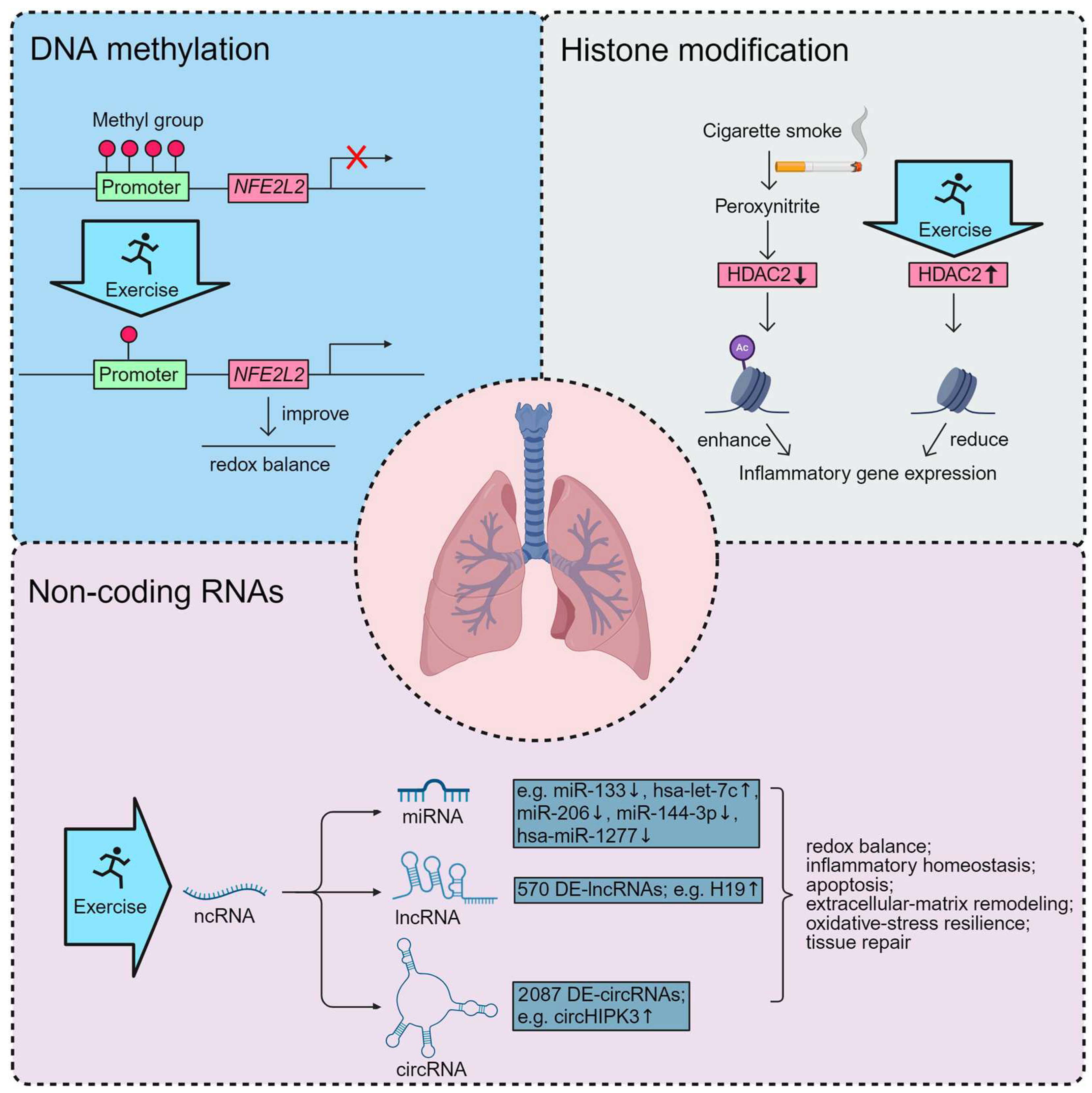
Recent studies suggest that aerobic exercise can restore HDAC2 activity, thereby enhancing anti-inflammatory responses, while resistance training and interval training may modulate DNA methylation profiles of genes related to oxidative stress and mitochondrial biogenesis. In addition, exercise-induced non-coding RNAs have been implicated in regulating pathways of apoptosis, autophagy, and tissue repair. Collectively, these findings highlight that the benefits of exercise extend beyond functional adaptations to include epigenetic remodeling of key molecular networks that underlie COPD pathology (Figure 3).

4.1. Exercise-Induced DNA Methylation in COPD

Evidence directly linking exercise to DNA methylation changes in COPD remains limited, but several studies provide preliminary and mechanistic insights. In a pilot study of COPD patients enrolled in an eight-week supervised pulmonary rehabilitation program, da Silva et al. reported an acute decrease in global DNA methylation after the first exercise session and a progressive reduction in baseline circulating pro-inflammatory cytokines (e.g., IL-6 and IL-8) after completion of the program, suggesting that exercise produces measurable changes in peripheral epigenetic markers alongside systemic inflammatory modulation [146]. To more accurately reflect the available evidence, this pilot study represents the only direct human COPD dataset assessing DNA methylation responses to exercise, and it measured global rather than locus-specific methylation. Thus, no published human study has demonstrated promoter-resolved methylation remodeling in COPD lung or airway tissues following exercise.
Independent work has demonstrated that hypermethylation of the NFE2L2 promoter contributes to reduced Nrf2 expression and impaired antioxidant defenses in COPD lungs, linking locus-specific methylation changes to disease-relevant redox dysfunction [23]. Complementary animal experiments demonstrate that treadmill exercise increases muscle-derived mediators (for example, irisin), upregulates pulmonary Nrf2 and HO-1 expression, and mitigates CS–induced emphysematous changes, thereby providing tissue-level evidence that exercise engages Nrf2-centered antioxidant pathways relevant to COPD protection [147]. These preclinical findings support the biological plausibility that exercise could influence methylation states of antioxidant genes, although direct evidence for demethylation of NFE2L2 or other loci in COPD lung tissue following exercise is currently lacking.
Mechanistically, exercise has been shown in non-pulmonary models to reverse promoter hypermethylation of NFE2L2 and to reduce aberrant DNA methyltransferase (DNMT) expression, leading to restored Nrf2 transcription and improved redox balance [148]. In addition, several intervention studies report locus-specific methylation or expression changes that connect epigenetic modulation to clinically relevant targets. For example, exercise-associated demethylation and transcriptional recovery of antioxidant genes (NFE2L2, SOD2) and reduced promoter methylation at genes involved in mitochondrial function have been documented in training studies in non-pulmonary tissues and are congruent with the upregulation of Nrf2-dependent transcripts observed in smoke-exposed animals following exercise [148,149]. However, because these observations originate from non-pulmonary or non-COPD models, they should be considered supportive rather than indicative of a confirmed mechanism in COPD. While these findings jointly suggest that exercise can influence redox- and inflammation-related pathways that are also epigenetically regulated in COPD, they do not establish a causal sequence linking exercise to locus-specific DNA methylation remodeling in COPD lung tissue. Therefore, the current evidence should be viewed as hypothesis-generating, and definitive demonstration of methylation-mediated protection will require studies that pair well-defined exercise interventions with tissue-resolved epigenomic profiling in human COPD.

4.2. Exercise-Induced Histone Modification in COPD

Current evidence directly demonstrating that exercise ameliorates COPD through regulated histone modifications is limited. Clinical pulmonary rehabilitation trials in COPD have documented reductions in systemic inflammatory mediators and alterations in peripheral epigenetic markers, but locus-specific histone mark mapping at pro-inflammatory or antioxidant gene promoters has not been reported in these interventions [146]. To our knowledge, no study has evaluated histone acetylation or methylation patterns in lung or airway tissues before and after exercise in COPD patients, representing a major evidence gap.
Observational studies have consistently documented reduced histone deacetylase activity in COPD, notably decreased HDAC2 expression and lowered class III deacetylase SIRT1 levels in lung and peripheral tissues, changes that are mechanistically linked to heightened inflammatory signaling, oxidative stress and corticosteroid resistance in patients [42,150,151,152]. Exercise and physical activity are known to influence histone marks in non-pulmonary tissues. Human and animal studies have reported exercise-associated increases in activating histone acetylation (e.g., H3K9ac, H3 acetylation) and enrichment of activating methylation marks (e.g., H3K4me3) at genes governing mitochondrial biogenesis and metabolic adaptation [153,154,155]. While these findings underscore the plausibility that exercise may also influence chromatin states in COPD, the absence of direct histone mapping in lung tissues precludes mechanistic conclusions. Importantly, although HDAC2 and SIRT1 reductions are central to COPD pathobiology, no available study has demonstrated that exercise restores the expression or activity of these enzymes in COPD lung tissue, and therefore they should be regarded as potential mechanistic targets rather than established mediators of exercise benefit.
In CS–exposed rodent models, exercise interventions reproducibly activate cytoprotective pathways such as Nrf2/HO-1 and ameliorate emphysematous pathology, providing tissue-level evidence of benefit. However, these reports have generally not included ChIP-based or other locus-specific assays of histone marks at pro-inflammatory or antioxidant gene promoters, so the chromatin-level mediators remain uncharacterized [147]. Mechanistic animal work linking histone-modifying enzymes to smoke-induced muscle wasting and inflammation further underscores candidate mediators (for example, HDAC2 involvement in skeletal muscle dysfunction), suggesting targets for focused chromatin analyses in intervention studies [100]. Taken together, the literature supports the hypothesis that exercise can reprogram chromatin states in ways that favor antioxidant and anti-inflammatory gene expression, but definitive demonstration in COPD requires intervention studies that pair well-defined exercise prescriptions with tissue-resolved, locus-specific histone profiling and matched functional endpoints.

4.3. Exercise-Induced ncRNAs in COPD

Emerging evidence indicates that exercise modulates the expression of ncRNAs, including miRNAs, lncRNAs, and circRNAs, which may contribute to protective effects in COPD. Recent human data provide preliminary but direct evidence that exercise modulates specific miRNAs associated with COPD pathophysiology. In an observational cohort, miR-133 and miR-206, two muscle-enriched myomiRs involved in skeletal-muscle differentiation and regeneration, were negatively correlated with daily physical activity levels in patients with COPD, suggesting a link between reduced activity and muscle dysfunction–related miRNA expression [53]. In a pilot interventional study, 12 weeks of individualized aerobic training in patients with moderate-to-severe COPD led to downregulation of miR-144-3p and hsa-miR-1277 and upregulation of hsa-let-7c in peripheral blood [156]. These alterations coincided with improvements in exercise tolerance and reductions in circulating IL-6 and TNF-α. Notably, the upregulation of let-7c, a miRNA known to target inflammatory mediators [157], and the suppression of miR-144-3p, previously linked to oxidative stress and erythroid dysfunction in COPD [158], imply that aerobic training exerts systemic anti-inflammatory and cytoprotective effects through miRNA regulation. Although the sample sizes were small and locus-specific mechanistic validation is lacking, these studies provide the currently available evidence that structured exercise interventions can reprogram disease-relevant miRNA networks in COPD patients, potentially contributing to improved redox and inflammatory homeostasis.
Evidence from animal models corroborates these findings and extends them to lncRNAs and circRNAs. Recent animal studies provide direct evidence that exercise modulates lncRNA expression in smoke-exposed COPD models and that these changes may contribute to tissue protection. For example, in a mouse model of CS-induced COPD, aerobic training was shown to reduce lung expression of lncRNA H19, a lncRNA previously found to promote ROS generation, epithelial–mesenchymal transition (EMT) and lung injury, and this reduction correlated with decreased inflammation, suppressed EMT marker α-SMA, and preservation of alveolar architecture [95]. Moreover, a comprehensive transcriptomic profiling study of COPD patients after 12 weeks of aerobic training revealed differential expression of 570 lncRNAs in peripheral blood, concurrent with changes in mRNA and circRNA networks, implying that exercise broadly alters lncRNA landscapes in COPD contexts [156]. In parallel, 2087 circRNAs were found to be differentially expressed in peripheral blood post-training in this study, supporting the idea that exercise affects circRNA networks in COPD [156]. Functional studies in related injury models indicate that circRNAs (for example circHIPK3) can sponge pro-inflammatory miRNAs (such as miR-21), thereby reducing NF-κB activation and oxidative stress in target cells [95,159]. However, these functional observations derive from related injury models rather than COPD itself, and thus cannot be taken as evidence that similar ncRNA-dependent mechanisms mediate exercise-induced protection in COPD lung tissue.
Collectively, these studies provide direct evidence that exercise remodels ncRNA networks in COPD, influencing pathways involved in inflammation, oxidative stress, apoptosis, and tissue repair. While the data remain limited, these findings support the concept that ncRNA-mediated regulation represents a potential mechanism by which exercise confers protective effects in COPD. Future studies employing longitudinal human cohorts and integrated multi-omics approaches will be critical to delineate the precise ncRNA networks modulated by exercise and their functional relevance to clinical outcomes.

5. Discussion

This review synthesizes emerging evidence that structured physical activity influences molecular processes relevant to COPD through multiple epigenetic layers. Clinical pulmonary rehabilitation and supervised exercise consistently reduce systemic inflammatory mediators and improve functional outcomes in COPD; in several small human studies these clinical benefits have been accompanied by changes in peripheral epigenetic markers, most notably at the level of global DNA methylation and circulating ncRNAs [146,156]. Complementary preclinical work demonstrates that exercise upregulates cytoprotective signaling such as the Nrf2/HO-1 axis and attenuates CS–induced emphysematous changes in rodent models, establishing tissue-level molecular and morphological correlates of exercise benefit [147]. Independent molecular studies have documented disease-associated epigenetic substrates in COPD lungs and airways, including promoter hypermethylation of antioxidant regulators and perturbations in histone-modifying enzymes such as HDAC2 and SIRT1 [23,42,95,150]. Together, these observations identify epigenetic substrates that could represent mechanistic points of interaction for exercise, although direct evidence linking exercise to targeted remodeling of these marks in COPD lungs remains unavailable.
Despite these convergent lines of evidence, the mechanistic picture remains incomplete. No published study to date has established the full causal chain—exercise intervention, locus-specific epigenetic remodeling in the lung, and consequent, reproducible improvement in COPD clinical end points—in a single, well-powered human or animal experiment. Most human trials that incorporate epigenetic measurements have been small and focused on peripheral samples [146,156]. Even when epigenetic changes are observed in PBMCs or plasma, whether these alterations parallel lung-resident epigenetic remodeling remains unknown, as concordance between blood and lung tissues has not been systematically evaluated in COPD. Moreover, because epigenetic marks are inherently dynamic, most existing studies lack multi-time-point sampling, preventing differentiation between transient exercise-induced fluctuations and durable adaptations that may mediate clinical improvement. Animal studies more readily combine intervention and tissue analysis, but most report pathway activation and phenotypic rescue without concurrent, genome-wide or targeted histone/chromatin profiling, limiting the ability to determine whether specific histone modifications mediate exercise responses [147]. Studies of ncRNAs are promising and include both human and murine data showing exercise-associated modulation of miRNAs, lncRNAs and circRNAs. However, these datasets lack longitudinal resolution and functional perturbation (gain- or loss-of-function) evidence that could establish causal mediation [95,156,160].
In addition to epigenetic mechanisms, COPD pathogenesis is strongly shaped by protease–antiprotease imbalance, chronic oxidative stress, and persistent inflammation. Elevated levels of matrix metalloproteinase-9 (MMP-9) and an increased MMP-9/TIMP-1 ratio are consistently associated with emphysema severity and extracellular matrix degradation in COPD [161,162]. Likewise, impaired Nrf2-dependent antioxidant responses contribute to heightened oxidant burden and tissue injury [163]. Exercise interventions, although not directly shown to modulate protease activity at the epigenetic level, have been reported to attenuate systemic inflammation and improve redox homeostasis in COPD, partly through upregulation of cytoprotective pathways such as Nrf2/HO-1 in preclinical smoke-exposed models [147]. Furthermore, evidence from non-COPD contexts indicates that resistance training can modulate MMP-2 and MMP-9 expression in skeletal muscle, visceral adipose tissue and circulation, supporting a plausible link between improved protease–antiprotease balance and regular physical activity, though studies directly evaluating these pathways in COPD remain lacking [164]. Integrating these findings, exercise may exert multi-layered regulatory effects including reduce inflammatory signaling, enhance antioxidant capacity, and potentially influence protease-related pathways, thereby creating a biological context in which epigenetic remodeling could contribute to durable improvements in pulmonary homeostasis. However, definitive mechanistic links among exercise, epigenetic remodeling, and protease/oxidant homeostasis in COPD remain to be demonstrated.
These limitations point to specific priorities for future research. Clinical trials should prospectively embed standardized epigenetic end points, collect appropriately matched biospecimens (for example, PBMCs, bronchial brushings or BAL cells, and skeletal muscle biopsies) at harmonized timepoints (baseline, early response, end of intervention and follow-up), and pair molecular profiling with rigorous clinical outcomes (spirometry, symptom scores, exercise capacity, and quantitative imaging). Multi-omics approaches that combine whole-genome bisulfite sequencing or array-based methylome profiling, ChIP-seq for key histone marks and chromatin modifiers, and small/total RNA sequencing for ncRNAs will be essential to map coordinated regulatory networks. In parallel, mechanistic animal studies should integrate locus-specific epigenetic assays with genetic or epigenetic perturbation (e.g., DNMT/HDAC/SIRT modulation, CRISPR-Cas9–based epigenome editing) to test whether reversing a given epigenetic lesion is necessary and sufficient for exercise-mediated phenotypic rescue. Careful attention to exercise prescription (modality, intensity, duration), participant phenotyping (inflammatory status, comorbidity, medication use), and statistical power will be critical to resolve heterogeneity and to identify subgroups most likely to derive epigenetic and clinical benefit.
From a translational perspective, the current literature supports cautious optimism rather than immediate clinical application. Epigenetic signatures, if validated in larger, longitudinal cohorts, could become useful biomarkers for stratifying response to pulmonary rehabilitation or for monitoring biological responses to exercise. Likewise, combinatorial strategies that pair rehabilitation with pharmacologic modulators of epigenetic enzymes remain an intriguing hypothesis, but such approaches should be pursued only after mechanistic proof of concept is obtained. In the near term, investigators running pulmonary rehabilitation trials should consider routine collection of biospecimens and standardized epigenetic assays to build the evidence base required for future precision rehabilitation.
In summary, exercise-mediated modulation of epigenetic mechanisms represents a biologically plausible route by which physical activity can influence inflammation, redox balance and tissue integrity in COPD. Existing clinical and preclinical studies provide initial support for this model but do not yet establish locus-specific causal mediation. Addressing this gap will require adequately powered, tissue-resolved, and mechanistically informed studies that integrate multi-omics profiling with robust phenotyping and interventional perturbations.

6. Conclusions

Exercise provides multifaceted benefits in COPD, extending beyond improvements in physical performance to modulation of key molecular pathways involved in disease progression. Emerging evidence demonstrates that these protective effects are mediated, at least in part, through epigenetic mechanisms, including DNA methylation, histone modifications, and ncRNA regulation. Exercise may restore aberrant methylation patterns, rebalance histone acetylation and methylation at critical gene loci, and fine-tune ncRNA networks, collectively mitigating inflammation, oxidative stress, and tissue remodeling. The integration of clinical and preclinical findings underscores the potential of exercise as a disease-modifying intervention, offering both functional and molecular benefits. These insights highlight the importance of incorporating structured physical activity into COPD management and provide a foundation for personalized rehabilitation strategies informed by epigenetic profiling.

7. Future Directions

Future research should focus on elucidating the causal relationships between exercise-induced epigenetic modifications and clinical outcomes in COPD. Longitudinal studies with larger patient cohorts and multi-tissue analyses are needed to capture the systemic impact of exercise and to identify biomarkers predictive of individual responsiveness. Multi-omics approaches, integrating methylomics, histone modification mapping, transcriptomics, and ncRNA profiling, will be critical for constructing a comprehensive view of exercise-mediated epigenetic regulation. Additionally, comparative studies exploring different exercise modalities, intensities, and durations will help optimize rehabilitation protocols. Investigating combinatorial strategies that integrate pharmacological epigenetic modulators with exercise interventions may further enhance therapeutic efficacy. Ultimately, a deeper understanding of epigenetic mechanisms will facilitate precision rehabilitation, enabling tailored exercise prescriptions that maximize molecular and functional benefits in COPD patients.

Author Contributions

Conceptualization, Y.L. and X.Z.; review of literature, Y.Z. and X.C.; writing—review and editing, Y.Z., J.X., X.C., Y.L. and X.Z. All authors have read and agreed to the published version of the manuscript.

Funding

This research was funded by the Natural Science Foundation of Fujian Province (2025J01132343).

Institutional Review Board Statement

Not applicable.

Informed Consent Statement

Not applicable.

Data Availability Statement

No new data were created or analyzed in this study.

Conflicts of Interest

The authors declare no conflicts of interest.

Abbreviations

The following abbreviations are used in this manuscript:
circRNAsCircular RNAs
COPDChronic Obstructive Pulmonary Disease
CSCigarette Smoke
DNMTDNA Methyltransferase
EMTEpithelial–Mesenchymal Transition
EWASEpigenome-Wide Association Studies
HDACsHistone Deacetylases
HIITHigh-Intensity Interval Training
lncRNAsLong Non-Coding RNAs
MICTModerate-Intensity Continuous Training
miRNAsMicroRNAs
ncRNAsNon-Coding RNAs

References

  1. Christenson, S.A.; Smith, B.M.; Bafadhel, M.; Putcha, N. Chronic Obstructive Pulmonary Disease. Lancet 2022, 399, 2227–2242. [Google Scholar] [CrossRef]
  2. Berg, K.; Wright, J.L. The Pathology of Chronic Obstructive Pulmonary Disease: Progress in the 20th and 21st Centuries. Arch. Pathol. Lab. Med. 2016, 140, 1423–1428. [Google Scholar] [CrossRef]
  3. Lareau, S.C.; Fahy, B.; Meek, P.; Wang, A. Chronic Obstructive Pulmonary Disease (Copd). Am. J. Respir. Crit. Care Med. 2019, 199, P1–P2. [Google Scholar] [CrossRef] [PubMed]
  4. Sahoo, K.C.; Subhankar, S.; Mohanta, P.C.; Jagaty, S.K.; Dutta, P.; Pothal, S. Prevalence of Metabolic Syndrome in Chronic Obstructive Pulmonary Disease and Its Correlation with Severity of Disease. J. Fam. Med. Prim. Care. 2022, 11, 2094–2098. [Google Scholar] [CrossRef]
  5. Barreiro, E.; Jaitovich, A. Muscle Atrophy in Chronic Obstructive Pulmonary Disease: Molecular Basis and Potential Therapeutic Targets. J. Thorac. Dis. 2018, 10, S1415–S1424. [Google Scholar] [CrossRef]
  6. Franssen, F.M.; Rochester, C.L. Comorbidities in Patients with Copd and Pulmonary Rehabilitation: Do They Matter? Eur. Respir. Rev. 2014, 23, 131–141. [Google Scholar] [CrossRef]
  7. Halpin, D.M. Systemic Effects of Chronic Obstructive Pulmonary Disease. Expert. Rev. Respir. Med. 2007, 1, 75–84. [Google Scholar] [CrossRef] [PubMed]
  8. Vucic, E.A.; Chari, R.; Thu, K.L.; Wilson, I.M.; Cotton, A.M.; Kennett, J.Y.; Zhang, M.; Lonergan, K.M.; Steiling, K.; Brown, C.J.; et al. DNA Methylation Is Globally Disrupted and Associated with Expression Changes in Chronic Obstructive Pulmonary Disease Small Airways. Am. J. Respir. Cell Mol. Biol. 2014, 50, 912–922. [Google Scholar] [CrossRef]
  9. Shakespear, M.R.; Halili, M.A.; Irvine, K.M.; Fairlie, D.P.; Sweet, M.J. Histone Deacetylases as Regulators of Inflammation and Immunity. Trends Immunol. 2011, 32, 335–343. [Google Scholar] [CrossRef] [PubMed]
  10. Mortaz, E.; Masjedi, M.R.; Barnes, P.J.; Adcock, I.M. Epigenetics and Chromatin Remodeling Play a Role in Lung Disease. Tanaffos 2011, 10, 7–16. [Google Scholar]
  11. Tuncel, O.; Kara, M.; Yaylak, B.; Erdogan, I.; Akgul, B. Noncoding Rnas in Apoptosis: Identification and Function. Turk. J. Biol. 2022, 46, 1–40. [Google Scholar] [PubMed]
  12. Wang, H.; Sun, K.; Peng, H.; Wang, Y.; Zhang, L. Emerging Roles of Noncoding Rnas in Idiopathic Pulmonary Fibrosis. Cell Death Discov. 2024, 10, 443. [Google Scholar] [CrossRef]
  13. Xie, N.; Liu, G. Ncrna-Regulated Immune Response and Its Role in Inflammatory Lung Diseases. Am. J. Physiol. Lung Cell Mol. Physiol. 2015, 309, L1076–L1087. [Google Scholar] [CrossRef]
  14. Jiang, M.; Li, P.; Wang, Y.; Cao, Y.; Han, X.; Jiang, L.; Liu, X.; Wu, W. Role of Nrf2 and Exercise in Alleviating Copd-Induced Skeletal Muscle Dysfunction. Ther. Adv. Respir. Dis. 2023, 17, 17534666231208633. [Google Scholar] [CrossRef]
  15. Frinchi, M.; Morici, G.; Mudo, G.; Bonsignore, M.R.; Di Liberto, V. Beneficial Role of Exercise in the Modulation of Mdx Muscle Plastic Remodeling and Oxidative Stress. Antioxidants. 2021, 10, 558. [Google Scholar] [CrossRef]
  16. Han, L.; Li, P.; He, Q.; Yang, C.; Jiang, M.; Wang, Y.; Cao, Y.; Han, X.; Liu, X.; Wu, W. Revisiting Skeletal Muscle Dysfunction and Exercise in Chronic Obstructive Pulmonary Disease: Emerging Significance of Myokines. Aging Dis. 2023, 15, 2453–2469. [Google Scholar] [CrossRef]
  17. Henrot, P.; Dupin, I.; Schilfarth, P.; Esteves, P.; Blervaque, L.; Zysman, M.; Gouzi, F.; Hayot, M.; Pomies, P.; Berger, P. Main Pathogenic Mechanisms and Recent Advances in Copd Peripheral Skeletal Muscle Wasting. Int. J. Mol. Sci. 2023, 24, 6454. [Google Scholar] [CrossRef]
  18. Sellami, M.; Bragazzi, N.; Prince, M.S.; Denham, J.; Elrayess, M. Regular, Intense Exercise Training as a Healthy Aging Lifestyle Strategy: Preventing DNA Damage, Telomere Shortening and Adverse DNA Methylation Changes over a Lifetime. Front. Genet. 2021, 12, 652497. [Google Scholar] [CrossRef] [PubMed]
  19. Swiatowy, W.J.; Drzewiecka, H.; Kliber, M.; Sasiadek, M.; Karpinski, P.; Plawski, A.; Jagodzinski, P.P. Physical Activity and DNA Methylation in Humans. Int. J. Mol. Sci. 2021, 22, 12989. [Google Scholar] [CrossRef]
  20. Widmann, M.; Niess, A.M.; Munz, B. Physical Exercise and Epigenetic Modifications in Skeletal Muscle. Sports Med. 2019, 49, 509–523. [Google Scholar] [CrossRef]
  21. Zheng, X.; Liu, X.; Guo, Y.; Lv, Y.; Lin, C.; Wang, D.; Wang, S.; Liu, Y.; Hu, X. Physical Exercise and Epigenetic Modifications in Skeletal Muscle, Brain, and Heart. Epigenetics Chromatin. 2025, 18, 12. [Google Scholar] [CrossRef] [PubMed]
  22. Jones, P.A.; Takai, D. The Role of DNA Methylation in Mammalian Epigenetics. Science 2001, 293, 1068–1070. [Google Scholar] [CrossRef]
  23. Zhang, Z.; Fu, C.; Liu, J.; Sai, X.; Qin, C.; Di, T.; Yang, Y.; Wu, Y.; Bian, T. Hypermethylation of the Nrf2 Promoter Induces Ferroptosis by Inhibiting the Nrf2-Gpx4 Axis in Copd. Int. J. Chron. Obstruct. Pulmon. Dis. 2021, 16, 3347–3362. [Google Scholar] [CrossRef]
  24. He, L.X.; Tang, Z.H.; Huang, Q.S.; Li, W.H. DNA Methylation: A Potential Biomarker of Chronic Obstructive Pulmonary Disease. Front. Cell Dev. Biol. 2020, 8, 585. [Google Scholar] [CrossRef]
  25. Gao, X.; Jia, M.; Zhang, Y.; Breitling, L.P.; Brenner, H. DNA Methylation Changes of Whole Blood Cells in Response to Active Smoking Exposure in Adults: A Systematic Review of DNA Methylation Studies. Clin. Epigenetics 2015, 7, 113. [Google Scholar] [CrossRef]
  26. Guida, F.; Sandanger, T.M.; Castagne, R.; Campanella, G.; Polidoro, S.; Palli, D.; Krogh, V.; Tumino, R.; Sacerdote, C.; Panico, S.; et al. Dynamics of Smoking-Induced Genome-Wide Methylation Changes with Time since Smoking Cessation. Hum. Mol. Genet. 2015, 24, 2349–2359. [Google Scholar] [CrossRef]
  27. Dogan, M.V.; Shields, B.; Cutrona, C.; Gao, L.; Gibbons, F.X.; Simons, R.; Monick, M.; Brody, G.H.; Tan, K.; Beach, S.R.; et al. The Effect of Smoking on DNA Methylation of Peripheral Blood Mononuclear Cells from African American Women. BMC Genom. 2014, 15, 151. [Google Scholar] [CrossRef]
  28. Elliott, H.R.; Tillin, T.; McArdle, W.L.; Ho, K.; Duggirala, A.; Frayling, T.M.; Davey Smith, G.; Hughes, A.D.; Chaturvedi, N.; Relton, C.L. Differences in Smoking Associated DNA Methylation Patterns in South Asians and Europeans. Clin. Epigenetics 2014, 6, 4. [Google Scholar] [CrossRef]
  29. Shenker, N.S.; Ueland, P.M.; Polidoro, S.; van Veldhoven, K.; Ricceri, F.; Brown, R.; Flanagan, J.M.; Vineis, P. DNA Methylation as a Long-Term Biomarker of Exposure to Tobacco Smoke. Epidemiology 2013, 24, 712–716. [Google Scholar] [CrossRef] [PubMed]
  30. Zeilinger, S.; Kuhnel, B.; Klopp, N.; Baurecht, H.; Kleinschmidt, A.; Gieger, C.; Weidinger, S.; Lattka, E.; Adamski, J.; Peters, A.; et al. Tobacco Smoking Leads to Extensive Genome-Wide Changes in DNA Methylation. PLoS ONE 2013, 8, e63812. [Google Scholar] [CrossRef] [PubMed]
  31. Sun, Y.V.; Smith, A.K.; Conneely, K.N.; Chang, Q.; Li, W.; Lazarus, A.; Smith, J.A.; Almli, L.M.; Binder, E.B.; Klengel, T.; et al. Epigenomic Association Analysis Identifies Smoking-Related DNA Methylation Sites in African Americans. Hum. Genet. 2013, 132, 1027–1037. [Google Scholar] [CrossRef]
  32. Zong, D.; Liu, X.; Li, J.; Ouyang, R.; Chen, P. The Role of Cigarette Smoke-Induced Epigenetic Alterations in Inflammation. Epigenetics Chromatin. 2019, 12, 65. [Google Scholar] [CrossRef]
  33. Beetch, M.; Harandi-Zadeh, S.; Shen, K.; Lubecka, K.; Kitts, D.D.; O’Hagan, H.M.; Stefanska, B. Dietary Antioxidants Remodel DNA Methylation Patterns in Chronic Disease. Br. J. Pharmacol. 2020, 177, 1382–1408. [Google Scholar] [CrossRef]
  34. Morrow, J.D.; Glass, K.; Cho, M.H.; Hersh, C.P.; Pinto-Plata, V.; Celli, B.; Marchetti, N.; Criner, G.; Bueno, R.; Washko, G.; et al. Human Lung DNA Methylation Quantitative Trait Loci Colocalize with Chronic Obstructive Pulmonary Disease Genome-Wide Association Loci. Am. J. Respir. Crit. Care Med. 2018, 197, 1275–1284. [Google Scholar] [CrossRef] [PubMed]
  35. Clifford, R.L.; Fishbane, N.; Patel, J.; MacIsaac, J.L.; McEwen, L.M.; Fisher, A.J.; Brandsma, C.A.; Nair, P.; Kobor, M.S.; Hackett, T.L.; et al. Altered DNA Methylation Is Associated with Aberrant Gene Expression in Parenchymal but Not Airway Fibroblasts Isolated from Individuals with Copd. Clin. Epigenetics 2018, 10, 32. [Google Scholar] [CrossRef] [PubMed]
  36. Casas-Recasens, S.; Cassim, R.; Mendoza, N.; Agusti, A.; Lodge, C.; Li, S.; Bui, D.; Martino, D.; Dharmage, S.C.; Faner, R. Epigenome-Wide Association Studies of Chronic Obstructive Pulmonary Disease and Lung Function: A Systematic Review. Am. J. Respir. Crit. Care Med. 2024, 210, 766–778. [Google Scholar] [CrossRef]
  37. Chen, Y.C.; Tsai, Y.H.; Wang, C.C.; Liu, S.F.; Chen, T.W.; Fang, W.F.; Lee, C.P.; Hsu, P.Y.; Chao, T.Y.; Wu, C.C.; et al. Epigenome-Wide Association Study on Asthma and Chronic Obstructive Pulmonary Disease Overlap Reveals Aberrant DNA Methylations Related to Clinical Phenotypes. Sci. Rep. 2021, 11, 5022. [Google Scholar] [CrossRef]
  38. Lee, M.K.; Hong, Y.; Kim, S.Y.; Kim, W.J.; London, S.J. Epigenome-Wide Association Study of Chronic Obstructive Pulmonary Disease and Lung Function in Koreans. Epigenomics 2017, 9, 971–984. [Google Scholar] [CrossRef] [PubMed]
  39. Lawrence, M.; Daujat, S.; Schneider, R. Lateral Thinking: How Histone Modifications Regulate Gene Expression. Trends Genet. 2016, 32, 42–56. [Google Scholar] [CrossRef]
  40. Ito, K.; Ito, M.; Elliott, W.M.; Cosio, B.; Caramori, G.; Kon, O.M.; Barczyk, A.; Hayashi, S.; Adcock, I.M.; Hogg, J.C.; et al. Decreased Histone Deacetylase Activity in Chronic Obstructive Pulmonary Disease. N. Engl. J. Med. 2005, 352, 1967–1976. [Google Scholar] [CrossRef]
  41. Barnes, P.J.; Ito, K.; Adcock, I.M. Corticosteroid Resistance in Chronic Obstructive Pulmonary Disease: Inactivation of Histone Deacetylase. Lancet 2004, 363, 731–733. [Google Scholar] [CrossRef]
  42. Rajendrasozhan, S.; Yang, S.R.; Edirisinghe, I.; Yao, H.; Adenuga, D.; Rahman, I. Deacetylases and Nf-Kappab in Redox Regulation of Cigarette Smoke-Induced Lung Inflammation: Epigenetics in Pathogenesis of Copd. Antioxid. Redox Signal. 2008, 10, 799–811. [Google Scholar] [CrossRef]
  43. Sundar, I.K.; Yao, H.; Rahman, I. Oxidative Stress and Chromatin Remodeling in Chronic Obstructive Pulmonary Disease and Smoking-Related Diseases. Antioxid. Redox Signal. 2013, 18, 1956–1971. [Google Scholar] [CrossRef]
  44. Bartling, T.R.; Drumm, M.L. Oxidative Stress Causes Il8 Promoter Hyperacetylation in Cystic Fibrosis Airway Cell Models. Am. J. Respir. Cell Mol. Biol. 2009, 40, 58–65. [Google Scholar] [CrossRef]
  45. Ito, K.; Hanazawa, T.; Tomita, K.; Barnes, P.J.; Adcock, I.M. Oxidative Stress Reduces Histone Deacetylase 2 Activity and Enhances Il-8 Gene Expression: Role of Tyrosine Nitration. Biochem. Biophys. Res. Commun. 2004, 315, 240–245. [Google Scholar] [CrossRef]
  46. Trejo-Villegas, O.A.; Heijink, I.H.; Avila-Moreno, F. Preclinical Evidence in the Assembly of Mammalian Swi/Snf Complexes: Epigenetic Insights and Clinical Perspectives in Human Lung Disease Therapy. Mol. Ther. 2024, 32, 2470–2488. [Google Scholar] [CrossRef]
  47. Xie, L.; Wu, M.; Lin, H.; Liu, C.; Yang, H.; Zhan, J.; Sun, S. An Increased Ratio of Serum Mir-21 to Mir-181a Levels Is Associated with the Early Pathogenic Process of Chronic Obstructive Pulmonary Disease in Asymptomatic Heavy Smokers. Mol. Biosyst. 2014, 10, 1072–1081. [Google Scholar] [CrossRef]
  48. Zeng, Z.; He, S.; Lu, J.; Liu, C.; Lin, H.; Xu, C.; Xie, L.; Sun, S. Microrna-21 Aggravates Chronic Obstructive Pulmonary Disease by Promoting Autophagy. Exp. Lung Res. 2018, 44, 89–97. [Google Scholar] [CrossRef] [PubMed]
  49. Zeng, Z.; Liu, X.; Xiang, F.; He, X.; Li, J.; Liu, H.; Xie, L. Microrna-21 Plays a Role in Exacerbating Chronic Obstructive Pulmonary Disease by Regulating Necroptosis and Apoptosis in Bronchial Epithelial Cells. Tob. Induc. Dis. 2025, 23. [Google Scholar] [CrossRef] [PubMed]
  50. Sato, T.; Liu, X.; Nelson, A.; Nakanishi, M.; Kanaji, N.; Wang, X.; Kim, M.; Li, Y.; Sun, J.; Michalski, J.; et al. Reduced Mir-146a Increases Prostaglandin E(2)in Chronic Obstructive Pulmonary Disease Fibroblasts. Am. J. Respir. Crit. Care Med. 2010, 182, 1020–1029. [Google Scholar] [CrossRef]
  51. Schembri, F.; Sridhar, S.; Perdomo, C.; Gustafson, A.M.; Zhang, X.; Ergun, A.; Lu, J.; Liu, G.; Zhang, X.; Bowers, J.; et al. Micrornas as Modulators of Smoking-Induced Gene Expression Changes in Human Airway Epithelium. Proc. Natl. Acad. Sci. USA 2009, 106, 2319–2324. [Google Scholar] [CrossRef]
  52. Ezzie, M.E.; Crawford, M.; Cho, J.H.; Orellana, R.; Zhang, S.; Gelinas, R.; Batte, K.; Yu, L.; Nuovo, G.; Galas, D.; et al. Gene Expression Networks in Copd: Microrna and Mrna Regulation. Thorax 2012, 67, 122–131. [Google Scholar] [CrossRef] [PubMed]
  53. Lewis, A.; Riddoch-Contreras, J.; Natanek, S.A.; Donaldson, A.; Man, W.D.; Moxham, J.; Hopkinson, N.S.; Polkey, M.I.; Kemp, P.R. Downregulation of the Serum Response Factor/Mir-1 Axis in the Quadriceps of Patients with Copd. Thorax 2012, 67, 26–34. [Google Scholar] [CrossRef]
  54. Donaldson, A.; Natanek, S.A.; Lewis, A.; Man, W.D.; Hopkinson, N.S.; Polkey, M.I.; Kemp, P.R. Increased Skeletal Muscle-Specific Microrna in the Blood of Patients with Copd. Thorax 2013, 68, 1140–1149. [Google Scholar] [CrossRef] [PubMed]
  55. Christenson, S.A.; Brandsma, C.A.; Campbell, J.D.; Knight, D.A.; Pechkovsky, D.V.; Hogg, J.C.; Timens, W.; Postma, D.S.; Lenburg, M.; Spira, A. Mir-638 Regulates Gene Expression Networks Associated with Emphysematous Lung Destruction. Genome Med. 2013, 5, 114. [Google Scholar] [CrossRef] [PubMed]
  56. Sai, X.; Qin, C.; Zhang, Z.; Yu, H.; Bian, T. A Mirna-21-Mediated Pten/Akt/Nf-Kappab Axis Promotes Chronic Obstructive Pulmonary Disease Pathogenesis. Int. J. Chron. Obstruct. Pulmon. Dis. 2024, 19, 1141–1151. [Google Scholar] [CrossRef]
  57. Geisler, S.; Coller, J. Rna in Unexpected Places: Long Non-Coding Rna Functions in Diverse Cellular Contexts. Nat. Rev. Mol. Cell Biol. 2013, 14, 699–712. [Google Scholar] [CrossRef]
  58. Ge, J.; Geng, S.; Jiang, H. Long Noncoding Rnas Antisense Noncoding Rna in the Ink4 Locus (Anril) Correlates with Lower Acute Exacerbation Risk, Decreased Inflammatory Cytokines, and Mild Gold Stage in Patients with Chronic Obstructive Pulmonary Disease. J. Clin. Lab. Anal. 2019, 33, e22678. [Google Scholar] [CrossRef]
  59. Tang, W.; Shen, Z.; Guo, J.; Sun, S. Screening of Long Non-Coding Rna and Tug1 Inhibits Proliferation with Tgf-Beta Induction in Patients with Copd. Int. J. Chron. Obstruct. Pulmon. Dis. 2016, 11, 2951–2964. [Google Scholar] [CrossRef]
  60. Mo, R.; Li, J.; Chen, Y.; Ding, Y. Lncrna Gas5 Promotes Pyroptosis in Copd by Functioning as a Cerna to Regulate the Mir-223-3p/Nlrp3 Axis. Mol. Med. Rep. 2022, 26, 219. [Google Scholar] [CrossRef]
  61. Xie, J.; Wu, Y.; Tao, Q.; Liu, H.; Wang, J.; Zhang, C.; Zhou, Y.; Wei, C.; Chang, Y.; Jin, Y.; et al. The Role of Lncrna in the Pathogenesis of Chronic Obstructive Pulmonary Disease. Heliyon 2023, 9, e22460. [Google Scholar] [CrossRef]
  62. Manevski, M.; Devadoss, D.; Long, C.; Singh, S.P.; Nasser, M.W.; Borchert, G.M.; Nair, M.N.; Rahman, I.; Sopori, M.; Chand, H.S. Increased Expression of Lasi Lncrna Regulates the Cigarette Smoke and Copd Associated Airway Inflammation and Mucous Cell Hyperplasia. Front. Immunol. 2022, 13, 803362, Erratum in: Front Immunol. 2022, 13, 988069. [Google Scholar]
  63. Bamodu, O.A.; Wu, S.M.; Feng, P.H.; Sun, W.L.; Lin, C.W.; Chuang, H.C.; Ho, S.C.; Chen, K.Y.; Chen, T.T.; Tseng, C.H.; et al. Lnc-Il7r Expression Reflects Physiological Pulmonary Function and Its Aberration Is a Putative Indicator of Copd. Biomedicines 2022, 10, 786. [Google Scholar] [CrossRef]
  64. Luo, J.; Li, L.; Hu, D.; Zhang, X. Linc00612/Mir-31-5p/Notch1 Axis Regulates Apoptosis, Inflammation, and Oxidative Stress in Human Pulmonary Microvascular Endothelial Cells Induced by Cigarette Smoke Extract. Int. J. Chron. Obstruct. Pulmon. Dis. 2020, 15, 2049–2060. [Google Scholar] [CrossRef] [PubMed]
  65. Zhou, A.Y.; Zhao, Y.Y.; Zhou, Z.J.; Duan, J.X.; Zhu, Y.Z.; Cai, S.; Chen, P. Microarray Analysis of Long Non-Coding Rnas in Lung Tissues of Patients with Copd and Hoxa-As2 Promotes Hpmecs Proliferation Via Notch1. Int. J. Chron. Obstruct. Pulmon. Dis. 2020, 15, 2449–2460. [Google Scholar] [CrossRef]
  66. Zhang, H.; Guan, R.; Zhang, Z.; Li, D.; Xu, J.; Gong, Y.; Chen, X.; Lu, W. Lncrna Nqo1-As1 Attenuates Cigarette Smoke-Induced Oxidative Stress by Upregulating Its Natural Antisense Transcript Nqo1. Front. Pharmacol. 2021, 12, 729062. [Google Scholar] [CrossRef] [PubMed]
  67. Gu, W.; Wang, L.; Deng, G.; Gu, X.; Tang, Z.; Li, S.; Jin, W.; Yang, J.; Guo, X.; Li, Q. Knockdown of Long Noncoding Rna Miat Attenuates Cigarette Smoke-Induced Airway Remodeling by Downregulating Mir-29c-3p-Hif3a Axis. Toxicol. Lett. 2022, 357, 11–19. [Google Scholar] [CrossRef] [PubMed]
  68. Hao, W.; Lin, F.; Shi, H.; Guan, Z.; Jiang, Y. Long Non-Coding Rna Oip5-As1 Regulates Smoke-Related Chronic Obstructive Pulmonary Disease Via Targeting Micro Rna -410-3p/Il-13. Bioengineered 2021, 12, 11664–11676. [Google Scholar] [CrossRef]
  69. Liu, P.; Zhang, H.; Zeng, H.; Meng, Y.; Gao, H.; Zhang, M.; Zhao, L. Lncrna Casc2 Is Involved in the Development of Chronic Obstructive Pulmonary Disease Via Targeting Mir-18a-5p/Igf1 Axis. Ther. Adv. Respir. Dis. 2021, 15, 17534666211028072. [Google Scholar] [CrossRef] [PubMed]
  70. Hansen, T.B.; Jensen, T.I.; Clausen, B.H.; Bramsen, J.B.; Finsen, B.; Damgaard, C.K.; Kjems, J. Natural RNA Circles Function as Efficient MicroRNA Sponges. Nature 2013, 495, 384–388. [Google Scholar] [CrossRef]
  71. Wang, Y.; Cui, Y.; Li, X.; Jin, S.H.; Wang, H.; Gaipl, U.S.; Ma, H.; Wang, S.; Zhou, J.G. Circrnas: Functions and Emerging Roles in Cancer and Immunotherapy. BMC Med. 2025, 23, 477. [Google Scholar] [CrossRef] [PubMed]
  72. Duan, R.; Niu, H.; Yu, T.; Cui, H.; Yang, T.; Hao, K.; Wang, C. Identification and Bioinformatic Analysis of Circular Rna Expression in Peripheral Blood Mononuclear Cells from Patients with Chronic Obstructive Pulmonary Disease. Int. J. Chron. Obstruct. Pulmon. Dis. 2020, 15, 1391–1401. [Google Scholar] [CrossRef] [PubMed]
  73. Xie, T.; Yang, Z.; Xian, S.; Lin, Q.; Huang, L.; Ding, Y. Hsa_Circ_0008833 Promotes Copd Progression Via Inducing Pyroptosis in Bronchial Epithelial Cells. Exp. Lung Res. 2024, 50, 1–14. [Google Scholar] [CrossRef] [PubMed]
  74. Liu, P.; Wang, Y.; Zhang, N.; Zhao, X.; Li, R.; Wang, Y.; Chen, C.; Wang, D.; Zhang, X.; Chen, L.; et al. Comprehensive Identification of Rna Transcripts and Construction of Rna Network in Chronic Obstructive Pulmonary Disease. Respir. Res. 2022, 23, 154. [Google Scholar] [CrossRef]
  75. Shen, X.R.; Liu, Y.Y.; Qian, R.Q.; Zhang, W.Y.; Huang, J.A.; Zhang, X.Q.; Zeng, D.X. Circular Rna Expression of Peripheral Blood Mononuclear Cells Associated with Risk of Acute Exacerbation in Smoking Chronic Obstructive Pulmonary Disease. Int. J. Chron. Obstruct. Pulmon. Dis. 2024, 19, 789–797. [Google Scholar] [CrossRef]
  76. Tang, S.; Ding, Y.; Zhou, Z.; Yang, W. Identification and Bioinformatic Analysis of Circrnas in the Plasma of Patients with Very Severe Chronic Obstructive Pulmonary Disease. BMC Pulm. Med. 2023, 23, 211. [Google Scholar] [CrossRef]
  77. Zhang, C.; Gu, S.; Kang, X. Circrna Circ_0006892 Regulates Mir-24/Phlpp2 Axis to Mitigate Cigarette Smoke Extract-Induced Bronchial Epithelial Cell Injury. Biotechnol. Appl. Biochem. 2022, 69, 735–748. [Google Scholar] [CrossRef]
  78. Wang, Z.; Zuo, Y.; Gao, Z. Circankrd11 Knockdown Protects Hpmecs from Cigarette Smoke Extract-Induced Injury by Regulating Mir-145-5p/Brd4 Axis. Int. J. Chron. Obstruct. Pulmon. Dis. 2021, 16, 887–899. [Google Scholar] [CrossRef]
  79. Hansen, E.S.H.; Pitzner-Fabricius, A.; Toennesen, L.L.; Rasmusen, H.K.; Hostrup, M.; Hellsten, Y.; Backer, V.; Henriksen, M. Effect of Aerobic Exercise Training on Asthma in Adults: A Systematic Review and Meta-Analysis. Eur. Respir. J. 2020, 56, 2000146. [Google Scholar] [CrossRef]
  80. Armstrong, M.; Vogiatzis, I. Personalized Exercise Training in Chronic Lung Diseases. Respirology 2019, 24, 854–862. [Google Scholar] [CrossRef]
  81. Troosters, T.; Janssens, W.; Demeyer, H.; Rabinovich, R.A. Pulmonary Rehabilitation and Physical Interventions. Eur. Respir. Rev. 2023, 32, 220222. [Google Scholar] [CrossRef]
  82. Gloeckl, R.; Marinov, B.; Pitta, F. Practical Recommendations for Exercise Training in Patients with Copd. Eur. Respir. Rev. 2013, 22, 178–186. [Google Scholar] [CrossRef]
  83. Sallam, N.; Laher, I. Exercise Modulates Oxidative Stress and Inflammation in Aging and Cardiovascular Diseases. Oxid. Med. Cell. Longev. 2016, 2016, 7239639. [Google Scholar] [CrossRef]
  84. Alcazar, J.; Losa-Reyna, J.; Rodriguez-Lopez, C.; Navarro-Cruz, R.; Alfaro-Acha, A.; Ara, I.; Garcia-Garcia, F.J.; Alegre, L.M.; Guadalupe-Grau, A. Effects of Concurrent Exercise Training on Muscle Dysfunction and Systemic Oxidative Stress in Older People with Copd. Scand. J. Med. Sci. Sports 2019, 29, 1591–1603. [Google Scholar] [CrossRef]
  85. Pitta, F.; Troosters, T.; Spruit, M.A.; Probst, V.S.; Decramer, M.; Gosselink, R. Characteristics of Physical Activities in Daily Life in Chronic Obstructive Pulmonary Disease. Am. J. Respir. Crit. Care Med. 2005, 171, 972–977. [Google Scholar] [CrossRef] [PubMed]
  86. Spruit, M.A.; Burtin, C.; De Boever, P.; Langer, D.; Vogiatzis, I.; Wouters, E.F.; Franssen, F.M. Copd and Exercise: Does It Make a Difference? Breathe 2016, 12, e38–e49. [Google Scholar] [CrossRef] [PubMed]
  87. Wang, L.; Wu, X. Clinical Effects of Exercise Combined with Respiratory Training in the Rehabilitation Treatment of Patients with Chronic Obstructive Pulmonary Disease. Altern. Ther. Health Med. 2024, 30, 188–194. [Google Scholar]
  88. Kantorowski, A.; Wan, E.S.; Homsy, D.; Kadri, R.; Richardson, C.R.; Moy, M.L. Determinants and Outcomes of Change in Physical Activity in Copd. ERJ Open Res. 2018, 4, 00054–2018. [Google Scholar] [CrossRef]
  89. Marquez-Martin, E.; Ruiz, F.O.; Ramos, P.C.; Lopez-Campos, J.L.; Azcona, B.V.; Cortes, E.B. Randomized Trial of Non-Invasive Ventilation Combined with Exercise Training in Patients with Chronic Hypercapnic Failure Due to Chronic Obstructive Pulmonary Disease. Respir. Med. 2014, 108, 1741–1751. [Google Scholar] [CrossRef]
  90. Abd El-Kader, S.M.; Al-Jiffri, O.H. Exercise Alleviates Depression Related Systemic Inflammation in Chronic Obstructive Pulmonary Disease Patients. Afr. Health Sci. 2016, 16, 1078–1088. [Google Scholar] [CrossRef]
  91. Abd El-Kader, S.M.; Al-Jiffri, O.H.; Al-Shreef, F.M. Plasma Inflammatory Biomarkers Response to Aerobic Versus Resisted Exercise Training for Chronic Obstructive Pulmonary Disease Patients. Afr. Health Sci. 2016, 16, 507–515. [Google Scholar] [CrossRef]
  92. Menegali, B.T.; Nesi, R.T.; Souza, P.S.; Silva, L.A.; Silveira, P.C.; Valenca, S.S.; Pinho, R.A. The Effects of Physical Exercise on the Cigarette Smoke-Induced Pulmonary Oxidative Response. Pulm. Pharmacol. Ther. 2009, 22, 567–573. [Google Scholar] [CrossRef]
  93. Nesi, R.T.; de Souza, P.S.; Dos Santos, G.P.; Thirupathi, A.; Menegali, B.T.; Silveira, P.C.; da Silva, L.A.; Valenca, S.S.; Pinho, R.A. Physical Exercise Is Effective in Preventing Cigarette Smoke-Induced Pulmonary Oxidative Response in Mice. Int. J. Chron. Obstruct. Pulmon. Dis. 2016, 11, 603–610. [Google Scholar] [CrossRef]
  94. Wang, X.; Wang, Z.; Tang, D. Aerobic Exercise Alleviates Inflammation, Oxidative Stress, and Apoptosis in Mice with Chronic Obstructive Pulmonary Disease. Int. J. Chron. Obstruct. Pulmon. Dis. 2021, 16, 1369–1379, Erratum in: Int. J. Chron. Obstruct. Pulmon. Dis. 2021, 16, 3201–3202. [Google Scholar] [CrossRef]
  95. Liu, P.; Gao, H.; Wang, Y.; Li, Y.; Zhao, L. Lncrna H19 Contributes to Smoke-Related Chronic Obstructive Pulmonary Disease by Targeting Mir-181/Pdcd4 Axis. COPD 2023, 20, 119–125. [Google Scholar] [CrossRef]
  96. Denham, J.; Sellami, M. Exercise Training Increases Telomerase Reverse Transcriptase Gene Expression and Telomerase Activity: A Systematic Review and Meta-Analysis. Ageing Res. Rev. 2021, 70, 101411. [Google Scholar] [CrossRef] [PubMed]
  97. Arsenis, N.C.; You, T.; Ogawa, E.F.; Tinsley, G.M.; Zuo, L. Physical Activity and Telomere Length: Impact of Aging and Potential Mechanisms of Action. Oncotarget 2017, 8, 45008–45019. [Google Scholar] [CrossRef]
  98. Pellegrino, D.; Casas-Recasens, S.; Faner, R.; Palange, P.; Agusti, A. When Getomics Meets Aging and Exercise in Copd. Respir. Med. 2023, 216, 107294. [Google Scholar] [CrossRef] [PubMed]
  99. So, B.; Park, J.; Jang, J.; Lim, W.; Imdad, S.; Kang, C. Effect of Aerobic Exercise on Oxidative Stress and Inflammatory Response During Particulate Matter Exposure in Mouse Lungs. Front. Physiol. 2021, 12, 773539. [Google Scholar] [CrossRef] [PubMed]
  100. To, M.; Swallow, E.B.; Akashi, K.; Haruki, K.; Natanek, S.A.; Polkey, M.I.; Ito, K.; Barnes, P.J. Reduced Hdac2 in Skeletal Muscle of Copd Patients. Respir. Res. 2017, 18, 99. [Google Scholar] [CrossRef]
  101. Simpson, K.; Killian, K.; McCartney, N.; Stubbing, D.G.; Jones, N.L. Randomised Controlled Trial of Weightlifting Exercise in Patients with Chronic Airflow Limitation. Thorax 1992, 47, 70–75. [Google Scholar] [CrossRef]
  102. Spruit, M.A.; Gosselink, R.; Troosters, T.; De Paepe, K.; Decramer, M. Resistance Versus Endurance Training in Patients with Copd and Peripheral Muscle Weakness. Eur. Respir. J. 2002, 19, 1072–1078. [Google Scholar] [CrossRef]
  103. Iepsen, U.W.; Jorgensen, K.J.; Ringbaek, T.; Hansen, H.; Skrubbeltrang, C.; Lange, P. A Systematic Review of Resistance Training Versus Endurance Training in Copd. J. Cardiopulm. Rehabil. Prev. 2015, 35, 163–172. [Google Scholar] [CrossRef]
  104. Klijn, P.; van Keimpema, A.; Legemaat, M.; Gosselink, R.; van Stel, H. Nonlinear Exercise Training in Advanced Chronic Obstructive Pulmonary Disease Is Superior to Traditional Exercise Training. A Randomized Trial. Am. J. Respir. Crit. Care Med. 2013, 188, 193–200. [Google Scholar] [CrossRef]
  105. Frykholm, E.; Klijn, P.; Saey, D.; van Hees, H.W.H.; Stal, P.; Sandstrom, T.; Sorlin, A.; Maltais, F.; Nyberg, A. Effect and Feasibility of Non-Linear Periodized Resistance Training in People with Copd: Study Protocol for a Randomized Controlled Trial. Trials 2019, 20, 6. [Google Scholar] [CrossRef]
  106. Loughran, K.J.; Emerson, J.; Avery, L.; Suri, S.; Flynn, D.; Kaner, E.; Rapley, T.; Martin, D.; McPhee, J.; Fernandes-James, C.; et al. Exercise-Based Interventions Targeting Balance and Falls in People with Copd: A Systematic Review and Meta-Analysis. Eur. Respir. Rev. 2024, 33, 240003. [Google Scholar] [CrossRef] [PubMed]
  107. Menon, M.K.; Houchen, L.; Harrison, S.; Singh, S.J.; Morgan, M.D.; Steiner, M.C. Ultrasound Assessment of Lower Limb Muscle Mass in Response to Resistance Training in Copd. Respir. Res. 2012, 13, 119. [Google Scholar] [CrossRef] [PubMed]
  108. O’Shea, S.D.; Taylor, N.F.; Paratz, J.D. Progressive Resistance Exercise Improves Muscle Strength and May Improve Elements of Performance of Daily Activities for People with Copd: A Systematic Review. Chest 2009, 136, 1269–1283. [Google Scholar] [CrossRef] [PubMed]
  109. Lim, C.; Nunes, E.A.; Currier, B.S.; McLeod, J.C.; Thomas, A.C.Q.; Phillips, S.M. An Evidence-Based Narrative Review of Mechanisms of Resistance Exercise-Induced Human Skeletal Muscle Hypertrophy. Med. Sci. Sports Exerc. 2022, 54, 1546–1559. [Google Scholar] [CrossRef]
  110. Paoli, A.; Cerullo, G.; Bianco, A.; Neri, M.; Gennaro, F.; Charrier, D.; Moro, T. Not Only Protein: Dietary Supplements to Optimize the Skeletal Muscle Growth Response to Resistance Training: The Current State of Knowledge. J. Hum. Kinet. 2024, 91, 225–244. [Google Scholar] [CrossRef]
  111. Figueiredo, V.C. Revisiting the Roles of Protein Synthesis During Skeletal Muscle Hypertrophy Induced by Exercise. Am. J. Physiol. Regul. Integr. Comp. Physiol. 2019, 317, R709–R718. [Google Scholar] [CrossRef]
  112. Deldicque, L. Protein Intake and Exercise-Induced Skeletal Muscle Hypertrophy: An Update. Nutrients 2020, 12, 2023. [Google Scholar] [CrossRef]
  113. Marillier, M.; Bernard, A.C.; Verges, S.; Neder, J.A. Locomotor Muscles in Copd: The Rationale for Rehabilitative Exercise Training. Front. Physiol. 2019, 10, 1590. [Google Scholar] [CrossRef]
  114. Lourenço, Í.; Neto, W.K.; Amorim, L.d.S.P.; Ortiz, V.M.M.; Geraldo, V.L.; Ferreira, G.H.d.S.; Caperuto, É.C.; Gama, E.F. Muscle Hypertrophy and Ladder-Based Resistance Training for Rodents: A Systematic Review and Meta-Analysis. Physiol. Rep. 2020, 8, e14502. [Google Scholar] [CrossRef]
  115. Krause Neto, W.; Silva, W.; Oliveira, T.; Vilas Boas, A.; Ciena, A.; Caperuto, E.C.; Gama, E.F. Ladder-Based Resistance Training with the Progression of Training Load Altered the Tibial Nerve Ultrastructure and Muscle Fiber Area without Altering the Morphology of the Postsynaptic Compartment. Front. Physiol. 2024, 15, 1371839. [Google Scholar] [CrossRef] [PubMed]
  116. Kim, K.; Ahn, N.; Jung, S.; Park, S. Effects of Intermittent Ladder-Climbing Exercise Training on Mitochondrial Biogenesis and Endoplasmic Reticulum Stress of the Cardiac Muscle in Obese Middle-Aged Rats. Korean J. Physiol. Pharmacol. 2017, 21, 633–641. [Google Scholar] [CrossRef] [PubMed]
  117. Ribeiro, M.B.T.; Guzzoni, V.; Hord, J.M.; Lopes, G.N.; Marqueti, R.C.; de Andrade, R.V.; Selistre-de-Araujo, H.S.; Durigan, J.L.Q. Resistance Training Regulates Gene Expression of Molecules Associated with Intramyocellular Lipids, Glucose Signaling and Fiber Size in Old Rats. Sci. Rep. 2017, 7, 8593, Erratum in: Sci. Rep. 2019, 9, 6383. [Google Scholar] [CrossRef]
  118. Ramirez-Venegas, A.; Ward, J.L.; Olmstead, E.M.; Tosteson, A.N.; Mahler, D.A. Effect of Exercise Training on Dyspnea Measures in Patients with Chronic Obstructive Pulmonary Disease. J. Cardiopulm. Rehabil. 1997, 17, 103–109. [Google Scholar] [CrossRef]
  119. Strasser, B.; Siebert, U.; Schobersberger, W. Effects of Resistance Training on Respiratory Function in Patients with Chronic Obstructive Pulmonary Disease: A Systematic Review and Meta-Analysis. Sleep Breath. 2013, 17, 217–226. [Google Scholar] [CrossRef]
  120. Nejatian Hoseinpour, A.; Bassami, M.; Ahmadizad, S.; Donath, L.; Setayesh, S.; Mirzaei, M.; Mohammad Rahimi, G.R. The Influence of Resistance Training on Inflammatory Markers, Body Composition and Functional Capacity in Healthy Older Adults: A Systematic Review and Meta-Analysis. Arch. Gerontol. Geriatr. 2025, 130, 105731. [Google Scholar] [CrossRef]
  121. Flynn, M.G.; McFarlin, B.K.; Markofski, M.M. The Anti-Inflammatory Actions of Exercise Training. Am. J. Lifestyle Med. 2007, 1, 220–235. [Google Scholar] [CrossRef] [PubMed]
  122. Calle, M.C.; Fernandez, M.L. Effects of Resistance Training on the Inflammatory Response. Nutr. Res. Pract. 2010, 4, 259–269. [Google Scholar] [CrossRef]
  123. Okamura, M.; Shimizu, M.; Yamamoto, S.; Nishie, K.; Konishi, M. High-Intensity Interval Training Versus Moderate-Intensity Continuous Training in Patients with Heart Failure: A Systematic Review and Meta-Analysis. Heart Fail. Rev. 2023, 28, 1113–1128. [Google Scholar] [CrossRef]
  124. Atakan, M.M.; Li, Y.; Kosar, S.N.; Turnagol, H.H.; Yan, X. Evidence-Based Effects of High-Intensity Interval Training on Exercise Capacity and Health: A Review with Historical Perspective. Int. J. Environ. Res. Public. Health 2021, 18, 7201. [Google Scholar] [CrossRef]
  125. Li, Y.; Zhao, W.; Yang, Q. Effects of High-Intensity Interval Training and Moderate-Intensity Continuous Training on Mitochondrial Dynamics in Human Skeletal Muscle. Front. Physiol. 2025, 16, 1554222. [Google Scholar] [CrossRef]
  126. Wang, H.; Liu, Q.; Liu, L.; Cao, J.; Liang, Q.; Zhang, X. High-Intensity Interval Training Improves the Outcomes of Patients with Chronic Obstructive Pulmonary Disease: A Meta-Analysis of Randomized Controlled Trials. Respir. Med. 2023, 208, 107128. [Google Scholar] [CrossRef]
  127. Wang, X.; Lu, J.; Niu, J.; Zhang, X.; Li, M. Effectiveness of High-Intensity Interval Training in Rehabilitation Nursing for Mild-to-Moderate Stable Copd Patients: A Randomized Controlled Clinical Trial. BMC Sports Sci. Med. Rehabil. 2025, 17, 28. [Google Scholar] [CrossRef]
  128. Liu, S.; Yang, A.; Yu, Y.; Xu, B.; Yu, G.; Wang, H. Exercise Prescription Training in Chronic Obstructive Pulmonary Disease: Benefits and Mechanisms. Int. J. Chron. Obstruct. Pulmon. Dis. 2025, 20, 1071–1082. [Google Scholar] [CrossRef]
  129. Wang, Y.; Li, P.; Cao, Y.; Liu, C.; Wang, J.; Wu, W. Skeletal Muscle Mitochondrial Dysfunction in Chronic Obstructive Pulmonary Disease: Underlying Mechanisms and Physical Therapy Perspectives. Aging Dis. 2023, 14, 33–45. [Google Scholar] [CrossRef] [PubMed]
  130. Little, J.P.; Safdar, A.; Wilkin, G.P.; Tarnopolsky, M.A.; Gibala, M.J. A Practical Model of Low-Volume High-Intensity Interval Training Induces Mitochondrial Biogenesis in Human Skeletal Muscle: Potential Mechanisms. J. Physiol. 2010, 588, 1011–1022. [Google Scholar] [CrossRef] [PubMed]
  131. Ryrso, C.K.; Thaning, P.; Siebenmann, C.; Lundby, C.; Lange, P.; Pedersen, B.K.; Hellsten, Y.; Iepsen, U.W. Effect of Endurance Versus Resistance Training on Local Muscle and Systemic Inflammation and Oxidative Stress in Copd. Scand. J. Med. Sci. Sports 2018, 28, 2339–2348. [Google Scholar] [CrossRef]
  132. Han, C.; Lu, P.; Yan, S.Z. Effects of High-Intensity Interval Training on Mitochondrial Supercomplex Assembly and Biogenesis, Mitophagy, and the Amp-Activated Protein Kinase Pathway in the Soleus Muscle of Aged Female Rats. Exp. Gerontol. 2022, 158, 111648. [Google Scholar] [CrossRef]
  133. Pengam, M.; Goanvec, C.; Moisan, C.; Simon, B.; Albacete, G.; Feray, A.; Guernec, A.; Amerand, A. Moderate Intensity Continuous Versus High Intensity Interval Training: Metabolic Responses of Slow and Fast Skeletal Muscles in Rat. PLoS ONE 2023, 18, e0292225. [Google Scholar] [CrossRef]
  134. Torma, F.; Gombos, Z.; Jokai, M.; Takeda, M.; Mimura, T.; Radak, Z. High Intensity Interval Training and Molecular Adaptive Response of Skeletal Muscle. Sports Med. Health Sci. 2019, 1, 24–32. [Google Scholar] [CrossRef]
  135. Brusselle, G.G.; Joos, G.F.; Bracke, K.R. New Insights into the Immunology of Chronic Obstructive Pulmonary Disease. Lancet 2011, 378, 1015–1026. [Google Scholar] [CrossRef]
  136. Barnes, P.J. Oxidative Stress in Chronic Obstructive Pulmonary Disease. Antioxidants. 2022, 11, 965. [Google Scholar] [CrossRef] [PubMed]
  137. Egan, B.; Zierath, J.R. Exercise Metabolism and the Molecular Regulation of Skeletal Muscle Adaptation. Cell Metab. 2013, 17, 162–184. [Google Scholar] [CrossRef]
  138. Barres, R.; Yan, J.; Egan, B.; Treebak, J.T.; Rasmussen, M.; Fritz, T.; Caidahl, K.; Krook, A.; O’Gorman, D.J.; Zierath, J.R. Acute Exercise Remodels Promoter Methylation in Human Skeletal Muscle. Cell Metab. 2012, 15, 405–411. [Google Scholar] [CrossRef]
  139. Turner, D.C.; Seaborne, R.A.; Sharples, A.P. Comparative Transcriptome and Methylome Analysis in Human Skeletal Muscle Anabolism, Hypertrophy and Epigenetic Memory. Sci. Rep. 2019, 9, 4251. [Google Scholar] [CrossRef] [PubMed]
  140. Seaborne, R.A.; Strauss, J.; Cocks, M.; Shepherd, S.; O’Brien, T.D.; Someren, K.A.V.; Bell, P.G.; Murgatroyd, C.; Morton, J.P.; Stewart, C.E.; et al. Methylome of Human Skeletal Muscle after Acute & Chronic Resistance Exercise Training, Detraining & Retraining. Sci. Data 2018, 5, 180213. [Google Scholar] [CrossRef] [PubMed]
  141. Arany, Z. Pgc-1 Coactivators and Skeletal Muscle Adaptations in Health and Disease. Curr. Opin. Genet. Dev. 2008, 18, 426–434. [Google Scholar] [CrossRef] [PubMed]
  142. Furrer, R.; Hawley, J.A.; Handschin, C. The Molecular Athlete: Exercise Physiology from Mechanisms to Medals. Physiol. Rev. 2023, 103, 1693–1787. [Google Scholar] [CrossRef] [PubMed]
  143. Seaborne, R.A.; Strauss, J.; Cocks, M.; Shepherd, S.; O’Brien, T.D.; van Someren, K.A.; Bell, P.G.; Murgatroyd, C.; Morton, J.P.; Stewart, C.E.; et al. Human Skeletal Muscle Possesses an Epigenetic Memory of Hypertrophy. Sci. Rep. 2018, 8, 1898. [Google Scholar] [CrossRef]
  144. Schenk, A.; Koliamitra, C.; Bauer, C.J.; Schier, R.; Schweiger, M.R.; Bloch, W.; Zimmer, P. Impact of Acute Aerobic Exercise on Genome-Wide DNA-Methylation in Natural Killer Cells-a Pilot Study. Genes 2019, 10, 380. [Google Scholar]
  145. Robson-Ansley, P.J.; Saini, A.; Toms, C.; Ansley, L.; Walshe, I.H.; Nimmo, M.A.; Curtin, J.A. Dynamic Changes in Dna Methylation Status in Peripheral Blood Mononuclear Cells Following an Acute Bout of Exercise: Potential Impact of Exercise-Induced Elevations in Interleukin-6 Concentration. J. Biol. Regul. Homeost. Agents. 2014, 28, 407–417. [Google Scholar]
  146. da Silva, I.R.V.; de Araujo, C.L.P.; Dorneles, G.P.; Peres, A.; Bard, A.L.; Reinaldo, G.; Teixeira, P.J.Z.; Lago, P.D.; Elsner, V.R. Exercise-Modulated Epigenetic Markers and Inflammatory Response in Copd Individuals: A Pilot Study. Respir. Physiol. Neurobiol. 2017, 242, 89–95. [Google Scholar] [CrossRef]
  147. Kubo, H.; Asai, K.; Kojima, K.; Sugitani, A.; Kyomoto, Y.; Okamoto, A.; Yamada, K.; Ijiri, N.; Watanabe, T.; Hirata, K.; et al. Exercise Ameliorates Emphysema of Cigarette Smoke-Induced Copd in Mice through the Exercise-Irisin-Nrf2 Axis. Int. J. Chron. Obstruct. Pulmon. Dis. 2019, 14, 2507–2516. [Google Scholar] [CrossRef]
  148. Chen, X.; Zhu, X.; Wei, A.; Chen, F.; Gao, Q.; Lu, K.; Jiang, Q.; Cao, W. Nrf2 Epigenetic Derepression Induced by Running Exercise Protects against Osteoporosis. Bone Res. 2021, 9, 15. [Google Scholar] [CrossRef]
  149. Done, A.J.; Traustadottir, T. Nrf2 Mediates Redox Adaptations to Exercise. Redox Biol. 2016, 10, 191–199. [Google Scholar] [CrossRef]
  150. Barnes, P.J. Reduced Histone Deacetylase in Copd: Clinical Implications. Chest 2006, 129, 151–155. [Google Scholar] [CrossRef]
  151. Barnes, P.J. Role of Hdac2 in the Pathophysiology of Copd. Annu. Rev. Physiol. 2009, 71, 451–464. [Google Scholar] [CrossRef] [PubMed]
  152. Yanagisawa, S.; Papaioannou, A.I.; Papaporfyriou, A.; Baker, J.R.; Vuppusetty, C.; Loukides, S.; Barnes, P.J.; Ito, K. Decreased Serum Sirtuin-1 in Copd. Chest 2017, 152, 343–352. [Google Scholar] [CrossRef]
  153. Lim, C.; Shimizu, J.; Kawano, F.; Kim, H.J.; Kim, C.K. Adaptive Responses of Histone Modifications to Resistance Exercise in Human Skeletal Muscle. PLoS ONE 2020, 15, e0231321. [Google Scholar] [CrossRef]
  154. McGee, S.L.; Fairlie, E.; Garnham, A.P.; Hargreaves, M. Exercise-Induced Histone Modifications in Human Skeletal Muscle. J. Physiol. 2009, 587, 5951–5958. [Google Scholar] [CrossRef]
  155. Li, J.; Zhang, S.; Li, C.; Zhang, X.; Shan, Y.; Zhang, Z.; Bo, H.; Zhang, Y. Endurance Exercise-Induced Histone Methylation Modification Involved in Skeletal Muscle Fiber Type Transition and Mitochondrial Biogenesis. Sci. Rep. 2024, 14, 21154. [Google Scholar] [CrossRef] [PubMed]
  156. Liu, P.; Zhang, M.; Gao, H.; Han, S.; Liu, J.; Sun, X.; Zhao, L. Regulation of Whole-Transcriptome Sequencing Expression in Copd after Personalized Precise Exercise Training: A Pilot Study. Respir. Res. 2023, 24, 156. [Google Scholar] [CrossRef]
  157. Liu, T.; Zhang, Z.; Shen, W.; Wu, Y.; Bian, T. Microrna Let-7 Induces M2 Macrophage Polarization in Copd Emphysema through the Il-6/Stat3 Pathway. Int. J. Chron. Obs. Pulm. Dis. 2023, 18, 575–591. [Google Scholar] [CrossRef] [PubMed]
  158. Li, Y.; Lu, X.; Li, W.; Shi, Z.; Du, W.; Xu, H.; Liu, Z.; Wu, Y. The Circrere/Mir-144-3p/Tlr2/Mmp9 Signaling Axis in Copd Pulmonary Monocytes Promotes the Emt of Pulmonary Epithelial Cells. Biochem. Biophys. Res. Commun. 2022, 625, 1–8. [Google Scholar] [CrossRef]
  159. Sweef, O.; Mahfouz, R.; Tascioglu, T.; Albowaidey, A.; Abdelmonem, M.; Asfar, M.; Zaabout, E.; Corcino, Y.L.; Thomas, V.; Choi, E.S.; et al. Decoding Lncrna in Copd: Unveiling Prognostic and Diagnostic Power and Their Driving Role in Lung Cancer Progression. Int. J. Mol. Sci. 2024, 25, 9001. [Google Scholar] [CrossRef]
  160. Liu, X.; Ali, M.K.; Dua, K.; Mao, Y.; Liu, J. Circular Rnas: Emerging Players in Asthma and Copd. Front. Cell Dev. Biol. 2023, 11, 1267792. [Google Scholar] [CrossRef]
  161. Dey, T.; Kalita, J.; Weldon, S.; Taggart, C.C. Proteases and Their Inhibitors in Chronic Obstructive Pulmonary Disease. J. Clin. Med. 2018, 7, 244. [Google Scholar] [CrossRef] [PubMed]
  162. Christopoulou, M.E.; Papakonstantinou, E.; Stolz, D. Matrix Metalloproteinases in Chronic Obstructive Pulmonary Disease. Int. J. Mol. Sci. 2023, 24, 3786. [Google Scholar] [CrossRef] [PubMed]
  163. Suzuki, M.; Betsuyaku, T.; Ito, Y.; Nagai, K.; Nasuhara, Y.; Kaga, K.; Kondo, S.; Nishimura, M. Down-Regulated Nf-E2-Related Factor 2 in Pulmonary Macrophages of Aged Smokers and Patients with Chronic Obstructive Pulmonary Disease. Am. J. Respir. Cell Mol. Biol. 2008, 39, 673–682. [Google Scholar] [CrossRef]
  164. de Sousa Neto, I.V.; Tibana, R.A.; da Cunha Nascimento, D.; Vieira, D.C.; Durigan, J.L.; Pereira, G.B.; Navalta, J.W.; de Cassia Marqueti, R.; Prestes, J. Effects of Resistance Training Volume on Mmps in Circulation, Muscle and Adipose Tissue. Int. J. Sports Med. 2017, 38, 307–313. [Google Scholar] [CrossRef] [PubMed]
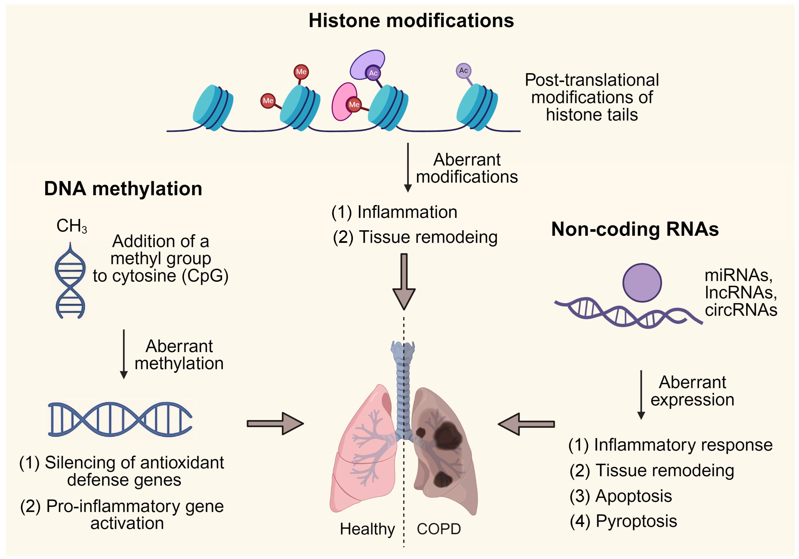
Figure 1. Epigenetic regulation in the pathogenesis of COPD. Created with BioRender.com, der, b. (2025) https://BioRender.com/oikyxt2 (accessed on 4 November 2025).
Ijms 26 11392 g001
Figure 2. Exercise modalities in COPD rehabilitation: outcomes and mechanisms.
Ijms 26 11392 g002
Figure 3. Exercise-induced epigenetic modulation in COPD. Created with BioRender.com, der, b. (2025) https://BioRender.com/my4a89c (accessed on 4 November 2025).
Ijms 26 11392 g003
Disclaimer/Publisher’s Note: The statements, opinions and data contained in all publications are solely those of the individual author(s) and contributor(s) and not of MDPI and/or the editor(s). MDPI and/or the editor(s) disclaim responsibility for any injury to people or property resulting from any ideas, methods, instructions or products referred to in the content.