Article

Modeling Human Protein Physical Interactions Involved in HIV Attachment In Silico

by
Vladimir S. Davydenko
,
Alexander N. Shchemelev
*,
Yulia V. Ostankova
,
Ekaterina V. Anufrieva
and
Areg A. Totolian
Saint Petersburg Pasteur Institute, 197101 St. Petersburg, Russia
*
Author to whom correspondence should be addressed.
Int. J. Mol. Sci. 2025, 26(22), 11209; https://doi.org/10.3390/ijms262211209
Submission received: 16 October 2025 / Revised: 8 November 2025 / Accepted: 18 November 2025 / Published: 20 November 2025
(This article belongs to the Section Molecular Microbiology)

Abstract

The human immunodeficiency virus (HIV) remains a major global health challenge. A promising therapeutic strategy involves identifying human proteins capable of physically blocking viral entry by interacting with key components of the HIV attachment system. To address this challenge systematically, we developed a computational pipeline for prioritizing protein–protein interaction and applied it to identify host proteins interacting with the viral glycoprotein gp120 and cellular receptors (CD4, CCR5, CXCR4, CCR2). Our approach combined large-scale interaction modeling using AlphaFold 3 with a comprehensive comparative analysis framework. We screened a panel of 55 candidate human proteins selected through integrated bioinformatics analysis. The pipeline incorporated model confidence assessment, quantitative contact analysis, and normalization against reference interactions to generate a robust ranking of candidates. Key findings reveal several important patterns. Chemokine CCL27 uniquely demonstrated high binding potential to both CCR5 co-receptor and viral gp120, suggesting its potential for dual-blockade capability. Analysis of natural ligand interactions with chemokine receptors showed marked disparity: CC-chemokine family members exhibited significantly greater binding capacity for CCR5 and CCR2 receptors compared to CXC-family ligand interactions with CXCR4. This binding imbalance may potentially drive selective viral pressure and influence tropism evolution during disease progression. We also identified potential interactions between HIV entry components and neuropeptides including PNOC and NPY, as well as various membrane receptors beyond classical coreceptors. Furthermore, cluster analysis revealed clear separation between receptor-type and ligand-type interactors, supporting the biological plausibility of our predictions. While acknowledging limitations related to model refinement, this study provides a systematically ranked set of candidate targets for HIV therapeutic development. Beyond identifying specific HIV interaction candidates, this study establishes a generalizable computational pipeline for the prioritization of protein–protein interaction in pathogen-host systems, effectively bridging large-scale modeling.

1. Introduction

The human immunodeficiency virus (HIV) remains one of the most significant global health challenges [1]. Despite advances in antiretroviral therapy (ART) targeting key stages of the viral replication cycle [2], infection persistence is largely driven by the emergence of drug-resistant viral mutations, reducing the efficacy of existing treatment regimens [3,4]. This underscores the urgent need to discover new bioactive compounds and therapeutic strategies to overcome current protocol limitations.
HIV research relevance extends beyond developing drugs targeting various viral cycle stages to identifying novel molecular targets and endogenous infection control mechanisms. This necessity stems from the extreme complexity of virus–human immune system interactions [5], which significantly constrains existing therapeutic strategies’ effectiveness [6,7]. A promising direction involves identifying endogenous innate immunity factors with antiviral activity. Studies demonstrate that certain human proteins, like chemokine CCL3, can inhibit infection by binding chemokine co-receptors and competitively blocking viral entry [8]. Crucially, such inhibition targets cellular rather than viral components. Key advantages of these endogenous inhibitors include their preexistence in the human body, partially understood physiological roles and safety profiles, and known interaction mechanisms with potential target proteins for some. A prominent example is APOBEC3G, which inhibits HIV-1 replication by inducing lethal hypermutation in newly synthesized viral DNA, thereby blocking reverse transcription and integration processes [9]. However, the systematic identification of such endogenous inhibitors among broad panels of host proteins remains challenging.
Current approaches for interaction discovery include public databases enable reconstruction of large-scale protein–protein interaction (PPI) networks. Such network analysis helps identify key protein hubs associated with HIV pathogenesis for further in-depth study [10]. However, these networks often reflect functional associations or co-expression rather than direct physical interaction, potentially yielding false-positive results. Even for physical interactions, network analysis typically lacks molecular mechanism details, spatial characteristics (complex conformation and stoichiometry), binding affinity, or process dynamics. Identifying physical interactions involving direct atom-atom contact between molecules is critical, as they underpin most fundamental cellular processes: signal transduction, enzymatic catalysis, viral particle assembly, and function blocking. Therefore, establishing physical contact between host proteins and viral proteins and/or established cellular cofactors opens avenues for designing targeted low-molecular-weight inhibitors or peptidomimetics that can specifically disrupt this interaction, thereby interrupting the pathogen’s life cycle. Simultaneously, this research direction deepens understanding of molecular pathogenesis mechanisms by revealing key target proteins and regulatory pathways involved in disease development. However, conventional PPI networks typically cannot distinguish between direct physical interactions and indirect functional associations.
The comprehensive experimental determination of physical interactions across the full spectrum of potential host–pathogen protein pairs presents substantial practical challenges due to the exceptional resource intensity involved. Such large-scale experimental screening demands immense investments of time, specialized equipment, and materials, rendering exhaustive experimental approaches impractical for initial candidate discovery. In this context, preliminary computer modeling offers a powerful strategy to optimize the search process through sophisticated in silico assessment. Modern computational methods can generate rationally justified priority target lists for subsequent experimental validation, thereby concentrating research efforts on the most promising candidates. Recent advances in structural bioinformatics, particularly the development of AlphaFold and related deep learning architectures [11], have revolutionized our ability to predict protein–protein interaction with unprecedented accuracy. These approaches enable detailed characterization of binding interface formation between viral and cellular proteins at atomic resolution, predicting spatial architecture of complexes and identifying specific amino acid residues that dominate binding energy contributions. The resulting structural insights provide critical foundation for multiple downstream applications, including virtual screening of low-molecular-weight compound libraries to identify candidates capable of sterically or allosterically disrupting pathogenic complex formation. However, the efficient processing and prioritization of hundreds of predicted complexes generated by these methods require specialized analytical pipelines. Thus, despite the power of computational modeling, a pressing need remains for integrated frameworks that can systematically evaluate and rank large volumes of predicted protein complexes to maximize research efficiency [12,13,14]. The primary aim of this study was to develop a computational pipeline for predicting protein–protein interaction and to apply it to identify human proteins capable of physically interacting with the viral glycoprotein gp120 and/or major HIV cellular co-receptors (CD4, CCR5, CXCR4, CCR2).

2. Results

The study proceeded through several key stages: initial validation of protein structures, modeling of binary interactions, quantitative analysis of interfaces, and finally, a comparative ranking of candidates based on a composite metric. The results of each stage are detailed below.
To validate the modeling workflow, we first generated single-protein structures for each host receptor and the HIV gp120 glycoprotein (five single models of HCBGPs/HRP and one CCR5-Δ32 model). The parameters of these models are presented in Table 1. The obtained protein structures visually correspond to models available in protein databases, while pTM and RS parameter values indicate close approximation to the native structures of the analyzed proteins.
The subsequent stage involved modeling interactions between receptors/coreceptors and the HRP (5 models). Model parameters are presented in Table 2.
In line with the expected limitations of our simplified modeling approach (Section 4.2.4), the generated models for biologically established reference complexes did not achieve high interface confidence values (ipTM < 0.6 for all HCBGP-HRP pairs), despite their plausible visual appearance and correspondence with known interaction patterns (Figure 1). This consistent result across all reference pairs confirms that absolute ipTM scores are not reliable discriminators in this specific screening context. It thereby reinforces the validity of our decision to employ a comparative analysis framework based on the composite area metric, which normalizes predicted interactions against these internal reference benchmarks.
We generated interaction models for each background protein paired with every candidate protein, resulting in a total of 275 protein–protein interaction models. Model confidence results are presented in Appendix A. Among these, 68 models showed reliable pTM but unreliable RS, while 37 models demonstrated both reliable RS and pTM but unreliable ipTM. Two models (ADRA2C, FPR3) exhibited reliable RS but unreliable ipTM and pTM.
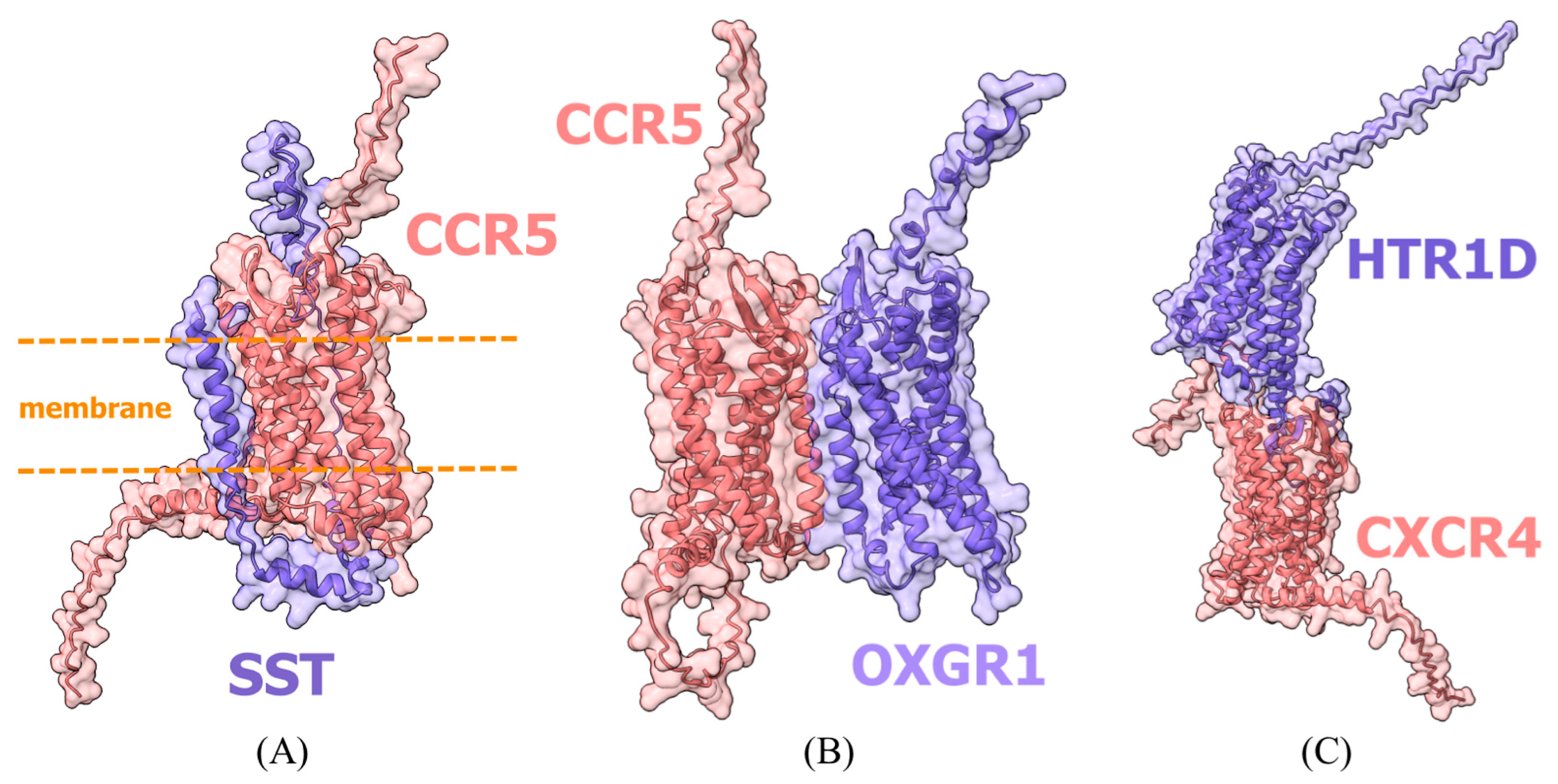
It should be noted that despite satisfactory confidence metrics for several predicted models, physical interactions between these proteins in the presented conformations remain unlikely due to steric constraints and electrostatic incompatibility (Figure 2). While these artifacts represent known limitations of the simplified docking system, we intentionally retained all models in subsequent analysis to demonstrate the pipeline’s ability to handle diverse prediction scenarios. A visual assessment of model plausibility was performed, though this evaluation necessarily remains subjective; its results are provided in Appendix B.
Having established a set of complex models, we proceeded to quantitatively characterize the protein-protein interfaces. We analyzed atomic contacts, steric clashes, and hydrogen bonds to derive a normalized interaction score (In) for each candidate complex. This provided a biophysical characterization of the interaction interfaces, complementing the structural confidence metrics. The resulting contact data for the background genes are presented in Table 3.
Contact data for HICGP interactions with each background protein and their normalized metrics are presented in Appendix C, Table A3 and Table A4.
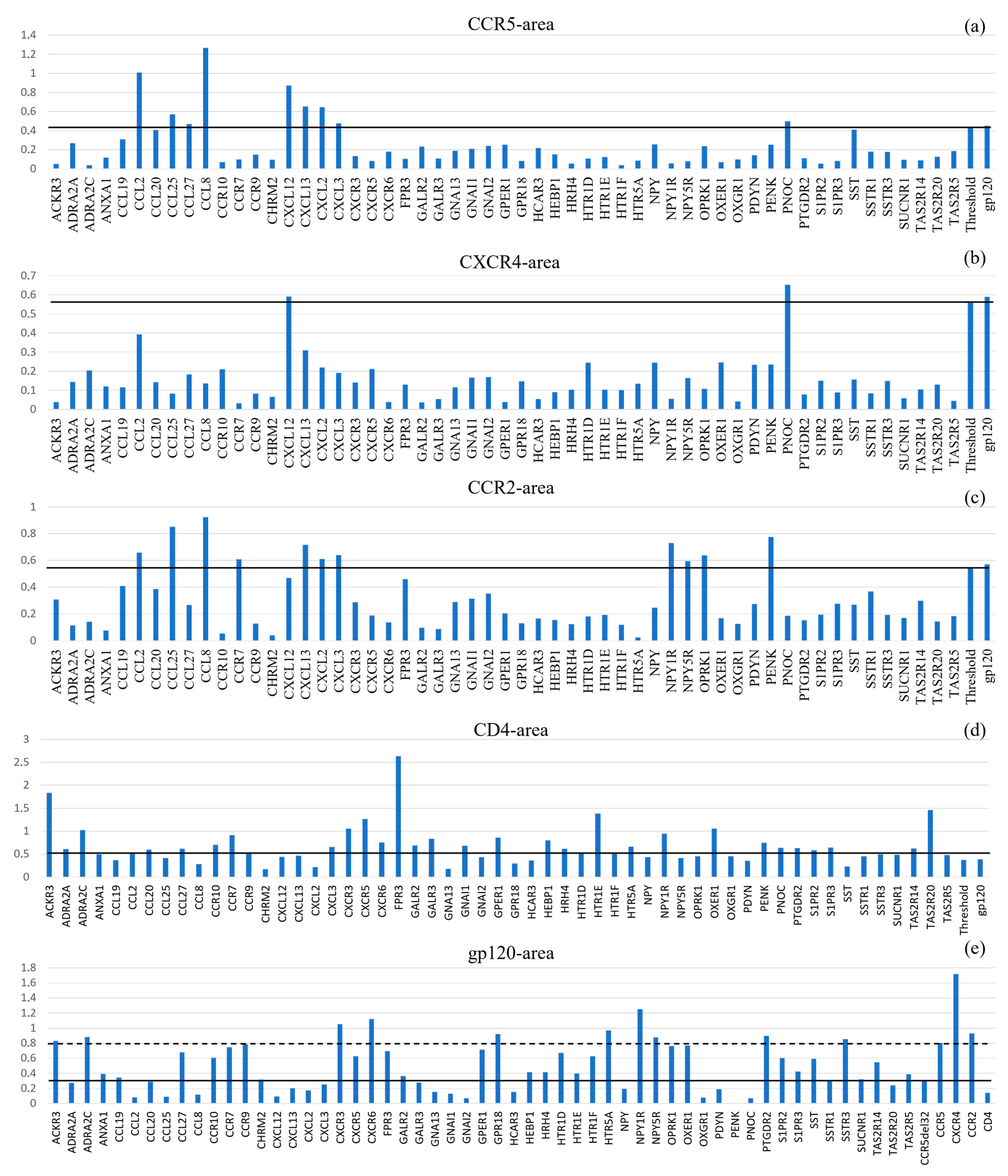
To integrate both the model confidence (RS) and the interface quality (In) into a single prioritization metric, we calculated the composite Normalized Interaction Area (A = RS × In). This approach allowed for the systematic ranking of all candidate interactions against our internal reference set (gp120-HCBGPs). The resulting landscape of interaction areas revealed distinct clusters and high-priority candidates (Figure 3, Table 4).
For standardization, interactions between HICGPs and background proteins were considered significant when their area values exceeded 95% of the area value for the corresponding background protein’s interaction with gp120 (excluding the gp120 interaction itself). Complete calculation data are presented in Appendix D and Appendix E. Table 4 presents HICGPs with the most significant area values.
Significant proteins based on calculated interaction area with CCR5 were: CCL2, CCL25, CCL27, CCL8, CXCL12, CXCL13, CXCL2, CXCL3, and PNOC. Significant proteins based on calculated interaction area with CXCR4 were: CXCL12 and PNOC. Significant proteins based on calculated interaction area with CCR2 were: CCL2, CCL25, CCL8, CCR7, CXCL13, CXCL2, CXCL3, NPY1R, NPY5R, OPRK1, and PENK.
Analysis of CD4-gp120 interactions reveals a limited number of atomic contacts and insignificant binding surface area. Consequently, the diagnostic value of this complex for comparative analysis is substantially reduced, as most investigated HICGPs demonstrate area values comparable to or exceeding that of the CD4+gp120 system. Similarly to the CD4+gp120 complex, the area in the gp120+CCR5-Δ32 system also proved insufficient for use as a threshold to exclude HICGPs with low significance. When considering the interaction area threshold of CCR5 with gp120, the candidate list is as follows: CCL27, CCR7, NPY1R, NPY5R, OPRK1, ACKR3, ADRA2C, CCR10, CCR9, CXCR3, CXCR5, CXCR6, FPR3, GPER1, HTR1D, HTR1F, HTR5A, OXER1, PTGDR2, S1PR2, SSTR3, TAS2R14, GPR18, and SST.
The primary proteins interacting with gp120 are receptors, a finding which is further supported by cluster analysis results presented in Table 5. A clear separation into receptor and ligand clusters is evident. Particular attention should be given to HICGP interaction models with HCBGPs/HRP that demonstrated area values exceeding established thresholds. No candidate proteins interacting with all background human proteins analyzed in this study were identified. HICGPs interacting with three HCBGPs (CCR5, CXCR4, CD4) were identified: PNOC and CXCL12. HICGPs interacting with one of the main coreceptors (CCR5 or CXCR4) and one or two other HCBGPs (CCR2 and/or CD4) were identified: CCL2, CCL8, CCL25, CCL27, CXCL13, CXCL3, and CXCL2. Interaction models are presented in files in the Supplementary Materials.
Among HICGPs, only CCL27 showed area values above thresholds for interaction models with both CCR5 and gp120 (Figure 4). Interaction models are presented in files in the Supplementary Materials.

3. Discussion

3.1. Overview of the Computational Approach and Key Findings

Top-ranked HICGPs based on comparative Area analysis (values exceeding operational thresholds for prioritization are indicated in bold). This study aimed to conduct an in silico screen of a panel of 55 HICGPs to identify molecules potentially capable of modulating a key stage of the HIV life cycle, namely the interaction between viral glycoprotein gp120 and cellular receptors [16]. The application of the AlphaFold 3 algorithm enabled the reconstruction and comprehensive analysis of 275 molecular complexes, resulting in the identification of both expected and previously undescribed potential targets for therapeutic intervention in HIV infection.

3.2. Validation of the Pipeline: Separation of Ligands and Receptors

The clear separation of candidate proteins into distinct receptor-type and ligand-type clusters, as revealed by our k-means cluster analysis (Table 5), provides internal validation for our computational pipeline and prioritization strategy. This recapitulation of fundamental biological categories demonstrates that our comparative framework, based on the composite Area metric, effectively captures biologically relevant features of protein–protein interaction. The clear dichotomy suggests that the predicted interaction models respect basic biological principles, where ligands (such as chemokines and neuropeptides) and receptors occupy distinct functional and structural niches, even within the simplified in silico environment. This successful separation reinforces the biological plausibility of the top-ranking candidates identified by our screening approach and supports the robustness of our method in distinguishing between different modes of potential interaction with the HIV entry machinery.

3.3. Chemokine Ligands: Expected and Discordant Results

As anticipated based on their known biological functions, C-C family chemokines (CCL2, CCL8, CCL25, CCL27) and C-X-C family chemokines (CXCL12, CXCL13, CXCL2, CXCL3) demonstrated high interaction potential with their natural receptors CCR5 and CCR2 in our models. Although normalized contact parameters for most did not exceed those of the gp120-coreceptor complexes, their binding capacity suggests these chemokines may act as natural competitive antagonists, potentially blocking viral glycoprotein binding sites.
Of particular note is the case of chemokine CCL2. Our model predicts high-affinity binding to CCR5, suggesting a potential for direct competitive inhibition of viral entry. This finding, however, appears to contradict experimental studies reporting that CCL2 can enhance HIV replication in vivo and ex vivo [17,18]. We propose that this discrepancy underscores the distinction between a direct physical interaction, captured by our structural models, and a protein’s net biological effect within a complex physiological environment.
The primary physiological role of CCL2 is the chemotaxis of monocytes and other immune cells to sites of inflammation [19]. This recruitment significantly expands the pool of target cells (e.g., CD4+ T cells, macrophages) available for HIV infection, an indirect proviral effect that likely dominates the net outcome in many experimental and physiological contexts. Thus, the in silico prediction and experimental observations can be reconciled within a dual-activity framework: CCL2 may possess an intrinsic, direct antiviral potential via coreceptor blockade (as predicted by our model), which is masked in vivo by its potent, indirect proviral effect via target cell recruitment.
This case highlights a critical principle for interpreting computational screens: a predicted physical interaction signifies mechanistic potential, but the net biological outcome is determined by the broader cellular and systemic context [20].
Analysis revealed a significant disproportion: the relative number of ligands interacting with CXCR4 was substantially lower than with CCR5/CCR2. These findings suggest that within the employed model, CC-type chemokines exhibit more pronounced inhibitory activity against viral utilization of CCR5 co-receptor. The observed binding disparity is consistent with the hypothesis that an imbalance in available natural ligands could contribute to selective pressures influencing coreceptor switching and the emergence of CXCR4-tropic variants in later stages of infection [18,21].

3.4. Potential gp120 Interactions with Non-Canonical Receptors

Beyond the classical co-receptors, our modeling suggests the capacity of the viral glycoprotein gp120 for direct interaction with a broad spectrum of cellular membrane receptors. Although overall prediction reliability was moderate—as expected given gp120’s high conformational plasticity and the challenges of predicting its binding sites—our comparative analysis identified two significant subgroups of potential interactors.
The first group comprises chemokine superfamily receptors (CCR10, CCR7, CCR9, CXCR3, CXCR5, CXCR6), which demonstrated contact quantities with gp120 comparable to reference coreceptors. These findings align with publications suggesting that some of these receptors may serve as alternative or supplementary viral entry portals into specific cell types [22,23]. The specific targeting of this receptor class by gp120 may represent a viral adaptation to broaden cellular tropism.
The second, more diverse group consisted of various neuroreceptors and other membrane proteins (HRH4, HTR1D, HTR1F, HTR5A, NPY1R, NPY5R, OPRK1, OXER1, OXGR1, PTGDR2, S1PR2, S1PR3, SSTR1, SSTR3, SUCNR1). For several of these, existing data suggest possible associations with HIV-associated neuropathologies [24], making them promising targets for further investigation in the context of neuroinvasion and neuropathogenesis.
Somatostatin (SST) merits particular attention in this context, as its expression level has been reported to correlate with HIV progression [25,26], although earlier studies refuted its substantial role [27], indicating ambiguity in existing data that warrants further clarification.
The G-protein family (GNAI1, GNAI2, GNA13) exhibited high structural confidence values (pTM) but low contact quantities with coreceptors, which is expected since they typically interact with receptor cytoplasmic domains. Their potential influence on infection is likely mediated through complex intracellular signaling cascades and cannot be adequately assessed within our binary interaction modeling methodology [28].

3.5. Hypothesis-Generating Predictions for Neuropeptides

The high ranking of several neuropeptides (PNOC, NPY, PDYN, PENK) among the candidate interactors warrants specific discussion. Their prioritization should be interpreted with particular caution due to inherent methodological considerations. The small size and inherent structural flexibility of neuropeptides pose a particular challenge for reliable modeling using static docking approaches, potentially allowing for multiple conformations and leading to overestimated confidence in some binding poses. Indeed, some models showed potential binding to sterically inaccessible sites, such as intracellular domains. Therefore, while these candidates ranked highly in our screen, they should be classified as the most speculative predictions, serving primarily to generate hypotheses for rigorous experimental validation.
Notably, the literature analysis provides indirect support for the potential biological relevance of these systems in the context of HIV infection. For instance, anterior cingulate cortex samples from Patients with HIV showed decreased PDYN (prodynorphin) gene mRNA levels alongside increased OPRK1 (kappa-opioid receptor) mRNA expression compared to controls [24]. We hypothesize that reduced PDYN expression may represent a compensatory mechanism aimed at limiting monocyte recruitment and mitigating neuroinflammatory processes, while enhanced OPRK1 expression might be associated with attempts to modulate proinflammatory signaling pathways. Furthermore, increased neuropeptide Y (NPY)-like immunoreactivity has been observed in the cerebrospinal fluid of Patients with HIV, suggesting a potential link to HIV encephalopathy [29]. Thus, while the direct physical interactions predicted by our models remain highly speculative, the involved neuropeptide systems appear to be engaged in the host response to HIV infection, particularly within the nervous system.

3.6. Limitations of the Study

While our computational pipeline provides a systematic approach for prioritizing protein interactions, several limitations should be acknowledged. First, the low ipTM scores observed for biologically validated complexes are a direct consequence of our simplified binary modeling strategy, which traded atomic-level refinement for screening throughput. This inherent trade-off is why our comparative analysis framework, rather than absolute confidence scores, forms the core of the prioritization pipeline. Second, our models represent simplified binary interactions without key biophysical contexts such as explicit lipid membranes, gp120 glycosylation, or physiological ionic conditions. Third, the operational thresholds and confidence intervals used for candidate selection were derived from a comparative analysis with biologically verified reference interactions rather than from rigorous statistical distributions of null models. While this practical approach allowed for large-scale prioritization, it lacks a formal statistical foundation. Finally, and most importantly, all predictions—particularly those involving neuropeptides and novel receptor interactions—require experimental validation (e.g., SPR, BLI, cellular assays) before any firm biological conclusions can be drawn. We reiterate that the term “significant” throughout this manuscript refers specifically to candidates that surpassed our operational, comparative thresholds for prioritization within this computational screen. These thresholds provide a systematic ranking for guiding future research but do not constitute statistical or biological validation of the interactions.

3.7. Concluding Remarks

This in silico study successfully demonstrates a generalizable computational pipeline for the prioritization of protein–protein interaction in pathogen-host systems. By applying this framework to HIV-1 attachment, we have systematically narrowed a broad panel of candidates to a focused set of high-priority targets. Our results not only recapitulate known biology, validating our approach, but also generate novel and sometimes unexpected hypotheses regarding viral engagement with chemokine and neuromodulatory systems. The predictions presented here, most notably the dual-binding candidate CCL27, establish a robust and prioritized foundation for guiding future experimental efforts aimed at validating these interactions and exploring their therapeutic potential. This work underscores the power of integrated computational modeling to illuminate complex host–pathogen interaction landscapes.

4. Materials and Methods

4.1. Materials for Modeling Physical Interactions

The analysis utilized a panel of 55 candidate proteins selected using our previously developed integrated scoring system [30]. This system weighted gene-disease associations based on tissue-specific expression levels, subcellular localization, biological pathway annotations, and participation in relevant biological processes. The study included key cellular receptors associated with HIV entry (CCR5, CXCR4, CCR2, CD4) [31,32] and the viral glycoprotein gp120, which mediates initial virus-target cell binding [16]. The scoring algorithm assigned greater weight to associations with the primary co-receptors CCR5 and CXCR4.
Primary amino acid sequences were obtained from the NCBI database for: HIV-interacting candidate gene proteins, HICGPs (ACKR3, ADRA2A, ADRA2C, ANXA1, CCL19, CCL2, CCL20, CCL25, CCL27, CCL8, CCR10, CCR7, CCR9, CHRM2, CXCL12, CXCL13, CXCL2, CXCL3, CXCR3, CXCR5, CXCR6, FPR3, GALR2, GALR3, GNA13, GNAI1, GNAI2, GPER1, GPR18, HCAR3, HEBP1, HRH4, HTR1D, HTR1E, HTR1F, HTR5A, NPY, NPY1R, NPY5R, OPRK1, OXER1, OXGR1, PDYN, PENK, PNOC, PTGDR2, S1PR2, S1PR3, SST, SSTR1, SSTR3, SUCNR1, TAS2R14, TAS2R20, TAS2R5); HIV coreceptor background gene proteins, HCBGPs (CCR5, CXCR4, CCR2, CD4); and the HIV reference protein, HRP (gp120). The complete sequence list appears in Appendix F.
We selected a CCR5-tropic gp120 variant for modeling, with its amino acid sequence corresponding to the HIV subtype A6 isolate predominant in Russia, ensuring relevance to regional epidemiological characteristics. The analysis incorporated the Δ32 nonsense mutation (CCR5-del32), which causes a 32-base pair deletion in the CCR5 gene resulting in a truncated, non-functional protein. Physical interaction modeling included this structurally modified CCR5 protein form. Computational analysis employed an in silico reconstructed CCR5 amino acid sequence featuring the specific deletion, enabling assessment of structural alterations on protein–protein interaction interfaces.

4.2. Physical Protein–Protein Interaction Modeling

To address the study objectives, we reconstructed three-dimensional structures for all specified proteins. Subsequent interaction modeling between HICGPs and HCBGPs/HRP employed the AlphaFold 3 algorithm (https://alphafoldserver.com/, developed by Google DeepMind, London, UK, accessed on 17 October 2025), recognized as one of the most effective tools for in silico protein structure reconstruction. This algorithm demonstrates high accuracy in predicting individual polypeptide structures and enables heterodimeric protein complex prediction, albeit with slightly reduced confidence compared to monomeric predictions [11].
The modeling results enabled candidate protein filtering, selecting only those forming statistically significant complexes with target structures for further investigation. Constructed models included confidence metrics for each protein complex. We utilized the highest-confidence model for subsequent analysis. To standardize the research approach, molecular modeling assessed interactions between HCBGPs and the viral glycoprotein gp120 (HRP). The same in silico approach analyzed the CCR5Δ32 variant receptor containing the Δ32 deletion, which served to establish computational validation thresholds.

4.2.1. Model Confidence Assessment

The initial analysis stage involved evaluating model reliability. We assessed output data quality using predicted template modeling (pTM) and interface predicted template modeling (ipTM) metrics. These parameters were evaluated both individually and as components of the composite ranking score (RS), calculated according to Equation (1).
R S = 0.8 × i p T M + 0.2 × p T M + 0.5 × d 100 × N c ;
wherein RS—ranking score;
pTM—predicted template modeling;
ipTM—interface predicted template modeling;
d—disorders;
Nc—number of clusters.
According to the AlphaFold manual, a pTM score below 0.5 generally indicates that the predicted model may not closely resemble the true structure. Similarly, ipTM values below 0.6 suggest potentially unreliable modeling of the protein-protein interface region. The RS serves as a unified metric, where values ≥ 0.5 indicate models with acceptable reliability [11]. These RS values were utilized for all subsequent analyses.
As anticipated for highly flexible and context-dependent systems like the gp120 glycoprotein, which requires CD4 binding for its mature co-receptor interaction state [15,33], even reference complexes with established biological validity (e.g., gp120-CD4, gp120-CCR5) did not achieve high ipTM scores (>0.6) in our simplified binary modeling approach. This expected outcome directly reflects our methodological choice to prioritize large-scale screening over modeling complex biophysical contexts and underscores the necessity of our comparative framework, which relies on relative rather than absolute confidence metrics for candidate prioritization.

4.2.2. Analysis of Contact Quantification in Predicted Models

The next stage involved analyzing contacts in the obtained structures. Using ChimeraX software v1.10.1, developed by the Resource for Biocomputing, Visualization, and Informatics (San Francisco, CA, USA) [34], we analyzed three categories: contacts, clashes, and hydrogen bonds. The total number of protein interactions (Interaction) was calculated as follows (Equation (2)):
I = N c t N c + N h ;
wherein I—interactions;
Nct—numbers of contacts;
Nc—numbers of clashes;
Nh—numbers of H-bonds.
This metric penalizes non-physical steric clashes while rewarding specific favorable interactions (hydrogen bonds), providing a more biophysically meaningful estimate of interface quality compared to our initial formulation.
For HICGP interactions with background proteins (HCBGPs), the obtained data were normalized against the interaction count (In) between that specific background protein and gp120. For HICGP interactions with gp120 (HRP), normalization was performed using the gp120-CCR5Δ32 interaction count, representing the minimal observed interaction value. It should be noted that gp120 affinity for CCR5 increases due to conformational changes induced by viral protein interaction with CD4 [15,33]. However, reliable computational modeling of such three-protein complexes remains challenging [11]. The per-residue confidence scores (pLDDT) for all predicted models are provided in the Supplementary Materials. While these data offer valuable granular insight into local model quality, they were not incorporated into our primary ranking pipeline due to the scale of the study. Our comparative framework was designed to utilize global and interface-level metrics (pTM, ipTM, RS) for high-throughput prioritization. The provided pLDDT data are intended for researchers who wish to perform a more detailed, residue-level assessment of the specific models of interest identified by our screen.

4.2.3. Comprehensive Model Comparison Based on Derived Parameters

For comprehensive model comparison, the resulting set of models was plotted on a coordinate plane with axes corresponding to RS and In. The integrative metric, accounting for both parameters, was selected as the area (A) of the rectangle formed between the coordinate axes and the perpendicular distances from the data point to these axes (Equation (3)).
A = R S × I n ;
wherein A—area;
RS—ranking score;
In—normalized Interaction.
To establish a robust operational threshold for candidate prioritization, we defined a cutoff at 95% of the Area value for each background protein’s interaction with gp120. This threshold was selected based on established practices in biological screening and aligns with the standard statistical significance level (p < 0.05) widely adopted in biomedical sciences [35]. This approach creates a normalized, comparative benchmark derived from the internal reference set of known biological interactions (gp120-HCBGPs), rather than relying on arbitrary absolute thresholds. By using the 95th percentile, we aim to select candidates whose composite interaction score (A = RS × In) is nearly equivalent to or exceeds that of our positive controls, ensuring a high degree of confidence for subsequent experimental validation. For interactions with gp120 itself, we analyzed all Area values exceeding that of the gp120-CCR5Δ32 interaction, representing the minimal observed interaction in our reference set. It is important to emphasize that these thresholds serve specifically for comparative ranking within this screen; they indicate a candidate’s relative promise for further experimental validation, not its absolute biological significance.
Concurrently, all areas for HICGP interactions with background proteins underwent cluster analysis to identify patterns in HICGP involvement during viral entry. This analysis employed k-means clustering in IBM SPSS Statistics 27 software. Thus, the comparative analysis methodology involved data normalization to a unified scale relative to control interactions and statistical significance evaluation of identified contacts.

4.2.4. Limitations of the Computational Approach

The methodology employed several intentional simplifications to enable a computationally tractable large-scale screening pipeline. The primary goal was the comparative ranking of a large set of candidate interactions, not the production of refined, atomic-resolution structural models. Consequently, to ensure feasibility and consistency across the screen, all AlphaFold models were used in their raw, unrefined output state. This means we deliberately abstained from:
-
Post-processing refinement using tools like HADDOCK or RosettaDock, which would be computationally prohibitive at this scale but are essential for obtaining stable, energetically minimized complexes.
-
Incorporation of key biophysical contexts, such as explicit lipid membranes, glycosylation of gp120, or physiological ionic conditions. Our models represent simplified binary interactions in vacuo.
These choices were necessary for feasibility but mean that the models should be viewed as rough scaffolds identifying potential interaction interfaces. The ranking provides a priority list for future, more focused experimental and computational validation.

5. Conclusions

The computational modeling and comparative ranking pipeline developed in this study enabled the systematic identification and prioritization of human proteins with the potential to physically interact with key components of the HIV attachment system. Our analysis highlights several promising candidates and mechanistic insights. Notably, the viral glycoprotein gp120 demonstrated a predicted capacity to interact with a broad spectrum of membrane receptors beyond the classical coreceptors, suggesting a more complex landscape of viral entry portals. It is important to note that the presented models, while useful for comparative ranking, represent simplifications that do not incorporate key biophysical contexts such as explicit lipid membranes, gp120 glycosylation, or physiological ionic conditions. Furthermore, we observed marked disparity in the binding potential of natural chemokine ligands, which may influence coreceptor selectivity and viral tropism. The identification of specific neuropeptides, such as PNOC and NPY, as potential interactors with the HIV entry machinery opens new avenues for investigating their role in infection and pathogenesis. Most importantly, the case of CCL27 emerged as a particularly compelling candidate, with a unique predicted capability for dual interaction with both the CCR5 coreceptor and viral gp120. Collectively, these findings provide a systematically ranked set of candidate targets and establish a robust computational framework for future studies of pathogen-host protein interactions, guiding subsequent in-depth bioinformatic analyses and prioritizing targets for experimental validation.

Supplementary Materials

The following supporting information can be downloaded at https://www.mdpi.com/article/10.3390/ijms262211209/s1.

Author Contributions

Conceptualization, V.S.D. and Y.V.O.; methodology, V.S.D. and A.N.S.; software, V.S.D. and E.V.A.; validation, V.S.D. and A.N.S.; formal analysis, V.S.D.; investigation, V.S.D.; writing—original draft preparation, V.S.D.; writing—review and editing, A.N.S. and Y.V.O.; visualization, V.S.D. and E.V.A.; supervision, A.A.T.; project administration, Y.V.O. and A.A.T. All authors have read and agreed to the published version of the manuscript.

Funding

This research was supported by the Russian Science Foundation grant 24-25-00479 (Assessing the potential significance of host genetic factors in infection with human immunodeficiency virus infection and disease progression https://rscf.ru/project/24-25-00479/, accessed on 17 October 2025).

Institutional Review Board Statement

Not applicable.

Informed Consent Statement

Not applicable.

Data Availability Statement

The original contributions presented in this study are included in the article/Supplementary Materials. Further inquiries can be directed to the corresponding author.

Conflicts of Interest

The authors declare no conflicts of interest.

Abbreviations

The following abbreviations are used in this manuscript:
HICGPsHIV-interacting candidate gene proteins
HCBGPsHIV coreceptor background gene proteins
HRPHIV reference protein
HIVhuman immunodeficiency virus
gp120glycoprotein 120
CCR5C-C chemokine receptor type 5
CXCR4C-X-C chemokine receptor type 4
CD4cluster of differentiation 4
NCBINational Center for Biotechnology Information
PPIprotein–protein interaction
pTMpredicted template modeling
ipTMinterface predicted template modeling
RSranking score

Appendix A

Table A1. Model confidence metrics according to AlphaFold. Values exceeding the reliability threshold are indicated in bold.
HICGPHCBGPs/HRP
CCR5CXCR4CCR2CD4gp120
ipTMpTMScoreipTMpTMScoreipTMpTMScoreipTMpTMScoreipTMpTMScore
ACKR30.150.460.280.160.460.290.260.50.410.240.470.380.30.630.41
ADRA2A0.180.410.380.160.40.360.140.390.370.190.390.380.150.480.37
ADRA2C0.140.410.330.170.420.370.140.40.350.370.420.530.170.510.38
ANXA10.130.480.250.250.540.370.180.490.310.120.450.250.540.650.6
CCL190.530.70.680.180.620.370.510.650.670.340.570.50.230.460.36
CCL20.620.750.740.370.660.540.610.70.750.130.540.320.110.430.25
CCL200.570.740.710.270.650.450.60.690.760.240.560.420.150.470.29
CCL250.630.720.790.170.580.390.550.670.740.140.510.360.140.410.32
CCL270.540.730.690.190.610.40.460.650.650.30.560.470.350.450.47
CCL80.670.770.780.290.640.470.630.710.770.120.520.320.110.430.26
CCR100.20.480.350.210.430.350.130.420.30.190.430.330.340.610.46
CCR70.130.430.280.130.420.280.150.420.310.220.440.350.350.630.46
CCR90.160.460.30.140.440.280.120.420.280.170.410.30.340.610.43
CHRM20.150.410.350.130.390.330.140.390.350.170.380.360.150.470.36
CXCL120.520.730.670.660.760.790.360.660.540.260.510.430.120.440.27
CXCL130.510.720.670.350.650.540.50.680.670.110.550.320.150.450.3
CXCL20.540.730.690.220.630.430.550.690.720.160.540.360.10.440.26
CXCL30.420.680.590.320.650.510.570.70.740.110.540.320.10.420.26
CXCR30.130.430.290.140.430.30.130.410.290.210.450.350.280.60.42
CXCR50.170.460.30.120.430.280.140.430.30.220.440.360.40.60.51
CXCR60.110.440.260.120.430.270.140.440.30.240.470.370.470.640.56
FPR30.240.50.370.210.450.330.340.460.450.480.490.570.490.630.56
GALR20.120.460.30.20.460.360.120.440.320.190.450.360.190.460.36
GALR30.150.460.30.130.440.290.150.440.320.210.440.360.130.440.29
GNA130.610.650.680.580.630.650.590.630.680.140.390.270.160.470.26
GNAI10.740.690.790.710.670.760.670.670.740.550.40.590.150.420.23
GNAI20.750.70.790.720.680.770.680.670.750.340.50.450.130.430.21
GPER10.280.450.410.140.420.290.50.60.630.190.440.340.210.540.34
GPR180.140.480.260.20.460.330.140.440.30.180.440.310.350.580.44
HCAR30.160.440.310.120.430.280.180.440.350.180.430.330.180.560.33
HEBP10.290.610.420.250.570.370.160.530.320.120.480.280.260.540.33
HRH40.130.440.290.150.440.320.120.410.30.170.40.320.130.510.31
HTR1D0.170.470.330.180.440.340.140.440.330.180.410.340.270.540.42
HTR1E0.170.460.320.140.440.30.180.460.350.370.530.510.210.560.36
HTR1F0.130.440.280.180.460.320.140.440.30.250.420.380.220.560.35
HTR5A0.170.470.330.130.420.30.160.430.340.250.450.420.310.580.44
NPY0.20.650.430.370.670.510.20.60.450.160.560.390.110.480.34
NPY1R0.160.450.310.180.460.330.340.460.470.230.430.380.330.580.46
NPY5R0.150.420.320.170.420.340.220.420.390.160.380.320.310.560.47
OPRK10.230.490.380.150.430.30.230.440.390.160.40.310.270.590.41
OXER10.150.450.320.210.440.360.130.420.30.220.440.380.290.580.42
OXGR10.20.50.330.130.450.260.220.480.360.170.420.30.110.590.24
PDYN0.210.510.470.230.510.510.150.480.450.120.470.40.170.360.45
PENK0.180.510.460.370.530.630.30.50.580.490.520.70.080.360.39
PNOC0.150.570.40.550.630.720.150.530.420.310.530.520.120.410.36
PTGDR20.140.450.290.170.470.320.160.450.330.210.460.360.140.570.29
S1PR20.240.490.370.180.470.320.220.480.360.20.440.340.20.580.33
S1PR30.190.470.340.170.430.320.290.50.440.180.430.330.240.580.38
SST0.250.660.520.210.620.480.220.610.490.140.550.40.310.530.56
SSTR10.190.440.350.160.420.320.190.410.360.170.410.330.240.560.39
SSTR30.180.440.350.250.430.410.160.40.350.20.410.380.260.550.43
SUCNR10.170.490.290.180.490.310.250.510.380.20.470.330.290.660.39
TAS2R140.270.530.380.120.450.260.220.450.350.210.440.330.330.620.41
TAS2R200.120.460.240.20.450.310.140.460.270.280.50.380.270.620.35
TAS2R50.150.480.270.130.460.270.120.450.260.220.440.330.150.520.25

Appendix B

Table A2. Manual structural classification of predicted protein complexes. Structural classification symbols: c—contact; ?—ambiguous; *—membrane penetration; **—side-by-side receptors; ***—vertical stacking.
ProteinsVisual Assessment
CCR5CXCR4CCR2CD4gp120
ACKR3*******cc
ADRA2A******?c
ADRA2C*******c
ANXA1ccccc
CCL19ccccc
CCL2ccccc
CCL20ccccc
CCL25ccccc
CCL27ccccc
CCL8ccccc
CCR10********c
CCR7******?c
CCR9*******c
CHRM2*********c
CXCL12ccc?c
CXCL13ccccc
CXCL2ccccc
CXCL3ccccc
CXCR3******cc
CXCR5*******c
CXCR6*******c
FPR3******?c
GALR2*******c
GALR3******cc
GNA13ccccc
GNAI1ccc*c
GNAI2ccccc
GPER1*******c
GPR18*******c
HCAR3*******c
HEBP1ccccc
HRH4*******c
HTR1D********c
HTR1E*******c
HTR1F*******c
HTR5A******cc
NPYccccc
NPY1R******cc
NPY5R*******c
OPRK1********c
OXER1******cc
OXGR1*******c
PDYNcc??c
PENKcc*c?
PNOC**ccc
PTGDR2*******c
S1PR2*******c
S1PR3*******c
SST*cc*c
SSTR1*******c
SSTR3******cc
SUCNR1*******c
TAS2R14*******c
TAS2R20******?c
TAS2R5*******c

Appendix C

Table A3. Contact, clash, and hydrogen bond metrics for HICGP interactions with HCBGPs/HRP, normalized against background interactions.
ProteinsContactsClashesHydrogen BondsTotal
CCR5CXCR4CCR2CD4gp120CCR5CXCR4CCR2CD4gp120CCR5CXCR4CCR2CD4gp120CCR5CXCR4CCR2CD4gp120
ACKR34555154236265465322600513164149154217255
ADRA2A1671607673991720124987032158147647292
ADRA2C2525099102337152151857140312252028487292
ANXA11131225394831113496310345105119528982
CCL1910112911930115716604821231010211512533121
CCL230227718579402325127226157223052671807440
CCL20123112996812663481011794912811610464125
CCL2515480236673567151601451510162782365235
CCL2714117178601895122220161081131521698459182
CCL83731062613853335263324511563641062464056
CCR1050223409716151638140130719452203796166
CCR7784548513620635982194216277942403117204
CCR9127115105822271811114111513141101099581230
CHRM2628931221146176294001660722521111
CXCL123042551974745307222518273142922751784644
CXCL132192142146891121910810111515542182102196585
CXCL2211192172259181191107711322101881742783
CXCL3180137166112142875212198161218113817792123
CXCR3114212229137318134134328118226102172203136316
CXCR56639013516315471168111224161361278128158155
CXCR61905799922563957817503813156529592252
FPR3651512172201593131318111766863145210208156
GALR218838658812619254661227175376286127
GALR389725811212297213713155816857104120
GNA13596187317022522565166265873074
GNAI15471754769001155913665980875270
GNAI261698739410010071210416881964342
GPER1138546412028210611226913691374966114265
GPR187917895452957207443052212721639043264
HCAR316570109506616114389322015872974958
HEBP186961031581529953343324108090100129158
HRH443120909019236962715434411198587169
HTR1D73284125782017331491161412117226511271201
HTR1E9313112511814678155111339487126113122139
HTR1F3016983652383525725304212301178260225
HTR5A6020916652707470212430819571651671277
NPY1381751086668111021816116251331761125072
NPY1R41673641093580658730121210154263318112343
NPY5R58205330592464293242412153135517831358235
OPRK114313939266232101067110631011213913233566234
OXER153286129135246444181626010361149252114125231
OXGR1696077704142542100226658726841
PDYN651721223957216617412923671681254053
PENK11313831341021145101210680123137274480
PNOC29832690512334204001527541279333915524
PTGDR2999510184491138891070334586909679389
S1PR2321991148024622855262222103217311177230
S1PR3551071359713449179834100145410212888140
SST186125112301292013858118811217712011226133
SSTR1119982307211078251111365111159621062100
SSTR3116139122532721015110296915711213311258250
SUCNR1788098681016106530003572709266103
TAS2R14521711928817613019513162255214717585168
TAS2R2012616211517384101110542355611815411017386
TAS2R51747016571207219211116101541546114565195
Table A4. Interaction values for HICGPs with HCBGPs/gp120 relative to background interactions.
ProteinsNormalized Interaction Values
CCR5CXCR4CCR2CD4gp120
ACKR30.183040.133510.751224.822222.02381
ADRA2A0.705360.400540.312201.600000.73016
ADRA2C0.111610.550410.409761.933332.31746
ANXA10.468750.324250.253661.977780.65079
CCL190.455360.313350.609760.733330.96032
CCL21.361610.727520.878051.644440.31746
CCL200.571430.316080.507321.422220.99206
CCL250.723210.212531.151221.155560.27778
CCL270.678570.460490.409761.311111.44444
CCL81.625000.288831.200000.888890.44444
CCR100.200890.599460.180492.133331.31746
CCR70.352680.114441.965852.600001.61905
CCR90.491070.297000.463411.800001.82540
CHRM20.267860.196190.121950.466670.88095
CXCL121.303570.749320.868291.022220.34921
CXCL130.973210.572211.068291.444440.67460
CXCL20.937500.512260.848780.600000.65873
CXCL30.808040.376020.863412.044440.97619
CXCR30.455360.468660.990243.022222.50794
CXCR50.272320.757490.624393.511111.23016
CXCR60.696430.141690.463412.044442.00000
FPR30.281250.395101.024394.622221.23810
GALR20.781250.100820.302441.911111.00794
GALR30.361610.185290.278052.311110.95238
GNA130.276790.177110.424390.666670.58730
GNAI10.263390.217980.424391.155560.55556
GNAI20.303570.220710.468290.955560.33333
GPER10.611610.133510.321952.533332.10317
GPR180.321430.444140.439020.955562.09524
HCAR30.705360.196190.473171.088890.46032
HEBP10.357140.245230.487802.866671.25397
HRH40.183040.324250.414631.933331.34127
HTR1D0.321430.722070.546341.577781.59524
HTR1E0.388390.343320.551222.711111.10317
HTR1F0.133930.318800.400001.333331.78571
HTR5A0.254460.449590.078051.577782.19841
NPY0.593750.479560.546341.111110.57143
NPY1R0.187500.171661.551222.488892.72222
NPY5R0.245540.485011.526831.288891.86508
OPRK10.620540.359671.634151.466671.85714
OXER10.218750.686650.556102.777781.83333
OXGR10.294640.158040.351221.511110.32540
PDYN0.299110.457770.609760.888890.42063
PENK0.549110.373301.336591.06667-
PNOC1.245540.907360.443901.222220.19048
PTGDR20.383930.245230.468291.755563.08730
S1PR20.142860.471390.541461.711111.82540
S1PR30.241070.277930.624391.955561.11111
SST0.790180.326980.546340.577781.05556
SSTR10.513390.261581.024391.377780.79365
SSTR30.500000.362400.546341.288891.98413
SUCNR10.321430.190740.448781.466670.81746
TAS2R140.232140.400540.853661.888891.33333
TAS2R200.526790.419620.536593.844440.68254
TAS2R50.687500.166210.707321.444441.54762

Appendix D

Table A5. HCBGP area thresholds.
HCBGPAreaThreshold (95% Area)
CCR50.450.4275
CXCR40.590.5605
CCR20.570.5415
CD40.390.3705
CCR5del320.31

Appendix E

Table A6. Area values for HICGPs. For AlphaFold Score, values exceeding thresholds are indicated in bold. For HCBG Area, values exceeding Threshold are indicated in bold. For gp120 area, values exceeding the CCR5Δ32-based threshold are indicated in bold; values exceeding the CCR5+gp120 area threshold are indicated in italics.
HICGPsAlphafold ScoreContacts NormalArea
CCR5CXCR4CCR2CD4gp120CCR5CXCR4CCR2CD4gp120CCR5CXCR4CCR2CD4gp120
ACKR30.280.290.410.380.410.183040.133510.751224.822222.023810.051250.038720.308001.832440.82976
ADRA2A0.380.360.370.380.370.705360.400540.312201.600000.730160.268040.144200.115510.608000.27016
ADRA2C0.330.370.350.530.380.111610.550410.409761.933332.317460.036830.203650.143411.024670.88063
ANXA10.250.370.310.250.60.468750.324250.253661.977780.650790.117190.119970.078630.494440.39048
CCL190.680.370.670.50.360.455360.313350.609760.733330.960320.309640.115940.408540.366670.34571
CCL20.740.540.750.320.251.361610.727520.878051.644440.317461.007590.392860.658540.526220.07937
CCL200.710.450.760.420.290.571430.316080.507321.422220.992060.405710.142230.385560.597330.28770
CCL250.790.390.740.360.320.723210.212531.151221.155560.277780.571340.082890.851900.416000.08889
CCL270.690.40.650.470.470.678570.460490.409761.311111.444440.468210.184200.266340.616220.67889
CCL80.780.470.770.320.261.625000.288831.200000.888890.444441.267500.135750.924000.284440.11556
CCR100.350.350.30.330.460.200890.599460.180492.133331.317460.070310.209810.054150.704000.60603
CCR70.280.280.310.350.460.352680.114441.965852.600001.619050.098750.032040.609410.910000.74476
CCR90.30.280.280.30.430.491070.297000.463411.800001.825400.147320.083160.129760.540000.78492
CHRM20.350.330.350.360.360.267860.196190.121950.466670.880950.093750.064740.042680.168000.31714
CXCL120.670.790.540.430.271.303570.749320.868291.022220.349210.873390.591960.468880.439560.09429
CXCL130.670.540.670.320.30.973210.572211.068291.444440.674600.652050.308990.715760.462220.20238
CXCL20.690.430.720.360.260.937500.512260.848780.600000.658730.646880.220270.611120.216000.17127
CXCL30.590.510.740.320.260.808040.376020.863412.044440.976190.476740.191770.638930.654220.25381
CXCR30.290.30.290.350.420.455360.468660.990243.022222.507940.132050.140600.287171.057781.05333
CXCR50.30.280.30.360.510.272320.757490.624393.511111.230160.081700.212100.187321.264000.62738
CXCR60.260.270.30.370.560.696430.141690.463412.044442.000000.181070.038260.139020.756441.12000
FPR30.370.330.450.570.560.281250.395101.024394.622221.238100.104060.130380.460982.634670.69333
GALR20.30.360.320.360.360.781250.100820.302441.911111.007940.234380.036290.096780.688000.36286
GALR30.30.290.320.360.290.361610.185290.278052.311110.952380.108480.053730.088980.832000.27619
GNA130.680.650.680.270.260.276790.177110.424390.666670.587300.188210.115120.288590.180000.15270
GNAI10.790.760.740.590.230.263390.217980.424391.155560.555560.208080.165670.314050.681780.12778
GNAI20.790.770.750.450.210.303570.220710.468290.955560.333330.239820.169950.351220.430000.07000
GPER10.410.290.630.340.340.611610.133510.321952.533332.103170.250760.038720.202830.861330.71508
GPR180.260.330.30.310.440.321430.444140.439020.955562.095240.083570.146570.131710.296220.92190
HCAR30.310.280.350.330.330.705360.196190.473171.088890.460320.218660.054930.165610.359330.15190
HEBP10.420.370.320.280.330.357140.245230.487802.866671.253970.150000.090740.156100.802670.41381
HRH40.290.320.30.320.310.183040.324250.414631.933331.341270.053080.103760.124390.618670.41579
HTR1D0.330.340.330.340.420.321430.722070.546341.577781.595240.106070.245500.180290.536440.67000
HTR1E0.320.30.350.510.360.388390.343320.551222.711111.103170.124290.103000.192931.382670.39714
HTR1F0.280.320.30.380.350.133930.318800.400001.333331.785710.037500.102020.120000.506670.62500
HTR5A0.330.30.340.420.440.254460.449590.078051.577782.198410.083970.134880.026540.662670.96730
NPY0.430.510.450.390.340.593750.479560.546341.111110.571430.255310.244580.245850.433330.19429
NPY1R0.310.330.470.380.460.187500.171661.551222.488892.722220.058130.056650.729070.945781.25222
NPY5R0.320.340.390.320.470.245540.485011.526831.288891.865080.078570.164900.595460.412440.87659
OPRK10.380.30.390.310.410.620540.359671.634151.466671.857140.235800.107900.637320.454670.76143
OXER10.320.360.30.380.420.218750.686650.556102.777781.833330.070000.247190.166831.055560.77000
OXGR10.330.260.360.30.240.294640.158040.351221.511110.325400.097230.041090.126440.453330.07810
PDYN0.470.510.450.40.450.299110.457770.609760.888890.420630.140580.233460.274390.355560.18929
PENK0.460.630.580.70.390.549110.373301.336591.066670.000000.252590.235180.775220.746670.00000
PNOC0.40.720.420.520.361.245540.907360.443901.222220.190480.498210.653300.186440.635560.06857
PTGDR20.290.320.330.360.290.383930.245230.468291.755563.087300.111340.078470.154540.632000.89532
S1PR20.370.320.360.340.330.142860.471390.541461.711111.825400.052860.150840.194930.581780.60238
S1PR30.340.320.440.330.380.241070.277930.624391.955561.111110.081960.088940.274730.645330.42222
SST0.520.480.490.40.560.790180.326980.546340.577781.055560.410890.156950.267710.231110.59111
SSTR10.350.320.360.330.390.513390.261581.024391.377780.793650.179690.083710.368780.454670.30952
SSTR30.350.410.350.380.430.500000.362400.546341.288891.984130.175000.148580.191220.489780.85317
SUCNR10.290.310.380.330.390.321430.190740.448781.466670.817460.093210.059130.170540.484000.31881
TAS2R140.380.260.350.330.410.232140.400540.853661.888891.333330.088210.104140.298780.623330.54667
TAS2R200.240.310.270.380.350.526790.419620.536593.844440.682540.126430.130080.144881.460890.23889
TAS2R50.270.270.260.330.250.687500.166210.707321.444441.547620.185630.044880.183900.476670.38690

Appendix F

Table A7. Proteins nomenclature and GenBank numbers used in the study.
Proteins
Abbreviation
GenBank
Number
Proteins Name
ACKR3XP_054199043.1atypical chemokine receptor 3 isoform X1 [Homo sapiens]
ADRA2ANP_000672.3alpha-2A adrenergic receptor [Homo sapiens]
ADRA2CABY87522.1adrenergic alpha-2C-receptor [Homo sapiens]
ANXA1XP_054218828.1annexin A1 isoform X1 [Homo sapiens]
CCL19EAW58416.1chemokine (C-C motif) ligand 19 [Homo sapiens]
CCL2AAP35993.1chemokine (C-C motif) ligand 2 [Homo sapiens]
CCL20AAH20698.1Chemokine (C-C motif) ligand 20 [Homo sapiens]
CCL25AAI44464.1CCL25 protein [Homo sapiens]
CCL27EAW58421.1chemokine (C-C motif) ligand 27 [Homo sapiens]
CCL8AAI26243.1Chemokine (C-C motif) ligand 8 [Homo sapiens]
CCR10NP_057686.2C-C chemokine receptor type 10 [Homo sapiens]
CCR7EAW60669.1chemokine (C-C motif) receptor 7 [Homo sapiens]
CCR9NP_001373376.1C-C chemokine receptor type 9 isoform A [Homo sapiens]
CHRM2NP_001365901.1muscarinic acetylcholine receptor M2 [Homo sapiens]
CXCL12AAV49999.1chemokine (C-X-C motif) ligand 12 (stromal cell-derived factor 1) [Homo sapiens]
CXCL13AAH12589.1Chemokine (C-X-C motif) ligand 13 [Homo sapiens]
CXCL2CAG46968.1CXCL2 [Homo sapiens]
CXCL3AAH65743.1Chemokine (C-X-C motif) ligand 3 [Homo sapiens]
CXCR3NP_001495.1C-X-C chemokine receptor type 3 isoform 1 [Homo sapiens]
CXCR5XP_054225616.1C-X-C chemokine receptor type 5 isoform X1 [Homo sapiens]
CXCR6NP_001373364.1C-X-C chemokine receptor type 6 [Homo sapiens]
FPR3XP_054176392.1N-formyl peptide receptor 3 isoform X1 [Homo sapiens]
GALR2XP_054173638.1galanin receptor type 2 isoform X1 [Homo sapiens]
GALR3EAW60191.1galanin receptor 3 [Homo sapiens]
GNA13NP_006563.2guanine nucleotide-binding protein subunit alpha-13 isoform 1 [Homo sapiens]
GNAI1AAM12619.1guanine nucleotide binding protein alpha i1 [Homo sapiens]
GNAI2XP_054202176.1guanine nucleotide-binding protein G(i) subunit alpha-2 isoform X1 [Homo sapiens]
GPER1EAL23938.1G protein-coupled receptor 30 [Homo sapiens]
GPR18AFF59486.1G protein-coupled receptor 18 [Homo sapiens]
HCAR3APT70330.1hydroxycarboxylic acid receptor 3 [Homo sapiens]
HEBP1EAW96296.1heme binding protein 1 isoform CRA_a [Homo sapiens]
HRH4ACA05997.1histamine H4 receptor [Homo sapiens]
HTR1DEAW95037.15-hydroxytryptamine (serotonin) receptor 1D [Homo sapiens]
HTR1EAAH69751.1HTR1E protein [Homo sapiens]
HTR1FNP_001309138.15-hydroxytryptamine receptor 1F [Homo sapiens]
HTR5AEAX04526.15-hydroxytryptamine (serotonin) receptor 5A [Homo sapiens]
NPYAAA59944.1neuropeptide Y [Homo sapiens]
NPY1REAX04841.1neuropeptide Y receptor Y1 [Homo sapiens]
NPY5RXP_054206099.1neuropeptide Y receptor type 5 isoform X1 [Homo sapiens]
OPRK1EAW86723.1opioid receptor kappa 1 [Homo sapiens]
OXER1NP_683765.2oxoeicosanoid receptor 1 [Homo sapiens]
OXGR1NP_001333126.12-oxoglutarate receptor 1 [Homo sapiens]
PDYNXP_054179481.1proenkephalin-B isoform X1 [Homo sapiens]
PENKCAG46607.1PENK [Homo sapiens]
PNOCAAV38141.1prepronociceptin [Homo sapiens]
PTGDR2NP_004769.2prostaglandin D2 receptor 2 [Homo sapiens]
S1PR2NP_004221.3sphingosine 1-phosphate receptor 2 [Homo sapiens]
S1PR3NP_001382777.1sphingosine 1-phosphate receptor 3 [Homo sapiens]
SSTAAH32625.1Somatostatin [Homo sapiens]
SSTR1EAW65836.1somatostatin receptor 1 [Homo sapiens]
SSTR3EAW60148.1somatostatin receptor 3 [Homo sapiens]
SUCNR1ABY87909.1succinate receptor 1 [Homo sapiens]
TAS2R14EAW96222.1taste receptor, type 2, member 14 [Homo sapiens]
TAS2R20NP_795370.2taste receptor type 2 member 20 [Homo sapiens]
TAS2R5EAW83984.1taste receptor type 2 member 5 [Homo sapiens]
CCR5NP_001381712.1C-C chemokine receptor type 5 [Homo sapiens]
CXCR4EAX11616.1chemokine (C-X-C motif) receptor 4 [Homo sapiens]
CD4QDC22486.1CD4 [Homo sapiens]
CCR2AAI26453.1Chemokine (C-C motif) receptor 2 [Homo sapiens]
gp120AWU79409.1envelope glycoprotein, partial [Human immunodeficiency virus 1]

References

  1. Global HIV & AIDS Statistics—Fact Sheet. Available online: https://www.unaids.org/en/resources/fact-sheet (accessed on 14 August 2024).
  2. Bekker, L.-G.; Beyrer, C.; Mgodi, N.; Lewin, S.R.; Delany-Moretlwe, S.; Taiwo, B.; Masters, M.C.; Lazarus, J.V. HIV Infection. Nat. Rev. Dis. Prim. 2023, 9, 42. [Google Scholar] [CrossRef]
  3. Schemelev, A.N.; Ostankova, Y.V.; Zueva, E.B.; Semenov, A.V.; Totolian, A.A. Detection of Patient HIV-1 Drug Resistance Mutations in Russia’s Northwestern Federal District in Patients with Treatment Failure. Diagnostics 2022, 12, 1821. [Google Scholar] [CrossRef]
  4. Schemelev, A.N.; Ostankova, Y.V.; Valutite, D.E.; Serikova, E.N.; Zueva, E.B.; Semenov, A.V.; Totolian, A.A. Risk Assessment of First-Line Treatment Failure in Untreated HIV Patients in Northwestern Federal District of the Russian Federation. Russ. J. Infect. Immun. 2023, 13, 302–308. [Google Scholar] [CrossRef]
  5. Schemelev, A.N.; Davydenko, V.S.; Ostankova, Y.V.; Reingardt, D.E.; Serikova, E.N.; Zueva, E.B.; Totolian, A.A. Involvement of Human Cellular Proteins and Structures in Realization of the HIV Life Cycle: A Comprehensive Review. Viruses 2024, 16, 1682. [Google Scholar] [CrossRef]
  6. Deeks, S.G. HIV Infection, Inflammation, Immunosenescence, and Aging. Annu. Rev. Med. 2011, 62, 141–155. [Google Scholar] [CrossRef]
  7. Kulabukhova, E.I.; Kravchenko, A.V.; Zimina, V.N.; Pokrovskaya, A.V. Genetic Markers of HIV Infection Progression and Development of Secondary Diseases. Epidemiologiya i Infektsionnye Bolezni. Aktual. Vopr. 2016, 3, 41–48. [Google Scholar]
  8. Ansari, A.W.; Heiken, H.; Moenkemeyer, M.; Schmidt, R.E. Dichotomous effects of C-C chemokines in HIV-1 pathogenesis. Immunol. Lett. 2007, 110, 1–5. [Google Scholar] [CrossRef]
  9. Imahashi, M.; Nakashima, M.; Iwatani, Y. Antiviral Mechanism and Biochemical Basis of the Human APOBEC3 Family. Front. Microbiol. 2012, 3, 250. [Google Scholar] [CrossRef] [PubMed]
  10. Han, Y.; Cheng, L.; Sun, W. Analysis of Protein-Protein Interaction Networks through Computational Approaches. Protein Pept. Lett. 2020, 27, 265–278. [Google Scholar] [CrossRef]
  11. Abramson, J.; Adler, J.; Dunger, J.; Evans, R.; Green, T.; Pritzel, A.; Ronneberger, O.; Willmore, L.; Ballard, A.J.; Bambrick, J.; et al. Accurate structure prediction of biomolecular interactions with AlphaFold 3. Nature 2024, 630, 493–500. [Google Scholar] [CrossRef]
  12. Rudometova, N.B.; Rudometov, A.P.; Fando, A.A.; Vahitov, D.I.; Ilyichev, A.A.; Agafonov, A.P.; Karpenko, L.I. Production of a New Variant of Soluble Trimer Env of HIV-1 CRF63_02A6 SOSIP.664. J. Microbiol. Epidemiol. Immunobiol. 2024, 101, 641–649. [Google Scholar] [CrossRef]
  13. La Verde, V.; Dominici, P.; Astegno, A. Towards Understanding Plant Calcium Signaling through Calmodulin-Like Proteins: A Biochemical and Structural Perspective. Int. J. Mol. Sci. 2018, 19, 1331. [Google Scholar] [CrossRef]
  14. Chai, H.; Gu, Q.; Hughes, J.; Robertson, D.L. In Silico Prediction of HIV-1-Host Molecular Interactions and Their Directionality. PLoS Comput. Biol. 2022, 18, e1009720. [Google Scholar] [CrossRef]
  15. Jacquemard, C.; Koensgen, F.; Colin, P.; Lagane, B.; Kellenberger, E. Modeling of CCR5 Recognition by HIV-1 gp120: How the Viral Protein Exploits the Conformational Plasticity of the Coreceptor. Viruses 2021, 13, 1395. [Google Scholar] [CrossRef]
  16. Yoon, V.; Fridkis-Hareli, M.; Munisamy, S.; Lee, J.; Anastasiades, D.; Stevceva, L. The GP120 Molecule of HIV-1 and Its Interaction with T Cells. Curr. Med. Chem. 2010, 17, 741–749. [Google Scholar] [CrossRef] [PubMed]
  17. Packard, T.A.; Schwarzer, R.; Herzig, E.; Rao, D.; Luo, X.; Egedal, J.H.; Hsiao, F.; Widera, M.; Hultquist, J.F.; Grimmett, Z.W.; et al. CCL2: A Chemokine Potentially Promoting Early Seeding of the Latent HIV Reservoir. mBio 2022, 13, e0189122. [Google Scholar] [CrossRef]
  18. Campbell, G.R.; Spector, S.A. CCL2 Increases X4-Tropic HIV-1 Entry into Resting CD4+ T Cells. J. Biol. Chem. 2008, 283, 30745–30753. [Google Scholar] [CrossRef]
  19. Gschwandtner, M.; Derler, R.; Midwood, K.S. More Than Just Attractive: How CCL2 Influences Myeloid Cell Behavior Beyond Chemotaxis. Front. Immunol. 2019, 10, 2759. [Google Scholar] [CrossRef]
  20. He, J.; Huo, X.; Pei, G.; Jia, Z.; Yan, Y.; Yu, J.; Qu, H.; Xie, Y.; Yuan, J.; Zheng, Y.; et al. Dual-role transcription factors stabilize intermediate expression levels. Cell 2024, 187, 2746–2766.e25. [Google Scholar] [CrossRef]
  21. Van Montfort, T.; Thomas, A.A.; Pollakis, G.; Paxton, W.A. Dendritic Cells Preferentially Transfer CXCR4-Using Human Immunodeficiency Virus Type 1 Variants to CD4+ T Lymphocytes in Trans. J. Virol. 2008, 82, 7886–7896. [Google Scholar] [CrossRef] [PubMed]
  22. Nedellec, R.; Coetzer, M.; Shimizu, N.; Hoshino, H.; Polonis, V.R.; Morris, L.; Mårtensson, U.E.; Binley, J.; Overbaugh, J.; Mosier, D.E. Virus Entry via the Alternative Coreceptors CCR3 and FPRL1 Differs by Human Immunodeficiency Virus Type 1 Subtype. J. Virol. 2009, 83, 8353–8363. [Google Scholar] [CrossRef] [PubMed]
  23. D’huys, T.; Claes, S.; Van Loy, T.; Schols, D. CXCR7/ACKR3-Targeting Ligands Interfere with X7 HIV-1 and HIV-2 Entry and Replication in Human Host Cells. Heliyon 2018, 4, e00557. [Google Scholar] [CrossRef]
  24. Yuferov, V.; Butelman, E.R.; Ho, A.; Morgello, S.; Kreek, M.J. Neurocognitive and Neuroinflammatory Correlates of PDYN and OPRK1 mRNA Expression in the Anterior Cingulate in Postmortem Brain of HIV-Infected Subjects. J. Neuroinflamm. 2014, 11, 5. [Google Scholar] [CrossRef]
  25. Everall, I.P.; Salaria, S.; Atkinson, J.H.; Young, C.; Corbeil, J.; Grant, I.; Masliah, E.; HNRC (HIV Neurobehavioral Research Center). Diminished somatostatin gene expression in individuals with HIV and major depressive disorder. Neurology 2006, 67, 1867–1869. [Google Scholar] [CrossRef]
  26. Fox, L.; Alford, M.; Achim, C.; Mallory, M.; Masliah, E. Neurodegeneration of somatostatin-immunoreactive neurons in HIV encephalitis. J. Neuropathol. Exp. Neurol. 1997, 56, 360–368. [Google Scholar] [CrossRef]
  27. Mercure, L.; Phaneuf, D.; Wainberg, M.A. Differential effect of the immunomodulatory hormone somatostatin on replication of human immunodeficiency virus type 1 in CD4+ and CD8+ T lymphocytes. Clin. Diagn. Lab. Immunol. 1995, 2, 192–198. [Google Scholar] [CrossRef]
  28. Moreira, I.S. Structural Features of the G-Protein/GPCR Interactions. Biochim. Biophys. Acta 2014, 1840, 16–33. [Google Scholar] [CrossRef]
  29. Malessa, R.; Heimbach, M.; Brockmeyer, N.H.; Hengge, U.; Rascher, W.; Michel, M.C. Increased Neuropeptide Y-like Immunoreactivity in Cerebrospinal Fluid and Plasma of Human Immunodeficiency Virus-Infected Patients: Relationship to HIV Encephalopathy. J. Neurol. Sci. 1996, 136, 154–158. [Google Scholar] [CrossRef]
  30. Davydenko, V.S.; Ostankova, Y.V.; Shchemelev, A.N.; Anufrieva, E.V.; Kushnareva, V.V.; Totolian, A.A. Identification of human genes interacting with HIV attachment receptors and potentially involved in disease pathogenesis based on multi-network bioinformatics analysis. HIV Infect. Immunosuppr. Disord. 2024, 16, 28–44. [Google Scholar] [CrossRef]
  31. Moore, J.; Trkola, A.; Dragic, T. Co-receptors for HIV-1 entry. Curr. Opin. Immunol. 1997, 9, 551–562. [Google Scholar] [CrossRef] [PubMed]
  32. Frade, J.; Llorente, M.; Mellado, M.; Alcamí, J.; Gutiérrez-Ramos, J.; Zaballos, A.; Real, G.; Martínez-A, C. The amino-terminal domain of the CCR2 chemokine receptor acts as coreceptor for HIV-1 infection. J. Clin. Investig. 1997, 100, 497–502. [Google Scholar] [CrossRef] [PubMed]
  33. Shaik, M.; Peng, H.; Lu, J.; Rits-Volloch, S.; Xu, C.; Liao, M.; Chen, B. Structural basis of coreceptor recognition by HIV-1 envelope spike. Nature 2018, 565, 318–323. [Google Scholar] [CrossRef]
  34. Meng, E.C.; Goddard, T.D.; Pettersen, E.F.; Couch, G.S.; Pearson, Z.J.; Morris, J.H.; Ferrin, T.E. UCSF ChimeraX: Tools for Structure Building and Analysis. Protein Sci. 2023, 32, e4792. [Google Scholar] [CrossRef] [PubMed]
  35. Hazra, A. Using the Confidence Interval Confidently. J. Thorac. Dis. 2017, 9, 4125–4130. [Google Scholar] [CrossRef] [PubMed]
Figure 1. Correspondence between predicted interaction models for CCR5+gp120 and CD4+gp120 with experimental data. Panels: (A) AlphaFold CD4 (pink)+gp120 (purple); (B) AlphaFold CCR5 (pink)+gp120 (purple); (C) representation of CCR5, CD4, and gp120 interaction based on experimental data [15].
Ijms 26 11209 g001
Figure 2. Examples of primary errors in protein interaction modeling. Panels: (A) protein contacts both receptor termini, effectively penetrating the cell membrane, SST (purple) + CCR5 (pink); (B) receptors positioned adjacent to each other, OXGR1 (purple) + CCR5 (pink); (C) receptors stacked vertically, HTR1D (purple) + CXCR4 (pink). This conformation is possible but highly improbable.
Ijms 26 11209 g002
Figure 3. Comparative analysis of candidate interactions based on the composite metric Normalized Interaction Area (A = RS × In), which integrates model confidence (Ranking Score, RS) and normalized interface contact count (In). Panels: (a) Interaction areas with CCR5; (b) with CXCR4; (c) with CCR2; (d) with CD4; (e) Interaction areas between gp120 and candidate/background proteins. For upper panels (ad), the solid line represents the prioritization threshold based on the gp120+HCBGPs area. For panel (e), the solid line represents the threshold based on the gp120+CCR5Δ32 area; the dashed line represents the gp120+CCR5 area.
Ijms 26 11209 g003
Figure 4. Interaction models with CCL27: (A) CCL27 with CCR5; (B) CCL27 with gp120. CCL27 represents the most promising candidate showing dual-binding potential, with high confidence scores across multiple metrics (RS = 0.69 with CCR5, 0.47 with gp120).
Ijms 26 11209 g004
Table 1. Model data for HCBGPs and CCR5-Δ32.
ProteinpTMRS
CCR50.810.9
CCR5-del320.680.83
CXCR40.790.87
CCR20.750.87
CD40.690.79
gp120 HIV0.780.81
Table 2. Parameters of HCBGP-HRP interaction models.
ProteinipTMpTMRS
CCR5+gp1200.340.620.45
CCR5-del32+gp1200.170.460.31
CXCR4+gp1200.490.610.59
CCR2+gp1200.450.60.57
CD4+gp1200.250.540.39
Note: Parameter values exceeding the threshold are indicated in bold.
Table 3. Contact quantification between background proteins and viral gp120 protein.
ProteinContacts
(Including Clashes)
ClashesHydrogen BondsTotal
CCR52101226224
CXCR43642225367
CCR21961120205
CD4431345
CCR5del32 *129 *12 *9 *126 *
* CCR5del32—model unreliable.
Table 4. Top-ranked HICGPs based on comparative Area analysis (values exceeding operational thresholds for prioritization are indicated in bold). For gp120 area, values exceeding the CCR5Δ32-based threshold are indicated in bold; values exceeding the CCR5+gp120 area threshold are indicated in italics.
HICGPsArea
CCR5CXCR4CCR2CD4gp120
ACKR30.051250.0387190.3081.8324440.82976
ADRA2C0.036830.2036510.1434151.0246670.88063
CCL21.0075890.3928610.6585370.5262220.079365
CCL250.5713390.0828880.8519020.4160.088889
CCL270.4682140.1841960.2663410.6162220.678889
CCL81.26750.1357490.9240.2844440.115556
CCR70.098750.0320440.6094150.910.744762
CXCL120.8733930.5919620.4688780.4395560.094286
CXCL130.6520540.3089920.7157560.4622220.202381
CXCL20.6468750.2202720.6111220.2160.17127
CXCL30.4767410.1917710.6389270.6542220.25381
CXCR30.1320540.1405990.2871711.0577781.05333
CXCR60.1810710.0382560.1390240.7564441.12
GPR180.0835710.1465670.1317070.2962220.9219
HTR5A0.0839730.1348770.0265370.6626670.9673
NPY1R0.0581250.0566490.7290730.9457781.25222
NPY5R0.0785710.1649050.5954630.4124440.87659
OPRK10.2358040.1079020.6373170.4546670.761429
PENK0.2525890.2351770.775220.7466670
PNOC0.4982140.6532970.1864390.6355560.068571
PTGDR20.1113390.0784740.1545370.6320.89532
SSTR30.1750.1485830.191220.4897780.85317
Table 5. Clustering results of HICGPs based on area parameter for their interaction models with HCBGPs and HRP. The clear separation into receptor and ligand clusters provides internal validation of our prioritization approach, demonstrating its ability to recapitulate fundamental biological categories.
ProteinClusterDistance (Arb. Units)
ACKR320.302
ADRA2A20.341
ADRA2C20.357
ANXA120.236
CCL1920.323
CCL210.412
CCL2010.375
CCL2510.347
CCL2720.349
CCL810.754
CCR1020.220
CCR720.427
CCR920.247
CHRM220.322
CXCL1210.408
CXCL1310.148
CXCL210.107
CXCL310.224
CXCR320.493
CXCR520.138
CXCR620.571
FPR320.264
GALR220.273
GALR320.332
GNA1320.417
GNAI120.450
GNAI210.469
gp12010.348
GPER120.203
GPR1820.379
HCAR320.429
HEBP120.172
HRH420.208
HTR1D20.178
HTR1E20.175
HTR1F20.171
HTR5A20.458
NPY20.406
NPY1R20.854
NPY5R20.484
OPRK120.457
OXER120.263
OXGR120.507
PDYN20.394
PENK10.428
PNOC10.549
PTGDR220.345
S1PR220.113
S1PR320.164
SST20.270
SSTR120.292
SSTR320.295
SUCNR120.266
TAS2R1420.089
TAS2R2020.338
TAS2R520.203
Note: The ‘Distance’ value for each protein represents the Euclidean distance to the centroid of its assigned cluster. It is a measure of how well the object fits its cluster and should not be used to compare cohesion between different clusters, as clusters can vary significantly in their inherent size and density within the multidimensional feature space.
Disclaimer/Publisher’s Note: The statements, opinions and data contained in all publications are solely those of the individual author(s) and contributor(s) and not of MDPI and/or the editor(s). MDPI and/or the editor(s) disclaim responsibility for any injury to people or property resulting from any ideas, methods, instructions or products referred to in the content.