Abstract
KRAS mutations occur in over 90% of pancreatic ductal adenocarcinomas (PDACs), most commonly at codon 12, but the clinical implications of codon-specific subtypes in advanced disease remain unclear. We retrospectively analyzed 269 patients with advanced PDAC who underwent next-generation sequencing between 2020 and 2024 at a single tertiary cancer center. Clinical features, co-mutations, treatment outcomes, and survival were evaluated. KRAS mutations were detected in 92% of patients, most frequently G12V (36%) and G12D (34%), followed by G12R (10%) and non-G12 variants (9%). TP53 was the most frequent co-mutation (67%) and was significantly enriched in G12D tumors compared with wild type (74.2% vs. 31.8%). Mutations in homologous recombination and DNA damage response genes were more common in non-G12 and wild-type tumors, although not statistically significant. Serum CA 19-9 was elevated in most G12-mutant tumors, whereas approximately 40% of non-G12 and wild-type patients showed normal levels despite advanced disease. No significant survival differences were observed among KRAS subtypes in the overall or treated cohorts. However, patients with G12V mutations achieved significantly longer survival with fluorouracil-based than gemcitabine-based chemotherapy. These findings suggest that, while KRAS subtyping alone lacks prognostic value, the G12V subtype may inform chemotherapy selection and warrants further prospective validation.
1. Introduction
Pancreatic ductal adenocarcinoma (PDAC) is currently the third leading cause of cancer-related mortality and is projected to rise to the second leading cause by 2030 [,]. Most patients present with advanced-stage disease, with a median survival of less than one year []. Two phase III randomized trials have demonstrated that FOLFIRINOX and gemcitabine plus nab-paclitaxel are superior to gemcitabine monotherapy, establishing them as standard first-line therapies for advanced PDAC [,]. Although selecting the optimal regimen can improve survival outcomes, treatment decisions are primarily based on clinical factors such as performance status, comorbidities, and patient preference, as no validated biomarkers are available to guide personalized therapy.
Among the genetic alterations identified in PDAC, oncogenic KRAS mutations are the most prevalent, occurring in more than 90% of cases and frequently coexisting with alterations in TP53, CDKN2A, and SMAD4 [,]. These mutations drive oncogenic cellular processes and influence prognosis, with the KRAS oncoprotein playing a central role in signaling by linking growth factor receptors to intracellular pathways []. The majority of KRAS missense mutations occur at codon 12, with p.G12D being the most common (35–40%), followed by p.G12V (20–30%), p.G12R (10–20%), and other rare variants [,] (subsequently abbreviated as G12D, G12V, and G12R).
Although KRAS was historically considered an undruggable target, recent advancements have led to the development of small-molecule inhibitors targeting mutant KRAS []. Sotorasib and adagrasib, the most clinically advanced KRAS G12C inhibitors, have received FDA approval for the treatment of KRAS G12C-mutated lung cancer [,]. However, KRAS G12C mutations are rare in PDAC, occurring in less than 2% of cases, thereby limiting the therapeutic relevance of these agents in pancreatic cancer. In contrast, inhibitors targeting the more prevalent KRAS G12D mutation have demonstrated promising tumor regression in preclinical studies and are currently being evaluated in clinical trials [,]. Consequently, codon-specific KRAS mutations are emerging as predictive biomarkers to guide treatment decisions.
Differences in KRAS allele-specific mutations may influence distinct signaling pathways and lead to variations in gene expression during PDAC carcinogenesis. Although each KRAS allele subtype exhibits unique biochemical and clinicopathological features, differences among KRAS allele subtypes in PDAC have not been well studied [,]. While evidence on the prognostic impact of KRAS-mutated PDAC subtypes remains limited, recent studies suggest that the KRAS G12 subtype is associated with poorer outcomes than KRAS non-G12 variants [], with KRAS G12D linked to a worse prognosis than the G12R subtype []. However, prior analyses combined heterogeneous disease stages and did not assess relationships with systemic treatment response, leaving the clinical relevance of KRAS subtypes uncertain. To properly evaluate codon-specific prognosis, studies in advanced PDAC should determine whether KRAS mutation subtypes are associated with survival outcomes and the relative effectiveness of systemic chemotherapy.
In this study, we characterized the prevalence of KRAS mutation subtypes and their co-mutations in advanced PDAC and evaluated their associations with clinicopathologic features and prognostic outcomes. Additionally, we further investigated the impact of KRAS mutation subtypes on systemic chemotherapy effectiveness and survival outcomes.
2. Results
2.1. Patients
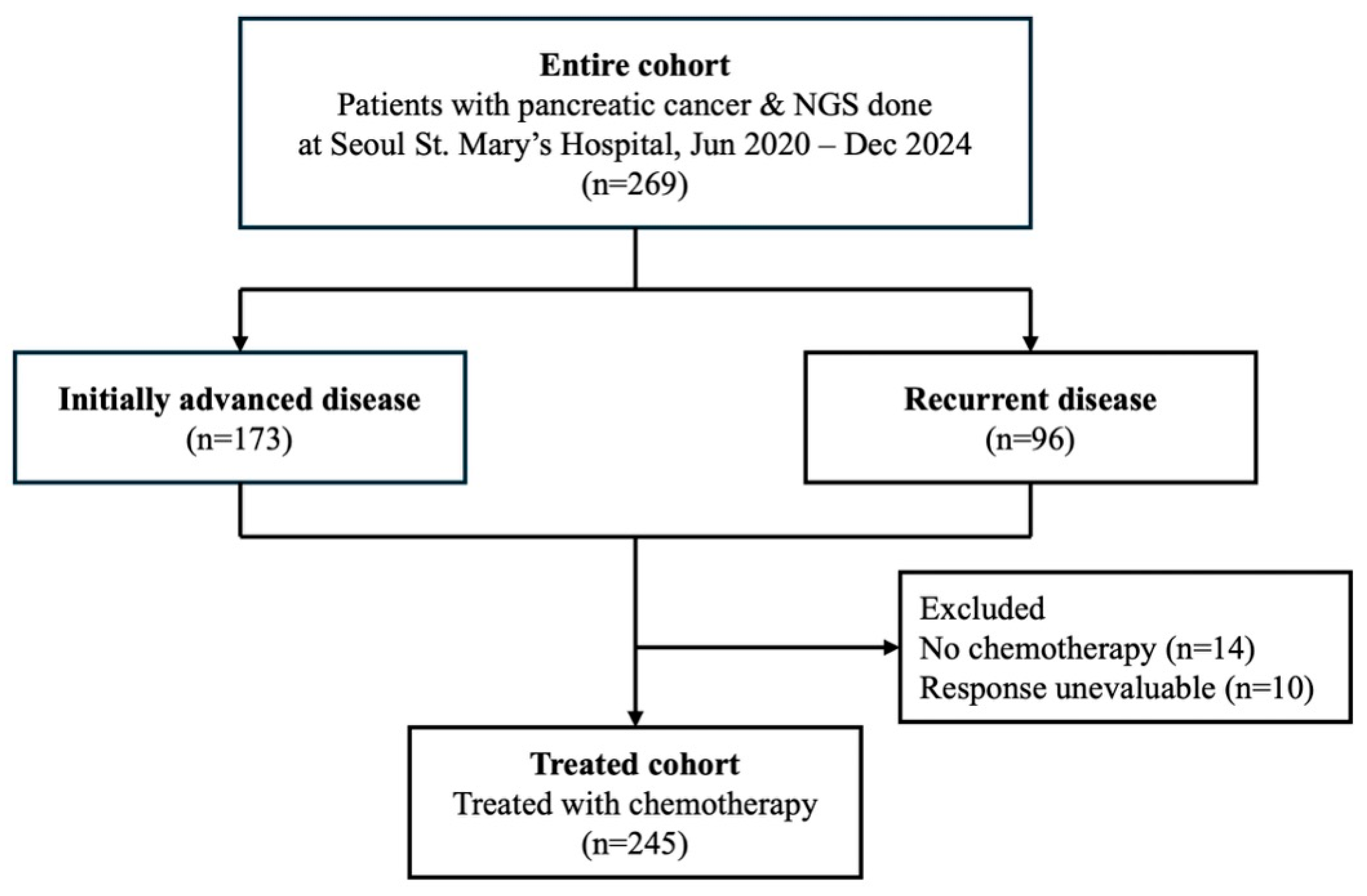
A total of 269 patients with pathologically confirmed advanced pancreatic cancer who underwent next-generation sequencing (NGS) between June 2020 and December 2024 at Seoul St. Mary’s Hospital were included in this retrospective study (Figure 1).
Figure 1.
Study flowchart diagram. The flowchart shows cohort patient selection.
Among them, 245 patients received systemic chemotherapy and underwent radiological response evaluation, comprising the treated cohort. Baseline characteristics of the entire cohort are summarized in Table 1. The median age was 66 years (IQR, 61–73), and 52.4% of patients were male. A total of 64.3% had initially advanced disease, while 35.7% had recurrent disease, and most patients (82.2%) presented with metastatic disease. The primary tumor was located in the pancreatic head in 50.2% and in the body or tail in 49.8% of patients, and the majority of tumors (89.2%) were histologically classified as ductal adenocarcinoma. Regarding the specimen source for NGS, 65.4% were obtained from primary tumor tissues, 28.3% from metastatic sites, and 6.3% from liquid biopsies, with specimens acquired by surgical resection (42.8%), needle biopsy (50.9%), or peripheral blood (6.3%). KRAS mutations were identified in 91.8% of patients, while TP53, CDKN2A, and SMAD4 mutations were detected in 64.3%, 29.4%, and 12.3% of cases, respectively. Among the 22 patients with KRAS wild-type tumors, actionable genomic alterations are summarized in Table S1, with druggable alterations such as BRAF V600E detected in three patients.
Table 1.
Baseline characteristics of entire cohort.
2.2. KRAS Mutation Subtypes and Survival Outcomes
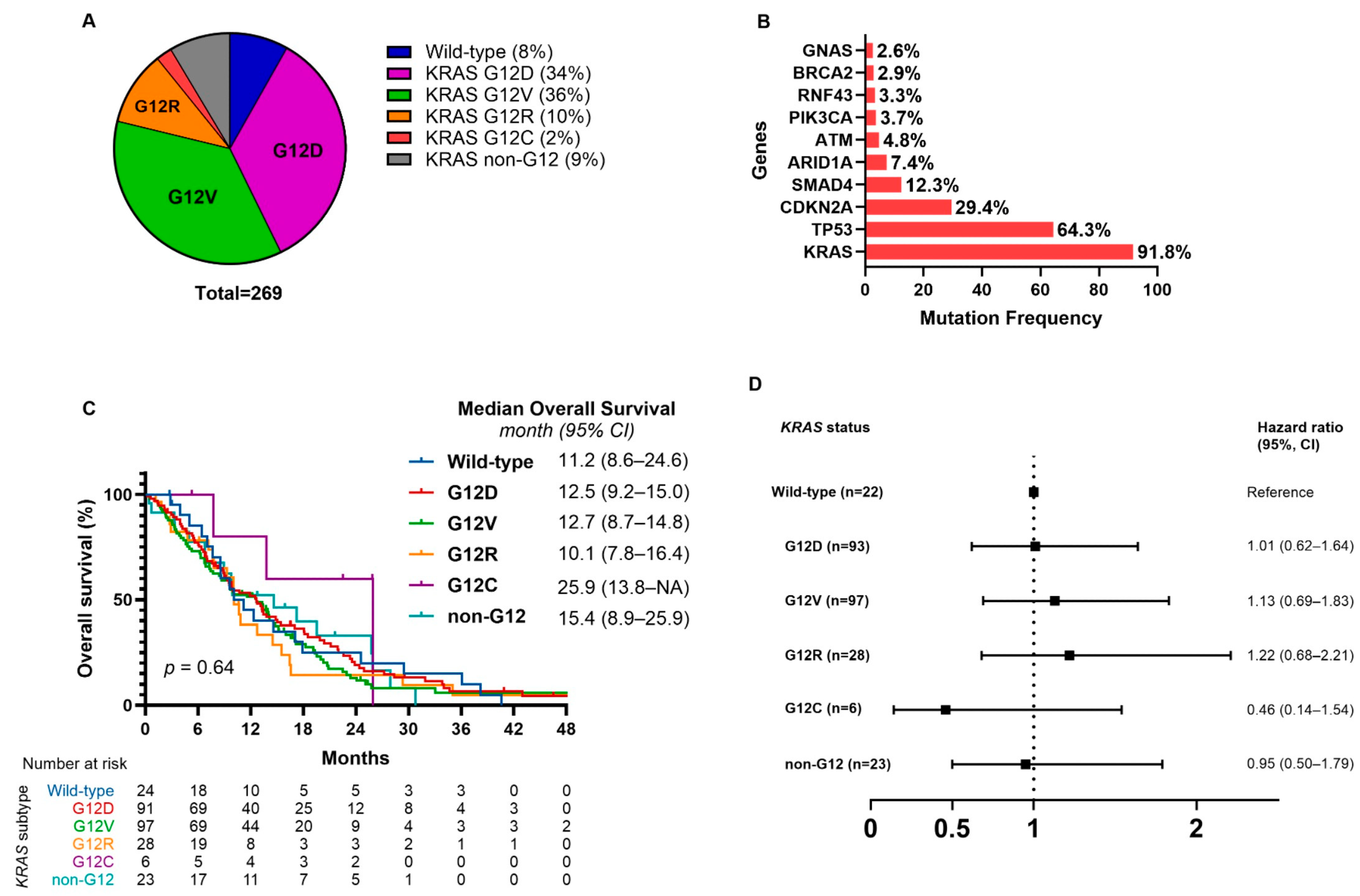
Among the 269 patients, 247 (92%) harbored KRAS mutations. The most prevalent subtype was G12V (96 patients, 36%), followed by G12D (93 patients, 34%), G12R (28 patients, 10%), and non-G12 codon mutations (23 patients, 9%) (Figure 2A). Two patients had co-occurring G12D and non-G12 mutations. G12C mutations were identified in 6 patients, representing 2% of the total cohort. The median follow-up duration for the entire cohort was 9.9 months (95% CI, 8.9–11.9), and 218 patients (81.0%) had died at the time of analysis. The median overall survival (OS) for the entire cohort was 12.5 months (95% CI, 9.9–13.8). OS according to KRAS mutation subtypes revealed no statistically significant difference across groups (p = 0.64, Figure 2C). The median OS was 11.2 months (95% CI, 8.6–24.6) in patients with wild-type KRAS, 12.5 months (95% CI, 9.2–15.0) in those with G12D mutations, 12.7 months (95% CI, 8.7–14.8) in G12V, 10.1 months (95% CI, 7.8–16.4) in G12R, and 15.4 months (95% CI, 8.9–25.9) in non-G12 mutations. The G12C subgroup showed a numerically longer median OS of 25.9 months (95% CI, 13.8–not reached), though the sample size was small and the difference was not statistically significant. Compared with patients carrying wild-type KRAS, none of the specific KRAS subtypes showed a statistically significant difference in OS (Figure 2D).
Figure 2.
Distribution of KRAS mutation subtypes, frequently mutated genes, and associated overall survival in pancreatic cancer. (A) Distribution of KRAS mutation subtypes among patients with pancreatic cancer. (B) Bar plot illustrating the frequency distribution of the most prevalent gene mutations in our cohort. (C) Kaplan–Meier curves for overall survival (OS) according to KRAS mutation subtype. (D) Comparison of median OS between wild-type KRAS and specific KRAS mutation subtypes. (A) shows mutation-level distribution; co-occurring KRAS alterations within a single patient are counted separately. (C,D) are patient-level analyses, with each patient assigned to one codon group to avoid double-counting. KRAS G12D/V/R/C denote KRAS p.G12D/V/R/C. Variants follow HGVS at first mention and are abbreviated thereafter.
2.3. KRAS Allele Specific Mutations and Co-Mutations
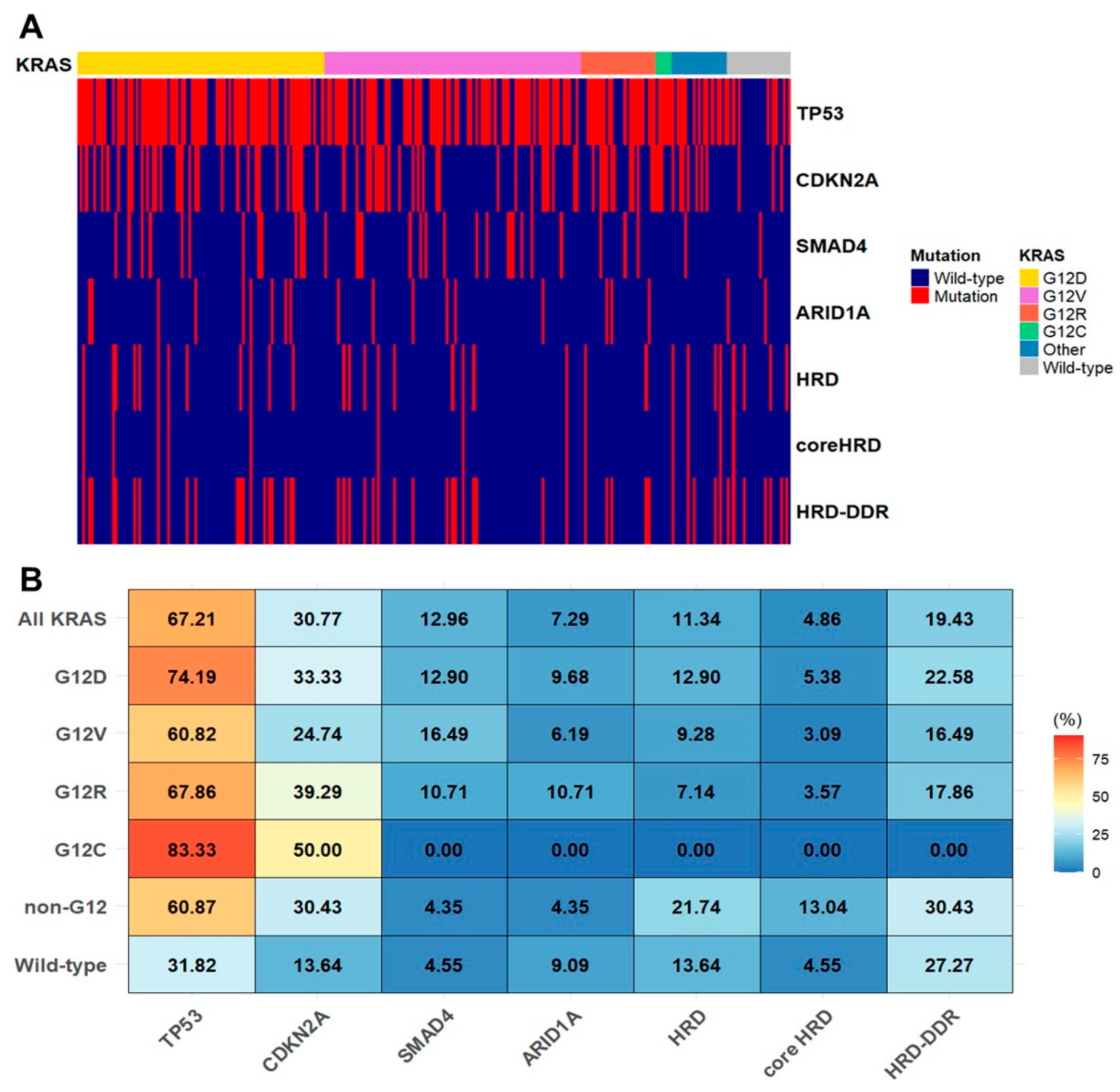
To characterize the patterns of co-occurring genomic alterations across different KRAS alleles, we generated an oncoplot and a heatmap stratified by KRAS mutation subtypes (Figure 3). TP53, CDKN2A, and SMAD4 were the most frequently co-altered genes, regardless of KRAS subtype. The heatmap displays the frequency of mutations in TP53, CDKN2A, SMAD4, ARID1A, and genes associated with homologous recombination deficiency (HRD) and DNA damage response (DDR) pathways across KRAS subgroups (Figure 3B). TP53 was the most commonly altered gene overall, with particularly high frequencies in the G12D and G12R subgroups (74.2% and 67.9%, respectively), whereas its mutation rate was substantially lower in the wild-type group (31.8%). Among all evaluated genes, only TP53 showed a statistically significant difference in mutation frequency across KRAS subtypes (p = 0.0074), whereas other genes showed no significant variation, although mutations in HRD- or DDR-related genes tended to be more frequent in the non-G12 and wild-type subgroups. To evaluate subgroup-specific differences in TP53 mutation frequency, pairwise comparisons demonstrated a significantly higher frequency in the G12D subgroup compared to wild-type KRAS (adjusted p = 0.0089), whereas no other comparisons showed statistically significant differences.
Figure 3.
Co-occurring genomic alterations by allele specific KRAS mutations in pancreatic cancer. (A) An oncoplot illustrating somatic gene alterations stratified by KRAS mutation subtypes in patients with pancreatic cancer. (B) A heatmap showing the frequency of co-occurring genomic alterations involving TP53, CDKN2A, SMAD4, ARID1A, and genes related to homologous recombination deficiency (HRD) or DNA damage repair (DDR) across KRAS mutation subgroups.
2.4. KRAS Mutation Subtypes and Clinical Characteristics
Baseline clinical characteristics were compared across KRAS mutation subtypes, including G12D, G12V, G12R, non-G12 mutations, and wild type (Table S2). Most characteristics were evenly distributed among the groups, with no statistically significant differences observed. However, serum carbohydrate antigen 19-9 (CA 19-9) levels at the time of advanced disease diagnosis differed significantly according to KRAS mutation subtype (p = 0.011). A higher proportion of patients with G12D, G12V, or G12R mutations exhibited elevated CA 19-9 levels (77.8%, 85.4%, and 85.2%, respectively), whereas a greater proportion of patients with non-G12 mutations or wild-type KRAS showed normal CA 19-9 levels (42.1% and 40.9%, respectively). Although not statistically significant, several trends of interest were noted. Tumors in the wild-type group were more frequently located in the pancreatic head (63.6%, p = 0.459), while tumors in the G12R group were more often found in the body or tail of the pancreas (60.7%). Elevated carcinoembryonic antigen (CEA) levels were more frequently observed in the non-G12 group (63.2%, p = 0.395), and the neutrophil-to-lymphocyte ratio (NLR) similarly showed no significant differences across the subgroups (p = 0.201).
2.5. KRAS Mutation Subtypes and Treatment Outcomes
A total of 245 patients were included in the treated cohort, and their baseline characteristics are summarized in Table 2. The majority of patients (69.4%) had 0–1 metastatic organ site, while 30.6% had two or more. The liver was the most frequent site of metastasis (53.4%), followed by the peritoneum (24.9%) and lungs (16.3%). Elevated baseline CA 19-9 levels were observed in approximately three-quarters of patients (71.8%). As first-line palliative chemotherapy, gemcitabine-based regimens were administered to 56.3% of patients, the majority of whom received gemcitabine plus nab-paclitaxel (53.8%), while fluorouracil-based regimens were given to 43.7%, including FOLFIRINOX in 38.7%.
Table 2.
Baseline characteristics of treated cohort.
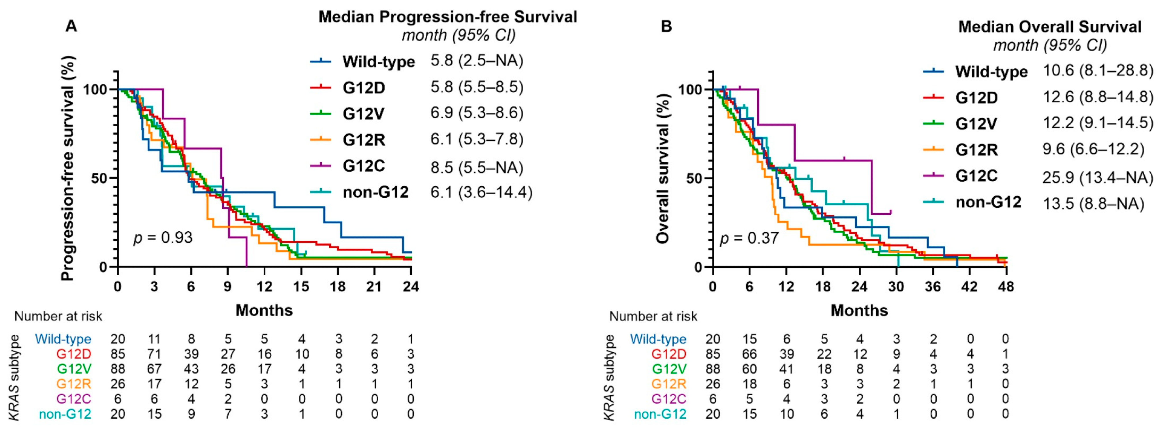
In the overall treated cohort, the median progression-free survival (PFS) was 6.3 months (95% CI, 5.5–7.3), and the median OS was 11.2 months (95% CI, 9.6–13.1). Survival analysis revealed no statistically significant difference in PFS among the KRAS mutation subtypes (p = 0.93; Figure 4A). Similarly, no significant difference in OS was observed between the subgroups (p = 0.37; Figure 4B). Findings from very small subgroups are exploratory and should be interpreted with caution. Compared with KRAS wild-type, none of the codon-specific KRAS subtypes exhibited statistically significant differences in PFS or OS (Figure S1). Treatment outcomes across KRAS mutation subtypes are summarized in Table 3. In the overall cohort, the objective response rate (ORR) was 35.5%, with the highest rate observed in the non-G12 group (55.0%), followed by the G12D (38.8%), G12V (34.1%), and G12R (26.9%) subgroups, and the lowest in the wild-type group (20.0%). Compared with the G12D subgroup, no statistically significant differences in ORR were found for any other subtype. The disease control rate (DCR) was 85.3% in the overall cohort, with no significant differences relative to the G12D subgroup. Although numerically lower DCRs were noted in the G12R (73.1%) and wild-type (75.0%) groups, these differences were not statistically significant. The 6-month PFS rate and 12-month OS rate in the total cohort were 51.8% (95% CI, 45.7–58.6) and 48.0% (95% CI, 41.9–54.8), respectively, with no significant differences in these time-specific survival rates when comparing the G12D subgroup with other mutation subtypes.
Figure 4.
Kaplan–Meier curves for progression-free survival and overall survival by KRAS mutation subtype in the treated cohort. (A) Progression-free survival (PFS) and (B) overall survival (OS) according to KRAS mutation subtype in patients who received systemic treatment. No statistically significant differences in PFS (p = 0.93) or OS (p = 0.37) were observed among the subgroups.
Table 3.
Treatment outcomes according to KRAS mutation subtypes.
2.6. Effectiveness of Treatment Regimens
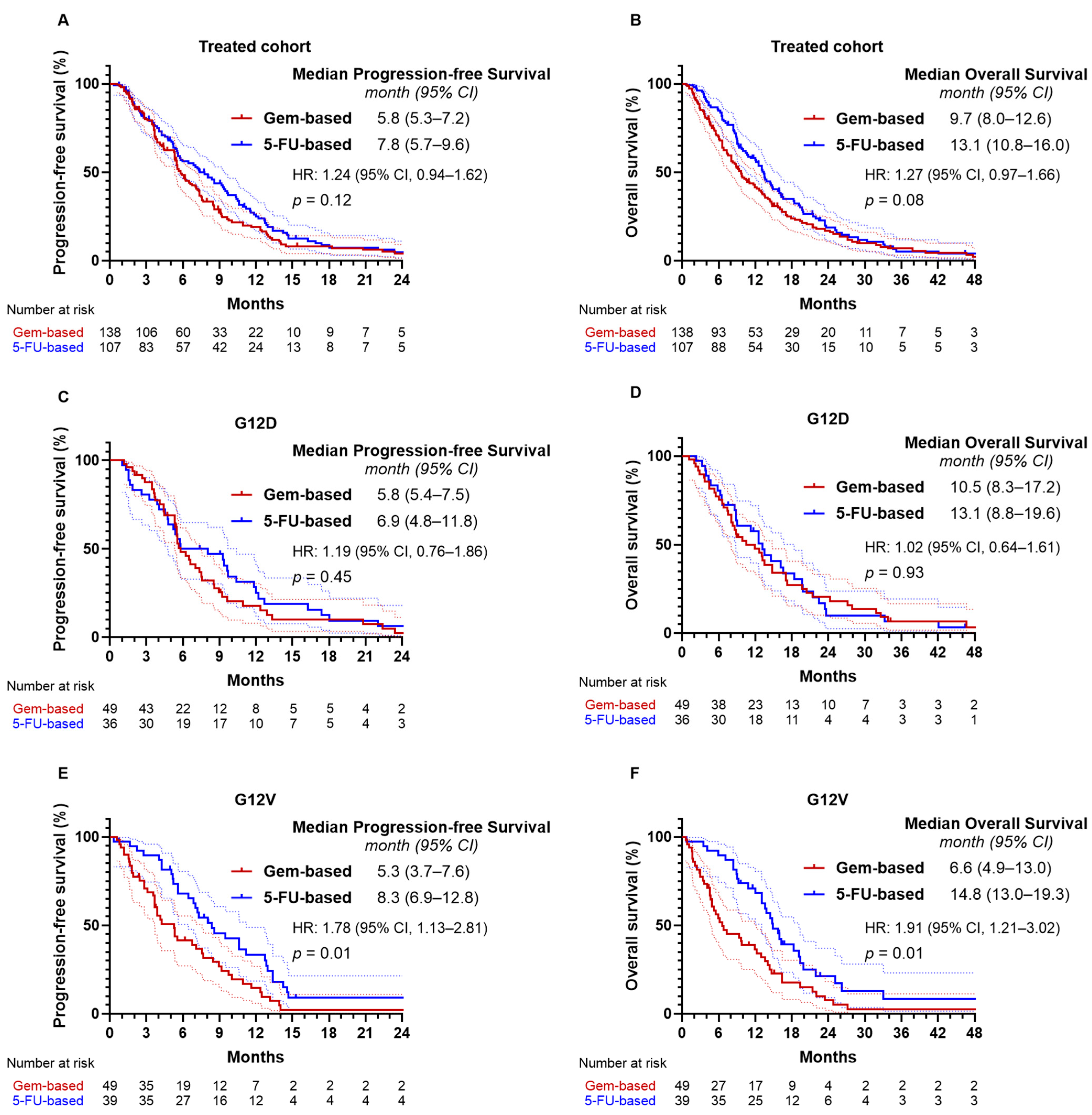
In the overall treated cohort, patients receiving fluorouracil-based chemotherapy had a numerically longer median PFS of 7.8 months (95% CI, 5.7–9.6) compared with 5.8 months (95% CI, 5.3–7.2) for those receiving gemcitabine-based regimens, although the difference was not statistically significant (p = 0.12; Figure 5A). Median OS was 13.1 months (95% CI, 10.8–16.0) with fluorouracil-based therapy versus 9.7 months (95% CI, 8.0–12.6) with gemcitabine-based therapy (p = 0.08; Figure 5B). No significant differences were observed in ORR (32.7% vs. 37.7%, p = 0.50) or DCR (86.0% vs. 84.8%, p = 0.86) between the two regimens (Table S3). In the G12D subgroup, survival outcomes were similar between regimens. Median PFS was 6.9 months (95% CI, 4.8–11.8) with fluorouracil-based therapy and 5.8 months (95% CI, 5.4–7.5) with gemcitabine-based therapy (p = 0.45; Figure 5C), while median OS was 13.1 months (95% CI, 8.8–19.6) and 10.5 months (95% CI, 8.3–17.2), respectively (p = 0.93; Figure 5D). In the G12V subgroup, patients treated with gemcitabine-based regimens had a significantly inferior median PFS of 5.3 months (95% CI, 3.7–7.6) compared with 8.3 months (95% CI, 6.9–12.8) for those receiving fluorouracil-based regimens (HR = 1.78; 95% CI, 1.13–2.81; p = 0.01; Figure 5E). Median OS was also shorter with gemcitabine-based therapy (6.6 months [95% CI, 4.9–13.0]) than with fluorouracil-based therapy (14.8 months [95% CI, 13.0–19.3]) (HR = 1.91; 95% CI, 1.21–3.02; p = 0.01; Figure 5F). Although ORR (38.5% vs. 30.6%) and DCR (92.3% vs. 79.6%) were numerically higher with fluorouracil-based therapy, these differences were not statistically significant.
Figure 5.
Progression-free and overall survival by first-line chemotherapy regimen in the treated cohort and KRAS G12D/G12V subgroups. Progression-free survival and overall survival according to first-line chemotherapy regimen in (A,B) the overall treated cohort, (C,D) the KRAS G12D subgroup, and (E,F) the KRAS G12V subgroup.
3. Discussion
In this retrospective study of patients with advanced PDAC, we comprehensively characterized the distribution of KRAS mutation subtypes, their co-mutation profiles, and associated clinical implications, including response to chemotherapy and survival outcomes. Unlike previous studies that assessed the prognostic implications of KRAS mutation subtypes but included surgically resected cases, our analysis focused exclusively on patients with advanced disease, thereby reflecting the majority of real-world clinical presentations. No statistically significant differences in OS or PFS were observed across KRAS subtypes, both in the overall cohort and within the treated cohort. However, subgroup analysis revealed that patients harboring KRAS G12V mutations derived significantly greater survival benefit from fluorouracil-based chemotherapy compared to gemcitabine-based regimens, suggesting a potential subtype-specific therapeutic interaction.
Consistent with prior reports, KRAS mutations were observed in over 90% of cases, with G12D and G12V comprising the most frequent subtypes [,]. The KRAS G12D subtype has historically been associated with more aggressive disease biology and poorer prognosis [,]. However, in our study, no statistically significant differences in survival outcomes or treatment effectiveness were observed among the KRAS subtypes. These findings align with recent studies reporting no significant OS differences among codon 12 KRAS mutants in patients with advanced-stage PDAC []. Although previous studies have suggested that the G12R subtype is associated with better survival compared to G12D in advanced disease [], our analysis did not reveal any significant differences in OS, PFS, or treatment response between the two subtypes. Further validation in larger cohorts with multivariable adjustment for additional prognostic factors is needed to clarify the clinical implications of the G12R subtype. While limited by the very small sample size, the G12C subgroup demonstrated numerically longer OS, which may reflect the inclusion of patients who received KRAS G12C-targeted therapies.
Analysis of co-occurring genomic alterations revealed that TP53 was the most frequently co-mutated gene, present in 67% of KRAS-mutated cases. The G12D subtype, in particular, exhibited a significantly higher frequency of TP53 mutations (74.2%) compared with the KRAS wild-type group (31.8%), a finding consistent with previous reports []. Co-mutation of KRAS and TP53 has been implicated in promoting PDAC progression and metastasis, suggesting a potential molecular synergy that may underlie the poorer OS associated with the G12D subtype []. Alterations in HRD- and DDR-related genes were observed more often in non-G12 and KRAS wild-type tumors than in G12-mutant tumors, although this difference was not statistically significant. These findings indicate that distinct KRAS alleles may influence the broader genomic landscape and warrant further investigation, particularly regarding the role of HRD and DDR mutations as biomarkers for platinum-based therapy.
No significant differences in clinical characteristics, including histologic differentiation, were observed across KRAS subtypes. Serum CA 19-9 levels were significantly elevated in patients with G12 mutations, whereas a substantial proportion of patients with non-G12 or wild-type tumors (~40%) exhibited normal levels at the time of advanced disease diagnosis. This finding, which may relate to differences in KRAS-driven oncogenesis and tumor marker production, suggests that in non-G12 or wild-type cases, CA 19-9 may not reliably reflect treatment response.
In the treated cohort, no significant differences in PFS or OS were observed across KRAS subtypes, and ORR was slightly higher in the G12D subtype compared with the G12R and wild-type groups, suggesting that the prognostic significance of KRAS subtypes in advanced PDAC remains controversial. By treatment regimen, no difference in efficacy was observed between fluorouracil- and gemcitabine-based therapies in the G12D group, whereas patients with the G12V subtype showed significantly improved PFS and OS with fluorouracil-based therapy. These findings suggest that the G12V subtype may be less sensitive to gemcitabine and highlight the importance of investigating associations between KRAS mutation subtypes and transcriptome-based predictive signatures, such as GemPred, which predicts benefit from gemcitabine treatment []. While these observations require prospective validation, KRAS G12V mutation status may serve as a predictive biomarker for fluorouracil-based first-line chemotherapy in pancreatic cancer.
Our study has several limitations. First, its retrospective design and single-institution setting may introduce ascertainment bias and limit the generalizability of the findings. Second, the sample size of certain KRAS subgroups, such as G12C and non-G12 mutations, was small, reducing the statistical power to detect meaningful differences. Taken together, these two issues render the findings hypothesis-generating and warrant confirmation in adequately powered, prospective multicenter cohorts. Third, potential confounders such as performance status, disease burden, treatment dose intensity, and subsequent therapies may have influenced treatment outcomes, but multivariable analysis could not be performed owing to the limited sample size. Finally, tumor heterogeneity and temporal evolution of mutational profiles may not have been fully captured by single-sample NGS, and in a subset of patients, liquid biopsy was used, which may further affect consistency of genomic profiling.
Despite these limitations, our study provides comprehensive insights into the clinical and molecular characteristics of KRAS codon-specific mutations in advanced PDAC and their relationship with treatment outcomes. Prognostic differences across KRAS subtypes were not observed in our cohort, and although a differential response to chemotherapy was noted in the G12V subgroup, the biological rationale remains insufficient and requires further validation. Therefore, KRAS subtyping may not yet be essential for therapeutic decision-making in clinical practice. Nevertheless, identifying KRAS G12C mutations is important given the availability of effective targeted therapies [,], and genomic profiling of KRAS wild-type tumors remains essential to detect actionable alterations []. Furthermore, as novel KRAS-targeted therapies, including G12D-selective inhibitors, enter clinical trials [,], defining biologically and clinically meaningful subgroups may become increasingly important.
4. Materials and Methods
4.1. Patients
Patients with histologically or cytologically confirmed pancreatic exocrine cancer were eligible for inclusion in this study if they had NGS results obtained at Seoul St. Mary’s Hospital. The study was conducted in accordance with Korean regulations and the principles of the Declaration of Helsinki. The Institutional Review Board (IRB) of The Catholic University of Korea, Seoul St. Mary’s Hospital, approved data acquisition (protocol code: KC24RASI0307) and granted a waiver of informed consent due to the retrospective nature of the analysis. Retrospective analysis was conducted on patient demographics, clinical characteristics, pathological findings, genomic profiles, treatment history, and outcomes using data extracted from electronic medical records.
4.2. Molecular Alteration Analyses
Molecular profiling and KRAS status were determined through tumor tissue analysis using the Oncomine Comprehensive Assay Plus or liquid biopsy via AlphaLiquid100. These panels included 517 and 118 genes, respectively, covering oncogenes, tumor suppressor genes, gene rearrangements, microsatellite stability status, and tumor mutational burden. Formalin-fixed, paraffin-embedded (FFPE) tumor specimens or plasma samples were collected for NGS either at the time of diagnosis or upon disease recurrence. For patient-level analyses (survival and clinicopathologic comparisons), if more than one KRAS alteration was detected in a single patient, we assigned a single codon category based on the highest variant allele frequency to avoid double-counting.
Genomic DNA and RNA were extracted from FFPE tumor samples and analyzed using the Oncomine Comprehensive Assay Plus (Thermo Fisher Scientific, Waltham, MA, USA) on the IonTorrent™ S5 XL platform, following the manufacturer’s protocols. Sequencing quality was assessed using plug-in coverage analysis, and data were processed through dedicated bioinformatic workflows within the Ion Reporter v5.12 server (IonTorrent, Waltham, MA, USA). Variants, including single nucleotide variants (SNVs), insertions/deletions (indels), copy number variations (CNVs), and gene fusions, were filtered based on the following thresholds: a minimum allele frequency of ≥1.5% for hotspot SNVs and indels, a minimum read count of 100 for fusions, and CNV thresholds of ≥6 for gains and ≤0.7 for losses. Variants were classified into clinical relevance tiers according to established guidelines [].
Cell-free DNA (cfDNA) was extracted from plasma samples prior to systemic treatment. DNA NGS library preparation and solution-based target enrichment were performed at IMBdx, Inc. (Seoul, Republic of Korea) using AlphaLiquid100, as previously described []. A targeted panel of 118 cancer-related genes was used to detect four variant classes (SNVs, indels, CNVs, and fusions). Captured DNA libraries were sequenced on the Illumina NovaSeq 6000 platform (Illumina, San Diego, CA, USA) in 2 × 150 bp paired-end mode. For AlphaLiquid100, variants were called using thresholds of at least 0.1% allele fraction for SNVs and indels (high-confidence at 0.5% or higher), 2.4 copies or higher for CNVs, and 0.2% or higher for fusions.
4.3. Mutational Status of HRD- and DDR-Related Genes
To investigate the association between KRAS mutation subtypes and genomic alterations related to HRD and DDR pathways, we evaluated the prevalence of HRD-related, core HRD, and DDR-associated gene mutations across each KRAS mutation subgroup. Genomic alterations associated with HRD and DDR pathways were identified according to a curated list of genes implicated in these repair mechanisms. HRD-related alterations were defined as pathogenic variants in genes functionally involved in homologous recombination repair, including ATM, BAP1, BARD1, BLM, BRCA1, BRCA2, BRIP1, CHEK2, FAM175A, FANCA, FANCC, NBN, PALB2, RAD50, RAD51, RAD51C, and RTEL1 []. Among these, BRCA1, BRCA2, and PALB2 were designated as core HRD genes, reflecting their pivotal roles in double-strand break repair and their clinical relevance as biomarkers for predicting response to platinum and PARP inhibitor therapy. In addition, pathogenic variants in genes involved in homologous recombination and DDR pathways, including ARID1A, ATM, ATRX, BAP1, BARD1, BLM, BRCA1, BRCA2, BRIP1, CHEK1, CHEK2, FANCA, FANCC, FANCD2, FANCE, FANCF, FANCG, FANCL, MRE11A, NBN, PALB2, RAD50, RAD51, RAD51B, and WRN, were evaluated as HRD and DDR-associated alterations [].
4.4. Assessments
Tumor assessments were performed in accordance with the Response Evaluation Criteria in Solid Tumors (RECIST) version 1.1. Imaging evaluations were conducted using computed tomography (CT) of the thorax, abdomen, and pelvis at 6- to 8-week intervals following the initiation of chemotherapy. Additional imaging was performed as clinically indicated. Radiologic response-evaluable patients were defined as those who received at least one cycle of chemotherapy and underwent at least one post-baseline disease assessment using imaging modalities. Serum tumor markers, including CA 19-9 and CEA, were measured concurrently with radiologic assessments. For stratified analyses, we applied a CA 19-9 threshold of 59× the upper limit of normal consistent with landmark clinical trials [,].
4.5. Statistical Analysis
Descriptive statistics are reported as proportions or medians with interquartile ranges (IQR). Categorical variables were compared using the chi-square test or Fisher’s exact test, while continuous variables were analyzed using Student’s t-test. For pairwise comparisons of categorical variables, Fisher’s exact test was employed with false discovery rate (FDR) adjustment to account for multiple testing, with an FDR-adjusted p value of less than 0.1 considered statistically significant. To explore co-mutation patterns across KRAS mutation subtypes, an oncoplot and heatmap were generated using the MAFtools and ComplexHeatmap packages in R. OS was defined as the time from diagnosis of advanced disease to death in the entire cohort, and from the initiation of first-line palliative chemotherapy to death in the treated cohort. PFS was defined as the time from initiation of first-line palliative chemotherapy to documented disease progression or death from any cause. Kaplan–Meier estimates were used to evaluate OS and PFS, with subgroup comparisons performed using the log-rank test. Unstratified Cox proportional hazards regression was used to estimate hazard ratios (HRs) and their corresponding 95% confidence intervals (CIs). ORR and DCR were compared across subgroups using Fisher’s exact test. All tests were two-sided, and statistical significance was identified by a p-values < 0.05. Statistical analyses and data visualization were performed using GraphPad Prism version 10.3 (GraphPad Software, San Diego, CA, USA) and RStudio (version 2024, RStudio, PBC, Boston, MA) with R version 4.4.3.
5. Conclusions
In conclusion, while KRAS mutation subtypes were not significantly associated with survival outcomes in the overall cohort, our findings suggest that certain alleles, such as G12V, may be linked to differential sensitivity to standard chemotherapy. Although KRAS subtyping may not be universally essential for therapeutic decision-making in advanced PDAC, it could still hold clinical relevance in selected patients. Further prospective studies and functional investigations are warranted to validate these observations and clarify their implications for precision oncology.
Supplementary Materials
The following supporting information can be downloaded at: https://www.mdpi.com/article/10.3390/ijms262210908/s1.
Author Contributions
Conceptualization, D.C., M.L., S.-H.H., K.S. and S.J.P.; Methodology, D.C., M.L., S.-H.H., K.S. and S.J.P.; Investigation, D.C., M.L., S.-H.H., K.S., I.-H.K., S.H.L., Y.K., T.H.H. and S.J.P.; Formal Analysis, D.C., M.L., S.-H.H., K.S., I.-H.K., S.H.L., Y.K., T.H.H. and S.J.P.; Data Curation, D.C., M.L., S.-H.H., K.S., I.-H.K., S.H.L., Y.K., T.H.H. and S.J.P.; Writing—Original Draft Preparation, D.C., M.L., S.-H.H., K.S. and S.J.P.; Writing—Review & Editing, D.C., M.L., S.-H.H., K.S., I.-H.K., S.H.L., Y.K., T.H.H. and S.J.P.; Visualization, D.C. and S.J.P.; Supervision, S.J.P. All authors have read and agreed to the published version of the manuscript.
Funding
This research was supported by the Catholic Medical Center Research Foundation in the program year of 2024 and 2025.
Institutional Review Board Statement
The study was conducted in accordance with the Declaration of Helsinki and approved by the Institutional Review Board of The Catholic University of Korea, Seoul St. Mary’s Hospital (protocol code KC24RASI0307; approval date: 17 May 2024).
Informed Consent Statement
Patient consent was waived due to the retrospective nature of the study, as approved by the IRB (KC24RASI0307).
Data Availability Statement
The majority of the data generated or analyzed during this study are included in this article and its Supplementary Information Files. Additional datasets used in the current study are available from the corresponding author on request.
Conflicts of Interest
All authors declare that they have no conflicts of interest.
Abbreviations
The following abbreviations are used in this manuscript:
| PDAC | Pancreatic Ductal Adenocarcinoma |
| KRAS | Kirsten rat sarcoma viral oncogene homolog |
| TP53 | Tumor protein p53 |
| CDKN2A | Cyclin-Dependent Kinase Inhibitor 2A |
| SMAD4 | Mothers against decapentaplegic homolog 4 |
| NGS | Next-Generation Sequencing |
| SNV | Single Nucleotide Variant |
| CNV | Copy Number Variation |
| cfDNA | Cell-free DNA |
| HRD | Homologous Recombination Deficiency |
| DDR | DNA Damage Response |
| CA 19-9 | Carbohydrate Antigen 19-9 |
| CEA | Carcinoembryonic Antigen |
| OS | Overall Survival |
| PFS | Progression-Free Survival |
| NLR | Neutrophil-to-Lymphocyte Ratio |
| 5-FU | 5-Fluorouracil |
| Nal-IRI/FL | Nanoliposomal Irinotecan + 5-Fluorouracil/Leucovorin |
| ORR | Objective Response Rate |
| DCR | Disease Control Rate |
| PARP | Poly(ADP-ribose) Polymerase |
References
- Siegel, R.L.; Giaquinto, A.N.; Jemal, A. Cancer statistics, 2024. CA Cancer J. Clin. 2024, 74, 12–49. [Google Scholar]
- Mizrahi, J.D.; Surana, R.; Valle, J.W.; Shroff, R.T. Pancreatic cancer. Lancet 2020, 395, 2008–2020. [Google Scholar] [CrossRef]
- Cronin, K.A.; Lake, A.J.; Scott, S.; Sherman, R.L.; Noone, A.M.; Howlader, N.; Henley, S.J.; Anderson, R.N.; Firth, A.U.; Ma, J.; et al. Annual Report to the Nation on the Status of Cancer, part I: National cancer statistics. Cancer 2018, 124, 2785–2800. [Google Scholar] [CrossRef]
- Conroy, T.; Desseigne, F.; Ychou, M.; Bouché, O.; Guimbaud, R.; Bécouarn, Y.; Adenis, A.; Raoul, J.L.; Gourgou-Bourgade, S.; de la Fouchardière, C.; et al. FOLFIRINOX versus gemcitabine for metastatic pancreatic cancer. N. Engl. J. Med. 2011, 364, 1817–1825. [Google Scholar] [PubMed]
- Von Hoff, D.D.; Ervin, T.; Arena, F.P.; Chiorean, E.G.; Infante, J.; Moore, M.; Seay, T.; Tjulandin, S.A.; Ma, W.W.; Saleh, M.N.; et al. Increased survival in pancreatic cancer with nab-paclitaxel plus gemcitabine. N. Engl. J. Med. 2013, 369, 1691–1703. [Google Scholar] [CrossRef]
- Biankin, A.V.; Waddell, N.; Kassahn, K.S.; Gingras, M.C.; Muthuswamy, L.B.; Johns, A.L.; Miller, D.K.; Wilson, P.J.; Patch, A.M.; Wu, J.; et al. Pancreatic cancer genomes reveal aberrations in axon guidance pathway genes. Nature 2012, 491, 399–405. [Google Scholar] [CrossRef] [PubMed]
- Hosein, A.N.; Dougan, S.K.; Aguirre, A.J.; Maitra, A. Translational advances in pancreatic ductal adenocarcinoma therapy. Nat. Cancer 2022, 3, 272–286. [Google Scholar] [CrossRef]
- Cicenas, J.; Kvederaviciute, K.; Meskinyte, I.; Meskinyte-Kausiliene, E.; Skeberdyte, A.; Cicenas, J. KRAS, TP53, CDKN2A, SMAD4, BRCA1, and BRCA2 Mutations in Pancreatic Cancer. Cancers 2017, 9, 42. [Google Scholar] [CrossRef]
- Bailey, P.; Chang, D.K.; Nones, K.; Johns, A.L.; Patch, A.M.; Gingras, M.C.; Miller, D.K.; Christ, A.N.; Bruxner, T.J.; Quinn, M.C.; et al. Genomic analyses identify molecular subtypes of pancreatic cancer. Nature 2016, 531, 47–52. [Google Scholar] [CrossRef] [PubMed]
- Yousef, A.; Yousef, M.; Chowdhury, S.; Abdilleh, K.; Knafl, M.; Edelkamp, P.; Alfaro-Munoz, K.; Chacko, R.; Peterson, J.; Smaglo, B.G.; et al. Impact of KRAS mutations and co-mutations on clinical outcomes in pancreatic ductal adenocarcinoma. NPJ Precis. Oncol. 2024, 8, 27. [Google Scholar]
- Ostrem, J.M.; Shokat, K.M. Direct small-molecule inhibitors of KRAS: From structural insights to mechanism-based design. Nat. Rev. Drug Discov. 2016, 15, 771–785. [Google Scholar] [CrossRef] [PubMed]
- Skoulidis, F.; Li, B.T.; Dy, G.K.; Price, T.J.; Falchook, G.S.; Wolf, J.; Italiano, A.; Schuler, M.; Borghaei, H.; Barlesi, F.; et al. Sotorasib for Lung Cancers with KRAS p.G12C Mutation. N. Engl. J. Med. 2021, 384, 2371–2381. [Google Scholar] [CrossRef] [PubMed]
- Jänne, P.A.; Riely, G.J.; Gadgeel, S.M.; Heist, R.S.; Ou, S.I.; Pacheco, J.M.; Johnson, M.L.; Sabari, J.K.; Leventakos, K.; Yau, E.; et al. Adagrasib in Non-Small-Cell Lung Cancer Harboring a KRAS(G12C) Mutation. N. Engl. J. Med. 2022, 387, 120–131. [Google Scholar] [CrossRef] [PubMed]
- Kemp, S.B.; Cheng, N.; Markosyan, N.; Sor, R.; Kim, I.K.; Hallin, J.; Shoush, J.; Quinones, L.; Brown, N.V.; Bassett, J.B.; et al. Efficacy of a Small-Molecule Inhibitor of KrasG12D in Immunocompetent Models of Pancreatic Cancer. Cancer Discov. 2023, 13, 298–311. [Google Scholar] [CrossRef]
- Hallin, J.; Bowcut, V.; Calinisan, A.; Briere, D.M.; Hargis, L.; Engstrom, L.D.; Laguer, J.; Medwid, J.; Vanderpool, D.; Lifset, E.; et al. Anti-tumor efficacy of a potent and selective non-covalent KRAS(G12D) inhibitor. Nat. Med. 2022, 28, 2171–2182. [Google Scholar] [CrossRef]
- Johnson, C.; Burkhart, D.L.; Haigis, K.M. Classification of KRAS-Activating Mutations and the Implications for Therapeutic Intervention. Cancer Discov. 2022, 12, 913–923. [Google Scholar] [CrossRef]
- Hobbs, G.A.; Der, C.J. RAS Mutations Are Not Created Equal. Cancer Discov. 2019, 9, 696–698. [Google Scholar] [CrossRef]
- Boilève, A.; Rousseau, A.; Hilmi, M.; Tarabay, A.; Mathieu, J.R.R.; Cartry, J.; Bedja, S.; Goudarzi, N.; Nicotra, C.; Ngo-Camus, M.; et al. Codon-specific KRAS mutations predict survival in advanced pancreatic cancer. ESMO Gastrointest. Oncol. 2024, 3, 100030. [Google Scholar] [CrossRef]
- Bournet, B.; Muscari, F.; Buscail, C.; Assenat, E.; Barthet, M.; Hammel, P.; Selves, J.; Guimbaud, R.; Cordelier, P.; Buscail, L. KRAS G12D Mutation Subtype Is A Prognostic Factor for Advanced Pancreatic Adenocarcinoma. Clin. Transl. Gastroenterol. 2016, 7, e157. [Google Scholar] [CrossRef]
- Ardalan, B.; Ciner, A.; Baca, Y.; Darabi, S.; Kasi, A.; Lou, E.; Azqueta, J.I.; Xiu, J.; Nabhan, C.; Shields, A.F.; et al. Prognostic indicators of KRAS G12X mutations in pancreatic cancer. J. Clin. Oncol. 2023, 41 (Suppl. S4), 735. [Google Scholar] [CrossRef]
- Kim, M.P.; Li, X.; Deng, J.; Zhang, Y.; Dai, B.; Allton, K.L.; Hughes, T.G.; Siangco, C.; Augustine, J.J.; Kang, Y.; et al. Oncogenic KRAS Recruits an Expansive Transcriptional Network through Mutant p53 to Drive Pancreatic Cancer Metastasis. Cancer Discov. 2021, 11, 2094–2111. [Google Scholar] [CrossRef] [PubMed]
- Nicolle, R.; Gayet, O.; Duconseil, P.; Vanbrugghe, C.; Roques, J.; Bigonnet, M.; Blum, Y.; Elarouci, N.; Armenoult, L.; Ayadi, M.; et al. A transcriptomic signature to predict adjuvant gemcitabine sensitivity in pancreatic adenocarcinoma. Ann. Oncol. 2021, 32, 250–260. [Google Scholar] [CrossRef] [PubMed]
- Strickler, J.H.; Satake, H.; George, T.J.; Yaeger, R.; Hollebecque, A.; Garrido-Laguna, I.; Schuler, M.; Burns, T.F.; Coveler, A.L.; Falchook, G.S.; et al. Sotorasib in KRAS p.G12C-Mutated Advanced Pancreatic Cancer. N. Engl. J. Med. 2023, 388, 33–43. [Google Scholar] [CrossRef]
- Linehan, A.; O’Reilly, M.; McDermott, R.; O’Kane, G.M. Targeting KRAS mutations in pancreatic cancer: Opportunities for future strategies. Front. Med. 2024, 11, 1369136. [Google Scholar] [CrossRef]
- Ben-Ammar, I.; Rousseau, A.; Nicolle, R.; Tarabay, A.; Boige, V.; Valery, M.; Pudlarz, T.; Malka, D.; Gelli, M.; Fernandez-De-Sevilla, E.; et al. Precision medicine for KRAS wild-type pancreatic adenocarcinomas. Eur. J. Cancer 2024, 197, 113497. [Google Scholar] [CrossRef]
- Li, M.M.; Datto, M.; Duncavage, E.J.; Kulkarni, S.; Lindeman, N.I.; Roy, S.; Tsimberidou, A.M.; Vnencak-Jones, C.L.; Wolff, D.J.; Younes, A.; et al. Standards and Guidelines for the Interpretation and Reporting of Sequence Variants in Cancer: A Joint Consensus Recommendation of the Association for Molecular Pathology, American Society of Clinical Oncology, and College of American Pathologists. J. Mol. Diagn. 2017, 19, 4–23. [Google Scholar] [CrossRef] [PubMed]
- Jeong, S.H.; Kyung, D.; Yuk, H.D.; Jeong, C.W.; Lee, W.; Yoon, J.K.; Kim, H.P.; Bang, D.; Kim, T.Y.; Lim, Y.; et al. Practical Utility of Liquid Biopsies for Evaluating Genomic Alterations in Castration-Resistant Prostate Cancer. Cancers 2023, 15, 2847. [Google Scholar] [CrossRef]
- Park, W.; Chen, J.; Chou, J.F.; Varghese, A.M.; Yu, K.H.; Wong, W.; Capanu, M.; Balachandran, V.; McIntyre, C.A.; El Dika, I.; et al. Genomic Methods Identify Homologous Recombination Deficiency in Pancreas Adenocarcinoma and Optimize Treatment Selection. Clin. Cancer Res. 2020, 26, 3239–3247. [Google Scholar] [CrossRef]
- Heeke, A.L.; Pishvaian, M.J.; Lynce, F.; Xiu, J.; Brody, J.R.; Chen, W.J.; Baker, T.M.; Marshall, J.L.; Isaacs, C. Prevalence of Homologous Recombination-Related Gene Mutations Across Multiple Cancer Types. JCO Precis. Oncol. 2018, 2, 1–13. [Google Scholar] [CrossRef]
Disclaimer/Publisher’s Note: The statements, opinions and data contained in all publications are solely those of the individual author(s) and contributor(s) and not of MDPI and/or the editor(s). MDPI and/or the editor(s) disclaim responsibility for any injury to people or property resulting from any ideas, methods, instructions or products referred to in the content. |
© 2025 by the authors. Licensee MDPI, Basel, Switzerland. This article is an open access article distributed under the terms and conditions of the Creative Commons Attribution (CC BY) license (https://creativecommons.org/licenses/by/4.0/).