Article

First In Silico Study of Two Echinococcus granulosus Glyceraldehyde-3-Phosphate Dehydrogenase Isoenzymes Recognized by Liver Cystic Echinococcosis Human Sera

by
Facundo Ariel Agüero
1,2,
Andrea Maglioco
1,2,
María Pía Valacco
2,3,
Alejandra Yaqueline Juárez Valdez
2,4,
Emilio Roldán
1,
Margot Paulino
5,* and
Alicia Graciela Fuchs
1,4,*
1
Centro de Altos Estudios en Ciencias Humanas y de la Salud, Universidad Abierta Interamericana, Buenos Aires C1270AAH, Argentina
2
Consejo Nacional de Investigaciones Científicas y Técnicas, Buenos Aires C1270AAH, Argentina
3
Centro de Estudios Químicos y Biológicos por Espectrometría de Masa, Facultad de Ciencias Exactas y Naturales, Universidad de Buenos Aires, Buenos Aires C1121ABG, Argentina
4
Instituto Nacional de Parasitología “Dr. Mario Fatala-Chaben”, Administración Nacional de Salud “Dr. Carlos Malbrán”, Buenos Aires C1121ABG, Argentina
5
Facultad de Química, Departamento de Experimentación y Teoría de la Materia y sus Aplicaciones, Área Bioinformática, Universidad de la República, Montevideo CP 12900, Uruguay
*
Authors to whom correspondence should be addressed.
Int. J. Mol. Sci. 2025, 26(21), 10622; https://doi.org/10.3390/ijms262110622
Submission received: 9 September 2025 / Revised: 27 October 2025 / Accepted: 29 October 2025 / Published: 31 October 2025

Abstract

Cystic echinococcosis (CE) is an endemic zoonotic disease caused by Echinococcus granulosus, which forms cysts in ungulates’ intermediate hosts. Humans are accidental hosts, and CE affects more than one million people worldwide. Imaging remains the diagnostic gold standard, outperforming serological methods. This study presents an in silico analysis of two glyceraldehyde-3-phosphate dehydrogenase (GAPDH) isoenzymes from E. granulosus (EgGAPDH), isolated from a parasite cell line (EGPE). EgGAPDHs were recognized by sera from CE patients, identified through LC-MS/MS and PCR of metacestodes from cattle liver. One isoenzyme is intracellular (IC) (UniProt: W6UJ19), and the other is extracellular (EC) (UniProt: W6V1T8). GAPDH is involved in host–parasite interactions and metabolic processes. We characterized the physicochemical properties; linear epitopes (LEPs); and amino acid domains of EgGAPDH, its hosts, and other parasites. W6UJ19 emerged as the most promising isoenzyme as a marker of infection. Molecular dynamics simulations of isoenzymes, performed in the presence or absence of two bisphosphonates (BPs), revealed how drug binding alters conformational epitopes (CEPs) and suggested that W6UJ19 is more responsive to BP modulation. Binding affinity analysis using the MMPBSA method revealed that etidronate (EHDP) binds EgGAPDH with greater affinity than phosphate (Pi) and alendronate (AL), in the following order: EHDP > Pi > AL.

Graphical Abstract

1. Introduction

Cystic echinococcosis (CE) is a zoonotic endemic disease produced by the Echinococcus granulosus s.s (E. granulosus) in the intermediate host. E. granulosus s.l. belongs to the class Cestoda, and based on the last consensus, it is classified into four genotype clusters: E. granulosus sensu stricto (G1–G3), E. equinus (G4), E. ortleppi (G5), and E. canadensis (G6–8/10) [1]. E. granulosus s.s (G1/3) is prevalent in both human and livestock populations in Argentina [2]. The World Health Organization (WHO) classifies CE as a neglected disease. In 2020, it was estimated that one million people worldwide were infected, with an incidence of 50 cases per 100,000 inhabitants in hyperendemic areas. However, prevalence rates reached 5–10% in certain regions of Argentina, Peru, Central Africa, and China [3].
The life cycle of the parasite involves an intermediate host, specifically ungulate animals, and accidental hosts such as humans and cats [4]. In the intermediate host, the parasite develops cystic echinococcosis (CE) characterized by the development of a larval stage unilocular cyst, most commonly located in the liver (65–75%) and lung (23–30%) [5] and, less frequently, in the heart, bone, and pelvic organs, and other tissues. The intermediate host is infected by ingesting the oncosphere present on dust, vegetables, dog jaws, or fur. The definitive host, a member of the Canidae family, becomes infected by ingesting raw, infected viscera from the intermediate host. Within the intestinal lumen, the embryos develop into hermaphroditic worms that release oncospheres, which are excreted in feces after approximately 5 months of growth and maturation.
Parasitic cysts developed in the intermediate host are composed of two membranes. The outer membrane is acellular, in direct contact with the host’s tissues, and is composed primarily of glycoproteins and mucins. The inner membrane is a germinal, proliferative layer, where parasite embryos, or protoscoleces (pe), are formed through asexual sprouting. These pe are released into the cyst’s internal cavity, which is filled with hydatid fluid (HF) containing salts, proteins, and cells. The presence of the cyst induces a chronic inflammatory response in host tissue, leading to the formation of a poorly vascularized external membrane. In humans, cysts can grow up to five centimeters per year and may disseminate if ruptured due to trauma or other complications.
Cyst evolution in humans is monitored and diagnosed by abdominal ultrasonography, showing characteristic images classified according to the Gharbi classification [6], in active (I, II, and III) and inactive (IV and V). Serology is used to support diagnosis, but it is not a reliable tool for CE diagnosis or monitoring patient follow-up due to the occurrence of false-positive and false-negative results, and cross-reactions [7]. E. granulosus releases factors within the host that interfere with the immune response [8]. The well-characterized antigens used in CE diagnosis are AgB and Ag5. Serological methods, such as ELISA and Western blot, are performed using a homogenate of ex vivo larval tissues or HF collected from infected ungulate animals at slaughterhouses. However, the use of recombinant E. granulosus major antigen, rAgB, has not significantly improved the diagnostic performance of serological tests compared to native antigens [9,10].
Our group is working on the identification of new antigens from E. granulosus s.l. The antigenic profile of the EGPE cell line, derived from bovine E. granulosus G1 pe [11], demonstrated greater sensitivity in detecting serum reactivity from patients compared to HF obtained from sheep infected with E. granulosus G1 [12]. EGPE cells were grown in liquid medium and biphasic agarose medium, forming cystic colonies [11]. This cellular model enables the study of proteins primarily in intracellular (IC) and extracellular (EC) compartments. Using immune-identification, by CE patient sera, and LC-MS/MS methodologies, we identified four histones eluted by immune affinity from different subcellular localizations, IC and EC compartments [13].
Glyceraldehyde 3-phosphate dehydrogenase (GAPDH) is a homotetrameric enzyme of the glycolytic pathway composed of four subunits of 36 kDa. Each subunit contains an active center with the coenzyme NAD+ and the substrate-binding regions responsible for reducing NAD+ to NADH, necessary for the function of the electron transport chain and the conversion of the glyceraldehyde-3-phosphate (G3P) into 1,3-bisphosphoglycerate, which is later metabolized in the glycolytic pathway to produce ATP. GAPDH is known as a “housekeeping” gene and is localized in the cytoplasm of all animals’ somatic cells at high concentrations, accounting for approximately 5 to 15% of total soluble protein [14]. In cestodes, this enzyme has been colocalized with tegmental calcareous corpuscles, as observed in Taenia solium metacestodes [15]. Additionally, other roles have been attributed to GAPDH, such as vesicle and exosome biogenesis [16,17] and the binding to lactoferrin, ferritin [18], mucins [19], and fibronectin [20]. GAPDH is sensitive to free oxygen radicals, which trigger its dissociation and its later migration to the nucleus, contributing to cell death and apoptosis [21]. In Trichomonas vaginalis, iron availability has been shown to regulate both the synthesis and surface localization of the enzyme [22]. Moreover, multiple GAPDH isoenzymes and isoforms have also been described in various species with different organelle associations, subcellular localization, and different roles. The impact of GAPDH on parasite–host relations is probably contributing to the parasite adhesion and development, considering its presence in vesicles and exosomes secreted by the parasite [17].
BPs are synthetic analogs of bisphosphoric acid, in which the central oxygen atom is replaced by a carbon atom. These compounds exhibit pleiotropic mechanisms of action. Antiparasitic activity of BP has been proposed in Apicomplexa, primarily through the inhibition of farnesyl diphosphate synthase [23], interference with acidocalcisomes by mimicking the hydroxyapatite surface [24,25], and inhibition of mitochondrial ubiquinone [26]. In Trypanosoma bruceii, BPs have been shown to inhibit solenyl biphosphate synthase [27]. As a biological effect, BPs reduce the size of Toxoplasma gondii cysts in the central nervous system [28,29]. Furthermore, amino-BP have been reported to inhibit the synthesis of GAPDH in cancer cell lines [30]. The antiparasitic effects of BPs depend on both the specific compound used and the biological characteristics of the parasite.
Our research group has studied the effects of five BPs, commonly used in humans and veterinary medicine, on the EGPE cell line. We observed the inhibition of cell growth and cystic colony development upon treatment with ibandronate, etidronate (EHDP), and olpadronate. These effects were accompanied by decreases in intracellular ATP and free calcium levels [31,32]. The chemical structure of BPs determines their molecular targets. Non-amino BP, which constitute the first compound generation (e.g., EHDP), act by forming insoluble complexes with ATP. In contrast, amino-BP, such as AL, inhibit the mevalonate pathways, affecting growth factor signaling and cellular metabolism [33]. The pleiotropic actions of BPs are attributed to their chemical simplicity.
In this work, we studied two out of five GAPDH isoenzymes described in E. granulosus recognized by CE patient sera and identified by LC-MS/MS methodology. One was found in the IC and the other in the EC compartment. To date, no bibliographic references have been found that provide a detailed molecular characterization of EgGAPDH beyond its genetic sequences. However, studies have reported inhibitory effects of praziquantel and albendazole on capsular GAPDH in E. granulosus metacestodes [34]. Additionally, growth inhibition of metacestodes by antibodies against recombinant Echinococcus multilocularis GAPDH [35] highlights the relevance of studying this enzyme in Echinococcus spp.
GAPDH is one of the most representative constitutive cellular proteins, known for its multiple biological functions. In this study, two isoenzymes of EgGAPDH were investigated, one predominantly IC and the other EC. Both isoenzymes were recognized in sera from CE patients and identified by LC-MS/MS. The corresponding genes were amplified by PCR from ex vivo metacestodes of E. granulosus s.s. (G1 genotype). The physicochemical properties and amino acid sequence differences in identified EgGAPDH isoenzymes were analyzed and compared with GAPDH sequences from other described E. granulosus GAPDH, other parasites, and host organisms.
In this work, we characterized the EgGAPDH for the first time. In silico tridimensional models of both EgGAPDH isoenzymes were constructed, and LEP and CEP were predicted and mapped onto the modeled structures. Previous in vitro studies indicated that EHDP inhibited parasite cell growth, while AL did not. Then, EgGAPDH models were subjected to molecular dynamics simulations to assess whether EgGAPDH is a target of BP. After assessing their molecular stability, molecular models were used to study interactions with two BPs commonly used in human and veterinary medicine: EHDP and AL. The molecular interactions between BP and EgGAPDH may modulate the molecular dynamics of the enzyme’s quaternary structure in complex with its substrate and cofactors and alter the exposure of epitopes on the protein surface.
These results contribute to a better understanding of the structure, physicochemical properties, and immunological characteristics of EgGAPDH isoenzymes. Furthermore, this study provides insights into potential differences in the interaction of different GAPDH isoenzymes with BP, which may explain the variable efficacy of BP in inhibiting parasite cell growth in vitro and support their potential use in antiparasitic therapy.

2. Results

2.1. Characterization of EgGAPDH

2.1.1. Genomic Sequences Identification

EGPE cell proteins and supernatant proteins from EGPE colonies were recognized by antibodies from CE human sera but not by sera from patients with fasciolasis or cysticercosis. Two GAPDH isoenzymes were identified by LC-MS/MS (Project accession: PXD069559) (Table S1). Previously these enzymes were identified in ex vivo metacestodes (PXD067276). One enzyme was present in the cell homogenate (IC; UniProt W6UJ19) and another in the colony supernatant (EC; UniProt W6V1T8). DNA sequences of both GADPDH isoenzymes were obtained from E. granulosus ss/G1 metacestode isolated from cow liver. The nucleotide identity between the two isoenzymes was 74.11%. The PCR results are shown in Figure 1a,b.
The obtained product sequences were analyzed using BLAST (version 2.17.0) against published E. granulosus sequences. For isoenzyme W6UJ19, the forward sequence showed 86.74% identity with 99% coverage relative to the NCBI Reference Sequence XM_024492989.1. The reverse sequence displayed 93.53% identity with 78% coverage. In the case of isoenzyme W6V1T8, the forward sequence exhibited 81.31% identity and 72% coverage compared to the NCBI Reference Sequence XM_024494574.1. The reverse sequence showed 98.54% identity with 83% coverage (Figure S1a–d).

2.1.2. Amino Acid Sequence’s Identity

Amino acid sequence identity with other E. granulosus GAPDH isoenzymes, none of which were recognized by human serum in reactivity assays, was evaluated. Isoenzymes W6UPZ5 and A0A068WMZ6 (UniProt) showed 26.42% and 28.63% identity, respectively, with W6UJ19 and 25.87% and 27.53% identity with W6V1T8. In contrast, isoenzyme A0A058WSX5 showed 99.70% identity with W6UJ19 and 73.13% identity with W6V1T8. Isoenzymes A0A068WKQ2 (UniProt) and A0A096ZQK3 (UniProt) exhibited 71.64% and 73.12% identity with the IC isoenzyme (W6UJ19), and 96.13% and 99.79% identity with the EC isoenzyme (W6V1T8).
The amino acid sequences of the EgGAPDH isoenzymes W6UJ19 and W6V1T8 were compared with GAPDH sequences from other species. W6UJ19 showed the following sequence identities: E. multilocularis 71.73%, T. solium 75.6%, Fasciola hepatica 73.94%, L. mexicana 53.43%, H. sapiens (UniProt P04406) 53.43%, Bos taurus 70.57% and Ovis aries 70.81%. In contrast, W6V1T8 shared higher sequence identities with most of these species: E. multilocularis 97%, T. solium 91.96%, F. hepatica 81.91%, H. sapiens 70.78%, B. taurus 75.38% and O. aries 75.78%. The exception was L. mexicana, with which W6V1T8 shared a lower identity (51.80%) compared to W6UJ19.

2.1.3. Physicochemical Characterization

All the GAPDH isoenzymes analyzed were predicted to be thermostable and to lack a classical signal peptide for secretion. The predictive half-lives for W6UJ19, W6V1T8, and L. mexicana GAPDH were similar, with estimated values of 30 h in vitro in mammalian reticulocytes, over 20 h in yeast, and more than 10 h in Escherichia coli, according to ProtParam analysis. Despite these similarities, notable differences were observed in the predicted physicochemical properties between the E. granulosus isoenzymes. W6V1T8 exhibited a higher net positive charge, greater hydrophobicity, and a higher isoelectric point compared to W6UJ19 (Table 1).

2.2. EgGAPDH Secondary and Tertiary Structure Stability

2.2.1. Secondary Structure

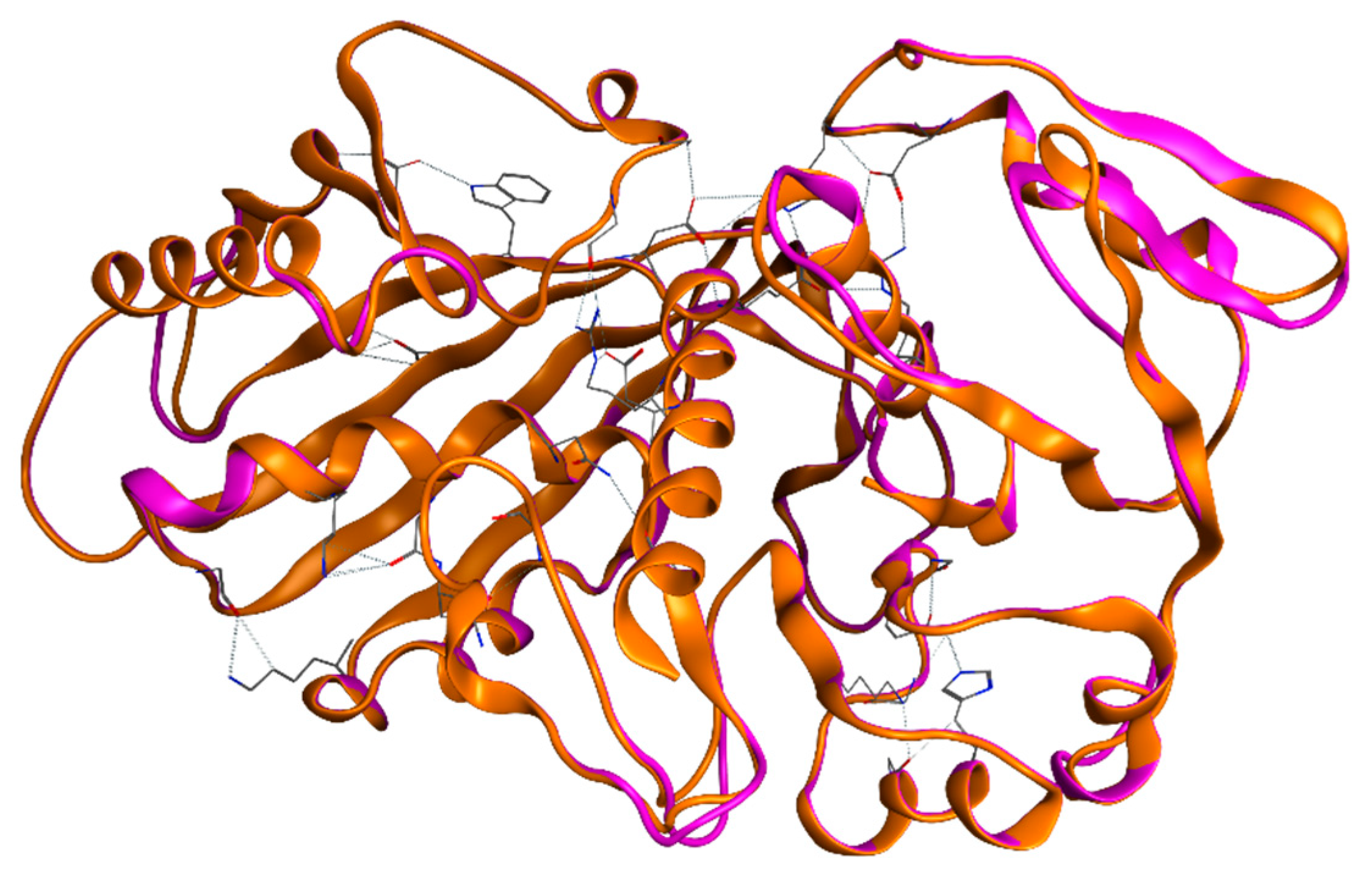
The secondary structures of both EgGAPDH were found to be highly similar. For W6UJ19 and W6V1T8, the predicted structures were as follows: α-helix, 33.80% and 33.93%; extended strand, 23.67% and 24.70%; β-turn, 9.17% and 8.33%; and random coil, 31.36% and 33.04%, respectively (Figure 2).

2.2.2. Tertiary Structure of EgGAPDH Isoenzymes Stability

The subunit of each isoenzyme was assayed for molecular structure stability. The root mean square deviation (RMSD) between the ab initio models generated with Phyre2 and the AlpHafold3 models was 1.27 Å for W6UJ19 and 1.42 Å for W6V1T8. After 100 ns of molecular dynamics simulation, the tertiary structures remained within a stable conformational range, indicating consistent structural stability throughout the simulation (Figure S2).

2.2.3. Quaternary Structures and LEP of Isoenzymes

Monomers of both isoenzymes were assembled in a quaternary structure through non-covalent bonds (Figure 3a,b).
The sequences of the predicted EgLEP were compared with the amino acid sequences of GAPDH from other species, including E. multilocularis, T. solium, F. hepatica, L. mexicana, H. sapiens, B. taurus, and O. aries. No similarities were found with the intracellular isoenzyme of GAPDH. However, the extracellular isoenzyme W6V1T8 showed similarity with T. solium (UniProt A8R8Q4) in the LEP region K138-T153, and with E. multilocularis (UniProt Q27652) in regions K138-T153 and F286-I301.
  • Post-translational LEP modifications.
The complete enzyme is formed by the assembly of four identical subunits without covalent bonds. LEPs were predicted for both W6UJ19 and W6V1T8. Table 2 shows the amino acid sequences of the LEP along with their predicted post-translational modifications.

2.3. Isoenzymes Docking and Molecular Dynamics Simulation

2.3.1. Isoenzymes Docking

GAPDH is composed of four identical subunits, each containing an active site for substrate binding, G3P, and binding sites for NAD+ and Pi (Figure 4). These functional sites were conserved among the GAPDH proteins analyzed in this study. However, L. mexicana and O. aries showed greater variability in the conformational binding sites for NAD+ and Pi (Table S2).
  • Isoenzymes potential energy
The potential energy of both isoenzyme control systems (quaternary structure of EgGAPDH with the substrate, Pi, and NAD+) through 100 ns of dynamic simulation is summarized in Table 3. Notably, the total potential energy exhibits a negative value and remains stable across all the simulations. The electrostatic partition constitutes the largest and most negative component. Conversely, the Van der Waals energy, although serving as a positive counterpart, possesses a relatively minor absolute value. Consequently, the overall energy persists in a negative state, underscoring the stability of the system. This outcome is consistent across all the simulations, reinforcing the notion that the electrostatic contribution plays a dominant role in determining the system’s energetic properties. Overall, the potential energy profiles of both isoenzymes in the control, EHDP, and AL systems are shown in Figure S3.

2.3.2. Amino Acids of EgGAPDH Involved in Substrate, Pi, and NAD+ Binding During Dynamics Simulation

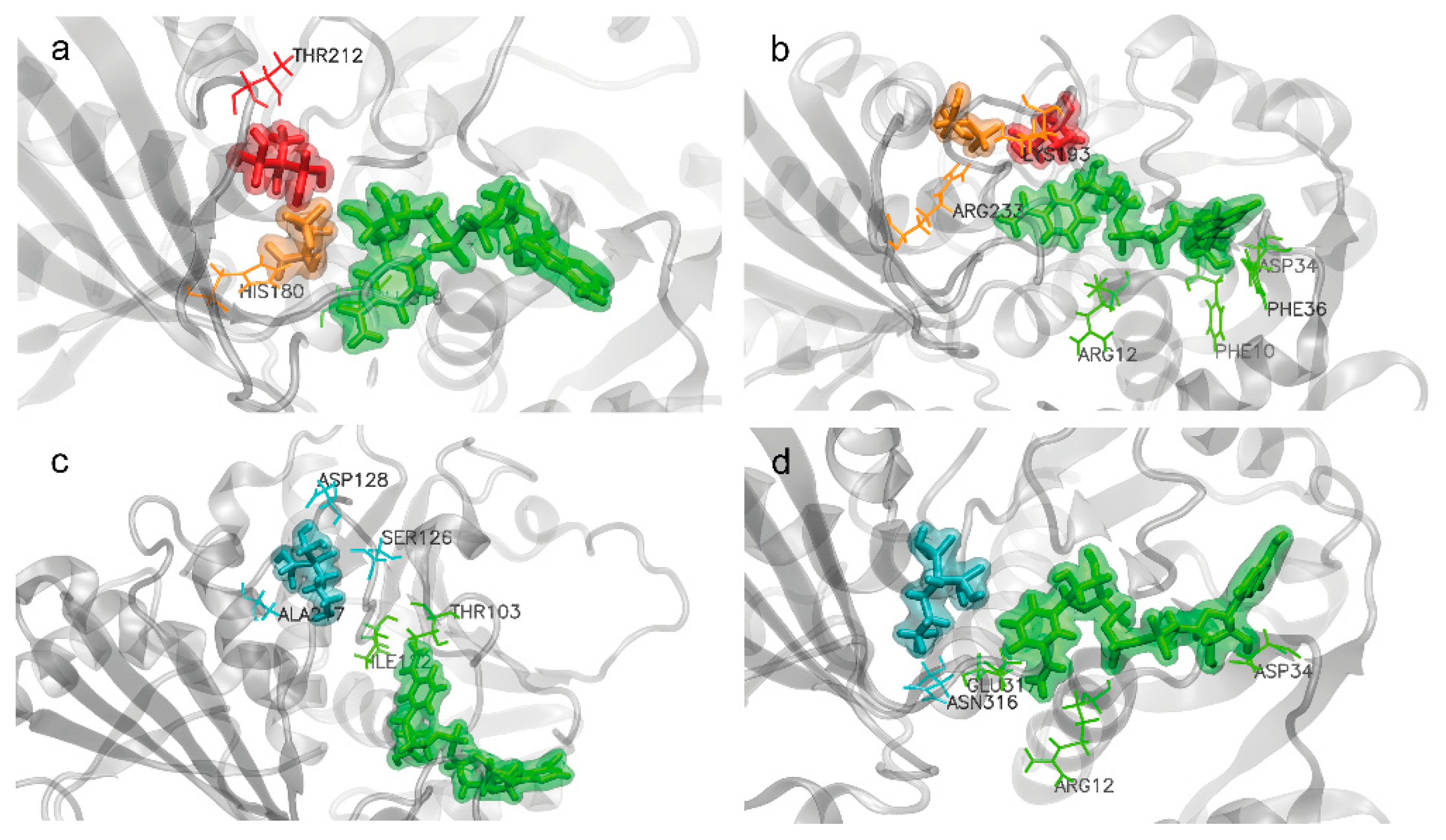
Throughout the molecular dynamics simulations, each subunit of EgGAPDH showed two binding sites corresponding to the substrate, Pi, and NAD+. These two sites corresponded to the beginning or the end of the 100 ns simulation. While the binding sites involved similar amino acids overall, differences were observed between isoenzymes and among subunits. The amino acids involved in substrate, Pi, and NAD+ binding for the IC isoform are shown in Figure 5a, and for the EC isoenzyme in Figure 5b. Interaction details in the 2D ligand interaction plot are shown in Figures S4 and S5.
  • Substrate binding
The residue R233 or R235 was involved in substrate binding of the monomers A, B, and C in the IC and monomers A and B of the EC isoenzyme at the beginning of the simulation. At the end of the simulation, this binding was maintained in monomers A and B in the IC, and appeared in all the monomers (A, B, C, and D) of the EC isoenzyme.
  • Inorganic phosphorus binding
For Pi binding, R233 was involved at the end of the simulation in monomers A, B, and C of the IC isoenzyme. In contrast, for the EC isoenzyme, R233 was involved in Pi interaction only in subunit C, with no Pi binding observed in the other monomers at the end of the simulation.
  • NAD+ binding
NAD+ binding interactions presented greater variability between subunits at the initial time point than at the end of the simulation, particularly in subunit A of the IC isoenzyme. Subunit C of the EC isoenzyme displayed different NAD+ interacting residues between the start and the end of the simulation, whereas subunit D showed no interaction with NAD+ at the end of the simulation.

2.3.3. Binding Free Energy (∆G) for Substrate, Pi, and NAD+

The binding free energy (∆G) values were calculated using the MMPBSA method based on the last 100 frames of the simulation. The binding free energy varied among the subunits of each EgGAPDH isoenzyme, highlighting that the amino acid interaction pattern may differ between subunits (Table S3). The binding of the substrate to monomer A yielded a ∆G binding: −10.6968 ± CI 95% 1.005 for W6UJ19 and −4.72 ± CI 95% 0.64 for W6V1T8. In contrast, NAD+ binding resulted in negative ∆G values only in monomers B (−4.0176, CI 95% ± 0.725) and D (−0.61, CI 95% ± 0.55) of W6V1T8. The calculated ∆G values for Pi were positive across all the subunits and isoenzymes.

2.4. Interaction of BP with Both EgGAPDH Isoenzymes

Two BPs, AL and EHDP, were docked in silico, showing stable molecular interactions with both EgGAPDH isoenzymes. These interactions did not alter the interaction potential energy, which remained consistent with the values shown in Table 3 and Figure S3. The structural stability of both enzymes in the presence of each BP is shown in Figure S6a,b through RMSD profiles.
Despite similar structural stability, differences were observed in the interactions between the two BPs and EgGAPDH. AL did not support co-binding of the substrate and Pi, whereas EHDP was able to dock with G3P at the active site, although it did not support Pi interaction.

2.4.1. Docking Affinity

  • EHDP increased Pi docking
After docking the EHDP in the presence of NAD+, Pi, and G3P, establishing an interaction zone (docking site) consisting of a sphere at 4.5 A centered on G3P, we observed that the EHDP was positioned superimposed on the Pi, so a new situation was modeled in which the EHDP is positioned in the same place as the Pi, having displaced the Pi.
Using the “placement none” resource, which allows the static interaction situation to be measured in each case, the interaction energy of the Pi in the NAD+, Pi, G3P complex in the absence of the EHDP was measured (Table 4):
  • AL increased NAD+ docking
After docking with AL in both enzymes, containing G3P, Pi, and NAD+, it was observed that AL was positioned in the same area as G3P and Pi. Therefore, a putative model was generated of an enzyme that is blocked by AL, having displaced itself during the interaction of all components to G3P and Pi. This does not mean that the docking has caused such displacement. This only proposes a possible model that could explain the interaction between Al and NAD+.
In these putative models, AL showed different influences on the binding of NAD+ at the site in both EgGAPDH isoenzymes (Table 5).

2.4.2. BP Effect on the Isoenzyme Substrate Affinity Studied by Molecular Dynamics

A measurement of the free energy differences (∆G) of ligands and enzymes was calculated by the MMPBSA method. Table 6 shows that the addition of BP changed the subunit affinity for the substrate, NAD+ and Pi, accordingly with EHDP or AL presence.
  • Substrate affinity.
Upon EHDP addition, substrate binding affinity became evident in both isoenzymes. Conversely, AL inhibited G3P binding at the active site, showing only weak binding in subunit B of W6UJ19 and subunit D of W6V1T8. Both GAPDH isoenzymes exhibited a variable substrate affinity, with some subunits showing weak or no binding for the substrate depending on the studied subunit (G3P) (Table 6). This instantaneous measurement makes evident the diversity among subunits with respect to their binding.
  • Pi affinity
The Pi interaction may be very transient with both isoenzymes, and its binding could not be reliably detected under control conditions. However, EHDP was able to bind to the Pi site of both isoenzymes, although binding to W6V1T8 occurred with lower affinity. In contrast, AL exhibited weak binding to the Pi conformational site on both W6UJ19 and W6V1T8 (Table 6).
  • NAD+ affinity
No NAD+ binding was detected in W6UJ19, either in the presence or absence of EHDP. In W6V1T8, NAD+ binding was observed, and upon EHDP addition, detectable NAD+ binding was observed in another subunit. However, AL promoted NAD+ binding in W6UJ19, specifically in subunits C and D, and in W6V1T8 in subunit D, although with a weaker value than in the control subunit B (Table 6).

2.4.3. Amino Acids Involved in BP Interactions

In Table 7 and Table 8, all amino acids interacting with all ligands are listed.
  • EHDP and AL interactions with EgGAPDH W6UJ19 and W6V1T8
In Figure 6, the main interactions of EHDP and AL with EgGAPDH W6UJ19 and W6V1T8 are shown. Figure S7 shows a 2D ligand interaction plot between EHDP and W6UJ19. In Figure S8 a 2D ligand interaction plot between EHDP and W6V1T8 is shown. In Figures S9 and S10, the 2D ligand interaction plots between AL with W6UJ19 and W6V1T8 are shown.

2.5. CEP and Its Modification by the BP Interactions

CEPs

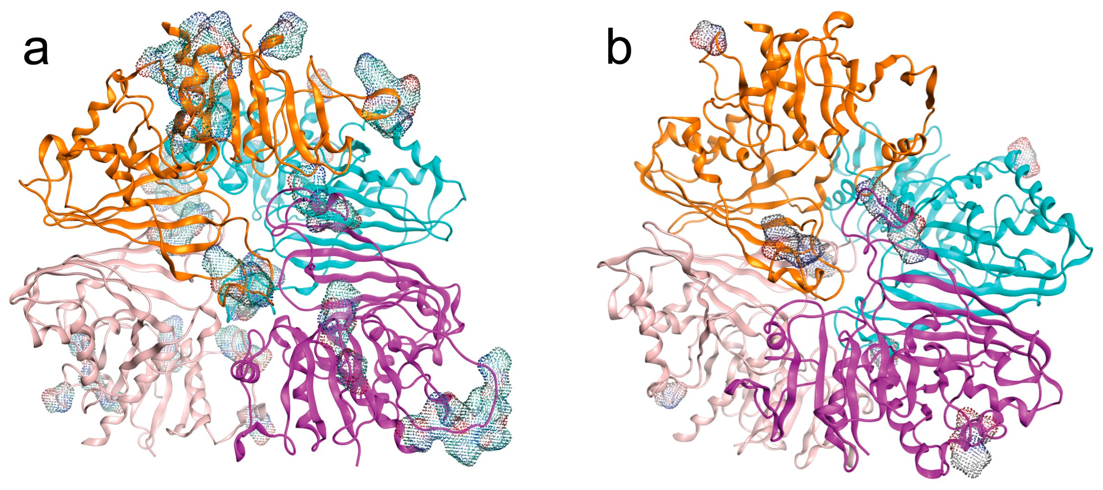
Surface CEPs were identified in both isoenzymes (Figure 7a,b)
The IC isoenzyme (UniProt W6UJ19) exhibited a greater number of conformational antigenic sites than the EC isoenzyme (UniProt W6V1T8). Subunit A was the most antigenic in the intracellular isoenzyme, while subunit B was the most antigenic in the extracellular isoenzyme based on the number of amino acid residues exposed in CEP (Table 9).
Although BP established non-covalent interactions with amino acid residues, they altered the surface exposure of the CEP. The effect of BP on the number and composition of amino acids in the CEP was most evident with EHDP on W6UJ19. The interaction of BP with CEPs of both isoenzymes is summarized in Table 9.

3. Discussion

GAPDH is a housekeeping protein, highly represented in cells with regulated enzymatic activity whose transcription and intracellular localization are regulated by feeding or cell proliferation [36]. GAPDH is a highly conserved protein throughout evolution, functioning in the glycolysis pathway by catalyzing the conversion of G3P, NAD+, and Pi into 1,3-bisphosphoglycerate and NADH, thereby connecting glycolysis with the respiratory chain. The enzymatic activity depends on the integrity of its quaternary structure, sustained by NAD+ interaction and the absence of negative cooperativity among its monomers, as demonstrated in studies on yeast GAPDH [37]. However, several additional functions have been attributed to GAPDH in cellular biology, including roles in endocytosis [38], fusion of plasma [39] and nuclear membranes [40], and autophagy [41]. Furthermore, partially oxidized GAPDH acylphosphatase activity, which may contribute to glycolysis by coupling with oxidative phosphorylation [42]. Depending on its subcellular localization, GAPDH can perform distinct functions. Nuclear GAPDH, whose localization is cell cycle-dependent, is proposed to play a protective role against DNA damage at telomeres caused by ceramide and toxins [43].
In addition, GAPDH contributes to cellular adaptability and defense against pathogens by participating in protein biogenesis and the regulation of gene expression [44]. Extracellular GAPDH has also been implicated in host–parasite interaction; for example, during leishmaniasis infection, it has been shown to downregulate TNF-α expression in host cells [45].
Given its multiple roles and physiological functions, GAPDH has been proposed as a pharmacological target in cancer, neurological diseases, and chronic parasitic infections [34]. Despite its high evolutionary conservation, GAPDH can elicit an immune response. Recombinant E. multilocularis GAPDH has been tested in laboratory animals to prevent metacestode development [35].
Standardized and reliable serological markers are needed for the diagnosis, prognosis, and post-treatment follow-up of patients with CE infection. Moreover, if the antigens are related to metabolic processes, parasite viability, or growth, such markers could also aid in CE infection stratification. Our group is searching for novel antigenic proteins to serve as serological markers for the diagnosis and monitoring of CE. This is the first work presenting the in silico model of two GAPDH isoenzymes from E. granulosus ss/G1, recognized by sera from CE patients with liver cysts. These isoenzymes were found in different subcellular localizations, W6UJ19 (UniProt) in IC and W6V1T8 (UniProt) in EC. Physicochemical differences between the two isoenzymes were observed, particularly in their isoelectric points and net molecular charges, both of which were higher in the EC isoenzyme. Both isoenzymes were detected by PCR in ex vivo metacestode samples. The genetic identity between IC and EC isoenzymes was approximately 74%. In contrast, other EgGAPDH isoenzymes not recognized by immunosera (UniProt W6UPZ5 or A0A068WMZ6) shared only ~20% identity with the IC and EC isoenzymes.
Regarding LEP singularity, the IC isoform W6UJ19 is more specific for E. granulosus than the EC W6V1T8. In fact, the EC EgGAPDH is likely responsible for the initial activation of the host immune system, producing immune cross-reaction among E. granulosus, E. multilocularis, and T. solium, while the IC isoform, which is more specific, may be released as a consequence of parasite cell death. The IC isoenzyme (UniProt W6UJ19) emerged as the main candidate for differential diagnosis, showing approximately 53% sequence identity with the human GAPDH and approximately 70% identity with GAPDH sequences from E. multilocularis, T. solium, and F. hepatica, and 55% identity with L. mexicana. The IC isoenzyme (UniProt W6UJ19) is also the most promising candidate for constructing a recombinant multiepitope antigen. Notably, it does not share predicted LEP with E. multilocularis or T. solium in the regions spanning residues S123-L138 and F288-I303. Additionally, no post-translational modifications were predicted in the sequences containing these epitopes. Moreover, an antibody reactivity rate between W6V1T8 and W6UJ19 could be useful to determine infection prognosis and treatment success [46]. Due to analysis of metacestode vesicles by the LC-MS/MS method with different metabolic activities, correlation was found between W6V1T8 abundance (emPAI) and vesicle metabolic activity. Instead, W6UJ19 showed consistent relative abundance regardless of metabolic activity [46]. Enzymes from the glycolytic pathway are localized in calcareous corpuscles [15], where EgGAPDH EC is probably localized.
CEP exhibited results conceptually similar to those described for LEP. Those results, regarding LEP and CEP, showed that the IC isoform could be more antigenic than the EC isoform. The IC isoenzyme (UniProt W6UJ19) exhibited a greater number of conformational antigenic sites than the EC isoenzyme (UniProt W6V1T8). Moreover, changes in the number of amino acids involved in CEP upon non-covalent binding of EHDP were more evident in the IC isoform. The EC isoform immunoreactivity may correlate with parasite metabolic activity. EHDP and AL decreased the number of CEP-related amino acids on the EC isoform, which may produce a reduction in CEP immunoreactivity. In contrast, BP effect on W6UJ19 increased CEP-related amino acid, potentially enhancing CEP antigenicity and renewing the immune recognition in chronic infections. The EHDP effect could be related to the PPi released from the calcareous body. These PPi would bind the EC enzyme in the Pi site and consequently decrease its antigenicity.
Molecular dynamics simulations showed that the IC isoenzyme presented a greater affinity for both the substrate and Pi, maintaining more stable amino acid interactions throughout the simulation compared to the EC isoenzyme. Although enzymatic activity assays were not performed, the simulation results suggest that the IC isoform is more likely to play a metabolic role in the E. granulosus metacestode regarding the MMPBSA results, where substrate affinity is higher in W6UJ19. In contrast, the EC isoform, likely localized in secretion vesicles and/or calcareous corpuscles, appears to participate primarily in host–parasite interaction [15], potentially through protein–protein interactions that facilitate parasite attachment. According to our findings, the quaternary structure of the enzyme remained stable, according to the RMSD results, even when the conformational sites for NAD+ or Pi binding were asymmetrically lost by the end of the molecular dynamics simulations. While the quaternary structure of the enzyme is essential for enzymatic activity, it is likely also required for the protein’s non-enzymatic functions, particularly those related to intercellular or intracellular interactions. GAPDH lacks a classical secretion signal peptide, suggesting that its ability to be secreted may depend on interactions with carrier proteins that facilitate its translocation to other cellular compartments [47,48].
Among the BP studied, EHDP reduced cell growth, whereas AL did not produce any measurable effect on in vitro cell behavior, beyond a reduction in ATP levels. This BP acts via different mechanisms: EHDP leads to the accumulation of non-degradable ATP analogs [49], while AL inhibits the protein prenylation pathway. EHDP showed stronger binding to both GAPDH isoenzymes than Pi, although pyrophosphate (PPi) exhibited the highest binding affinity (p < 0.005 concerning EHDP, calculated by docking). However, PPi was unable to substitute Pi in the catalytic activity of GAPDH [50]. Unlike PPi, EHDP is not degraded by pyrophosphatases. EHDP also enhanced substrate binding to the IC enzyme and modified the conformational sites for substrate, Pi, and NAD+ binding throughout the molecular dynamics simulation, from initiation to completion, suggesting a potential modulation of enzymatic activity. Consequently, we can assume that the GAPDH model with EHDP and without Pi has increased affinity for the substrate in both isoenzymes.
In contrast, AL exhibited lower affinity for GAPDH compared to both the substrate and Pi. EHDP bound more strongly than Pi, and previous studies have reported that phosphonates can interact with GAPDH by binding after Pi removal; the acyl-enzyme intermediate is expected to enhance activity [51]. However, an increase in GAPDH activity may also lead to elevated production of reactive oxygen species, which could overwhelm the cell’s antioxidant defenses and modify the enzyme’s active site, affecting its ability to bind substrate or Pi [42].
One of the known metabolic effects of EHDP is the reduction in glycolysis rate and the promotion of glycogen storage [52] without altering intracellular Pi concentration or uptake [53]. In previous experiments, we observed a decrease in EGPE cell growth after 72 H of EHDP treatment. This selective effect on parasite cells could be attributed to the limited antioxidant capacity of E. granulosus, as neither superoxide dismutase nor the thioredoxin system was overexpressed in response to H2O2 challenge [54]. In fact, EHDP is the BP with the least effect on bone metabolism at a dose used in humans and animals [49,55,56]. Due to its slight effect on bone metabolism and selective effect on EgGAPDH, EHDP, which has been shown to inhibit EGPE cell growth, could be evaluated as an adjuvant treatment for CE localized in bone. As a member of the BP compound family, EHDP exhibits more affinity for bone tissue than for other organs.
Several studies have explored the immunological effects of BP, demonstrating their potential as adjuvant therapies [57] and their ability to promote prolonged antigen persistence within immune cells. However, these studies often did not distinguish whether the immunological modulation was due to BP interference in the mevalonate pathway or a direct effect on the antigenic molecule itself [58].

4. Materials and Methods

4.1. Ethics Statement and Serum Samples

Serum samples from CE patients were obtained by Dr. Jorge Gentile at the Hospital Municipal Ramón Santamarina, Tandil, Argentina. Dr. Elizabeth Luz Sánchez Romaní, from the Laboratorio de Zoonosis Parasitaria CNSP-INS, Peru, provided sera samples from patients with cysticercosis and fascioliasis. The Ethics Committee of the Universidad Abierta Interamericana, Buenos Aires, Argentina, approved all the protocols and procedures (number 01011) date 28 October 2014, and the ethical protocol approval has continued up to today [12].

4.2. Characterization of EgGAPDH

4.2.1. EGPE Cell Culture, Protein Extracts, and Supernatant

EGPE is a cell line derived from the liver bovine metacestode of E. granulosus ss/G1 pe. The cell line was obtained in 2008–2010 in our laboratory (Centro de Altos Estudios en Ciencias Humanas y de la Salud, Universidad Abierta Interamericana). Parasite’s pe was digested with papain, and cells cultured in liquid medium 199, antibiotics, β-mercaptoethanol, sodium pyruvate, and 10% filtrated cystic fluid for over one month were discarded because mammalian cells contaminated the parasite cell culture. Afterward, the cystic fluid was changed with 10% inactivated BFS. Cystic colonies were obtained by seeding cells in semisolid 2% agarose medium in 5 mM buffer phosphate and 5mM HEPES (1:1; v/v), pH 7.5. After 30 min of cell seeding, liquid medium was added, and the plates were cultured in a humid tissue culture incubator 95% air/5% CO2. Cells were maintained in our laboratory in liquid nitrogen. Cells used in the experiments corresponded to passages 35 to 40. EGPE cells were cultured, as previously described [11], in medium 199 (Sigma, St. Louis, MO, USA), 1 mM sodium pyruvate (sodium salt, extra pure, Anhedra, Beijing, China), and 78 μg/mL β-mercaptoethanol (Merck, Darmstadt, Germany) at pH 7.9 (37 °C; CO2:air; 5/95%). EGPE cell colonies were performed in 2% agarose (20,000 cells/well) [11]. After 20 days of cell growth in a liquid medium, the protein extracts were obtained. EGPE cells were washed five times with DPBS and incubated in lysis buffer (8 mmol/L CHAPS, MP Biomedicals, 10 mmol/L Tris–HCl, pH 8, 2 mmol/L EDTA, 0.1% B-mercaptoethanol, MP Biomedicals, and 1/100 protease inhibitor cocktail, Sigma-Aldrich) at 4 °C for 2 h. The samples were then frozen-thawed three times and spun down at 10,000× g. [12].
Cell colonies were grown for 5 days without medium change, supernatants were obtained, and cell debris was removed by centrifuging the supernatants three times (3000 rpm). Finally, samples were aliquoted and stored at −20 °C until use.

4.2.2. Protein Identification

The methodology was previously described by Maglioco et al., 2022 [13]. Briefly, protein fractions were obtained by separating cell extracts based on molecular weight using a Sephacryl S200 HR column (GE Healthcare, Hertfordshire, UK). Eluted protein fractions were identified by absorbance (205–280 nm) using a spectrophotometer (Biowave II+, Biochrome Ltd., Cambridge, UK). Each protein fraction and EGPE supernatant were concentrated through a 3 kDa cut-off membrane concentrator (Pierce, Thermo Scientific, Waltham, MA, USA). Antigenic protein fractions and EGPE supernatant were analyzed by Western blot using a pool of sera from 11 patients with hepatic CE. Reactive protein fractions were subsequently purified by affinity chromatography using Protein G HP SpinTrap columns (GE Healthcare). Sera from patients with cysticercosis and fascioliasis were used as controls. Eluted proteins were concentrated through a 3 kDa cut-off membrane concentrator (Pierce, Thermo Scientific).
A 15% SDS–PAGE was performed to concentrate and clean up the protein extracts before in-gel digestion. Protein bands were visualized by Coomassie blue staining, excised, destained, washed, reduced, alkylated, and then digested in-gel with 100 ng Trypsin (Promega V5111). The resulting peptides were recovered by elution and analyzed by nano LC-MS/MS at the Proteomics Core Facility of the CEQUIBIEM, Faculty of Exact Sciences, University of Buenos Aires/IQUIBICEN CONICET. Protein identification was performed using the E. granulosus database from UniProt.org [59], following the procedure described by Maglioco et al., 2022 [13]. Spectrum data was deposited in Project accession: PXD069559 (Table S1). Previously, both GAPDHs were found in ex vivo E. granulosus metacestode [46] (PXD067276).

4.2.3. EgGAPDH Genomic Sequence

E. granulosus DNA was extracted from a G1 genotype metacestode isolated from the liver of a cow obtained at a local slaughterhouse in Buenos Aires, Argentina (provided by Dr. Tatiana Aronowicz, SENASA). Primers for amplification of the complete GAPDH gene sequence were designed using the Primer-Blast tool available on the NCBI website [60]. The fragment size ranged from ≈800–1000 bp.
For W6UJ19 (IC), the forward primer was GAGGCCCAACACCGGAATTA and the reverse CCACGGGTTTCAGTAATGAGC (Tm 54.36 °C; extension time: 1 min 3 s). For W6V1T8 (EC), the forward primer was TCGAGAAGGCCTCGGTAAGA and the reverse primer was TCCAGCGGGAGCCTTAATGA (Tm 54 °C; extension time: 50 s). Primers were purchased from Gene Biotech SRL, Buenos Aires, Argentina. Each reaction tube contained: 1.5 mM MgCl2 (5x, Colorless GoTaq® Reaction Buffer, Promega, Madison, WI, USA), 0.2 mM of dNTPs mix (dGTP, dCTP, dTTP and dATP, Promega, Madison, WI, USA), 1 µM forward primer, 1 µM reverse primer, 1.25 units of DNA polymerase (GoTaq® polymerase, Promega, Madison, WI, USA), 18 ng of DNA template and nuclease-free water up to 25 µL (ultrapure, PB-L, Productos Bio-lógicos). DNA amplification was performed with an initial denaturation step at 95 °C for 2 min, followed by 35 cycles of denaturation at 95 °C for 1 min, primer annealing at the respective temperature for each primer pair for 1 min, and extension at 72 °C for a time determined by the expected product size. A final extension step was carried out at 72 °C for 5 min. Reactions were conducted using a Mastercycler gradient thermocycler (Eppendorf, Hamburg, Germany). Then, 17.5 µL of each product was analyzed by agarose gel electrophoresis (LE molecular biology grade, PB-L, Productos Bio-Lógicos, Buenos Aires, Argentina) using a 100–1100 bp ladder (Dongsheng Biotech Co., Ltd., Guangzhou, China) as a size marker. DNA bands were excised and purified using the Wizard® SV Gel and PCR Clean Up System (Promega Co., USA). Purified PCR products were sequenced at the CEDIE “Dr César Bergadá (CONICET- Hospital de Niños Ricardo Gutiérrez, Buenos Aires, Argentina). The resulting sequences were analyzed using BLAST and manual alignment. Amino acid sequences corresponding to predicted epitopes were identified from the full-length protein sequences retrieved from the BLAST results.

4.2.4. Amino Acid Sequence, Protein Structure, and Physicochemical Analysis

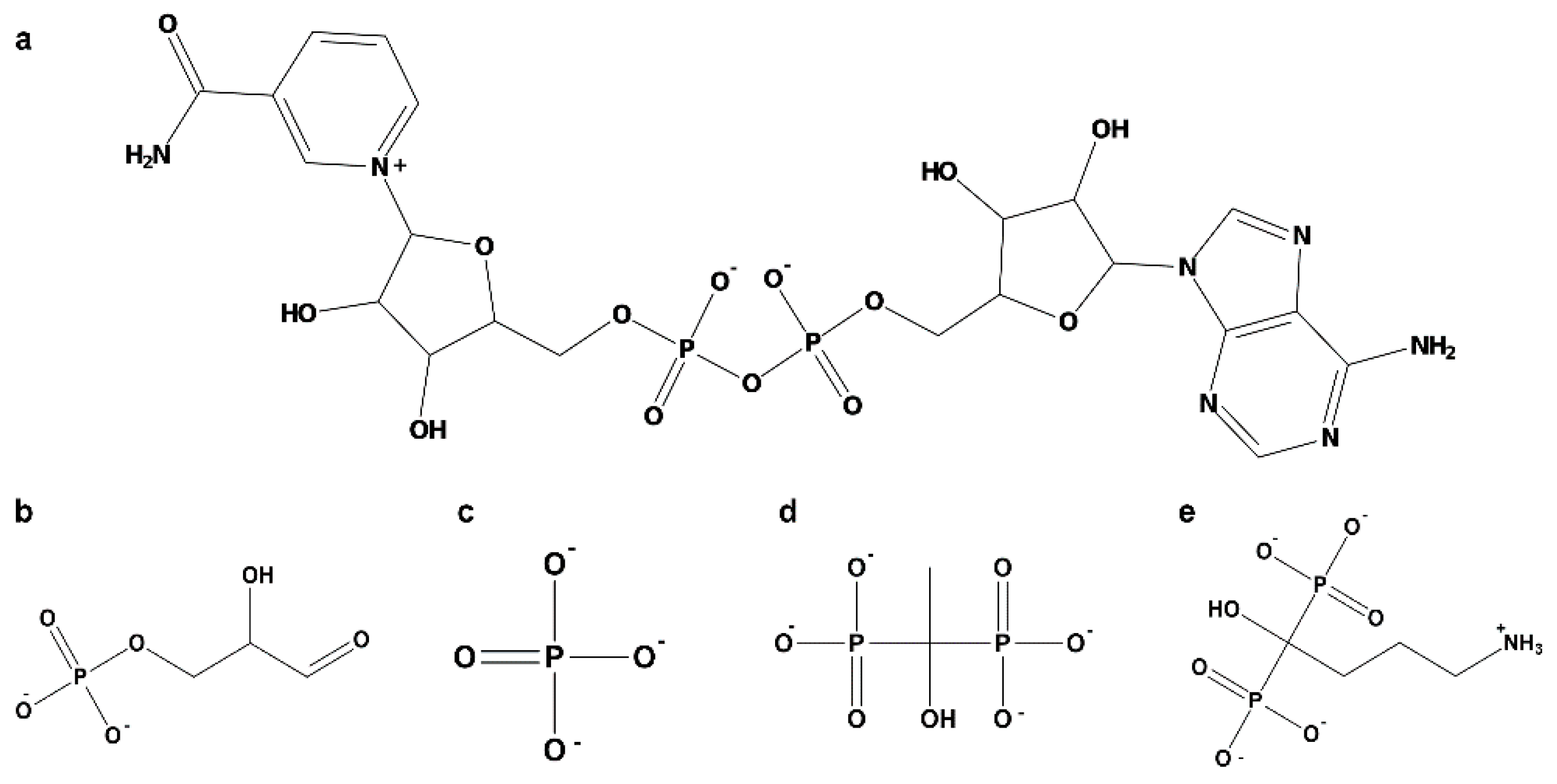
The complete amino acid sequences of both GAPDHs were obtained from UniProt. Analysis of differences in GAPDH protein sequences was carried out using BLAST-P, using the complete sequence of each isoenzyme and a non-redundant database and organism as inputs. Physicochemical properties were analyzed using the ProtParam tool from the Expasy server [61]. Secondary structure prediction of E. granulosus GAPDH was conducted using SOPMA [62]. GAPDH domains were predicted using Pfam [63], NCBI [64], and Interpro [65]. Modeling of the tertiary structure was performed using Phyre2 normal mode [66]. The protein models obtained by Phyre2 have been superposed to AlphaFold3 models [67], and the root mean square deviation (RMSD) between the two models was evaluated. Model quality was assessed by ProSA-web [68]. To assemble GAPDH monomers into a quaternary structure, we modeled the 222 symmetry homotetramer (biological assembly) using Leishmania mexicana GAPDH (PDB code: 1I32) as a template. The model of Acropora millepora (PDB code: 6PX2) was used to map the binding sites for substrates Pi, and NAD+. The protonation and tautomeric states of titratable residues, and the orientation of hydrogen atoms, were assigned using the Protonate3D module implemented in MOE (MOE 2022.02). This procedure simultaneously optimizes ionization, tautomeric, and hydrogen-bonding states based on a free-energy model including Van der Waals, Coulomb, solvation, and titration terms [69]. Calculations were performed at pH 7 and a saline concentration of 0.1 M. Histidine residues were protonated at Nε2 (HIE) unless hydrogen bonding suggested an alternative tautomeric state. Ligands (G3P, Pi, EHDP, and AL) were parameterized in their predominant ionization states at neutral pH: G3P as a dianion, EHDP as a tetraanionic species, and Pi and AL as trianionic species. The resulting protonation states were used to generate the fully protonated structures for subsequent energy minimization, docking, and molecular dynamics simulations, and are shown in Figure 8.

4.3. Molecular Dynamics Simulation

Native Interactions of GAPD

The GAPDH molecular structures were validated through molecular dynamics simulation using the Nanoscale Molecular Dynamics software (version 3.0, NAMD2) [70]. GAPDH was solvated with explicit solvent by using the TIP3 water model. For W6UJ19, the water box size was 114.83 × 122.65 × 107.08 Å (x, y, z), containing 40,077 water molecules. For W6V1T8, the water box was 116.29 × 117.06 × 110.21 Å (x, y, z), containing 40,085 water molecules. System neutralization was performed to reflect the distinct localization of both enzymes; KCl was used for the IC (W6UJ19) and NaCl for the EC isoenzyme (W6V1T8). The ionic concentration was 0.15 mol/L in both cases. All simulations were performed under the periodic boundary conditions. The counterion content was 112 Cl and 128 K+ atoms and 112 Cl and 129 Na+, for W6UJ19 and W6V1T8, respectively. All molecular dynamics simulations were performed using the CHARMM36 force field under NPT Ensemble (Isothermal-Isobaric) conditions: constant particle number, pressure (1 atm), and temperature (300 K). The simulation protocols involved 20,000 steps for minimization by the conjugate gradient method plus 144,000 steps (0.288 ns) of heating from 60 to 300 K, maintaining the absolute temperature at 300 K for the following steps. Afterward, 500,000 steps (1 ns) were performed to equilibrate with restrictions in the backbone, and finally, a 100 ns unrestrained trajectory was obtained for production. The time of each step was 2 fs. The potential energy throughout the simulation was monitored to confirm the thermodynamic equilibrium of each system. The root mean square deviation (RMSD) and root mean square fluctuation (RMSF) were calculated. Structures were analyzed and visualized with Visual Molecular Dynamics (version 1.9.3) [71] and Molecular Operating Environment (MOE 2022.20) [72].
  • Free energy binding estimation.
The free energy binding between the protein chains and the ligand molecules was estimated by the MMPBSA method using the last 100 frames of each molecular dynamic simulation with the VMD CAFE1.0 tool and the APBS program. For each estimation, the entire molecular system was defined as the complex, the target molecule as the ligand, and all remaining molecules in the system (excluding the ligand) as the receptor.

4.4. Docking and Enzymatic Dynamic Interaction with BP

4.4.1. Docking

Molecular docking simulations of the natural substrate of GAPDH, glyceralde-hyde-3-phosphate (G3P), Pi, and two BPs, EHDP (PubChem: CID3305) and AL (PubChem: CID2088), were performed using the Dock module of MOE 2022.20. The catalytic site corresponding to the substrate-binding region of each GAPDH enzyme with NAD+ positioned in its corresponding binding site was defined by selecting residues within a sphere of 4.5 Å radius centered on the catalytic cysteine.
Ligand conformations were generated by systematic bond rotations prior to placement. Poses were generated using the Alpha Triangle placement method, which aligns triplets of ligand atoms with triplets of α-sphere centers within the defined binding site. Each ligand yielded up to ten poses, initially scored with the London ΔG function and subsequently refined through energy minimization using the GBVI/WSA ΔG scoring scheme. The top-ranked pose for each ligand was selected for subsequent molecular dynamics simulations. Differences among docking scores were analyzed using the Student t-test, considering that each enzyme has 4 subunits; each subunit was considered a single unit for analysis.

4.4.2. Molecular Dynamics Interaction

Protein-ligand complexes were prepared for molecular dynamics simulation using MOE software (version 2024.06) and the QwikMD tool [73] available in VMD 1.9.3, as previously described. All molecular dynamics simulations were performed on the homotetrameric quaternary structures of both GAPDH isoenzymes, each composed of four identical subunits arranged in 222 symmetry, as described for other GAPDH enzymes. The tetrameric assemblies were generated based on the L. mexicana GAPDH template (PDB ID: 1I32) and validated for structural stability during the simulations. Ligand three-dimensional geometries were manually built and optimized in MOE 2022.20. Initial geometry optimization was performed using the MMFF94x force field to refine bond lengths, angles, and torsions. For molecular dynamics simulations, the ligands were parameterized using the CHARMM General Force Field (CGenFF), ensuring full compatibility with the CHARMM36 force field employed for the protein systems. Each optimized ligand structure was visually inspected to confirm correct bond orders, protonation states, and stereochemistry prior to docking and simulation.
A 100 ns production simulation was performed without backbone restraints. Molecular dynamics settings were the same as outlined in Section Native Interactions of GAPD, with modifications in the number of water molecules and ions for each system. For the W6UJ19-EHDP complex, the solvation box contained 40,078 water molecules along with 112 Cl and 116 K+ ions. For the W6UJ19-AL complex, the system included 40,081 water molecules and 116 Cl, with 112 K+ ions. In the W6V1T8-EHDP system, 40,084 water molecules were used, along with 112 Cl and 116 Na+ ions. The W6V1T8-AL system contained 39,980 water molecules and 116 Cl with 112 Na+ ions. All simulations were executed remotely using NAMD2 on high-performance computing equipment at the Department of Experimentation and Theory of the Structure of Matter and its Applications (DETEMA, Facultad de Química, Universidad de la República, Montevideo, Uruguay). Hardware specifications included: AMD Ryzen 9 5950X processor, RAM 64 GB, RTX 3080 TI GPU, running Ubuntu 22.04.1.

4.5. Searching for Epitopes

4.5.1. B-Cell LEP

LEP prediction was conducted on GAPDH sequences from E. granulosus, T. solium, F. hepatica, E. multilocularis, Bos taurus, Ovis aries, Homo sapiens, and L. mexicana using eight epitope prediction programs. The B-cell LEP for each GAPDH were selected from ABCpred (with a score above 0.85) [74], identified in regions of at least five adjacent amino acids by Bepipred Linear Epitope Prediction 2.0 (threshold: 0.5) or by Bepipred Linear Epitope Prediction (threshold: 0.35), and by at least three of the following software: Chou & Fasman Beta-Turn Prediction (threshold indicated by the server for each protein sequence), Emini Surface Accessibility Prediction (threshold: 1.0), Karplus & Schulz Flexibility Prediction (threshold: 1.0), Kolaskar & Tongaonkar Antigenicity (threshold indicated by the server for each protein sequence), and Parker Hydrophilicity Prediction (threshold indicated by the server for each protein sequence) in IEDB (Immune Epitope Database and Analysis Resource) [75]. Post-translational modifications were predicted using MusiteDeep [76] (cut off 0.50) and DeepNitro [77] (cut off medium score).

4.5.2. B-Cell CEP and BP

B-cell CEP for each GAPDH protein was predicted using DiscoTope 2.0, applying a threshold of −3.7 (sensitivity = 0.47 and specificity = 0.75). Predictions were based on the PDB structures of each GAPDH system at both the beginning and end of the molecular dynamics simulation, including IC and EC isoenzymes under control conditions and after binding with EHDP and AL.

5. Conclusions

We performed the first in silico studies on E. granulosus GAPDH, an essential enzyme of the glycolytic pathway involved in host–parasite interactions, recognized by sera from CE patients. Based on the predicted LEP in two GAPDH isoenzymes, the IC isoenzyme (W6UJ19) emerged as the best candidate for the design of multiepitope constructs with potential applications in CE diagnosis, and the ratio between the IC and EC isoenzymes will be useful to evaluate treatment success. Moreover, the CEP modification by EHDP on W6UJ19 could enhance immunological surveillance, although it may decrease antigenicity of W6V1T8.
Molecular dynamics simulations suggest that the IC isoenzyme has a greater probability of being modulated by BP compared to the extracellular (EC) isoenzyme (W6V1T8), supporting its potential as a pharmacological target. Simulations examining the interaction of two BPs, EHDP and AL, with both isoenzymes showed that EHDP (a first-generation BP) exhibits stronger binding affinity than AL. This finding aligns with our previous in vitro results using EGPE. Indeed, EHDP, due to its limited effect on bone metabolism and its potential antiparasitic effect, could be a candidate for translational research in infections caused by E. granulosus in intermediate hosts. Notably, EHDP shares a structural similarity with PPi, suggesting that endogenous PPi could potentially interact with GAPDH and modulate its enzymatic activity or interfere with EHDP’s effect; however, only PPi is metabolized by endogenous pyrophosphatase. The simulations also provided insights into molecular stability and substrate interactions affected or unaffected by BP binding. Furthermore, we demonstrated how CEP could be altered upon binding of those interactive compounds.

Supplementary Materials

The following supporting information can be downloaded at https://www.mdpi.com/article/10.3390/ijms262110622/s1.

Author Contributions

F.A.A. performed PCR experiments, in silico and data analysis, and contributed to manuscript writing. A.M. contributed to in silico data analysis and manuscript writing. M.P.V. performed proteomic protein identification and analysis. A.Y.J.V. contributed to PCR experiments and manuscript writing. E.R. contributed to manuscript writing. M.P. was a director of in silico experiments and data analysis and contributed to manuscript writing. A.G.F. was the research director, conceived and designed the results analysis, and wrote the manuscript. All authors have read and agreed to the published version of the manuscript.

Funding

This work was supported by the Fundación Iberoamericana de Estudios Superiores. Chacabuco 90, B Buenos Aires, Argentina, 2024.

Institutional Review Board Statement

The study was conducted in accordance with the Declaration of Helsinki and approved by the Ethics Committee of the Universidad Abierta Interamericana, Buenos Aires, Argentina (protocol code 01011 and date of 28 October 2014).

Informed Consent Statement

Informed consent was obtained from all the subjects involved in the study.

Data Availability Statement

The original contributions presented in this study are included in the article/Supplementary Material. Further inquiries can be directed to the corresponding authors.

Acknowledgments

The authors appreciate Claudia Nose for the preparation of the figures, from the National Institute of Parasitology “Dr. Mario Fatala-Chaben,” ANLIS-Malbrán, CABA, Argentina.

Conflicts of Interest

The authors declare no conflicts of interest.

References

  1. Vuitton, D.A.; McManus, D.P.; Rogan, M.T.; Romig, T.; Gottstein, B.; Naidich, A.; Tuxun, T.; Wen, H.; da Silva, A.M.; World Association of Echinococcosis. International consensus on terminology to be used in the field of echinococcoses. Parasite 2020, 27, 41. [Google Scholar] [CrossRef]
  2. Cucher, M.A.; Macchiaroli, N.; Baldi, G.; Camicia, F.; Prada, L.; Maldonado, L.; Avila, H.G.; Fox, A.; Gutiérrez, A.; Negro, P.; et al. Cystic echinococcosis in South America: Systematic review of species and genotypes of Echinococcus granulosus sensu lato in humans and natural domestic hosts. Trop. Med. Int. Health 2016, 21, 166–175. [Google Scholar] [CrossRef]
  3. Equinococosis. Available online: https://www.who.int/es/news-room/fact-sheets/detail/echinococcosis (accessed on 19 April 2023).
  4. Avila, H.G.; Maglioco, A.; Getiser, M.L.; Ferreyra, M.P.; Ferrari, F.; Klinger, E.; Barbery Venturi, M.S.; Agüero, F.A.; Fuchs, A.G.; Jensen, O. First report of Cystic echinococcosis caused by Echinococcus granulosus sensu stricto/G1 in Felix catus from the Patagonian region of Argentina. Parasitol. Res. 2021, 120, 747–750. [Google Scholar] [CrossRef]
  5. Tercero Gutiérrez, M.J.; Olala Herbosa, R. Hidatidosis una zoonosis de distribución mundial. ofFARM 2008, 27, 88–94. [Google Scholar]
  6. Tévez Craise, L.; Daiana Vaccaro, R.; De Luca, P.A.; Vásquez Guillén, M.E.; Calaramo, O.A.; Logioco, F. Hydatidosis: Clinical-imaging classification according to Gharbi and the World Health Organization. Rev. Argent. Radiol. 2022, 86, 41–48. [Google Scholar]
  7. Tamarozzi, F.; Silva, R.; Fittipaldo, V.A.; Buonfrate, D.; Gottstein, B.; Siles Lucas, M. Serology for the diagnosis of human hepatic cystic echinococcosis and its relation with cyst staging: A systematic review of the literature with meta-analysis. PLoS Negl. Trop. Dis. 2021, 15, e0009370. [Google Scholar] [CrossRef]
  8. Pittini, A.; Martínez Acosta, Y.E.; Casaravilla, C.; Seoane, P.I.; Rückerl, D.; Quijano, C.; Allen, J.E.; Díaz, A. Particles from the Echinococcus granulosus laminated layer Inhibit CD40 upregulation in dendritic cells by interfering with Akt activation. Infect. Immun. 2019, 87, e0064. [Google Scholar] [CrossRef]
  9. Barbieri, M.; Fernández, V.; González, G.; Luaces, V.M.; Nieto, A. Diagnostic evaluation of a synthetic peptide derived from a novel antigen B subunit as related to other available peptides and native antigens used for serology of cystic hydatidosis. Parasite Immunol. 1998, 20, 51–61. [Google Scholar] [CrossRef] [PubMed]
  10. Sadjjadi, S.F.; Tahereh Mohammadzadeh, T.; Fatemeh Hafezi, F.; Sadjjadi, S.M. Evaluation of the ability of antigen B originated from Echinococcus granulosus sensu stricto and E. Canadensis for the diagnosis of confirmed human cystic echinococcosis using ELISA. Iran J. Parasitol. 2022, 17, 358–465. [Google Scholar] [CrossRef]
  11. Echeverría, C.I.; Isolabella, D.M.; Prieto Gonzalez, E.A.; Leonardelli, A.; Prada, L.; Perrone, A.; Fuchs, A.G. Morphological and biological characterization of cell line developed from bovine Echinococcus granulosus. Vitr. Cell. Dev. Biol. Anim. 2010, 46, 781–792. [Google Scholar] [CrossRef]
  12. Maglioco, A.; Gentile, J.; Barbery Venturi, M.S.; Jensen, O.; Hernández, C.; Gertiser, M.L.; Poggio, V.; Canziani, G.; Fuchs, A.G. Detection of Echinococcus granulosus sensu lato infection by using extracts derived from a protoscoleces G1 cell line. Parasite Immunol. 2019, 41, e12674. [Google Scholar] [CrossRef] [PubMed]
  13. Maglioco, M.; Agüero, F.A.; Valacco, M.P.; Juàrez Valdez, A.; Paulino, M.; Fuchs, A.G. Characterization of the B-Cell Epitopes of Echinococcus granulosus histones H4 and H2A recognized by sera from patients with liver cysts. Front. Cell. Infect. Microbiol. 2022, 12, 901994. [Google Scholar] [CrossRef]
  14. Muronetz, V.I.; Kuravsky, M.L.; Barinova, K.V.; Schmalhausen, E.V. Sperm-specific glyceraldehyde-3-phosphate dehydrogenase—An evolutionary acquisition of mammals. Biochemistry 2015, 80, 1672–1689. [Google Scholar] [CrossRef]
  15. Ahn, C.S.; Kim, J.G.; Bae, Y.A.; Kim, S.H.; Shin, J.H.; Yang, Y.; Kang, I.; Kong, Y. Fasciclin-calcareous corpuscle binary complex mediated protein-protein interactions in Taenia solium metacestode. Parasit. Vectors. 2017, 10, 438. [Google Scholar] [CrossRef]
  16. Dar, G.H.; Mendes, C.C.; Kuan, W.L.; Speciale, A.A.; Conceição, M.; Gorgens, A.; Uliyakina, I.; Lobo, M.J.; Lim, W.F.; EL Andaloussi, S.; et al. GAPDH controls extracellular vesicle biogenesis and enhances the therapeutic potential of EV mediated siRNA delivery to the brain. Nat. Commun. 2021, 12, 6666, Erratum in Nat. Commun. 2021, 12, 7357. [Google Scholar] [CrossRef]
  17. Liang, P.; Mao, L.; Zhang, S.; Guo, X.; Liu, G.; Wang, L.; Hou, J.; Zheng, Y.; Luo, X. Identification and molecular characterization of exosome-like vesicles derived from the Taenia asiatica adult worm. Acta Trop. 2019, 198, 105036. [Google Scholar] [CrossRef]
  18. Quail, E.A.; Yeoh, G.C. The effect of iron status on glyceraldehyde 3-phosphate dehydrogenase expression in rat liver. FEBS Lett. 1995, 359, 126–128. [Google Scholar] [CrossRef] [PubMed]
  19. Alvarez, R.A.; Blaylock, M.W.; Baseman, J.B. Surface localized glyceraldehyde-3-phosphate dehydrogenase of Mycoplasma genitalium binds mucin. Mol. Microbiol. 2003, 48, 1417–1425. [Google Scholar] [CrossRef]
  20. Huang, K.Y.; Huang, P.J.; Ku, F.M.; Lin, R.; Alderete, J.F.; Tang, P. Comparative transcriptomic and proteomic analyze of Trichomonas vaginalis following adherence to fibronectin. Infect. Immun. 2012, 8, 3900–3911. [Google Scholar] [CrossRef]
  21. Rizvi, S.H.M.; Shao, D.; Tsukahara, Y.; Pimentel, D.R.; Weisbrod, R.M.; Hamburg, M.N.; McComb, M.E.; Matsui, R.; Bachschmid, M.M. Oxidized GAPDH transfers S-glutathionylation to a nuclear protein Sirtuin-1 leading to apoptosis. Free Radic. Biol. Med. 2021, 174, 73–83. [Google Scholar] [CrossRef] [PubMed]
  22. Lama, A.; Kucknoor, A.; Mundodi, V.; Alderete, J.F. Glyceraldehyde-3-phosphate dehydrogenase is a surface-associated, fibronectin-binding protein of Trichomonas vaginalis. Infect. Immun. 2009, 77, 2703–2711. [Google Scholar] [CrossRef] [PubMed]
  23. Sun, S.T.; McKenna, C.E. Farnesyl pyrophosphate synthase modulators: A patent review (2006–2010). Expert. Opin. Ther. Pat. 2011, 21, 1433–1451. [Google Scholar] [CrossRef]
  24. Docampo, R.; Moreno, S.N.J. Acidocalcisomes. Cell Calcium. 2011, 50, 113–119. [Google Scholar] [CrossRef]
  25. Docampo, R.; de Souza, W.; Miranda, K.; Rohloff, P.; Moreno, S.N.J. Acidocalcisomes—Conserved from bacteria to man. Nat. Rev. Microbiol. 2005, 3, 251–261. [Google Scholar] [CrossRef]
  26. Doggett, J.S.; Schultz, T.; Miller, A.J.; Bruzual, I.; Pou, S.; Winter, R.; Dodean, R.; Zakharov, L.N.; Nilsen, A.; Riscoe, M.K. Orally bioavailable endochin-like quinolone carbonate ester prodrug reduces Toxoplasma gondii brain cysts. Antimicrob. Agents Chemother. 2020, 64, e00535-20. [Google Scholar] [CrossRef]
  27. Lai, D.H.; Poropat, E.; Pravia, C.; Landoni, M.; Couto, A.S.; Rojo, F.G.; Fuchs, A.G.; Lukeš, J. Solanesyl diphosphate synthase, an enzyme of the ubiquinone synthetic pathway, is required throughout the life cycle of Trypanosoma brucei. Eukaryot. Cell 2014, 13, 320–328. [Google Scholar] [CrossRef]
  28. Sleda, M.A.; Li, Z.H.; Behera, R.; Baierna, B.; Li, C.; Jumpathong, J.; Malwal, S.R.; Kawamukai, M.; Oldfield, E.; Moreno, S.N.J. The heptaprenyl diphosphate synthase (Coq1) is the target of a lipophilic bisphosphonate that protects mice against Toxoplasma gondii Infection. mBio 2022, 13, e0196622. [Google Scholar] [CrossRef]
  29. Sleda, M.A.; Pitafi, Z.F.; Song, W.; Oldfield, E.; Moreno, S.N.J. Lipophilic bisphosphonates reduced cyst burden and ameliorated hyperactivity of mice chronically infected with Toxoplasma gondii. mBio 2024, 15, e0175624. [Google Scholar] [CrossRef] [PubMed]
  30. Valenti, M.T.; Bertoldo, F.; Dalle Carbonare, L.; Azzarello, G.; Zenari, S.; Zanatta, M.; Balducci, E.; Vinante, O.; Lo Cascio, V. The effect of bisphosphonates on gene expression: GAPDH as a housekeeping or a new target gene? BMC Cancer 2006, 6, 49. [Google Scholar] [CrossRef] [PubMed]
  31. Fuchs, A.G.; Echeverría, C.I.; Pérez Rojo, F.G.; Prieto González, E.A.; Roldán, E.J. Proline modulates the effect of bisphosphonate on calcium levels and adenosine triphosphate production in cell lines derived from bovine Echinococcus granulosus protoscoleces. J. Helminthol. 2014, 88, 459–467. [Google Scholar] [CrossRef]
  32. Ferrulli, M.; Pérez Rojo, F.G.; Granada Herrera, L.A.; Maglioco, A.F.; Roldán, E.A.J.; Fuchs, A.G. Effect of etidronate and ibandronate on cytosolic Ca2+ in HT29 and parasite cell line from Echinococcus granulosus sensu lato. Open Parasitol. J. 2019, 7, 19–25. [Google Scholar] [CrossRef]
  33. Ebetino, F.H.; Hogan, A.M.; Sun, S.; Tsoumpra, M.K.; Duan, X.; Triffitt, J.T.; Kwaasi, A.A.; Dunford, J.E.; Barnett, B.L.; Oppermann, U.; et al. The relationship between the chemistry and biological activity of the bisphosphonates. Bone 2011, 49, 20–33. [Google Scholar] [CrossRef]
  34. You, J.Q.; Xiao, S.H.; Guo, H.F.; Jiao, P.Y.; Mei, J.Y.; Yao, M.Y. Effect of mebendazole and praziquantel on glucosephosphate isomerase and glyceraldehydephosphate dehydrogenase in Echinococcus granulosus cyst wall harbored in mice. Zhongguo Yao Li Xue Bao 1997, 18, 75–77. [Google Scholar]
  35. Mülle Schollenberger, V.; Beyer, W.; Schnitzler, P.; Merckelbach, A.; Roth, S.; Kalinna, B.H.; Lucius, R. Immunisation with Salmonella typhimurium-delivered glyceraldehyde-3-phosphate dehydrogenase protects mice against challenge infection with Echinococcus multilocularis eggs. Int. J. Parasitol. 2001, 31, 1441–1449. [Google Scholar] [CrossRef]
  36. Corbin, I.R.; Gong, Y.; Zhang, M.; Minuk, G.Y. Proliferative and nutritional dependent regulation of glyceraldehyde-3-phosphate dehydrogenase expression in the rat liver. Cell Prolif. 2002, 35, 173–182. [Google Scholar] [CrossRef]
  37. Herzfeld, J.; Schlesinger, P.A. Analysis of the allosteric basis for positive and negative co-operativity and half-of-the-sites reactivity in yeast and rabbit muscle glyceraldehyde 3-phosphate dehydrogenase. J. Mol. Biol. 1975, 87, 483–517. [Google Scholar] [CrossRef]
  38. Robbins, A.R.; Ward, R.D.; Oliver, C. A mutation in glyceraldehyde 3-phosphate dehydrogenase alters endocytosis in CHO cells. J. Cell Biol. 1995, 130, 1093–1104. [Google Scholar] [CrossRef]
  39. Iwai, K.; Shibukawa, Y.; Yamazaki, N.; Wada, Y. Transglutaminase 2-dependent deamidation of glyceraldehyde-3-phosphate dehydrogenase promotes trophoblastic cell fusion. J. Biol. Chem. 2014, 289, 4989. [Google Scholar] [CrossRef] [PubMed]
  40. Nakagawa, T.; Hirano, Y.; Inomata, A.; Yokota, S.; Miyachi, K.; Kaneda, M.; Umeda, M.; Furukawa, K.; Omata, S.; Horigome, T. Participation of a fusogenic protein, glyceraldehyde-3-phosphate dehydrogenase, in nuclear membrane assembly. J. Biol. Chem. 2003, 278, 20395–20404. [Google Scholar] [CrossRef] [PubMed]
  41. Huo, J.; Dong, W.; Xu, J.; Ma, L.; You, C. Role of glyceraldehyde-3-phosphate dehydrogenase (GAPDH) in autophagy activation following subarachnoid hemorrhage. Exp. Neurol. 2024, 371, 114577. [Google Scholar] [CrossRef] [PubMed]
  42. Dan’shina, P.V.; Schmalhausen, E.V.; Arutiunov, D.Y.; Pleten’, A.P.; Muronetz, V.I. Acceleration of glycolysis in the presence of the non-phosphorylating and the oxidized phosphorylating glyceraldehyde-3-phosphate dehydrogenases. Biochemistry 2003, 68, 593–600. [Google Scholar] [CrossRef] [PubMed]
  43. Sundararajj, K.P.; Wood‡, R.E.; Ponnusamy, S.; Salas, A.M.; Szulc, Z.; Bielawska, A.; Obeid, L.M.; Hannun, Y.A.; Ogretmen, B. Rapid shortening of telomere length in response to ceramide involves the inhibition of telomere binding activity of nuclear glyceraldehyde-3-phosphate dehydrogenase. J. Biol. Chem. 2004, 279, 6152–6162. [Google Scholar] [CrossRef]
  44. White, M.R.; Khan, M.M.; Deredge, D.; Ross, C.R.; Quintyn, R.; Zucconi, B.E.; Wysocki, V.H.; Wintrode, P.L.; Wilson, G.M.; Garcin, E.D. A dimer interface mutation in glyceraldehyde-3-phosphate dehydrogenase regulates its binding to AU-rich RNA. J. Biol. Chem. 2015, 290, 1770–1785, Erratum in J. Biol. Chem. 2015, 290, 4129. [Google Scholar] [CrossRef]
  45. Das, P.; Mukherjee, A.; Adak, S. Glyceraldehyde-3-phosphate dehydrogenase present in extracellular vesicles from Leishmania major suppresses host TNF-alpha expression. J. Biol. Chem. 2021, 297, 01198. [Google Scholar] [CrossRef]
  46. Maglioco, A.; Miana, V.; Valacco, M.P.; Agüero, F.A.; Gertiser, M.L.; Avila, H.G.; Barbery Venturi, M.S.; Jensen, O.; Juárez Valdez, A.Y.; Prieto González, E.A.; et al. First comparative proteomic and in vitro behavioral study of Echinococcus granulosus metacestodes in Felis catus. Front. Veter-Sci. 2025, 12, 1546420. [Google Scholar] [CrossRef] [PubMed]
  47. Wang, J.; Li, Y.; Pan, L.; Li, J.; Yu, Y.; Liu, B.; Zubair, M.; Wei, Y.; Pillay, B.; Olaniran, A.O.; et al. Glyceraldehyde-3-phosphate dehydrogenase (GAPDH) moonlights as an adhesin in Mycoplasma hyorhinis adhesion to epithelial cells as well as a plasminogen receptor mediating extracellular matrix degradation. Vet. Res. 2021, 52, 80. [Google Scholar] [CrossRef]
  48. Barbosa, M.S.; Báo, S.N.; Andreotti, P.F.; de Faria, F.P.; Felipe, M.S.; dos Santos Feitosa, L.; Mendes-Giannini, M.J.; Soares, C.M. Glyceraldehyde-3-phosphate dehydrogenase of Paracoccidioides brasiliensis is a cell surface protein involved in fungal adhesion to extracellular matrix proteins and interaction with cells. Infect. Immun. 2006, 74, 382–389. [Google Scholar] [CrossRef]
  49. Russell, R.G. Bisphosphonates: Mode of action and pharmacology. Pediatrics 2007, 2, S150–S162. [Google Scholar] [CrossRef] [PubMed]
  50. Duff, S.M.; Moorhead, G.B.; Lefebvre, D.D.; Plaxton, W.C. Phosphate starvation inducible; bypasses’ of adenylate and phosphate dependent glycolytic enzymes in Brassica nigra suspension cells. Plant Physiol. 1989, 90, 1275–1278. [Google Scholar] [CrossRef]
  51. Li, Y.K.; Boggaram, J.; Byers, L.D. Alkylation of glyceraldehyde-3-phosphate dehydrogenase with haloacetylphosphonates. An unusual pH-dependence. Biochem. J. 1991, 275, 767–773. [Google Scholar] [CrossRef]
  52. Felix, R.; Fast, D.K.; Sallis, J.D.; Fleisch, H. Effect of diphosphonates on glycogen content of rabbit ear cartilage cells in culture. Calcif. Tissue Int. 1980, 30, 163–166. [Google Scholar] [CrossRef]
  53. Felix, R.; Fleisch, H. Effect of diphosphonates on ATP and Pi content, Pi uptake and energy charge of cultured calvaria cells. Experientia 1982, 8, 644–646. [Google Scholar] [CrossRef] [PubMed]
  54. Cancela, M.; Paes, J.A.; Moura, H.; Barr, J.R.; Zaha, A.; Ferreira, H.B. Unraveling oxidative stress response in the cestode parasite Echinococcus granulosus. Sci. Rep. 2019, 9, 5876. [Google Scholar] [CrossRef]
  55. Fleisch, H. Bisphosphonates in Bone Disease: From the Laboratory to the Patients, 4th ed.; Academic Press: Cambridge, MA, USA, 2000. [Google Scholar]
  56. Russell, R.G.G.; Wats, N.B.; Ebetino, F.H.; Rogers, M.J. Mechanisms of action of bisphosphonates: Similarities and differences and their potential influence on clinical efficacy. Osteoporos. Int. 2007, 19, 733–759. [Google Scholar] [CrossRef] [PubMed]
  57. Ye, P.; Yang, Y.; Qu, Y.; Yang, W.; Tan, J.; Zhang, C.; Sun, D.; Zhang, J.; Zhao, W.; Guo, S.; et al. LL-37 and bisphosphonate co-delivery 3D-scaffold with antimicrobial and antiresorptive activities for bone regeneration. Int. J. Biol. Macromol. 2024, 277, 134091. [Google Scholar] [CrossRef]
  58. Xia, Y.; Xie, Y.; Yu, Z.; Xiao, H.; Jiang, G.; Zhou, X.; Yang, Y.; Li, X.; Zhao, M.; Li, L.; et al. The mevalonate pathway is a druggable target for vaccine adjuvant discovery. Cell 2018, 175, 1059–1073.e21. [Google Scholar] [CrossRef]
  59. Available online: https://www.uniprot.org/uniprotkb?query=%22Glyceraldehyde%203-phosphate%20dehydrogenase%22%20AND%20(taxonomy_id:6210) (accessed on 28 June 2023).
  60. Ye, J.; Coulouris, G.; Zaretskaya, I.; Cutcutache, I.; Rozen, S.; Madden, T.L. Primer-BLAST: A tool to design target-specific primers for polymerase chain reaction. BMC Bioinform. 2012, 3, 134. [Google Scholar] [CrossRef] [PubMed]
  61. Wilkins, M.R.; Gasteiger, E.; Bairoch, A.; Sanchez, J.C.; Williams, K.L.; Appel, R.D.; Hochstrasser, D.F. Protein identification and analysis tools in the ExPASy server. Methods Mol. Biol. 1999, 12, 531–552. [Google Scholar] [CrossRef]
  62. Combet, C.; Blanchet, C.; Geourjon, C.; Deléage, G. NPS@: Network protein sequence analysis. Trends Biochem. Sci. 2000, 25, 147–150. [Google Scholar] [CrossRef]
  63. Mistry, J.; Chuguransky, S.; Williams, L.; Qureshi, M.; Salazar, G.A.; Sonnhammer, E.L.L.; Tosatto, S.C.E.; Paladin, L.; Raj, S.; Richardson, L.J.; et al. Pfam: The protein families database in 2021. Nucleic Acids Res. 2021, 49, D412–D419. [Google Scholar] [CrossRef]
  64. Lu, S.; Wang, J.; Chitsaz, F.; Derbyshire, M.K.; Geer, R.C.; Gonzales, N.R.; Hurwitz, D.I.; Marchler, G.H.; Song, J.S.; Thanki, N.; et al. CDD/SPARCLE: The conserved domain database in 2020. Nucleic Acids Res. 2020, 48, D265–D268. [Google Scholar] [CrossRef]
  65. Paysan Lafosse, T.; Blum, M.; Chuguransky, S.; Grego, T.; Pinto, B.L.; Salazar, G.A.; Bileschi, M.L.; Bork, P.; Bridge, A.; Colwell, L.; et al. InterPro in 2022. Nucleic Acids Res. 2023, 51, D418–D427. [Google Scholar] [CrossRef]
  66. Kelley, L.A.; Mezulis, S.; Yates, C.M.; Wass, M.N.; Sternberg, M.J. The Phyre2 web portal for protein modeling, prediction and analysis. Nat. Protoc. 2015, 10, 845–858. [Google Scholar] [CrossRef]
  67. Döpner, P.; Kemnitz, S.; Doerr, M.; Schulig, L. af3cli: Streamlining AlphaFold3 Input Preparation. J. Chem. Inf. Model. 2025, 5, 3886–3891. [Google Scholar] [CrossRef] [PubMed]
  68. Wiederstein, M.; Sippl, M.J. ProSA-web: Interactive web service for the recognition of errors in three-dimensional structures of proteins. Nucleic Acids Res. 2007, 35, W407–W410. [Google Scholar] [CrossRef]
  69. Labute, P. Protonate3D: Assignment of ionization states and hydrogen coordinates to macromolecular structures. Proteins 2009, 75, 187–205. [Google Scholar] [CrossRef]
  70. Phillips, J.C.; Hardy, D.J.; Maia, J.D.C.; Stone, J.E.; Ribeiro, J.V.; Bernardi, R.C.; Buch, R.; Fiorin, G.; Hénin, J.; Jiang, W.; et al. Scalable molecular dynamics on CPU and GPU architectures with NAMD. J. Chem. Phys. 2020, 153, 044130. [Google Scholar] [CrossRef]
  71. Humphrey, W.; Dalke, A.; Schulten, K. VMD: Visual molecular dynamics. J. Mol. Graph. 1996, 14, 33–38. [Google Scholar] [CrossRef] [PubMed]
  72. Molecular Operating Environment. Available online: https://www.chemcomp.com/en/Products.htm (accessed on 26 October 2025).
  73. Ribeiro, J.V.; Bernardi, R.C.; Rudack, T.; Stone, J.E.; Phillips, J.C.; Freddolino, P.L.; Schulten, K. QwikMD - Integrative Molecular Dynamics Toolkit for Novices and Experts. Sci. Rep. 2016, 6, 26536. [Google Scholar] [CrossRef] [PubMed]
  74. Saha, S.; Raghava, G.P. Prediction of continuous B-cell epitopes in an antigen using recurrent neural network. Proteins 2006, 65, 40–48. [Google Scholar] [CrossRef]
  75. Vitam, R.; Blazeska, N.; Marrama, D.; IEDB Curation Team Members; Duesing, S.; Bennett, J.; Greenbaum, J.; De Almeida Mendes, M.; Mahita, J.; Wheeler, D.K.; et al. The Immune Epitope Database (IEDB): 2024 update. Nucleic Acids Res. 2025, 53, D436–D443. [Google Scholar] [CrossRef] [PubMed]
  76. Wang, D.; Liu, D.; Yuchi, J.; He, F.; Jiang, Y.; Cai, S.; Li, J.; Xu, D. MusiteDeep: A deep-learning based webserver for protein post-translational modification site prediction and visualization. Nucleic Acids Res. 2020, 48, W140–W146. [Google Scholar] [CrossRef] [PubMed]
  77. Xie, Y.; Luo, X.; Li, Y.; Chen, L.; Ma, W.; Huang, J.; Cui, J.; Zhao, Y.; Xue, Y.; Zuo, Z.; et al. DeepNitro: Prediction of Protein Nitration and Nitrosylation Sites by Deep Learning. Genom. Proteom. Bioinf. 2018, 16, 294–306. [Google Scholar] [CrossRef] [PubMed]
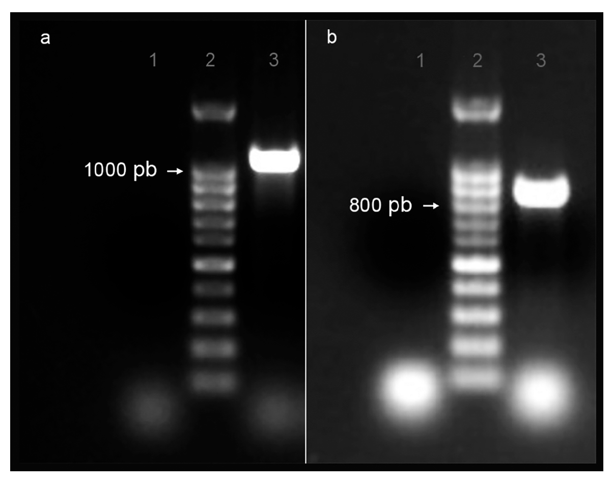
Figure 1. PCR products of EgGAPDH isoenzyme DNA were extracted from ex vivo liver cattle metacestode E. granulosus G1/s.s. (a) Amplification of isoenzyme W6UJ19; (b) amplification of isoenzyme W6V1T8. Lane 1: negative control; lane 2: Base pair marker (pb) and lane 3: PCR product.
Ijms 26 10622 g001
Figure 2. Secondary structure of EgGAPDH monomer. The image shows the superimposition of one subunit of each EgGAPDH isoenzyme: W6UJ19 (intracellular, shown in pink) and W6V1T8 (extracellular, shown in orange), as obtained using MOE.
Ijms 26 10622 g002
Figure 3. Quaternary structure and LEP mapping of EgGAPDH isoenzymes. In (a), the intracellular isoenzyme W6UJ19 is shown; in (b), the extracellular isoenzyme W6V1T8 is shown. Both quaternary structures are shown as homotetramers, with the four subunits depicted in colors. LEPs are highlighted in opaque. Each color represents one monomer.
Ijms 26 10622 g003
Figure 4. EgGAPDH and ligands docked in subunit A. Top row: GAPDH W6UJ19 (intracellular) from left to right: (a) protein with G3P (orange), Pi (red), and NAD+ (green); (b) G3P (orange), EHDP (red), and NAD+ (green); (c) AL (cyan) and NAD+ (green). Bottom row: GAPDH W6V1T8 (extracellular) from left to right: (d) protein with G3P (orange), Pi (red), and NAD+ (green); (e) G3P (orange), EHDP (red), and NAD+ (green); (f) AL (cyan) and NAD+ (green).
Ijms 26 10622 g004
Figure 5. Ligand interactions of Pi (red), G3P (orange), and NAD+ (green) in subunit A of EgGAPDH: (a) IC isoenzyme W6UJ19 and (b) EC isoenzyme W6V1T8. Residues are highlighted in sticks, in the same color as the ligands they interact with.
Ijms 26 10622 g005
Figure 6. Ligand interaction at the end of simulation (100 ns). G3P (orange), EHDP (red), and NAD+ (green) are shown interacting with EgGAPDH W6UJ19 (a) and W6V1T8 (b). AL (cyan) and NAD+ (green) are shown interacting with EgGAPDH W6UJ19 (c) and W6V1T8 (d). Residues are highlighted in sticks in the same color as the ligand they interact with.
Ijms 26 10622 g006
Figure 7. CEPs of EgGAPDH isoenzymes. Homotetramers are illustrated with CEPs mapped onto the molecular surface as dots colored according to atomic elements. Chain A is shown in orange, chain B in purple, chain C in cyan, and chain D in gray. (a) IC isoenzyme W6UJ19 and (b) EC isoenzyme W6V1T8.
Ijms 26 10622 g007
Figure 8. Two-dimensional chemical structure of the studied ligands in the ionization state used in computational simulations. (a) NAD+, (b) Glyceraldehyde-3-phosphate (G3P), (c) inorganic phosphate (Pi), (d) etidronate (EHDP), (e) alendronate (AL).
Ijms 26 10622 g008
Table 1. Comparative GAPDH physicochemical properties from different species.
GAPDH
Specie (UniProt)
Amino Acid Number
/
M.W.
ChargeIsoelectric PointProtein InstabilityAliphatic IndexGravy Index
Echinococcus granulosus (W6UJ19)338
/
36.6
+1 (R + K 37; N + E: 36)7.5624.4586.55−0.100
Echinococcus granulosus (W6V1T8)336
/
36.1
+3 (R + L 35;
N + E: 32)
8.4421.8285.890.004
Echinococcus multilocularis (Q27652)336
/
36.4
+7 (R + K: 39; N + E: 32)9.0227.1084.40−0.090
Taenia solium (A8R8Q4)336
/
36.3
+3 (R + K: 35; N + E: 32)8.4419.7386.19−0.029
Fasciola hepatica (A0A068LJN3)338
/
36.9
0 (R + K: 37;
N + E: 37)
7.1025.1584.76−0.109
Leishmania mexicana
(Q27890)
361
/
39.0
+7 (R + K: 42; N + E: 35)9.0527.6383.96−0.115
Homo sapiens (P04406)335
/
36.0
+3 (R + K: 36; N + E: 33)8.5715.0485.55−0.112
Bos taurus (P10096)333
/
35.9
+3 (R + K: 36; N + E: 33)8.5117.7084.89−0.078
Ovis aries (Q28554)322
/
34.7
+1 (R + K: 34; N + E: 33)7.8317.3885.09−0.091
Table 2. Glyceraldehyde-3-phosphate dehydrogenase LEP and the post-translational modifications.
GAPDH
Isoenzyme
LEPResiduePredicted
Modification(s)
W6UJ19A82-EAIPWDKDGVYYVV-E97W87Nitration
K89Ubiquitination
D88-KDGVYYVVESTGVN-T103K89Ubiquitination
S123-APSKDAPTFVVGVN-L138--
K140-YDPSMTIVSNASCT-T155S149Phosphorylation
N150Glycosylation
S152Phosphorylation
T154Phosphorylation
Q186-KLVDGPNPKGWRDG-R201K195Acetylation
Methylation
D282-VVSMDFRTSTASST-F297S285Phosphorylation
F288-RTSTASSTFDANAG-I303--
W6V1T8K138-YDPSMKVVSNASCT-T153S147Phosphorylation
N148Glycosylation
C151Nitrosylation
S150Phosphorylation
T152Phosphorylation
T153Phosphorylation
F286-LSTTCSSTFDARAG-I301--
Table 3. Total potential energy of both EgGAPDH isoenzymes during molecular dynamics simulation after equilibration step (100 ns of unrestrained production), partitioned into electrostatic and Van der Waals contributions; all the values are in kcal/mol.
GAPDHElectrostaticVan der WaalsTotal Potential Energy
MeanSDMeanSDMeanSD
W6UJ19MD−498,905.11484.604335,705.02302.8204−433,534.22357.6353
W6V1T8MD−500,354.14483.128335,967.32303.1194−435,622.22344.5178
Table 4. ∆G of binding estimated for Pi and EHDP to IC and EC EgGAPDH in each simulation system.
Ligand∆G of Binding (kcal/mol)
IC W6UJ19IC W6V1T8
Pi (system GAPDH: Pi, G3P, NAD+)−2.136 ± 1.366−3.477 ± 0.098
EHDP (system GAPDH: EHDP, G3P, NAD+)−4.413 ± 0.313−4.363 ± 0.331
All measured averages were analyzed using Student’s t-test, resulting in p < 0.05, confirming that the differences were statistically significant.
Table 5. ∆G of binding estimated for NAD+ to IC and EC EgGAPDH in control and AL in each simulation system.
Ligand∆G of Binding (kcal/mol)
IC W6UJ19EC W6V1T8
NAD+ (system GAPDH: Pi, G3P, NAD+)−7.727 ± 0.674−9.274 ± 0.419
NAD+ (system GAPDH: AL, NAD+)−9.418 ± 0.644−10.376 ± 0.771
In consequence, the substitution of AL by G3P and Pi resulted in an energy interaction significantly increased for the NAD+ molecule (Student’s t-test p < 0.05).
Table 6. Binding free energy (∆G, kcal/mol) estimated by the MMPBSA method for EgGAPDH isoenzymes in the presence and absence of BP.
MMPBSA of EgGAPDH With and Without BP
CONTROLW6UJ19W6V1T8
SubunitLigandsMean±sd±CI 95%Mean±sd±CI 95%
AG3P−10.69685.12291.005−4.7243.26190.64
B15.45065.14491.013.96594.46190.875
C16.45564.80650.94544.98317.60671.49
D24.0283.85830.7556.26374.55380.895
ANAD+22.46886.71121.3156.65775.49921.08
B32.85927.39921.45−4.01763.69960.725
C13.59464.36310.85523.78095.52711.08
D6.09624.27390.835−0.612.81360.55
APi29.64586.74451.3256.70743.00.59
B39.19847.66471.51.224510.3122.025
C22.2356.12431.20545.34455.93141.165
D37.28897.06521.3852.139910.7412.11
EHDP
AG3P−16.99654.76220.935−7.91123.98410.78
B5.27063.81020.75−7.58773.84380.755
C−5.19537.18691.405−7.93173.17450.62
D−9.10295.55891.09−14.5874.18190.82
ANAD+2.472510.07951.9751.72235.93181.16
B7.38544.77540.9356.2215.07630.995
C27.65378.75311.715−4.60313.90270.765
D28.91975.19061.024.49284.51070.885
AEHDP−6.60673.35390.655−4.07243.04220.595
B−7.17152.93010.5750.50113.97650.78
C0.8155.10881.005−11.81483.06830.605
D2.07674.41940.865−1.36163.28441.29
AL
ANAD+24.19477.31841.4359.14076.05821.19
B2.19414.44010.878.86465.43371.065
C−3.72583.58320.7055.27854.28280.84
D−3.02832.0030.39−3.25833.98060.78
AAL19.15983.95250.777.6243.24520.635
B−1.29682.20080.430.37862.34060.46
C4.39723.51140.6856.95413.38360.665
D8.25114.19110.82−1.882313.84392.715
Table 7. List of amino acids interacting with ligands in the IC EgGAPDH in each simulation system. In bold: amino acid that makes frequent contacts with ligands.
W6UJ19
Control
InitialFinal
G3PPiNAD+G3PPiNAD+
Subunit AR235(2)T154
S152
T212
G12(2)
N318(2)
S123(2)
R235(3)
His180(2)
T183
C153
K195(3)
R235(2)
S123
Subunit BC153
H180
T183
C153(2)
T154
H180
G12
R13
D35
N318(2)
R235(2)
H180(2)
T183
R235(3)
K195(2)
R13
D35
D190
Subunit CR235(2)T154R13
H180
EA319
K187
T212(2)
G213
R235(2)
H180(2)
E319(2)
D190
N193
K187
Subunit DH180T154(2)
H180
G213
D35
S98
C153
C153K195
R235
D35
T99
W6UJ19
EHDP
InitialFinal
G3PEHDPNAD+G3PEHDPNAD+
Subunit AR235(2)T154(2)
Q211
T212
G213
R235
G12(2)
S98
S123(2)
N318(2)
T183
H180(3)
Q211
R235
D337
Subunit BC153
H180
T183
C153
T154
H180
G12
R13
D35
N318(2)
C153D199(2)
Q211(2)
P209
K195
Subunit CR235(2)T154(2)
T212
R13
H180
K187
E319
H180(2)T212P192
E319
Subunit DC153T154
Q211
T212(2)
G213
D35
S98
C153
S123
C153(2)
H180(2)
T183
N318
Q211
T212
R235
D35
E319
W6UJ19
AL
InitialFinal
ALNAD+ALNAD+
Subunit AT154(2)
Q211
T212(2)
G213
R235
G12(2)
S98
S123(2)
N318(2)
S123A184
E319
K187
D190
Subunit BC153
T154
H180
G12
R13
D35
N318(2)
I38-
Subunit CT212
T154(2)
R13
H180
K187
E319
S126
D128(2)
A217
T103(2)
I122
K187
Subunit DT154
Q211
T212(2)
G213
D35
S98
C153
A124
S149
S152
-
Table 8. List of amino acids interacting with ligands in the EC EgGAPDH in each simulation system. In bold: amino acid that makes frequent contacts with ligands.
W6V1T8
Control
InitialFinal
G3PPiNAD+G3PPiNAD+
Subunit AT181
C151
R233(2)
S150
C151(2)
T152(2)
R12
I13
S97
S121(2)
N316(2)
R233(3)
K193(2)
-R12(2)
D34(2)
T98
S121
E317
Subunit BC151
R233
C151
T152(2)
R12(2)
D34(2)
S121
N316(2)
R233(2)
K193(2)
-G11
D34(2)
Subunit CC151S150
C151(2)
T152
T210
R12
I13
S97(2)
Y320
R233R233(2)
K233(3)
T181
A182
P237
Subunit DCys151(2)S150
C151
T152(2)
G11(2)
R12
I13
N101
K193
R233(3)
--
W6V1T8
EHDP
InitialFinal
G3PEHDPNAD+G3PEHDPNAD+
Subunit AC151
T181
R233
T152(2)
T210
G11
R12
I13
S121(2)
N316(2)
K193(2)
R233(2)
-F10
R12
D34(2)
F36
Subunit BS150
C151
R233
T152(2)R12(2)
D34
E78
S121
N316(2)
E317
K193(2)
R233(2)
-F10
R12
D34
(2)
S121
Subunit CC151C151(2)
T152
H178
R12
I13
P35
E78
S97(2)
Y320
K193(2)G99G11
R12
D34(2)
Subunit DC151
T183
C151
T152
G11
R12
I13
P123(2)
K193
R233(2)
T152
T210
P35
S97
T98
S121
P123
T181
T183
K192
W6V1T8
AL
InitialFinal
ALNAD+ALNAD+
Subunit AC151G11
R12
I13
S121(2)
D316(2)
T181F10
D34(2)
F36
Subunit BC151R12
I13
D34
E78
C151
E317
D165
N166
G11
R12(2)
I13
D34(2)
Subunit CC151(2)
E317
R12
I13
S97(2)
Y320
N316R12
D34(2)
E317
Subunit DC151(2)
H178
G11(2)
R12
I13
D34
-P190
K185
K192
Table 9. CEPs, post-translational modifications, and changes induced by BP on EgGAPDH W6UJ19 intracellular and W6V1T8 extracellular isoenzymes. System simulations: 1—control: Pi, G3P, and NAD+. 2—EHDP: EHDP, G3P, and NAD+. 3—AL: AL and NAD+. Post-translational modifications: -CH3: methylation; -CH3CO: acetylation; Ph: phosphorylation; and Ubiq: ubiquitination.
IsoenzymeSystemChain AChain BChain CChain D
W6UJ19ControlK63, D64, G65, K66, L80, K89, A106, K107, G109, A110, L112, K113, N114, N115, S144, N193, P194, K195L80, N81, E83, A108, G109, A110, H111, L112, K113, N114, N115, S144, N193, P194E79, L80, N81, A82, E83, K89, K113, S144, T185, N193, P194, K195, G200, D337I38, D39, D64, N81, A82, A106, G109, K113, S144, P192, N193, P194
ALD64, G65, K66, L80, N81, K89, A106, K107, G109, A110, L112, K113, N114, N115, S144, P192, N193, P194, K195L80, N81, A82, A84, I85, P86, W87, D88, K89, D90, A108, G109, A110, H111, L112, K113, N114, S144, N193, P194, K195L80, N81, A82, E83, K89, H111, S144, P192, N193, P194, K195, G200N81, A106, A110, K113, N114, N115, S144, T185, N193, P194, K195
EHDPD39, K63, D64, G65, K66, K107, G109, A110, K113, N114, N115, S144, N193, P194, K195, G196, D337T41, L112, K113, S144, P192, N193, P194, K195L80, N81, A82, E83, A106, S144, N193, P194, K195N81, K107, A110, H111, K113, N114, N115, S144, P194, K195, G196, G200
Post-translational
modifications
-CH3/-CH3COK66 and K195K195K195K195
UbiqK89K89K89-
Ph---T185
W6V1T8Control S142, K192, K193P141, S142, K192, K193K192, D335S142, K192
ALP141, S142, K192S142, K192S142P141, K193
EHDPK192S142, K192D38, K138, K192, K193P190, K192
Post-translational
modifications
-CH3K192K192 K192K192
-CH3/-CH3COK193K193K193K193
Disclaimer/Publisher’s Note: The statements, opinions and data contained in all publications are solely those of the individual author(s) and contributor(s) and not of MDPI and/or the editor(s). MDPI and/or the editor(s) disclaim responsibility for any injury to people or property resulting from any ideas, methods, instructions or products referred to in the content.