Article

Integrated In Vitro and In Silico Evaluation of the Antimicrobial and Cytotoxic Potential of Calotropis procera Leaf Ethanolic Extract: From GC-MS Profiling to Molecular Docking and Dynamics

by
Juan David Rodríguez-Macías
1,*,
Oscar Saurith-Coronell
2,
Laura Martínez Parra
3,
Domingo César Carrascal-Hernández
2,
Fabio Fuentes-Gandara
4,
Daniel Insuasty
2 and
Edgar A. Márquez-Brazón
2
1
Facultad de Ciencias de la Salud, Exactas y Naturales, Universidad Libre Seccional Barranquilla, Barranquilla 080001, Colombia
2
Facultad de Ciencias Básicas, Departamento de Química y Biología, Universidad del Norte, Barranquilla 080003, Colombia
3
Estudiante de Maestría en Biotecnología, Universidad Libre Seccional Barranquilla, Barranquilla 080001, Colombia
4
Department of Natural and Exact Sciences, Universidad de la Costa, Barranquilla 080002, Colombia
*
Author to whom correspondence should be addressed.
Int. J. Mol. Sci. 2025, 26(21), 10574; https://doi.org/10.3390/ijms262110574
Submission received: 3 July 2025 / Revised: 2 September 2025 / Accepted: 5 September 2025 / Published: 30 October 2025

Abstract

Calotropis procera, a drought-tolerant shrub widely used in folk medicine, was evaluated for its antimicrobial potential and safety using an integrative in vitro/in silico workflow. Ethanolic leaf extract (EE-CP) displayed a dose-dependent inhibition of Staphylococcus aureus ATCC 2913 and Escherichia coli ATCC 35218, reaching 93% and 52% of the amoxicillin control, respectively (MIC 207 µg mL−1 and 149 µg mL−1). GC-MS and LC-HRMS profiling revealed cardenolides (strophanthidin, gitoxigenin) and indole derivatives as major constituents. Pharmacophore mapping highlighted the essential glycosyltransferase MurG as a likely bacterial target; molecular docking showed that strophanthidin and NCGC00384918 bind MurG more strongly than the native substrate UDP-GlcNAc (ΔG ≤ −9.4 kcal mol−1), a result corroborated by 100 ns molecular dynamics simulations and MM-PBSA binding energies (−96.4 and −49.3 kcal mol−1). EE-CP caused <10% hemolysis up to 1.5 mg mL−1 and exhibited LC50 values of 302 µg mL−1 (human lymphocytes) and 247 µg mL−1 (BHK-21 cells), indicating a narrow but exploitable therapeutic window. Collectively, these findings constitute the first report on Colombian C. procera demonstrating potent anti-Staphylococcus activity, MurG-targeted cardenolides, and acceptable erythrocyte compatibility. This study supports EE-CP as a promising source of lead molecules and antibiotic adjuvants, warranting guided fractionation and in vivo validation to optimize efficacy and mitigate cytotoxicity.

1. Introduction

Medicinal plants have long been recognized as valuable sources of antimicrobial compounds, primarily due to their vast and structurally diverse arsenal of secondary metabolites. These include alkaloids, flavonoids, terpenoids, glycosides, and phenolics, many of which have evolved as part of the plant’s natural defense mechanisms against pathogens. Unlike synthetic antibiotics, which often target a single bacterial protein or pathway, phytochemicals can disrupt microbial viability through multiple modes of action, such as membrane disruption, the inhibition of nucleic acid or protein synthesis, and metal ion chelation, thereby reducing the likelihood of resistance development. This polypharmacological behavior not only enhances their antimicrobial efficacy but also makes plant-derived compounds particularly attractive in the fight against multidrug-resistant pathogens [1,2,3].
Beyond their pharmacodynamic versatility, plant-based antimicrobials offer practical advantages in terms of accessibility, sustainability, and ethnopharmacological validation. In many regions, especially where access to conventional healthcare is limited, traditional medicinal plants such as Calotropis procera have been used empirically for centuries to manage infections and inflammatory conditions. These uses, rooted in local knowledge systems, provide a meaningful framework for bioprospecting and scientific validation. Moreover, plants with invasive potential, like C. procera, present an ecologically responsible resource for drug discovery, as their exploitation may contribute to ecological control while yielding valuable bioactive molecules. This convergence of ecological utility, biochemical richness, and cultural relevance positions medicinal plants as a vital frontier in the development of next-generation antimicrobials [4,5].
Also known as Sodom apple, calotrope, and giant milkweed, C. procera is a shrub native to Africa, the Arabian Peninsula, Western Asia, South Asia, and Indochina. It belongs to the Apocynaceae family, typically grows in arid and semi-arid habitats and reaches a height of 2.5–4 m. This plant, characterized by large pale green leaves and a toxic milky sap in its green fruits, has potential economic and medicinal value, making it of interest for its ecological, economic, and antimicrobial properties [1]. Extracts of C. procera have been widely used in traditional medicine due to their pharmacologically active compounds found in their flowers, roots, and leaves, as well as in milky latex. These extracts, particularly from the aerial parts of the plant, have been commonly employed for the treatment of various diseases, including fever, tumors, joint pain, ulcers, muscle spasms, and constipation. The antimicrobial and cytotoxic properties of C. procera have been extensively studied, with some researchers suggesting potential applications in developing new eco-friendly antibacterial and antiviral drugs [2].
Recently, Ahmad Nejhad et al. (2023) [3] studied the antimicrobial effects of C. procera leaf extracts against E. coli, observing a zone of inhibition (ZI) of 14.1 mm. The extracts contained moderate amounts of flavonoids (1781.7 μg/g), phenols (174.82 mg/g), and p-coumaric acid [3]. Another study identified 31 compounds from C. procera via gas chromatography-mass spectrometry (GC-MS), including phytol (13.32%), α-amyrin (39.36%), lupeol acetate (17.94%), gombasterol A (2.14%), linolenic acid (3.04%), stigmasterol (3.16%), and hexadecanoic acid (5.55%). The ethanolic extracts demonstrated antimicrobial activity against Klebsiella pneumoniae, E. coli, and Staphylococcus aureus, with zones of inhibition of 18.66 mm, 21.26 mm, and 21.93 mm, respectively [4].
Previous reports also describe the chemometric profile and antimicrobial activity of C. procera leaf extracts, which are rich in palmitic acid ester (10.24%), linoleic acid (7.43%), fatty acid ethyl ester (21.36%) and amino acids (8.1%), with a zone of inhibition of 11 mm against E. coli [5]. Additionally, ethanolic and aqueous extracts of C. procera leaves and latex have been tested against Bacillus subtilis, Candida albicans, and Salmonella typhi [6,7]. In these studies, ethanolic extracts showed higher efficacy, with a zone of inhibition of 21 mm against B. subtilis, whereas the aqueous extracts showed no activity [8].
Given the growing interest in plant-derived antimicrobials, Calotropis procera has emerged as a promising candidate due to its rich phytochemical profile and traditional medicinal relevance. While several studies have reported its antimicrobial potential, few have integrated a comprehensive approach that combines both experimental validation and computational modeling to elucidate its bioactive mechanisms.
In this context, the present study offers a novel contribution by not only evaluating the antimicrobial and cytotoxic effects of C. procera leaf ethanolic extract through in vitro assays, but also by characterizing its chemical constituents via gas chromatography-mass spectrometry (GC-MS) and assessing their molecular interactions with microbial targets using in silico techniques. Specifically, molecular docking and molecular dynamics (MD) simulations have been employed to predict the binding affinity, stability, and potential mechanism of action of the major phytochemicals identified.
This integrative strategy provides a more robust and mechanistic understanding of the antimicrobial potential of C. procera, bridging the gap between empirical observations and molecular-level insights. By combining the strengths of both experimental and computational methodologies, this work not only enhances the predictive power of phytochemical screening but also contributes to the rational design of plant-based antimicrobial agents with improved efficacy and safety profiles.

2. Results

2.1. Solubility Test for the Ethanolic Extract of C. procera Leaves

A solubility test is a crucial step in the evaluation of the properties and potential applications of the ethanolic extract of C. procera leaves [6]. The test consists of evaluating the solubility of the extract in different solvents with different polarities, such as water, chloroform, and ethanol [8]. Important information about the potential pharmaceutical, antimicrobial, and antioxidant properties of a wide range of compounds can be obtained from these tests. For example, previous studies have shown that the ethanolic extract of C. procera leaves exhibits a significant inhibition of key enzymes related to diabetes mellitus, indicating its potential for pharmaceutical applications [9,10].
Table 1 reports the solubility results of the ethanolic extracts of C. procera leaves; solvents with different polarities such as xylol, chloroform, and heptane were used to deduce the presence of secondary metabolites in the extracts. The extract was fully soluble in ethanol (96%) and dimethyl sulfoxide (DMSO) and partially soluble in methanol. These results suggest that the major constituents of the extract are predominantly polar to moderately polar compounds, which aligns with the known phytochemical profile of C. procera, including flavonoids, phenolic acids, and triterpenoids [11].
In contrast, the extract was insoluble in distilled water and acetone, and only slightly soluble in ethyl acetate, chloroform, xylol, and heptane. This limited solubility in non-polar and aprotic solvents indicates a relatively low abundance of highly lipophilic compounds such as long-chain hydrocarbons or non-polar sterols. These findings are consistent with previous GC-MS analyses of C. procera extracts, which report a dominance of semi-polar compounds like lupeol acetate, α-amyrin, and phytol [12].
The high solubility in ethanol and DMSO is particularly advantageous for pharmaceutical and antimicrobial applications. Ethanol is widely used in herbal extraction due to its ability to dissolve a broad spectrum of bioactive compounds, while DMSO is a preferred solvent in in vitro assays for its excellent solubilizing capacity and cell permeability. These properties support the extract’s suitability for downstream biological testing, including enzyme inhibition, antimicrobial screening, and cytotoxicity assays [13]. Moreover, the poor solubility in water underscores the importance of solvent selection for both extraction and formulation. This characteristic may also influence the bioavailability and delivery strategies for therapeutic applications, suggesting that encapsulation or emulsification techniques may be required for aqueous formulations [14,15].

2.2. Phytochemical Test of the Ethanolic Extract of C. procera Leaves

An extract yield of 8.6% was obtained. The results obtained from the preliminary phytochemical screening of the ethanolic extract of C. procera leaves revealed the presence of cardiac glycosides, flavonoids, saponins, alkaloids, tannins, and phenols as shown in Table 2.
The qualitative phytochemical screening of the ethanolic extract of C. procera leaves revealed the presence of several key secondary metabolites, including alkaloids, flavonoids, phenols, cardiac glycosides, and saponins, while tannins were absent. These findings are consistent with previous reports on the phytochemical richness of C. procera, particularly in its aerial parts.
The detection of alkaloids is noteworthy, as these nitrogen-containing compounds are widely recognized for their antimicrobial, analgesic, and cytotoxic properties. Alkaloids from C. procera have been previously reported to exhibit significant antibacterial activity, supporting their potential role in the observed bioactivity of the extract [16,17].
Flavonoids and phenolic compounds, both detected in this extract, are well-known for their antioxidant and antimicrobial properties. These polyphenolic compounds can disrupt microbial membranes and inhibit key enzymes, contributing to the plant’s defense mechanisms. Their presence in C. procera has been linked to its traditional use in treating infections and inflammation [18,19,20].
The presence of cardiac glycosides, as indicated by the Kiliani test, is particularly interesting. While these compounds are primarily known for their cardiotonic effects, they have also demonstrated antimicrobial and cytotoxic activities in various plant species. In C. procera, cardiac glycosides such as calotropin and uscharin have been isolated and shown to possess potent biological activity [21,22,23].
On the other hand, saponins, detected at a high level (4+ foaming), are amphiphilic glycosides known for their ability to form stable foams and disrupt lipid membranes. Their surfactant properties contribute to antimicrobial and antifungal effects, and they may also enhance the bioavailability of other phytochemicals by increasing membrane permeability [24]. Interestingly, the phytochemistry screening showed an absence of tannins. This result may reflect the solvent selectivity, as ethanol tends to favor the extraction of moderately polar compounds over highly astringent polyphenols. This absence does not diminish the extract’s therapeutic potential but rather highlights the importance of solvent choice in targeting specific metabolite classes.
The presence of alkaloids, flavonoids, phenols, cardiac glycosides, and saponins in the ethanolic extract of C. procera leaves provides a strong biochemical basis for many of its traditional medicinal uses. These classes of compounds are frequently associated with antimicrobial, anti-inflammatory, analgesic, and cytotoxic activities, properties that align with the ethnobotanical applications of C. procera in treating infections, pain, fever, and inflammatory conditions [25].

2.3. High-Performance Liquid Chromatography Coupled to Electrospray Ionization and Quadrupole Time-of-Flight Mass Spectrometry (HPLC-ESI-QTOF-MS)

The HPLC-ESI-QTOF-MS analysis of the ethanolic extract of Calotropis procera leaves led to the tentative identification of six compounds, including cardenolides, indole derivatives, and other bioactive phytochemicals (Table 3, Figure 1). These findings provide valuable insights into the chemical complexity and potential pharmacological relevance of the extract.
Among the identified compounds, strophanthidin (C23H34O6) was detected at a retention time of 10.62 min with an experimental m/z of 407.2510. Strophanthidin is a well-known cardiotonic steroid belonging to the cardenolide class, which has been previously reported in C. procera latex and leaves [26]. Its presence supports the traditional use of the plant in treating cardiovascular and inflammatory conditions and aligns with earlier reports of cytotoxic and antimicrobial activity associated with cardenolides [27,28,29].
The detection of anthranilate (C7H7NO2), indole-3-carboxylic acid (C9H7NO2), and indole-3-acetic acid (C10H9NO2) further highlights the presence of indole-based metabolites, which are known to play roles in plant defense and microbial inhibition. Indole-3-acetic acid is a plant hormone (auxin) that also exhibits antimicrobial and anti-inflammatory properties [30,31,32].
These compounds may contribute to the observed antimicrobial activity of the extract and suggest a broader ecological role for C. procera in its native arid environments.
Two additional compounds labeled NCC20203915 (C19H24O4) and Meli5001779351-O1 (C16H21N2O) were tentatively annotated based on their mass spectra. While these compounds are not yet fully characterized in the context of C. procera, their detection suggests the presence of novel or less-studied secondary metabolites. The identification of such compounds opens new avenues for pharmacological exploration, particularly in the context of antimicrobial and cytotoxic screening.
According to date reported in the literature, most metabolomic studies on C. procera have been conducted in regions such as India, Saudi Arabia, and North Africa. The current findings represent, to the best of our knowledge, the first report of the HPLC-ESI-QTOF-based metabolite profiling of C. procera leaves collected in Colombia. This regional novelty is significant, as environmental factors such as soil composition, climate, and altitude can influence the secondary metabolite profile of medicinal plants [33]. These findings not only validate traditional uses but also provide a chemical foundation for future bioactivity-guided fractionation and drug discovery efforts.

2.4. Antibiogram Tests on the Ethanolic Extract of C. procera Leaves

2.4.1. Disk Diffusion Test: Staphylococcus aureus and Escherichia coli

The selection of Staphylococcus aureus (a Gram-positive bacterium) and Escherichia coli (a Gram-negative bacterium) as test organisms in antimicrobial assays is both strategic and scientifically justified. These two species are among the most clinically significant pathogens worldwide, frequently implicated in community-acquired and nosocomial infections [34,35,36]. For example, Staphylococcus aureus is known for its ability to cause a wide range of infections, from superficial skin conditions to life-threatening diseases such as pneumonia, endocarditis, and sepsis. Its increasing resistance to antibiotics, particularly methicillin-resistant S. aureus (MRSA), has made it a high-priority target for new antimicrobial agents [37,38]. Escherichia coli, on the other hand, is a leading cause of urinary tract infections, gastrointestinal diseases, and bloodstream infections. Its capacity to acquire resistance genes, including extended-spectrum β-lactamases (ESBLs), has raised global concern regarding treatment efficacy [39,40,41]. Therefore, by including both Gram-positive and Gram-negative strains, this study ensures a broader evaluation of the antimicrobial spectrum of the C. procera extract, which is essential for assessing its potential as a therapeutic agent.
Previous studies have reported the antimicrobial activity of C. procera extracts against both S. aureus and E. coli, although the extent of activity varies depending on the extraction method, plant part used, and geographical origin. For instance, ethanolic extracts of C. procera leaves have shown zones of inhibition ranging from 14 to 22 mm against these pathogens, suggesting the presence of potent bioactive compounds such as cardenolides, flavonoids, and alkaloids [42,43,44].
However, to date, there are no published reports on the antibacterial activity of C. procera extracts collected in Colombia, making this study the first to explore its efficacy against S. aureus and E. coli in this regional context. This not only adds to the global phytochemical and pharmacological knowledge of the species but also highlights the potential of Colombian C. procera as a source of novel antimicrobial agents.
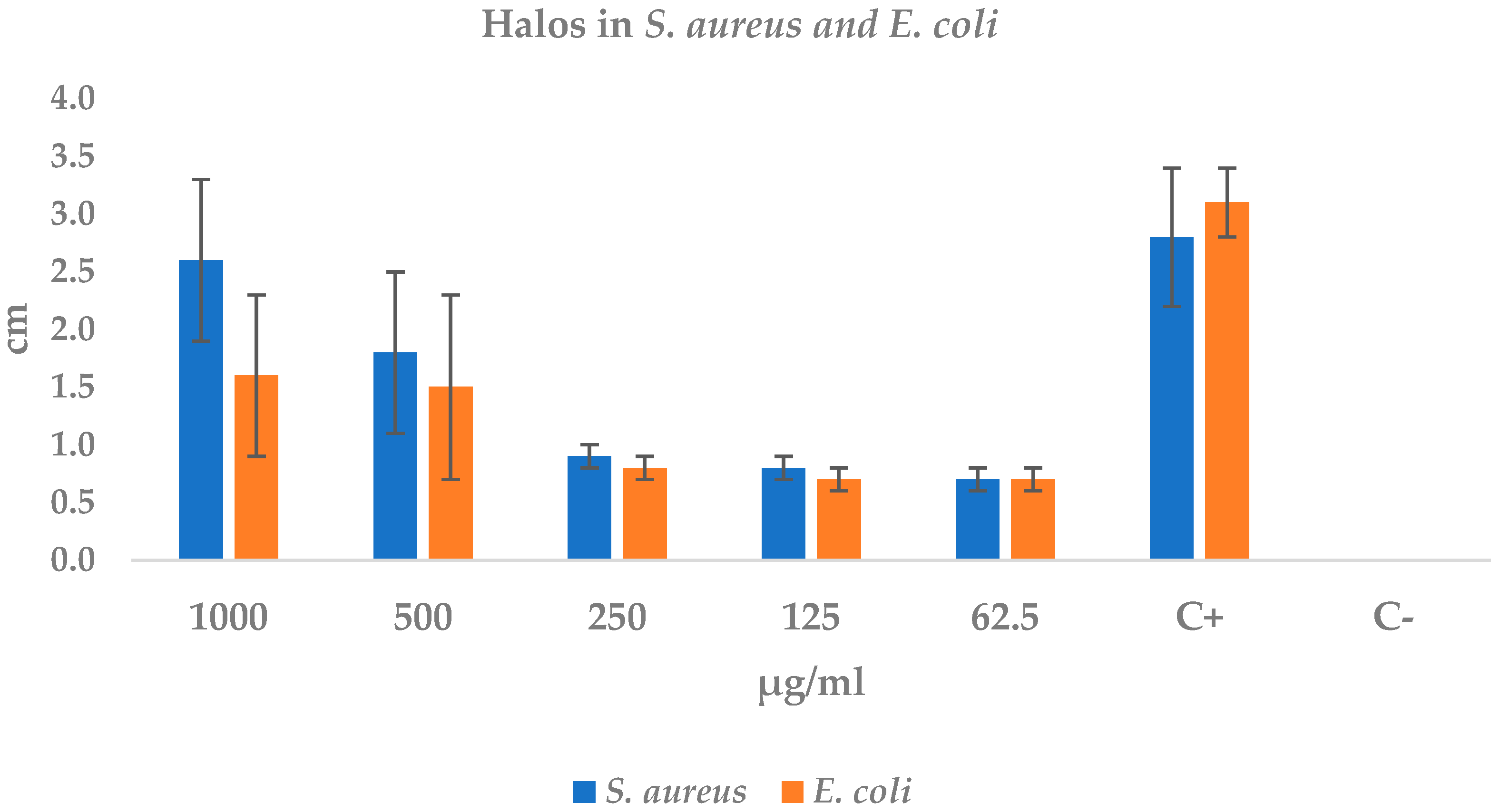
The extract exhibited dose-dependent antibacterial activity against both pathogens (Table 4, Figure 2). In S. aureus ATCC 2913, halo diameters increased from 0.7 ± 0.1 cm at 62.5 µg mL−1 to 2.6 ± 0.7 cm at 1000 µg mL−1, reaching 93% of the inhibition produced by the positive control (2.8 ± 0.6 cm). The linear fit (R2 = 0.97; r = 0.99) indicates an approximate increase of 0.21 cm for every 100 µg mL−1 of extract.
In E. coli ATCC 35218 the response was similar but less pronounced: diameters rose from 0.7 ± 0.1 cm at 62.5 µg mL−1 to 1.6 ± 0.7 cm at 1000 µg mL−1, representing 52% of the positive control halo (3.1 ± 0.3 cm). The dose-effect correlation was also high (r ≈ 0.98), with an average increment of 0.18 cm per 100 µg mL−1.
No inhibition was detected in the negative control in any assay, confirming the reliability of the method. These findings demonstrate that the extract displays high efficacy against S. aureus from 500 µg mL−1 onward and moderate activity against E. coli only at higher concentrations.

2.4.2. Minimum Inhibitory Concentration Test of S. aureus and E. coli

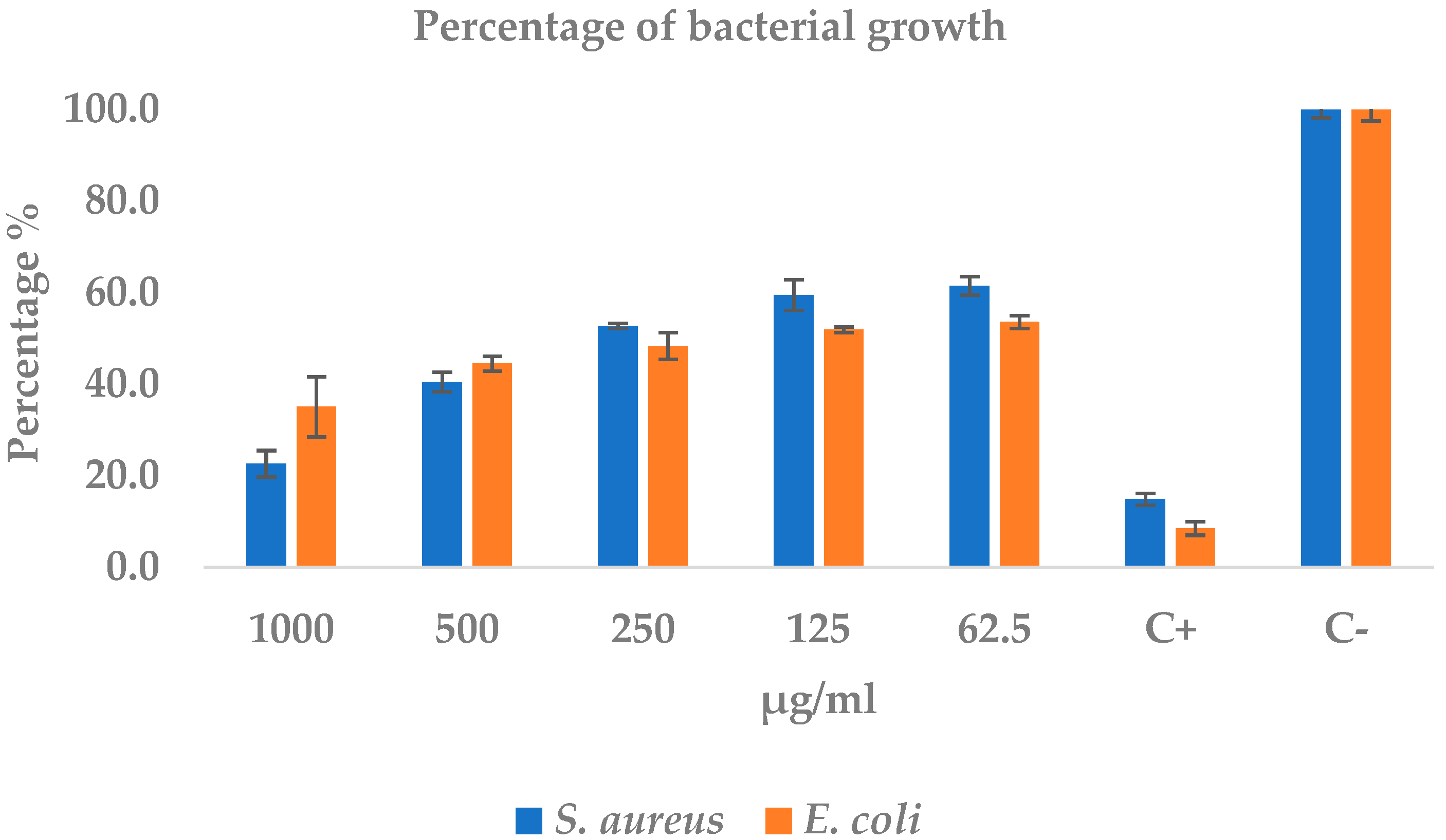
The extract exhibited a robust, clearly dose-dependent bactericidal effect: at 1000 µg mL−1 it reduced the growth of S. aureus by 77% and that of E. coli by 65%, approaching the reference effect of the positive control (600 mg amoxicillin), which left only 15% and 8% viability, respectively (see Table 5 and Figure 3). The regression analysis revealed a very strong negative correlation between concentration and growth (r ≥ −0.95; p < 0.02), with slopes of −0.21 and −0.18 absorbance units for every 100 µg mL−1 increase in extract for S. aureus and E. coli, respectively. These slopes account for the stepwise absorbance decrease from 0.46 ± 0.02 to 0.07 ± 0.02 in S. aureus and from 0.38 ± 0.01 to 0.25 ± 0.05 in E. coli when the concentration rose from 62.5 to 1000 µg mL−1 (Table 5). The estimated IC50 values, 207 µg mL−1 for S. aureus and 149 µg mL−1 for E. coli, position the Gram-positive strain as more susceptible at high doses, although the Gram-negative strain requires less extract to achieve 50% inhibition. Standard deviations below 10% of the mean for most treatments underscore experimental precision, while the complete lack of inhibition in the negative control (sterile water) rules out vehicle-related artifacts. These findings confirm that the extract attains 70–90% of the antibacterial potency of amoxicillin against S. aureus, shows moderate efficacy against E. coli, and emerges as a promising candidate for alternative therapies, particularly targeting resistant Gram-positive pathogens (Figure 3).

2.5. Evaluation of the Hemolytic Activity of EEHCP

The ethanolic extract of C. procera (EE-CP) caused only marginal damage to human erythrocyte membranes. As summarized in Table 6, hemolysis remained below 10% at every concentration tested. The highest value—9.3 ± 0.1% at 1000 µg mL−1—still falls under the generally accepted non-hemolytic threshold, whereas all other doses (32–1500 µg mL−1) produced ≤ 2.2% lysis, comparable to the negative control (PBS, 2.2 ± 0.3%; p > 0.05). In sharp contrast, the positive control (1% SDS) induced complete membrane rupture (100.6 ± 1.2%). These findings confirm that EE-CP is essentially non-cytotoxic toward human erythrocytes over a 32–1500 µg mL−1 range, supporting its biocompatibility for further pharmacological development.

2.6. Cytotoxicity of the Ethanolic Extract of C. procera Leaves on Lymphocytes

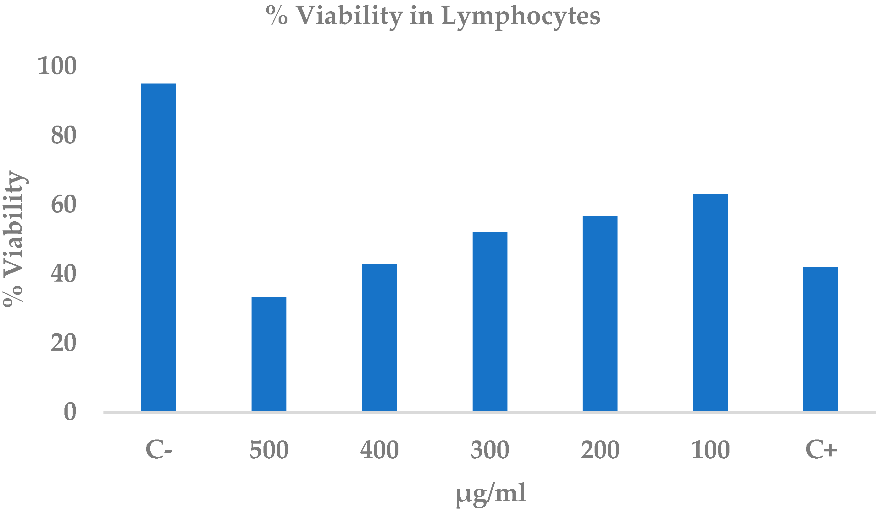
The ethanolic extract of C. procera (EE-CP) elicited a clear, concentration-dependent cytotoxic effect on human lymphocytes after 24 h, as quantified by the trypan blue exclusion assay (Table 7). Viability dropped from 95.0 ± 3.8% in the negative control to 63.1 ± 0.7% at 100 µg mL−1 and fell below the positive control level (41.9 ± 5.9%) at 400 µg mL−1, reaching a minimum of 33.2 ± 4.5% at 500 µg mL−1. Statistically significant cytotoxicity emerged at concentrations ≥300 µg mL−1 (p < 0.05; Figure 4). Linear regression revealed a strong inverse relationship between extract concentration and cell viability (R2 = 0.97), and the resulting equation yielded an LC50 of 301.9 ± 10.6 µg mL−1. These data demonstrate that EE-CP exerts moderate but measurable toxicity on lymphocytes, with an appreciable loss of viability only at the upper end of the concentration range tested.

2.7. Cytotoxicity of Ethanolic Extract of C. procera Leaves on Normal Cell Line Baby Hamster Kidney-21 Cells (BHK-21)

The ethanolic extract of C. procera (EE-CP) produced a marked, dose-dependent cytotoxic effect on the non-tumor BHK-21 cell line after 24 h (Table 8; Figure 5). Viability plummeted from 100.5 ± 2.0% in the negative control to 16.7 ± 2.3% at 500 µg mL−1—an 83% loss comparable to the positive control injury (6.2 ± 1.1%). Even the lowest concentration tested (62.5 µg mL−1) reduced viability to 76.7 ± 17.0%, indicating that all doses differed significantly from the untreated cells (p < 0.05). A linear regression of viability versus log-concentration revealed an almost perfect inverse relationship (R2 = 0.99), underscoring the consistency of the toxic response. Extrapolation from the regression line yielded an LC50 of 246.8 ± 22.6 µg mL−1, indicating that half-maximal lethality is reached at roughly one quarter of the highest dose. Collectively, these data demonstrate that EE-CP is substantially more cytotoxic to (BHK-21) cells than to lymphocytes and that its safety margin must be carefully considered in future formulations.

2.8. Data Collection

One of the main goals of this study was to determine which compounds in the Calotropis procera extract might have antibacterial properties. To do this, we first recreated the 2D structures of these compounds using the JSME molecular editor. This allowed us to generate structure data files (.sdf) for each molecule. We also downloaded .sdf files for a few reference compounds UDP-GlcNAc, β-1,4-GALT1-IN-1, Alendronate, and compound 6 from the PubChem database. Next, we evaluated the drug-like properties of these compounds using Lipinski’s Rule of Five and the ADME-Tox protocol, which looks at how a compound is absorbed, distributed, metabolized, and excreted by the body, as well as its potential toxicity using ProTox-3.0 [45,46]. The results of these assessments are shown in Table 9. Most of the compounds were found to fall into acute toxicity categories 4 to 6. According to the Globally Harmonized System (GHS) for classifying chemicals, there are six levels of acute toxicity. These are based on the LD50 value, which estimates the dose needed to kill half of a test population. It is a standard way to measure how toxic a substance is. The GHS considers three main ways a substance can enter the body: by swallowing (oral), through the skin (dermal), or by breathing it in (inhalation). Compounds in category 4, for example, are considered harmful if taken in any of these ways. They typically have an oral LD50 between 300 and 2000 mg/kg, a dermal LD50 between 1000 and 2000 mg/kg, and an inhalation LD50 between 2500 and 5000 ppm. Understanding these toxicity levels is crucial in the early stages of drug development [47]. It helps researchers choose compounds that are less likely to cause harm and more likely to succeed in later testing.
The compounds strophanthidin and NCGC00384918 were classified as toxicity class 2, meaning they are considered fatal if swallowed. Because of this, they could be strong candidates for future structural modifications aimed at reducing their toxicity and improving their safety for potential clinical use. This approach has already been successfully applied in various studies, particularly in the development of anticancer agents [48,49,50].
A notable example is the work conducted to reduce the toxicity and improve the bioavailability of Norcantharidin, a demethylated derivative of cantharidin. Norcantharidin has attracted significant research interest due to its broad range of biological activities, including potential treatments for systemic lupus erythematosus, platelet aggregation disorders, renal interstitial fibrosis, and cancer. However, despite its therapeutic promise, Norcantharidin has been associated with high levels of nephrotoxicity, hepatotoxicity, urotoxicity, and cardiotoxicity. To address these issues, researchers have implemented various structural modification strategies to enhance the compound’s solubility and reduce its toxicity. Interestingly, these modifications have not only improved its safety profile but also led to increased antitumor activity. This highlights how structural changes can positively influence a compound’s ADME properties, potentially boosting its overall biological effectiveness [51].

2.9. Target Identification

To explore the potential molecular targets of the principal metabolites identified in C. procera, we employed PharmMapper, a robust and widely recognized web-based platform for reverse pharmacophore mapping [52]. This tool predicts likely biological targets by comparing the pharmacophoric features of small molecules against a comprehensive database of pharmacophore models derived from experimentally validated protein structures. PharmMapper has been successfully applied across diverse domains, including natural product research, synthetic drug discovery, and drug repurposing, demonstrating its versatility and predictive power in early-stage drug development [53,54].
Therefore, in our study, the 2D structures of the selected compounds were first optimized to ensure accurate pharmacophore alignment. For each compound, a maximum of 300 potential targets were considered. The selection of potential targets followed a two-step strategy that combined computational confidence with biological plausibility. Initially, PharmMapper predictions were filtered using a normalized score greater than 0.85, ensuring that only targets with a strong pharmacophoric similarity to known ligands were considered. Among these high-scoring candidates, MurG glycosyltransferase was prioritized because it catalyzes the final step in lipid II biosynthesis, a critical precursor for peptidoglycan assembly in bacterial cell walls. This enzyme is indispensable for both Gram-positive and Gram-negative bacteria and is widely recognized as a validated antibacterial target [55]. Previous studies have demonstrated that MurG inhibition results in bactericidal effects in Escherichia coli and Staphylococcus aureus [56]. The antimicrobial profile observed in this study, marked by strong activity against S. aureus and the moderate inhibition of E. coli, is consistent with this mechanism, as Gram-positive bacteria are generally more susceptible to disruptions in cell wall synthesis. Furthermore, the cardenolides identified in the extract, such as strophanthidin and gitoxigenin, are known to interact with membrane-associated enzymes, and their high docking scores combined with stable molecular dynamics interactions with MurG reinforce their potential role as inhibitors. Regarding cytotoxicity, cardenolides are well-documented inhibitors of Na+/K+-ATPase in mammalian cells [57], which explains the narrow therapeutic window observed in the assays of Calotropis procera metabolites [58].
By integrating PharmMapper into our computational pipeline, we aimed to bridge the gap between phytochemical identification and mechanistic insight, enabling a more rational and hypothesis-driven evaluation of the antimicrobial and cytotoxic potential of C. procera metabolites. The predicted targets, which form the basis for subsequent molecular docking and dynamics simulations, are summarized in Table 10.
The pharmacophoric mapping results presented in Table 10 reveal the most probable protein targets for the compounds, with the highest scoring PharmMapper compounds being NCGC00384918 and strophanthidin. These predictions, based on high-confidence pharmacophore alignments (scores > 0.85), offer valuable insights into the potential mechanisms of action of these metabolites. PharmMapper, a widely used reverse pharmacophore mapping tool, enables the identification of likely biological targets by comparing the structural features of small molecules against a comprehensive database of pharmacophore models derived from experimentally validated protein structures [59].
For NCGC00384918, the top-ranked targets include proteins from Escherichia coli, Pseudomonas aeruginosa, and Pseudomonas syringae, many of which are involved in essential bacterial processes. These include transport system kinases (PDB: 2p67), DNA repair enzymes (PDB: 1ghh), and Type III secretion system (T3SS) proteins (PDB: 1o9y)—the latter being key virulence factors in Gram-negative pathogens and attractive targets for anti-virulence strategies [60,61,62,63,64,65,66].
Similarly, strophanthidin, a well-characterized cardenolide, was predicted to interact with several of the same bacterial targets, including glucosyltransferases (PDB: 1nlm) [67,68,69], nitrate reductases [70,71,72], (PDB: 2nya), and lipoamino dehydrogenases [73,74,75] (PDB: 1lv1). These enzymes are involved in cell wall biosynthesis, nitrogen metabolism, and energy production, all of which are critical for bacterial viability. The recurrence of targets such as glucosyltransferases and transport kinases across both compounds suggests a convergent mechanism of action, potentially enhancing the antimicrobial efficacy of the extract through multi-target synergy, a hallmark of polypharmacology [76].
These computational predictions are consistent with previous in vitro studies demonstrating the antibacterial activity of C. procera extracts against both E. coli and Staphylococcus aureus [77]. The ability of its metabolites to potentially inhibit key bacterial enzymes and virulence factors provides a molecular-level explanation for these observations. For instance, the inhibition of T3SS can attenuate bacterial virulence without necessarily inducing cell death, a promising strategy to reduce selective pressure for resistance. Likewise, targeting glucosyltransferases and nitrate reductases can disrupt fundamental metabolic pathways, leading to bacterial growth inhibition.
Importantly, many of the predicted targets are associated with Gram-negative bacteria, which are notoriously difficult to treat due to their impermeable outer membranes and multidrug resistance mechanisms. The ability of C. procera metabolites to potentially engage with such targets underscores their therapeutic promise and supports the traditional use of this plant in managing infectious diseases.
However, the focus was placed on identifying targets from Gram-negative bacteria that play essential roles in survival or replication, particularly those previously recognized as critical for bacterial viability when inhibited. One of the most promising targets identified was the essential glycosyltransferase (MurG). This enzyme belongs to the NDP-glycosyltransferase superfamily and plays a key role in the biosynthesis of glycolipids, glycoproteins, and other important metabolites involved in the bacterial life cycle. Specifically, MurG catalyzes the addition of the sugar nucleotide UDP-GlcNAc to lipid I, forming lipid II, a crucial precursor in the synthesis of peptidoglycan, the main component of the bacterial cell wall [78,79,80]. Compounds derived from vancomycin and other antibiotics, such as Murgocil, have shown strong antibacterial activity by targeting peptidoglycan synthesis in bacteria like Staphylococcus aureus and Escherichia coli. These compounds interfere with the normal function of MurG, making it a highly attractive target for the development of new antibiotics aimed at disrupting cell wall formation in both Gram-positive and Gram-negative bacteria [81,82,83].

2.10. Molecular Docking

The compounds NCGC00384918 and strophanthidin, both derived from the Calotropis procera extract and predicted to have potential affinity for the MurG enzyme, were subjected to molecular docking simulations. These were analyzed alongside reference compounds β-1,4-GALT1-IN-6, Alendronate, and compound 6 which have already shown inhibitory activity against MurG in both in silico and in vitro studies. Based on the PharmMapper predictions and the biological relevance of MurG, this enzyme was selected as the primary target for further investigation [84].
As previously mentioned, MurG plays a crucial role in bacterial cell wall synthesis. Any disruption to its function can significantly impair the formation of the peptidoglycan layer, ultimately leading to bacterial cell death [85]. The docking results for the Calotropis compounds and the reference molecules are summarized in Table 11. Strophanthidin showed a binding energy of −9.43 kcal/mol, while NCGC00384918 scored −9.12 kcal/mol. Both values indicate stronger interactions than the enzyme’s natural ligand, UDP-GlcNAc, which had a binding affinity of −8.41 kcal/mol. These findings suggest that the two Calotropis compounds may bind more tightly to MurG than its native substrate, highlighting their potential as promising antibacterial agents.
On the other hand, the reference compounds showed a range of interesting binding affinities. The compound β-1,4-GALT1-IN-6 had a binding energy of −9.29 kcal/mol, which is stronger than that of the natural ligand. This result supports previous in vitro findings where this compound effectively inhibited bacterial growth. In contrast, Alendronate and compound 6 showed lower binding energies of −3.99 and −6.30 kcal/mol, respectively. These values suggest weaker interactions with the MurG enzyme compared with the other compounds. However, it is important to note that a lower binding energy does not necessarily rule out the inhibitory potential of these molecules, as other factors may contribute to their biological activity [86].
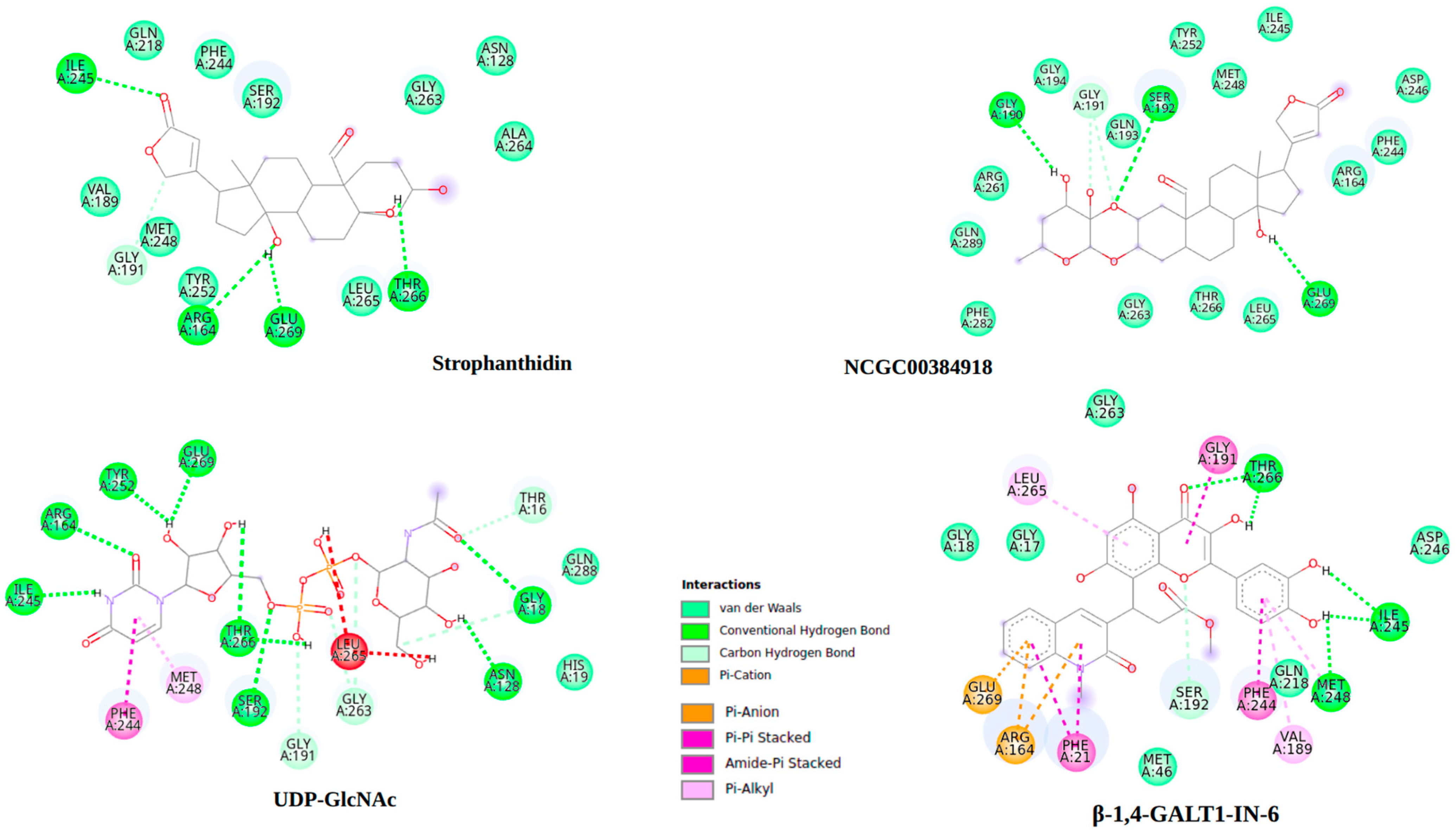
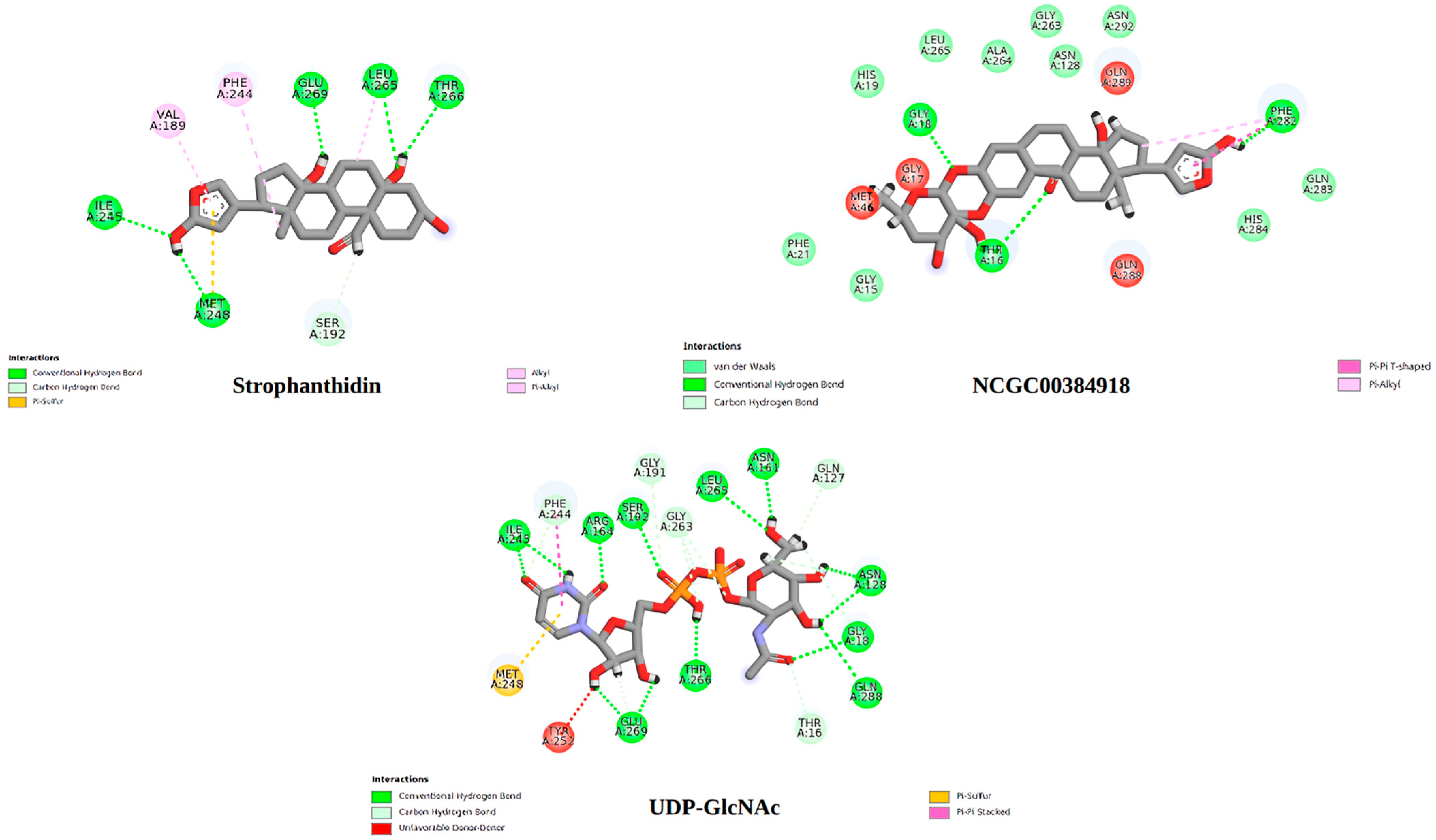
To better understand how these compounds interact with MurG, 2D interaction diagrams were generated for the complexes with the strongest binding energies. These visualizations, created using Discovery Studio Visualizer 4.5 (BIOVIA, Dassault Systèmes, San Diego, 2025), are shown in Figure 6 and help illustrate the key intermolecular interactions involved.
As shown in Figure 6, strophanthidin forms hydrogen bonds with key residues ILE245, ARG164, THR266, and GLY191, like the interactions observed with the natural ligand UDP-GlcNAc. This highlights the importance of these amino acids in the inhibition process of the MurG enzyme and suggests a possible shared mechanism of action between these compounds. Likewise, NCGC00384918 also shares hydrogen bond interactions with the natural ligand, particularly with residues SER192, GLU269, and GLY191. These findings point to the potential significance of GLU269 and GLY191 in MurG inhibition. When comparing the interaction profiles of the different MurG inhibitors, it becomes clear that both the Calotropis compounds and β-1,4-GALT1-IN-6, which showed the strongest binding energy among the reference compounds, exhibit a notable number of hydrogen bonds and van der Waals interactions. These involve hydrogen donor oxygens and aromatic rings, suggesting that the number, distance, and type of these interactions play a crucial role in the binding affinity and stability of the enzyme-ligand complexes [87].
The similarity in interaction patterns between the Calotropis compounds (strophanthidin and NCGC00384918), the natural ligand, and β-1,4-GALT1-IN-6 suggests a potentially comparable mechanism of action. This structural and energetic stability supports the idea that these natural compounds could act as effective MurG inhibitors.

2.11. Molecular Dynamics

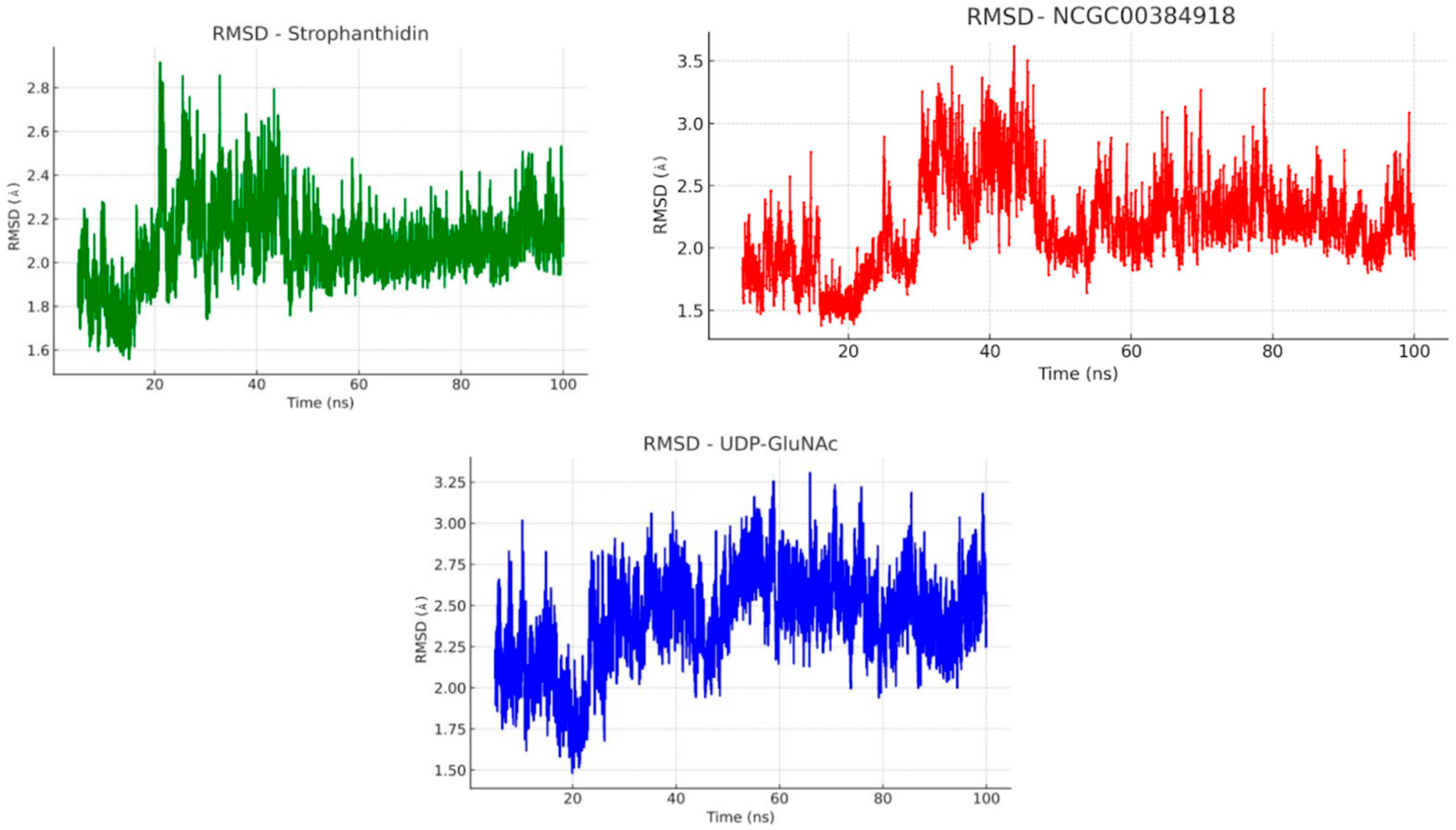
To better understand how stable the ligand-protein complexes are under physiological conditions, we ran molecular dynamics simulations in a water and ion environment that mimics the conditions inside the body [88]. We used the Root Mean Square Deviation (RMSD) method, which is widely applied to track how much the structure of a protein changes over time both in its backbone and in all its heavy atoms [89]. A low RMSD value generally indicates that the protein remains stable and does not undergo significant structural shifts during the simulation. After completing the docking protocol, we focused on evaluating the stability of the complexes formed between the MurG enzyme and three ligands: strophanthidin, NCGC00384918, and the natural substrate UDP-GlcNAc. Each complex was simulated for 100 nanoseconds.
Figure 7 shows the RMSD plots for these MurG-ligand complexes. As illustrated, all three systems, UDP-GlcNAc, strophanthidin, and NCGC00384918, reached a stable equilibrium around 50 nanoseconds into the simulation. Before this equilibration phase, the RMSD values hovered around 2 Å, suggesting that the complexes underwent some initial structural rearrangements to reach a more energetically favorable conformation.
Based on the RMSD values analyzed in Figure 7, the natural ligand UDP-GlcNAc showed an average RMSD of around 1 Å after 50 nanoseconds of simulation, indicating that the system reached a stable equilibrium. Both strophanthidin and NCGC00384918 displayed similar behavior to the natural ligand, suggesting that their interactions with MurG are comparably stable. Notably, strophanthidin stood out with an RMSD of approximately 0.6 Å. After the initial stabilization phase, this complex exhibited only minor conformational fluctuations, and from around 60 ns onward, it maintained the lowest RMSD among all simulated compounds, indicating an even greater binding stability than UDP-GlcNAc.
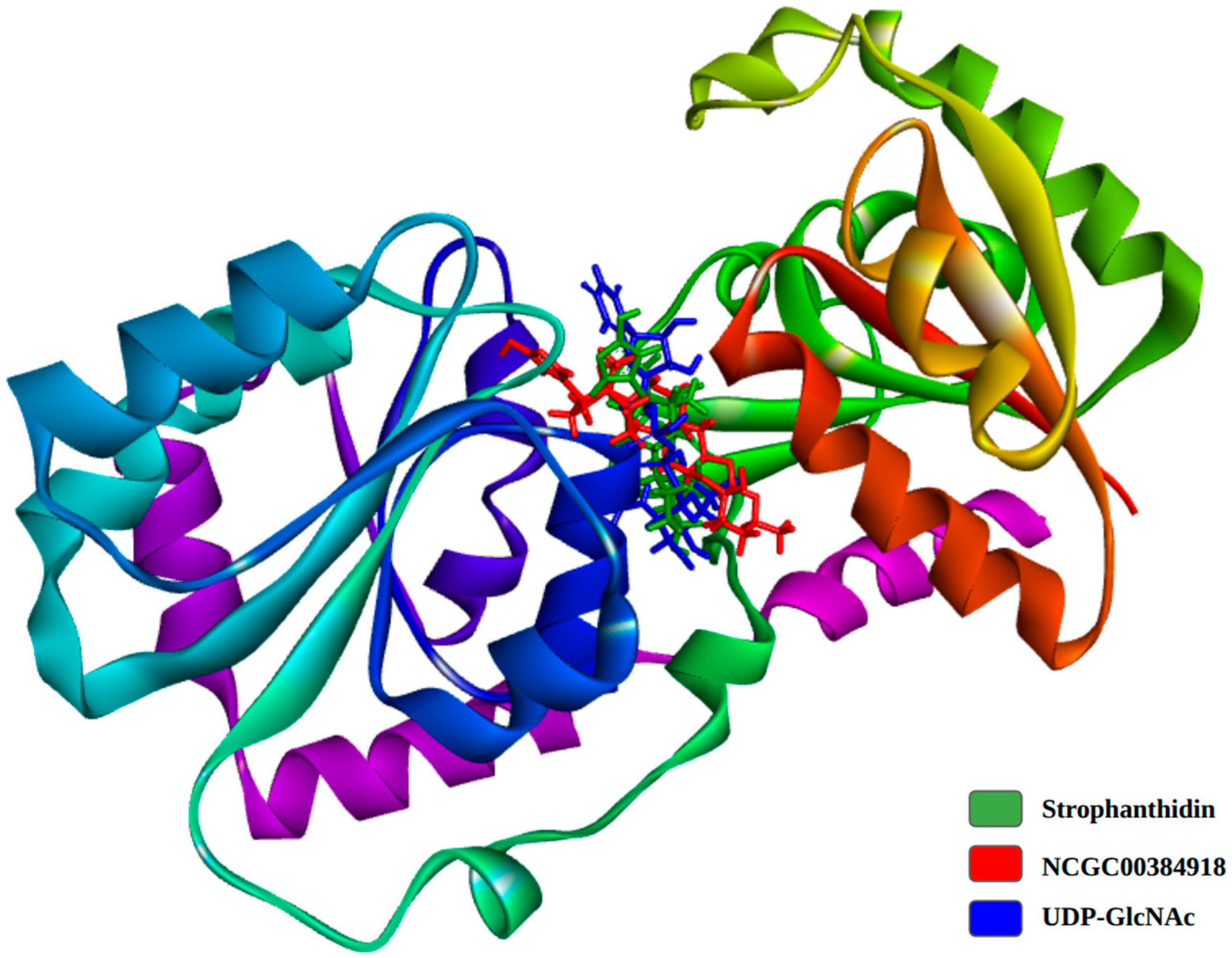
The stabilization phase during molecular dynamics simulations is crucial for understanding the strength and reliability of ligand binding, which in turn reflects the compound’s potential inhibitory effectiveness [89]. Figure 8 illustrates the binding poses of the simulated compounds at their lowest energy states. As shown, there is a clear structural overlap between the Calotropis compounds and the natural ligand, suggesting that they may share a similar binding mode. This overlap supports the idea that these natural compounds could act as effective inhibitors of the MurG enzyme.
Figure 9 shows the 2D interaction diagrams of the complexes at their lowest energy states. As mentioned earlier, the main types of interactions between MurG and its natural ligand are hydrogen bonds and van der Waals forces. What stands out is the preservation of certain key interactions throughout the simulation. For example, both the natural ligand and strophanthidin consistently maintained a hydrogen bond with the amino acid residue GLU269. This suggests that this specific interaction may play a crucial role in stabilizing the complex and could potentially enhance the compound’s biological activity.
To better understand how the ligands interact with the MurG enzyme, we calculated their binding energies using the MM-PBSA (Molecular Mechanics Poisson-Boltzmann Surface Area) approach. This method provides a more detailed view of the interaction process by analyzing the molecular dynamics simulation trajectories [90,91,92]. These simulations were carried out using the YASARA structure 2025.1 which includes a macro called md_analyzebindenergy.mcr. This macro estimates binding energy using Equation 1.
It is important to note that in YASARA’s default setup, more positive values indicate stronger binding affinities. However, negative values do not necessarily mean there is no interaction as they can still reflect meaningful binding, depending on the context. To make the interpretation of our results more intuitive and consistent with previous studies, we inverted and rewrote the original equation from the YASARA macro, as shown in Equation 2 [93].
B i n d E n e r g y = E R e c e p t E s o l v + E p o t + E L i g a n d E s o l v + E p o t E C o m p l e x E s o l v + E p o t
where ERecept is the receptor (protein) energy which is the sum of the solvation and potential contributions; Eligand is the energy of the ligand, and Ecomplex is the energy of the ligand-receptor (protein) complex.
B i n d E n e r g y = E C o m p l e x E S o l v + E P o t E L i g a n d E S o l v + E P o t E R e c e p t E S o l v + E P o t
This macro calculates the average binding energy by using the solvation energy (ESolv) and potential energy (EPot) of the ligand, the receptor, and the ligand-receptor complex. The results of these binding energy calculations are summarized in Table 12.
As shown in Table 12, the binding energies between MurG and the different simulated compounds were calculated and compared. UDP-GlcNAc was used as a reference point for interpreting the results, since binding energy is a key indicator of complex stability. Lower values generally suggest more stable interactions, which often correlate with better biological activity.
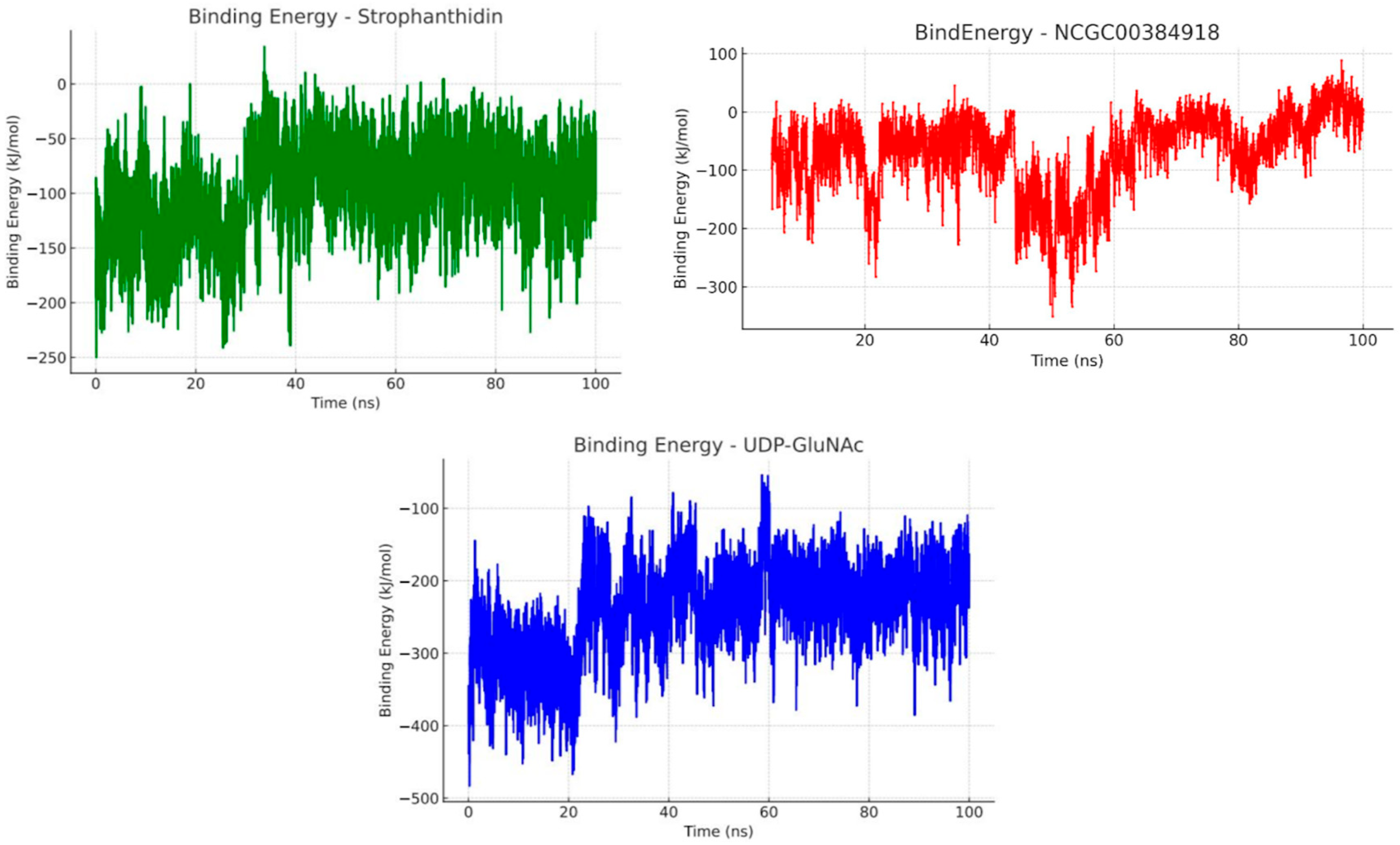
UDP-GlcNAc showed a binding energy of −237.56 kJ/mol. While this value is positive, as mentioned earlier, it does not mean that there is no stable interaction. Instead, it reflects the estimated stability of the ligand-protein complex. Strophanthidin, on the other hand, had a binding energy of −96.44 kJ/mol, which is significantly lower than that of the natural ligand, suggesting a more stable and potentially stronger interaction over time. Even more promising was NCGC00394918, which achieved the lowest binding energy of −49.34 kJ/mol in the molecular dynamic simulations.
Both Calotropis compounds outperformed the natural ligand in terms of binding energy. NCGC00394918 showed the most favorable result, followed by strophanthidin. These findings suggest that these natural compounds may play a role in inhibiting bacterial growth and could be strong candidates for the development of new antibiotics. To better visualize how binding energy fluctuates over the course of the simulation, Figure 10 presents a graph showing the changes in interaction energy over time.
As shown in Figure 10, UDP-GlcNAc exhibited only minor fluctuations throughout the simulation, indicating a stable interaction with the MurG protein. This aligns with the RMSD plot, where the complex reached a more energetically favorable and stable state around 25 ns and maintained that equilibrium for the remainder of the simulation. A similar pattern was observed with strophanthidin, which stabilized around 30 ns and remained steady, suggesting a comparable level of interaction stability to the natural ligand. This supports the idea that strophanthidin may possess inhibitory properties against MurG.
In contrast, NCGC00384918 displayed a different behavior. Although it showed the lowest average binding energy around −49.34 kJ/mol, its interaction energy dropped sharply around 45 ns, likely due to conformational changes in the protein-ligand complex that temporarily weakened the interaction. However, from 60 ns onward, the binding energy gradually increased, eventually surpassing the initial values. This suggests a structural rearrangement that led to more favorable interactions. This trend is consistent with the RMSD data, which also showed a decrease around the same time, indicating a more stable complex. These findings provide strong evidence that both strophanthidin and NCGC00384918 are promising antibacterial candidates. The results support the hypothesis that their primary mechanism of action involves the inhibition of the MurG glycosyltransferase, an essential enzyme in the peptidoglycan biosynthesis pathway, which is critical for bacterial survival.
While these in silico results align with previous in vitro findings, further validation is necessary. Specific experimental assays are needed to confirm the direct interaction of these compounds with MurG and to fully understand their mechanism of action. It is also important to acknowledge the limitations of computational models. These simulations often simplify biological systems, which can lead to discrepancies between predicted and actual interactions. Additionally, the accuracy of these models depends on the quality of the structural data and the simulation conditions, which may not fully reflect real biological environments [94,95]. Therefore, experimental confirmation is essential. Further molecular modifications should also be explored to reduce toxicity, minimize side effects, and enhance the efficacy of these candidate molecules [96,97]. Overall, the approach used in this study provides valuable insights into the biological systems involved and offers a strong foundation for the development of new antibacterial agents

3. Discussion

Calotropis procera (“giant milkweed”) has shifted from an ornamental curiosity to a high-risk invasive species in Colombia. Herbarium records and iNaturalist observations already place it in at least ten departments across the Caribbean lowlands, the Andes, and the Magdalena Valley, colonizing roadsides, coastal dunes, and arid savannas thanks to its tolerance of drought and salinity. Vigorous resprouting after cutting or fire, wind-dispersed cotton-like seeds, and allelopathic effects make control challenging [98,99,100].
Yet the plant is still exploited locally: latex for treating warts, leaves as an analgesic, stems for fiber and fuel, and even as an alternative host for monarch butterflies. Colombian research, however, is almost nonexistent, being limited to a single undergraduate thesis with preliminary antimicrobial assays. No peer-reviewed national studies address its phytochemistry, toxicity, or ecological impacts. This gap contrasts sharply with numerous foreign reports highlighting its antibacterial, antifungal, and cytotoxic activities. The findings presented here therefore provide the first local evidence and are essential for weighing ecological risks against its pharmacological promise [98,101,102,103].
The remarkable breadth of secondary metabolites in Calotropis procera underpins the robust antibacterial activity observed for its crude extracts and purified fractions. Phytochemical profiling by GC-MS and HPLC consistently reveals high levels of methoxylated flavonoids (e.g., quercetin- and isorhamnetin-glycosides), triterpenes such as α/β-amirine and lupeol, and a rich arsenal of cardenolides including calotropina, calactina, and uzarigenina. Added to this small-molecule repertoire are a suite of bioactive proteins and enzymes—most notably a 14.4 kDa latex lysozyme (LL) and several cysteine proteases (procerain, CpCP1-3)—that together endow the plant with a multi-modal defense system.
This chemical complexity translates into a broad yet differential antibacterial spectrum. Crude ethanolic extracts of the leaves suppress Staphylococcus aureus (MRSA) with IC50 values close to 200 µg mL−1, reaching up to 93% of the inhibition achieved by amoxicillin, while Escherichia coli and Pseudomonas aeruginosa require higher doses (MIC ≥ 1 mg mL−1) to elicit comparable effects. In contrast, the purified latex lysozyme exhibits single-digit MICs (13–14 µg mL−1) and inhibition zones of 15–24 mm against a panel of 20 pathogenic strains, including multidrug-resistant Klebsiella and Salmonella species. Electron-microscopy studies confirm that LL perforates the peptidoglycan meshwork, causing catastrophic membrane collapse and the leakage of cytoplasmic contents, whereas flavonoids and triterpenes appear to compromise membrane integrity through lipophilic insertion, the chelation of essential metals, and reactive oxygen modulation. Unsaturated fatty acid methyl esters present in flower and leaf extracts provide a further layer of membrane destabilization, broadening the antibacterial portfolio of the crude drug.
Importantly, several fractions act as resistance modulators. Ethanolic leaf extract lowers the MIC of gentamicin against S. aureus by 65% and of imipenem against P. aeruginosa by almost two orders of magnitude, while remaining antagonistic to certain fluoroquinolones. Such synergistic interactions point to the extract’s value as an antibiotic adjuvant capable of resensitizing recalcitrant pathogens. The overall pattern that emerges is one of preferential potency toward Gram-positive bacteria—owing to easier access to the peptidoglycan layer—but with the capacity, through enzymatic or combinatorial mechanisms, to overcome the outer membrane barrier of Gram-negative species.
Taken together, these findings position C. procera as a chemically versatile source of antimicrobial leads. The coexistence of potent enzymatic weapons (latex lysozyme), membrane-active flavonoids and triterpenes, and high-affinity cardenolides offers multiple points of attack against resistant pathogens. However, the narrow therapeutic window noted for some crude fractions and the higher cytotoxicity toward mammalian BHK-21 cells underscore the need for guided fractionation and structural optimization. The targeted isolation of the most active yet least cytotoxic constituents, coupled with in vivo validation and mechanistic dissection, will be essential before C. procera-derived molecules can progress toward pre-clinical development as standalone antimicrobials or synergy-boosting adjuvants.
Multiple reports concur that the antimicrobial activity of Calotropis procera depends on both the extraction solvent and the class of metabolites concentrated. In the literature, ethanolic and methanolic extracts—rich in methoxylated flavonoids, triterpenes, and cardenolides—produce inhibition zones of 8.5–35.4 mm and MIC values ranging from 0.6 µg mL−1 (latex lysozyme) to 40 mg mL−1 in semipolar fractions, clearly outperforming aqueous extracts, whose potency seldom falls below 6 mg mL−1.
The present study confirms this trend: the ethanolic extract generated maximum halos of 2.6 ± 0.7 cm (≈26 mm) against Staphylococcus aureus and 1.6 ± 0.7 cm against Escherichia coli, equivalent to 93% and 52% of the activity of amoxicillin, respectively. The IC50 values obtained (207 µg mL−1 for S. aureus and 149 µg mL−1 for E. coli) lie at the lower end of the MIC values reported for crude extracts, confirming that the ethanolic preparation concentrates on the most active compounds.
The comparative target analysis is equally enlightening. Previous docking studies identified binding affinities ≤ −10 kcal mol−1 for lupeol, L-rhamnose, and calotroproceryl acetate A toward the S. aureus tyrosyl-tRNA synthetase, and values of −6.0 to −6.7 kcal mol−1 for root diterpenes against E. coli DNA gyrase B. In this work, the cardenolides strophanthidin (−9.43 kcal mol−1) and NCGC00384918 (−9.12 kcal mol−1) exhibited even stronger affinities for the MurG glycosyltransferase. Moreover, 100 ns simulations revealed stable complexes (RMSD < 2 Å) and far lower MM-PBSA energies (−96.4 and −49.3 kcal mol−1) than the natural ligand, with GLU269 acting as a key anchor residue—strengthening MurG’s candidacy as a therapeutic target in both Gram-positive and Gram-negative bacteria.
Regarding safety, hemolysis remained below 10%, between 32 and 1500 µg mL−1, and lymphocytes exhibited lower cytotoxicity (LC50 ≈ 302 µg mL−1) than BHK-21 cells (LC50 ≈ 247 µg mL−1), confirming the relatively narrow therapeutic window reported for other extracts yet supporting the initial biocompatibility of the ethanolic fraction. Together with the in vitro synergy reported with gentamicin and imipenem, these results endorse cardenolide-enriched fractions as potential adjuvants or lead antimicrobial prototypes against resistant Gram-positive and Gram-negative pathogens, provided that structural optimization is pursued to mitigate the cardiotoxicity typical of this family.

4. Materials and Methods

4.1. Preparation of the Ethanolic Extract of Calotropis procera

Four kilograms of fresh leaves of flowering C. procera leaves were collected during the rainy season on the Universidad Libre campus, Barranquilla (11°01’19” N 74°51’54” W). After discarding damaged material, 3325.5 g were dehydrated in an oven at 60 °C to a constant weight, yielding 331.2 g of dry leaves (≈90% moisture loss) [1]
The dried leaves were ground to a powder and mixed with 500 mL of absolute ethanol (Sigma-Aldrich, St. Louis, MO, USA) under gentle stirring for 24 h. The ethanolic supernatant was filtered, and the solvent was removed on a rotary evaporator (40 °C, 120 mbar), producing a green semi-solid residue. This residue was resuspended in sterile DMSO (Merck, Darmstadt, Germany) to 10 mg mL−1 (final DMSO ≤ 1% v/v) and passed through a 0.22 µm membrane filter.
The resulting ethanolic extract of C. procera leaves (EEHCP) was stored at 4 °C and used directly in the biological assays described in this study [104].

4.2. Disk Diffusion Assay

Eighteen-hour cultures of both bacteria were adjusted to 0.5 McFarland and lawn-spread on Mueller-Hinton agar (Millipore, Merck, Darmstadt, Germany). Sterile paper disks (6 mm) were impregnated with 10 µL of extract at 1000, 500, 250, 125, and 62.5 µg mL−1 and placed on the agar alongside amoxicillin disks (10 µg, positive control) and disks containing 10 µL of 1% DMSO (negative control). Plates were incubated for 24 h at 37 °C and inhibition zones (mm) measured with a digital caliper. All assays were run in independent triplicate [105,106].

4.3. Minimum Inhibitory Concentration (MIC)

MIC values were determined by broth microdilution in Mueller-Hinton broth following CLSI M07-A10. The concentrations of the extracts used in our bioassays were prepared starting from a stock solution at 20,000 ppm (μg/mL). An aliquot of this stock was added into the medium to achieve the desired test concentrations. When further dilutions were necessary, additional medium was used to adjust the concentration accordingly. This approach ensured that the extracts were initially dissolved in a solvent and then appropriately diluted into the testing medium for each experiment.
Two-fold serial dilutions of the extract (62.5–1000 µg mL−1) were prepared in sterile 96-well plates. Wells were inoculated with 100 µL of S. aureus ATCC 2913 or E. coli ATCC 35218 suspensions adjusted to 0.5 McFarland and diluted to 1:100, yielding 5 × 10⁵ CFU mL−1. After 18 h at 37 °C, the MIC was recorded as the lowest concentration without visible turbidity. The absorbance was measured at 600 nm. Amoxicillin 10 µg mL−1 and broth + 0.5% DMSO were used as positive and negative controls, respectively [107]. The absorbance values obtained in the assays were converted to percentages using the negative control as the reference, since it represents the maximum bacterial growth in this medium. Although this does not alter the MIC value, it enables a clearer interpretation of the results.

4.4. Cytotoxicity in Human Lymphocytes

Peripheral blood was collected from healthy donors. After a 1:1 dilution with PBS, mononuclear cells were isolated on a Ficoll-Paque™ gradient (400× g, 30 min), washed twice and resuspended in RPMI-1640 (Sigma-Aldrich, St. Louis, MO, USA) containing 10% FBS (Sigma-Aldrich, St. Louis, MO, USA) and 1% penicillin/streptomycin. Cell suspensions (1 × 10⁶ cells mL−1) were incubated for 24 h with the extract (100–500 µg mL−1). Viability was assessed by trypan blue exclusion, mixing 10 µL of cell suspension with 10 µL of 0.4% trypan blue and counting viable/non-viable cells in a Neubauer chamber. RPMI + 0.1% DMSO served as the negative control and SDS 0.05% w/v as the positive control. LC50 was determined by the linear regression of the dose-response curve [108,109].

4.5. Cytotoxicity of the Extract on the BHK-21 Cell Line

BHK-21 cells (ATCC® CCL-10™) were maintained in RPMI-1640 supplemented with 10% FBS, 1% penicillin/streptomycin (PAA, Toronto, ON, Canada) (100 U mL−1/100 µg mL−1), and 2 mM L-glutamine at 37 °C, with 5% CO2. Cells (1 × 10⁴ per well) were seeded in 96-well plates; after 24 h adherence they were treated with the extract (62.5–500 µg mL−1, 1:2 dilutions). Following 24 h exposure, the medium was removed, 100 µL of MTT solution (Sigma-Aldrich, St. Louis, MO, USA) (0.5 mg mL−1 in PBS) was added and the plates were incubated for 4 h. Formazan crystals were dissolved in 100 µL DMSO and absorbance read at 570 nm. The negative control was RPMI + 0.1% DMSO; the positive control was 1 mM H2O2 [110]. Viability percentages were calculated relative to the negative control and LC50 values obtained by logit fitting (IBM® SPSS® Statistics, version 22).

4.6. Hemolytic Activity

Human erythrocytes (EDTA anticoagulated) were washed three times with PBS (pH 7.4) and adjusted to a 4% suspension (v/v). A 100 µL aliquot of this suspension was mixed with 100 µL of extract (32–1500 µg mL−1) and incubated for 60 min at 37 °C. After centrifugation (1000× g, 5 min) the supernatant absorbance was recorded at 540 nm [111]. Percentage hemolysis was calculated using PBS as the negative control (NC) and 1% SDS as the positive control (PC) as follows:
% Hemolysis = (Asame − ACN)/(ACP − ACN) × 100
Samples with < 10% hemolysis were considered biocompatible.

4.7. Data Collection

To begin, the molecular structures of the compounds found in the Calotropis procera extract were modeled using the JSME platform, a JavaScript-based molecular structure editor that allows for interactive and intuitive molecule design. Using this tool, the structures were exported in .sdf format for further analysis [112,113]. Meanwhile, the structures of UDP-GlcNAc, β-1,4-GALT1-IN-6, Alendronate, and compound 6 were retrieved directly from the PubChem database. The 3D structures of all these compounds including those from Calotropis procera and the reference molecules were then generated using OpenBabel 3.0.0. [114]. A genetic algorithm was applied to optimize the molecular conformations, followed by energy minimization using the MMFF94s force field. The final energy-minimized structures were saved in .pdb format and prepared for molecular docking simulations [115].

4.8. Target Identification

To identify potential molecular targets, we used the PharmMapper platform (http://lilab.ecust.edu.cn/pharmmapper/; accessed on 2 September 2025). This software begins by generating 3D conformations of the input molecules provided in .sdf format. An MMFF94 force field is first applied to adjust the energy parameters, which are then used to generate up to 300 conformations per molecule using a genetic algorithm. Target prediction is carried out using a druggable pharmacophore model, which identifies likely protein targets for small molecules based on reverse pharmacophore matching. This process involves screening against several databases, including DrugBank, BindingDB, PDBBind, and PDTD, which together contain around 7000 receptor-based pharmacophore models. For each compound, the top 300 predicted targets were retrieved [116].
From these results, we selected only those targets with a normalized score higher than 0.85. The selected targets were then organized into tables, ranked in descending order of binding probability. Special attention was given to targets from Gram-negative bacteria, particularly Escherichia coli, prioritizing those involved in essential biological functions.

4.9. ADME-Tox Properties

The term ADME-Tox refers to a set of key properties that describe how a compound behaves in the body: absorption, distribution, metabolism, excretion, and toxicity. These characteristics are essential in drug development because they help researchers understand how a drug will act once it’s inside the body [117]. ADME-Tox analysis also helps define the safety profile of a compound, including the safest route of administration and the dosage range within which it can be used without causing harm [118]. By comparing a compound’s ADME-Tox properties to those of known substances in established databases, researchers can quickly identify molecules with poor pharmacokinetic or toxicological profiles. This allows them to either discard unsuitable candidates early on or make structural modifications to improve the safety and effectiveness of promising compounds that have shown good biological activity in silico or in vitro [119].
For this study, we used the ProTox 3.0 web platform to evaluate the ADME-Tox profiles of the compounds found in the Calotropis procera extract. The results were interpreted using the classification guidelines from the Globally Harmonized System (GHS) for chemical labeling and safety [120,121,122]. According to these guidelines, special attention was given to compounds classified under toxicity class 2, which are considered potentially fatal at doses between 5 and 50 mg/kg. These were flagged as high risk and treated with caution during the analysis. It also points out which molecules might need to be tweaked to make them safer and helps determine the best way to deliver them in a potential treatment [123].

4.10. Docking Protocol

Molecular docking is a powerful computational technique used to predict how two molecules, typically a ligand and a target protein, might interact. It plays a key role in drug discovery by helping identify new therapeutic candidates and potential mechanisms of action. Thanks to its ability to screen large libraries of compounds quickly and efficiently, molecular docking has become one of the most widely used tools in modern drug development. It is also valuable for uncovering unknown therapeutic targets and predicting possible side effects [124,125]. In this study, we used AutoDock-GPU 1.6 (AD-GPU) and AutoDock Vina 1.2.0 to analyze the molecular interactions between the compounds found in the Calotropis procera extract, UDP-GlcNAc, and known glycosyltransferase inhibitors. AD-GPU leverages GPU hardware to significantly speed up docking simulations by implementing a gradient-based search algorithm known as ADADELTA [126,127,128].
To better understand the potential mechanism of action of the Calotropis compounds, we first searched for molecules previously reported to inhibit glycosyltransferase activity both in silico and in vitro. From this search, three reference compounds were identified: β-1,4-GALT1-IN-1, Alendronate, and compound 6. These were used as benchmarks to validate the docking results of the C. procera compounds [129].
The ligands and target protein were prepared using AutoDockTools and OpenBabel. [130] First, water molecules and any non-essential compounds not part of the primary structure of the protein or ligand were removed. Then, Gasteiger charges were calculated and added to ensure proper electrostatic interactions during the docking simulations. Both ligands and targets were saved in .pdbqt format for compatibility with the docking software. For molecular docking studies, ligand flexibility was enabled. This allowed the simulation to account for possible conformational changes the ligand might undergo to achieve a more energetically favorable interaction with the protein. Meanwhile, the protein was kept rigid throughout the simulation. The docking process was carried out using a custom AutoDock-GPU script, employing the ADADELTA search algorithm. For each docking run, 100 different conformations were generated, based on a maximum of 42,000 generations and 2,500,000 evaluations. The center and dimensions of the docking grid box are detailed below in Table 13.

4.11. Molecular Dynamics

To evaluate how well the candidate compounds might perform as potential drugs, it is essential to understand the stability of their interactions with their target proteins. Molecular dynamics (MD) simulations are a widely used computational technique that models how proteins and small molecules behave under physiological conditions [131,132]. These simulations help assess the type, strength, and duration of intermolecular interactions within a ligand-receptor complex. The insights gained from MD simulations are crucial for identifying compounds with the most favorable binding properties [133]. Beyond drug discovery, MD simulations are also valuable for studying how mutations affect protein behavior and their links to various diseases. They are especially useful in understanding how mutations influence drug-receptor interactions, which are critical in contexts like antibiotic resistance [133].
For this study, we used the software YASARA Dynamics 32.12.24 and the AMBER14 force field to run the simulations. Each complex was placed in a cubic simulation box with a 5 Å buffer around the system’s atoms and periodic boundary conditions were applied. To mimic physiological conditions, the system was set at pH 7.4, a temperature of 298 K, and a pressure of 1 atm. A 0.9% NaCl ion concentration was used, and the system was solvated with water at a density of 0.997 mg/mL. Each simulation was run for 100 nanoseconds. After the simulations, we analyzed the results using YASARA’s md_analyze.mcr macro, which calculates key structural parameters such as RMSD (Root Mean Square Deviation) to assess protein stability. We also used the md_analyzebindenergy.mcr macro to estimate binding energy, applying a modified Poisson-Boltzmann equation. In this context, more negative binding energy values indicate stronger, more stable interactions often correlating with a higher potential efficacy of the compound.
These results allow us to compare the performance of different candidate drugs against the natural ligand, helping to identify the most promising molecules for further development.

5. Conclusions

The present study provides compelling evidence that the ethanolic extract of Calotropis procera leaves possesses significant antibacterial activity, particularly against Staphylococcus aureus, a pathogen of high clinical relevance. Through a combination of in vitro assays and in silico modeling, we demonstrated that the extract not only inhibits bacterial growth in a dose-dependent manner but also interacts favorably with key molecular targets such as MurG glycosyltransferase, an enzyme essential for peptidoglycan biosynthesis. In addition, the integration of high-performance liquid chromatography coupled to electrospray ionization and quadrupole time-of-flight mass spectrometry (HPLC-ESI-QTOF-MS) allowed for the identification of bioactive cardenolides, including strophanthidin and NCGC00384918, which exhibited strong binding affinities and stable interactions with MurG in molecular dynamics simulations. These findings suggest a plausible mechanism of action and reinforce the therapeutic potential of these compounds. From a safety perspective, the extract showed acceptable hemocompatibility, with hemolysis remaining below the 10% threshold across tested concentrations. However, cytotoxicity assays revealed a relatively narrow therapeutic window, particularly in non-tumor BHK-21 cells, highlighting the need for careful dose optimization and further fractionation to isolate the most promising constituents. Finally, this work represents the first comprehensive report on Colombian C. procera integrating phytochemical profiling, antimicrobial screening, cytotoxicity evaluation, and computational modeling. The results underscore the plant’s potential as a source of antimicrobial leads and antibiotic adjuvants, especially in the context of rising resistance among Gram-positive pathogens. Future studies should focus on guided fractionation, the structural modification of lead compounds to reduce toxicity, and in vivo validation to advance these findings toward preclinical development.

Author Contributions

Conceptualization, J.D.R.-M., L.M.P. and E.A.M.-B.; methodology, J.D.R.-M., L.M.P., O.S.-C. and E.A.M.-B.; software, J.D.R.-M., F.F.-G. and O.S.-C.; validation, J.D.R.-M. and E.A.M.-B.; formal analysis, J.D.R.-M., L.M.P., D.C.C.-H. and D.I.; investigation, J.D.R.-M. and E.A.M.-B.; resources, D.I., E.A.M.-B., D.C.C.-H. and F.F.-G.; data curation, E.A.M.-B. and F.F.-G.; writing—original draft preparation, L.M.P., D.C.C.-H. and J.D.R.-M.; writing—review and editing, E.A.M.-B. and D.I.; visualization, L.M.P., E.A.M.-B. and F.F.-G.; supervision, D.I. and E.A.M.-B.; project administration, J.D.R.-M.; funding acquisition, J.D.R.-M., D.I. and E.A.M.-B. All authors have read and agreed to the published version of the manuscript.

Funding

This research received no external funding.

Institutional Review Board Statement

Not applicable.

Informed Consent Statement

Written informed consent has been obtained from the patient(s) to publish this paper.

Data Availability Statement

Data are contained within the article.

Acknowledgments

The authors would like to express their sincere gratitude to Universidad del Norte, Universidad San Francisco de Quito, and Universidad Libre for providing all the computational resources necessary to carry out this project. Additionally, they would like to thank Minciencias for providing grant number RC-746-2017 and for their invaluable support in facilitating the J.D.M.R. scholarship through the 909 call.

Conflicts of Interest

The authors declare no conflicts of interest.

References

  1. Kaur, A.; Batish, D.R.; Kaur, S.; Chauhan, B.S. An Overview of the Characteristics and Potential of Calotropis procera From Botanical, Ecological, and Economic Perspectives. Front. Plant Sci. 2021, 12, 690806. [Google Scholar] [CrossRef]
  2. Velmurugan, S.; Viji, V.T.; Babu, M.M.; Punitha, M.J.; Citarasu, T. Antimicrobial Effect of Calotropis procera Active Principles against Aquatic Microbial Pathogens Isolated from Shrimp and Fishes. Asian Pac. J. Trop. Biomed. 2012, 2, S812–S817. [Google Scholar] [CrossRef]
  3. Ahmad Nejhad, A.; Alizadeh Behbahani, B.; Hojjati, M.; Vasiee, A.; Mehrnia, M.A. Identification of Phytochemical, Antioxidant, Anticancer and Antimicrobial Potential of Calotropis procera Leaf Aqueous Extract. Sci. Rep. 2023, 13, 14716. [Google Scholar] [CrossRef] [PubMed]
  4. Saddiq, A.A.; Tag, H.M.; Doleib, N.M.; Salman, A.S.; Hagagy, N. Antimicrobial, Antigenotoxicity, and Characterization of Calotropis procera and Its Rhizosphere-Inhabiting Actinobacteria: In Vitro and In Vivo Studies. Molecules 2022, 27, 3123. [Google Scholar] [CrossRef] [PubMed]
  5. Pattnaik, P.K.; Kar, D.; Chhatoi, H.; Shahbazi, S.; Ghosh, G.; Kuanar, A. Chemometric Profile & Antimicrobial Activities of Leaf Extract of Calotropis procera and Calotropis Gigantea. Nat. Prod. Res. 2017, 31, 1954–1957. [Google Scholar] [CrossRef]
  6. Olu, J.; Yusuf, H.O.; Dantanko, F.; Adetunji, O.A.; Ajenifujah-Solebo, S.O. Calotropis procera Leaves and Fruits Phytochemical and GCMS Analysis and Evaluation of Their Methanolic Extracts Antimicrobial Effect on Isolated Bacteria. Plant Sci. J. 2022, 1, 16–31. [Google Scholar] [CrossRef]
  7. Falana, M.B.; Nurudeen, Q.O. Evaluation of Phytochemical Constituents and in Vitro Antimicrobial Activities of Leaves Extracts of Calotropis procera against Certain Human Pathogens. Not. Sci. Biol. 2020, 12, 208–221. [Google Scholar] [CrossRef]
  8. Shobowale, O.O.; Ogbulie, N.J.; Itoandon, E.E.; Oresegun, M.O.; Olatope, S.O.A. Phytochemical and Antimicrobial Evaluation of Aqueous and Organic Extracts of Calotropis procera Ait Leaf and Latex. Niger. Food J. 2013, 31, 77–82. [Google Scholar] [CrossRef]
  9. Amini, M.H.; Ashraf, K.; Salim, F.; Meng Lim, S.; Ramasamy, K.; Manshoor, N.; Sultan, S.; Ahmad, W. Important Insights from the Antimicrobial Activity of Calotropis procera. Arab. J. Chem. 2021, 14, 103181. [Google Scholar] [CrossRef]
  10. Kazeem, M.I.; Mayaki, A.M.; Ogungbe, B.F.; Ojekale, A.B. In-Vitro Studies on Calotropis procera Leaf Extracts as Inhibitors of Key Enzymes Linked to Diabetes Mellitus. Iran. J. Pharm. Res. 2016, 15, 37. [Google Scholar]
  11. Ihegboro, G.O.; Owolarafe, T.A.; Ononamadu, C.J.; Bello, H.; Kufre-Akpan, M. Calotropis procera Root Extract’s Anti-Diabetic and Hepatoprotective Therapeutic Activity in Alloxan-Induced Pancreatic Toxicity in Wistar Rats. Iran. J. Toxicol. 2022, 16, 285–296. [Google Scholar] [CrossRef]
  12. Chundattu, S.J.; Agrawal, V.K.; Ganesh, N.; Chundattu, S.J.; Agrawal, V.K.; Ganesh, N. Phytochemical Investigation of Calotropis procera. Arab. J. Chem. 2011, 9, S230–S234. [Google Scholar] [CrossRef]
  13. Wadhwani, B.D.; Mali, D.; Vyas, P.; Nair, R.; Khandelwal, P. A Review on Phytochemical Constituents and Pharmacological Potential of Calotropis procera. RSC Adv. 2021, 11, 35854–35878. [Google Scholar] [CrossRef]
  14. Aljamal, M.A.; Hasan, H.M.; Sonosy, H.A.A. Antibacterial Activity Investigation and Anti-Biotic Sensitive’s for Different Solvents (Ethanol, Propanol, DMSO and di Ethel Ether) Extracts of Seeds, Leafs and Stems of (Laurus azorica and Avena sterilis) Plants. Int. J. Curr. Microbiol. Appl. Sci. 2024, 13, 175–190. [Google Scholar] [CrossRef]
  15. Adebisi, A.O.; Kaialy, W.; Hussain, T.; Al-Hamidi, H.; Nokhodchi, A.; Conway, B.R.; Asare-Addo, K. Freeze-Dried Crystalline Dispersions: Solid-State, Triboelectrification and Simultaneous Dissolution Improvements. J. Drug Deliv. Sci. Technol. 2021, 61, 102173. [Google Scholar] [CrossRef]
  16. Hemrajani, C.; Negi, P.; Parashar, A.; Gupta, G.; Jha, N.K.; Singh, S.K.; Chellappan, D.K.; Dua, K. Overcoming Drug Delivery Barriers and Challenges in Topical Therapy of Atopic Dermatitis: A Nanotechnological Perspective. Biomed. Pharmacother. 2022, 147, 112633. [Google Scholar] [CrossRef] [PubMed]
  17. Zhang, N.; Wang, M.; Li, Y.; Zhou, M.; Wu, T.; Cheng, Z. TLC–MS Identification of Alkaloids in Leonuri Herba and Leonuri Fructus Aided by a Newly Developed Universal Derivatisation Reagent Optimised by the Response Surface Method. Phytochem. Anal. 2021, 32, 242–251. [Google Scholar] [CrossRef] [PubMed]
  18. Du, Y.; Habert, G.; Brumaud, C. Influence of Tannin and Iron Ions on the Water Resistance of Clay Materials. Constr. Build. Mater. 2022, 323, 126571. [Google Scholar] [CrossRef]
  19. Wang, R.; Li, W.; Fang, C.; Zheng, X.; Liu, C.; Huang, Q. Extraction and identification of new flavonoid compounds in dandelion Taraxacum mongolicum Hand.-Mazz. with evaluation of antioxidant activities. Sci. Rep. 2023, 13, 2166. [Google Scholar] [CrossRef]
  20. Pérez, M.; Dominguez-López, I.; Lamuela-Raventós, R.M. The Chemistry Behind the Folin-Ciocalteu Method for the Estimation of (Poly)Phenol Content in Food: Total Phenolic Intake in a Mediterranean Dietary Pattern. J. Agric. Food Chem. 2023, 71, 17543–17553. [Google Scholar] [CrossRef]
  21. Patel, S. Plant-Derived Cardiac Glycosides: Role in Heart Ailments and Cancer Management. Biomed. Pharmacother. 2016, 84, 1036–1041. [Google Scholar] [CrossRef] [PubMed]
  22. Góral, I.; Wojciechowski, K. Surface Activity and Foaming Properties of Saponin-Rich Plants Extracts. Adv. Colloid Interface Sci. 2020, 279, 102145. [Google Scholar] [CrossRef] [PubMed]
  23. Abbassi, K.; Atay-Kadiri, Z.; Ghaout, S. Biological Effects of Alkaloids Extracted from Three Plants of Moroccan Arid Areas on the Desert Locust. Physiol. Entomol. 2003, 28, 232–236. [Google Scholar] [CrossRef]
  24. Mohamed, M.A.; Hamed, M.M.; Ahmed, W.S.; Abdou, A.M. Antioxidant and Cytotoxic Flavonols from Calotropis procera. Z. Naturforschung Sect. C J. Biosci. 2011, 66, 547–554. [Google Scholar] [CrossRef] [PubMed]
  25. Salih, A.M.; Al-Qurainy, F.; Nadeem, M.; Tarroum, M.; Khan, S.; Shaikhaldein, H.O.; Al-Hashimi, A.; Alfagham, A.; Alkahtani, J. Optimization Method for Phenolic Compounds Extraction from Medicinal Plant (Juniperus procera) and Phytochemicals Screening. Molecules 2021, 26, 7454. [Google Scholar] [CrossRef]
  26. Nenaah, G.E. Potential of Using Flavonoids, Latex and Extracts from Calotropis procera (Ait.) as Grain Protectants against Two Coleopteran Pests of Stored Rice. Ind. Crops Prod. 2013, 45, 327–334. [Google Scholar] [CrossRef]
  27. Crout, D.H.G.; Curtis, R.F.; Hassall, C.H.; Jones, T.L. The Cardiac Glycosides of Calotropis procera. Tetrahedron Lett. 1963, 4, 63–67. [Google Scholar] [CrossRef]
  28. Moustafa, A.M.Y.; Ahmed, S.H.; Nabil, Z.I.; Hussein, A.A.; Omran, M.A. Extraction and Phytochemical Investigation of Calotropis procera: Effect of Plant Extracts on the Activity of Diverse Muscles. Pharm. Biol. 2010, 48, 1080–1190. [Google Scholar] [CrossRef]
  29. Johnson, I.T.; Gee, J.M.; Price, K.; Curl, C.; Fenwick, G.R. Influence of Saponins on Gut Permeability and Active Nutrient Transport in Vitro. J. Nutr. 1986, 116, 2270–2277. [Google Scholar] [CrossRef]
  30. Nchimbi, H.Y. Ethnopharmacology of Calotropis procera: Traditional Knowledge and Scientific Insights from Dodoma Region, Tanzania. Adv. Integr. Med. 2025, 12, 100535. [Google Scholar] [CrossRef]
  31. Bou Malhab, L.J.; Bajbouj, K.; Shehab, N.G.; Elayoty, S.M.; Sinoj, J.; Adra, S.; Taneera, J.; Saleh, M.A.; Abdel-Rahman, W.M.; Semreen, M.H.; et al. Potential Anticancer Properties of Calotropis procera: An Investigation on Breast and Colon Cancer Cells. Heliyon 2023, 9, e16706. [Google Scholar] [CrossRef]
  32. Tian, X.; Gu, L.; Zeng, F.; Liu, X.; Zhou, Y.; Dou, Y.; Han, J.; Zhao, Y.; Zhang, Y.; Luo, Q.; et al. Strophanthidin Induces Apoptosis of Human Lung Adenocarcinoma Cells by Promoting TRAIL-DR5 Signaling. Molecules 2024, 29, 877. [Google Scholar] [CrossRef]
  33. Van Quaquebeke, E.; Simon, G.; André, A.; Dewelle, J.; El Yazidi, M.; Bruyneel, F.; Tuti, J.; Nacoulma, O.; Guissou, P.; Decaestecker, C.; et al. Identification of a Novel Cardenolicle (2′-Oxovoruscharin) from Calotropis procera and the Hemisynthesis of Novel Derivatives Displaying Potent in Vitro Antitumor Activities and High in Vivo Tolerance: Structure-Activity Relationship Analyses. J. Med. Chem. 2005, 48, 849–856. [Google Scholar] [CrossRef] [PubMed]
  34. Rajkovic, J.; Novakovic, R.; Grujic-Milanovic, J.; Ydyrys, A.; Ablaikhanova, N.; Calina, D.; Sharifi-Rad, J.; Al-Omari, B. An Updated Pharmacological Insight into Calotropin as a Potential Therapeutic Agent in Cancer. Front. Pharmacol. 2023, 14, 1160616. [Google Scholar] [CrossRef] [PubMed]
  35. Alaimo, A.; Ponce, A.; Flores-Maldonado, C.; Contreras, R.G. Cardiac Glycosides: From Natural Defense Molecules to Emerging Therapeutic Agents. Biomolecules 2025, 15, 885. [Google Scholar] [CrossRef] [PubMed]
  36. Sikorskaya, T.V.; Efimova, K.V.; Imbs, A.B. Lipidomes of Phylogenetically Different Symbiotic Dinoflagellates of Corals. Phytochemistry 2021, 181, 112579. [Google Scholar] [CrossRef]
  37. Li, H.; Xia, W.; Guo, H.; Wang, Y.; Wang, Q.; Zhang, G.; Ma, Q.; Zhou, J.; Wang, T. Auxin Inhibited Colonization of Antibiotic Resistant Bacteria in Soybean Sprouts and Spread of Resistance Genes to Endophytic Bacteria: Highlighting Energy Metabolism and Immunity Mechanism. J. Hazard. Mater. 2025, 493, 138280. [Google Scholar] [CrossRef]
  38. Pant, P.; Pandey, S.; Dall’Acqua, S. The Influence of Environmental Conditions on Secondary Metabolites in Medicinal Plants: A Literature Review. Chem. Biodivers. 2021, 18, e2100345. [Google Scholar] [CrossRef]
  39. Temkin, E.; Wulffhart, L.; Pollington, T.M.; Chudasama, D.; Schechner, V.; Nutman, A.; Schwaber, M.J.; Carmeli, Y. A Benchmarking Study of Thirty-Day Mortality Following Staphylococcus aureus or Escherichia coli Bacteremia. J. Infect. 2025, 91, 106529. [Google Scholar] [CrossRef]
  40. Frickmann, H.; Hahn, A.; Berlec, S.; Ulrich, J.; Jansson, M.; Schwarz, N.G.; Warnke, P.; Podbielski, A. On the Etiological Relevance of Escherichia Coli and Staphylococcus aureus in Superficial and Deep Infections—A Hypothesis-Forming, Retrospective Assessment. Eur. J. Microbiol. Immunol. 2019, 9, 124–130. [Google Scholar] [CrossRef]
  41. Poolman, J.T.; Anderson, A.S. Escherichia coli and Staphylococcus aureus: Leading Bacterial Pathogens of Healthcare Associated Infections and Bacteremia in Older-Age Populations. Expert Rev. Vaccines 2018, 17, 607–618. [Google Scholar] [CrossRef]
  42. Tong, S.Y.C.; Davis, J.S.; Eichenberger, E.; Holland, T.L.; Fowler, V.G. Staphylococcus aureus Infections: Epidemiology, Pathophysiology, Clinical Manifestations, and Management. Clin. Microbiol. Rev. 2015, 28, 603–661. [Google Scholar] [CrossRef]
  43. Kaku, N.; Ishige, M.; Yasutake, G.; Sasaki, D.; Ota, K.; Mitsumoto-Kaseida, F.; Kosai, K.; Hasegawa, H.; Izumikawa, K.; Mukae, H.; et al. Long-Term Impact of Molecular Epidemiology Shifts of Methicillin-Resistant Staphylococcus aureus on Severity and Mortality of Bloodstream Infection. Emerg. Microbes Infect. 2025, 14, 2449085. [Google Scholar] [CrossRef]
  44. Fakhri-Demeshghieh, A.; Hasannejad, H.; Khoramian, P.; Rahmanian, V.; Bahonar, A. Tetracycline Resistance among Escherichia Coli Isolates from Broilers in Iran: A Systematic Review and Meta-Analysis. Iran. J. Public Health 2025, 54, 101–111. [Google Scholar] [CrossRef]
  45. Monroy, I.; Catalá-Gregori, P.; Sevilla-Navarro, S. Assessment of Antibiotic Resistance and Virulence in Escherichia Coli Strains Isolated from Poultry in Spain. Poult. Sci. 2025, 104, 104838. [Google Scholar] [CrossRef]
  46. Suberviola, B.; Andaluz-Ojeda, D. Antibiotic Resistance and Mortality in ICU Patients: A Retrospective Analysis of First Culture Growth Results. Antibiotics 2025, 14, 290. [Google Scholar] [CrossRef]
  47. Saher, U.; Ovais Omer, M.; Javeed, A.; Ahmad Anjum, A.; Rehman, K.; Awan, T. Soluble Laticifer Proteins from Calotropis procera as an Effective Candidates for Antimicrobial Therapeutics. Saudi J. Biol. Sci. 2023, 30, 103659. [Google Scholar] [CrossRef]
  48. Radwan, A.M.; Alghamdi, H.A.; Kenawy, S.K.M. Effect of Calotropis procera L. Plant Extract on Seeds Germination and the Growth of Microorganisms. Ann. Agric. Sci. 2019, 64, 183–187. [Google Scholar] [CrossRef]
  49. Banerjee, P.; Kemmler, E.; Dunkel, M.; Preissner, R. ProTox 3.0: A Webserver for the Prediction of Toxicity of Chemicals. Nucleic Acids Res. 2024, 52, W513–W520. [Google Scholar] [CrossRef] [PubMed]
  50. Bueno, J. ADMETox: Bringing Nanotechnology Closer to Lipinski’s Rule of Five; Springer: Berlin/Heidelberg, Germany, 2020; pp. 61–74. [Google Scholar] [CrossRef]
  51. Kim, K.-H.; Song, D.-J.; Yu, M.-H.; Park, Y.-S.; Noh, H.-R.; Kim, H.-J.; Choi, J.-W. Hazard Classification of Household Chemical Products in Korea According to the Globally Harmonized System of Classification and Labeling of Chemicals. Ann. Occup. Environ. Med. 2013, 25, 11. [Google Scholar] [CrossRef] [PubMed]
  52. Nassar, A.E.F.; Kamel, A.M.; Clarimont, C. Improvingthe Decision-Making Process in Structural Modification of Drug Candidates: Reducing Toxicity. Drug Discov. Today 2004, 9, 1055–1064. [Google Scholar] [CrossRef]
  53. Zhang, X.Y.; Elfarra, A.A. Toxicity Mechanism-Based Prodrugs: Glutathione-Dependent Bioactivation as a Strategy for Anticancer Prodrug Design. Expert Opin. Drug Discov. 2018, 13, 815–824. [Google Scholar] [CrossRef]
  54. Liu, Q.; Sun, H.; Li, X.; Sheng, H.; Zhu, L. Strategies for Solubility and Bioavailability Enhancement and Toxicity Reduction of Norcantharidin. Molecules 2022, 27, 7740. [Google Scholar] [CrossRef]
  55. Wang, X.; Shen, Y.; Wang, S.; Li, S.; Zhang, W.; Liu, X.; Lai, L.; Pei, J.; Li, H. PharmMapper 2017 Update: A Web Server for Potential Drug Target Identification with a Comprehensive Target Pharmacophore Database. Nucleic Acids Res. 2017, 45, W356–W360. [Google Scholar] [CrossRef]
  56. Osher, E.L.; Castillo, F.; Elumalai, N.; Waring, M.J.; Pairaudeau, G.; Tavassoli, A. A Genetically Selected Cyclic Peptide Inhibitor of BCL6 Homodimerization. Bioorg Med. Chem. 2018, 26, 3034–3038. [Google Scholar] [CrossRef]
  57. Nabinger, D.D.; Altenhofen, S.; Buatois, A.; Facciol, A.; Peixoto, J.V.; da Silva, J.M.K.; Chatterjee, D.; Rübensam, G.; Gerlai, R.; Bonan, C.D. Acute Administration of a Dopamine D2/D3 Receptor Agonist Alters Behavioral and Neural Parameters in Adult Zebrafish. Prog. Neuropsychopharmacol. Biol. Psychiatry 2023, 125, 110753. [Google Scholar] [CrossRef] [PubMed]
  58. Horna, G.; Ruiz, J. Type 3 Secretion System as an Anti-Pseudomonal Target. Microb. Pathog. 2021, 155, 104907. [Google Scholar] [CrossRef] [PubMed]
  59. He, L.-L.; Xiong, L.-T.; Wang, X.; Li, Y.-Z.; Li, J.-B.; Shi, Y.; Deng, X.; Cui, Z.-N. Application of Inhibitors Targeting the Type III Secretion System in Phytopathogenic Bacteria. Chin. Chem. Lett. 2025, 36, 110044. [Google Scholar] [CrossRef]
  60. Shreya; Pant, T.; Kailoo, S.; Kumar, Y. Targeting Type III Secretion System Using Salmonella: A Promising Candidate for Innovative Antibacterial Therapy. Indian J. Microbiol. 2025, 65, 898–912. [Google Scholar] [CrossRef]
  61. Zhou, J.; Cai, Y.; Liu, Y.; An, H.; Deng, K.; Ashraf, M.A.; Zou, L.; Wang, J. Breaking down the Cell Wall: Still an Attractive Antibacterial Strategy. Front. Microbiol. 2022, 13, 952633. [Google Scholar] [CrossRef] [PubMed]
  62. Süssmuth, R.D.; Kulike-Koczula, M.; Gao, P.; Kosol, S. Fighting Antimicrobial Resistance: Innovative Drugs in Antibacterial Research. Angew. Chem. Int. Ed. 2025, 64, e202414325. [Google Scholar] [CrossRef]
  63. Theuretzbacher, U.; Blasco, B.; Duffey, M.; Piddock, L.J.V. Unrealized Targets in the Discovery of Antibiotics for Gram-Negative Bacterial Infections. Nat. Rev. Drug Discov. 2023, 22, 957–975. [Google Scholar] [CrossRef]
  64. Hirakawa, H.; Kurushima, J.; Hashimoto, Y.; Tomita, H. Progress Overview of Bacterial Two-Component Regulatory Systems as Potential Targets for Antimicrobial Chemotherapy. Antibiotics 2020, 9, 635. [Google Scholar] [CrossRef]
  65. Zhang, Q.; Ma, Q.; Wang, Y.; Wu, H.; Zou, J. Molecular Mechanisms of Inhibiting Glucosyltransferases for Biofilm Formation in Streptococcus Mutans. Int. J. Oral Sci. 2021, 13, 30. [Google Scholar] [CrossRef]
  66. Shtaiwi, A.; Khan, S.U.; Khedraoui, M.; Alaraj, M.; Samadi, A.; Chtita, S. A Comprehensive Computational Study to Explore Promising Natural Bioactive Compounds Targeting Glycosyltransferase MurG in Escherichia coli for Potential Drug Development. Sci. Rep. 2024, 14, 7098. [Google Scholar] [CrossRef] [PubMed]
  67. Liu, Y.; Huang, Y.; Fan, C.; Chi, Z.; Bai, M.; Sun, L.; Yang, L.; Yu, C.; Song, Z.; Yang, X.; et al. Ursolic Acid Targets Glucosyltransferase and Inhibits Its Activity to Prevent Streptococcus Mutans Biofilm Formation. Front. Microbiol. 2021, 12, 743305. [Google Scholar] [CrossRef]
  68. Jiang, X.; Jiang, S.; Huang, H.; Li, D.; Yang, R.; Yang, Y.; Wang, D.; Song, B.; Chen, Z. Multi-Omics Analysis Reveals That the Antimicrobial Kasugamycin Potential Targets Nitrate Reductase in Didymella Segeticola to Achieve Control of Tea Leaf Spot. Phytopathology 2022, 112, 1894–1906. [Google Scholar] [CrossRef] [PubMed]
  69. Liu, H.; Huang, Y.; Huang, M.; Wang, M.; Ming, Y.; Chen, W.; Chen, Y.; Tang, Z.; Jia, B. From Nitrate to NO: Potential Effects of Nitrate-Reducing Bacteria on Systemic Health and Disease. Eur. J. Med. Res. 2023, 28, 425. [Google Scholar] [CrossRef]
  70. Palomares-Navarro, J.J.; Bernal-Mercado, A.T.; González-Pérez, C.J.; Martínez-Tellez, M.A.; Gonzalez-Aguilar, G.A.; Ortega-Ramirez, L.A.; Ayala-Zavala, J.F. Inhibition of Salmonella Typhimurium Biofilm and Polysaccharide Production via Eugenol-Glucosyltransferase Interactions. Biofouling 2025, 41, 113–130. [Google Scholar] [CrossRef]
  71. Brinkmann, S.; Spohn, M.S.; Schäberle, T.F. Bioactive Natural Products from Bacteroidetes. Nat. Prod. Rep. 2022, 39, 1045–1065. [Google Scholar] [CrossRef] [PubMed]
  72. Fenn, S.; Dubern, J.F.; Cigana, C.; De Simone, M.; Lazenby, J.; Juhas, M.; Schwager, S.; Bianconi, I.; Döring, G.; Elmsley, J.; et al. Nira Is an Alternative Nitrite Reductase from Pseudomonas Aeruginosa with Potential as an Antivirulence Target. mBio 2021, 12, 10–1128. [Google Scholar] [CrossRef]
  73. Legood, S.; Boneca, I.G.; Buddelmeijer, N. Mode of Action of Lipoprotein Modification Enzymes—Novel Antibacterial Targets. Mol. Microbiol. 2021, 115, 356–365. [Google Scholar] [CrossRef]
  74. Mignani, S.; Huber, S.; Tomás, H.; Rodrigues, J.; Majoral, J.P. Compound High-Quality Criteria: A New Vision to Guide the Development of Drugs, Current Situation. Drug Discov. Today 2016, 21, 573–584. [Google Scholar] [CrossRef] [PubMed]
  75. Ashokkumar, K.; Murugan, M.; Dhanya, M.K.; Warkentin, T.D. Botany, Traditional Uses, Phytochemistry and Biological Activities of Cardamom [Elettaria cardamomum (L.) Maton]—A Critical Review. J. Ethnopharmacol. 2020, 246, 112244. [Google Scholar] [CrossRef]
  76. Mengin-Lecreulx, D.; Texier, L.; Rousseau, M.; Van Heijenoort, J. The MurG Gene of Escherichia Coli Codes for the UDP-N-Acetylglucosamine:N-Acetylmuramyl-(Pentapeptide) Pyrophosphoryl-Undecaprenol N-Acetylglucosamine Transferase Involved in the Membrane Steps of Peptidoglycan Synthesis. J. Bacteriol. 1991, 173, 4625–4636. [Google Scholar] [CrossRef]
  77. Hu, Y.; Chen, L.; Ha, S.; Gross, B.; Falcone, B.; Walker, D.; Mokhtarzadeh, M.; Walker, S. Crystal Structure of the MurG:UDP-GlcNAc Complex Reveals Common Structural Principles of a Superfamily of Glycosyltransferases. Proc. Natl. Acad. Sci. USA 2003, 100, 845–849. [Google Scholar] [CrossRef]
  78. Chen, L.; Men, H.; Ha, S.; Ye, X.Y.; Brunner, L.; Hu, Y.; Walker, S. Intrinsic Lipid Preferences and Kinetic Mechanism of Escherichia Coli MurG. Biochemistry 2002, 41, 6824–6833. [Google Scholar] [CrossRef]
  79. Mann, P.A.; Müller, A.; Xiao, L.; Pereira, P.M.; Yang, C.; Ho Lee, S.; Wang, H.; Trzeciak, J.; Schneeweis, J.; Dos Santos, M.M.; et al. Murgocil Is a Highly Bioactive Staphylococcal-Specific Inhibitor of the Peptidoglycan Glycosyltransferase Enzyme MurG. ACS Chem. Biol. 2013, 8, 2442–2451. [Google Scholar] [CrossRef]
  80. Liu, Y.; Breukink, E. The Membrane Steps of Bacterial Cell Wall Synthesis as Antibiotic Targets. Antibiotics 2016, 5, 28. [Google Scholar] [CrossRef] [PubMed]
  81. Eggert, U.S.; Ruiz, N.; Falcone, B.V.; Branstrom, A.A.; Goldman, R.C.; Silhavy, T.J.; Kahne, D. Genetic Basis for Activity Differences Between Vancomycin and Glycolipid Derivatives of Vancomycin. Science 2001, 294, 361–364. [Google Scholar] [CrossRef] [PubMed]
  82. Kranjc, J.; Tomašič, T.; Pajk, S.; Brinc, M.; Pišlar, A.; Anderluh, M. New Inhibitors of β-1,4-Galactosyltransferase I Discovered by Virtual Screening. ChemMedChem 2025, 20, e202400896. [Google Scholar] [CrossRef]
  83. Mohammadi, T.; Karczmarek, A.; Crouvoisier, M.; Bouhss, A.; Mengin-Lecreulx, D.; Den Blaauwen, T. The Essential Peptidoglycan Glycosyltransferase MurG Forms a Complex with Proteins Involved in Lateral Envelope Growth as Well as with Proteins Involved in Cell Division in Escherichia coli. Mol. Microbiol. 2007, 65, 1106–1121. [Google Scholar] [CrossRef]
  84. Yunta, M.J.R. Using Molecular Modelling to Study Interactions Between Molecules with Biological Activity. In Bioinformatics; Oxford University Press: Oxford, UK, 2012; Volume 92. [Google Scholar] [CrossRef]
  85. Nazari, S.; Abdelrasoul, A. Simulation-Based Assessment of Zwitterionic Pendant Group Variations on the Hemocompatibility of Polyethersulfone Membranes. Funct. Compos. Mater. 2024, 5, 12. [Google Scholar] [CrossRef]
  86. Li, S.; Xu, Y.; Shen, Q.; Liu, X.; Lu, J.; Chen, Y.; Lu, T.; Luo, C.; Luo, X.; Zheng, M.; et al. Non-Covalent Interactions with Aromatic Rings: Current Understanding and Implications for Rational Drug Design. Curr. Pharm. Des. 2013, 19, 6522–6533. [Google Scholar] [CrossRef]
  87. Arcon, J.P.; Defelipe, L.A.; Modenutti, C.P.; López, E.D.; Alvarez-Garcia, D.; Barril, X.; Turjanski, A.G.; Martí, M.A. Molecular Dynamics in Mixed Solvents Reveals Protein-Ligand Interactions, Improves Docking, and Allows Accurate Binding Free Energy Predictions. J. Chem. Inf. Model. 2017, 57, 846–863. [Google Scholar] [CrossRef]
  88. Maruyama, Y.; Igarashi, R.; Ushiku, Y.; Mitsutake, A. Analysis of Protein Folding Simulation with Moving Root Mean Square Deviation. J. Chem. Inf. Model. 2023, 63, 1529–1541. [Google Scholar] [CrossRef]
  89. Srivastava, H.K.; Sastry, G.N. Molecular Dynamics Investigation on a Series of HIV Protease Inhibitors: Assessing the Performance of MM-PBSA and MM-GBSA Approaches. J. Chem. Inf. Model. 2012, 52, 3088–3098. [Google Scholar] [CrossRef] [PubMed]
  90. Genheden, S.; Ryde, U. The MM/PBSA and MM/GBSA Methods to Estimate Ligand-Binding Affinities. Expert Opin. Drug Discov. 2015, 10, 449–461. [Google Scholar] [CrossRef] [PubMed]
  91. Rafi, S.B.; Cui, G.; Song, K.; Cheng, X.; Tonge, P.J.; Simmerling, C. Insight through Molecular Mechanics Poisson-Boltzmann Surface Area Calculations into the Binding Affinity of Triclosan and Three Analogues for FabI, the E. Coli Enoyl Reductase. J. Med. Chem. 2006, 49, 4574–4580. [Google Scholar] [CrossRef] [PubMed]
  92. Hou, T.; Wang, J.; Li, Y.; Wang, W. Assessing the Performance of the MM/PBSA and MM/GBSA Methods. 1. The Accuracy of Binding Free Energy Calculations Based on Molecular Dynamics Simulations. J. Chem. Inf. Model. 2011, 51, 69–82. [Google Scholar] [CrossRef]
  93. Sierra-Hernandez, O.; Saurith-Coronell, O.; Rodríguez-Macías, J.; Márquez, E.; Mora, J.R.; Paz, J.L.; Flores-Sumoza, M.; Mendoza-Mendoza, A.; Flores-Morales, V.; Marrero-Ponce, Y.; et al. In Silico Identification of Potential Clovibactin-like Antibiotics Binding to Unique Cell Wall Precursors in Diverse Gram-Positive Bacterial Strains. Int. J. Mol. Sci. 2025, 26, 1724. [Google Scholar] [CrossRef]
  94. van Gunsteren, W.F.; Daura, X.; Fuchs, P.F.J.; Hansen, N.; Horta, B.A.C.; Hünenberger, P.H.; Mark, A.E.; Pechlaner, M.; Riniker, S.; Oostenbrink, C. On the Effect of the Various Assumptions and Approximations Used in Molecular Simulations on the Properties of Bio-Molecular Systems: Overview and Perspective on Issues. ChemPhysChem 2021, 22, 264–282. [Google Scholar] [CrossRef]
  95. Bentley, P.J. Methods for Improving Simulations of Biological Systems: Systemic Computation and Fractal Proteins. J. R. Soc. Interface 2009, 6, S451–S466. [Google Scholar] [CrossRef]
  96. Kundu, P.; Das, S.; Chattopadhyay, N. Managing Efficacy and Toxicity of Drugs: Targeted Delivery and Excretion. Int. J. Pharm. 2019, 565, 378–390. [Google Scholar] [CrossRef]
  97. Westhouse, R.A. Safety Assessment Considerations and Strategies for Targeted Small Molecule Cancer Therapeutics in Drug Discovery. Toxicol. Pathol. 2010, 38, 165–168. [Google Scholar] [CrossRef]
  98. Pompelli, M.F.; Mendes, K.R.; Ramos, M.V.; Santos, J.N.B.; Youssef, D.T.A.; Pereira, J.D.; Endres, L.; Jarma-Orozco, A.; Solano-Gomes, R.; Jarma-Arroyo, B.; et al. Mesophyll Thickness and Sclerophylly among Calotropis procera Morphotypes Reveal Water-Saved Adaptation to Environments. J. Arid. Land. 2019, 11, 795–810. [Google Scholar] [CrossRef]
  99. Gonçalves-Oliveira, R.C.; Rodrigues, H.B.; Benko-Iseppon, A.M. Range Distribution of the Invasive Alien Species Calotropis procera in South America Dry Environments under Climatic Change Scenarios. J. Arid. Environ. 2022, 205, 104819. [Google Scholar] [CrossRef]
  100. Alam, M.S.; Rajhi, B.R.; Alam, M.F.; Hamzi, M.M.; Albratty, M.; Alhazmi, H.A.; Najmi, A.; Zoghebi, K.; Rehman, Z.; Moni, S.S. Spectral Characterization, Analgesic, and Anti-Inflammatory Effects of Ethanolic Extract of Calotropis procera Leaf and Dry Latex from Jazan, Kingdom of Saudi Arabia. Not. Bot. Horti Agrobot. Cluj. Napoca 2024, 52, 13626. [Google Scholar] [CrossRef]
  101. Habeeb, A.; Ramesh, S.; Shanmugam, R. Calotropis procera and the Pharmacological Properties of Its Aqueous Leaf Extract: A Review. Cureus 2024, 16, e60354. [Google Scholar] [CrossRef]
  102. Joshi, T.; Pandey, N. Phytochemical and Antibacterial Screening of Leaves and Latex of Calotropis procera: A Comparison. Univ. J. Phytochem. Ayurvedic Heights 2024, 2, 1–14. [Google Scholar] [CrossRef] [PubMed]
  103. Andrew, O.E.; Umeh, E.U.; Elabor, I.; Zakari, S.; Samuel, E.; Anna, S.F. Antibacterial Potential and Time-Kill Kinetics of Calotropis procera Extracts. UMYU Sci. 2024, 3, 459–468. [Google Scholar] [CrossRef]
  104. Kastrat, E.; Cheng, H.P. Escherichia Coli Has an Undiscovered Ability to Inhibit the Growth of Both Gram-Negative and Gram-Positive Bacteria. Sci. Rep. 2024, 14, 7420. [Google Scholar] [CrossRef] [PubMed]
  105. Høiby, N.; Henneberg, K.Å.; Wang, H.; Stavnsbjerg, C.; Bjarnsholt, T.; Ciofu, O.; Johansen, U.R.; Sams, T. Formation of Pseudomonas Aeruginosa Inhibition Zone during Tobramycin Disk Diffusion Is Due to Transition from Planktonic to Biofilm Mode of Growth. Int. J. Antimicrob. Agents 2019, 53, 564–573. [Google Scholar] [CrossRef]
  106. Melo, R.S.; Azevedo, Á.M.A.; Pereira, A.M.G.; Rocha, R.R.; Cavalcante, R.M.B.; Matos, M.N.C.; Lopes, P.H.R.; Gomes, G.A.; Rodrigues, T.H.S.; Dos Santos, H.S.; et al. Chemical Composition and Antimicrobial Effectiveness of Ocimum Gratissimum L. Essential Oil Against Multidrug-Resistant Isolates of Staphylococcus aureus and Escherichia coli. Molecules 2019, 24, 3864. [Google Scholar] [CrossRef]
  107. Chometon, T.Q.; Da Silva Siqueira, M.; Sant’anna, J.C.; Almeida, M.R.; Gandini, M.; De Almeida Nogueira, A.C.M.; Antas, P.R.Z. A Protocol for Rapid Monocyte Isolation and Generation of Singular Human Monocyte-Derived Dendritic Cells. PLoS ONE 2020, 15, e0231132. [Google Scholar] [CrossRef]
  108. Grievink, H.W.; Luisman, T.; Kluft, C.; Moerland, M.; Malone, K.E. Comparison of Three Isolation Techniques for Human Peripheral Blood Mononuclear Cells: Cell Recovery and Viability, Population Composition, and Cell Functionality. Biopreserv Biobank 2016, 14, 410–415. [Google Scholar] [CrossRef] [PubMed]
  109. Supandi, S.K.; Michael, Y.C.; Bargowo, L. Viability Testof Lemongrass Extract (Cymbopogon Citratus) on BHK-21 Fibroblast Cell Culture. Med. Leg. Update 2020, 20, 820–824. [Google Scholar] [CrossRef]
  110. Muñoz-Acevedo, A.; González, M.C.; De Moya, Y.S.; Rodríguez, J.D. Volatile Metabolites of Piper Eriopodon (Miq.) C.DC. from Northern Region of Colombia and Assessment of In Vitro Bioactivities of the Leaf Essential Oil. Molecules 2023, 28, 2594. [Google Scholar] [CrossRef]
  111. Ertl, P. Molecular Structure Input on the Web. J. Cheminform 2010, 2, 1. [Google Scholar] [CrossRef]
  112. Bienfait, B.; Ertl, P. JSME: A Free Molecule Editor in JavaScript. J. Cheminform 2013, 5, 24. [Google Scholar] [CrossRef]
  113. O’Boyle, N.M.; Banck, M.; James, C.A.; Morley, C.; Vandermeersch, T.; Hutchison, G.R. Open Babel: An Open Chemical Toolbox. J. Cheminform 2011, 3, 33. [Google Scholar] [CrossRef]
  114. Vainio, M.J.; Johnson, M.S. Generating Conformer Ensembles Using a Multiobjective Genetic Algorithm. J. Chem. Inf. Model. 2007, 47, 2462–2474. [Google Scholar] [CrossRef]
  115. Cannady, E.; Katyayan, K.; Patel, N. ADME Principles in Small Molecule Drug Discovery and Development: An Industrial Perspective. Toxicol. Pathol. 2022, 1, 51–76. [Google Scholar] [CrossRef]
  116. Joshi, N.; Arya, R.K.K.; Bisht, D.; Joshi, D.C. Role of Drug Metabolism & Disposition in Discovery and Development of New Drugs. World J. Pharm. Sci. 2021, 9, 180–190. [Google Scholar] [CrossRef]
  117. Saunders, A.; Harrington, P. de B. Advances in Activity/Property Prediction from Chemical Structures. Crit. Rev. Anal. Chem. 2024, 54, 135–147. [Google Scholar] [CrossRef] [PubMed]
  118. Banerjee, P.; Eckert, A.O.; Schrey, A.K.; Preissner, R. ProTox-II: A Webserver for the Prediction of Toxicity of Chemicals. Nucleic Acids Res. 2018, 46, W257–W263. [Google Scholar] [CrossRef] [PubMed]
  119. Segall, M.D.; Barber, C. Addressing Toxicity Risk When Designing and Selecting Compounds in Early Drug Discovery. Drug Discov. Today 2014, 19, 688–693. [Google Scholar] [CrossRef] [PubMed]
  120. Kolb, P.; Ferreira, R.S.; Irwin, J.J.; Shoichet, B.K. Docking and Chemoinformatic Screens for New Ligands and Targets. Curr. Opin. Biotechnol. 2009, 20, 429–436. [Google Scholar] [CrossRef]
  121. Wang, G.; Zhu, W. Molecular Docking for Drug Discovery and Development: A Widely Used Approach but Far from Perfect. Future Med. Chem. 2016, 8, 1710. [Google Scholar] [CrossRef]
  122. Santos-Martins, D.; Solis-Vasquez, L.; Tillack, A.F.; Sanner, M.F.; Koch, A.; Forli, S. Accelerating A Uto D Ock 4 with GPUs and Gradient-Based Local Search. J. Chem. Theory Comput. 2021, 17, 1060–1073. [Google Scholar] [CrossRef]
  123. Eberhardt, J.; Santos-Martins, D.; Tillack, A.F.; Forli, S. AutoDock Vina 1.2.0: New Docking Methods, Expanded Force Field, and Python Bindings. J. Chem. Inf. Model. 2021, 61, 3891–3898. [Google Scholar] [CrossRef] [PubMed]
  124. Trott, O.; Olson, A.J. AutoDock Vina: Improving the Speed and Accuracy of Docking with a New Scoring Function, Efficient Optimization, and Multithreading. J. Comput. Chem. 2010, 31, 455–461. [Google Scholar] [CrossRef]
  125. Morris, G.M.; Ruth, H.; Lindstrom, W.; Sanner, M.F.; Belew, R.K.; Goodsell, D.S.; Olson, A.J. Software News and Updates AutoDock4 and AutoDockTools4: Automated Docking with Selective Receptor Flexibility. J. Comput. Chem. 2009, 30, 2785–2791. [Google Scholar] [CrossRef]
  126. Salo-Ahen, O.M.H.; Alanko, I.; Bhadane, R.; Alexandre, A.M.; Honorato, R.V.; Hossain, S.; Juffer, A.H.; Kabedev, A.; Lahtela-Kakkonen, M.; Larsen, A.S.; et al. Molecular Dynamics Simulations in Drug Discovery and Pharmaceutical Development. Processes 2020, 9, 71. [Google Scholar] [CrossRef]
  127. Vidal-Limon, A.; Aguilar-Toalá, J.E.; Liceaga, A.M. Integration of Molecular Docking Analysis and Molecular Dynamics Simulations for Studying Food Proteins and Bioactive Peptides. J. Agric. Food Chem. 2022, 70, 934–943. [Google Scholar] [CrossRef]
  128. AlRawashdeh, S.; Barakat, K.H. Applications of Molecular Dynamics Simulations in Drug Discovery. Methods Mol. Biol. 2024, 2714, 127–141. [Google Scholar] [CrossRef]
  129. Wu, X.; Xu, L.Y.; Li, E.M.; Dong, G. Application of Molecular Dynamics Simulation in Biomedicine. Chem. Biol. Drug Des. 2022, 99, 789–800. [Google Scholar] [CrossRef]
  130. Nugraha, G.; Istyastono, E.P. Virtual Target Construction for Structure-Based Screening in the Discovery of Histamine H2 Receptor Ligands. Int. J. Appl. Pharm. 2021, 13, 239–241. [Google Scholar] [CrossRef]
  131. Huai, Z.; Shen, Z.; Sun, Z. Binding Thermodynamics and Interaction Patterns of Inhibitor-Major Urinary Protein-I Binding from Extensive Free-Energy Calculations: Benchmarking AMBER Force Fields. J. Chem. Inf. Model. 2021, 61, 284–297. [Google Scholar] [CrossRef] [PubMed]
  132. Vögele, M.; Zhang, B.W.; Kaindl, J.; Wang, L. Is the Functional Response of a Receptor Determined by the Thermodynamics of Ligand Binding? J. Chem. Theory Comput. 2023, 19, 8414–8422. [Google Scholar] [CrossRef] [PubMed]
  133. Sapundzhi, F.; Popstoilov, M.; Lazarova, M. RMSD Calculations for Comparing Protein Three-Dimensional Structures. Lecture Notes in Computer Science (including subseries Lecture Notes in Artificial Intelligence and Lecture Notes in Bioinformatics). In Numberical Methoda and Applications; Spinger: Berlin/Heidelberg, Germany, 2023; pp. 279–288. [Google Scholar] [CrossRef]
Figure 1. Chemical structures of the eleven compounds tentatively identified by LC-HRMS. The illustration depicts the molecular structures corresponding to the main LC-HRMS signals: (1) strophanthidin, (2) anthranilate, (3) indole-3-carboxylic acid, (4) indole-3-acetic acid, (5) NCGC00384918, (6) MLS001077351-01, (7) tryptamine-C5:0, (8) gitoxigenin, (9) NCGC00179704, (10) 10S-hydroxypheophorbide a, (11) pyropheophorbide a.
Ijms 26 10574 g001
Figure 2. Bar graph of the average diameter of S. aureus and E. coli. halos compared to positive control (C+: Amoxicillin 600 mg) and negative control (C−: sterile water).
Ijms 26 10574 g002
Figure 3. Percentage growth of S. aureus and E. coli after 24 h exposure to the extract. Blue bars (S. aureus) and orange bars (E. coli) represent residual growth (mean ± SD) normalized to the negative control (C-; sterile water, 100%). Five extract concentrations (62.5–1000 µg mL−1), a positive control (C+; amoxicillin 600 mg L−1), and the negative control were evaluated. Error bars denote the standard deviation. Lower values indicate greater inhibitory activity.
Ijms 26 10574 g003
Figure 4. Percentage of cell viability at different concentrations of ethanolic extract of C. procera leaves on human lymphocytes at 24 h. C+: positive control (methanol), C−: Negative control (PBS). The results shown are an average of 5 experiments.
Ijms 26 10574 g004
Figure 5. Percentage of cell viability at different concentrations of ethanolic extract of C. procera leaves in the BHK-21 cell line for 24 h. C+: control positive (hygromycin), C−: control negative (DMSO 0.5%).
Ijms 26 10574 g005
Figure 6. Comparison of the molecular interactions of UDP-GlcNAc, the two compounds from Calotropis extract, and β-1,4-GALT1-IN-6.
Ijms 26 10574 g006
Figure 7. Structural stability analysis: RMSD of UDP-GlcNAc and Calotropin extract compounds in complex with MurG.
Ijms 26 10574 g007
Figure 8. Superposition of the structures of UDP-GlcNAc and the compounds of Calotropis extract during the minimum energy state in the molecular dynamics simulation.
Ijms 26 10574 g008
Figure 9. Comparison of the molecular interactions of UDP-GlcNAc, the two compounds from Calotropis extract, and β-1,4-GALT1-IN-6, during the state of minimum energy in molecular dynamics simulations.
Ijms 26 10574 g009
Figure 10. Binding energy during the dynamics of UDP-GlcNAc ligands and Calotropis extract compounds over time.
Ijms 26 10574 g010
Table 1. Ethanolic extract of C. procera solubility test.
SolventResult
Distilled waterInsoluble
MethanolPartially soluble
Ethanol 96%fully soluble
Ethyl acetatePartially soluble
AcetoneInsoluble
ChloroformFully soluble
XylolPartially soluble
HeptaneSlighty soluble
DMSOFully soluble
Table 2. Results of the phytochemical screening of the ethanolic extract of C. procera leaves.
ProofResultMethodology
alkaloids
Wagner’s test
+[16]
tannins
Iron chloride (FeCl)
[17]
flavonoids
Hydrochloric acid and magnesium tape
+[18]
phenols
Folin-Ciocalteau and sodium carbonate
+[19]
cardiac glycosides
Kiliani’s test
+[20]
saponins
Foaming
+[21]
Table 3. Exact mass characteristic positive ions of compounds identified by HPLC-ESI-QTOF in C. procera extract.
No.tr, minTentative AnnotationFormulaObserved IonExperimental m/zTheoretical m/zΔ ppm
110.902StrophanthidinC23H32O6[M + H]+405.2180405.2271−22.6169
211.605AnthranilateC7H7NO2[M + H]+138.0540138.0549−6.9175
311.659Indole-3-carboxylic acidC9H7NO2[M + H]+162.0530162.0549−12.0638
412.015Indole-3-acetic acidC10H9NO2[M + H]+176.0680176.0706−14.7952
512.523NCGC00384918C29H40O9[M-H2O + H]+515.2510515.2639−25.1230
612.892MLS001077351-01C14H12N2O2[M + H]+241.0930241.0971−17.2295
713.551Tryptamine-C5:0C15H20N2O[M + H]+245.1610245.1648−15.6629
814.055GitoxigeninC23H34O5[M + H-H2O]+373.2300373.2373−19.6550
914.198NCGC00179704C15H24O3[M-H2O + H]+235.1650235.1692−18.0976
1021.22610S-Hydroxypheophorbide aC35H36N4O6[M + H]+609.2540609.2707−27.5099
1124.474Pyropheophorbide aC33H34N4O3[M + H]+535.2570535.2703−24.9724
Table 4. Average and standard deviation of the diameter measurement of halos in S. aureus and E. coli.
Treatments (µg/mL)S. aureusE. coli
10002.6 ± 0.71.6 ± 0.7
5001.8 ± 0.71.5 ± 0.8
2500.9 ± 0.10.8 ± 0.1
1250.8 ± 0.10.7 ± 0.1
62.50.7 ± 0.10.7 ± 0.1
Positive control2.8 ± 0.63.1 ± 0.3
Negative control0.0 ± 0.00.0 ± 0.0
Table 5. Mean and standard deviation of absorbance and normalized percentage growth of S. aureus and E. coli.
Treatments (µg/mL)S. aureus Absorbance (Mean ± SD)S. aureus % Growth
(Mean ± SD)
E. coli
Absorbance (Mean ± SD)
E. coli %
Growth
(Mean ± SD)
10000.07 ± 0.0222.61 ± 2.920.25 ± 0.0535.09 ± 6.55
5000.18 ± 0.0240.50 ± 2.100.32 ± 0.0144.49 ± 1.59
2500.31 ± 0.0452.71 ± 0.550.35 ± 0.0248.35 ± 2.88
1250.41 ± 0.0359.50 ± 3.380.37 ± 0.0051.93 ± 0.60
62.50.46 ± 0.0261.50 ± 2.010.38 ± 0.0153.61 ± 1.39
Positive control0.12 ± 0.0114.90 ± 1.280.06 ± 0.018.49 ± 1.49
Negative control0.77 ± 0.01100.00 ± 1.830.71 ± 0.02100.00 ± 2.48
Table 6. Percentages of hemolytic activity and standard deviations according to the concentrations used.
Treatments µg/mL% Hemolysis
Negative control2.2 ± 0.3
10009.3 ± 0.1
l5001.8 ± 1.1
2501.9 ± 1.0
1251.4 ± 0.6
630.8 ± 0.5
321.3 ± 0.4
Positive control100.6 ± 1.2
Table 7. Percentages of cell viability and standard deviations at different concentrations of the ethanolic extract of C. procera leaves on human lymphocytes at 24 h. * = Statistically significant difference with respect to the negative control (PBS).
Treatments µg/mL% Viability
50033.2 ± 4.5 *
40042.8 ± 9.4 *
30052.0 ± 8.8 *
20056.7 ± 19.5
10063.1 ± 0.7
Methanol41.9 ± 5.9
PBS95.0 ± 3.8
Table 8. Cell viability percentages and standard deviations at different concentrations of the ethanolic extract of C. procera leaves on the BHK-21 cell line for 24 h. Values are shown as mean ± standard deviation. *: statistically significant difference with respect to the negative control.
Treatments
µg/ mL
% Vitality
Negative control100.5 ± 2.0
50016.7 ± 2.3 *
25047.7 ± 3.4 *
12571.5 ± 8.0 *
62.576.7 ± 17.0 *
Positive control6.2 ± 1.1 *
Table 9. The natural ligand of the MurG enzyme, UDP-GlcNAc, and the compounds belonging to the Calotropis extract with their respective Iso-SMILES codes and ADME-Tox level.
CompoundIso-SMILESTox Level
UDP-GlcNAcCC(=O)N[C@@H]1[C@H]([C@@H]([C@H](O[C@@H]1OP(=O)(O)OP(=O)(O)OC[C@@H]2[C@H]([C@H]([C@@H](O2)N3C=CC(=O)NC3=O)O)O)CO)O)O6
β-1,4-GALT1-IN-6[H]Oc1c([H])c([H])c(-c2oc3c(C@@(c4c([H])c5c([H])c([H])c([H])c([H])c5n(C([H])([H])[H])c4=O)C([H])([H])C(=O)OC([H])([H])[H])c(O[H])c([H])c(O[H])c3c(=O)c2O[H])c([H])c1O[H]5
Alendronate [H]OC(C([H])([H])C([H])([H])C([H])([H])N([H])[H])(P@TB1(O[H])O[H])P@SP3(O[H])O[H]4
C6[H]O[C@@H]1C@HOC@(C([H])([H])OP@SP3([O-])OP@SP3([O-])[O-])[C@@H]1O[H]6
Strophanthidin[H]OC12C([H])([H])C([H])([H])[C@]3([H])C@@(C([H])([H])C([H])([H])[C@]4(C([H])([H])[H])[C@@]3(O[H])C([H])([H])C([H])([H])[C@@]4([H])C3=C([H])C(=O)OC3([H])[H])[C@]1(C([H])=O)C([H])([H])C([H])([H])C@@(O[H])C2([H])[H]2
Anthranilate[H]OC(=O)c1c([H])c([H])c([H])c([H])c1N([H])[H]4
Tryptamine-C5[H]c1c([H])c([H])c2c(c1[H])c(C([H])([H])C([H])([H])N([H])C(=O)C([H])([H])C([H])([H])C([H])([H])C([H])([H])[H])c([H])n2[H]3
NCGC00384918[H]O[C@@]12O[C@@]3([H])C([H])([H])[C@@]4(C([H])=O)C@@(C([H])([H])C([H])([H])[C@@]5([H])[C@]4([H])C([H])([H])C([H])([H])[C@@]4(C([H])([H])[H])[C@]5(O[H])C([H])([H])C([H])([H])[C@]4([H])C4=C([H])C(=O)OC4([H])[H])C([H])([H])[C@@]3([H])O[C@@]1([H])OC@@(C([H])([H])[H])C([H])([H])[C@@]2([H])O[H]2
MLS001077351-01[H]c1c([H])c([H])c2c3c([H])c(C(=O)OC([H])([H])C([H])([H])[H])n([H])c([H])c-3nc2c1[H]4
Indole-3-carboxylic acid[H]OC(=O)c1c([H])n([H])c2c([H])c([H])c([H])c([H])c125
NCGC00179704[H]OC(=O)C(=C([H])[H])[C@]1([H])C([H])([H])C([H])([H])[C@@]2(C([H])([H])[H])C([H])([H])C([H])([H])C([H])([H])C@(C([H])([H])[H])[C@]2([H])C1([H])[H]5
Gitoxigenin[H]O[C@@]1([H])C([H])([H])C([H])([H])[C@@]2(C([H])([H])[H])C@@(C([H])([H])C([H])([H])[C@@]3([H])[C@]2([H])C([H])([H])C([H])([H])[C@]2(C([H])([H])[H])C@@(C4=C([H])C(=O)OC4([H])[H])C@@(O[H])C([H])([H])[C@]32O[H])C1([H])[H]2
Indole-3-acetic acid[H]OC(=O)C([H])([H])c1c([H])n([H])c2c([H])c([H])c([H])c([H])c124
Table 10. Results obtained from the pharmacophoric search performed with PharmMapper.
PDBOrganismFunctionScorePDB
Code
OrganismFunctionScore
NCGC00384918Strophanthidin
3qfwR. palustrisUnknown function0.9961lvlP. putidaLipoamino dehydrogenase0.929
2p67E. coli K12Transport system kinase0.9521nlmE. coliEssential Glucosyltransferasa0.933
2zm5E. coli K12Transferase0.9511o9yP. syringaeT3SS0.921
1ghhE. coliDNA repair0.9512p67E. coli K12Transport system kinase0.92
1o9yP. syringaeT3SS0.9472zm5E. coli K12Transferase0.916
1tu1P. aeruginosaUnknown function0.9461ghhE. coliDNA repair0.916
2nyaE. coli K12Nitrate reductase 0.9291tu1P. aeruginosaUnknown function0.909
1nlmE. coliEssential Glucosyltransferase0.9082nyaE. coli K12Nitrate reductase0.886
Table 11. Docking scores of UDP-GlcNAc, the two compounds selected from the Calotropis extract and the reference compounds.
LigandBinding Energy (Kcal/mol)
UDP-GlcNAc−8.41
Strophanthidin−9.43
NCGC00384918−9.12
β-1,4-GALT1-IN-6−9.29
Alendronate−3.99
Compound 6−6.30
Table 12. Average binding energy between ligands and MurG during molecular dynamics simulations.
Complex Compound-MurGBinding Energy (kJ/mol)
UDP-GlcNAc−237.56
Strophantidin−96.44
NCGC00394918−49.34
Table 13. Center and box sizes in Cartesian coordinates of the MurG target.
TargetX CenterX SizeY CenterY SizeZ CenterZ Size
MurG3850−4502150
Disclaimer/Publisher’s Note: The statements, opinions and data contained in all publications are solely those of the individual author(s) and contributor(s) and not of MDPI and/or the editor(s). MDPI and/or the editor(s) disclaim responsibility for any injury to people or property resulting from any ideas, methods, instructions or products referred to in the content.