Review

Bioprotective Strategies to Control Listeria monocytogenes in Food Products and Processing Environments

1
Département des Sciences des Aliments, Faculté des Sciences de l’Agriculture et de l’Alimentation, Université Laval, Québec, QC G1V 0A6, Canada
2
Faculté de Pharmacie, Université Laval, Québec, QC G1V 0A6, Canada
3
Centre de Recherche du CHU de Québec-Université Laval, Québec, QC G1V 4G2, Canada
4
Institute of Nutrition and Functional Foods, Université Laval, Québec, QC G1V 0A6, Canada
5
Research Center in Infectious Diseases, Université Laval, Québec, QC G1V 0A6, Canada
6
PROTEO, The Quebec Network for Research on Protein Function, Engineering, and Applications, Montréal, QC H2X 3Y7, Canada
*
Authors to whom correspondence should be addressed.
Int. J. Mol. Sci. 2025, 26(21), 10481; https://doi.org/10.3390/ijms262110481
Submission received: 23 September 2025 / Revised: 19 October 2025 / Accepted: 22 October 2025 / Published: 28 October 2025
(This article belongs to the Special Issue Mechanism Research on Listeria monocytogenes and Its Control)

Abstract

Listeria monocytogenes is a highly pathogenic foodborne bacteria that is responsible for listeriosis, a serious infectious disease characterized by a high mortality rate among vulnerable populations such as the immunocompromised, pregnant women and the elderly. Moreover, its pathogenicity, its capacity to persist in food processing environments and proliferate in adverse conditions like low temperatures and high salt concentrations, and its ability to generate biofilms make it a major contaminant affecting ready-to-eat food products. In response to this potential public health threat, the agrifood industry has traditionally adopted conventional control methods including thermal treatment and chemical preservatives. However, these approaches have their limitations, especially in terms of efficacy, organoleptic impact and consumer acceptability. In this context, innovative biocontrol strategies are increasingly attracting interest among scientific and industrial stakeholders. This review reports a global overview of the mechanisms involved in the pathogenicity and survival abilities of Listeria monocytogenes in food commodities and processing equipment, as well as a current state of the use of protective cultures and antimicrobial peptides as promising biological-based approaches to control and prevent Listeria monocytogenes in food products and food processing.

Graphical Abstract

1. Introduction

The management of microbiological contamination in ready-to-eat (RTE) food products constitutes a major concern in the agrifood industry [1]. The development of effective safety solutions, particularly targeting foodborne pathogens such as Listeria monocytogenes represents an important challenge in an increasingly complex and globalized manufacturing and extended supply chain [1]. L. monocytogenes is a Gram-positive ubiquitous bacterium that is able to survive and proliferate under extreme environmental conditions, such as low temperatures, acidic pH and high salt concentrations [2,3]. These characteristics provide a remarkable ability for this microorganism to persist through food processing environments and contaminate a wide range of food matrices, including dairy products, processed meats, smoked seafood and vegetables [4]. Clinically, L. monocytogenes causes listeriosis, a major foodborne infectious disease characterized by a high mortality rate among vulnerable populations such as pregnant women, newborns, the elderly and the immunocompromised [5,6]. This pathogenicity relies on a complex arsenal of molecular factors such as internalin A and B responsible for host cell invasion, listeriolysin O facilitating phagosome evasion, as well as the ActA protein promoting intracellular motility, controlled through the central PrfA regulator [7]. Furthermore, the biofilm formation capacity of L. monocytogenes strengthens its resistance to antimicrobial agents as well as to conventional cleaning and disinfection processes, thereby complicating its elimination, particularly under food transportation process [8]. Facing these microbiological threats, conventional preservation methods such as pasteurization, refrigeration, drying and chemical preservatives have demonstrated some limitations, particularly due to the emergence of resistant strains, the potential alteration of organoleptic properties of food products and the increased consumer concerns regarding some chemical additives suspected to have potential toxic effects [9,10]. Simultaneously, the increasing demand for “clean label”, low processing, safer and more natural ingredients has stimulated the emergence of biological preservation strategies, under the concept of biopreservation [10,11,12]. Biopreservation is essentially based on the use of beneficial microorganisms, referring to protective cultures as well as their antimicrobial metabolites, such as bacteriocins and other antimicrobial peptides (AMPs) of microbial origin to inhibit pathogenic or spoilage microorganisms [10,11,12,13,14]. Bacterial cultures, particularly lactic acid bacteria (LAB), have been used for thousands of years for preservation, including bread, meat and dairy products, by preventing the proliferation of pathogenic microorganisms responsible for food spoilage [15]. Based on their GRAS (Generally Recognized As Safe) designation, these cultures represent the principal candidates for these applications [15]. These bacteria can produce a wide range of antimicrobial substances such as organic acids, hydrogen peroxide and diacetyl, as well as peptides specifically targeting Gram-positive bacteria including L. monocytogenes [15,16]. In this context, bacteriocins such as nisin (additive E234) and pediocin PA-1 have been successfully adopted in several industrial applications for preserving and protecting processed meats, dairy products and smoked fish [11]. Nevertheless, their effectiveness depends significantly on food matrix nature, storage conditions, peptide stability and peptide production regulation, as well as their interaction with other food components [17]. Consequently, current research is exploring synergies between protective cultures, antimicrobial peptides and other gentle methods such as modified atmosphere, active packaging, or moderate physical and chemical barriers through an integrated approach known as barrier technology.
In this context, the following review provides a critical and integrated overview of the current state of knowledge regarding the mechanisms involved in Listeria monocytogenes pathogenicity and survival in foods as well as innovative bio-based approaches used to control this pathogen in the agrifood industry. Specific focus is devoted to protective cultures and antimicrobial compounds (notably bacteriocins and lipopeptides) with description of their modes of action, diversity and proven efficacy under practical conditions, as well as their level of technological and commercial status. In addition, this review highlights the current limitations of these approaches, discussing potential improvement within a regulatory, technological and economic framework, in order to assess their real potential as a sustainable alternative to conventional preservation methods.

2. Listeria monocytogenes: Resilience and Pathogenicity Mechanisms

2.1. Generalities on Listeria monocytogenes

Listeria monocytogenes, a pathogen, was originally identified in 1910 by the researcher Gustav Hülpheres through a study conducted on rabbit liver necrosis in Sweden that initially attributed the name Bacillus hepatis to this bacterium [18]. Microbiologically, this microorganism is classified as a Gram-positive, motile, catalase-positive, facultatively aero-anaerobic, non-spore-forming bacillus (0.5 µm wide and 1 µm–1.5 µm wide) belonging to the Bacteria group, Firmicutes phylum, Bacilli class, Bacillales order, Listeriaceae family and the Listeria genus [3]. Taxonomically, this bacterium belongs to the Clostridium-Lactobacillus sub-branch with Brochothrix thermosphacta [19]. Typically, L. monocytogenes remains motile around a temperature ranging from 24 °C to 28 °C, using a peritrichous flagella, while remaining immobile at temperatures above 30 °C [20]. In addition to its capability to grow in a saline environment, it is also known for its ability to proliferate across an extended pH scale ranging from 4.4 to 9.6, as well as growing at temperatures that extend from −0.4 °C to 45 °C, with an optimum growth temperature of 37 °C [3,21]. Furthermore, it may also proliferate under low-water activity (AW) conditions [22]. These factors are crucial for the survival and development of this pathogen under the extreme conditions typically associated with food processing equipment, contributing to the bacterial spread and subsequent public health threat [23].
L. monocytogenes is classified into 13 serotypes, namely ½ 1/2a, 1/2b, 1/2c, 3a, 3b, 3c, 4a, 4ab, 4b, 4c, 4d, 4e and 7, based on somatic and flagellar antigenic properties [24]. Additionally, these serotypes were also grouped into four lineages according to their genetic diversity (Table 1). The lineage I includes serotypes 1/2b, 3b, 4b, 4d, 4e and 7, among others, 1/2b and 4 serotypes have been identified to be responsible for the expression of the virulence factor listeriolysin S, as well as their involvement in human infections [25]. The lineage II hosts the 1/2a, 1/2c, 3a and 3c serotypes, which generally harbor several plasmids, providing heavy metal resistance [24]. The 1/2a, 4a, 4b and 4c serotypes were grouped together in lineage III. However, 4a, 4c and the atypical serotype 4b have been grouped as lineage IV isolates [26]. In addition, the last two serotypes have been rarely identified and demonstrated specific genetic and phenotypic characteristics [27]. Table 1 summarizes the different L. monocytogenes lineages and serotypes.
Table 1. Overview of the different L. monocytogenes lineages and serotypes.
LineageSerotypesCharacteristicsDistribution and OriginsReferences
I1/2b, 3b, 4b, 4d, 4e and 7
  • ▪ Hypervirulent; related to human invasive forms (septicemia, meningitis).
  • ▪ The prevalence of LIPI-3 virulence island and enhanced PrfA-factor activity.
Predominantly isolated from humans and infrequently detected in the environment or food products.[28]
II1/2a, 1/2c, 3a and 3c
  • ▪ Lower virulence compared to lineage I, may however lead to sporadic or widespread infections.
  • ▪ The only presence of the LIPI-1 virulence island.
Frequently occurs in processed foods and food processing equipment.[25,28,29]
III1/2a, 4a, 4b and 4c
  • ▪ Uncommonly involved in human listeriosis and relatively non-virulent.
  • ▪ Presence of the LIPI-1 virulence island with absence of LIPI-3 and LIPI-4 islands.
Frequently detected in livestock and agricultural environment.[25]
IV4a, 4c and 4b
  • ▪ Infrequently isolated strains with low virulence among humans.
  • ▪ Presence of the LIPI-1 virulence island with absence of LIPI-3 and LIPI-4 islands.
Detected in agricultural and livestock environments.[25,28]

2.2. Human Listeriosis

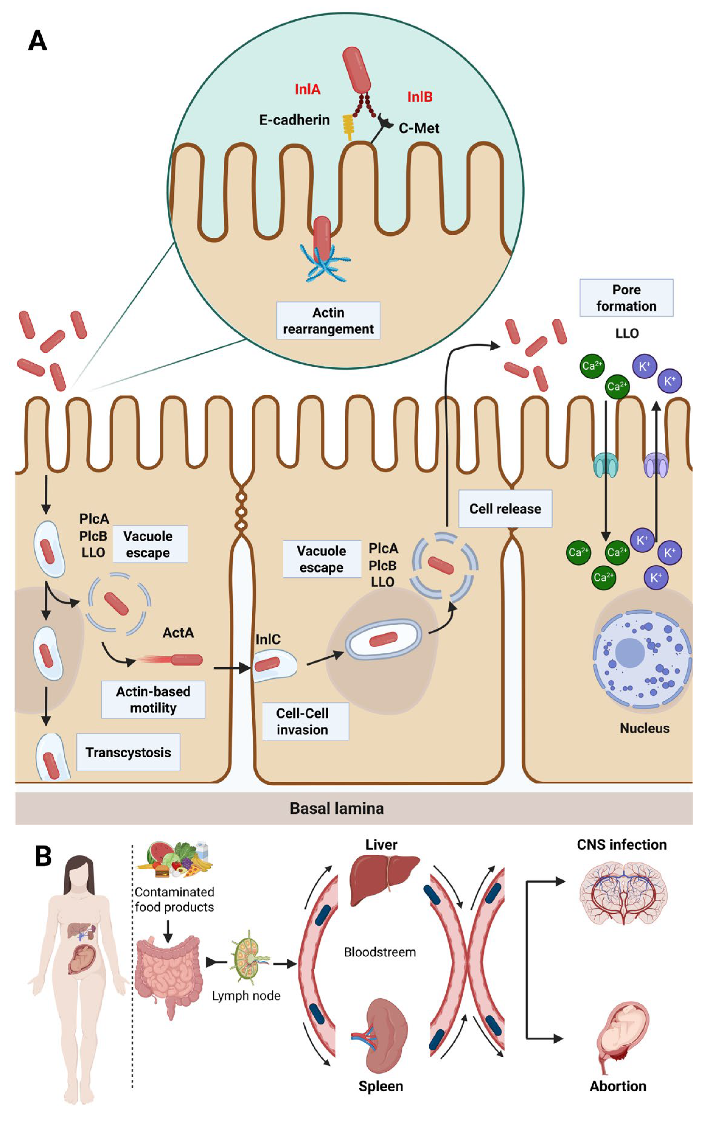
Listeriosis is a notifiable zoonotic disease caused by L. monocytogenes, a ubiquitous microorganism highly pathogenic to both humans and animals [30]. Essentially, this disease can be transmitted through the consumption of contaminated food products or through direct contact with carrier livestock, as illustrated in Figure 1 [30,31]. Listeriosis effects remain extremely limited for the general population but can be lethal for specific populations with a hospitalization rate over 95% among vulnerable individuals such as newborns, pregnant women, the elderly and immunocompromised patients [32]. Invasive listeriosis is categorized into two main classes, namely severe invasive listeriosis, involving a potentially fatal infection, and non-invasive febrile gastroenteritis, representing a relatively low severity infection [33]. In general, severe invasive listeriosis appears as sepsis, meningitis, endocarditis, encephalitis and cerebral infections among immunocompromised peoples, causing a 22% death rate in adults, and 10% for those suffering from endocarditis [6,34]. Meanwhile, non-invasive febrile gastroenteritis occurs as an infection commonly associated with influenza-like symptoms, either with or without gastroenteritis [6]. Moreover, this category occurs in generalized atypical meningitis, septicemia or febrile gastroenteritis, characterized by fever and diarrhea and associated with muscular pain and headache affecting adults [35]. The expression of these two listeriosis forms essentially depends on individual state of health, age, immune system response, infection mode and dose, as well as infectious strain virulence [36]. From a global epidemiological perspective, L. monocytogenes has been responsible for numerous epidemics, usually involving the consumption of ready-to-eat food products [37]. These infectious diseases have been mainly related to changes in consumers’ behavior, in particular, their dietary intake that is increasingly oriented towards the consumption of ready-to-eat food products [35]. Furthermore, worldwide trade globalization, the growing prevalence of age-related vulnerable populations and the emergence of numerous infectious diseases impairing consumers’ immune systems have contributed to the increased risk of contracting listeriosis [35].

2.3. Virulence Factors and Pathogenicity

Listeriosis essentially involves the consumption of contaminated food products, where the gastrointestinal tract is the pathogen’s main delivery pathway into the host organism [4,32,38]. The pathogenicity of foodborne L. monocytogenes is essentially related to its extensive ability to promote internalization in host cells [18]. This pathogen possesses an extensive capacity to overcome four crucial defensive barriers in humans, namely, the enzymes in the gastrointestinal tract, the intestinal epithelium, the blood–brain barrier and the blood–placenta barrier in pregnant women, and subsequently disseminate through other organs such as the liver and spleen [39]. Host infection involves a multi-step process that initially starts with gastrointestinal transit, followed by target cell adhesion and invasion and subsequent lysis of the protective vacuole to promote intracellular dissemination and propagation to adjacent cells (Figure 2) [40].
Following the ingestion of contaminated food matrixes, pathogenic L. monocytogenes exhibits the ability to establish colonies in the gastrointestinal tract, through bacterial resistance to the stomach’s and duodenum’s range of acidic conditions, bile salts, pancreatic secretions and host-generated proteolytic enzymes [41]. The mechanism of tolerance and adaptive response to acid stress (ATR), the glutamate decarboxylase (GAD) system, arginine deaminase (ADI) and the F0F1-ATPase complex are the main systems involved in the resistance capacity to low-pH environments [42]. Activated through pre-exposure to sublethal pH values, ATR system supports the endurance of L. monocytogenes by protecting the microorganism against both osmotic stress and high temperatures [43]. Additionally, this adaptation is triggered by the involvement of the glutamate decarboxylase system or an internal proton pump promoting an increased cytoplasmic buffering capacity of the bacterial cells [44]. The GAD system is encoded through five genes, namely gaD1, gaD2 and gaD3 encoding decarboxylases, as well as gadT1 and gadT2 antiporters [45]. These genes are hosted in the SS-1 genomic cluster responsible for L. monocytogenes stress survival, a cluster which simultaneously encodes a protective penicillin V acylase essential for bile fluids tolerance [46]. The ADI system is a further mechanism involved in the protection of L. monocytogenes against relatively low pH levels [47]. This mechanism involves two enzymes encoded by the arcABC operon, carbamylotransferase and carbamate kinase, responsible for maintaining cytoplasmic pH and protecting the bacterium against acidic environments [48,49]. The F0F1-ATPase complex is an additional system implicated in acid resistance through two essential components, namely, the catalytic component (F1) responsible for ATP synthesis and hydrolysis, as well as the membrane domain (F0) playing a proton channel role [50]. The ATP aerobic hydrolysis carried out by this complex relies on protons flowing either into or out of cell membranes and induces a proton motility that ensures bacterial propagation under adverse acidic conditions [50]. To ensure both bacterial survival and adaptation to the various stress conditions induced from high-acid environments, these gastrointestinal resistance mechanisms operate in synchronization [41].
In addition, this foodborne pathogen demonstrates a high capacity to tolerate bile fluids, recognized for their ability to disrupt bacterial cell-walls and proteins, subsequently promoting the destruction of genetic material, and increase in oxidative stress in microbial cells [51]. Bile salt resistance is essentially mediated by hydrolase production that enables the cleavage of specific amino acids present in bile salts, thereby reducing their inhibitory activity and improving bacterial growth in the gastrointestinal tract [52]. The Bsh gene is responsible for hydrolase production, and the expression of this gene is directly correlated with the Sigma B (sigB) factor that subsequently activates a number of other protective factors under extreme stress conditions [53]. Moreover, another identified mechanism of bile fluid tolerance involves a bile exclusion protein (BilE) regulated by the PrfA virulence factor, which is already implicated in the expression of the listeriolysin regulatory protein [46]. Additionally, the pathogenicity, virulence, survival and replication of this microorganism essentially depend on the PrfA transcriptional and regulatory factor, a thermally regulated factor with an optimal operating temperature of 37 °C [40,54].
Following successful colonization under gastrointestinal stress conditions, invasiveness is initiated by the adhesion and penetration of L. monocytogenes into host epithelial cells through three different surface proteins, namely, internalines InlA and InlB that bind to membrane receptors, E-cadherin and C-Met, respectively, as well as the cellular adhesion protein (LAP) encoded by the lap gene that is responsible for the cleavage of cellular junctions via binding to the Hsp60 receptor (Figure 2) [55]. Furthermore, invasin A encoded by the lmo1413 gene is another internalizing factor that essentially facilitates the penetration of L. monocytogenes through the host’s intestinal mucus [56]. The post-internalization phase is followed by the temporary encapsulation of L. monocytogenes cells in the primary phagosomale vacuole, ensuring the bacterium’s survival by evading the host cell’s phagocytic (autophagy) mechanisms and contributing to the pathogenicity of this microorganism [3]. The encapsulation step is followed by a pore-forming vacuolar escape mediated by a cytolysin enzyme known as listeriolysin O (LLO), encoded by the hly gene in conjunction with a specific phospholipase (plcA and plcB) and a metalloprotease protein responsible for the bacterial cells release into the epithelial cell cytosol in the context of LLO deficiency [57]. Moreover, two additional vacuolar release mechanisms could be involved, namely, the lysosomal thiol reductase-inducible gamma-interferon (GILT) that promotes vacuole degradation and the pheromone-encoding lipoprotein A (pPplA), that cooperates specifically with LLO [58]. Inside the cytosol, the bacterial cell multiplication machinery is activated using the host’s nutritional resources [47]. Subsequently, this pathogen disseminates towards adjacent cells through actin polymerization (ActA) as polar filaments. This has been identified as the L. monocytogenes genetic determinant that promotes motility and intracellular dissemination in host cells [59]. By disseminating to adjacent cells, bacterial cells internalize into new cells to start another life cycle through a secondary bilayer vacuole, followed by another escape cycle supported by LLO, InlC (internalin C), as well as the two plcA and plcB phospholipases [60]. Bacterial cells crossing the intestinal barrier are then carried through the lymphatic and blood circulatory systems to the mesenteric lymph nodes and further to vital organs, including the spleen and liver, where they are immediately neutralized by the host’s immune system through macrophages, neutrophils and dendritic cells [40,61]. However, infectious cells can spread into vital organs and cross the placental barrier during pregnancy, leading to serious health consequences among vulnerable individuals such as the immunocompromised, pregnant women and newborns [62]. L. monocytogenes infectious mechanisms are illustrated in Figure 2. The following sections summarize the different infectious mechanisms affecting the targeted organs and the health impact of listeriosis on hosts.

2.3.1. Brain

To date, a comprehensive overview regarding the infectious process associated with brain penetration of pathogenic L. monocytogenes remains incomplete, primarily due to the unavailability of scientific data based on in vivo tests [47]. Nevertheless, two infection mechanisms have been identified as being directly implicated in the invasive process of the central nervous system (CNS) in both humans and animals [63]. Hematogenous uptake provides a mechanism enabling rhombencephalitis (infection of the brain stem) and brain abscesses, involving the transport of L. monocytogenes cells through the bloodstream and their crossing of the blood–brain barrier to reach cellular targets [64]. Retrograde axonal transport is an additional infection pathway enabling pathogens to be transmitted through axoplasmic efflux to reach the brain through two different transport pathways, namely, a cranial nerve-mediated transport pathway, principally, the trigeminal nerve, where the nerves are infected, and an olfactory epithelium-mediated transport pathway using the mucous membrane of the nasal cavity [65,66]. Alternatively, a further virulence factor (Vip) encoding a surface protein can interact directly with the CNS through a specific receptor, Gp96, located in cerebral microvessels, promoting bacterial colonization in the brain [45]. Furthermore, data from a study conducted by Vázquez-Boland et al. [67] demonstrate the implication of the surface protein from the internalin family (lnlF) in promoting L. monocytogenes colonization in nerve cells, where recognition between the target cell and the pathogen is mediated by cell hots vimentin, a surface receptor localized on the microvascular endothelial brain cells.

2.3.2. Maternal–Fetal Listeriosis

In pregnancy, L. monocytogenes has the capability to cross the blood–placental barrier, and subsequently cause abortion, stillbirth or lethal neonatal infection [68]. Fetal infection in pregnant women can occur by two pathways, either by cellular propagation through maternal phagocytes carrying the pathogen, or through infected trophoblasts subsequently carried by the blood circulatory system to the fetal placental villi [62]. In addition, numerous studies have shown that placental infection by L. monocytogenes depends on the virulence of the infecting strain, where most virulent strains harbor the inlP gene encoding an internalizing InlP internalin protein, which is responsible for enhancing interaction between InlP and the cytoplasmic cell junction protein afadin; thus, reinforcing transcytosis through epithelial cells and promoting the placental infection [69].

2.3.3. Liver

Once they have cleared the intestinal barrier, L. monocytogenes cells are transported towards the liver through the blood and/or lymphatic system [47]. Kupffer cells have been identified as part of the anti-listeriosis immune system, essentially responsible for capturing and eliminating bacterial cells from the liver [40,54]. These specialized liver cells cannot completely eradicate the pathogenic cells, allowing bacterial persistence population to continue growing and multiplying inside the hepatocyte [62]. During internalization and cell multiplication, individual cells interact with neutrophils, resulting in microabscess formation and hepatic cell necrosis [70]. Ensuring survival and persistence in hepatic cells essentially depends on the Mpl virulence factor, a factor associated with metalloprotease synthesis responsible for the pathogen’s protection against the host immune system [62].

2.4. Antibiotic Resistance

Antibiotic-resistant L. monocytogenes in foods is an emerging threat for both food safety and public health [71]. Even though this species has historically been considered susceptible to the principal classes of clinically used antibiotics, such as ampicillin and gentamicin [72,73]. However, an increasing number of reports have revealed the emergence of multi-resistant food strains, particularly in RTE products, unpasteurized dairy products, cold meats and smoked fish [4,74]. These resistance patterns could be acquired through horizontal gene transfer involving conjugative plasmids, transposons, integrons or resistance islands, such as Listeria genomic island 1 (LGI1) that carries multiple resistance genes [30]. Among the genes involved in L. monocytogenes resistance, the most commonly reported are the: tet(M) and tet(S) (tetracyclines), erm(B) and erm(C) (macrolides), aac(6′)-aph(2′′) (aminoglycosides), cat (chloramphenicol) and dfrD (trimethoprim) genes [75]. The presence of these resistances genes in L. monocytogenes is not only of concern for the treatment of infections in humans but also raises serious concerns regarding selective pressures in the food environment [75,76]. The following section summarizes the reported and identified resistance incidents alongside the reported mutations that have been associated with resistance acquisition across different antibiotic classes in L. monocytogenes:

2.4.1. Resistance to Quinolones

Quinolones and fluoroquinolones are widespread treatments for a wide range of infectious diseases of bacterial origin affecting both humans and animals that were clinically introduced for the first time between 1962 and 1980 in the form of nalidixic acid for quinolones and in veterinary medicine for fluoroquinolones, respectively [77]. Nevertheless, their widespread utilization has been followed by the emergence of acquired resistance in several pathogens, including L. monocytogenes [78]. Resistance to ciprofloxacin is mainly associated with mutations found in both targeted receptors and genes encoding topoisomerases [78]. Several L. monocytogenes isolates have shown numerous modifications located in the DNA gyrase subunit (A) encoding gene, conferring quinolone resistance pattern by reducing interaction between the antibiotic and its target [79]. Efflux pumps such as Lde, MdrL and FepA were identified as a mechanism associated with fluoroquinolones resistance in L. monocytogenes where macrolides and cefotaxime are also exported via the MdrL pump through an activated ion flux [80].

2.4.2. Resistance to ß-Lactams

Penicillins and cephalosporins are ß-lactam-based antibiotics that inhibit the bacterial cell wall membrane assembly [81]. Penicillin resistance is an extremely rare occurrence, reported in only 0.1% of identified Listeria spp. strains [82]. This relatively low level of penicillin resistance is principally attributed to the non-existence of selective pressure in food environments, where this class of antibiotic is rarely used, thereby limiting the emergence and fixation of mutations or resistance genes [83]. Nevertheless, L. monocytogenes has been identified as naturally resistant to oxacillin, monobactams and broad-spectrum cephalosporins such as ceftazidime and cefotaxime [84]. Intrinsic resistance to cefotaxime and ceftazidime (cephalosporins) is essentially related to the ineffectiveness of these antibiotics against PBP3, indicating that this class of binding proteins maintain its ability to build the bacterial cell wall even if the other classes of PBP are inhibited [85]. In addition, the oatA gene encodes an O-acetyltransferase, a catalytic enzyme playing a key role in cephalosporin resistance through cell wall modification via muramic acid acetylation at the peptidoglycan level that prevents the antibiotic from binding effectively to PBPs, resulting in limited efficacy against the bacteria [86]. Moreover, CesR and LiaSR two-component systems represent a further resistant mechanism in the reduced cephalosporins susceptibility [87]. Both systems are involved in antibiotic-induced stress detection and response through the activation of regulators response that stimulate defense mechanisms via cell wall permeability reinforcement [87].

2.4.3. Resistance to Tetracyclines

Resistance to tetracyclines is the most frequently detected resistance phenotype in L. monocytogenes as a result of the widespread use of this antibiotic in agriculture, exerting strong selective pressure on bacterial strains and promoting the horizontal transfer of resistance genes carried by plasmids or transposons, thereby increasing the spread of resistance [80]. These molecules essentially inhibit protein synthesis by targeting the 30S ribosomal subunit [82]. Among the most frequently reported genes encoding resistance to tetracyclines: tet(A), tet(K), tet(L), tet(M) and tet(S) have been consistently detected in L. monocytogenes [80]. Tetracyclines resistance is essentially mediated through two mechanisms, one involving efflux pumps that evacuate antibiotics via proton pumps encoded by tet(A), tet(K), tet(L), and the other via a ribosomal protection mechanism encoded by the tet(M) and tet(S) resistance genes [88]. The TetM active efflux pump regulator is associated with Tn1545 conjugative transposons responsible for horizontal gene transfer between Enterococcus, Staphylococcus and Listeria strains. Additionally, the tet(S) gene has been frequently identified in L. monocytogenes and most probably transferred via the Tn6000 transposon that belongs to the Tn916 transposons family responsible for tetracyclines resistance patterns [89,90].

2.4.4. Resistance to Phenicols

Phenicols like chloramphenicol or florfenicol constitute a class of antibiotics mainly targeting the peptidyltransferase of the microbial 50S subunit [91]. In Listeria spp., the acquired resistance pattern to phenicol’s is essentially due to antibiotic enzymatic inactivation mediated by the chloramphenicol type A acetyltransferase as well as through protonic active efflux (chloramphenicol/florfenicol) [92]. This mechanism of resistance is the most common in phenicol-resistant bacteria [92]. The enzymatic mechanism of antibiotic inactivation involves an A-8 type cat gene encoding proteases located in the plM78 conjugative plasmid which is transferable between both L. monocytogenes and Staphylococcus aureus [93]. Furthermore, the plasmid pIP811 (37 kb) has been identified as being able to confer chloramphenicol resistance in several microorganisms and also as an auto transferable genetic element is numerous Gram-positive and Gram-negative bacteria including L. monocytogenes, Enterococcus faecalis, Bacillus subtilis and Escherichia coli [94]. In addition, florfenicol resistance has been associated with floR gene expression encoding an efflux pump and this mechanism has also been identified to be responsible for chloramphenicol acquired resistance in 50% of floR-positive L. monocytogenes isolates from dairy farms [95].

2.4.5. Resistance to Macrolides

Macrolides, lincosamides and striptogramin B (MLSB) bind on the bacterial 50S subunit to inhibit protein synthesis essential for cell survival [96]. The genetic elements underlying macrolide resistance involve rRNA methylases responsible for the modification of the MLSB binding site, ATP-binding transporters (ABC), MFS efflux pumps family and enzymatic inactivation encoding genes [91]. The modification of the 23S ribosomal RNA with the rRNA methylase (encoded by the erm gene) is the most common resistance mechanism among MLSB [93]. Among the 92 genes conferring resistance to MLSB, only the erm(A), erm(B) and erm(C) genes have been found in L. monocytogenes [97]. The InC, pIP50, pKUB3007 and pAMß1 plasmids have been identified as the only erm(B) harborers that confer erythromycin resistance in several bacteria, including L. monocytogenes, Enterococcus and Staphylococcus [98,99]. In addition, the presence of macrolide efflux pumps involving ABC such as msr(A) and mef(A) has also been identified in several macrolide-resistant L. monocytogenes isolated from food products [97,100].

2.4.6. Resistance to Trimethoprim

Trimethoprim is an inhibitor of the folate pathway that blocks nucleic acid synthesis, notably adenine and thymine [82]. Few incidents of trimethoprim resistance have been reported among L. monocytogenes isolates, and they have mainly been linked to the acquisition of genes (dfrD or drfG) encoding dihydrofolate reductases, as well as mutations occurring in the dhfr gene, resulting in moderately low or high levels of trimethoprim resistance [91]. The dfrD gene was first identified in 1995 on several plasmids such as pIP823, pUB110 and pC194 as a transferable element between several Gram-positive pathogens, including Staphylococcus spp., Streptococcus spp., Enterococcus spp. and L. monocytogenes [94]. The drfG gene has recently been detected in Tn916 (Tn6198) type transposon among L. monocytogenes isolates as well as in TN5801 type transposon and pMG1 plasmid in Enterococcus and Staphylococcus strains isolated in India [101]. Furthermore, the observed dhfr gene mutations have been associated with excessive and repeated bacterial exposure to trimethoprim concentration, resulting in several amino acid sequences undergoing single or double substitutions, affecting the dihydrofolate reductase genomic region [102].

2.4.7. Resistance to Aminoglycosides

Aminoglycosides inhibiting the protein synthesis process in the by targeting the 30S bacterial subunit [82]. Resistance to this class of antibiotics is rarely reported, according to several studies carried out on L. monocytogenes [103]. Resistance to aminoglycosides has only been documented in a Listeria food isolate from a meningitis patient in Greece [103]. In addition, several studies have shown that aminoglycosides resistance is associated with the presence of genes encoding inactivation enzymes able to destroy the antibiotic as soon as it is internalized in the bacterial cytosol [80]. Generally, these genes have been acquired through horizontal transfer of mobile genetic elements, such as Tn3760 transposon that harbor the aac6-aph2 gene encoding 6-N-streptomycin adenylytransferase, responsible for streptomycin resistance and disseminated among Staphylococcus spp. and Enterococcus spp. strains [82].
The evolution of antibiotic resistance among L. monocytogenes isolates has raised a major concern in both clinical and food safety context [38,104]. Medically, resistant isolates impair the effectiveness of first-line antibiotic treatment with ampicillin and gentamicin, particularly among vulnerable populations such as pregnant women, the elderly and the immunocompromised, where listeriosis can potentially lead to very severe or life-threatening consequences [5,6,7]. The emergence of even moderately resistant L. monocytogenes isolates can significantly compromise the therapeutic options, notably in β-lactam allergy context [105,106]. Environmentally, antibiotic-resistant L. monocytogenes isolates appear to be effectively persistent in agrifood processing environments. This persistence is enhanced through their capacity to generate biofilm over inert surfaces and their increased tolerance to oxidative stress, as well as their cross-resistance with disinfectants that promote their proliferation despite the conventional industrial sanitizing process [107,108,109,110]. In addition, some studies have demonstrated that some persistent L. monocytogenes isolates from agrifood environments also displayed antibiotic-resistant profiles, suggesting a relationship between environmental persistence and the ability to cope with antimicrobial pressure [8,37]. This situation exacerbates the related risk of contaminated RTE food products and consequently, widespread L. monocytogenes outbreaks. For example, hypervirulent and resistant 4b serotype L. monocytogenes strains have been responsible for more than 1000 humans listeriosis cases in South Africa between 2017 and 2018 [111]. Such outbreaks highlight the important challenge associated with antibiotic-resistant L. monocytogenes in foods as an effective dissemination vector of listeriosis among general population and consumers.

2.5. Survival of Listeria monocytogenes Under Food Processing Conditions

The capacity of L. monocytogenes cells to survive, adapt and persist in a wide range of adverse physicochemical conditions during food processing, cleaning and preservation constitutes a major challenge for the food industry [76]. The combination of specialized metabolic, structural and transcriptional mechanisms ensures the bacteria’s capacity to tolerate a wide range of stress factors while maintaining their pathogenic potential [47]. Among the most problematic features of this microorganism, its ability to proliferate under low temperatures and growth in refrigerated environment is particularly challenging since they are frequently considered as microbiological safety zones [76]. This adaptation to low-temperature environments is based on the adjustment of membrane phospholipids composition through an increase in the unsaturated fatty acids content, production cold shock proteins (CSPs) such as CspA and CspD and the accumulation of cryoprotective solutes such as betaine and carnitine through gbuABC and opuCABCD systems, regulated by SigmaB factor (SigB) [76,112]. This multifactorial response represents a profound metabolic transformation, involving the mobilization of significant energy resources and is much more than a simple survival strategy [113]. Moreover, the physiological costs of this approach could affect virulence or growth under higher temperatures, illustrating an adaptive dynamic that is potentially modulated by the food matrix nature and condition [113]. Meanwhile, L. monocytogenes demonstrates an increased tolerance to the thermal stresses encountered during pasteurization or cooking processes performed in several agrifood sectors. This thermal resistance is based on the coordinated activation of heat shock proteins (HSPs) such as DnaK, GroEL and ClpC/P/E that are responsible for the assembly, repair or degradation of the denatured proteins. These proteins are regulated by transcriptional repressors HrcA and CtsR, which are sensitive to temperature variations [109,114].
This adaptive capacity is reinforced through high-acid, -alkali and -salt tolerance. For example, the GAD system appears to play a critical role in the intracellular neutralization of protons in acidic environments, while the F0F1-ATPase pump participates in the active expulsion of H+ ions [42,43]. Similarly, in the presence of NaCl, L. monocytogenes cells initiate an osmoprotective response by accumulation of K+ protons, followed by an importation of compatible solutes (betaine, proline) via σ^B-regulated transporters [43]. Tolerating high saline concentrations enables some strains to successfully persist in specific food niches such as salted products, salted products and cured cheeses, thereby conferring an ecological advantage in specific food ecosystems [76]. In addition, the high alkaline tolerance of L. monocytogenes is essentially due to coordinated physiological and molecular responses. To maintain intracellular homeostasis under high-alkaline conditions, the bacterium relies on proton expulsion systems (H+-ATPases), while simultaneously modulating the fatty acid composition of its membrane to reinforce its barrier against the influx of hydroxide ions [76,115]. In addition, the sigma B regulon (SigB) orchestrates the expression of stress-responsive genes, thereby improving survival under alkaline stress [116]. L. monocytogenes also exhibits an alarming tolerance level towards chemical disinfectants, particularly quaternary ammonium compounds (QACs) such as benzalkonium chloride [117,118]. This resistance is mainly attributable to the expression of efflux pumps regulated through the bcrABC, qacH, emrE, emrC and mdrL genes and frequently distributed on plasmids or mobile genomic islands such as LGI1, promoting their dissemination through horizontal transfer [117]. This resistance profile is enhanced by biofilm formation, trapping the disinfectants inside the extracellular matrix [119]. In addition, cross-resistance has been reported towards heavy metals, suggesting co-selection of tolerant characteristics under industrial environments [119]. Excessive exposure to biocides, particularly involving sublethal concentrations, exerts selection pressure, leading ultimately to the emergence of L. monocytogenes strains with enhanced tolerance [120]. This situation highlights the importance of a comprehensive re-evaluation of existing disinfection procedures, particularly in relation to the adaptive mechanisms and the potential risks of cross-resistance, as well as the long-term effectiveness of conventional cleaning–disinfection strategies [121].
The ability to form biofilms is one of the most critical factors in L. monocytogenes′ persistence in food processing operations. These multicellular structures are composed of encapsulated bacterial cells by a complex extracellular microbial material incorporating polysaccharides, extracellular DNA (eDNA), proteins and lipids [8]. This microbiological structure provides bacterial resistance against disinfectants, temperature fluctuations, ultra-violet rays, heavy metals and dehydration [122]. Biofilms can develop on a wide variety of surfaces, including stainless steel, plastic and glass at temperatures as low as 4 °C [8]. The formation of biofilms is regulated through several genes, including bapL, agr, sigB, and influenced by numerous environmental factors such as hydric flux, nutrient availability and surface characteristics [8]. Some hypervirulent clonal lineages (CC1, CC2, CC4) display a particular ability to develop a persistent form of biofilms, often associated with genomic islands such as LGI1 and LGI2 [47,107]. The high tolerance of biofilms to disinfectants is exacerbated by slowed physiological cell growth, reducing their susceptibility to chemical disinfectant agents specifically targeting growing bacteria [123]. Consequently, biofilms become a chronic recontamination reservoir that is particularly challenging to eradicate despite successive physicochemical treatments [76,123]. These factors highlight both the limitations of conventional sanitation practices and the necessity to develop more effective biological control strategies, involving a combination of enzymatic, mechanical and targeted antimicrobial approaches.

3. Protective Cultures to Control Listeria monocytogenes

Protective cultures have been defined as specifically selected microorganisms able to inhibit the growth of pathogenic or spoilage bacteria while significantly maintaining the sensory, physicochemical, and nutritional properties of foods [124]. However, starter cultures may also play a dual role in fermented food systems, not only as technological agents responsible for fermentation and the development of desirable sensory characteristics but also as protective cultures that improve microbial safety and product stability. For example, Lactococcus lactis and Pediococcus acidilactici were traditionally used as starter cultures, a dairy fermentation process, which also acts as a protective culture by producing bacteriocins such as nisin Z and pediocin PA-1, which are known to be effective inhibitors of Gram-positive pathogens [125,126].
Based on their consistent utilization, protective cultures have been recognized as GRAS by the Food and Drug Administration (FDA) for human consumption [127]. Generally, LAB are associated with fermented food products, including bacterial strains such as Lactobacillus spp., Lactococcus spp., Leuconostoc spp., Pediococcus spp., Streptococcus spp. and Enterococcus spp. [15]. Numerous antimicrobial compounds produced by LAB have been characterized for their inhibitory potential against a wide range of foodborne pathogens such as organic acids (acetic, lactic, succinic, phenyllactic and propionic acid), hydrogen peroxide (H2O2), diacetyl, reuterin and antimicrobial peptides (AMPs) including bacteriocins and lipopeptides [128,129].

3.1. Modes of Action of Protective Cultures

The preservation effect through the LAB cultures has been associated with the accelerated acidification of the raw ingredients through the accumulation of organic acids, mainly lactic acids [130]. As relatively weak acids, organic acids exhibit significant antimicrobial properties in food matrices which have a pH value above 3 [129]. This activity is based on several mechanisms, including membrane protein denaturation and transmembrane transport disruption, as well as proton gradient interference, enzymatic inhibition and ROS (reactive oxygen species) production, resulting in unbalanced metabolism and microbial growth inhibition [129]. The antimicrobial activity of organic acids is enhanced in highly acidic environments and predominantly characterized by the undissociated state [13]. The apolar form penetrates the cytoplasmic membrane and dissociates further in the cytosol, leading to intracellular acidification, disruption of cellular metabolism via protein denaturation and enzymatic inhibition, and eventually microbial death [129]. The low molecular weight and neutral nature of hydrogen peroxide allow it to penetrate the bacterial cytoplasmic membrane. Its reduction in the cytosol results in the formation of hydroxyl radicals, extremely reactive species capable of irreversibly damaging enzymes and nucleic acids [131,132]. The mechanism of action of diacetyl remains incompletely elucidated, but some studies suggest that they can interact with arginine residues to alter protein structure. In addition, diacetyl may bind to DNA, resulting in its misfolding [133]. Until the early 2000s, the specific mechanism of action of reuterin (3-hydroxypropionaldehyde), an aldehyde produced by Lactobacillus reuteri, remained undefined [134]. This compound possesses a highly reactive aldehyde group that generates several derivatives in aqueous solutions, thereby complicating its study [134]. Subsequent investigations have demonstrated that reuterin induces oxidative stress among target bacteria, particularly through the modification of thiol groups on proteins or small intracellular molecules, disrupting essential cellular functions [134,135]. On the other hand, the bacteriocins have been extensively investigated as potential bioconservation agents [136]. These antimicrobial peptides produced by LAB exhibit significant inhibitory activity against a wide range of foodborne pathogens, including L. monocytogenes [137,138]. The principal mechanism of action involves disruption of cytoplasmic membrane integrity on targeted bacteria, inducing intracellular leakage, membrane potential disruption, and, ultimately, cell death [139].
In addition, another mechanism of action of protective cultures, besides their antimicrobial activity, involves the attenuation of virulence factors of pathogens. Some studies have demonstrated that exposure of L. monocytogenes and Staphylococcus aureus strains to protective bacterial cultures has the potential to significantly reduce the expression of some key virulence factors, thereby reducing their infectious potential following the ingestion of contaminated food. Among L. monocytogenes, this principally involves listeriolysin O and internalines, key factors responsible for host cell adhesion, invasion, and intracellular translocation [140]. For example, co-culturing L. monocytogenes and Lactobacillus plantarum significantly downregulated the hly gene (encoding listeriolysin O) and the inlA/inlB genes (encoding internalines), resulting in decreased bacterial uptake by epithelial cells in vitro and reduced listerial virulence in murine infection models [140]. However, in the case of S. aureus, this virulence decreased effect has been associated with quorum-sensing modulation as well as toxin (enterotoxins and hemolysins) production inhibition, thereby limiting pathogenicity and increasing antimicrobial susceptibility [141]. These effects are modulated through mechanisms, including the disruption of quorum sensing and the alteration of intercellular communication, disturbing the regulation of virulence genes, biofilm formation, and immune system evasion [140,141,142]. This approach is particularly relevant in terms of food safety, given that it minimizes the risk of infection while maintaining food quality and enhancing microbial safety without compromising the sensory or physicochemical properties of the food matrices.

3.2. Uses of Protective Cultures in the Agrifood Industry to Control Listeria

As a result, on their proven safety and effectiveness against a wide range of spoilage and/or pathogenic microorganisms, LAB and their bacteriocins are attracting growing interest as natural preservatives in the agrifood industry (Table 2) [139]. Their uses can be based on different approaches involving the direct inoculation on food products using bacteriocinogenic LAB strains as starter culture able to produce antimicrobial compounds in situ [15,127]. Alternatively, purified or semi-purified bacteriocins can be incorporated as preservation additives, or as an ingredient derived from fermentation using a bacteriocinogenic strain, naturally harboring antimicrobial cluster gene production during the food processing process [127]. Table 2 summarizes different protective cultures used in a variety of food products to control L. monocytogenes.
Table 2. Major protective cultures and their antimicrobial metabolites used in different food products.
Protective CultureBacteriocinsFood ProductsReferences
Lactococcus lactis DF04MiNisinFresh goat’s cheese[143]
Lactococcus lactis LL56NisinFresh cheese[144]
Lactococcus lactis DPC4275Lactacin 3147Cottage cheese[143]
Lactobacillus sakei CTC494Sakacin (G/P)Vacuum-cooked ham[145]
Lactobacillus sakei LAK-23Sakacin (non-specified)Smoked fish[146]
Latilactobacillus curvatus CRL705Lactocin 705 and Lactocin AL705Cooked vacuum-packed beef meat[147]
Lactobacillus curvatus ACU-1Sakacin QCooked meat[148]
Lactobacillus plantarumPlantaricin A, EF, JK and SApples, fresh vegetables, lettuce, cold meats[149]
Lactobacillus pentosus MS031Non-specifiedFresh chopped fruits[150]
Lactobacillus caseiNon-specifiedReady-to-eat salads[151]
Pediococcus pentosaceus DT016Pediocin (non-specified)Fresh vegetables[152]
Enterococcus hirae ST57ACCNon-specifiedSkim milk[153]
Leuconostoc mesenteroidesLeucocin CApples and lettuce[154]
Carnobacterium maltaromaticumCarnocin and piscicolinFish and smoked salmon[155]
Carnobacterium divergens M35Divergicin M35Smoked salmon[156]
Paenibacillus polymyxaPolymyxin-like peptidesCanned vegetables, meat[157]
Streptococcus salivarius K12Salivaricin and Subtilin AFermented food products[158]
Lacticaseibacillus rhamnosus GGRhamnosin-like peptidesFresh lettuce and ready-to-eat products[159]
Enterococcus durans M3-3Duracin-likeRaw milk[160]
Leuconostoc carnosum 4010CarnosinRefrigerated raw meat and meat products[161]
Lactobacillus helveticus CNRZ32Helveticin JPressed cheese[162]
Weissella hellenica 4M13Weissellin A and BPickled vegetables[163]
Using bacteriocingenic LAB strains provides an attractive alternative to chemical preservatives to control L. monocytogenes proliferation in a wide range of food products due to their biopreservative and fermentative properties [164]. These cultures also allow in situ bacteriocins production, leading to a reduction in purification costs and minimized organoleptic alterations [11]. For example, the use of Lactococcus lactis strains DF04Mi and DPC4275 as lactacin 3147 producers in dairy products showed a 1000-fold reduction in listerial cells in fresh cheeses [143]. Specific bacterial strains of Pediococcus acidilactici UL5 and Lactococcus lactis UL719, respectively, producing pediocin PA-1 and nisin Z, were remarkably effective as a protective culture applied in raw milk by allowing total inhibition of L. monocytogenes combined with a preservation of the milk’s physicochemical characteristics [137]. For meat and fish products, Lactobacillus sakei (sakacin P, G) and Latilactobacillus curvatus (lactocin 705) strains displayed significant anti-listeria activity while ensuring sensorial integrity [145,165]. As an example, L. sakei CTC494 was incorporated as a protective culture in vacuum-packed cooked ham. Mathematical modeling demonstrated a significant inhibition of L. monocytogenes development at 4 °C, thereby reducing growth rate and maximum population, particularly in high culture concentrations [145]. Carnobacterium maltaromaticum, isolated from seafood products, represents another natural protective culture used for smoked salmon preservation [155]. This strain produces the bacteriocins carnocin and piscicolin that are effective peptides against Listeria spp. under low storage temperatures, which make them suitable for protein-rich and neutral-pH food products [155]. Furthermore, the use of Lactobacillus plantarum, Lactococcus lactis or Pediococcus pentosaceus cultures in fresh or lightly processed fruits and vegetables has shown a significant L. monocytogenes load reduction from 1.4 to 2.7 log CFU/g, while simultaneously inhibiting other pathogenic bacteria such as E. coli and Salmonella spp. [127,154]. The use of protective cultures contributes to a sustainable approach by limiting the release of chemical residues into the environment and thereby reducing the risk of developing preservative-resistant strains [136]. However, introducing a protective culture directly into a food processing system requires rigorous validation to ensure the safety and stability of the culture, as well as the prevention of undesirable interactions with beneficial microbiota or the organoleptic properties of the food product [136].

4. Antimicrobial Peptides Produced by Bacteria: Promising Biocontrol Agents

4.1. Bacteriocins and Lipopeptides
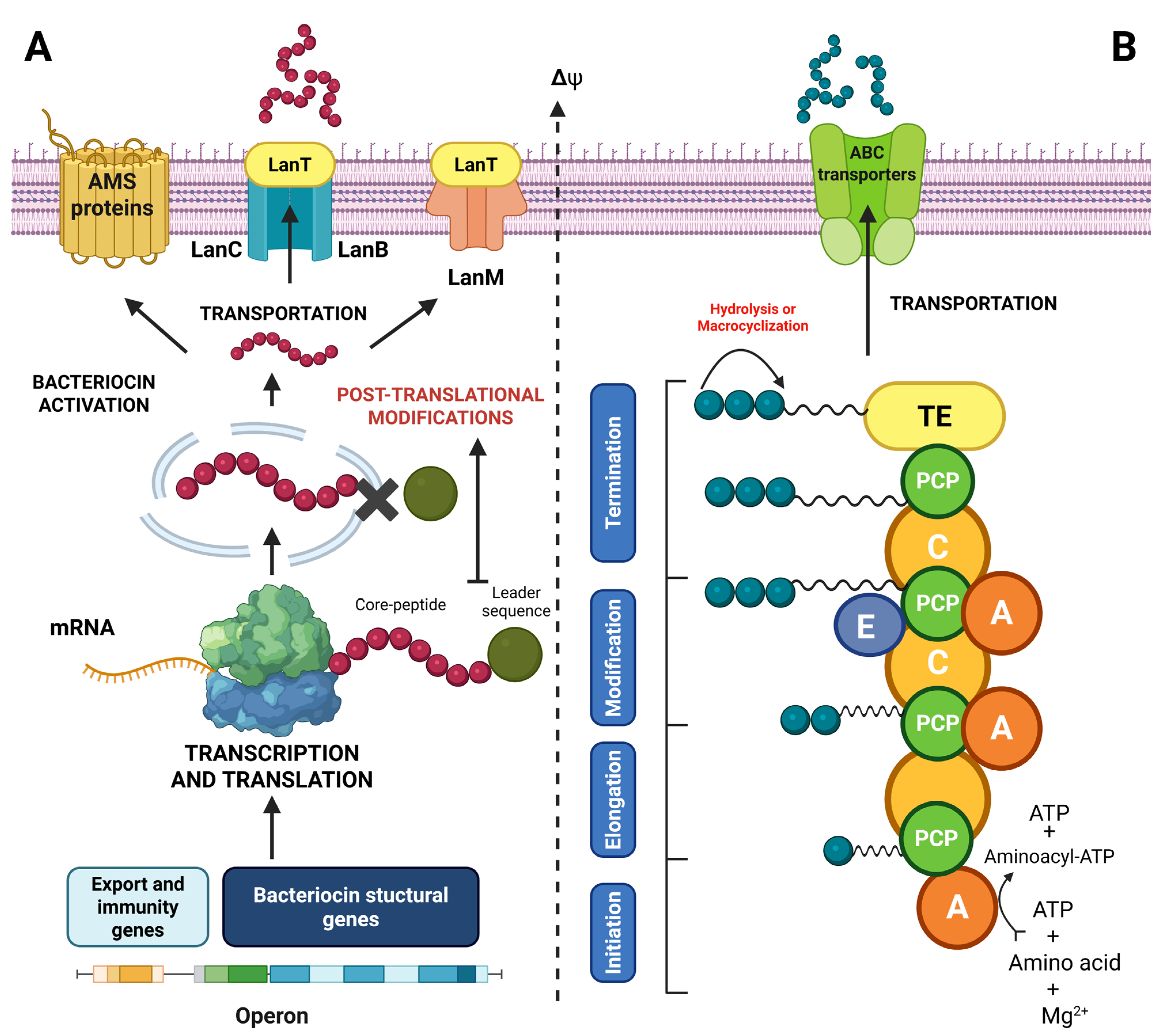
Bacteriocins and bacterial lipopeptides constitute two major classes of AMPs naturally produced by a wide range of bacteria, principally to maintain their ecological niche and compete with other microorganisms [139,166]. Bacteriocins are ribosomally synthesized peptides (RiPPs), characterized by a specific antimicrobial activity and a narrow spectrum of action, most commonly targeting bacteria related phylogenetically to the producer strain [16]. Their stability, low toxicity and effectiveness at low concentrations make them very promising candidates for use as bioconservation agents in foods [136]. In general, these molecules are distinguished by significant inhibitory effect against several pathogens of interest, including L. monocytogenes [167]. In general, bacteriocins are synthesized as a biologically inactivated precursor containing a N-terminal sequence protecting the peptide from enzyme-induced proteolytic degradation occurring in the cytoplasmic environment of the bacterial producing strains (Figure 3A) [168]. The produced precursor peptides generally require post-translational modification, a crucial step for establishing an active three-dimensional structure before being extracellularly exploited [15]. The post-translational maturation process is essentially followed by an extracellular exportation step of the bioactive peptide through a self-defense system involving encapsulation protein secretion as an autoimmunity process or active efflux pumps, or a simultaneous combination of both systems [169,170].
Figure 3. Schematic illustration of the different stages for the biosynthesis of bacteriocins (A) and lipopeptides (B) in bacteria (created with BioRender.com) [171,172,173]. Δψ represents the transmembrane potential.
Ijms 26 10481 g003
On the other hand, lipopeptides are non-ribosomally synthesized peptides (NRPs) displaying a wide range of spectra of action and characterized by the addition of a fatty acid chain on a cyclic or linear polypeptide [174]. This amphiphilic nature ensures their direct insertion into phospholipid membranes, causing depolarization and cell lysis of targeted bacteria [166]. Lipopeptide biosynthesis involves non-ribosomal peptide synthetases, a metabolic pathway distinguished from classical messenger RNA-encoded translational pathways (Figure 3B) [173]. This system provides extensive structural flexibility and enables the incorporation of non-proteinogenic amino acids, as well as various postsynthesis modifications such as N-methylation, cyclization and epimerization, thereby conferring a wide range of biochemical and functional diversity on lipopeptides [172]. In general, the synthesis process involves the activation of the amino acid substrate with an adenylation domain (A), thereby selecting and activating the amino acid by ATP binding to create an aminoacyl-AMP intermediate [173]. A peptidyl carrier protein (PCP), also known as the thiolation domain (T), is then transferred through a thioester bond to phosphopantetheine [172]. The subsequent condensation domain (C) is then used to catalyze peptide bond formation between the activated residues on successive modules. Other specific modules may include an epimerization domain (E) to convert L amino acids to D, or an N-methyltransferase domain (MT) to incorporate methyl groups [173]. Finally, a thioesterase (TE) domain enables peptide cleavage and cyclization in several models [175]. In lipopeptides, an essential step involving N-terminal acyl chain addition is usually performed through an activated fatty acid (e.g., acyl-CoA), and incorporated through a specific acylation domain frequently located at the initiation module [176]. This acylation confers the amphiphilic character of lipopeptides that is crucial for their biological activity and interaction with cell membranes [177]. The different stages of bacteriocin and lipopeptide biosynthesis are presented in Figure 3.

4.2. Classification of Bacteriocins and Lipopeptides

Bacteriocins and lipopeptides classification has been continually reviewed in accordance with new scientific advances, particularly through the discovery of new chemical structures, as well as novel specific mechanisms of action for each AMP class [178,179]. These developments reflect the growing complexity and functional diversity among the identified compounds. This section will focus on the main classes of molecules showing selective antimicrobial properties against L. monocytogenes, particularly Gram-positive bacteriocins and microbial lipopeptides demonstrated to be effective against this foodborne pathogen. The following Gram-positive bacteriocin classification is based on Cotter et al. [16]. The structural and functional diversity of Gram-positive bacteriocins are significantly more extensive than those produced by Gram-negative bacteria. In general, they are classified into three main categories according to their structure, mechanism of action and mode of biosynthesis.

4.2.1. Class I Bacteriocins

Bacteriocins subjected to post-translational modifications during the maturation process have been included in this class [180]. They are further subdivided into several subclasses based on the nature of the modification (Table 3) [181]. Lantibiotics are described as peptides containing at least one lanthionine residue, a dehydrated amino acid formed through threonine or serine cross-linking with a cysteine residue [182]. A second subclass includes the labyrinthopeptins characterized by the presence of a labionine, a post-traditionally modified carbocyclic amino acid residue [183]. The sactibiotics subclass contains cyclic bioactive peptides bearing a linkage between a cysteine side chain and the Cα of another amino acid residue [184]. Glycocins are bacteriocins distinguished by the presence of S-glycosylation and O-glycosylation of serine and/or threonine residues [185]. Another suggested subclass includes the darobactins, peptides characterized by their unusual double-ring structure formed through tryptophan–tryptophan, lysine or arginine bonds [186]. The cyanobactins have also been proposed as a subclass, defined by the involvement of an N-terminal proteolysis enzyme in their maturation [187]. In addition, circular bacteriocins are characterized by the covalent linkage of their N- and C-terminal residues, resulting in a large cyclic peptide backbone [188]. For bottromycins, the presence of a macrolactamidine moiety portrays this subclass [188]. Finally, lasso peptides were proposed as a distinct subclass, characterized by a macrolactam ring structure through which the C-terminal tail is threaded to form a unique lasso-shaped topology [189]. With recent advances in bacteriocin discovery, several other subclasses have been and will be added within Class I as described in more details by Sugrue et al. [188].

4.2.2. Class II Bacteriocins

These bacteriocins represent a heterogeneous group of peptides typically measuring under 10 kilo Dalton (KDa) in size and without undergoing a post-translational modification process during the maturation stage [180]. This class is further subdivided into four subclasses [180]. Firstly, class IIa contains “pediocin-like” bacteriocins that are, characterized by their anti-listeria properties and used as food additives to control several pathogenic bacteria [180]. Class IIb includes bacteriocins combining two unmodified peptides exhibiting synergistic activity [190]. Circular bacteriocins are characterized by a post-translational modification, resulting in a covalent bond formation between the N-terminus and the C-terminus, constituting class IIc of bacteriocins [16]. Class IId bacteriocins constitute a heterogeneous group of “non-pediocin-like” linear leaderless or non-two component peptides exhibiting a no post-translational modifications process [191].

4.2.3. Class III Bacteriocins

This class includes recently identified AMPs like bacteriolysins and tailocins [192]. Bacteriolysins are thermolabile antimicrobial proteins synthesized by several Gram-positive bacteria and characterized by a domain-like structure, conferring lytic activity on the targeted bacterial cell wall [192]. On the other hand, tailocins are distinguished by their phage-like tail structure [193]. Table 3 summarizes the most documented bacteriocin classes produced by Gram-positive bacteria.
Table 3. Gram-positive bacteriocin classification according to Cotter et al. [16].
ClassSubclassCharacteristicsBacteriocinsReferences
ILantibioticsPost-translational modificationNisin Z[194]
LabyrinthopeptinsLabyrithopeptin A2[195]
SactibioticsSubtilisin A[196]
GlycocinsPallidocin[197]
DarobactinDarobactin A[198]
CayanobactinKawaguchipeptin B[187]
CircularPumilarin[199]
BottromycinBottromycin A2[200]
Lasso peptideUbonodin[189]
IIIIaUnmodified, low-molecular-weight peptides
(<10 KDa)
Pediocin PA-1[201]
IIbLactacin F[202]
IIcCarnocyclin A[203]
IIdEnterocin L50A/B[204]
IIIBacteriolysinsThermolabile, unmodified, high-molecular-weight peptidesHelveticin J[205]
TailocinsPhage tail structureMonocin J25[206]

4.2.4. Lipopeptides

Lipopeptides are generally low-molecular-weight molecules (500 to 1500 kDa) characterized by a fatty acyl chain anchored to peptide structure via an amino or hydroxyl group. Their hydrophobic tail and polar peptide structure provide an amphiphilic character that can contribute to their interesting emulsifying, foaming and mobilizing properties, as well as their inhibitory activity against bacteria, viruses, fungi and cancer cells [207,208]. As lipopeptides exhibit a high degree of heterogeneity, their classification can be based on chemical structure, natural origin and net charge [174]. This section focusses on lipopeptides produced by bacteria due to their antimicrobial potential, structural diversity and diverse modes of action [166]. Based on their chemical structure, these compounds are generally divided into two main groups, namely, linear and cyclic lipopeptides (Table 4) [166]. In addition, these molecules can be categorized as cationic, anionic, or non-ionic peptides according to their net charge [174]. Linear lipopeptides include cerexins, tridecapeptins, corrugatins and syringafactins, naturally produced by Paenibacillus spp., Bacillus spp. and Pseudomonas spp. [207,209]. Cationic cyclic peptides are distinguished by C-terminal cyclization through an ester or amide bond, and N-terminal acylation, enabling their integration through non-ribosomal peptide synthetases [174]. Moreover, non-cationic cyclic peptides are characterized by the presence of lactone or lactam cyclic structure including amino acid residues, subsequently conjugated to a lipophilic moiety. Their structure generally involves a dextrorotatory (D) and levorotatory amino acids complex arrangement, as well as non-proteinogenic residues [177]. Table 4 illustrates the classification of microbial lipopeptides based on their structure.
Table 4. Structural classification of bacterial lipopeptides.
TypeLipopeptideStructure and CharacteristicOriginReferences
CyclicSurfactinCyclic heptapeptide bonded to β-hydroxy-C13-C15 fatty acid through lactone bridgeBacillus subtilis[210]
Iturin Aβ-Amino fatty acid-linked cyclic heptapeptideBacillus subtilis[211]
DaptomycinCyclic decapeptide containing branched fatty acidsStreptomyces roseosporus[212]
LichenysinCyclic, surfactin-likeBacillus licheniformis[213]
Gramicidin SCyclic decapeptide compound with double symmetrical ringBacillus brevis[214]
Tyrocidin ACyclic decapeptide rich in hydrophobic amino acidsBacillus brevis[215]
Polymyxin B/ECyclic decapeptide containing N-terminal fatty acid, diaminobutyric acid-rich (Dab)Paenibacillus polymyxa[216]
LinearRamoplanin ALinear glycosylated lipopeptideActinoplanes spp.[217]
ActagardineModified linear lipopeptide (lanthionine)Actinoplanes garbadinensis[218]
Fusaricidin ALinear with a lipid tailPaenibacillus polymyxa[219]
Taromycin BLinear lipopeptideActinomadura spp.[220]
TelomycinLinear peptide, post-translationally modifiedStreptomyces spp.[221]

4.3. Mechanisms of Action

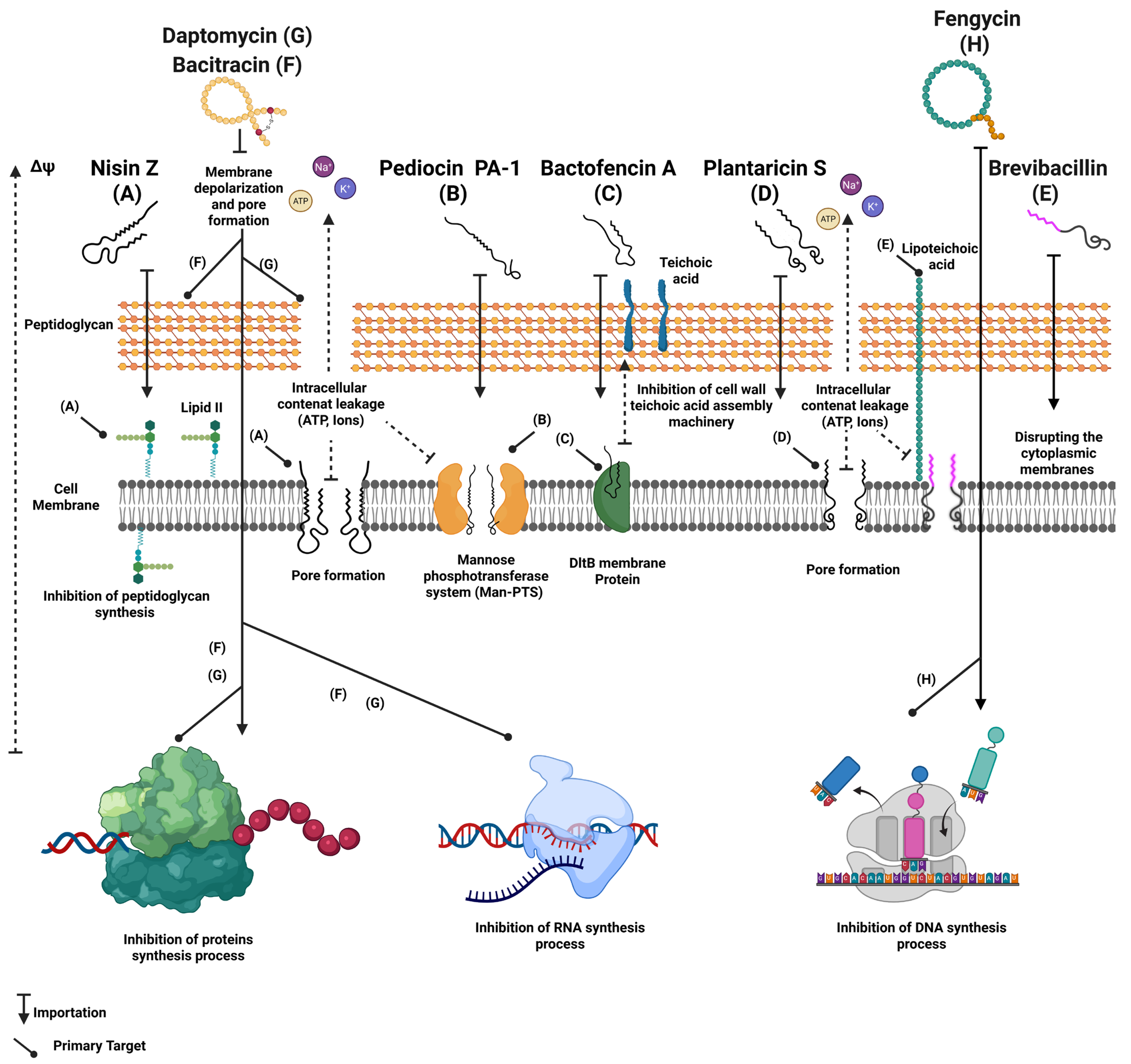
Gram-positive bacteriocins and bacterial lipopeptides act via a wide range of modes of action that are often different from those employed by clinically used antibiotics and chemical preservatives [178]. While antibiotics generally target specific bacterial cellular survival functions such as protein synthesis, DNA replication and cell wall synthesis, bacteriocins mostly exert their inhibitory effects through specific interactions with bacterial cell membranes, leading to their destructive effects [222]. For example, nisin Z, one of the most extensively studied anti-listeria bacteriocin, essentially interacts with lipid II to form a complex that blocks peptidoglycan synthesis and creates pores in the membrane [223]. This dual mechanism provides nisin significant effectiveness against L. monocytogenes and other Gram-positive pathogens [224]. Several class II bacteriocins act by disrupting the cytoplasmic membrane through pore formation, accompanied by a passive ions and metabolites flux crucial for cellular survival on targeted bacteria [139]. For example, the class II bacteriocin pediocin PA-1 has attracted significant interest due to its strong antimicrobial activity against several strains of Listeria spp., including antibiotic-resistant isolates [225]. Studies have demonstrated the capacity of pediocin PA-1 to inhibit L. monocytogenes proliferation through a specific membrane interaction mediated by the mannose–phosphotransferase system (Man-PTS), leading to subsequent membrane pore formation and intracellular component leakage [226]. Plantaricin S from Lactobacillus plantarum strains is also known for its capacity to interact with cell membranes and causing pore formation leading to cell lysis [227]. Some studies have revealed an increased efficacy of this compound against a wide range of foodborne pathogens, including Clostridium perfringens, L. monocytogenes and S. aureus [228]. In addition, some compounds like bactofencin A have been reported to disrupt cell membranes of the targeted bacteria through an interaction with the membrane protein DltB, leading to a significant loss of cellular integrity while inhibiting bacterial growth [229]. Other compounds, such as lactococcin 972, depolarize the cell membrane and disrupt septum formation, thereby inhibiting the bacterial scissiparity and cell multiplication processes (Figure 4) [230].
The modes of action of bacterial antimicrobial lipopeptides are primarily based primarily on their direct interaction with cytoplasmic membranes as a result of their amphiphilic nature, leading to structural disruption, depolarization, ions leakage and inhibition of essential cellular metabolism [231,232]. Surfactin and fengycin, produced by Bacillus subtilis, induce non-specific membrane permeabilization on L. monocytogenes isolates through direct penetration of the lipid bilayer, leading to leakage of essential intracellular components [210,233]. Furthermore, the brevibacillin, produced by Brevibacillus laterosporus is another lipopeptide known for its membrane activity, exerting its antimicrobial effect against Gram-positive bacteria by disrupting cytoplasmic membrane via an interaction with the lipoteichoic acids [234]. In addition, several lipopeptides exhibit multifactorial activity by associating membrane-based mechanism of action and interference with intracellular processes [14,235]. For example, daptomycin, a calcium-dependent cyclic lipopeptide produced by Streptomyces roseosporus [236] can insert itself into the cytoplasmic membrane and form oligomeric complexes, causing accelerated membrane depolarization, leading to potassium efflux while simultaneously inhibiting DNA, RNA and bacterial protein synthesis [212,237,238]. While lipopeptides such as gramicidin S can establish transmembrane pores allowing ions leakage, other peptides like polymyxin B are able to cross the bacterial membrane to directly interfere with nucleic acids, disrupting replication or transcription [173,239]. Nevertheless, these intracellular mechanisms appear to be relatively infrequent among lipopeptides, compared with those targeting cell membrane AMPs [173]. Figure 4 illustrates the different mechanisms of action of the described AMPs and their potential targets.

4.4. Use of Antimicrobial Peptides for the Biocontrol of Listeria monocytogenes in Foods

Antimicrobial peptides, particularly bacteriocins and lipopeptides produced by bacteria, represent an innovative and natural approach allowing an effective management of L. monocytogenes proliferation under agrifood processing environments [178,240]. Their natural aspect perfectly reflects consumers’ growing demand for chemical-free and preserved organic food [136]. From a technological perspective, these molecules exhibit several important characteristics, such as stability under acidic pH conditions, extreme temperatures and high salt concentrations, enabling their incorporation as preserving agents in food matrices [241]. AMPs can be used either as purified or semi-purified in a wide range of food matrices to provide a selective and adaptable biopreservation strategy [242]. Three major approaches have been described for their direct incorporation into food products, including the immersion of food products in bactericidal AMPs solutions, and a surface application by spraying or coating [242]. Table 5 summarizes the different applications of AMPs for the control of L. monocytogenes in the agrifood commodities.
Table 5. Examples of the use of antimicrobial peptides in the agrifood industry.
PeptidesProducer StrainsProductsApplicationEfficacyReferences
Nisin ALactococcus lactisNisaplin™Ingredients used through direct incorporation, active films or encapsulation in cheeses and UHT milk.2–4 log reduction in L. monocytogenes levels in Ricotta, Cottage and Galotiri cheeses.[124]
Nisin ZLactococcus lactisNiseen®-SiveeleIncorporation in Galotyri and Minas fresh cheeses.Extends products shelf-life up to 21 days under refrigeration conditions.[139]
MicocinCarnobacterium maltaromaticumMicocin®Surface application or coating on soft, raw milk cheeses.
Brining or direct application to chopped meat.
Complete reduction in L. monocytogenes under refrigeration for 14 days.
L. monocytogenes elimination for 14 days at 4 °C (<102 CFU/g).
[11]
Pediocin PA-1Pediococcus acidilacticiAlta™ 2341Direct incorporation or cultivation in pasteurized milk and soft cheeses.
Injections or incorporation into RTE sausages, hams and cooked meats.
L. monocytogenes elimination at 4 °C in 7–10 days.
3 to 5 log reduction in Listeria in ham at 4 °C in 10 days.
[11]
SurfactinBacillus subtilisInoviaTech, under developmentFood surface cleaning and disinfectant (stainless steel and plastic)L. monocytogenes biofilm inhibition, 4-log reduction in mature biofilm.[166]
FengycinBacillus subtilisBioBoom® CleanProduction line and sensitive surfaces decontamination.Synergistic effect and prolonged anti-Listeria activity with nisin.[243]
Iturine ABacillus subtilisUnder investigationActive packaging formulations and cutting surfaces.anti-listeria, suitable for encapsulation or coating.[243]
Lacticin 481Lactococcus lactis L3A21M1Under investigationApplication of purified lacticin 481 on fresh cheese3-log reduction in L. monocytogenes following 3 to 7 days at 4 °C.[244]
Leucocin K7Leuconostoc mesenteroides K7Under investigationIncorporated in UHT whole-fat milk80 UA/mL of Leucocin K7 combined with 5 mg/mL of glycine effectively inhibited L. monocytogenes growth for 7 days.[245]
Aureocin A70Staphylococcus aureus A70Under investigationIncorporated in UHT skim milkPartially purified aureocin formulation showed 5.5 log inhibition of L. monocytogenes after 7 days at refrigeration conditions.[124]
Using AMPs as biopreservatives to prevent L. monocytogenes proliferation in agrifood production processes could contribute to a reduction in economic losses associated with food spoilage and product recalls [12]. Nisin, produced by Lactococcus lactis and commercialized in particular as Nisaplin®, is the most widely used bacteriocin on an industrial scale [224]. Nisin is authorized as a food additive (E234) in several countries and has established a strong position as a solution for L. monocytogenes control in dairy products such as soft cheeses (Brie, Camembert), fresh cheese, and fermented dairy products [246]. This compound is either directly added to milk before coagulation or topically applied after ripening, thereby reducing initial contamination and limiting bacterial proliferation during storage [246]. Furthermore, Danisco has developed nisin formulations specifically for the cheese industry, enabling them to reduce the L. monocytogenes load on cheese surfaces by up to 3 logs while maintaining the same organoleptic properties [224]. Nevertheless, the combination of nisin with bovicin HC5 showed a significant L. monocytogenes reduction to non-detectable levels within 9 days at 4 °C in Minas Frescal cheese [124]. Enterocin S-48, despite being associated with safety concerns regarding virulent genes of Enterococcus spp., has demonstrated significant antibacterial activity against L. monocytogenes in both skim milk and fresh cheese [124]. The application of this molecule at 2000 AU/g in fresh cheese led to a complete reduction in L. monocytogenes level for 72 h and also demonstrated a very stable anti-listeria activity during 25 days under storage conditions [247]. Furthermore, lactococcin BZ and aureocin A70 displayed excellent effectiveness in both whole and skimmed milk models by reducing L. monocytogenes to undetectable levels during storage [247].
In the meats industry, pediocin PA-1 has been widely exploited as an effective bacteriocin against L. monocytogenes [124]. Commercialized in concentrated forms (e.g., ALTA™ 2341, Naarden, The Netherlands or Inneo, Innodal, Longueil, Canada), this product is either incorporated through injection or pulverization into processed products such as cooked hams and sausages, presenting high risk of post-cooking contamination [124]. Indeed, pediocin PA-1 is integrated through brine injections or product encapsulation, thereby ensuring anti-listeria long-lasting inhibition during storage conditions and thereby preserving food products organoleptic properties or avoiding the recourse to controversial chemical preservatives [124,223,225]. In addition, enterocins A, B and AS-48, from Enterococcus faecium and E. faecalis, respectively, demonstrated a significant antimicrobial activity against L. monocytogenes and other foodborne pathogens in various meat products, such as cooked ham, minced meat and fermented Spanish fuet sausage [248]. In fact, direct application of these bacteriocins in the fuet sausage generated a significant reduction in L. monocytogenes, exceeding 5 log CFU/g during ripening process [248]. In addition, the bacteriocin, pentocin 31-1, naturally produced by Lactobacillus pentosus 31-1, has been applied on tray-packed pork as an effective growth control agent for L. monocytogenes and extended shelf-life up to 15 days under refrigeration [242]. Among active packaging applications, several bacteriocins have been incorporated into antimicrobial films, such as ALTA™ 2341 pediocin in cellulose acetate films applied on vacuum-packed ham, or lactocin 705 and lactocin AL705 into wheat gluten-based and polymer biofilms for wiener sausages conservation [242]. Further innovative packaging systems incorporating nisin or enterocin 416K1 in cellulose, polyethylene films and lipid matrices have been developed for targeting contaminated surfaces, preventing negative interactions with the meat matrix and reducing bacteriocin consumption [242].
Lipopeptides produced by bacteria, mainly by Bacillus spp., have attracted considerable interest as biocontrol agents against L. monocytogenes in agrifood industry processes, due to their effectiveness, specificity, potential anti-biofilm activity, and enhanced safety profile [234,249]. These amphiphilic compounds, such as surfactin, iturin and fengycin, exhibit antimicrobial activity through direct interaction with the cytoplasmic membrane of targeted bacteria, leading to disruption of membrane integrity, leakage of cell contents and rapid cell death, while presenting a low probability of resistance development [166]. Compared to conventional chemical preservatives such as nitrite or paraben, lipopeptides are biodegradable, thermostable and effective across a wide range of pH, allowing them to be used in a wide range of food processes [250]. Despite their great potential, the commercialization of lipopeptides remains an emerging field with promising applications in antimicrobial active packaging and edible surfacing’s [250]. For example, the use of surfactin and iturin enriched biopolymer-based films, on ready-to-eat smoked fish filets, seafood products and fresh vegetables have been studied to prevent L. monocytogenes growth during refrigerated storage [251,252]. Moreover, surfactin has been used as a cleaning enzymatic formulation, as well as a biosurfactant for surface disinfection in agrifood facilities, promoting L. monocytogenes biofilm degradation, reducing bacterial retention and minimizing cross-contamination [253]. In addition, unpurified or semi-purified extracts rich in lipopeptides produced by B. subtilis or B. velezensis have been applied by pulverization or dipping techniques directly onto meat, cheese or vegetable products, resulting in 2 to 4 log reduction in L. monocytogenes under refrigeration conditions [208,254]. To enhance both their stability and effectiveness, some studies examined encapsulated formulations based on chitosan, liposomes or natural polymeric matrices providing controlled release on the food surface and better bioavailability and efficacy [255]. Despite their limited use as food additives due to restricted regulatory approval in several jurisdictions, their natural origin, biodegradability and efficacy against both planktonic and sessile L. monocytogenes forms provide a promising opportunity to enhance microbiological safety in the agrifood industry [250,253].

4.5. Synergy and Combination of Different Approaches

Nowadays, the combination of protective cultures and AMPs has emerged as an innovative and sustainable approach for L. monocytogenes management, mainly as a solution to the increasing limitations associated with chemical preservatives [256]. In this context, the integration of protective cultures such as Lactobacillus sakei, Carnobacterium maltaromaticum and Pediococcus acidilactici with natural or recombinant AMPs such as nisin, gallidermin and enterocins provides an effective direct bactericidal effect, while reducing the selective pressure on beneficial microflora and enhancing consumer acceptability [124]. For example, some studies have reported that an effective combination of a nisin-producing culture with high-pressure treatment successfully eradicated L. monocytogenes from cooked ham, while preserving the product’s texture and flavor (Table 6) [124]. Similarly, research conducted on soft cheeses showed that adding AMPs into an encapsulated active package increases the antimicrobial effectiveness while reducing essential oil volatilization and thereby avoiding organoleptic alterations to food [257]. This approach reflects the Hurdle Technology approach, characterized by combining several stress factors such as biological, physical and chemical stresses under low-intensity levels to maximize the bactericidal effect without compromising sensorial characteristics [11]. The synergy effects between AMPs and protective cultures or between these biocontrol approaches and moderate physical methods such as hydrostatic pressure, modified atmosphere and acidic pH, allow a more efficient control of L. monocytogenes growth or persistence under complex environmental conditions [11]. Nevertheless, a successful implementation of such strategies requires meticulously optimized parameters in terms of concentration, compatibility between different strains and peptide stability, as well as food matrix interaction involving lipids, proteins and water activity levels [11,258,259]. For example, a significant decrease in nisin activity in high-fat products was reported by Liang et al. [260] and found to result from its adsorption to lipids, demonstrating the importance of technological engineering in antimicrobial effectiveness.
Compared with traditional chemically based preservatives, frequently denounced for their potential adverse effects on human health and their negative impact on food product reputation, protective culture–AMPs combinations provide critical competitive advantages justifying their growing adoption in the agrifood industry [136]. First, these approaches display an enhanced specificity towards specific pathogens such as L. monocytogenes, as well as preserving the naturally beneficial microflora in fermented or minimally processed products [136]. Their natural origin, principally deriving from LAB, as well as other GRAS microorganisms, allows them to be more acceptable among consumers searching for “clean label” food products free from synthetic additives [261]. As opposed to certain chemical preservative agents such as quaternary ammoniums, suspected of promoting cross-resistance to conventional antibiotics, AMPs and protective cultures exert considerably reduced selective pressure, thereby minimizing the emergence of resistant bacterial strains [152]. However, their implementation on an industrial scale requires overcoming several technological and economic barriers. Despite being reduced by biotechnological developments, the production cost of AMPs remains substantial, along with the encapsulation and formulation costs of active packaging [136]. Moreover, regulatory uncertainties regarding some protective strains or recombinant peptides in different markets restrict their worldwide adoption [139]. Despite these limitations, recent formulation processes such as microencapsulation, lipid matrices and strains engineering provide promising avenues for overcoming these constraints [256,261].
Furthermore, combining protective bacterial cultures constitutes a distinct and particularly effective synergistic strategy for controlling foodborne pathogens. Rather than using individual bacterial strains, multi-strain protective cultures can improve antimicrobial efficacy, attenuate pathogen virulence, and promote better overall food safety through complementary mechanisms. For example, L. plantarum and P. acidilactici co-cultures were reported to be significantly more effective at reducing L. monocytogenes levels in RTE meat products than individual strains, as a result of their ability to produce biocompatible bacteriocins, increase acidification, and ensure competitive exclusion of pathogenic bacteria from their adhesion sites [262]. Moreover, L. sakei and L. curvatus combinations have displayed an enhanced inhibition levels against L. monocytogenes and S. aureus on fermented sausages, indicating additive or synergistic effects against several pathogens [263]. The synergistic effects of multi-strain protective cultures result from several complementary processes that enhance their antimicrobial potential and overall effectiveness in food preservation. These cultures can produce distinct antimicrobial peptides that target different cellular pathways, release several metabolites that inhibit virulent genes expression, and modulate the microbial community to promote beneficial bacterial growth while eliminating potentially pathogenic species [140,264,265]. These complementary interactions not only increase global antimicrobial efficacy but also at the same time decrease the risk of resistance development and provide a wider range of protection against pathogenic microorganisms [265]. Consequently, the strategic combination of protective cultures represents a potentially effective and flexible approach towards food preservation, providing significant improvements in terms of safety, stability, and quality of food products.
Table 6 illustrates the different synergistic approaches used to control L. monocytogenes in the agrifood industry.
Table 6. Synergistic approaches to control L. monocytogenes in the agrifood industry.
PeptideCulture and CompoundsTechnology and SupportSynergetic EffectApplicationReferences
NisinLactococcus lactisHigh pressure (400 MPa)Over 4 log CFU/g L. monocytogenes reduction.ham[124]
NisinThymol and carvacrol essential oilsNonencapsulated antimicrobial packagingExtended L. monocytogenes inhibition effect during storage.Soft cheeses[266]
Pediocin PA-1Pediococcus acidilacticiModified atmosphereIncreased L. monocytogenes inhibition under modified atmosphere.Packaged meats[266]
Enterocin AEnterococcus faeciumDirected fermentationSelective inhibition of Listeria and preserving technological microflora.Ripened cheeses[124]
Divergicin V41Carnobacterium divergens V41Combination of protective culture and AMPsSpecific L. monocytogenes inhibition while maintaining product sensory properties.Smoked salmon[267,268]
Divergicin M35Carnobacterium divergens M35Direct application under refrigeration conditionsReducing L. monocytogenes levels at low temperatures while preserving organoleptic characteristicsSmoked fish[156]
NisinCitric acidAMPs and citric acid combinationEffective antimicrobial effect against L. monocytogenes.Dairy products[269]
NisinReuterinSynergistic effect between AMPs and microbial secondary metabolite (aldehyde)Enhanced antimicrobial effectiveness against foodborne pathogens including L. monocytogenes.Raw milk[270]
Enterocin
416K1
Polyethylene terephthalate (PET) filmsActive packageSignificant decrease in viable L. monocytogenes bacterial cells.Seasoned cheese[11]
Overall, the strategy of combining protective cultures and/or AMPs with other approaches and their integration into a multi-barrier approach involving Hurdle Technology or smart packaging represents a promising and more sustainable alternative to chemical preservatives displaying a high adaptability potential according to the specific food matrix [11,261]. Moreover, the ability to control L. monocytogenes, including persistent or resistant strains, combined with demands for natural ingredients, establishes this combination approach as a strategic vector for future food safety.

5. Conclusions

The control of Listeria monocytogenes in agrifood commodities remains a major food safety concern, particularly in RTE food products. Confronted with the limitations of the conventional preservation methods, especially involving the use of thermal treatments, as well as controversial chemical additives, biologically based approaches involving protective cultures and antimicrobial peptides like bacteriocins and lipopeptides have emerged as strategic alternatives with enormous potential. With their ability to colonize foods ecological niches, produce metabolites and competitively inhibit pathogens, protective cultures constitute multifunctional strategies already applicable in a great number of food matrices. On the other hand, antimicrobial peptides, including those approved and used on an industrial scale, provide selective anti-listeria effects that frequently preserve food products with beneficial microflora and food organoleptic properties. Despite the promising results obtained with protective cultures, AMPs and different combinations, the widespread implementation of these approaches remains constrained by several challenges, including matrix-specific interaction, technological complexities, limited regulation for non-conventional AMPs, potential risks for bacterial resistance development, as well as the standardization requirements in terms of formulations and in situ evaluation protocols. Overall, protective cultures and antimicrobial peptides, either individually or synergistically combined, provide promising biotechnological approaches for a sustainable and natural control of L. monocytogenes in agrifood commodities, thereby promoting the development of a safer, innovative food supply system that is better adapted to consumer requirements.

Author Contributions

Conceptualization, O.F., I.F. and E.B.; methodology, O.F.; investigation, O.F.; visualization, O.F. and E.B.; writing—original draft preparation, O.F.; writing—review and editing, I.F. and E.B.; supervision, E.B. and I.F.; funding acquisition, E.B. and I.F. All authors have read and agreed to the published version of the manuscript.

Funding

This work was supported by the Natural Sciences and Engineering Research Council of Canada (NSERC) (RGPIN-2020-07217), the Fonds de recherche du Québec-Nature et Technologie (FRQNT) (Grant #2022-PR-191869) and the International Development Research Centre (IDRC)-InnoVet-Initiative as part of the Avibiocin II project (Grant #110298-001).

Institutional Review Board Statement

Not applicable.

Informed Consent Statement

Not applicable.

Data Availability Statement

No new data were created or analyzed in this study. Data sharing is not applicable to this article.

Acknowledgments

The authors thank the Natural Sciences and Engineering Research Council of Canada (NSERC), the Fonds de recherche du Québec-Nature et Technologie (FRQNT) and the International Development Research Centre (IDRC) for financial support.

Conflicts of Interest

The authors declare no conflicts of interest.

References

  1. Bhatia, V.; Nag, R.; Burgess, C.M.; Gaffney, M.; Celayeta, J.M.F.; Cummins, E. Microbial risks associated with Ready-to-Eat Fresh Produce (RTEFP)—A focus on temperate climatic conditions. Postharvest Biol. Technol. 2024, 213, 112924. [Google Scholar] [CrossRef]
  2. Benito, S.; López, A.; Lizana, X.; Lope, S.; Carbó, R.; Del Valle, L.J.; Marqués, A.M.; Piqué, N. Presence of Listeria monocytogenes in Prepared Foods: Analysis of Influencing Factors. J. Food Process. Preserv. 2017, 41, e12842. [Google Scholar] [CrossRef]
  3. Matle, I.; Mbatha, K.R.; Madoroba, E. A review of Listeria monocytogenes from meat and meat products: Epidemiology, virulence factors, antimicrobial resistance and diagnosis. Onderstepoort J. Vet. Sci. Anim. Ind. 2020, 87, e1–e20. [Google Scholar] [CrossRef] [PubMed]
  4. Farber, J.M.; Peterkin, P.I. Listeria monocytogenes, a food-borne pathogen. Microbiol. Rev. 1991, 55, 476. [Google Scholar] [CrossRef] [PubMed]
  5. Lamont, R.F.; Sobel, J.; Mazaki-Tovi, S.; Kusanovic, J.P.; Vaisbuch, E.; Kim, S.K.; Uldbjerg, N.; Romero, R. Listeriosis in human pregnancy: A systematic review. J. Perinat. Med. 2011, 39, 227–236. [Google Scholar] [CrossRef]
  6. Doganay, M. Listeriosis: Clinical presentation. FEMS Immunol. Med. Microbiol. 2003, 35, 173–175. [Google Scholar] [CrossRef]
  7. Radoshevich, L.; Cossart, P. Listeria monocytogenes: Towards a complete picture of its physiology and pathogenesis. Nat. Rev. Microbiol. 2018, 16, 32–46. [Google Scholar] [CrossRef]
  8. Mazaheri, T.; Cervantes-Huamán, B.R.H.; Bermúdez-Capdevila, M.; Ripolles-Avila, C.; Rodríguez-Jerez, J.J. Listeria monocytogenes Biofilms in the Food Industry: Is the Current Hygiene Program Sufficient to Combat the Persistence of the Pathogen? Microorganisms 2021, 9, 181. [Google Scholar] [CrossRef]
  9. Muncke, J.; Touvier, M.; Trasande, L.; Scheringer, M. Health impacts of exposure to synthetic chemicals in food. Nat. Med. 2025, 31, 1431–1443. [Google Scholar] [CrossRef]
  10. Kourkoutas, Y.; Proestos, C. Food Preservation: Challenges and Efforts for the Future. Foods 2020, 9, 391. [Google Scholar] [CrossRef]
  11. Castellano, P.; Melian, C.; Burgos, C.; Vignolo, G. Chapter Seven—Bioprotective cultures and bacteriocins as food preservatives. In Advances in Food and Nutrition Research; Toldrá, F., Ed.; Academic Press: Cambridge, MA, USA, 2023; Volume 106, pp. 275–315. [Google Scholar]
  12. Lucera, A.; Costa, C.; Conte, A.; Del Nobile, M.A. Food applications of natural antimicrobial compounds. Front. Microbiol. 2012, 3, 13. [Google Scholar] [CrossRef] [PubMed]
  13. Stanojević-Nikolić, S.; Dimić, G.; Mojović, L.; Pejin, J.; Djukić-Vuković, A.; Kocić-Tanackov, S. Antimicrobial Activity of Lactic Acid Against Pathogen and Spoilage Microorganisms. J. Food Process. Preserv. 2016, 40, 990–998. [Google Scholar] [CrossRef]
  14. Erdem Büyükkiraz, M.; Kesmen, Z. Antimicrobial peptides (AMPs): A promising class of antimicrobial compounds. J. Appl. Microbiol. 2022, 132, 1573–1596. [Google Scholar] [CrossRef]
  15. Mokoena, M.P. Lactic Acid Bacteria and Their Bacteriocins: Classification, Biosynthesis and Applications against Uropathogens: A Mini-Review. Molecules 2017, 22, 1255. [Google Scholar] [CrossRef]
  16. Cotter, P.D.; Ross, R.P.; Hill, C. Bacteriocins—A viable alternative to antibiotics? Nat. Rev. Microbiol. 2013, 11, 95–105. [Google Scholar] [CrossRef]
  17. Leroy, F.; De Vuyst, L. Lactic acid bacteria as functional starter cultures for the food fermentation industry. Trends Food Sci. Technol. 2004, 15, 67–78. [Google Scholar] [CrossRef]
  18. Carvalho, F.; Sousa, S.; Cabanes, D. How Listeria monocytogenes organizes its surface for virulence. Front. Cell. Infect. Microbiol. 2014, 4, 48. [Google Scholar] [CrossRef]
  19. Schmid, D.; Allerberger, F.; Huhulescu, S.; Pietzka, A.; Amar, C.; Kleta, S.; Prager, R.; Preußel, K.; Aichinger, E.; Mellmann, A. Whole genome sequencing as a tool to investigate a cluster of seven cases of listeriosis in Austria and Germany, 2011–2013. Clin. Microbiol. Infect. 2014, 20, 431–436. [Google Scholar] [CrossRef]
  20. Indrawattana, N.; Nibaddhasobon, T.; Sookrung, N.; Chongsa-Nguan, M.; Tungtrongchitr, A.; Makino, S.-I.; Tungyong, W.; Chaicumpa, W. Prevalence of Listeria monocytogenes in raw meats marketed in Bangkok and characterization of the isolates by phenotypic and molecular methods. J. Health Popul. Nutr. 2011, 29, 26–38. [Google Scholar] [CrossRef]
  21. Holch, A.; Webb, K.; Lukjancenko, O.; Ussery, D.; Rosenthal, B.M.; Gram, L. Genome Sequencing Identifies Two Nearly Unchanged Strains of Persistent Listeria monocytogenes Isolated at Two Different Fish Processing Plants Sampled 6 Years Apart. Appl. Environ. Microbiol. 2013, 79, 2944–2951. [Google Scholar] [CrossRef]
  22. Liu, D. Identification, subtyping and virulence determination of Listeria monocytogenes, an important foodborne pathogen. J. Med. Microbiol. 2006, 55, 645–659. [Google Scholar] [CrossRef]
  23. Ducey, T.F.; Page, B.; Usgaard, T.; Borucki, M.K.; Pupedis, K.; Ward, T.J. A single-nucleotide-polymorphism-based multilocus genotyping assay for subtyping lineage I isolates of Listeria monocytogenes. Appl. Environ. Microbiol. 2007, 73, 133–147. [Google Scholar] [CrossRef] [PubMed]
  24. Dhama, K.; Karthik, K.; Tiwari, R.; Shabbir, M.Z.; Barbuddhe, S.; Malik, S.V.S.; Singh, R.K. Listeriosis in animals, its public health significance (food-borne zoonosis) and advances in diagnosis and control: A comprehensive review. Vet. Q. 2015, 35, 211–235. [Google Scholar] [CrossRef] [PubMed]
  25. Orsi, R.H.; den Bakker, H.C.; Wiedmann, M. Listeria monocytogenes lineages: Genomics, evolution, ecology, and phenotypic characteristics. Int. J. Med. Microbiol. 2011, 301, 79–96. [Google Scholar] [CrossRef] [PubMed]
  26. Haase, J.K.; Didelot, X.; Lecuit, M.; Korkeala, H. L  monocytogenes MLST Study Group; Achtman, M. The ubiquitous nature of Listeria monocytogenes clones: A large-scale Multilocus Sequence Typing study. Environ. Microbiol. 2014, 16, 405–416. [Google Scholar] [CrossRef]
  27. Camargo, A.C.; Woodward, J.J.; Nero, L.A. The Continuous Challenge of Characterizing the Foodborne Pathogen Listeria monocytogenes. Foodborne Pathog. Dis. 2016, 13, 405–416. [Google Scholar] [CrossRef]
  28. Maury, M.M.; Tsai, Y.H.; Charlier, C.; Touchon, M.; Chenal-Francisque, V.; Leclercq, A.; Criscuolo, A.; Gaultier, C.; Roussel, S.; Brisabois, A.; et al. Uncovering Listeria monocytogenes hypervirulence by harnessing its biodiversity. Nat. Genet. 2016, 48, 308–313. [Google Scholar] [CrossRef]
  29. Moura, A.; Leclercq, A.; Vales, G.; Tessaud-Rita, N.; Bracq-Dieye, H.; Thouvenot, P.; Madec, Y.; Charlier, C.; Lecuit, M. Phenotypic and genotypic antimicrobial resistance of Listeria monocytogenes: An observational study in France. Lancet Reg. Health Eur. 2024, 37, 100800. [Google Scholar] [CrossRef]
  30. Hilliard, A.; Leong, D.; O’Callaghan, A.; Culligan, E.P.; Morgan, C.A.; DeLappe, N.; Hill, C.; Jordan, K.; Cormican, M.; Gahan, C.G.M. Genomic Characterization of Listeria monocytogenes Isolates Associated with Clinical Listeriosis and the Food Production Environment in Ireland. Genes 2018, 9, 171. [Google Scholar] [CrossRef]
  31. Vázquez-Sánchez, D.; Galvão, J.A.; Oetterer, M. Contamination sources, serogroups, biofilm-forming ability and biocide resistance of Listeria monocytogenes persistent in tilapia-processing facilities. J. Food Sci. Technol. 2017, 54, 3867–3879. [Google Scholar] [CrossRef]
  32. Scallan, E.; Hoekstra, R.M.; Angulo, F.J.; Tauxe, R.V.; Widdowson, M.A.; Roy, S.L.; Jones, J.L.; Griffin, P.M. Foodborne illness acquired in the United States—Major pathogens. Emerg. Infect. Dis. 2011, 17, 7–15. [Google Scholar] [CrossRef] [PubMed]
  33. Buchanan, R.L.; Gorris, L.G.M.; Hayman, M.M.; Jackson, T.C.; Whiting, R.C. A review of Listeria monocytogenes: An update on outbreaks, virulence, dose-response, ecology, and risk assessments. Food Control 2017, 75, 1–13, Erratum in Food Control 2018, 88, 236. [Google Scholar] [CrossRef]
  34. Mizuno, S.; Zendejas, I.R.; Reed, A.I.; Kim, R.D.; Howard, R.J.; Hemming, A.W.; Schain, D.C.; Soldevila-Pico, C.; Firpi, R.J.; Fujita, S. Listeria monocytogenes following orthotopic liver transplantation: Central nervous system involvement and review of the literature. World J. Gastroenterol. 2007, 13, 4391–4393. [Google Scholar] [CrossRef]
  35. Mateus, T.; Silva, J.; Maia, R.L.; Teixeira, P. Listeriosis during Pregnancy: A Public Health Concern. ISRN Obstet. Gynecol. 2013, 2013, 851712. [Google Scholar] [CrossRef] [PubMed]
  36. Poimenidou, S.V.; Dalmasso, M.; Papadimitriou, K.; Fox, E.M.; Skandamis, P.N.; Jordan, K. Virulence Gene Sequencing Highlights Similarities and Differences in Sequences in Listeria monocytogenes Serotype 1/2a and 4b Strains of Clinical and Food Origin From 3 Different Geographic Locations. Front. Microbiol. 2018, 9, 1103. [Google Scholar] [CrossRef]
  37. Shamloo, E.; Hosseini, H.; Abdi Moghadam, Z.; Halberg Larsen, M.; Haslberger, A.; Alebouyeh, M. Importance of Listeria monocytogenes in food safety: A review of its prevalence, detection, and antibiotic resistance. Iran. J. Vet. Res. 2019, 20, 241–254. [Google Scholar]
  38. Bagatella, S.; Tavares-Gomes, L.; Oevermann, A. Listeria monocytogenes at the interface between ruminants and humans: A comparative pathology and pathogenesis review. Vet. Pathol. 2022, 59, 186–210. [Google Scholar] [CrossRef]
  39. Chen, Y.; Chen, M.; Wang, J.; Wu, Q.; Cheng, J.; Zhang, J.; Sun, Q.; Xue, L.; Zeng, H.; Lei, T.; et al. Heterogeneity, Characteristics, and Public Health Implications of Listeria monocytogenes in Ready-to-Eat Foods and Pasteurized Milk in China. Front. Microbiol. 2020, 11, 642. [Google Scholar] [CrossRef]
  40. Vázquez-Boland, J.A.; Domínguez-Bernal, G.; González-Zorn, B.; Kreft, J.; Goebel, W. Pathogenicity islands and virulence evolution in Listeria. Microb. Infect. 2001, 3, 571–584. [Google Scholar] [CrossRef]
  41. Davis, M.; Ricke, S.; Donaldson, J. Establishment of Listeria monocytogenes in the Gastrointestinal Tract. Microorganisms 2019, 7, 75. [Google Scholar] [CrossRef]
  42. Smith, J.L.; Liu, Y.; Paoli, G.C. How does Listeria monocytogenes combat acid conditions? Can. J. Microbiol. 2012, 59, 141–152. [Google Scholar] [CrossRef] [PubMed]
  43. Chorianopoulos, N.; Giaouris, E.; Grigoraki, I.; Skandamis, P.; Nychas, G.-J. Effect of acid tolerance response (ATR) on attachment of Listeria monocytogenes Scott A to stainless steel under extended exposure to acid or/and salt stress and resistance of sessile cells to subsequent strong acid challenge. Int. J. Food Microbiol. 2011, 145, 400–406. [Google Scholar] [CrossRef] [PubMed]
  44. Francis, G.A.; Scollard, J.; Meally, A.; Bolton, D.J.; Gahan, C.G.; Cotter, P.D.; Hill, C.; O’Beirne, D. The glutamate decarboxylase acid resistance mechanism affects survival of Listeria monocytogenes LO28 in modified atmosphere-packaged foods. J. Appl. Microbiol. 2007, 103, 2316–2324. [Google Scholar] [CrossRef] [PubMed]
  45. Cabanes, D.; Sousa, S.; Cebriá, A.; Lecuit, M.; García-del Portillo, F.; Cossart, P. Gp96 is a receptor for a novel Listeria monocytogenes virulence factor, Vip, a surface protein. EMBO J. 2005, 24, 2827–2838. [Google Scholar] [CrossRef]
  46. Sleator, R.D.; Wemekamp-Kamphuis, H.H.; Gahan, C.G.M.; Abee, T.; Hill, C. A PrfA-regulated bile exclusion system (BilE) is a novel virulence factor in Listeria monocytogenes. Mol. Microbiol. 2004, 55, 1183–1195. [Google Scholar] [CrossRef]
  47. Osek, J.; Wieczorek, K. Listeria monocytogenes-How This Pathogen Uses Its Virulence Mechanisms to Infect the Hosts. Pathogens 2022, 11, 1491. [Google Scholar] [CrossRef]
  48. Cotter, P.D.; O’Reilly, K.; Hill, C. Role of the glutamate decarboxylase acid resistance system in the survival of Listeria monocytogenes LO28 in low pH foods. J. Food Prot. 2001, 64, 1362–1368. [Google Scholar] [CrossRef]
  49. Ryan, S.; Begley, M.; Hill, C.; Gahan, C.G. A five-gene stress survival islet (SSI-1) that contributes to the growth of Listeria monocytogenes in suboptimal conditions. J. Appl. Microbiol. 2010, 109, 984–995. [Google Scholar] [CrossRef]
  50. Cotter, P.D.; Gahan, C.G.; Hill, C. Analysis of the role of the Listeria monocytogenes F0F1-AtPase operon in the acid tolerance response. Int. J. Food Microbiol. 2000, 60, 137–146. [Google Scholar] [CrossRef]
  51. Begley, M.; Sleator, R.D.; Gahan, C.G.; Hill, C. Contribution of three bile-associated loci, bsh, pva, and btlB, to gastrointestinal persistence and bile tolerance of Listeria monocytogenes. Infect. Immun. 2005, 73, 894–904. [Google Scholar] [CrossRef]
  52. Bourgin, M.; Kriaa, A.; Mkaouar, H.; Mariaule, V.; Jablaoui, A.; Maguin, E.; Rhimi, M. Bile Salt Hydrolases: At the Crossroads of Microbiota and Human Health. Microorganisms 2021, 9, 1122. [Google Scholar] [CrossRef] [PubMed]
  53. Wemekamp-Kamphuis, H.H.; Wouters, J.A.; de Leeuw, P.P.; Hain, T.; Chakraborty, T.; Abee, T. Identification of sigma factor sigma B-controlled genes and their impact on acid stress, high hydrostatic pressure, and freeze survival in Listeria monocytogenes EGD-e. Appl. Environ. Microbiol. 2004, 70, 3457–3466. [Google Scholar] [CrossRef] [PubMed]
  54. Vázquez-Boland, J.A.; Kuhn, M.; Berche, P.; Chakraborty, T.; Domínguez-Bernal, G.; Goebel, W.; González-Zorn, B.; Wehland, J.; Kreft, J. Listeria pathogenesis and molecular virulence determinants. Clin. Microbiol. Rev. 2001, 14, 584–640. [Google Scholar] [CrossRef] [PubMed]
  55. Drevets, D.A. Dissemination of Listeria monocytogenes by infected phagocytes. Infect. Immun. 1999, 67, 3512–3517. [Google Scholar] [CrossRef]
  56. Mariscotti, J.F.; Quereda, J.J.; García-del Portillo, F.; Pucciarelli, M.G. The Listeria monocytogenes LPXTG surface protein Lmo1413 is an invasin with capacity to bind mucin. Int. J. Med. Microbiol. 2014, 304, 393–404. [Google Scholar] [CrossRef]
  57. Dramsi, S.; Cossart, P. Listeriolysin O-Mediated Calcium Influx Potentiates Entry of Listeria monocytogenes into the Human Hep-2 Epithelial Cell Line. Infect. Immun. 2003, 71, 3614–3618. [Google Scholar] [CrossRef]
  58. Xayarath, B.; Alonzo, F., 3rd; Freitag, N.E. Identification of a peptide-pheromone that enhances Listeria monocytogenes escape from host cell vacuoles. PLoS Pathog. 2015, 11, e1004707. [Google Scholar] [CrossRef]
  59. Kocks, C.; Gouin, E.; Tabouret, M.; Berche, P.; Ohayon, H.; Cossart, P.L. monocytogenes-induced actin assembly requires the actA gene product, a surface protein. Cell 1992, 68, 521–531. [Google Scholar] [CrossRef]
  60. Bonazzi, M.; Lecuit, M.; Cossart, P. Listeria monocytogenes internalin and E-cadherin: From structure to pathogenesis. Cell. Microbiol. 2009, 11, 693–702. [Google Scholar] [CrossRef]
  61. Witter, A.R.; Okunnu, B.M.; Berg, R.E. The Essential Role of Neutrophils during Infection with the Intracellular Bacterial Pathogen Listeria monocytogenes. J. Immunol. 2016, 197, 1557–1565. [Google Scholar] [CrossRef]
  62. Quereda, J.J.; Morón-García, A.; Palacios-Gorba, C.; Dessaux, C.; García-Del Portillo, F.; Pucciarelli, M.G.; Ortega, A.D. Pathogenicity and virulence of Listeria monocytogenes: A trip from environmental to medical microbiology. Virulence 2021, 12, 2509–2545. [Google Scholar] [CrossRef]
  63. Paranjape, N. Rhombencephalitis due to Listeria monocytogenes. IDCases 2021, 24, e01081. [Google Scholar] [CrossRef]
  64. Bartt, R. Listeria and Atypical Presentations of Listeria in the Central Nervous System. Semin. Neurol. 2000, 20, 361–374. [Google Scholar] [CrossRef]
  65. Disson, O.; Lecuit, M. Targeting of the central nervous system by Listeria monocytogenes. Virulence 2012, 3, 213–221. [Google Scholar] [CrossRef]
  66. Karlsson, W.K.; Harboe, Z.B.; Roed, C.; Monrad, J.B.; Lindelof, M.; Larsen, V.A.; Kondziella, D. Early trigeminal nerve involvement in Listeria monocytogenes rhombencephalitis: Case series and systematic review. J. Neurol. 2017, 264, 1875–1884. [Google Scholar] [CrossRef]
  67. Vázquez-Boland, J.A.; Wagner, M.; Scortti, M. Why Are Some Listeria monocytogenes Genotypes More Likely To Cause Invasive (Brain, Placental) Infection? mBio 2020, 11, e03126-20. [Google Scholar] [CrossRef]
  68. McLauchlin, J.; Amar, C.F.L.; Grant, K.A. Neonatal cross-infection due to Listeria monocytogenes. Epidemiol. Infect. 2022, 150, 1–31. [Google Scholar] [CrossRef]
  69. Faralla, C.; Rizzuto, G.A.; Lowe, D.E.; Kim, B.; Cooke, C.; Shiow, L.R.; Bakardjiev, A.I. InlP, a New Virulence Factor with Strong Placental Tropism. Infect. Immun. 2016, 84, 3584–3596. [Google Scholar] [CrossRef]
  70. Rogers, H.W.; Unanue, E.R. Neutrophils are involved in acute, nonspecific resistance to Listeria monocytogenes in mice. Infect. Immun. 1993, 61, 5090–5096. [Google Scholar] [CrossRef]
  71. Moreno, L.Z.; Paixão, R.; Gobbi, D.D.; Raimundo, D.C.; Ferreira, T.P.; Moreno, A.M.; Hofer, E.; Reis, C.M.; Matté, G.R.; Matté, M.H. Characterization of antibiotic resistance in Listeria spp. isolated from slaughterhouse environments, pork and human infections. J. Infect. Dev. Ctries. 2014, 8, 416–423. [Google Scholar] [CrossRef]
  72. Jensen, A.K.; Björkman, J.; Ethelberg, S.; Kiil, K.; Kemp, M.; Nielsen, E.M. Molecular Typing and Epidemiology of Human Listeriosis Cases, Denmark, 2002–2012. Emerg. Infect. Dis. J. 2016, 22, 625–633. [Google Scholar] [CrossRef] [PubMed]
  73. Walsh, D.; Duffy, G.; Sheridan, J.J.; Blair, I.S.; McDowell, D.A. Antibiotic resistance among Listeria, including Listeria monocytogenes, in retail foods. J. Appl. Microbiol. 2001, 90, 517–522. [Google Scholar] [CrossRef] [PubMed]
  74. Lungu, B.; O’Bryan, C.A.; Muthaiyan, A.; Milillo, S.R.; Johnson, M.G.; Crandall, P.G.; Ricke, S.C. Listeria monocytogenes: Antibiotic Resistance in Food Production. Foodborne Pathog. Dis. 2010, 8, 569–578. [Google Scholar] [CrossRef] [PubMed]
  75. Conter, M.; Paludi, D.; Zanardi, E.; Ghidini, S.; Vergara, A.; Ianieri, A. Characterization of antimicrobial resistance of foodborne Listeria monocytogenes. Int. J. Food Microbiol. 2009, 128, 497–500. [Google Scholar] [CrossRef]
  76. Bucur, F.I.; Grigore-Gurgu, L.; Crauwels, P.; Riedel, C.U.; Nicolau, A.I. Resistance of Listeria monocytogenes to Stress Conditions Encountered in Food and Food Processing Environments. Front. Microbiol. 2018, 9, 2700. [Google Scholar] [CrossRef]
  77. Jones, F.T.; Ricke, S.C. Observations on the history of the development of antimicrobials and their use in poultry feeds. Poult. Sci. 2003, 82, 613–617. [Google Scholar] [CrossRef]
  78. Kode, D.; Nannapaneni, R.; Bansal, M.; Chang, S.; Cheng, W.H.; Sharma, C.S.; Kiess, A. Low-Level Tolerance to Fluoroquinolone Antibiotic Ciprofloxacin in QAC-Adapted Subpopulations of Listeria monocytogenes. Microorganisms 2021, 9, 1052. [Google Scholar] [CrossRef]
  79. Lampidis, R.; Kostrewa, D.; Hof, H. Molecular characterization of the genes encoding DNA gyrase and topoisomerase IV of Listeria monocytogenes. J. Antimicrob. Chemother. 2002, 49, 917–924. [Google Scholar] [CrossRef]
  80. Luque-Sastre, L.; Arroyo, C.; Fox, E.M.; McMahon, B.J.; Bai, L.; Li, F.; Fanning, S. Antimicrobial Resistance in Listeria Species. Microbiol. Spectr. 2018, 6, 1128. [Google Scholar] [CrossRef]
  81. Zawadzka-Skomiał, J.; Markiewicz, Z.; Nguyen-Disteche, M.; Devreese, B.; Frère, J.-M.; Terrak, M. Characterization of the Bifunctional Glycosyltransferase/Acyltransferase Penicillin-Binding Protein 4 of Listeria monocytogenes. J. Bacteriol. 2006, 188, 1875–1881. [Google Scholar] [CrossRef]
  82. Baquero, F.; Lanza, V.F.; Duval, M.; Coque, T.M. Ecogenetics of antibiotic resistance in Listeria monocytogenes. Mol. Microbiol. 2020, 113, 570–579. [Google Scholar] [CrossRef]
  83. Goh, Y.-X.; Anupoju, S.M.B.; Nguyen, A.; Zhang, H.; Ponder, M.; Krometis, L.-A.; Pruden, A.; Liao, J. Evidence of horizontal gene transfer and environmental selection impacting antibiotic resistance evolution in soil-dwelling Listeria. Nat. Commun. 2024, 15, 10034. [Google Scholar] [CrossRef] [PubMed]
  84. Troxler, R.; Von Graevenitz, A.; Funke, G.; Wiedemann, B.; Stock, I. Natural antibiotic susceptibility of Listeria species: L. grayi, L. innocua, L. ivanovii, L. monocytogenes, L. seeligeri and L. welshimeri strains. Clin. Microbiol. Infect. 2000, 6, 525–535. [Google Scholar] [CrossRef] [PubMed]
  85. Vicente, M.F.; Pérez-Dáz, J.C.; Baquero, F.; Angel de Pedro, M.; Berenguer, J. Penicillin-binding protein 3 of Listeria monocytogenes as the primary lethal target for beta-lactams. Antimicrob. Agents Chemother. 1990, 34, 539–542. [Google Scholar] [CrossRef] [PubMed]
  86. Aubry, C.; Goulard, C.; Nahori, M.-A.; Cayet, N.; Decalf, J.; Sachse, M.; Boneca, I.G.; Cossart, P.; Dussurget, O. OatA, a Peptidoglycan O-Acetyltransferase Involved in Listeria monocytogenes Immune Escape, Is Critical for Virulence. J. Infect. Dis. 2011, 204, 731–740. [Google Scholar] [CrossRef]
  87. Kallipolitis, B.H.; Ingmer, H.; Gahan, C.G.; Hill, C.; Søgaard-Andersen, L. CesRK, a two-component signal transduction system in Listeria monocytogenes, responds to the presence of cell wall-acting antibiotics and affects beta-lactam resistance. Antimicrob. Agents Chemother. 2003, 47, 3421–3429. [Google Scholar] [CrossRef]
  88. Parra-Flores, J.; Holý, O.; Bustamante, F.; Lepuschitz, S.; Pietzka, A.; Contreras-Fernández, A.; Castillo, C.; Ovalle, C.; Alarcón-Lavín, M.P.; Cruz-Córdova, A.; et al. Virulence and Antibiotic Resistance Genes in Listeria monocytogenes Strains Isolated From Ready-to-Eat Foods in Chile. Front. Microbiol. 2022, 12, 3389. [Google Scholar] [CrossRef]
  89. Lancaster, H.; Roberts, A.P.; Bedi, R.; Wilson, M.; Mullany, P. Characterization of Tn916S, a Tn916-like element containing the tetracycline resistance determinant tet(S). J. Bacteriol. 2004, 186, 4395–4398. [Google Scholar] [CrossRef]
  90. Novais, C.; Freitas, A.R.; Silveira, E.; Baquero, F.; Peixe, L.; Roberts, A.P.; Coque, T.M. A tet(S/M) hybrid from CTn6000 and CTn916 recombination. Microbiology 2012, 158, 2710–2711. [Google Scholar] [CrossRef]
  91. van Hoek, A.H.; Mevius, D.; Guerra, B.; Mullany, P.; Roberts, A.P.; Aarts, H.J. Acquired antibiotic resistance genes: An overview. Front. Microbiol. 2011, 2, 203. [Google Scholar] [CrossRef]
  92. Schwarz, S.; Kehrenberg, C.; Doublet, B.; Cloeckaert, A. Molecular basis of bacterial resistance to chloramphenicol and florfenicol. FEMS Microbiol. Rev. 2004, 28, 519–542. [Google Scholar] [CrossRef]
  93. Li, L.; Olsen, R.H.; Shi, L.; Ye, L.; He, J.; Meng, H. Characterization of a plasmid carrying cat, ermB and tetS genes in a foodborne Listeria monocytogenes strain and uptake of the plasmid by cariogenic Streptococcus mutans. Int. J. Food Microbiol. 2016, 238, 68–71. [Google Scholar] [CrossRef] [PubMed]
  94. Charpentier, E.; Gerbaud, G.R.; Courvalin, P. Conjugative Mobilization of the Rolling-Circle Plasmid pIP823 from Listeria monocytogenes BM4293 among Gram-Positive and Gram-Negative Bacteria. J. Bacteriol. 1999, 181, 3368–3374. [Google Scholar] [CrossRef] [PubMed]
  95. Srinivasan, V.; Nam, H.M.; Nguyen, L.T.; Tamilselvam, B.; Murinda, S.E.; Oliver, S.P. Prevalence of Antimicrobial Resistance Genes in Listeria monocytogenes Isolated from Dairy Farms. Foodborne Pathog. Dis. 2005, 2, 201–211. [Google Scholar] [CrossRef]
  96. Roberts, M.C. Resistance to tetracycline, macrolide-lincosamide-streptogramin, trimethoprim, and sulfonamide drug classes. Mol. Biotechnol. 2002, 20, 261–283. [Google Scholar] [CrossRef] [PubMed]
  97. Granier, S.A.; Moubareck, C.; Colaneri, C.; Lemire, A.; Roussel, S.; Dao, T.T.; Courvalin, P.; Brisabois, A. Antimicrobial resistance of Listeria monocytogenes isolates from food and the environment in France over a 10-year period. Appl. Environ. Microbiol. 2011, 77, 2788–2790. [Google Scholar] [CrossRef]
  98. Maung, A.T.; Mohammadi, T.N.; Nakashima, S.; Liu, P.; Masuda, Y.; Honjoh, K.-i.; Miyamoto, T. Antimicrobial resistance profiles of Listeria monocytogenes isolated from chicken meat in Fukuoka, Japan. Int. J. Food Microbiol. 2019, 304, 49–57. [Google Scholar] [CrossRef]
  99. Yan, S.; Li, M.; Luque-Sastre, L.; Wang, W.; Hu, Y.; Peng, Z.; Dong, Y.; Gan, X.; Nguyen, S.; Anes, J.; et al. Susceptibility (re)-testing of a large collection of Listeria monocytogenes from foods in China from 2012 to 2015 and WGS characterization of resistant isolates. J. Antimicrob. Chemother. 2019, 74, 1786–1794. [Google Scholar] [CrossRef]
  100. Morvan, A.; Moubareck, C.; Leclercq, A.; Hervé-Bazin, M.; Bremont, S.; Lecuit, M.; Courvalin, P.; Le Monnier, A. Antimicrobial resistance of Listeria monocytogenes strains isolated from humans in France. Antimicrob. Agents Chemother. 2010, 54, 2728–2731. [Google Scholar] [CrossRef]
  101. Bertsch, D.; Uruty, A.; Anderegg, J.; Lacroix, C.; Perreten, V.; Meile, L.J. Tn6198, a novel transposon containing the trimethoprim resistance gene dfrG embedded into a Tn916 element in Listeria monocytogenes. J. Antimicrob. Chemother. 2013, 68, 986–991. [Google Scholar] [CrossRef]
  102. Korsak, D.; Krawczyk-Balska, A. Identification of the Molecular Mechanism of Trimethoprim Resistance in Listeria monocytogenes. Foodborne Pathog. Dis. 2017, 14, 696–700. [Google Scholar] [CrossRef]
  103. Tsakris, A.; Papa, A.; Douboyas, J.; Antoniadis, A. Neonatal meningitis due to multi-resistant Listeria monocytogenes. J. Antimicrob. Chemother. 1997, 39, 553–554. [Google Scholar] [CrossRef] [PubMed]
  104. Matereke, L.T.; Okoh, A.I. Listeria monocytogenes Virulence, Antimicrobial Resistance and Environmental Persistence: A Review. Pathogens 2020, 9, 528. [Google Scholar] [CrossRef] [PubMed]
  105. Aslan Kayiran, M.; Karadag, A.S.; Al-Khuzaei, S.; Chen, W.; Parish, L.C. Antibiotic Resistance in Acne: Mechanisms, Complications and Management. Am. J. Clin. Dermatol. 2020, 21, 813–819. [Google Scholar] [CrossRef] [PubMed]
  106. Olaimat, A.N.; Al-Holy, M.A.; Shahbaz, H.M.; Al-Nabulsi, A.A.; Abu Ghoush, M.H.; Osaili, T.M.; Ayyash, M.M.; Holley, R.A. Emergence of Antibiotic Resistance in Listeria monocytogenes Isolated from Food Products: A Comprehensive Review. Compr. Rev. Food Sci. Food Saf. 2018, 17, 1277–1292. [Google Scholar] [CrossRef]
  107. Ferreira, S.; Domingues, F. The antimicrobial action of resveratrol against Listeria monocytogenes in food-based models and its antibiofilm properties. J. Sci. Food Agric. 2016, 96, 4531–4535. [Google Scholar] [CrossRef]
  108. Wiśniewski, P.; Zakrzewski, A.J.; Zadernowska, A.; Chajęcka-Wierzchowska, W. Antimicrobial Resistance and Virulence Characterization of Listeria monocytogenes Strains Isolated from Food and Food Processing Environments. Pathogens 2022, 11, 1099. [Google Scholar] [CrossRef]
  109. Lou, Y.; Yousef, A.E. Resistance of Listeria monocytogenes to Heat after Adaptation to Environmental Stresses. J. Food Prot. 1996, 59, 465–471. [Google Scholar] [CrossRef]
  110. Carpentier, B.; Cerf, O. Review—Persistence of Listeria monocytogenes in food industry equipment and premises. Int. J. Food Microbiol. 2011, 145, 1–8. [Google Scholar] [CrossRef]
  111. Thomas, J.; Govender, N.; McCarthy, K.M.; Erasmus, L.K.; Doyle, T.J.; Allam, M.; Ismail, A.; Ramalwa, N.; Sekwadi, P.; Ntshoe, G.; et al. Outbreak of Listeriosis in South Africa Associated with Processed Meat. N. Engl. J. Med. 2020, 382, 632–643. [Google Scholar] [CrossRef]
  112. Chan, Y.C.; Raengpradub, S.; Boor, K.J.; Wiedmann, M. Microarray-based characterization of the Listeria monocytogenes cold regulon in log- and stationary-phase cells. Appl. Environ. Microbiol. 2007, 73, 6484–6498. [Google Scholar] [CrossRef]
  113. Flegler, A.; Iswara, J.; Mänz, A.T.; Schocke, F.S.; Faßbender, W.A.; Hölzl, G.; Lipski, A. Exogenous fatty acids affect membrane properties and cold adaptation of Listeria monocytogenes. Sci. Rep. 2022, 12, 1499. [Google Scholar] [CrossRef] [PubMed]
  114. Chastanet, A.; Derre, I.; Nair, S.; Msadek, T. clpB, a novel member of the Listeria monocytogenes CtsR regulon, is involved in virulence but not in general stress tolerance. J. Bacteriol. 2004, 186, 1165–1174. [Google Scholar] [CrossRef] [PubMed]
  115. Osek, J.; Lachtara, B.; Wieczorek, K. Listeria monocytogenes—How This Pathogen Survives in Food-Production Environments? Front. Microbiol. 2022, 13, 866462. [Google Scholar] [CrossRef] [PubMed]
  116. van der Veen, S.; Abee, T. Importance of SigB for Listeria monocytogenes static and continuous-flow biofilm formation and disinfectant resistance. Appl. Environ. Microbiol. 2010, 76, 7854–7860. [Google Scholar] [CrossRef]
  117. Müller, A.; Rychli, K.; Muhterem-Uyar, M.; Zaiser, A.; Stessl, B.; Guinane, C.M.; Cotter, P.D.; Wagner, M.; Schmitz-Esser, S. Tn6188—A novel transposon in Listeria monocytogenes responsible for tolerance to benzalkonium chloride. PLoS ONE 2013, 8, e76835. [Google Scholar] [CrossRef]
  118. Ratani, S.S.; Siletzky, R.M.; Dutta, V.; Yildirim, S.; Osborne, J.A.; Lin, W.; Hitchins, A.D.; Ward, T.J.; Kathariou, S. Heavy metal and disinfectant resistance of Listeria monocytogenes from foods and food processing plants. Appl. Environ. Microbiol. 2012, 78, 6938–6945. [Google Scholar] [CrossRef]
  119. Nightingale, K.K.; Ivy, R.A.; Ho, A.J.; Fortes, E.D.; Njaa, B.L.; Peters, R.M.; Wiedmann, M. inlA premature stop codons are common among Listeria monocytogenes isolates from foods and yield virulence-attenuated strains that confer protection against fully virulent strains. Appl. Environ. Microbiol. 2008, 74, 6570–6583. [Google Scholar] [CrossRef]
  120. Bland, R.; Brown, S.R.B.; Waite-Cusic, J.; Kovacevic, J. Probing antimicrobial resistance and sanitizer tolerance themes and their implications for the food industry through the Listeria monocytogenes lens. Compr. Rev. Food Sci. Food Saf. 2022, 21, 1777–1802. [Google Scholar] [CrossRef]
  121. Conficoni, D.; Losasso, C.; Cortini, E.; Di Cesare, A.; Cibin, V.; Giaccone, V.; Corno, G.; Ricci, A. Resistance to Biocides in Listeria monocytogenes Collected in Meat-Processing Environments. Front. Microbiol. 2016, 7, 1627. [Google Scholar] [CrossRef]
  122. Colagiorgi, A.; Bruini, I.; Di Ciccio, P.A.; Zanardi, E.; Ghidini, S.; Ianieri, A. Listeria monocytogenes Biofilms in the Wonderland of Food Industry. Pathogens 2017, 6, 41. [Google Scholar] [CrossRef]
  123. Lee, B.-H.; Cole, S.; Badel-Berchoux, S.; Guillier, L.; Felix, B.; Krezdorn, N.; Hébraud, M.; Bernardi, T.; Sultan, I.; Piveteau, P. Biofilm Formation of Listeria monocytogenes Strains Under Food Processing Environments and Pan-Genome-Wide Association Study. Front. Microbiol. 2019, 10, 2698. [Google Scholar] [CrossRef] [PubMed]
  124. Silva, C.C.G.; Silva, S.P.M.; Ribeiro, S.C. Application of Bacteriocins and Protective Cultures in Dairy Food Preservation. Front. Microbiol. 2018, 9, 594. [Google Scholar] [CrossRef] [PubMed]
  125. García-Díez, J.; Saraiva, C. Use of Starter Cultures in Foods from Animal Origin to Improve Their Safety. Int. J. Environ. Res. Public Health 2021, 18, 2544. [Google Scholar] [CrossRef] [PubMed]
  126. Hansen, E.B. Commercial bacterial starter cultures for fermented foods of the future. Int. J. Food Microbiol. 2002, 78, 119–131. [Google Scholar] [CrossRef]
  127. Castellano, P.; Pérez Ibarreche, M.; Blanco Massani, M.; Fontana, C.; Vignolo, G.M. Strategies for Pathogen Biocontrol Using Lactic Acid Bacteria and Their Metabolites: A Focus on Meat Ecosystems and Industrial Environments. Microorganisms 2017, 5, 38. [Google Scholar] [CrossRef]
  128. Scatassa, M.L.; Gaglio, R.; Cardamone, C.; Macaluso, G.; Arcuri, L.; Todaro, M.; Mancuso, I. Anti-Listeria Activity of Lactic Acid Bacteria in Two Traditional Sicilian Cheeses. Ital. J. Food. Saf. 2017, 6, 6191. [Google Scholar] [CrossRef]
  129. Souza, L.V.; Martins, E.; Moreira, I.; de Carvalho, A.F. Strategies for the Development of Bioprotective Cultures in Food Preservation. Int. J. Microbiol. 2022, 2022, 6264170. [Google Scholar] [CrossRef]
  130. Zapaśnik, A.; Sokołowska, B.; Bryła, M. Role of Lactic Acid Bacteria in Food Preservation and Safety. Foods 2022, 11, 1283. [Google Scholar] [CrossRef]
  131. Imlay, J.A. Cellular defenses against superoxide and hydrogen peroxide. Annu. Rev. Biochem. 2008, 77, 755–776. [Google Scholar] [CrossRef]
  132. Juan, C.A.; Pérez de la Lastra, J.M.; Plou, F.J.; Pérez-Lebeña, E. The Chemistry of Reactive Oxygen Species (ROS) Revisited: Outlining Their Role in Biological Macromolecules (DNA, Lipids and Proteins) and Induced Pathologies. Int. J. Mol. Sci. 2021, 22, 4642. [Google Scholar] [CrossRef]
  133. Jay, J.M. Antimicrobial properties of diacetyl. Appl. Environ. Microbiol. 1982, 44, 525–532. [Google Scholar] [CrossRef] [PubMed]
  134. Engels, C.; Schwab, C.; Zhang, J.; Stevens, M.J.A.; Bieri, C.; Ebert, M.-O.; McNeill, K.; Sturla, S.J.; Lacroix, C. Acrolein contributes strongly to antimicrobial and heterocyclic amine transformation activities of reuterin. Sci. Rep. 2016, 6, 36246. [Google Scholar] [CrossRef] [PubMed]
  135. Zupancic, K.; Kriksic, V.; Kovacevic, I.; Kovacevic, D. Influence of Oral Probiotic Streptococcus salivarius K12 on Ear and Oral Cavity Health in Humans: Systematic Review. Probiotics Antimicrob. Proteins 2017, 9, 102–110. [Google Scholar] [CrossRef]
  136. Gálvez, A.; Abriouel, H.; López, R.L.; Omar, N.B. Bacteriocin-based strategies for food biopreservation. Int. J. Food Microbiol. 2007, 120, 51–70. [Google Scholar] [CrossRef] [PubMed]
  137. Favaro, L.; Todorov, S.D. Bacteriocinogenic LAB Strains for Fermented Meat Preservation: Perspectives, Challenges, and Limitations. Probiotics Antimicrob. Proteins 2017, 9, 444–458. [Google Scholar] [CrossRef]
  138. Benítez-Chao, D.F.; León-Buitimea, A.; Lerma-Escalera, J.A.; Morones-Ramírez, J.R. Bacteriocins: An Overview of Antimicrobial, Toxicity, and Biosafety Assessment by in vivo Models. Front. Microbiol. 2021, 12, 695. [Google Scholar] [CrossRef]
  139. Soltani, S.; Hammami, R.; Cotter, P.D.; Rebuffat, S.; Said, L.B.; Gaudreau, H.; Bédard, F.; Biron, E.; Drider, D.; Fliss, I. Bacteriocins as a new generation of antimicrobials: Toxicity aspects and regulations. FEMS Microbiol. Rev. 2021, 45, fuaa039. [Google Scholar] [CrossRef]
  140. Aljasir, S.F.; D’Amico, D.J. Effect of pre-exposure to protective bacterial cultures in food on Listeria monocytogenes virulence. LWT 2021, 152, 112373. [Google Scholar] [CrossRef]
  141. Rodríguez-Sánchez, S.; Ramos, I.M.; Rodríguez-Pérez, M.; Poveda, J.M.; Seseña, S.; Palop, M.L. Lactic acid bacteria as biocontrol agents to reduce Staphylococcus aureus growth, enterotoxin production and virulence gene expression. LWT 2022, 170, 114025. [Google Scholar] [CrossRef]
  142. Meireles, D.; Pombinho, R.; Cabanes, D. Signals behind Listeria monocytogenes virulence mechanisms. Gut Microbes 2024, 16, 2369564. [Google Scholar] [CrossRef]
  143. Ross, R.P.; Galvin, M.; McAuliffe, O.; Morgan, S.M.; Ryan, M.P.; Twomey, D.P.; Meaney, W.J.; Hill, C. Developing applications for lactococcal bacteriocins. Antonie Van Leeuwenhoek 1999, 76, 337–346. [Google Scholar] [CrossRef]
  144. Pawlowska, A.M.; Zannini, E.; Coffey, A.; Arendt, E.K. Chapter 5—“Green Preservatives”: Combating Fungi in the Food and Feed Industry by Applying Antifungal Lactic Acid Bacteria. In Advances in Food and Nutrition Research; Henry, J., Ed.; Academic Press: Cambridge, MA, USA, 2012; Volume 66, pp. 217–238. [Google Scholar]
  145. Katla, T.; Møretrø, T.; Aasen, I.M.; Holck, A.; Axelsson, L.; Naterstad, K. Inhibition of Listeria monocytogenes in cold smoked salmon by addition of sakacin P and/or liveLactobacillus sakei cultures. Food Microbiol. 2001, 18, 431–439. [Google Scholar] [CrossRef]
  146. Iacumin, L.; Cappellari, G.; Pellegrini, M.; Basso, M.; Comi, G. Analysis of the Bioprotective Potential of Different Lactic Acid Bacteria Against Listeria monocytogenes in Cold-Smoked Sea Bass, a New Product Packaged Under Vacuum and Stored at 6 ± 2 °C. Front. Microbiol. 2021, 12, 796655. [Google Scholar] [CrossRef]
  147. Vignolo, G.; Fadda, S.; de Kairuz, M.N.; de Ruiz Holgado, A.A.P.; Oliver, G. Control of Listeria monocytogenes in ground beef by ‘Lactocin 705’, a bacteriocin produced by Lactobacillus casei CRL 705. Int. J. Food Microbiol. 1996, 29, 397–402. [Google Scholar] [CrossRef]
  148. Rivas, F.P.; Castro, M.P.; Vallejo, M.; Marguet, E.; Campos, C.A. Sakacin Q produced by Lactobacillus curvatus ACU-1: Functionality characterization and antilisterial activity on cooked meat surface. Meat Sci. 2014, 97, 475–479. [Google Scholar] [CrossRef]
  149. Trias, R.; Bañeras, L.; Montesinos, E.; Badosa, E. Lactic acid bacteria from fresh fruit and vegetables as biocontrol agents of phytopathogenic bacteria and fungi. Int. J. Microbiol. 2009, 11, 231–236. [Google Scholar] [CrossRef]
  150. Yi, L.; Qi, T.; Ma, J.; Zeng, K. Genome and metabolites analysis reveal insights into control of foodborne pathogens in fresh-cut fruits by Lactobacillus pentosus MS031 isolated from Chinese Sichuan Paocai. Postharvest Biol. Technol. 2020, 164, 111150. [Google Scholar] [CrossRef]
  151. Hill, D.; Sugrue, I.; Tobin, C.; Hill, C.; Stanton, C.; Ross, R.P. The Lactobacillus casei Group: History and Health Related Applications. Front. Microbiol. 2018, 9, 2107. [Google Scholar] [CrossRef]
  152. Ramos, B.; Brandão, T.R.S.; Teixeira, P.; Silva, C.L.M. Biopreservation approaches to reduce Listeria monocytogenes in fresh vegetables. Food Microbiol. 2020, 85, 103282. [Google Scholar] [CrossRef]
  153. Cavicchioli, V.Q.; Camargo, A.C.; Todorov, S.D.; Nero, L.A. Potential Control of Listeria monocytogenes by Bacteriocinogenic Enterococcus hirae ST57ACC and Pediococcus pentosaceus ST65ACC Strains Isolated from Artisanal Cheese. Probiotics Antimicrob. Proteins 2019, 11, 696–704. [Google Scholar] [CrossRef]
  154. Trias, R.; Badosa, E.; Montesinos, E.; Bañeras, L. Bioprotective Leuconostoc strains against Listeria monocytogenes in fresh fruits and vegetables. Int. J. Food Microbiol. 2008, 127, 91–98. [Google Scholar] [CrossRef]
  155. Dalgaard, P.; Vigel Jørgensen, L. Predicted and observed growth of Listeria monocytogenes in seafood challenge tests and in naturally contaminated cold-smoked salmon. Int. J. Food Microbiol. 1998, 40, 105–115. [Google Scholar] [CrossRef]
  156. Tahiri, I.; Desbiens, M.; Kheadr, E.; Lacroix, C.; Fliss, I. Comparison of different application strategies of divergicin M35 for inactivation of Listeria monocytogenes in cold-smoked wild salmon. Food Microbiol. 2009, 26, 783–793. [Google Scholar] [CrossRef]
  157. Dobrzyński, J.; Naziębło, A. Paenibacillus as a Biocontrol Agent for Fungal Phytopathogens: Is P. polymyxa the Only One Worth Attention? Microb. Ecol. 2024, 87, 134. [Google Scholar] [CrossRef]
  158. Burton, J.P.; Wescombe, P.A.; Moore, C.J.; Chilcott, C.N.; Tagg, J.R. Safety assessment of the oral cavity probiotic Streptococcus salivarius K12. Appl. Environ. Microbiol. 2006, 72, 3050–3053. [Google Scholar] [CrossRef]
  159. Xavier-Santos, D.; Scharlack, N.K.; Pena, F.d.L.; Antunes, A.E.C. Effects of Lacticaseibacillus rhamnosus GG supplementation, via food and non-food matrices, on children’s health promotion: A scoping review. Food Res. Int. 2022, 158, 111518. [Google Scholar] [CrossRef]
  160. Acar, B.C.; Yuksekdag, Z.; Yilmaz, E.S. Probiotic potential of three Enterococcus spp. isolated from raw milk: An in vitro assessment. Int. Dairy J. 2025, 164, 106196. [Google Scholar] [CrossRef]
  161. Lorenzo, J.M.; Munekata, P.E.; Dominguez, R.; Pateiro, M.; Saraiva, J.A.; Franco, D. Chapter 3—Main Groups of Microorganisms of Relevance for Food Safety and Stability: General Aspects and Overall Description. In Innovative Technologies for Food Preservation; Barba, F.J., Sant’Ana, A.S., Orlien, V., Koubaa, M., Eds.; Academic Press: Cambridge, MA, USA, 2018; pp. 53–107. [Google Scholar]
  162. El-Sayed, H.S.; Salama, H.H.; Edris, A.E. Survival of Lactobacillus helveticus CNRZ32 in spray dried functional yogurt powder during processing and storage. J. Saudi Soc. Agric. Sci. 2020, 19, 461–467. [Google Scholar] [CrossRef]
  163. Teixeira, C.G.; da Silva, R.R.; Fusieger, A.; Martins, E.; de Freitas, R.; de Carvalho, A.F. The Weissella genus in the food industry: A review. Res. Soc. Dev. 2021, 10, e8310514557. [Google Scholar] [CrossRef]
  164. Grigore-Gurgu, L.; Bucur, F.I.; Mihalache, O.A.; Nicolau, A.I. Comprehensive Review on the Biocontrol of Listeria monocytogenes in Food Products. Foods 2024, 13, 734. [Google Scholar] [CrossRef]
  165. Heir, E.; Liland, K.H.; Carlehög, M.; Holck, A.L. Reduction and inhibition of Listeria monocytogenes in cold-smoked salmon by Verdad N6, a buffered vinegar fermentate, and UV-C treatments. Int. J. Food Microbiol. 2019, 291, 48–58. [Google Scholar] [CrossRef]
  166. Meena, K.R.; Kanwar, S.S. Lipopeptides as the antifungal and antibacterial agents: Applications in food safety and therapeutics. BioMed Res. Int. 2015, 2015, 473050. [Google Scholar] [CrossRef]
  167. Chikindas, M.L.; Weeks, R.; Drider, D.; Chistyakov, V.A.; Dicks, L.M. Functions and emerging applications of bacteriocins. Curr. Opin. Biotechnol. 2018, 49, 23–28. [Google Scholar] [CrossRef] [PubMed]
  168. Kanmani, P.; Satish Kumar, R.; Yuvaraj, N.; Paari, K.A.; Pattukumar, V.; Arul, V. Probiotics and its functionally valuable products—A review. Crit. Rev. Food. Sci. Nutr. 2013, 53, 641–658. [Google Scholar] [CrossRef] [PubMed]
  169. de Freire Bastos, M.d.C.; Coelho, M.L.V.; Da Silva Santos, O.C. Resistance to bacteriocins produced by Gram-positive bacteria. Microbiology 2015, 161, 683–700. [Google Scholar] [CrossRef] [PubMed]
  170. Bountra, K.; Hagelueken, G.; Choudhury, H.G.; Corradi, V.; El Omari, K.; Wagner, A.; Mathavan, I.; Zirah, S.; Yuan Wahlgren, W.; Tieleman, D.P.; et al. Structural basis for antibacterial peptide self-immunity by the bacterial ABC transporter McjD. EMBO J. 2017, 36, 3062–3079. [Google Scholar] [CrossRef]
  171. Simons, A.; Alhanout, K.; Duval, R.E. Bacteriocins, Antimicrobial Peptides from Bacterial Origin: Overview of Their Biology and Their Impact against Multidrug-Resistant Bacteria. Microorganisms 2020, 8, 639. [Google Scholar] [CrossRef]
  172. Strieker, M.; Tanović, A.; Marahiel, M.A. Nonribosomal peptide synthetases: Structures and dynamics. Curr. Opin. Struct. Biol. 2010, 20, 234–240. [Google Scholar] [CrossRef]
  173. Süssmuth, R.D.; Mainz, A. Nonribosomal Peptide Synthesis—Principles and Prospects. Angew. Chem. Int. Ed. 2017, 56, 3770–3821. [Google Scholar] [CrossRef]
  174. Dini, S.; Oz, F.; Bekhit, A.E.-D.A.; Carne, A.; Agyei, D. Production, characterization, and potential applications of lipopeptides in food systems: A comprehensive review. Compr. Rev. Food Sci. Food Saf. 2024, 23, e13394. [Google Scholar] [CrossRef]
  175. Walsh, C.T. Polyketide and Nonribosomal Peptide Antibiotics: Modularity and Versatility. Science 2004, 303, 1805–1810. [Google Scholar] [CrossRef] [PubMed]
  176. Pang, Z.; Zhang, W.; Zhang, B.; Navaz, S.; Wang, F.; Liao, Y. Biosynthesis and modification strategies of novel cyclic lipopeptide secreted by Bacillus spp.: Research progress. Process Biochem. 2025, 151, 27–42. [Google Scholar] [CrossRef]
  177. Schneider, T.; Müller, A.; Miess, H.; Gross, H. Cyclic lipopeptides as antibacterial agents—Potent antibiotic activity mediated by intriguing mode of actions. Int. J. Med. Microbiol. 2014, 304, 37–43. [Google Scholar] [CrossRef] [PubMed]
  178. Bahar, A.A.; Ren, D. Antimicrobial Peptides. Pharmaceuticals 2013, 6, 1543–1575. [Google Scholar] [CrossRef] [PubMed]
  179. Mathur, H.; Field, D.; Rea, M.C.; Cotter, P.D.; Hill, C.; Ross, R.P. Bacteriocin-Antimicrobial Synergy: A Medical and Food Perspective. Front. Microbiol. 2017, 8, 3389. [Google Scholar] [CrossRef]
  180. Kumariya, R.; Garsa, A.K.; Rajput, Y.S.; Sood, S.K.; Akhtar, N.; Patel, S. Bacteriocins: Classification, synthesis, mechanism of action and resistance development in food spoilage causing bacteria. Microb. Pathog. 2019, 128, 171–177. [Google Scholar] [CrossRef]
  181. Alvarez Sieiro, P.; Montalban-Lopez, M.; Mu, D.; Kuipers, O. Bacteriocins of lactic acid bacteria: Extending the family. Appl. Microbiol. Biotechnol. 2016, 100, 2939–2951. [Google Scholar] [CrossRef]
  182. Bierbaum, G.; Sahl, H.G. Lantibiotics: Mode of action, biosynthesis and bioengineering. Curr. Pharm. Biotechnol. 2009, 10, 2–18. [Google Scholar] [CrossRef]
  183. Meindl, K.; Schmiederer, T.; Schneider, K.; Reicke, A.; Butz, D.; Keller, S.; Gühring, H.; Vértesy, L.; Wink, J.; Hoffmann, H.; et al. Labyrinthopeptins: A New Class of Carbacyclic Lantibiotics. Angew. Chem. Int. Ed. 2010, 49, 1151–1154. [Google Scholar] [CrossRef]
  184. Lohans, C.T.; Vederas, J.C. Structural characterization of thioether-bridged bacteriocins. J. Antibiot. 2014, 67, 23–30. [Google Scholar] [CrossRef] [PubMed]
  185. Norris, G.E.; Patchett, M.L. The glycocins: In a class of their own. Curr. Opin. Struct. Biol. 2016, 40, 112–119. [Google Scholar] [CrossRef] [PubMed]
  186. Dutta, A.; Sharma, P.; Dass, D.; Yarlagadda, V. Exploring the Darobactin Class of Antibiotics: A Comprehensive Review from Discovery to Recent Advancements. ACS Infect. Dis. 2024, 10, 2584–2599. [Google Scholar] [CrossRef] [PubMed]
  187. Sivonen, K.; Leikoski, N.; Fewer, D.P.; Jokela, J. Cyanobactins—Ribosomal cyclic peptides produced by cyanobacteria. Appl. Microbiol. Biotechnol. 2010, 86, 1213–1225. [Google Scholar] [CrossRef]
  188. Sugrue, I.; Ross, R.P.; Hill, C. Bacteriocin diversity, function, discovery and application as antimicrobials. Nat. Rev. Microbiol. 2024, 22, 556–571. [Google Scholar] [CrossRef]
  189. Cheung-Lee, W.L.; Parry, M.E.; Zong, C.; Cartagena, A.J.; Darst, S.A.; Connell, N.D.; Russo, R.; Link, A.J. Discovery of Ubonodin, an Antimicrobial Lasso Peptide Active against Members of the Burkholderia cepacia Complex. ChemBioChem 2020, 21, 1335–1340. [Google Scholar] [CrossRef]
  190. Nissen-Meyer, J.; Oppegård, C.; Rogne, P.; Haugen, H.S.; Kristiansen, P.E. The Two-Peptide (Class-IIb) Bacteriocins: Genetics, Biosynthesis, Structure, and Mode of Action. In Prokaryotic Antimicrobial Peptides: From Genes to Applications; Drider, D., Rebuffat, S., Eds.; Springer: New York, NY, USA, 2011; pp. 197–212. [Google Scholar]
  191. Nissen-Meyer, J.; Rogne, P.; Oppegård, C.; Haugen, H.S.; Kristiansen, P.E. Structure-function relationships of the non-lanthionine-containing peptide (class II) bacteriocins produced by gram-positive bacteria. Curr. Pharm. Biotechnol. 2009, 10, 19–37. [Google Scholar] [CrossRef]
  192. Sun, Z.; Wang, X.; Zhang, X.; Wu, H.; Zou, Y.; Li, P.; Sun, C.; Xu, W.; Liu, F.; Wang, D. Class III bacteriocin Helveticin-M causes sublethal damage on target cells through impairment of cell wall and membrane. J. Ind. Microbiol. Biotechnol. 2018, 45, 213–227. [Google Scholar] [CrossRef]
  193. Príncipe, A.; Fernandez, M.; Torasso, M.; Godino, A.; Fischer, S. Effectiveness of tailocins produced by Pseudomonas fluorescens SF4c in controlling the bacterial-spot disease in tomatoes caused by Xanthomonas vesicatoria. Microbiol. Res. 2018, 212–213, 94–102. [Google Scholar] [CrossRef]
  194. Saraiva, M.A.F.; Birri, D.J.; Brede, D.A.; Baracat-Pereira, M.C.; de Queiroz, M.V.; Nes, I.F.; de Moraes, C.A. Nisin Z Production by Wild Strains of Lactococcus lactis Isolated from Brazilian (Italian Type) Fermented Sausage. Int. J. Microbiol. 2020, 2020, 9309628. [Google Scholar] [CrossRef]
  195. Müller, W.M.; Schmiederer, T.; Ensle, P.; Süssmuth, R.D. In vitro biosynthesis of the prepeptide of type-III lantibiotic labyrinthopeptin A2 including formation of a C-C bond as a post-translational modification. Angew. Chem. Int. Ed. 2010, 49, 2436–2440. [Google Scholar] [CrossRef]
  196. Babasaki, K.; Takao, T.; Shimonishi, Y.; Kurahashi, K. Subtilosin A, a new antibiotic peptide produced by Bacillus subtilis 168: Isolation, structural analysis, and biogenesis. J. Biochem. 1985, 98, 585–603. [Google Scholar] [CrossRef]
  197. Kaunietis, A.; Buivydas, A.; Čitavičius, D.J.; Kuipers, O.P. Heterologous biosynthesis and characterization of a glycocin from a thermophilic bacterium. Nat. Commun. 2019, 10, 1115. [Google Scholar] [CrossRef]
  198. Wuisan, Z.G.; Kresna, I.D.M.; Böhringer, N.; Lewis, K.; Schäberle, T.F. Optimization of heterologous Darobactin A expression and identification of the minimal biosynthetic gene cluster. Metab. Eng. 2021, 66, 123–136. [Google Scholar] [CrossRef]
  199. van Heel, A.J.; Montalban-Lopez, M.; Oliveau, Q.; Kuipers, O.P. Genome-guided identification of novel head-to-tail cyclized antimicrobial peptides, exemplified by the discovery of pumilarin. Microb. Genom. 2017, 3, e000134. [Google Scholar] [CrossRef]
  200. Shimamura, H.; Gouda, H.; Nagai, K.; Hirose, T.; Ichioka, M.; Furuya, Y.; Kobayashi, Y.; Hirono, S.; Sunazuka, T.; Ōmura, S. Structure Determination and Total Synthesis of Bottromycin A2: A Potent Antibiotic against MRSA and VRE. Angew. Chem. Int. Ed. 2009, 48, 914–917. [Google Scholar] [CrossRef]
  201. Naghmouchi, K.; Fliss, I.; Drider, D.; Lacroix, C. Pediocin PA-1 production during repeated-cycle batch culture of immobilized Pediococcus acidilactici UL5 cells. J. Biosci. Bioeng. 2008, 105, 513–517. [Google Scholar] [CrossRef] [PubMed]
  202. Muriana, P.M.; Klaenhammer, T.R. Purification and partial characterization of lactacin F, a bacteriocin produced by Lactobacillus acidophilus 11088. Appl. Environ. Microbiol. 1991, 57, 114–121. [Google Scholar] [CrossRef] [PubMed]
  203. Martin-Visscher, L.A.; Gong, X.; Duszyk, M.; Vederas, J.C. The three-dimensional structure of carnocyclin A reveals that many circular bacteriocins share a common structural motif. J. Biol. Chem. 2009, 284, 28674–28681. [Google Scholar] [CrossRef] [PubMed]
  204. García-Vela, S.; Guay, L.D.; Rahman, M.R.T.; Biron, E.; Torres, C.; Fliss, I. Antimicrobial Activity of Synthetic Enterocins A, B, P, SEK4, and L50, Alone and in Combinations, against Clostridium perfringens. Int. J. Mol. Sci. 2024, 25, 1597. [Google Scholar] [CrossRef]
  205. Joerger, M.C.; Klaenhammer, T.R. Characterization and purification of helveticin J and evidence for a chromosomally determined bacteriocin produced by Lactobacillus helveticus 481. J. Bacteriol. 1986, 167, 439–446. [Google Scholar] [CrossRef]
  206. Curtis, G.D.W.; Mitchell, R.G. Bacteriocin (monocin) interactions among Listeria monocytogenes strains. Int. J. Food Microbiol. 1992, 16, 283–292. [Google Scholar] [CrossRef] [PubMed]
  207. Götze, S.; Stallforth, P. Structure, properties, and biological functions of nonribosomal lipopeptides from pseudomonads. Nat. Prod. Rep. 2020, 37, 29–54. [Google Scholar] [CrossRef] [PubMed]
  208. Mnif, I.; Ghribi, D. Review lipopeptides biosurfactants: Mean classes and new insights for industrial, biomedical, and environmental applications. Biopolymers 2015, 104, 129–147. [Google Scholar] [CrossRef] [PubMed]
  209. Sani, A.; Qin, W.-Q.; Li, J.-Y.; Liu, Y.-F.; Zhou, L.; Yang, S.-Z.; Mu, B.-Z. Structural diversity and applications of lipopeptide biosurfactants as biocontrol agents against phytopathogens: A review. Microbiol. Res. 2024, 278, 127518. [Google Scholar] [CrossRef]
  210. Chen, X.; Lu, Y.; Shan, M.; Zhao, H.; Lu, Z.; Lu, Y. A mini-review: Mechanism of antimicrobial action and application of surfactin. World J. Microbiol. Biotechnol. 2022, 38, 143. [Google Scholar] [CrossRef]
  211. Yaraguppi, D.A.; Bagewadi, Z.K.; Patil, N.R.; Mantri, N. Iturin: A Promising Cyclic Lipopeptide with Diverse Applications. Biomolecules 2023, 13, 1515. [Google Scholar] [CrossRef]
  212. Steenbergen, J.N.; Alder, J.; Thorne, G.M.; Tally, F.P. Daptomycin: A lipopeptide antibiotic for the treatment of serious Gram-positive infections. J. Antimicrob. Chemother. 2005, 55, 283–288. [Google Scholar] [CrossRef]
  213. Konz, D.; Doekel, S.; Marahiel, M.A. Molecular and biochemical characterization of the protein template controlling biosynthesis of the lipopeptide lichenysin. J. Bacteriol. 1999, 181, 133–140. [Google Scholar] [CrossRef]
  214. Swierstra, J.; Kapoerchan, V.; Knijnenburg, A.; van Belkum, A.; Overhand, M. Structure, toxicity and antibiotic activity of gramicidin S and derivatives. Eur. J. Clin. Microbiol. Infect. Dis. 2016, 35, 763–769. [Google Scholar] [CrossRef]
  215. Mootz, H.D.; Marahiel, M.A. The tyrocidine biosynthesis operon of Bacillus brevis: Complete nucleotide sequence and biochemical characterization of functional internal adenylation domains. J. Bacteriol. 1997, 179, 6843–6850. [Google Scholar] [CrossRef] [PubMed]
  216. Kwa, A.; Kasiakou, S.K.; Tam, V.H.; Falagas, M.E. Polymyxin B: Similarities to and differences from colistin (polymyxin E). Expert Rev. Anti Infect. Ther. 2007, 5, 811–821. [Google Scholar] [CrossRef] [PubMed]
  217. Farver, D.K.; Hedge, D.D.; Lee, S.C. Ramoplanin: A Lipoglycodepsipeptide Antibiotic. Ann. Pharmacother. 2005, 39, 863–868. [Google Scholar] [CrossRef] [PubMed]
  218. Zimmermann, N.; Metzger, J.W.; Jung, G. The Tetracyclic Lantibiotic Actagardine 1H-NMR and 13C-NMR Assignments and Revised Primary Structure. Eur. J. Biochem. 1995, 228, 786–797. [Google Scholar] [CrossRef]
  219. Kajimura, Y.; Kaneda, M. Fusaricidin A, a new depsipeptide antibiotic produced by Bacillus polymyxa KT-8. Taxonomy, fermentation, isolation, structure elucidation and biological activity. J. Antibiot. 1996, 49, 129–135. [Google Scholar] [CrossRef]
  220. Reynolds, K.A.; Luhavaya, H.; Li, J.; Dahesh, S.; Nizet, V.; Yamanaka, K.; Moore, B.S. Isolation and structure elucidation of lipopeptide antibiotic taromycin B from the activated taromycin biosynthetic gene cluster. J. Antibiot. 2018, 71, 333–338. [Google Scholar] [CrossRef]
  221. Resa, S.; González, M.; Reyes, F.; Pérez-Victoria, I. Revision of the full stereochemistry of telomycin. Org. Chem. Front. 2024, 11, 306–314. [Google Scholar] [CrossRef]
  222. Ołdak, A.; Zielińska, D. Bacteriocins from lactic acid bacteria as an alternative to antibiotics. Postep. Hig. Med. Dosw. 2017, 71, 328–338. [Google Scholar] [CrossRef]
  223. Barbosa, A.A.T.; de Melo, M.R.; da Silva, C.M.R.; Jain, S.; Dolabella, S.S. Nisin resistance in Gram-positive bacteria and approaches to circumvent resistance for successful therapeutic use. Crit. Rev. Microbiol. 2021, 47, 376–385. [Google Scholar] [CrossRef]
  224. Gharsallaoui, A.; Oulahal, N.; Joly, C.; Degraeve, P. Nisin as a Food Preservative: Part 1: Physicochemical Properties, Antimicrobial Activity, and Main Uses. Crit. Rev. Food Sci. Nutr. 2016, 56, 1262–1274. [Google Scholar] [CrossRef]
  225. Rodríguez, J.M.; Martínez, M.I.; Kok, J. Pediocin PA-1, a wide-spectrum bacteriocin from lactic acid bacteria. Crit. Rev. Food Sci. Nutr. 2002, 42, 91–121. [Google Scholar] [CrossRef] [PubMed]
  226. Diep, D.B.; Skaugen, M.; Salehian, Z.; Holo, H.; Nes, I.F. Common mechanisms of target cell recognition and immunity for class II bacteriocins. Proc. Natl. Acad. Sci. USA 2007, 104, 2384–2389. [Google Scholar] [CrossRef] [PubMed]
  227. Soliman, W.; Wang, L.; Bhattacharjee, S.; Kaur, K. Structure−Activity Relationships of an Antimicrobial Peptide Plantaricin S from Two-Peptide Class IIb Bacteriocins. J. Med. Chem. 2011, 54, 2399–2408. [Google Scholar] [CrossRef] [PubMed]
  228. Jiménez-Díaz, R.; Rios-Sánchez, R.M.; Desmazeaud, M.; Ruiz-Barba, J.L.; Piard, J.C. Plantaricins S and T, Two New Bacteriocins Produced by Lactobacillus plantarum LPCO10 Isolated from a Green Olive Fermentation. Appl. Environ. Microbiol. 1993, 59, 1416–1424. [Google Scholar] [CrossRef]
  229. Bédard, F.; Fliss, I.; Biron, E. Structure-Activity Relationships of the Bacteriocin Bactofencin A and Its Interaction with the Bacterial Membrane. ACS Infect. Dis. 2019, 5, 199–207. [Google Scholar] [CrossRef]
  230. Héchard, Y.; Sahl, H.G. Mode of action of modified and unmodified bacteriocins from Gram-positive bacteria. Biochimie 2002, 84, 545–557. [Google Scholar] [CrossRef]
  231. Janek, T.; Rodrigues, L.R.; Gudiña, E.J.; Czyżnikowska, Ż. Structure and mode of action of cyclic lipopeptide pseudofactin II with divalent metal ions. Colloids Surf. B Biointerfaces 2016, 146, 498–506. [Google Scholar] [CrossRef]
  232. Omardien, S.; Brul, S.; Zaat, S.A. Antimicrobial Activity of Cationic Antimicrobial Peptides against Gram-Positives: Current Progress Made in Understanding the Mode of Action and the Response of Bacteria. Front. Cell Dev. Biol. 2016, 4, 111. [Google Scholar] [CrossRef]
  233. Wang, Z.; Tang, M.; Chen, M.; Luo, X.; Xing, J.; Zhang, X.; Li, C.; Liu, Y. FengycinA-M3 Inhibits Listeria monocytogenes by Binding to Penicillin-Binding Protein 2B Targets to Disrupt Cell Structure. Probiotics Antimicrob. Proteins 2024. [Google Scholar] [CrossRef]
  234. Fliss, O.; Guay, L.-D.; Fliss, I.; Biron, É. Synthesis and structure–activity study of the antimicrobial lipopeptide brevibacillin. RSC Med. Chem. 2024, 15, 4168–4179. [Google Scholar] [CrossRef]
  235. Zhang, Q.Y.; Yan, Z.B.; Meng, Y.M.; Hong, X.Y.; Shao, G.; Ma, J.J.; Cheng, X.R.; Liu, J.; Kang, J.; Fu, C.Y. Antimicrobial peptides: Mechanism of action, activity and clinical potential. Mil. Med. Res. 2021, 8, 48. [Google Scholar] [CrossRef]
  236. Spanjaard, L.; Vandenbroucke-Grauls, C.M. Activity of daptomycin against Listeria monocytogenes isolates from cerebrospinal fluid. Antimicrob. Agents Chemother. 2008, 52, 1850–1851. [Google Scholar] [CrossRef]
  237. Müller, A.; Wenzel, M.; Strahl, H.; Grein, F.; Saaki, T.N.V.; Kohl, B.; Siersma, T.; Bandow, J.E.; Sahl, H.-G.; Schneider, T.; et al. Daptomycin inhibits cell envelope synthesis by interfering with fluid membrane microdomains. Proc. Natl. Acad. Sci. USA 2016, 113, E7077–E7086. [Google Scholar] [CrossRef] [PubMed]
  238. Silverman, J.A.; Perlmutter, N.G.; Shapiro, H.M. Correlation of daptomycin bactericidal activity and membrane depolarization in Staphylococcus aureus. Antimicrob. Agents Chemother. 2003, 47, 2538–2544. [Google Scholar] [CrossRef] [PubMed]
  239. Epand, R.M.; Vogel, H.J. Diversity of antimicrobial peptides and their mechanisms of action. Biochim. Biophys. Acta Biomembr. 1999, 1462, 11–28. [Google Scholar] [CrossRef]
  240. Abee, T.; Krockel, L.; Hill, C. Bacteriocins: Modes of action and potentials in food preservation and control of food poisoning. Int. J. Food Microbiol. 1995, 28, 169–185. [Google Scholar] [CrossRef]
  241. Vishweshwaraiah, Y.L.; Acharya, A.; Hegde, V.; Prakash, B. Rational design of hyperstable antibacterial peptides for food preservation. npj Sci. Food 2021, 5, 26. [Google Scholar] [CrossRef]
  242. Zhang, J.; Liu, G.; Li, P.; Qu, Y. Pentocin 31-1, a novel meat-borne bacteriocin and its application as biopreservative in chill-stored tray-packaged pork meat. Food Control 2010, 21, 198–202. [Google Scholar] [CrossRef]
  243. Ongena, M.; Jacques, P. Bacillus lipopeptides: Versatile weapons for plant disease biocontrol. Trends Microbiol. 2008, 16, 115–125. [Google Scholar] [CrossRef]
  244. Ribeiro, S.C.; O’Connor, P.M.; Ross, R.P.; Stanton, C.; Silva, C.C.G. An anti-listerial Lactococcus lactis strain isolated from Azorean Pico cheese produces lacticin 481. Int. Dent. J. 2016, 63, 18–28. [Google Scholar] [CrossRef]
  245. Shi, F.; Wang, Y.; Li, Y.; Wang, X. Mode of action of leucocin K7 produced by Leuconostoc mesenteroides K7 against Listeria monocytogenes and its potential in milk preservation. Biotechnol. Lett. 2016, 38, 1551–1557. [Google Scholar] [CrossRef] [PubMed]
  246. Guitián, M.V.; Ibarguren, C.; Soria, M.C.; Hovanyecz, P.; Banchio, C.; Audisio, M.C. Anti-Listeria monocytogenes effect of bacteriocin-incorporated agar edible coatings applied on cheese. Int. Dent. J. 2019, 97, 92–98. [Google Scholar] [CrossRef]
  247. Yildirim, Z.; Oncul, N.; Yıldırım, M.; Karabiyikli, S. Application of lactococcin BZ and enterocin KP against Listeria monocytogenes in milk as biopreservation agents. Acta Aliment. 2016, 45, 486–492. [Google Scholar] [CrossRef]
  248. Woraprayote, W.; Malila, Y.; Sorapukdee, S.; Swetwiwathana, A.; Benjakul, S.; Visessanguan, W. Bacteriocins from lactic acid bacteria and their applications in meat and meat products. Meat Sci. 2016, 120, 118–132. [Google Scholar] [CrossRef]
  249. Yang, X.; Yousef, A.E. Antimicrobial peptides produced by Brevibacillus spp.: Structure, classification and bioactivity: A mini review. World J. Microbiol. Biotechnol. 2018, 34, 57. [Google Scholar] [CrossRef]
  250. Zhang, B.; Xu, L.; Ding, J.; Wang, M.; Ge, R.; Zhao, H.; Zhang, B.; Fan, J. Natural antimicrobial lipopeptides secreted by Bacillus spp. and their application in food preservation, a critical review. Trends Food Sci. Technol. 2022, 127, 26–37. [Google Scholar] [CrossRef]
  251. Abhyankar, I.; Hirlekar, S.; Prabhune, A.; Nisal, A. Bridging the gap: An investigation of biosurfactants-polymer systems. Curr. Opin. Colloid Interface Sci. 2024, 72, 101806. [Google Scholar] [CrossRef]
  252. Abdullah; Cai, J.; Hafeez, M.A.; Wang, Q.; Farooq, S.; Huang, Q.; Tian, W.; Xiao, J. Biopolymer-based functional films for packaging applications: A review. Front. Nutri. 2022, 9, 1000116. [Google Scholar] [CrossRef]
  253. Adhikari, M.K.; Yadav, C.K.; Chaudhary, S.; Nath, D.; Bhattarai, A. An overview of novel biosurfactants and their potential for industrial applications from a biosurfactant perspective. Next Res. 2025, 2, 100305. [Google Scholar] [CrossRef]
  254. Krebs, B.; Höding, B.; Kübart, S.; Workie, M.; Junge, H.; Schmiedeknecht, G.; Grosch, R.; Bochow, H.; Hevesi, M. Use of Bacillus subtilis as Biocontrol Agent. I. Activities and Characterization of Bacillus subtilis Strains. J. Plant Dis. Prot. 1998, 105, 181–197. [Google Scholar]
  255. Raza, Z.A.; Khalil, S.; Ayub, A.; Banat, I.M. Recent developments in chitosan encapsulation of various active ingredients for multifunctional applications. Carbohydr. Res. 2020, 492, 108004. [Google Scholar] [CrossRef]
  256. Aljasir, S.F.; Gensler, C.; Sun, L.; D’Amico, D.J. The efficacy of individual and combined commercial protective cultures against Listeria monocytogenes, Salmonella, O157 and non-O157 shiga toxin-producing Escherichia coli in growth medium and raw milk. Food Control 2020, 109, 106924. [Google Scholar] [CrossRef]
  257. Roy, S.; Ramakrishnan, R.; Afzia, N.; Ghosh, T.; Zhang, W. Recent progress in the antimicrobial and antioxidant peptide activated film/coatings for food packaging applications: A Review. Food Biosci. 2024, 62, 105288. [Google Scholar] [CrossRef]
  258. Boparai, J.K.; Sharma, P.K. Mini Review on Antimicrobial Peptides, Sources, Mechanism and Recent Applications. Protein Pept. Lett. 2020, 27, 4–16. [Google Scholar] [CrossRef] [PubMed]
  259. Ye, Z.; Fu, L.; Li, S.; Chen, Z.; Ouyang, J.; Shang, X.; Liu, Y.; Gao, L.; Wang, Y. Synergistic collaboration between AMPs and non-direct antimicrobial cationic peptides. Nat. Commun. 2024, 15, 7319. [Google Scholar] [CrossRef]
  260. Liang, Q.; Liu, Z.; Liang, Z.; Fu, X.; Li, D.; Zhu, C.; Kong, Q.; Mou, H. Current challenges and development strategies of bacteriocins produced by lactic acid bacteria applied in the food industry. Compr. Rev. Food Sci. Food Saf. 2025, 24, e70038. [Google Scholar] [CrossRef]
  261. Fischer, S.W.; Titgemeyer, F. Protective Cultures in Food Products: From Science to Market. Foods 2023, 12, 1541. [Google Scholar] [CrossRef]
  262. Rossi, F.; Lathrop, A. Effects of Lactobacillus plantarum, Pediococcus acidilactici, and Pediococcus pentosaceus on the Growth of Listeria monocytogenes and Salmonella on Alfalfa Sprouts. J. Food Prot. 2019, 82, 522–527. [Google Scholar] [CrossRef]
  263. Castellano, P.; Aristoy, M.C.; Sentandreu, M.A.; Vignolo, G.; Toldrá, F. Lactobacillus sakei CRL1862 improves safety and protein hydrolysis in meat systems. J. Appl. Microbiol. 2012, 113, 1407–1416. [Google Scholar] [CrossRef]
  264. Pujato, S.A.; Mercanti, D.J.; Briggiler Marcó, M.; Capra, M.L.; Quiberoni, A.; Guglielmotti, D.M. Bacteriocins from lactic acid bacteria: Strategies for the bioprotection of dairy foods. Front. Food Sci. Technol. 2024, 4, 1439891. [Google Scholar] [CrossRef]
  265. Kwoji, I.D.; Aiyegoro, O.A.; Okpeku, M.; Adeleke, M.A. Multi-Strain Probiotics: Synergy among Isolates Enhances Biological Activities. Biology 2021, 10, 322. [Google Scholar] [CrossRef]
  266. Horita, C.N.; Baptista, R.C.; Caturla, M.Y.R.; Lorenzo, J.M.; Barba, F.J.; Sant’Ana, A.S. Combining reformulation, active packaging and non-thermal post-packaging decontamination technologies to increase the microbiological quality and safety of cooked ready-to-eat meat products. Trends Food Sci. Technol. 2018, 72, 45–61. [Google Scholar] [CrossRef]
  267. Connil, N.; Plissoneau, L.; Onno, B.; Pilet, M.-F.; Prévost, H.; Dousset, X. Growth of Carnobacterium divergens V41 and Production of Biogenic Amines and Divercin V41 in Sterile Cold-Smoked Salmon Extract at Varying Temperatures, NaCl Levels, and Glucose Concentrations. J. Food Prot. 2002, 65, 333–338. [Google Scholar] [CrossRef] [PubMed]
  268. Connil, N.; Prévost, H.; Dousset, X. Production of biogenic amines and divercin V41 in cold smoked salmon inoculated with Carnobacterium divergens V41, and specific detection of this strain by multiplex-PCR. J. Appl. Microbiol. 2002, 92, 611–617. [Google Scholar] [CrossRef]
  269. Zhao, X.; Zhen, Z.; Wang, X.; Guo, N. Synergy of a combination of nisin and citric acid against Staphylococcus aureus and Listeria monocytogenes. Food Addit. Contam. 2017, 34, 2058–2068. [Google Scholar] [CrossRef] [PubMed]
  270. Kumar, N.; Kumar, V.; Waheed, S.M.; Pradhan, D. Efficacy of Reuterin and Bacteriocins Nisin and Pediocin in the Preservation of Raw Milk from Dairy Farms. Food Technol. Biotechnol. 2020, 58, 359–369. [Google Scholar] [CrossRef]
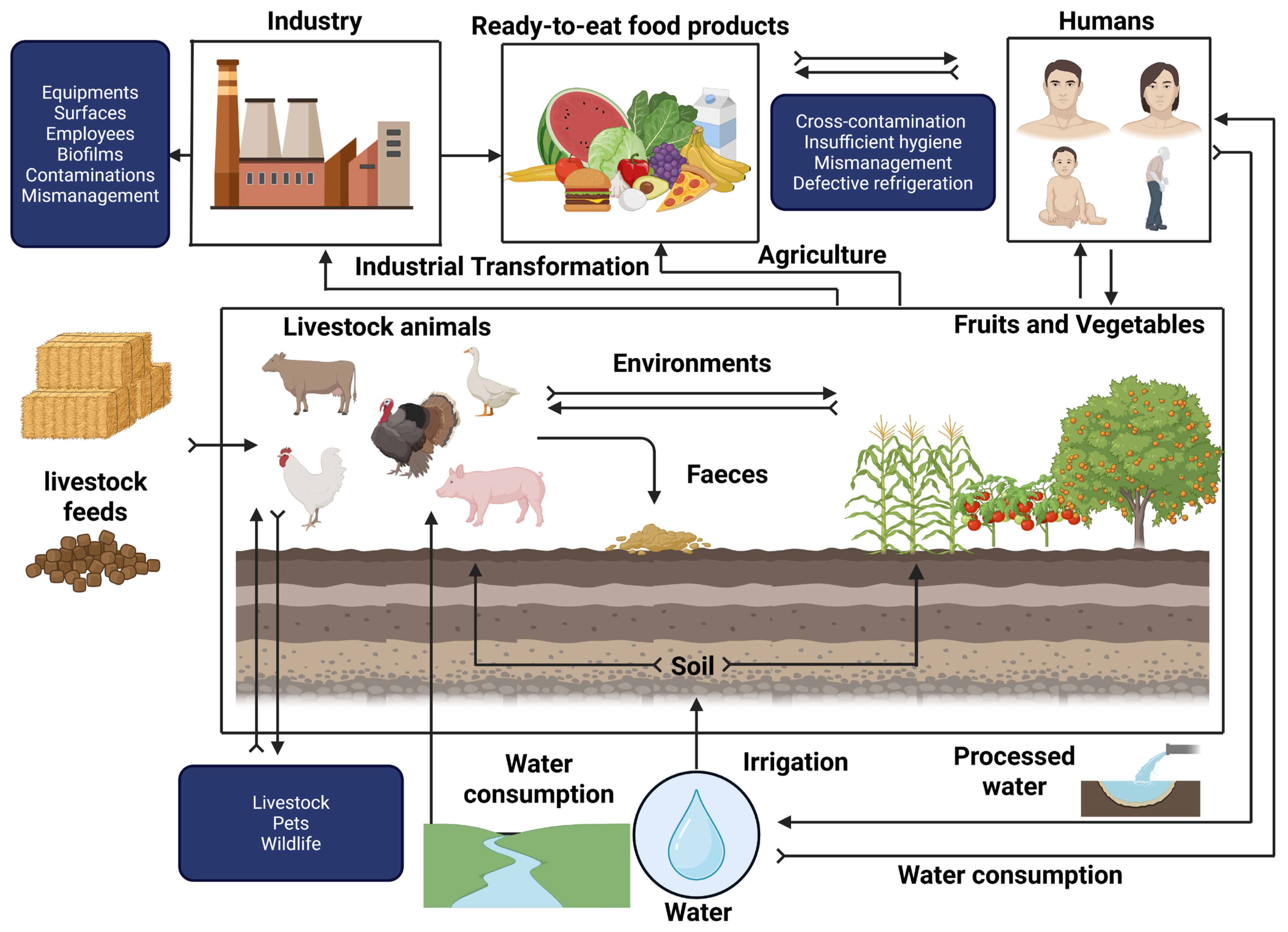
Figure 1. Contamination routes of the L. monocytogenes pathogen in the agrifood system. Transmission pathways across environmental and biological compartments (created with BioRender.com).
Ijms 26 10481 g001
Figure 2. General overview of Listeria monocytogenes infectious mechanisms. (A) Molecular mechanisms of invasion of non-phagocytic cells. This presentation provides an overview of the strategies used by L. monocytogenes to invade non-phagocytic cells, notably through the interaction of internal bacterial proteins (InlA, InlB) with host surface receptors (E-cadherin, Met), enabling the induction of bacterial endocytosis. (B) Schematic representation of the key stages of infection in the human host. This diagram illustrates the main steps in the infectious process of L. monocytogenes, including the crossing of epithelial barriers (intestinal, blood–brain, placental), cell invasion, intracellular survival and systemic dissemination within the host organism (created with BioRender.com).
Ijms 26 10481 g002
Figure 4. Schematic illustration of the mechanism of actions of antimicrobial peptides from bacteria (created with BioRender.com). Δψ represents the transmembrane potential. The letters A to H correspond, respectively, to the following peptides: nisin Z (A), pediocin PA-1 (B), bactofencin A (C), plantaricin S (D), brevibacillin (E), bacitracin (F), daptomycin (G), and fengycin (H).
Ijms 26 10481 g004
Disclaimer/Publisher’s Note: The statements, opinions and data contained in all publications are solely those of the individual author(s) and contributor(s) and not of MDPI and/or the editor(s). MDPI and/or the editor(s) disclaim responsibility for any injury to people or property resulting from any ideas, methods, instructions or products referred to in the content.