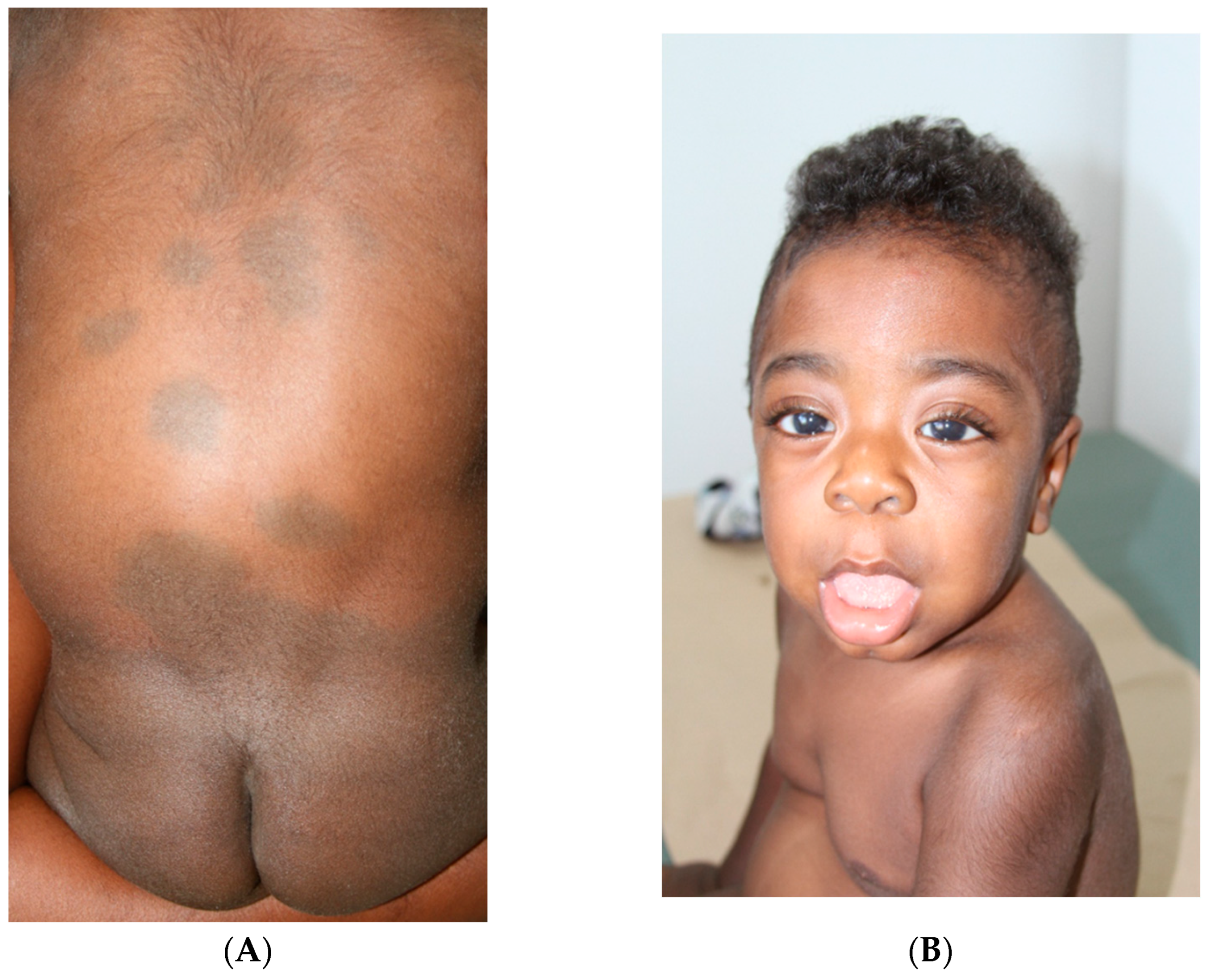
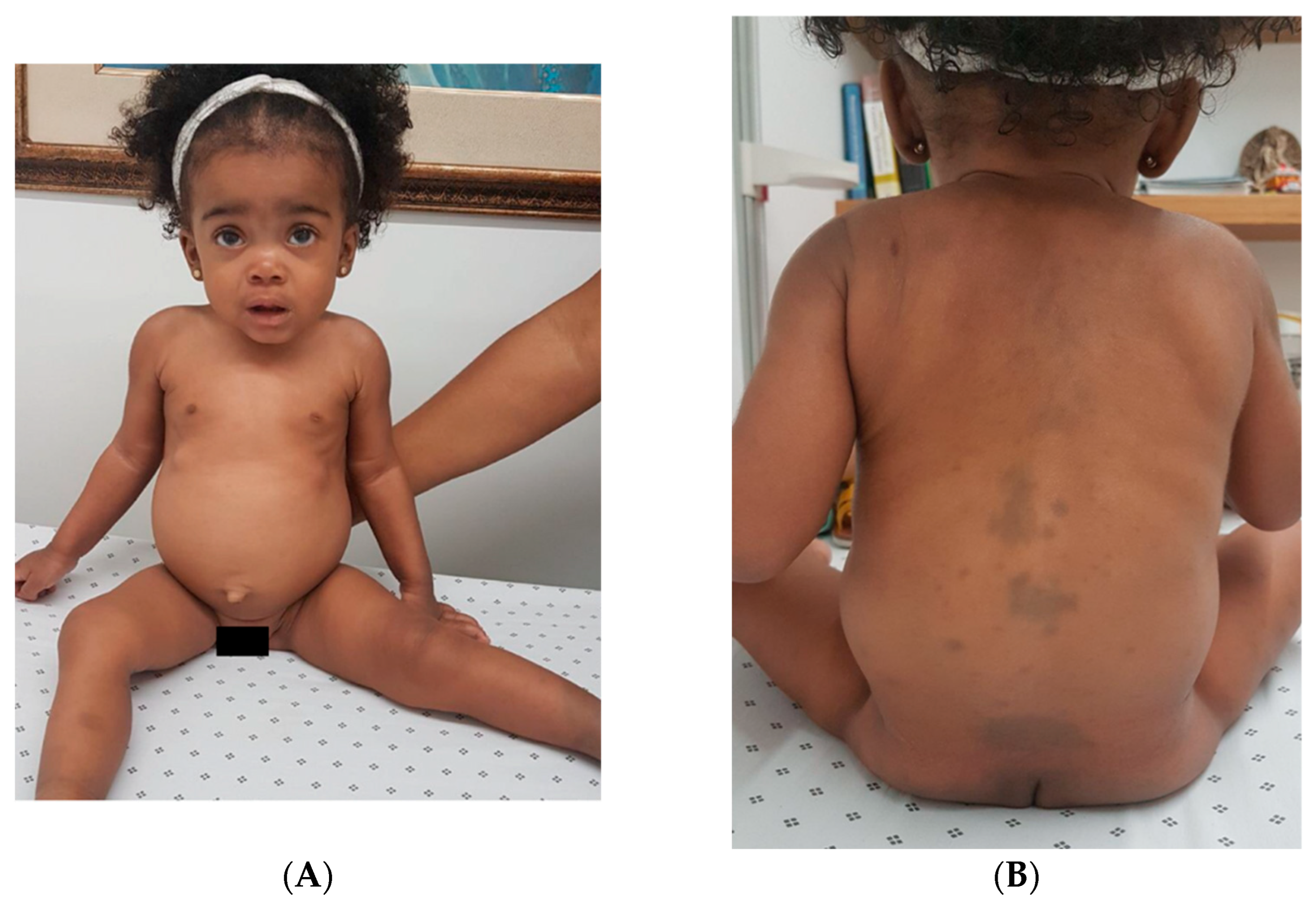
Congenital Dermal Melanocytosis Exhibited in Two Patients with Hurler Syndrome: Clinical Characterization and Report of a Recurrent IDUA Allele in Colombia
Abstract
1. Introduction
2. Case Presentation
2.1. Case 1
2.2. Case 2
3. Discussion
Author Contributions
Funding
Institutional Review Board Statement
Informed Consent Statement
Data Availability Statement
Acknowledgments
Conflicts of Interest
References
- Mendez, H.M.M.; Pinto, L.I.B.; Paskulin, G.A.; Ricachnevsky, N. Is there a relationship between inborn errors of metabolism and extensive mongolian spots? Am. J. Med. Genet. 1993, 47, 456–457. [Google Scholar] [CrossRef]
- Lee-Chen, G.J.; Lin, S.P.; Tang, Y.F.; Chin, Y.W. Mucopolysaccharidosis type I: Characterization of novel mutations affecting α- l-iduronidase activity. Clin. Genet. 1999, 56, 66–70. [Google Scholar] [CrossRef]
- Grech, R.; Galvin, L.; O’Hare, A.; Looby, S. Hurler syndrome (Mucopolysaccharidosis type I). BMJ Case Rep. 2013, 2013, bcr2012008148. [Google Scholar] [CrossRef]
- Hanson, M.; Lupski, J.R.; Hicks, J.; Metry, D. Association of Dermal Melanocytosis With Lysosomal Storage Disease: Clinical Features and Hypotheses Regarding Pathogenesis. Arch. Dermatol. 2003, 139, 916–920. [Google Scholar] [CrossRef]
- Ochiai, T.; Suzuki, Y.; Kato, T.; Shichino, H.; Chin, M.; Mugishima, H.; Orii, T. Natural history of extensive Mongolian spots in mucopolysaccharidosis type II (Hunter syndrome): A survey among 52 Japanese patients. J. Eur. Acad. Dermatol. Venereol. 2007, 21, 1082–1085. [Google Scholar] [CrossRef]
- Tran, M.C.; Lam, J.M. Cutaneous Manifestations of Mucopolysaccharidoses. Pediatr. Dermatol. 2016, 33, 594–601. [Google Scholar] [CrossRef]
- Gautam, M.; Shingala, K. A Review of Dermal Melanocytosis. Indian J. Paediatr. Dermatol. 2023, 24, 211–216. [Google Scholar] [CrossRef]
- Hambrick, G.W., Jr.; Scheie, H. Studies of the Skin in Hurler’s Syndrome: Mucopolysaccharidosis. Arch. Dermatol. 1962, 85, 455–471. [Google Scholar] [CrossRef]
- Grant, B.P.; Beard, J.S.; de Castro, F.; Guiglia, M.C.; Hall, B.D. Extensive Mongolian Spots in an Infant With Hurler Syndrome. Arch. Dermatol. 1998, 134, 108–109. [Google Scholar] [CrossRef]
- Richards, S.; Aziz, N.; Bale, S.; Bick, D.; Das, S.; Gastier-Foster, J.; Grody, W.W.; Hegde, M.; Lyon, E.; Spector, E.; et al. Standards and guidelines for the interpretation of sequence variants: A joint consensus recommendation of the American College of Medical Genetics and Genomics and the Association for Molecular Pathology. Genet. Med. 2015, 17, 405–424. [Google Scholar] [CrossRef]
- Cordova, A. The Mongolian Spot: A Study of Ethnic Differences and a Literature Review. Clin. Pediatr. 1981, 20, 714–719. [Google Scholar] [CrossRef]
- Kikuchi, I. What Is a Mongolian Spot? Int. J. Dermatol. 1982, 21, 131–133. [Google Scholar] [CrossRef]
- Kikuchi, I.; Inoue, S. Natural History of the Mongolian Spot. J. Dermatol. 1980, 7, 449–450. [Google Scholar] [CrossRef]
- Alimi, Y.; Iwanaga, J.; Loukas, M.; Oskouian, R.J.; Rizk, E.; Oakes, W.J.; Tubbs, R.S. A comprehensive review of Mongolian spots with an update on atypical presentations. Child’s Nerv. Syst. 2018, 34, 2371–2376. [Google Scholar] [CrossRef]
- Ashrafi, M.R.; Shabanian, R.; Mohammadi, M.; Kavusi, S. Extensive Mongolian Spots: A Clinical Sign Merits Special Attention. Pediatr. Neurol. 2006, 34, 143–145. [Google Scholar] [CrossRef]
- Weissbluth, M.; Esterly, N.B.; Caro, W.A. Report of an infant with GM1 gangliosidosis type I and extensive and unusual mongolian spots. Br. J. Dermatol. 1981, 104, 195–200. [Google Scholar] [CrossRef]
- Athanti, S.; Mouzam, S.; Ahmed, M.S. Atypical Mongolian Spots With Hurler’s Disease: A Case Report. Cureus 2024, 16, e58501. [Google Scholar] [CrossRef]
- Panteliadis, C.P.; Karatza, E.D.; Tzitiridou, M.K.; Koliouskas, D.E.; Spiroglou, K.S. Lissencephaly and mongolian spots in hurler syndrome. Pediatr. Neurol. 2003, 29, 59–62. [Google Scholar] [CrossRef]
- Alobaidy, H. Recent advances in the diagnosis and treatment of niemann-pick disease type C in children: A guide to early diagnosis for the general pediatrician. Int. J. Pediatr. 2015, 2015, 816593. [Google Scholar] [CrossRef]
- Ceccarini, M.R.; Codini, M.; Conte, C.; Patria, F.; Cataldi, S.; Bertelli, M.; Albi, E.; Beccari, T. Alpha-Mannosidosis: Therapeutic Strategies. Int. J. Mol. Sci. 2018, 19, 1500. [Google Scholar] [CrossRef]
- Wang, P.; Mazrier, H.; Caverly Rae, J.; Raj, K.; Giger, U. A GNPTAB nonsense variant is associated with feline mucolipidosis II (I-cell disease). BMC Vet. Res. 2018, 14, 416. [Google Scholar] [CrossRef]
- Galimberti, C.; Madeo, A.; Di Rocco, M.; Fiumara, A. Mucopolysaccharidoses: Early diagnostic signs in infants and children. Ital. J. Pediatr. 2018, 44, 133. [Google Scholar] [CrossRef]
- Wolf, R.; Wolf, D.; Ruocco, V.; Baroni, A.; Ruocco, E. Phacomatosis pigmento-pigmentaria: Should we add a new type of phacomatosis? Fact and controversies. Clin. Dermatol. 2013, 31, 464–466. [Google Scholar] [CrossRef]
- Nanda, A.; Al-Abdulrazzaq, H.K.; Habeeb, Y.K.R.; Zakkiriah, M.; Alghadhfan, F.; Al-Noun, R.; Al-Ajmi, H. Phacomatosis pigmentovascularis: Report of four new cases. Indian J. Dermatol. Venereol. Leprol. 2016, 82, 298–303. [Google Scholar] [CrossRef]
- Pedrosa, A.F.; Lopes, J.M.; Azevedo, F.; Mota, A. Spitz/Reed nevi: A review of clinical-dermatoscopic and histological correlation. Dermatol. Pract. Concept. 2016, 6, 37–41. [Google Scholar] [CrossRef]
- Mavropoulos, J.C.; Cohen, B.A. Chapter 6—Disorders of Pigmentation. In Pediatric Dermatology, 4th ed.; Content Repository Only! Elsevier: Amsterdam, The Netherlands, 2013; pp. 148–168. [Google Scholar] [CrossRef]
- Zayour, M.; Lazova, R. Congenital Melanocytic Nevi. Clin. Lab. Med. 2011, 31, 267–280. [Google Scholar] [CrossRef]
- Anderson, S. Cafe au Lait Macules and Associated Genetic Syndromes. J. Pediatr. Health Care 2020, 34, 71–81. [Google Scholar] [CrossRef]
- Kim, D.-H.; Choi, J.H.; Lee, J.H.; Kim, H.S. PHACE association with intracranial, oropharyngeal hemangiomas, and an atypical patent ductus arteriosus arising from the tortuous left subclavian artery in a premature infant. Korean J. Pediatr. 2012, 55, 29–33. [Google Scholar] [CrossRef]
- Shah, S.S.; Snelling, B.M.; Sur, S.; Ramnath, A.R.; Bandstra, E.S.; Yavagal, D.R. Scalp congenital hemangioma with associated high-output cardiac failure in a premature infant: Case report and review of literature. Interv. Neuroradiol. 2017, 23, 102–106. [Google Scholar] [CrossRef]
- Slominski, R.M.; Raman, C.; Jetten, A.M.; Slominski, A.T. Neuro–immuno–endocrinology of the skin: How environment regulates body homeostasis. Nat. Rev. Endocrinol. 2025, 21, 495–509, Correction in Nat. Rev. Endocrinol. 2025, 21, 513. [Google Scholar] [CrossRef]
- Slominski, A.T.; Slominski, R.M.; Raman, C.; Chen, J.Y.; Athar, M.; Elmets, C. Neuroendocrine Signaling in the Skin with a Special Focus on the Epidermal Neuropeptides. Am. J. Physiol.-Cell Physiol. 2022, 323, C1757–C1776. [Google Scholar] [CrossRef] [PubMed]
- Zanetti, A.; D’Avanzo, F.; Rigon, L.; Rampazzo, A.; Concolino, D.; Barone, R.; Volpi, N.; Santoro, L.; Lualdi, S.; Bertola, F.; et al. Molecular diagnosis of patients affected by mucopolysaccharidosis: A multicenter study. Eur. J. Pediatr. 2019, 178, 739–753. [Google Scholar] [CrossRef] [PubMed]


| Disease | Etiology/Gene | Inheritance | Clinical (Not Dermatologic) Features | Dermatologic Features | Ref. |
|---|---|---|---|---|---|
| Inborn Errors of Metabolism | |||||
| GM1 gangliosidosis (Type 1 and 2) | Pathogenic variants in GLB1 gene. | AR | Coarse facial features, gingival hypertrophy, corneal clouding, cherry-red macula, hepatosplenomegaly, vacuolated lymphocytes, and skeletal dysostosis in addition to a history of psychomotor regression. | Classical congenital dermal melanocytosis in the back, and gluteal region, and extremities. It presents with one or more macules with round, oval or angled morphology. Variable size from 1 to 20 cm with poorly defined edges (largest are better delimited). Homogeneous gray-blue coloration that is not accentuated on Wood’s lamp examination. The macules are present at birth and usually disappear at the age of 3 to 4 years. | [4] |
| MPS 1 | Pathogenic variants in IDUA gene. | AR | Coarse facial features (including macrocephaly with bulging frontal bones, depressed nasal bridge with broad nose with flared nostrils, thick lips), corneal clouding, skeletal dysostosis, thoracic-lumbar kyphosis, short stature, hepatosplenomegaly, hernias, cardiomyopathy and valve abnormalities, sensorineural hearing loss, enlarged tonsils and adenoids, upper airway recurrent infections with increased secretions. Neurologic involvement: delayed psychomotor development usually obvious by the age of 12 to 24 months, language skills very limited, with progressive mental decline leading to a severe intellectual disability. | [4] | |
| MPS 2 | Pathogenic variants in IDS gene | XLR | Coarse facial features, recurrent upper airway infections with increased secretions, hearing loss, hepatosplenomegaly, cardiac involvement, decrease joint mobility and global delay of developmental milestones by the age of 2 years. Neurological features of MPSII are represented by marked behavioral disturbances, such as hyperactivity, obstinacy, and aggressiveness. | [23] | |
| Niemann-Pick disease (Infantile form) | Pathogenic variants in NPC1 or NPC2 genes. | AR | The neonatal signs range from transient unexplained jaundice to severe cholestatic hepatopathy. Isolated hepatomegaly or splenomegaly in the infantile period associated with developmental delay in motor milestones and central hypotonia are the first neurologic symptoms. Also present are the loss of acquired motor skills, spasticity, intention tremor, and hearing loss. Brain imagery may show leukodystrophy. Frequently in late infantile period hepatosplenomegaly, ataxia, clumsiness, and frequent falling. Hearing loss, dysarthria with delayed speech and dysphagia are often present; focal or generalized seizures (sometimes fatal), cataplexy, and vertical supranuclear gaze palsy (VSGP) are usually present, while mental impairment and behavioral disturbances become more marked. | [20] | |
| Alpha-Mannosidosis. | Pathogenic variants in MAN2B1 gene | AR | Immunodeficiency, facial and skeletal abnormalities, hearing loss and intellectual disability. | [21] | |
| Mucolipidosis II (I-Cell disease) | Pathogenic variants in GNPTAB gene | AR | Stunted growth, skeletal joint abnormalities, coarse facial features, corneal clouding, intellectual disability, hepatomegaly, cardiomegaly and respiratory infections, similar to MPS. Severe progressive neuropathy and oculoskeletal dysfunction are recurrent features. | [22] | |
| MPS 4 | Pathogenic variants in GALNS gene. | AR | Coarse facial features, short stature, skeletal dysplasia with short stature, spinal cord compression, pectus carinatum, kyphoscoliosis, genu valgum with joints are usually lax and very flexible (hypermobile). In severe phenotypes, airway compromise (Restrictive pattern), and later valvular heart disease are the leading causes of morbidity and mortality. | [23] | |
| Sjogren-Larsson syndrome. | Pathogenic variants sin ALDH3A2 gene. | AR | Congenital ichthyosis, delayed psychomotor development, due to spastic diplegia, seizures, moderate or severe intellectual disability. | [24,25] | |
| Phacomatosis | |||||
| Phacomatosis pigmento-vascularis | Unknown | NA | Ocular dysfunction, usually bulbar melanosis, glaucoma, retinal hemangioma and hearing loss. | Disseminated vascular nevus (port wine stain type) associated with pigmentary nevi (epidermal nevus, nevus spilus, or dermal melanocytosis). | [24,25] |
| Phacomatosis pigmento- pigmentaria | Unknown | NA | Non-extracutaneous clinical features. | Two coexisting pigmentary nevi (epidermal nevus, nevus spilus, or dermal melanocytosis). | [24,25] |
| Congenital hemangioma | Unknown | NA | Visceral hemangiomas. As part of PHACE association: neurocutaneous condition with facial hemangiomas associated with a spectrum of posterior fossa malformations, arterial cerebrovascular anomalies, cardiovascular anomalies, and eye anomalies. Dysraphic myelodysplasias associated with urogenital and anorectal anomalies. | Most common benign tumor in childhood. It usually presents as a single flat, slightly rounded or oval lesion. Pink central area with less telangiectasias, and dark purple surrounding area with greater presence of telangiectasias. Whitish external area. Location mainly in the head and neck (43%) and extremities (38%). | [30,31] |
| Melanocytic Nevus | |||||
| Blue Nevus (BN) | Patogenic variants in GNA11 or GNAQ gene. | NA | Non-extracutaneous clinical features. | The common BN is a well-demarcated, slightly raised, or dome-shaped circumscribed symmetric bluish or bluish-black papule, usually measuring less than 1 cm in diameter. Preferred anatomic locations include the dorsal aspects of the hand and feet, the face and the scalp. Women > men. | [28] |
| Nevus of Ota/Nevus of Ito | Unknown | NA | It can be associated with vascular malformations in Sturge Weber syndrome and Klippel Trenaunay syndrome. | Nevus of Ota Blue represents a unilateral, patchy gray or brown irregular dermal melanosis, often spotted skin discoloration located on the face in the distribution of the first and second branches of the trigeminal nerve, the sclera on the affected side might also have a bluish discoloration. Nevus of Ito differs from nevus of Ota in its distribution that is confined to the neck, shoulder and proximal arm region. Can be in isolation or together. | [28] |
| Spitz Nevus | Unknown | NA | Non-extracutaneous clinical features. | Solitary, rounded or oval papule, with a smooth surface. It may also be verrucous, with mild scaling, crusting, or erosion. Its color can vary from pink to reddish-brown or purple-red and its growth may be slow or fast It appears in the first two decades of life, although it may appear in adulthood in 1/3 of the cases. This is rare at birth. | [26] |
| Becker Nevus Syndrome | Hormone dependent disorder (androgens) | NA | Unilateral hypoplasia of breast or other cutaneous, muscular or skeletal defects, involve the same side of the body as the nevus, ipsilateral hypoplasia of the shoulder girdle or abscence of the pectoralis mayor muscle and ipsilateral hypoplasia of a limb, hemivertebrae or spina bifida oculta, fused or accesory cervical ribs, pectus excavatum, pectus carinatum, internal tibial torsion, and scoliosis. | Hyperpigmented hairy macula lesion, with well-defined and irregular borders. It is located predominantly on the anterior trunk or on the scapular region, lesions appear around eight years of age and become more evident in puberty. Other cutaneous findings are granuloma annulare, basal cell carcinoma, malignant melanoma, lymphangioma osteoma cutis, and hypohidrosis. | [27] |
| Congenital Melanocytic Nevus | Pathogenic variants in NRAS or HRAS gene. | NA | High risk of melanoma, diffuse lipomatosis, hypertrophy of cranial bones, central nervous system malformations, such as arachnoid cysts, choroid plexus papilloma, cerebellar astrocytoma. | Brownish lesion with well-defined borders and hypertrichosis, the surface of the nevus may be papular, roughed, warty or cerebriform, most cases occur in the first two years of life. Its most frequent location is the torso, followed by the limbs and head, affects more than one body segment, some peculiar lesion led to the term in garment, described as bathing trunk, stole or coat sleeve. | [28] |
| café au lait spots | Pathogenic variants in NF1, PTPN11, BLM and GNAS gene. | AD | Neurofibromas and Lysch nodules are diagnostic hallmarks of neurofibromatosis. Short stature and cardiomyopathy could be seen in Noonan Syndrome. Precocious puberty and fibrous dysplasia should suggest McCune–Albright syndrome. | Flat hyperpigmented spots or macules, light brown in color, irregularly shaped skin lesions can occur anywhere on the body but appear most frequently on the trunk and extremities. Some have smooth, well-defined borders and others have more jagged edges. They may be present at birth but may also grow in number and size over time, most are benign. | [29] |
Disclaimer/Publisher’s Note: The statements, opinions and data contained in all publications are solely those of the individual author(s) and contributor(s) and not of MDPI and/or the editor(s). MDPI and/or the editor(s) disclaim responsibility for any injury to people or property resulting from any ideas, methods, instructions or products referred to in the content. |
© 2025 by the authors. Licensee MDPI, Basel, Switzerland. This article is an open access article distributed under the terms and conditions of the Creative Commons Attribution (CC BY) license (https://creativecommons.org/licenses/by/4.0/).
Share and Cite
Vanegas, S.; Ramírez-Montaño, D.; Padilla-Guzmán, A.; Pachajoa, H. Congenital Dermal Melanocytosis Exhibited in Two Patients with Hurler Syndrome: Clinical Characterization and Report of a Recurrent IDUA Allele in Colombia. Int. J. Mol. Sci. 2025, 26, 10418. https://doi.org/10.3390/ijms262110418
Vanegas S, Ramírez-Montaño D, Padilla-Guzmán A, Pachajoa H. Congenital Dermal Melanocytosis Exhibited in Two Patients with Hurler Syndrome: Clinical Characterization and Report of a Recurrent IDUA Allele in Colombia. International Journal of Molecular Sciences. 2025; 26(21):10418. https://doi.org/10.3390/ijms262110418
Chicago/Turabian StyleVanegas, Sara, Diana Ramírez-Montaño, Alejandro Padilla-Guzmán, and Harry Pachajoa. 2025. "Congenital Dermal Melanocytosis Exhibited in Two Patients with Hurler Syndrome: Clinical Characterization and Report of a Recurrent IDUA Allele in Colombia" International Journal of Molecular Sciences 26, no. 21: 10418. https://doi.org/10.3390/ijms262110418
APA StyleVanegas, S., Ramírez-Montaño, D., Padilla-Guzmán, A., & Pachajoa, H. (2025). Congenital Dermal Melanocytosis Exhibited in Two Patients with Hurler Syndrome: Clinical Characterization and Report of a Recurrent IDUA Allele in Colombia. International Journal of Molecular Sciences, 26(21), 10418. https://doi.org/10.3390/ijms262110418


