Review

Pathologic Signaling and Disease Implications of Insulin-like Growth Factor Binding Proteins in Cancer, Cardiovascular Disease, and Fibrosis

by
Zachary R. Sechrist
1,2,3,
Jaeden S. Cortés
2,3,
Nidhi R. Patel
2,3,
Zoe J. Pittman
2,3,
Gayathri Guru Murthy
3,4,5,
Guangzhen Zhu
3,5,
Calvin L. Cole
1,2,3,* and
Benjamin D. Korman
3,5,*
1
Department of Pathology and Laboratory Medicine, University of Rochester Medical Center, Rochester, NY 14642, USA
2
Department of Surgical Oncology, University of Rochester Medical Center, Rochester, NY 14642, USA
3
Center for Musculoskeletal Research, University of Rochester Medical Center, Rochester, NY 14642, USA
4
Translational Biomedical Science Program, University of Rochester Medical Center, Rochester, NY 14642, USA
5
Department of Allergy, Immunology & Rheumatology, University of Rochester Medical Center, Rochester, NY 14642, USA
*
Authors to whom correspondence should be addressed.
Int. J. Mol. Sci. 2025, 26(21), 10248; https://doi.org/10.3390/ijms262110248
Submission received: 31 August 2025 / Revised: 16 October 2025 / Accepted: 17 October 2025 / Published: 22 October 2025
(This article belongs to the Section Molecular Pathology, Diagnostics, and Therapeutics)

Abstract

The insulin-like growth factor binding protein (IGFBP) family consists of seven members, which are known for their roles in regulating canonical insulin-like growth factor (IGF) signaling and, more recently, a variety of non-canonical signaling pathways. This review will explore shared pathologic mechanisms amongst the IGFBP family members in diseases like cancer and fibrosis which reveal the unique and redundant properties of this critical family of proteins and provides unique insight into how their regulation is not only relevant to cell metabolism, but also plays an emerging role in diverse processes including immunity, TGF-β signaling, hypoxia and angiogenesis, and senescence. Moreover, these molecules have shown promise both as biomarkers and therapeutics, and a deeper understanding of this system is needed to appreciate how leveraging their regulation may be able to ameliorate diverse organ pathologies.

1. Introduction

The insulin-like growth factor binding protein (IGFBP) family consists of seven well-characterized members. Traditionally, they are known for their roles in regulating insulin-like growth factors (IGF-1 and IGF-2) and insulin signaling in a variety of tissue types helping to maintain homeostasis. More recently, these proteins have been found to signal through IGF-1R-independent signaling and are associated with a diverse group of signaling pathways which allow these molecules to alter cellular function beyond AKT/mTOR signaling and participate in the pathogenesis of a variety of otherwise seemingly divergent disease states. Biomarker studies have shown clear links between IGFBPs and a variety of pathologies including cancer, fibrosis, and vascular disease, but it is only in the last several years that mechanistic studies have begun to shed light on the role that these molecules play in disease. This review will identify the role that dysregulation of IGFBP family members have on disease progression and highlight conserved signaling mechanisms across independent pathological states. An enhanced understanding of IGFBPs can inform the development of new therapeutics for frequently intractable disease states.

2. IGFBP Structure and Function

In eukaryotes, IGF-1 and IGF-2 bind with high affinity to 6 homologous proteins known as IGFBPs. However, the seventh protein of the IGFBP family, IGFBP-7, binds to IGF-1 and IGF-2 with low affinity, and binds to insulin-growth factor 1 receptor (IGF-1R) and insulin more readily [1]. The IGFBPs are synthesized in the liver and are believed to have originated from a single ancestral IGFBP in chordates that then duplicated into two IGFBP genes. Following two genome duplications, eight IGFBP genes remained in placental mammals; however, two were lost, resulting in closely related IGFBPs 1-6 that have been successfully cloned and remain conserved in placental mammals to this day [2]. A total of 15 IGFBPs have since been discovered and categorized based on their affinity for IGFs. IGFBPs 1-6 are categorized as high-affinity binding proteins, whereas IGFBP-related proteins 1-10 (IGFBP-rP 1-10) are categorized as low-affinity binding proteins, with a higher affinity for insulin than to IGFs. Of the 10 IGFBP-related proteins, IGFBP-rp1 was the first discovered and successfully cloned as a gene and was classified as the seventh IGFBP [3,4]. Structurally, IGFBPs 1 through 6 comprise highly conserved N-terminus, central linker, and C-terminus domains unique to each protein. The N-terminus contains an IGFBP motif, which is an essential region for IGF binding, and 16–18 cysteine residues conserved across the IGFBPS that are bonded by intradomain disulfide bonds which contribute to the globular structure and high IGF affinity of these proteins. The C-terminus, while less conserved, comprises only six conserved cysteine residues. The central domain provides further specificity to each of the IGFBPs as it contains glycosylation, phosphorylation, and proteolytic cleavage sites [2,3]. While IGFBP-7 can bind IGFs with low affinity at its N-terminus, it lacks the conserved cysteine residue and the glycosylation and phosphorylation motifs present in the other IGFBPs, explaining its reduced specificity to bind IGF [3].

2.1. Canonical Functions and Maintenance of Homeostasis

Canonically, IGFBPs function to bind IGF-1 and 2, increasing the circulatory half-life of IGFs and modulating their binding to their principal receptor, IGF receptor 1 (IGF-1R). IGFBPs inhibit or augment IGF-1R signaling via post-translational modification or interactions with other regulatory proteins. This regulates signal transduction of IGF-1R and other processes including cellular growth, differentiation, and apoptosis [3,5]. Without inhibition of IGF/IGF-1R signaling, the PI3K-AKT/mTOR pathway, responsible for anti-apoptotic effects/protein synthesis, and the MAPK pathway, responsible for mitogenesis, are both activated [6,7]. When IGFs are bound to IGFBP binary complexes, their half-lives are extended by 90 min, up to 20 h for IGFBPs that can form a ternary complex with the acid-labile subunit (ALS). This allows for the accumulation of large reservoirs of IGFs that would otherwise be secreted immediately due to the lack of IGF intracellular tissue stores. IGFBP-7 is the only IGFBP that binds insulin, with a 100-fold lower affinity for IGF-1 and -2 [3].
IGF-1R signaling is not only inhibited through high-affinity binding of the IGFBPs to IGFs at the cell surface, but also by way of other mechanisms including phosphorylation. IGFBP-1, for example, has an increased affinity for IGF-1 due to hyperphosphorylation of its serine residues. This in turn reduces phosphorylation of IGF-1R and further reduces IGF-1R activation by IGF-1 [5]. IGFBP-1 has also been found to modulate IGF-1 availability metabolically. The production of IGFBP-1 in the liver is inversely related to insulin production, whereby insulin suppresses IGFBP-1 production. In a fasted state, as insulin levels decrease, there is a release of IGFBP-1 from the liver which in turn reduces IGF activity [2].
IGFBP-3 is the most abundant circulatory protein of the seven IGFBPs. IGFBP-3 enables endocrine signaling of IGFs by enhancing the stability of the protein in circulation. This allows IGF to reach distal tissue and induce IGF-1R signaling. The C-terminus of IGFBP-3, when bound to IGFs in a binary complex between 30 and 40 kDa, can bind the glycoprotein ALS to form a ternary complex of approximately 150 kDa. This complex is too large to migrate through vascular endothelium, resulting in increased circulatory IGF concentration [2,8]. Although approximately 90% of IGF:ALS ternary structures are assembled with IGFBP-3, IGFBP-5 is also capable of interacting with IGFs and ALS in circulation, with 50% of IGFBP-5 existing within the IGF:ALS ternary complex [2,5]. Proteolysis of the central linker domain of IGFBPs 3 and 5 results in the release of IGF from the ternary complexes, which increases the available concentration of biologically active IGF [9].
Proteolysis is not limited to IGFBP-3 and -5, as it is a mechanism through which all IGFBPs potentiate IGF-1R signaling by weakening IGF-IGFBP interactions. IGF signal transduction is initiated when IGFBP linker domains or C-terminals are cleaved by proteases, including matrix metalloproteases, kallikreins, cathepsins, and complement component C1. Recent studies have shown that pappalysin metalloproteinase PPAP-A is able to cleave IGFBPs 2, 4, and 5, whereas PPAP-A2 has been shown to cleave IGFBPs 3 and 5 [5,8,10].
The termination of cell replication, known as cellular senescence, is a regulatory mechanism induced in aged or stressed cells that can maintain homeostasis or be pathologic depending on cellular context, as often mobilized as an anti-cancer mechanism. IGFBPs 1-7 are part of the senescence-associated secretory phenotype (SASP), which constitutes proteins that are secreted by senescent cells. IGFBPs 4, 5, and 7 participate in a positive feedback loop in which senescent cells secrete reactive oxygen species (ROS), resulting in the secretion of IGFBPs which then induce senescence in neighboring cells via paracrine signaling [1,11]. Prolonged IGF activity has also been linked to promoting senescent phenotypes within cells, and IGFBP binding to either IGFs or IGFRs can modulate the development of cellular senescence. IGFBP-4’s recruitment of PAPPA tends to increase senescence, while IGFBP-3 tends to increase circulatory IGF levels, inhibiting IGF activity [10]. While IGFBP-7 exhibits relatively weak binding affinity to IGFs, it can bind IGF-1R, which is a binding partner of IGF-2. By competitively binding this receptor, IGF-2 is redirected to IGF-2R, which then promotes senescence. IGF-2-induced senescence via IGFBP-5 and 7 has also been shown to utilize the ERK and AKT signaling pathways [1,12].
Interestingly, by regulating the bioavailability of their canonical binding partners IGF-1 and -2, IGFBPs play a predominant role in folliculogenesis and embryogenesis [13]. IGFBP-2 is found to be expressed in highly proliferating cells and rapidly developing tissues during embryogenesis and enhances tissue development [14]. Similarly, IGFBP-3 is expressed by cells of the maternal reproductive tract and the embryo during pre-implantation, and has been shown to increase the developmental rate of embryos both in vivo and in vitro through IGF-1R signaling [15]. Importantly, disruptions to IGFBP signaling during embryogenesis can lead to impaired development. Elevated or phosphorylated IGFBP-1 leads to sequestration of IGF-1 and inhibits IGF-1R signaling, resulting in fetal growth restriction [16,17]. Lastly, IGFBP-4, a known inhibitor of the IGFs, has been observed to decrease in concentration in dominant bovine ovarian follicles and was upregulated in follicle atresia. Recombinant bovine IGFBP-4 significantly reduces hatching initiation and completion rates of blastocytes at the highest concentration [18]. Together, this highlights the importance of the IGFBP family of proteins in development and homeostatic functions. Better understanding the temporal regulation and intricate signaling of IGFBPs during embryogenesis can further identify their role in conditions like cancer, fibrosis and vascular disease.

2.2. Non-Canonical Functions and Pathological Pathways

While the canonical processes represent well-established functions of IGFBPs, this family of proteins also exhibits non-canonical functions that are independent of IGF activity such as integrin binding and nuclear translocation. Integrins are transmembrane proteins that allow for cell adhesion and communication between extracellular matrix proteins and cytoskeleton. Through non-canonical IGFBP binding, integrin activation modulates several cellular processes such as expression of genes, and ultimately tumorigenesis and disease outcomes. IGFBPs 1 and 2 contain the widely recognized integrin-binding tripeptide Arg-Gly-Asp (RGD), enabling them to bind to cell surfaces and modulate a number of cellular processes by inducing kinase phosphorylation [2,5]. IGFBP-1 has been shown to carry out cell-specific IGF-independent functions influencing cell motility via the RGD motif binding to alpha5- β1 integrin. While this complex enhances cell migration in cell types such as Chinese hamster ovary, smooth muscle, and schwannoma cells, cell invasion is inhibited in cell types such as mammary tumor cells and endometrial stromal cultures. Similarly, IGFBP-2 promotes tumorigenesis and enhances cell proliferation via integrin β1 activation and associated signaling in many cell types. Glioma cell proliferation that was promoted by exogenous IGFBP-2-induced integrin/ERK activation was inhibited by anti-integrin β1 antibody as well as knockdown of the protein [19]. IGFBP-2 also induces vasculogenic mimicry formation in glioma tissue via integrin alpha-5 and β1/RGD binding as adhesion proteins expressed in endothelial cells such as CD144 are upregulated [20]. Hepatic Stellate Cell (HSC) migration, a marker of liver fibrosis, is also promoted through IGFBP-induced integrin β1 signaling, more specifically integrin-β1-induced AKT phosphorylation. IGFBP-3 treated HSCs that are incubated with integrin β1-neutralizing antibody experience blocked cellular migration and attenuated AKT phosphorylation [21].
Nuclear translocation, another non-canonical action of IGFBPs, is the process by which cargo proteins are transported across the nuclear membrane, where they interact with nuclear hormones or receptors to modulate transcription of target genes. Select IGFBPs have been observed to participate in non-canonical signaling via nuclear translocation, in which they influence transcriptional activity by most commonly binding to the nuclear receptor retinoid X receptor alpha (RXR-α). RXR-α forms dimeric structures with nuclear receptors including retinoic acid receptor (RAR), peroxisome proliferator-activated receptor-λ (PPAR-λ), vitamin D receptor (VDR), and Nur77 [2]. IGFBP-3 contains a nuclear localization sequence (NLS) at the C-terminal that binds importin-β, a receptor protein responsible for transporting cargo proteins in and out of the nucleus via the nuclear pore complex (NPC) [2]. The N and C terminals of IGFBP-3 are then able to interact with nuclear receptors RXR-α or RAR existing as homodimers or heterodimers with their affiliated binding partners, whose response elements activate or inhibit transcription, respectively. Nur77, when associated with IGFBP-3 in a ternary complex, is phosphorylated and exported from the nucleus. This transport results in cytochrome c release, alongside caspase 3 and 7 activation, inducing apoptosis [2,22]. Like IGFBP-3, IGFBP-5 contains an NLS within its C-terminal that facilitates its nuclear translocation through interacting with importin α/β complexes and nucleon. IGFBP-5 has been shown to bind nuclear receptor VDR, preventing RXR/VDR heterodimerization and therefore attenuating the expression of bone differentiation markers in osteoblast-like cells [5,23]. IGFBP-5 has also been observed to bind RAR and interact with the natural RAR ligand all-trans retinoic acid (ARTA), impacting transcriptional activity associated with senescence [24]. IGFBP-6 is similarly translocated into the nucleus via an NLS located on the C-terminus, exhibiting preferential binding to importin-α. IGFBP-6 competitively binds to RXR-binding partners VDR and TRα1, preventing heterodimerization and thus attenuating VDR-dependent and TRα1-dependent transcriptional activity [5]. IGFBP-2 differs from the other IGFBPs capable of nuclear translocation in the sense that its NLS is located within the linker domain, which interacts with importin-α. Instead of binding nuclear receptors, IGFBP-2 is known to interact with the Vascular Endothelial Growth Factor (VEGF) promoter via transcription factor Fra-1, upregulating VEGF transcription and promoting angiogenesis [5].
While senescence is a normal cellular process, it can also occur non-canonically and promote tumorigenesis and disease progression. For example, mesenchymal stem cells (MSCs) treated with IGFBP-4 demonstrate an increase in SA-beta-gal, a biomarker for senescence [5]. Additionally, IGFBP-4 potentiates IGF-2’s induction of senescence as IGF-2 degradation is delayed in the presence of IGFBP-4 [11]. IGFBP-5 is also a promoter of senescence in human dermal fibroblasts (HDFs). Y-27632 is a compound that inhibits Rho-kinases that function to promote epidermal cell growth via ROCK 1 and ROCK 2. HDFs treated with y-27632 acquire a senescence-associated secretory phenotype (SASP), converting them to cancer-associated fibroblasts (CAF) which can be inhibited by IGFBP-5 knockdown [25]. Like IGFBP-4 and -5, IGFBP-7 can induce senescence in MSCs through release of SASP factors as well as through insulin and IGF interaction. When healthy MSCs were incubated with IGFBP-7, senescence was promoted. Additionally, unlike the other IGFBPs, IGFBP-7 can induce senescence in MSCs by binding insulin and blocking its anti-senescent properties [1]. IGFBP-7-induced senescence is exhibited in the progression of diseases such as scleroderma, in which human dermal fibroblasts cultured with both IGFBP-7 and TGF-β1 upregulated the expression of genes associated with myofibroblast differentiation and cellular senescence compared to healthy control fibroblasts [26].
In recent years, dysregulated canonical and non-canonical signaling of IGFBPs has gained interest for its role in disease states like cancer, vascular disease and fibrosis. Despite a well-established definition of IGFBP structure, pathological signaling is poorly understood and displays cell- and context-dependent differences in the literature. Interestingly, IGFBP family members display shared, complex roles in regulating the immune system, non-canonical TGF-β signaling, epithelial-to-mesenchymal transition, matrix remodeling, hypoxia, and angiogenesis. Importantly, dysregulation in these mechanisms is instrumental in the development of various cancers, vascular disease and fibrosis, which remain poorly treated diseases associated with poor patient quality of life. Understanding the dependence of IGFBP signaling and downstream mechanisms in disease may lead to improved clinical therapies.

3. IGFBP Signaling Pathways in Cancer

Cancer, in the most basic terms, is the uncontrolled growth of a cell through a dysregulation of homeostatic signaling pathways. As such, cancer is a multifactorial disease defined by reproducible and consistently dysfunctional mechanisms coined the hallmarks of cancer [27]. Understanding the key signaling factors responsible for the propagation of the hallmarks can lead to the development of novel therapeutics to reduce tumor burden in patients and potentially ameliorate crippling comorbidities. Interestingly, the IGFBP family members have been intimately connected to cancer progression and severity [28] and many of the family members display conserved mechanisms across cancer types. Herein, we describe how IGFBPs can regulate the Immune system, TGF-β growth signaling, hypoxia, angiogenesis, and epithelial-to-mesenchymal transition (EMT) (Figure 1), all of which have been described as hallmarks of cancer and are instrumental in tumor development and progression.

3.1. Immune System Modulation

There is a well-established relationship between immune cell dysfunction and tumor progression in various forms of cancer. Aberrant inflammation promotes tumorigenesis [29], and the tumor microenvironment secretes various cytokines that induce an anti-inflammatory environment, further promoting survival [30]. Understanding the signaling mechanisms that promote anti-inflammatory tumor-associated macrophages (TAMs), neutrophils, and regulatory T cells is crucial for developing successful cancer therapies [31]. Importantly, there is a defined relationship between increased immune cell infiltration into the tumor microenvironment and improved patient outcomes. A comprehensive TCGA screening study found a complex relationship between overall survival, immune cell infiltration and IGFBP-1 levels. Although context- and cancer-dependent, elevated IGFBP-1 correlated with reduced cell infiltration and therefore worsened patient outcomes in ovarian, lung, stomach and pancreatic cancer [32]. In contrast, low IGFBP-1 expression correlates with improved survival in stomach cancer and inversely correlates with CD4+ and CD8+ T cells, dendritic cells and neutrophils [33]. In renal cell carcinoma, elevated IGFBP-1 correlates with worsened survival and is inversely proportional to CD14 staining in tumor samples, indicating reduced macrophage and monocyte infiltration [34]. Interestingly, in breast cancer, where elevated IGFBP-6 correlates with improved survival, TCGA analysis found that IGFBP-6 expression also positively correlated with CD4+ and CD8+ T-cell and macrophage infiltration into the tumor microenvironment (TME) [35]. Lastly, although IGFBP-7 overexpression is associated with worsened patient survival in glioma [36], bladder [37], and stomach cancer [38], it induces both pro- and anti-inflammatory immune cell infiltration. More work is needed to define a role for IGFBP-7 in regulating phenotype switching of immune cells in cancer.
Another key factor in cancer progression and outcomes is the phenotypic state of immune cells within the TME. Two independent studies demonstrate that IGFBP-2 correlates with worsened survival in pancreatic ductal adenocarcinoma (PDAC) and that IGFBP-2 stimulates STAT3 signaling, leading to immune suppression phenotypes. Overexpression and knockdown studies of IGFBP-2 in PDAC cell lines show that IGFBP-2 activates STAT3 signaling in PDAC cells and elevates indoleamine 2, 3-dioxygenase (IDO) secretion, a known inducer of T-reg cells. As such, increased T-reg infiltration in the TME is associated with worsened survival [39]. Furthermore, similar experiments by this group define a role for IGFBP-2 in activating STAT3 signaling and subsequent macrophage polarization to an immunosuppressive M2 phenotype [40]. In colorectal cancer (CRC), a similar paradigm is reported. Fibroblasts from metastatic lesions in CRC secrete elevated IGFBP-2 that correlates with CD38 expression. In culture, these cells induced an immunosuppressive phenotype in macrophages and T cells, which was abrogated via siRNA gene silencing of IGFBP-2 [41]. Furthermore, the same group showed that in a murine model of CRC, CD38/IGFBP-2-expressing fibroblasts promote tumor growth and survival via lowered immune cell activation [42]. Fibroblast-secreted IGFBP-7 has also been shown to promote macrophage polarization through FGF2/AKT signaling mechanisms and reduce survival outcomes in gastric cancer [43]. Similar responses have been shown in glioma. IGFBP-2 and 6 promote M2 polarization, promoting tumor progression, and knockdown mouse models identified a phenotype switch to pro-inflammatory M1 cells and reduced tumor growth [44,45].
One interesting, common mechanism in the literature is the modulation of PD-L1 expression in cancer cells and select immune cells. PD-L1 or programmed cell death ligand 1 acts to inhibit a T-cell response, propagating tumor survival [46]. In brief, IGFBP-1 [47], IGFBP-2 [48], IGFBP-3 [49], and IGFBP-5 [50] have been shown to elevate PD-L1 expression on macrophages and tumor cells, leading to reduced immune response in gliomas, esophageal, and colorectal cancers. Furthermore, IGFBP-1 and PD-L1 co-overexpression sensitizes patients to checkpoint inhibitor therapy [47]. Collectively, there are convincing reports related to overexpression of IGFBP family members and induction of an immunosuppressive TME. Moreover, IGFBP overexpression may indicate a patient’s response to emerging immunotherapies. Collectively, IGFBPs have been shown to regulate both immune cell invasion in cancer and manipulate immune cell phenotype to induce a pro-tumor environment (Figure 1A). Moreover, there is an opportunity to combine previously established immunotherapies with anti-IGFBP therapies for the treatment of various cancer types.

3.2. TGF-β Signaling

There is an emerging field of research describing IGFBPs’ interactions with TGF-β signaling pathways, which are known to play a complex role in tumor progression [51]. Of note, there are two studies implicating a role for IGFBPs binding directly to TGF-β receptors. In glioblastoma, IGFBP-3 can interact directly with TGF-β receptors, stimulating SMAD2/3 signaling cascades and subsequent c-Jun activation. This promotes positive feedback on IGFBP-3 expression, and continual IGFBP-3 overexpression promotes cancer progression [52]. In vitro analysis using an esophageal carcinoma cell line indicated that increasing IGFBP-7 using an adenovirus system can directly induce SMAD2/3 activation and promote expression of TGF-β1, positively associated with tumor development [53]. Lastly, it is important to acknowledge a relationship whereby TGF-β regulates IGFBP expression. A complex mechanism has been described where CRC-derived tumor cells secrete TGF-β1 which induces IGFBP-3 expression in pericytes. This effect is negated in the presence of a TGF-β receptor antagonist. IGFBP-3 expression in pericytes positively regulates tumor cell proliferation and migration [54]. A similar crosstalk system has been found in gastric cancer. Tumor-derived TGF-β1 induces IGFBP-7 in myofibroblasts, which positively regulates tumor proliferation and metastasis and can be attenuated by TGF-β receptor antagonists [55]. Curiously, IGFBP-3 and IGFBP-7 have been found to be induced by TGF-β1 expression and activate TGF-β receptors in a tumor context. Given the complex nature of TGF-β signaling in cancer (Figure 1B), work must be conducted to further elucidate these complex interactions and identify potential therapeutic targets.

3.3. Epithelial-to-Mesenchymal Transition

IGFBP family members were first discovered for their roles in interacting with IGF proteins, potent regulators of various metabolic processes in cells [2]. Metabolic dysfunction is one of the original cancer hallmarks and plays a complex role in tumor progression [27,56]. As such, there is an abundance of literature identifying IGFBP members as metabolic regulators of tumor growth. Here we will focus on one metabolic mechanism, epithelial-to-mesenchymal transition (EMT), where, in fact, all but IGFBP-5 and 6 have been implicated. EMT is a process that allows epithelial cells to lose their polarity and acquire the phenotype of a mesenchymal-like cell and is a necessary step for a tumor to become metastatic. Importantly, EMT is characterized by a loss of epithelial markers like E-Cadherin and increased expression of mesenchymal markers like N-Cadherin and Vimentin among others [57]. To date, there are no cancer therapies that modify or inhibit EMT to reduce metastatic potential [58,59].
In lung adenocarcinoma cell lines where elevated IGFBP-1 correlates with reduced survival, knockout of IGFBP-1 significantly reduces cell proliferation and migratory potential in vitro. This is accompanied by reductions in pro-mesenchymal markers vimentin and N-cadherin and increases in the epithelial marker E-cadherin. It was reported that IGFBP-1 knockdown reduced PPARα expression and overexpressing PPARα pharmacologically overcame the IGFBP-1-dependent changes in proliferation and migration. However, subsequent in vivo models using IGFBP-1-null cells showed no significant reduction in metastatic nodules, indicating a compensatory mechanism in vivo [60]. Fibroblast secretion of IGFBP-2 in aged models has been found to be a potent promoter of melanoma metastasis in vivo and cell migration in vitro [61]. Two reports found IGFBP-2 to regulate EMT in hepatocellular carcinoma (HCC). One found that highly metastatic HCC tumors secrete extracellular vesicles rich in IGFBP-2. Administration of these vesicles further induced EMT in an HCC cell line with reduced metastatic potential and elevated vimentin and reduced E-cadherin. This elegantly identified IGFBP-2 as a primary inducer of EMT in HCC [62]. Moreover, overexpression of IGFBP-2 in mouse models of HCC increased markers of EMT vimentin and N-cadherin and was dependent on Wnt/β-catenin signaling and nuclear accumulation of β-catenin [63]. Interestingly, studies in melanoma demonstrate that in early phases of tumor growth and metastasis where IGFBP-3 expression is reduced due to promoter hypermethylation [64], elevated IGFBP-3 inhibits Wnt/β-catenin signaling, reducing metastatic potential [65]. However, during late-stage growth in highly metastatic melanoma cell lines, IGFBP-3-promoted cell migration and knockdown reduced cell motility [66]. Furthermore, an incredible study in PDAC using high-resolution immunofluorescence demonstrated that through SEMA7A and ATP1A1 interactions, tumor cells promote fibroblast secretion of IGFBP-3. The fibroblast-secreted IGFBP-3 subsequently acts on tumor cells to upregulate IL-17RB at the metastatic front in the tumor and promote invasion. Furthermore, this group confirmed IGFBP-3 interactions with TGF-β receptor 2 and determined that interaction is essential for IL-17RB secretion and invasion [67]. IGFBP-3 has also been implicated in promoting EMT and migration in tongue squamous cell carcinoma. Using cell tracking software and SAS-Fucci cells, IGFBP-3 knockdown has been shown to reduce cell motility and induce cell cycle arrest, slowing proliferation [12]. Likewise, IGFBP-7 has a biphasic response in gastric cancer proliferation and metastatic potential. Overexpression of IGFBP-7 accelerates tumor proliferation and invasion in vitro, whereas a knockdown reduces proliferation and metastatic potential. A similar trend was shown in in vivo models of gastric cancer [68]. On the contrary, much like in potentiating angiogenic potential described below, IGFBP-4 has a negative impact on EMT in HCC. IGFBP-4 overexpression inhibits EMT, induces E-cadherin, and reduces vimentin and N-cadherin expression. The opposite was true for models of IGFBP-4 silencing. In summation, IGFBPs play a crucial role in regulating tumor EMT and metastasis, a currently untreated consequence of tumor progression (Figure 1C). Further understanding the highlighted mechanisms of action may identify novel therapeutic targets for metabolic regulation and controlling metastasis.

3.4. Hypoxia and Angiogenesis

Hypoxia is a key feature of most solid tumors. Cancer cells readily adapt to the lack of oxygen in a growing tumor, enabling continued growth and metastasis. The best-defined transcription factors regulating cancer cells’ response to hypoxia are HIF-1α and HIF-1β [69]. In a model of endometrial cancer where IGFBP-2 overexpression correlates with disease progression, hypoxic environments significantly mediate IGFBP-2 overexpression and allow HIF-1α to bind to its promoter. HIF-1-induced overexpression of IGFBP-2 led to elevated PKM2 expression and a significant shift towards glycolysis, enabling tumor survival in hypoxic environments [70]. An independent study with anaplastic Wilms tumor demonstrated that HIF-1α interacts with the IGFBP-2 promoter and HIF-1 knockout significantly reduces IGFBP-2 expression. Moreover, a positive feedback loop exists where IGFBP-2 induces expression through IGF-1-mediated signaling [71]. Interestingly, tumor-expressed HIF-1 regulates skeletal muscle wasting via promotion of IGFBP-5 expression. In an elegant study using two model organisms for cancer cachexia, it was shown that ImpL2 in Drosophila, a direct homolog to IGFBP-5 in mammals, is under the regulation of HIF-1α and directly promotes muscle loss. Genetic deletion of HIF-1 in tumors in fly and mouse models significantly reduced IGFBP-5 expression and the loss phenotype [72]. Together, these studies demonstrate a need to interrogate HIF-1:IGFBP signaling mechanisms to identify combination therapies in the treatment of various cancers and potential comorbidities.
Hypoxia is intricately connected to angiogenesis which plays a complex role in tumor progression. Although largely context-dependent, inhibiting angiogenesis in tumors aids in slowing growth [69,73]. Recently, a few examples of IGFBP family members modulating angiogenesis have been presented. In hepatocellular carcinoma tumors treated with anti-angiogenic therapy, hypoxia increases IGFBP-1 and promotes angiogenesis and tumor growth. Dual treatment targeting angiogenesis and IGFBP-1 significantly reduced tumor burden, and the mechanism was attributed to IGFBP-1/α5β1 integrin interactions in endothelial cells [74]. In models of melanoma, it was found that HIF-1α activation downstream of melanoma differentiation-associated gene 9 induced expression of IGFBP-2, which promoted endothelial cell migration into the tumor, increasing VEGF-A expression and angiogenesis, enhancing tumor survival [75]. Moreover, novel interactions between tumor-secreted IGFBP-7 and CD93 on endothelial cells led to reduced angiogenesis and slowed tumor growth in in vitro models of melanoma, confirming the role of IGFBP-7:CD93 interactions in tumor development [76]. On the contrary, IGFBP-4 was identified as a potent inhibitor of angiogenesis in models of squamous cell carcinoma where IGFBP-4 neutralization improved angiogenesis and tumor outgrowth [77]. Collectively, a unique connection between hypoxia, IGFBP expression, and angiogenic potential has been established and defines a potential for targeted combination of IGFBP and hypoxia therapies in cancer (Figure 1D).

4. IGFBP Signaling Pathways in Vascular Homeostasis and Pathology

4.1. Hypoxia and Angiogenesis

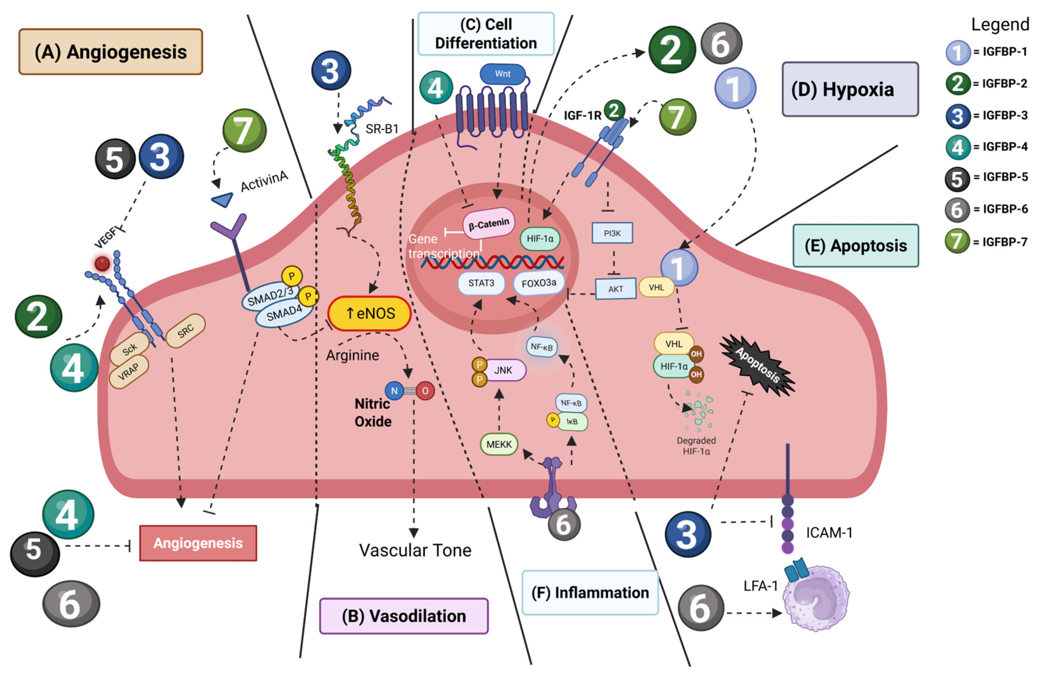
The IGF signaling pathway plays an essential role in cardiac development, with IGF2 representing a primary inducer of cardiomyocyte proliferation and morphogenesis of the myocardial wall [78]. Beyond this, IGFBPs can modulate pathologic cardiac development (Figure 2). For example, IGFBP-3 secreted by coronary endothelial cells contributes to decreased cardiomyocyte proliferation and increased maturation and the development of abnormal myocardial compaction. IGFBP-4 enhances cardiomyocyte differentiation from stem cells and promotes proliferation by inhibition of β-catenin signaling [79] (Figure 2C). Endothelial cells (ECs) express IGFBP-2, IGFBP-3, IGFBP-4, IGFBP-5 and IGFBP-6, with IGFBP-2 and IGFBP-3 predominating in microvascular ECs, and IGFBP-3 and IGFBP-4 playing larger roles in ECs from large vessels. IGFBP-2 is pro-angiogenic, IGFBP-4, IGFBP-5 and IGFBP-6 inhibit angiogenesis, and there is evidence for both pro-angiogenic and anti-angiogenic action by IGFBP-3 and IGFBP-7 [80] (Figure 2A). IGFBP-2 increases angiogenesis via transcriptional regulation of VEGF, whereas IGFBP-3 and IGFBP-5 inhibit VEGF signaling, and IGFBP-6 is induced by hypoxia and may serve as a negative regulator of HIF-1α signaling. IGFBP-3 and IGFBP-4 are important in sprouting angiogenesis, with IGFBP-3 loss of function leading to decreased angiogenesis, whereas knockdown of IGFBP-4 results in increased angiogenesis [81].

4.2. Cardiovascular Disease

Hypoxia upregulates IGFBP-1 in cardiomyocytes, which mediates their apoptosis, and this is regulated by HIF-1α [82]. Moreover, IGFBP-1 promotes neovascularization in response to ischemia with deletion of IGFBP-1 inhibiting endothelial regeneration following injury, while developmental angiogenesis is not affected by IGFBP-1 [83]. IGFBP-1 is also associated with increased risk of major cardiovascular events in patients with peripheral arterial disease [84] (Figure 2D).
In pulmonary hypertension, in addition to their IGF-dependent signaling, IGFBP-1 and IGFBP-2 have an IGF-1R-independent kinase activation pattern in smooth muscle cells, and IGFBP-2 induces EGFR and STAT3 signaling [85]. IGFBP-2 is also a potential biomarker for heart failure development and severity [86].
IGFBP-3 is downregulated in aortic tissues from patients with aortic dissection, and this is mediated by IGFBP-3 preserving aortic smooth muscle cells’ contractility and by reducing MMP9 activation [87]. In retinopathy of prematurity, IGFBP-3 increased endothelial migration, VEGFR1 and VEGFR2 expression, tube formation, and NO synthesis (Figure 2B) [88,89]. In diabetic retinopathy, IGFBP-3 reduced endothelial apoptosis and endothelial-mediated monocyte adhesion in an IGF-independent manner by suppressing ICAM1 [90] (Figure 2E). IGFBP-3 levels are increased in rats undergoing cardiac ischemia and represent a potential biomarker for coronary artery disease [91].
IGFBP-4 enhances cardiomyocyte differentiation while its absence impairs differentiation, and this is mediated through Wnt signaling [92]. Moreover, it can induce VEGF-mediated angiogenesis during myocardial infarction [93]. IGFBP-4’s actions in cardiovascular disease are also mediated by its cleavage by PAPP-A and MMPs, which causes accumulation of IGF on the outer membrane of cardiomyocytes [94].
Cardiomyocyte-specific IGFBP-5 knockdown inhibited myocardial apoptosis and increased cardiomyocyte proliferation in mice with myocardial infarction. IGFBP-5 also ameliorated pathological cardiac remodeling, and this was mediated by IGF-1-dependent signaling [95]. Functionally, IGFBP-5 deficiency protects against ischemic injury by promoting ATP metabolism and stabilizing HIF-1α [96].
IGFBP-6 plays an important role in regulating smooth muscle cell proliferation; knockdown of IGFBP-6 attenuated cell proliferation and increased cyclin E ubiquitination and phosphorylation of CDK2 [97]. Moreover, IGFBP-6 levels are reduced in atherosclerotic arteries, and a reduction in IGFBP-6 in endothelial cells increases inflammatory molecule expression and monocyte adhesion but has anti-inflammatory effects mediated via JNK and NF kappa B (NF-κB) signaling [98] (Figure 2F).
IGFBP-7 is upregulated in the angiogenic vasculature during physiological vessel formation via VEGF-A [99] and pathological processes such as tumor neovascularization [100]. Importantly, IGFBP-7 also plays a critical role in heart failure. In addition to being a sensitive and specific biomarker for heart failure, IGFBP-7 mediates cardiac senescence by stimulating IGF-1R-dependent suppression of FOXO3a [101,102] (Figure 2D). Excitingly, endothelial-specific IGFBP-7 knockout and therapeutic targeting of IGFBP-7 using a vaccine-based approach both ameliorated cardiac dysfunction in a pressure overload model of heart failure [103]. In acute lung injury, IGFBP-7 is overexpressed in endothelial cells, and this allows increased infiltration of Clec4nhi neutrophils to the lung which mediate barrier dysfunction [104].

5. IGFBP Signaling Pathways in Fibrosis

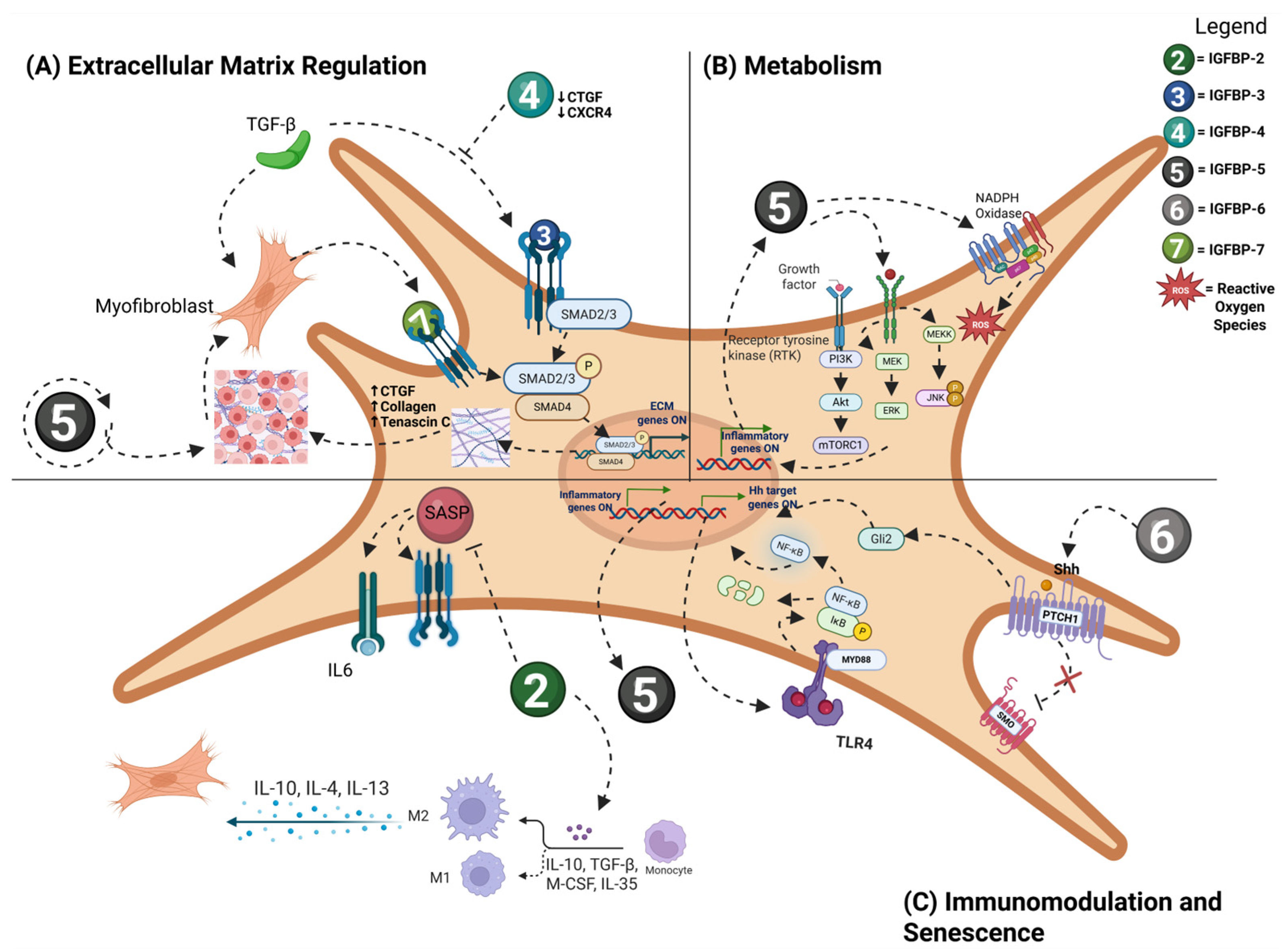
Fibrosis is a pathologic development of scar tissue which involves activation of fibroblasts, increased production and deposition of extracellular matrix (ECM) components, and differentiation of fibroblasts into myofibroblasts. Fibrotic responses are mediated by a variety of growth factors and cytokines, classically including transforming growth factor-β (TGF-β). There is an increasing recognition that several insulin-like growth factor-binding proteins are associated with fibrotic conditions, and emerging themes include IGFBP regulation of TGF biology, as well as a number of emerging pathways (Figure 3). Moreover, these molecules are secreted in the circulation and represent promising biomarkers for fibrosis. IGFBPs are implicated in the fibrotic response across organs and cell types (albeit with cell-specific and organ-specific manifestations), making the IGFBP system ripe for development of novel anti-fibrotic approaches.
IGFBP-1 does not have strong mechanistic data supporting its role in fibrogenesis. There has been evidence that it contributes to non-alcoholic fatty liver disease, a precursor to hepatic fibrosis, by reducing free-fatty-acid-induced lipid accumulation via interacting with integrins and ameliorating inflammation by inhibiting NF-κB and ERK signaling pathways, and has been shown to be a risk factor for more severe fibrosis in this disease [105,106]. Some studies have observed elevated serum IGFBP-1 levels in idiopathic pulmonary fibrosis (IPF), along with a decrease in IGFBP-1 following anti-fibrotic treatment [107]. However, other studies suggest that despite elevated IGFBP-1 levels, anti-fibrotic treatment does not cause a significant impact on IGFBP-1 expression in idiopathic pulmonary fibrosis (IPF) [108].
IGFBP-2 transgenic mice have reduced senescence in alveolar epithelial type 2 (AEC2) cells and have reduced bleomycin-induced lung fibrosis; moreover, recombinant IGFBP-2 is anti-fibrotic in bleomycin lung fibrosis, and IGFBP-2 expression is decreased in AEC2 cells isolated from fibrotic lung regions of patients with IPF. Interestingly, one mechanism by which IGFBP-2 may act is on myeloid cells; in cancer-associated fibroblasts, IGFBP-2 promotes glioma progression through induction of M2 macrophage polarization (Figure 3C) [44]. Clinical studies show that serum IGFBP-2 levels are elevated in patients with IPF [108]. Additionally, elevated serum IGFBP-2 levels have been detected in systemic sclerosis-associated interstitial lung disease (SSc-ILD), and are associated with deterioration of lung function, while treatment with anti-fibrotic agents pirfenidone and nintedanib reduces serum IGFBP-2 [109]. IGFBP-2 appears to be a strong candidate biomarker for early detection and disease monitoring in IPF and SSC-ILD. Notably, IGFBP-2 levels can be measured via ELISA using sputum samples, offering a less invasive method for diagnosis and disease monitoring [110].
IGFBP-3 is known to regulate TGF-β in cancer cells but has not been widely reported in fibrotic disease. IGFBP-3 is epigenetically regulated by METTL3 to promote cardiac fibroblast activation and fibrosis in a variety of cardiac fibrosis models [111]. IGFBP-3 treatment has been shown to stimulate human primary lung fibroblasts to produce tenascin-C and Collagen 1, supporting a role for IGFBP-3 in promoting ECM deposition in pulmonary fibrosis (Figure 3A) [112]. Increased levels of IGFBP-3 have been observed in primary skin fibroblast cultures from systemic sclerosis (SSc) patients [113]. Another study detected elevated IGFBP-3 in the bronchoalveolar lavage fluid of IPF patients [114].
IGFBP-4 blocks TGF-β-induced ECM production and inhibits ECM production in lung and skin cells in vitro, while IGFBP-4 knockout reduces bleomycin-induced fibrosis. IGFBP-4 also reduces the chemokine receptor CXCR4 and the pro-fibrotic factor CTGF [115]. IGFBP-4 has not been reported to be a biomarker in fibrotic disease.
IGFBP-5 knockdown blocks myofibroblast differentiation and decreases cardiac fibrosis in animal models through interactions with NFAT4, which stimulates the production of MFAP5 and enables SREBP2-dependent cholesterol synthesis [116]. In lung fibroblasts, IGFBP-5 promotes fibrosis by increasing the production of extracellular matrix (ECM) genes and the expression of pro-fibrotic genes including connective tissue growth factor (CTGF) and lysyl oxidase [117]. Reactive oxygen species (ROS) production was induced by IGFBP-5, regulated by MEK/ERK and JNK signaling, and mediated by NADPH oxidase. Notably, IGFBP-5 can amplify its own expression, functioning as a self-reinforcing driver of fibrosis [117]. Silencing IGFBP-5 in scleroderma and IPF fibroblasts reduced production of reactive oxygen species [118]. Upregulation of IGFBP-5 is detected in the skin of SSc patients and lung tissues of IPF patients (Figure 3B) [117].
IGFBP-6 expression is increased in myelofibrosis, and IGFBP-6-induced myofibroblast differentiation is associated with an upregulation of cancer-associated fibroblast markers. This process was mediated by TLR4 signaling and the sonic hedgehog (SHH) pathway [119]. IGFBP-6 has also been implicated in a wide variety of fibrotic diseases of the skin, heart, lung, kidney, and liver, and contributes to cancer stroma in the tumor microenvironment which is mediated at least partially by interactions with TGF- β [120,121]. In Dupuytren’s disease, a hereditary form of tendon fibrosis, IGFBP-6 is downregulated [122]. In hepatic fibrosis, IGFBP-6 has emerged as a key marker; reduced IGFBP-6 expression is seen in chronic hepatitis C as it progresses to fibrosis, and in NAFLD patients with fibrosis, elevated hepatic IGFBP-6 levels strongly correlate with advanced fibrosis [123,124]. Notably, IGFBP-6 levels are also sensitive to NAFLD treatments like tesamorelin, which prevents fibrosis progression, thus confirming its clinical relevance as a biomarker [120].
Urinary IGFBP-7 (along with TIMP-2) has been reported as a well-validated biomarker for renal fibrosis [125]. IGFBP-7 knockdown attenuates renal fibrosis and improves renal function through activation of ERK1/2 signaling [126]. Renal tubular epithelial cell IGFBP-7 interacts with PKM2 to drive renal lipid accumulation which promotes fibrosis [127]. In a zebrafish model of metabolic-associated liver fibrosis, IGFBP-7 promoted fibrosis by inhibiting hepatic ferroptosis [128]. Cardiac fibroblasts regulate cardiomyocyte homeostasis, but under pressure overload, they mediate cardiac fibrosis through the Htra3-TGF-β-IGFBP-7 pathway [129]. Importantly, there is a positive feedback loop between IGFBP-7 and TGF-β1: TGF-β inhibition significantly reduced IGFBP-7 expression, while IGFBP-7 knockout suppressed TGF-β1 expression [130]. IGFBP-7 is increased in the skin of patients with systemic sclerosis, and correlates with the modified Rodnan skin score, a validated measure of skin fibrosis severity [131]. IGFBP-7 is also a potential marker for early diagnosis of IPF in which elevated IGFBP-7 expression is seen in lung epithelial cells [130].

6. IGFBPs as Biomarkers of Disease

Given preclinical data suggesting that IGFBPs can regulate the progression of cancer, vascular disease, and fibrosis through both IGF-1-dependent and -independent mechanisms, their role as biomarkers in a clinical setting has been investigated. First, an understanding of which tissues express IGFBPs at homeostasis and during various pathological states, identified via The Human Protein Atlas, can aid in early detection and guide the development of targeted therapeutics to improve patient outcomes and quality of life. IGFBP-1 shows high RNA expression in the liver and lower levels in female reproductive tissues such as the ovary and endometrium, with protein expression predominantly localized to the placenta. Its moderate hepatic and endometrial expression patterns are consistent with reports linking IGFBP-1 to lung, endometrial, and breast cancers [132]. IGFBP-2 demonstrates moderate RNA expression in the brain, prostate, and liver, but exhibits higher levels in the pancreas and stomach. IGFBP-2 is frequently upregulated in gliomas, prostate cancer, and hepatocellular carcinoma, consistent with its basal expression in these organs [133,134]. IGFBP-3 shows the highest RNA expression in the placenta and liver, with basal expression in most other tissues except the cerebellum and bone marrow. Its broad expression in the breast, lung, colorectum, and prostate correlates with cancers of these organs [135,136]. IGFBP-4 displays high RNA levels in the liver and endometrium and moderate expression in the heart, lung, ovary, and uterus, strengthening its association with lung, ovarian, and uterine malignancy [137]. IGFBP-5 exhibits strong RNA expression in female reproductive tissues, including the cervix, ovary, and fallopian tube, with variable levels in other organs. This aligns with its reported roles in breast and lung cancers as well as in fibrotic conditions [137,138]. IGFBP-6 presents high protein expression in the brain, thyroid gland, lymphoid organs, and gastrointestinal tract, with RNA levels peaking in the choroid plexus and reproductive tissues. These patterns correspond with its reported associations with ovarian and brain tumors [139]. IGFBP-7 demonstrates moderate RNA expression in the prostate, lung, skin, and vascular tissues, and higher levels are observed in the choroid plexus, kidney, heart, and thyroid gland. Its tissue expression correlates with its reported roles in melanoma, prostate, and vascular cancers [140,141]. As such, Table 1 summarizes findings from 2020 to now on the clinical expression patterns, prognostic significance, and impact on survival outcomes of IGFBP family members in common cancer diagnoses: breast, lung, pancreatic, and gastrointestinal, and various vascular and fibrotic diseases.

7. Summary

The IGFBP family of proteins is traditionally known for their roles in regulating canonical IGF and insulin signaling in a variety of tissue types, helping to maintain metabolic homeostasis. Canonically, IGFBPs function to bind the mitogens IGF1/2 to regulate their binding to IGFRs which control biological mechanisms such as cellular growth, differentiation, and apoptosis. More recently, these proteins have gained attention for a variety of non-canonical signaling types in pathological pathways independent of IGF activity that have more disease potential. Their non-canonical binding of integrins and nuclear translocation results in several pathologies such as cellular senescence, apoptosis, inhibition of cell differentiation and gene transcription, tumorigenesis and tumor cell proliferation, angiogenesis, cardiovascular disease, and fibrosis. In addition to the canonical and non-canonical functions of IGFBP family members, research suggests that due to their role in the onset, development, and/or progression of certain diseases, they may act as effective biomarkers of disease state, severity, and prognosis. The dysregulation in IGFBP secretion is multifactorial and disease-specific, and a more nuanced understanding of this system is needed to understand how the similarities and differences in IGFBP family members relate to these unique and shared pathologies. However, the pathways that lead to disease are largely conserved across pathologies. Dysregulation in immune system function, TGF-β signaling, hypoxia and angiogenesis, and inflammation are commonly associated with pathological secretion of IGFBP family members. More preclinical and clinical research is needed to clearly define the role of IGFBPs in cancer diagnosis, cardiovascular disease, and fibrosis to determine their viability as therapeutic targets, especially because they do play vital roles in homeostasis. While anti-IGFBP antibodies have shown promise preclinically, there have yet to be clinical trials with these kinds of targeted therapies. Additionally, more information is needed to identify ways in which these complicated systems regulate disease. Specifically, as it pertains to cancer, research should focus on isolating the cells in the tumor microenvironment responsible for the secretion of IGFBPs and in which cancers targeting IGFBPs might have therapeutic potential to ameliorate disease, reduce disease-related toxicities, improve quality of life, and increase survival. Although much research has been performed on the physiological production and secretion of IGFBPs, more research is needed to elucidate the conditions in which the physiological production becomes pathological and leads to subsequent conditions of organ dysfunction such as interstitial lung disease, atherosclerosis, scleroderma, skeletal muscle wasting, and sarcopenia. Mechanistically, the identification of pathways that are dysregulated in the environment of increased or decreased IGFBP production may allow for the development of therapeutics that can target multiple proteins downstream of their secretion.

Author Contributions

Z.R.S., J.S.C., N.R.P., Z.J.P., G.G.M., G.Z., C.L.C. and B.D.K. contributed to conceptualization, writing—original draft preparation, and writing—review and editing. All authors have read and agreed to the published version of the manuscript.

Funding

This research received no external funding.

Data Availability Statement

No new data were created or analyzed in this study. Data sharing is not applicable to this article.

Conflicts of Interest

The authors declare no conflicts of interest.

Abbreviations

The following abbreviations are used in this manuscript:
AEC2Alveolar Epithelial Type 2
ALSAcid Labile Subunit
ARTAAll-Trans Retinoic Acid
ATP1A1ATPase Na+/K+-Transporting Subunit Alpha 1
CAFCancer-Associated Fibroblast
CDK2Cyclin-Dependent Kinase 2
CHFCongestive Heart Failure
Clec4nhiC-Type Lectin Domain Family 4
CRCColorectal Cancer
CTGFConnective Tissue Growth Factor
ECEndothelial Cell
ECMExtracellular Matrix
EGFREpidermal Growth Factor Receptor
EMTEpithelial-To-Mesenchymal Transition
ERKExtracellular Signal-Regulated Kinase
FGF2Fibroblast Growth Factor 2
FOXO3aFork-Head Box O3
Fra-1Fos-Related Antigen 1
HCCHepatocellular Carcinoma
HDFHuman Dermal Fibroblast
HIFHypoxia-Inducible Factor
HSCHepatic Stellate Cell
Htra3High-Temperature Requirement A3
ICAM1Intercellular Adhesion Molecule 1
IDOIndoleamine 2, 3-Dioxygenase
IGFInsulin-Like Growth Factor
IGFBPInsulin-Like Growth Factor Binding Protein
IGF-1RInsulin-Like Growth Factor Receptor 1
IGFBP-rPInsulin-like Growth Factor Binding Protein-Related Protein
IL-17RBInterleukin 17 Receptor B
ImpL2Ecdysone-Inducible Gene L2
IPFIdiopathic Pulmonary Fibrosis
JNKc-Jun N-Terminal Kinase
MACEMajor Adverse Cardiovascular Events
MAPKMitogen-Activated Protein Kinase
MEKMitogen-Activated Protein Kinase 1
METTL3Methyltransferase-Like 3
MFAP5Microfibril-Associated Protein 5
MMPMatrix Metalloproteinases
MRSSModified Rodnan Skin Score
mTORMechanistic Targeting of Rapamycin
MYBBP1AMYB Binding Protein 1a
NADPHNicotinamide Adenine Dinucleotide Phosphate (Reduced)
NAFLDNon-alcoholic Fatty Liver Disease
NFAT4Nuclear Factor of Activated T cells 4
NLSNuclear Localization Sequence
NONitric Oxide
NOTCH1Neurogenic Locus Notch Homolog Protein 1
NPCNuclear Pore Complex
NYHANew York Heart Association
PAHPulmonary Arterial Hypertension
PAPP-APregnancy-Associated Plasma Protein A
PDACPancreatic Ductal Adenocarcinoma
PD-L1Programmed Death-Ligand 1
PI3KPhosphoinositide 3-Kinase
PKM2Pyruvate Kinase
PPAR-λPeroxisome Proliferator-Activated Receptor-λ
ROCKRho-Associated Protein Kinase
ROSReactive Oxygen Species
RXR-αRetinoid X Receptor Alpha
SASPSenescence-Associated Secretory Phenotype
SEMA7ASemaphorin 7A
SHHSonic Hedgehog
SiRNASmall Interfering Ribonucleic Acid
SMADSuppressor of Mothers Against Decapentaplegic
SREBP2Sterol Regulatory Element-Binding Protein 2
SScSystemic Sclerosis
SSc-ILDSystemic Sclerosis-Associated Interstitial Lung Disease
STAT3Signal Transducer and Activator of Transcription 3
TAMTumor-Associated Macrophages
TCGAThe Cancer Genome Atlas Program
TGF-βTransforming Growth Factor
TIMP-2Tissue Inhibitor of Metalloproteinases 2
TLR4Toll-Like Receptor 4
TMETumor Microenvironment
TRα1Thyroid Hormone Receptor Alpha 1
VDRVitamin D Receptor
VEGFVascular Endothelial Growth Factor
VEGF-AVascular Endothelial Growth Factor-A
VEGFRVascular Endothelial Growth Factor receptor

References

  1. Siraj, Y.; Aprile, D.; Alessio, N.; Peluso, G.; Di Bernardo, G.; Galderisi, U. IGFBP7 is a key component of the senescence-associated secretory phenotype (SASP) that induces senescence in healthy cells by modulating the insulin, IGF, and activin A pathways. Cell Commun. Signal. 2024, 22, 540. [Google Scholar] [CrossRef] [PubMed]
  2. LeRoith, D.; Holly, J.M.P.; Forbes, B.E. Insulin-like growth factors: Ligands, binding proteins, and receptors. Mol. Metab. 2021, 52, 101245. [Google Scholar] [CrossRef]
  3. Chen, L.; Hui, L.; Li, J. The multifaceted role of insulin-like growth factor binding protein 7. Front. Cell Dev. Biol. 2024, 1, 1420862. [Google Scholar] [CrossRef]
  4. Song, F.; Zhou, X.X.; Hu, Y.; Li, G.; Wang, Y. The Roles of Insulin-Like Growth Factor Binding Protein Family in Development and Diseases. Adv. Ther. 2021, 38, 885–903. [Google Scholar] [CrossRef]
  5. Baxter, R.C. Signaling Pathways of the Insulin-like Growth Factor Binding Proteins. Endocr. Rev. 2023, 44, 753–778. [Google Scholar] [CrossRef]
  6. Forbes, B.E.; Blyth, A.J.; Wit, J.M. Disorders of IGFs and IGF-1R signaling pathways. Mol. Cell. Endocrinol. 2020, 518, 111035. [Google Scholar] [CrossRef]
  7. Werner, H. The IGF1 Signaling Pathway: From Basic Concepts to Therapeutic Opportunities. Int. J. Mol. Sci. 2023, 24, 14882. [Google Scholar] [CrossRef]
  8. Miller, B.S.; Rogol, A.D.; Rosenfeld, R.G. The History of the Insulin-Like Growth Factor System. Horm. Res. Paediatr. 2022, 95, 619–630. [Google Scholar] [CrossRef]
  9. Kim, H.; Fu, Y.; Hong, H.J.; Lee, S.-G.; Lee, D.S.; Kim, H.M. Structural basis for assembly and disassembly of the IGF/IGFBP/ALS ternary complex. Nat. Commun. 2022, 13, 4434. [Google Scholar] [CrossRef] [PubMed]
  10. Conover, C.A.; Oxvig, C. The IGF System and Aging. Endocr. Rev. 2025, 46, 214–223. [Google Scholar] [CrossRef] [PubMed]
  11. Alessio, N.; Squillaro, T.; Di Bernardo, G.; Galano, G.; De Rosa, R.; Melone, M.A.B.; Peluso, G.; Galderisi, U. Increase of circulating IGFBP-4 following genotoxic stress and its implication for senescence. Elife 2020, 9, e54523, Erratum in eLife 2022, 11, e80871. [Google Scholar] [CrossRef]
  12. Ng, E.F.Y.; Kaida, A.; Nojima, H.; Miura, M. Roles of IGFBP-3 in cell migration and growth in an endophytic tongue squamous cell carcinoma cell line. Sci. Rep. 2022, 12, 11503. [Google Scholar] [CrossRef]
  13. Choi, Y.S.; Ku, S.-Y.; Jee, B.-C.; Suh, C.-S.; Choi, Y.M.; Kim, J.G.; Moon, S.Y.; Kim, S.H. Comparison of follicular fluid IGF-I, IGF-II, IGFBP-3, IGFBP-4 and PAPP-A concentrations and their ratios between GnRH agonist and GnRH antagonist protocols for controlled ovarian stimulation in IVF-embryo transfer patients. Hum. Reprod. 2006, 21, 2015–2021. [Google Scholar] [CrossRef] [PubMed]
  14. Li, T.; Forbes, M.E.; Fuller, G.N.; Li, J.; Yang, X.; Zhang, W. IGFBP2: Integrative hub of developmental and oncogenic signaling network. Oncogene 2020, 39, 2243–2257. [Google Scholar] [CrossRef] [PubMed]
  15. Green, C.J.; Span, M.; Rayhanna, M.H.; Perera, M.; Day, M.L. Insulin-like Growth Factor Binding Protein 3 Increases Mouse Preimplantation Embryo Cleavage Rate by Activation of IGF1R and EGFR Independent of IGF1 Signalling. Cells 2022, 11, 3762. [Google Scholar]
  16. Gupta, M.B. The role and regulation of IGFBP-1 phosphorylation in fetal growth restriction. J. Cell Commun. Signal. 2015, 9, 111–123. [Google Scholar] [CrossRef] [PubMed]
  17. Kajimura, S.; Aida, K.; Duan, C. Insulin-like growth factor-binding protein-1 (IGFBP-1) mediates hypoxia-induced embryonic growth and developmental retardation. Proc. Natl. Acad. Sci. USA 2005, 102, 1240–1245. [Google Scholar] [CrossRef]
  18. Camacho de Gutiérrez, A.R.; Calisici, O.; Wrenzycki, C.; Gutiérrez-Añez, J.C.; Hoeflich, C.; Hoeflich, A.; Bajcsy, Á.C.; Schmicke, M. Effect of IGFBP-4 during In Vitro Maturation on Developmental Competence of Bovine Cumulus Oocyte Complexes. Animals 2024, 14, 673. [Google Scholar] [CrossRef]
  19. Wei, L.F.; Weng, X.F.; Huang, X.C.; Peng, Y.H.; Guo, H.P.; Xu, Y.W. IGFBP2 in cancer: Pathological role and clinical significance (Review). Oncol. Rep. 2021, 45, 427–438. [Google Scholar] [CrossRef]
  20. Liu, Y.; Li, F.; Yang, Y.T.; Xu, X.D.; Chen, J.S.; Chen, T.L.; Chen, H.J.; Zhu, Y.B.; Lin, J.Y.; Li, Y.; et al. IGFBP2 promotes vasculogenic mimicry formation via regulating CD144 and MMP2 expression in glioma. Oncogene 2019, 38, 1815–1831. [Google Scholar] [CrossRef]
  21. Yaqoob, U.; Luo, F.; Greuter, T.; Jalan Sakrikar, N.; Sehrawat, T.S.; Lu, J.; Hu, X.; Gao, J.; Kostallari, E.; Chen, J.; et al. GIPC-Regulated IGFBP-3 Promotes HSC Migration In Vitro and Portal Hypertension In Vivo Through a β1-Integrin Pathway. Cell. Mol. Gastroenterol. Hepatol. 2020, 10, 545–559. [Google Scholar] [CrossRef]
  22. Varma Shrivastav, S.; Bhardwaj, A.; Pathak, K.A.; Shrivastav, A. Insulin-Like Growth Factor Binding Protein-3 (IGFBP-3): Unraveling the Role in Mediating IGF-Independent Effects Within the Cell. Front. Cell Dev. Biol. 2020, 8, 286. [Google Scholar] [CrossRef]
  23. Poreba, E.; Durzynska, J. Nuclear localization and actions of the insulin-like growth factor 1 (IGF-1) system components: Transcriptional regulation and DNA damage response. Mutation Res. Rev. Mutat. Res. 2020, 784, 108307. [Google Scholar] [CrossRef] [PubMed]
  24. Alessio, N.; Aprile, D.; Peluso, G.; Mazzone, V.; Patrone, D.; Di Bernardo, G.; Galderisi, U. IGFBP5 is released by senescent cells and is internalized by healthy cells, promoting their senescence through interaction with retinoic receptors. Cell Commun. Signal. 2024, 22, 122. [Google Scholar] [CrossRef] [PubMed]
  25. Renna, F.; Ballarò, R.; Costelli, P. The Redox Balance: A Target for Interventions Against Muscle Wasting in Cancer Cachexia? Antioxid. Redox Signal. 2020, 33, 542–558. [Google Scholar] [CrossRef]
  26. Jeong, Y.J.; Murthy, G.G.; Wang, G.; Rahman, T.; Korman, B. Insulin-like Growth Factor Binding Protein-7 (IGFBP7) Plays a Pathogenic Role in Dermal Fibrosis and Is Increased in Systemic Sclerosis [Abstract]. Arthritis Rheumatol. 2024, 76 (Suppl. S9). Available online: https://acrabstracts.org/abstract/insulin-like-growth-factor-binding-protein-7-igfbp7-plays-a-pathogenic-role-in-dermal-fibrosis-and-is-increased-in-systemic-sclerosis/ (accessed on 16 October 2025).
  27. Hanahan, D. Hallmarks of Cancer: New Dimensions. Cancer Discov. 2022, 12, 31–46. [Google Scholar] [CrossRef]
  28. Liu, Y.; Shen, S.; Yan, Z.; Yan, L.; Ding, H.; Wang, A.; Xu, Q.; Sun, L.; Yuan, Y. Expression characteristics and their functional role of IGFBP gene family in pan-cancer. BMC Cancer 2023, 23, 371. [Google Scholar] [CrossRef]
  29. Nishida, A.; Andoh, A. The Role of Inflammation in Cancer: Mechanisms of Tumor Initiation, Progression, and Metastasis. Cells 2025, 14, 488. [Google Scholar] [CrossRef]
  30. Kartikasari, A.E.R.; Huertas, C.S.; Mitchell, A.; Plebanski, M. Tumor-Induced Inflammatory Cytokines and the Emerging Diagnostic Devices for Cancer Detection and Prognosis. Front. Oncol. 2021, 11, 692142. [Google Scholar] [CrossRef]
  31. Tie, Y.; Tang, F.; Wei, Y.-Q.; Wei, X.-W. Immunosuppressive cells in cancer: Mechanisms and potential therapeutic targets. J. Hematol. Oncol. 2022, 15, 61. [Google Scholar] [CrossRef]
  32. Yao, Z.; Han, J.; Wu, J.; Li, M.; Chen, R.; Jian, M.; Yang, Z.; Wang, X.; Zhang, Y.; Hu, J.; et al. Deciphering the multidimensional impact of IGFBP1 expression on cancer prognosis, genetic alterations, and cellular functionality: A comprehensive Pan-cancer analysis. Heliyon 2024, 10, e37402. [Google Scholar] [CrossRef] [PubMed]
  33. Luo, X.Y.; Zhang, Y.P.; Zheng, F.; Zhou, L. Multiple bioinformatics analysis identifies IGFBP1 as associated with the prognosis of stomach adenocarcinoma. Medicine 2023, 102, e33346. [Google Scholar] [CrossRef] [PubMed]
  34. Xu, T.; Gao, S.; Liu, J.; Huang, Y.; Chen, K.; Zhang, X. MMP9 and IGFBP1 Regulate Tumor Immune and Drive Tumor Progression in Clear Cell Renal Cell Carcinoma. J. Cancer 2021, 12, 2243–2257. [Google Scholar] [CrossRef]
  35. Lu, H.; Yu, X.; Xu, Z.; Deng, J.; Zhang, M.J.; Zhang, Y.; Sun, S. Prognostic Value of IGFBP6 in Breast Cancer: Focus on Glucometabolism. Technol. Cancer Res. Treat. 2024, 23, 15330338241271998. [Google Scholar] [CrossRef]
  36. Wang, H.; Wang, X.; Xu, L.; Zhang, J. Co-amplified with PDGFRA, IGFBP7 is a prognostic biomarker correlated with the immune infiltrations of glioma. Cancer Med. 2023, 12, 4951–4967. [Google Scholar] [CrossRef]
  37. Yi, X.; Zheng, X.; Xu, H.; Li, J.; Zhang, T.; Ge, P.; Liao, D.; Li, H.; Lyu, X.; Ai, J. IGFBP7 and the Tumor Immune Landscape: A Novel Target for Immunotherapy in Bladder Cancer. Front. Immunol. 2022, 13, 898493. [Google Scholar] [CrossRef]
  38. Zhao, Q.; Zhao, R.; Song, C.; Wang, H.; Rong, J.; Wang, F.; Yan, L.; Song, Y.; Xie, Y. Increased IGFBP7 Expression Correlates with Poor Prognosis and Immune Infiltration in Gastric Cancer. J. Cancer 2021, 12, 1343–1355. [Google Scholar] [CrossRef]
  39. Sun, L.; Zhang, Y.; Yang, T.; Chen, J.; Zhang, X.; Liang, X. IGFBP2 Drives Regulatory T Cell Differentiation through STAT3/IDO Signaling Pathway in Pancreatic Cancer. J. Pers. Med. 2022, 12, 2005. [Google Scholar] [CrossRef]
  40. Sun, L.; Zhang, X.; Song, Q.; Liu, L.; Forbes, E.; Tian, W.; Zhang, Z.; Kang, Y.; Wang, H.; Fleming, J.B.; et al. IGFBP2 promotes tumor progression by inducing alternative polarization of macrophages in pancreatic ductal adenocarcinoma through the STAT3 pathway. Cancer Lett. 2021, 500, 132–146. [Google Scholar] [CrossRef] [PubMed]
  41. Walterskirchen, N.; Müller, C.; Ramos, C.; Zeindl, S.; Stang, S.; Herzog, D.; Sachet, M.; Schimek, V.; Unger, L.; Gerakopoulos, V.; et al. Metastatic colorectal carcinoma-associated fibroblasts have immunosuppressive properties related to increased IGFBP2 expression. Cancer Lett. 2022, 540, 215737. [Google Scholar] [CrossRef]
  42. Ramos, C.; Walterskirchen, N.; Knöbl, V.; Zotter, C.; Müller, C.; Gerakopoulos, V.; Rauch, A.; Falk, L.; Sachet, M.; D’Angelo, E.; et al. Colorectal cancer peritoneal metastasis is promoted by tissue-specific fibroblasts that can arise in response to various local disorders. Cancer Lett. 2025, in press. [Google Scholar] [CrossRef] [PubMed]
  43. Li, D.; Xia, L.; Huang, P.; Wang, Z.; Guo, Q.; Huang, C.; Leng, W.; Qin, S. Cancer-associated fibroblast-secreted IGFBP7 promotes gastric cancer by enhancing tumor associated macrophage infiltration via FGF2/FGFR1/PI3K/AKT axis. Cell Death Discov. 2023, 9, 17. [Google Scholar] [CrossRef] [PubMed]
  44. Zhang, X.; Sun, X.; Guo, C.; Li, J.; Liang, G. Cancer-associated fibroblast-associated gene IGFBP2 promotes glioma progression through induction of M2 macrophage polarization. Am. J. Physiol. Cell Physiol. 2024, 326, C252–C268. [Google Scholar] [CrossRef]
  45. Longhitano, L.; Vicario, N.; Forte, S.; Giallongo, C.; Broggi, G.; Caltabiano, R.; Barbagallo, G.M.V.; Altieri, R.; Raciti, G.; Di Rosa, M.; et al. Lactate modulates microglia polarization via IGFBP6 expression and remodels tumor microenvironment in glioblastoma. Cancer Immunol. Immunother. 2023, 72, 1–20, Erratum in Cancer Immunol. Immunother. 2023, 72, 21. [Google Scholar] [CrossRef]
  46. Lin, X.; Kang, K.; Chen, P.; Zeng, Z.; Li, G.; Xiong, W.; Yi, M.; Xiang, B. Regulatory mechanisms of PD-1/PD-L1 in cancers. Mol. Cancer 2024, 23, 108. [Google Scholar] [CrossRef]
  47. Liu, M.; Yan, W.; Chen, D.; Luo, J.; Dai, L.; Chen, H.; Chen, K.N. IGFBP1(hi)WNT3A(lo) Subtype in Esophageal Cancer Predicts Response and Prolonged Survival with PD-(L)1 Inhibitor. Biology 2022, 11, 1575. [Google Scholar] [CrossRef]
  48. Wen, Z.; Sun, H.; Zhang, Z.; Zheng, Y.; Zheng, S.; Bin, J.; Liao, Y.; Shi, M.; Zhou, R.; Liao, W. High baseline tumor burden-associated macrophages promote an immunosuppressive microenvironment and reduce the efficacy of immune checkpoint inhibitors through the IGFBP2-STAT3-PD-L1 pathway. Cancer Commun. 2023, 43, 562–581. [Google Scholar] [CrossRef]
  49. Zhao, L.; Wang, Y.; Mu, P.; Zhang, X.; Qi, R.; Zhang, Y.; Zhang, H.; Zhu, X.; Dong, Z.; Dong, Y. IGFBP3 induces PD-L1 expression to promote glioblastoma immune evasion. Cancer Cell Int. 2024, 24, 60. [Google Scholar] [CrossRef] [PubMed]
  50. Lin, J.; Huang, G.; Zeng, Q.; Zhang, R.; Lin, Y.; Li, Y.; Huang, B.; Pan, H. IGFBP5, as a Prognostic Indicator Promotes Tumor Progression and Correlates with Immune Microenvironment in Glioma. J. Cancer 2024, 15, 232–250. [Google Scholar] [CrossRef]
  51. Baba, A.B.; Rah, B.; Bhat, G.R.; Mushtaq, I.; Parveen, S.; Hassan, R.; Hameed Zargar, M.; Afroze, D. Transforming Growth Factor-Beta (TGF-β) Signaling in Cancer-A Betrayal Within. Front. Pharmacol. 2022, 13, 791272. [Google Scholar] [CrossRef]
  52. Zhang, X.; Wang, G.; Gong, Y.; Zhao, L.; Song, P.; Zhang, H.; Zhang, Y.; Ju, H.; Wang, X.; Wang, B.; et al. IGFBP3 induced by the TGF-β/EGFRvIII transactivation contributes to the malignant phenotype of glioblastoma. iScience 2023, 26, 106639. [Google Scholar] [CrossRef]
  53. Li, X.; Zhang, J.; Wu, Y.; Ma, C.; Wei, D.; Pan, L.; Cai, L. IGFBP7 remodels the tumor microenvironment of esophageal squamous cell carcinoma by activating the TGFβ1/SMAD signaling pathway. Oncol. Lett. 2022, 24, 251. [Google Scholar] [CrossRef] [PubMed]
  54. Navarro, R.; Tapia-Galisteo, A.; Martín-García, L.; Tarín, C.; Corbacho, C.; Gómez-López, G.; Sánchez-Tirado, E.; Campuzano, S.; González-Cortés, A.; Yáñez-Sedeño, P.; et al. TGF-β-induced IGFBP-3 is a key paracrine factor from activated pericytes that promotes colorectal cancer cell migration and invasion. Mol. Oncol. 2020, 14, 2609–2628. [Google Scholar] [CrossRef]
  55. Hong, Z.; Xie, W.; Zhuo, H.; Wei, X.; Wang, K.; Cheng, J.; Lin, L.; Hou, J.; Chen, X.; Cai, J. Crosstalk between Cancer Cells and Cancer-Associated Fibroblasts Mediated by TGF-β1-IGFBP7 Signaling Promotes the Progression of Infiltrative Gastric Cancer. Cancers 2023, 15, 3965. [Google Scholar] [CrossRef]
  56. Martínez-Reyes, I.; Chandel, N.S. Cancer metabolism: Looking forward. Nat. Rev. Cancer 2021, 21, 669–680. [Google Scholar] [CrossRef]
  57. Yang, J.; Antin, P.; Berx, G.; Blanpain, C.; Brabletz, T.; Bronner, M.; Campbell, K.; Cano, A.; Casanova, J.; Christofori, G.; et al. Guidelines and definitions for research on epithelial–mesenchymal transition. Nat. Rev. Mol. Cell Biol. 2020, 21, 341–352, Erratum in Nat. Rev. Mol. Cell Biol. 2021, 22, 834. [Google Scholar] [CrossRef] [PubMed]
  58. Celià-Terrassa, T.; Kang, Y. How important is EMT for cancer metastasis? PLoS Biol. 2024, 22, e3002487. [Google Scholar] [CrossRef] [PubMed]
  59. Huang, Y.; Hong, W.; Wei, X. The molecular mechanisms and therapeutic strategies of EMT in tumor progression and metastasis. J. Hematol. Oncol. 2022, 15, 129. [Google Scholar] [CrossRef]
  60. Li, Y.; Yang, X.; Han, T.; Zhou, J.; Liu, Y.; Guo, J.; Liu, Z.; Bai, Y.; Xing, Y.; Ding, X.; et al. IGFBP1 promotes the proliferation and migration of lung adenocarcinoma cells through the PPARα pathway. Transl. Oncol. 2024, 49, 102095. [Google Scholar] [CrossRef]
  61. Alicea, G.M.; Patel, P.; Portuallo, M.E.; Fane, M.E.; Wei, M.; Chhabra, Y.; Dixit, A.; Carey, A.E.; Wang, V.; Rocha, M.R.; et al. Age-Related Increases in IGFBP2 Increase Melanoma Cell Invasion and Lipid Synthesis. Cancer Res. Commun. 2024, 4, 1908–1918. [Google Scholar] [CrossRef]
  62. Zheng, Y.; Li, W.; Huang, Y.; Cheng, H. Exosomal IGFBP2 derived from highly metastatic promotes hepatocellular carcinoma metastasis by inducing epithelial mesenchymal transition. Gene 2024, 913, 148374. [Google Scholar] [CrossRef]
  63. Chen, X.; Zhang, Y.; Zhang, P.; Wei, M.; Tian, T.; Guan, Y.; Han, C.; Wei, W.; Ma, Y. IGFBP2 drives epithelial-mesenchymal transition in hepatocellular carcinoma via activating the Wnt/β-catenin pathway. Infect. Agent Cancer 2023, 18, 73. [Google Scholar] [CrossRef]
  64. Dar, A.A.; Majid, S.; Nosrati, M.; de Semir, D.; Federman, S.; Kashani-Sabet, M. Functional modulation of IGF-binding protein-3 expression in melanoma. J. Investig. Dermatol. 2010, 130, 2071–2079. [Google Scholar] [CrossRef] [PubMed]
  65. Naspi, A.; Zingariello, M.; Sancillo, L.; Panasiti, V.; Polinari, D.; Martella, M.; Rosa Alba, R.; Londei, P. IGFBP-3 inhibits Wnt signaling in metastatic melanoma cells. Mol. Carcinog. 2017, 56, 681–693. [Google Scholar] [CrossRef] [PubMed]
  66. Xi, Y.; Nakajima, G.; Hamil, T.; Fodstad, O.; Riker, A.; Ju, J. Association of insulin-like growth factor binding protein-3 expression with melanoma progression. Mol. Cancer Ther. 2006, 5, 3078–3084. [Google Scholar] [CrossRef] [PubMed]
  67. Chen, Y.I.; Tien, S.C.; Ko, Y.L.; Chang, C.C.; Hsu, M.F.; Chien, H.J.; Peng, H.Y.; Jeng, Y.M.; Tien, Y.W.; Chang, Y.T.; et al. SEMA7A-mediated juxtacrine stimulation of IGFBP-3 upregulates IL-17RB at pancreatic cancer invasive front. Cancer Gene Ther. 2024, 31, 1840–1855. [Google Scholar] [CrossRef]
  68. Wang, J.; Wang, X.; Liu, Z.; Li, S.; Yin, W. IGFBP7 promotes gastric cancer by facilitating epithelial-mesenchymal transition of gastric cells. Heliyon 2024, 10, e30986. [Google Scholar] [CrossRef]
  69. Chen, Z.; Han, F.; Du, Y.; Shi, H.; Zhou, W. Hypoxic microenvironment in cancer: Molecular mechanisms and therapeutic interventions. Signal Transduct. Target. Ther. 2023, 8, 70. [Google Scholar] [CrossRef]
  70. Jin, Y.; Qi, M.; Si, L.; Shi, X.; Cai, M.; Fu, H.; Liu, Y.; Guo, R. IGFBP2 Promotes Proliferation and Glycolysis of Endometrial Cancer by Regulating PKM2/HIF-1α Axis. Cancer Sci. 2025, 116, 656–672. [Google Scholar] [CrossRef]
  71. Liu, Y.; Nelson, M.V.; Bailey, C.; Zhang, P.; Zheng, P.; Dome, J.S.; Liu, Y.; Wang, Y. Targeting the HIF-1α-IGFBP2 axis therapeutically reduces IGF1-AKT signaling and blocks the growth and metastasis of relapsed anaplastic Wilms tumor. Oncogene 2021, 40, 4809–4819. [Google Scholar] [CrossRef]
  72. Xiao, G.; Li, Y.; Hu, Y.; Tan, K.; Wang, M.; Zhu, K.; San, M.; Cheng, Q.; Tayier, D.; Hu, T.; et al. Intratumor HIF-1α modulates production of a cachectic ligand to cause host wasting. Cell Insight 2025, 4, 100247. [Google Scholar] [CrossRef]
  73. Liu, Z.-L.; Chen, H.-H.; Zheng, L.-L.; Sun, L.-P.; Shi, L. Angiogenic signaling pathways and anti-angiogenic therapy for cancer. Signal Transduct. Target. Ther. 2023, 8, 198. [Google Scholar] [CrossRef] [PubMed]
  74. Suzuki, H.; Iwamoto, H.; Seki, T.; Nakamura, T.; Masuda, A.; Sakaue, T.; Tanaka, T.; Imamura, Y.; Niizeki, T.; Nakano, M.; et al. Tumor-derived insulin-like growth factor-binding protein-1 contributes to resistance of hepatocellular carcinoma to tyrosine kinase inhibitors. Cancer Commun. 2023, 43, 415–434. [Google Scholar] [CrossRef]
  75. Das, S.K.; Bhutia, S.K.; Azab, B.; Kegelman, T.P.; Peachy, L.; Santhekadur, P.K.; Dasgupta, S.; Dash, R.; Dent, P.; Grant, S.; et al. MDA-9/syntenin and IGFBP-2 promote angiogenesis in human melanoma. Cancer Res. 2013, 73, 844–854. [Google Scholar] [CrossRef] [PubMed]
  76. Xu, Y.; Sun, Y.; Zhu, Y.; Song, G. Structural insight into CD93 recognition by IGFBP7. Structure 2024, 32, 282–291.e284. [Google Scholar] [CrossRef]
  77. Furusaka, Y.; Inoue, S.; Mizoguchi, I.; Hasegawa, H.; Katahira, Y.; Watanabe, A.; Sakamoto, E.; Sekine, A.; Miyakawa, S.; Umezu, T.; et al. Potent antitumor effects of the conditioned medium of bone marrow-derived mesenchymal stem cells via IGFBP-4. Cancer Sci. 2023, 114, 2499–2514. [Google Scholar] [CrossRef] [PubMed]
  78. Li, P.; Cavallero, S.; Gu, Y.; Chen, T.H.; Hughes, J.; Hassan, A.B.; Bruning, J.C.; Pashmforoush, M.; Sucov, H.M. IGF signaling directs ventricular cardiomyocyte proliferation during embryonic heart development. Development 2011, 138, 1795–1805. [Google Scholar] [CrossRef]
  79. Zhu, W.; Fan, Y.; Frenzel, T.; Gasmi, M.; Bartus, R.T.; Young, W.L.; Yang, G.Y.; Chen, Y. Insulin growth factor-1 gene transfer enhances neurovascular remodeling and improves long-term stroke outcome in mice. Stroke 2008, 39, 1254–1261. [Google Scholar] [CrossRef]
  80. Bach, L.A. Endothelial cells and the IGF system. J. Mol. Endocrinol. 2015, 54, R1–R13. [Google Scholar] [CrossRef]
  81. Dallinga, M.G.; Habani, Y.I.; Kayser, R.P.; Van Noorden, C.J.F.; Klaassen, I.; Schlingemann, R.O. IGF-binding proteins 3 and 4 are regulators of sprouting angiogenesis. Mol. Biol. Rep. 2020, 47, 2561–2572. [Google Scholar] [CrossRef]
  82. Tang, X.; Jiang, H.; Lin, P.; Zhang, Z.; Chen, M.; Zhang, Y.; Mo, J.; Zhu, Y.; Liu, N.; Chen, X. Insulin-like growth factor binding protein-1 regulates HIF-1alpha degradation to inhibit apoptosis in hypoxic cardiomyocytes. Cell Death Discov. 2021, 7, 242. [Google Scholar] [CrossRef]
  83. Haywood, N.J.; Slater, T.A.; Drozd, M.; Warmke, N.; Matthews, C.; Cordell, P.A.; Smith, J.; Rainford, J.; Cheema, H.; Maher, C.; et al. IGFBP-1 in Cardiometabolic Pathophysiology-Insights From Loss-of-Function and Gain-of-Function Studies in Male Mice. J. Endocr. Soc. 2020, 4, bvz006. [Google Scholar] [CrossRef]
  84. Li, B.; Shaikh, F.; Younes, H.; Abuhalimeh, B.; Zamzam, A.; Abdin, R.; Qadura, M. The Prognostic Potential of Insulin-like Growth Factor-Binding Protein 1 for Cardiovascular Complications in Peripheral Artery Disease. J. Cardiovasc. Dev. Dis. 2025, 12, 253. [Google Scholar] [CrossRef] [PubMed]
  85. Schlueter, B.C.; Quanz, K.; Baldauf, J.; Petrovic, A.; Ruppert, C.; Guenther, A.; Gall, H.; Tello, K.; Grimminger, F.; Ghofrani, H.A.; et al. The diverging roles of insulin-like growth factor binding proteins in pulmonary arterial hypertension. Vascul. Pharmacol. 2024, 155, 107379. [Google Scholar] [CrossRef]
  86. Barutaut, M.; Fournier, P.; Peacock, W.F.; Evaristi, M.F.; Caubere, C.; Turkieh, A.; Desmoulin, F.; Eurlings, L.W.M.; van Wijk, S.; Rocca, H.B.; et al. Insulin-like Growth Factor Binding Protein 2 predicts mortality risk in heart failure. Int. J. Cardiol. 2020, 300, 245–251. [Google Scholar] [CrossRef]
  87. Chen, S.; Chen, H.; Zhong, Y.; Ge, Y.; Li, C.; Qiao, Z.; Zhu, J. Insulin-like growth factor-binding protein 3 inhibits angiotensin II-induced aortic smooth muscle cell phenotypic switch and matrix metalloproteinase expression. Exp. Physiol. 2020, 105, 1827–1839. [Google Scholar] [CrossRef]
  88. Chang, K.H.; Chan-Ling, T.; McFarland, E.L.; Afzal, A.; Pan, H.; Baxter, L.C.; Shaw, L.C.; Caballero, S.; Sengupta, N.; Li Calzi, S.; et al. IGF binding protein-3 regulates hematopoietic stem cell and endothelial precursor cell function during vascular development. Proc. Natl. Acad. Sci. USA 2007, 104, 10595–10600. [Google Scholar] [CrossRef] [PubMed]
  89. Kielczewski, J.L.; Jarajapu, Y.P.; McFarland, E.L.; Cai, J.; Afzal, A.; Li Calzi, S.; Chang, K.H.; Lydic, T.; Shaw, L.C.; Busik, J.; et al. Insulin-like growth factor binding protein-3 mediates vascular repair by enhancing nitric oxide generation. Circ. Res. 2009, 105, 897–905. [Google Scholar] [CrossRef]
  90. Zhang, Q.; Jiang, Y.; Miller, M.J.; Peng, B.; Liu, L.; Soderland, C.; Tang, J.; Kern, T.S.; Pintar, J.; Steinle, J.J. IGFBP-3 and TNF-alpha regulate retinal endothelial cell apoptosis. Investig. Ophthalmol. Vis. Sci. 2013, 54, 5376–5384. [Google Scholar] [CrossRef] [PubMed]
  91. Lee, J.A.; Wise, J.; Raudsepp, S.D.; Paton, L.N.; Powell, J.; Jina, K.; Aldous, S.; Troughton, R.W.; Adamson, P.D.; Richards, A.M.; et al. Insulin-like growth factor binding protein-3 (IGFBP-3): A biomarker of coronary artery disease induced myocardial ischaemia. Eur. Heart J. Open 2025, 5, oeaf028. [Google Scholar] [CrossRef]
  92. Zhu, W.; Shiojima, I.; Ito, Y.; Li, Z.; Ikeda, H.; Yoshida, M.; Naito, A.T.; Nishi, J.; Ueno, H.; Umezawa, A.; et al. IGFBP-4 is an inhibitor of canonical Wnt signalling required for cardiogenesis. Nature 2008, 454, 345–349, Erratum in Nature 2014, 506, 254. [Google Scholar] [CrossRef]
  93. Wo, D.; Chen, J.; Li, Q.; Ma, E.; Yan, H.; Peng, J.; Zhu, W.; Fang, Y.; Ren, D.N. IGFBP-4 enhances VEGF-induced angiogenesis in a mouse model of myocardial infarction. J. Cell Mol. Med. 2020, 24, 9466–9471. [Google Scholar] [CrossRef]
  94. Adasheva, D.A.; Lebedeva, O.S.; Goliusova, D.V.; Postnikov, A.B.; Teriakova, M.V.; Kopylova, I.V.; Lagarkova, M.A.; Katrukha, A.G.; Serebryanaya, D.V. PAPP-A-Specific IGFBP-4 Proteolysis in Human Induced Pluripotent Stem Cell-Derived Cardiomyocytes. Int. J. Mol. Sci. 2023, 24, 8420. [Google Scholar] [CrossRef] [PubMed]
  95. Zhu, Q.; Lu, X.; Chen, M.; Zhang, T.; Shi, M.; Yao, W.; Zhang, H.; Gao, R.; Li, X.; Zhou, Y.; et al. IGFBP5 affects cardiomyocyte survival and functional recovery in mice following myocardial ischemia. Commun. Biol. 2024, 7, 1594. [Google Scholar] [CrossRef]
  96. Song, F.; Hu, Y.; Hong, Y.X.; Sun, H.; Han, Y.; Mao, Y.J.; Wu, W.Y.; Li, G.; Wang, Y. Deletion of endothelial IGFBP5 protects against ischaemic hindlimb injury by promoting angiogenesis. Clin. Transl. Med. 2024, 14, e1725. [Google Scholar] [CrossRef]
  97. Wang, Z.; Qi, Y.; Wang, R.; Wu, W.; Li, Z.; Wang, M.; Liu, R.; Zhang, C.; Li, W.; Wang, S. IGFBP6 regulates vascular smooth muscle cell proliferation and morphology via cyclin E-CDK2. J. Cell Physiol. 2020, 235, 9538–9556. [Google Scholar] [CrossRef] [PubMed]
  98. Su, M.; Zhao, W.; Jiang, H.; Zhao, Y.; Liao, Z.; Liu, Z.; Xu, M.; Jiang, S.; Wu, L.; Yang, Y.; et al. Endothelial IGFBP6 suppresses vascular inflammation and atherosclerosis. Nat. Cardiovasc. Res. 2025, 4, 145–162. [Google Scholar] [CrossRef]
  99. Hooper, A.T.; Shmelkov, S.V.; Gupta, S.; Milde, T.; Bambino, K.; Gillen, K.; Goetz, M.; Chavala, S.; Baljevic, M.; Murphy, A.J.; et al. Angiomodulin is a specific marker of vasculature and regulates vascular endothelial growth factor-A-dependent neoangiogenesis. Circ. Res. 2009, 105, 201–208. [Google Scholar] [CrossRef]
  100. Sun, Y.; Chen, W.; Torphy, R.J.; Yao, S.; Zhu, G.; Lin, R.; Lugano, R.; Miller, E.N.; Fujiwara, Y.; Bian, L.; et al. Blockade of the CD93 pathway normalizes tumor vasculature to facilitate drug delivery and immunotherapy. Sci. Transl. Med. 2021, 13, eabc8922. [Google Scholar] [CrossRef] [PubMed]
  101. Bracun, V.; van Essen, B.; Voors, A.A.; van Veldhuisen, D.J.; Dickstein, K.; Zannad, F.; Metra, M.; Anker, S.; Samani, N.J.; Ponikowski, P.; et al. Insulin-like growth factor binding protein 7 (IGFBP7), a link between heart failure and senescence. ESC Heart Fail. 2022, 9, 4167–4176. [Google Scholar] [CrossRef]
  102. Zhang, L.; Smyth, D.; Al-Khalaf, M.; Blet, A.; Du, Q.; Bernick, J.; Gong, M.; Chi, X.; Oh, Y.; Roba-Oshin, M.; et al. Insulin-like growth factor-binding protein-7 (IGFBP7) links senescence to heart failure. Nat. Cardiovasc. Res. 2022, 1, 1195–1214. [Google Scholar] [CrossRef]
  103. Katoh, M.; Nomura, S.; Yamada, S.; Ito, M.; Hayashi, H.; Katagiri, M.; Heryed, T.; Fujiwara, T.; Takeda, N.; Nishida, M.; et al. Vaccine Therapy for Heart Failure Targeting the Inflammatory Cytokine Igfbp7. Circulation 2024, 150, 374–389. [Google Scholar] [CrossRef]
  104. Yu, L.C.; He, R.; Wang, D.X.; Qi, D. Activated Clec4n(hi) Neutrophils Aggravate Lung Injury in an Endothelial IGFBP7-Dependent Manner. Am. J. Respir. Cell Mol. Biol. 2024, 71, 66–80. [Google Scholar] [CrossRef] [PubMed]
  105. Pan, J.; Cen, L.; Zhou, T.; Yu, M.; Chen, X.; Jiang, W.; Li, Y.; Yu, C.; Shen, Z. Insulin-like growth factor binding protein 1 ameliorates lipid accumulation and inflammation in nonalcoholic fatty liver disease. J. Gastroenterol. Hepatol. 2021, 36, 3438–3447. [Google Scholar] [CrossRef] [PubMed]
  106. Hagstrom, H.; Stal, P.; Hultcrantz, R.; Brismar, K.; Ansurudeen, I. IGFBP-1 and IGF-I as markers for advanced fibrosis in NAFLD—A pilot study. Scand. J. Gastroenterol. 2017, 52, 1427–1434. [Google Scholar] [CrossRef]
  107. Domvri, K.; Organtzis, I.; Apostolopoulos, A.; Fouka, E.; Kontakiotis, T.; Papakosta, D. Prognostic Value of Serum Biomarkers in Patients with Idiopathic Pulmonary Fibrosis in Relation to Disease Progression. J. Pers. Med. 2023, 13, 1307. [Google Scholar] [CrossRef]
  108. Guiot, J.; Bondue, B.; Henket, M.; Corhay, J.L.; Louis, R. Raised serum levels of IGFBP-1 and IGFBP-2 in idiopathic pulmonary fibrosis. BMC Pulm. Med. 2016, 16, 86. [Google Scholar] [CrossRef] [PubMed]
  109. Guiot, J.; Njock, M.S.; Andre, B.; Gester, F.; Henket, M.; de Seny, D.; Moermans, C.; Malaise, M.G.; Louis, R. Serum IGFBP-2 in systemic sclerosis as a prognostic factor of lung dysfunction. Sci. Rep. 2021, 11, 10882. [Google Scholar] [CrossRef]
  110. Guiot, J.; Henket, M.; Corhay, J.L.; Moermans, C.; Louis, R. Sputum biomarkers in IPF: Evidence for raised gene expression and protein level of IGFBP-2, IL-8 and MMP-7. PLoS ONE 2017, 12, e0171344. [Google Scholar] [CrossRef]
  111. Ding, J.F.; Sun, H.; Song, K.; Zhou, Y.; Tu, B.; Shi, K.H.; Lu, D.; Xu, S.S.; Tao, H. IGFBP3 epigenetic promotion induced by METTL3 boosts cardiac fibroblast activation and fibrosis. Eur. J. Pharmacol. 2023, 942, 175494. [Google Scholar] [CrossRef] [PubMed]
  112. Brissett, M.; Veraldi, K.L.; Pilewski, J.M.; Medsger, T.A., Jr.; Feghali-Bostwick, C.A. Localized expression of tenascin in systemic sclerosis-associated pulmonary fibrosis and its regulation by insulin-like growth factor binding protein 3. Arthritis Rheumatol. 2012, 64, 272–280. [Google Scholar] [CrossRef] [PubMed]
  113. Feghali, C.A.; Wright, T.M. Identification of multiple, differentially expressed messenger RNAs in dermal fibroblasts from patients with systemic sclerosis. Arthritis Rheumatol. 1999, 42, 1451–1457. [Google Scholar] [CrossRef]
  114. Aston, C.; Jagirdar, J.; Lee, T.C.; Hur, T.; Hintz, R.L.; Rom, W.N. Enhanced insulin-like growth factor molecules in idiopathic pulmonary fibrosis. Am. J. Respir. Crit. Care Med. 1995, 151, 1597–1603. [Google Scholar] [CrossRef]
  115. Su, Y.; Nishimoto, T.; Hoffman, S.; Nguyen, X.X.; Pilewski, J.M.; Feghali-Bostwick, C. Insulin-like growth factor binding protein-4 exerts antifibrotic activity by reducing levels of connective tissue growth factor and the C-X-C chemokine receptor 4. FASEB Bioadv. 2019, 1, 167–179. [Google Scholar] [CrossRef]
  116. Zhao, Q.; Shao, T.; Huang, S.; Zhang, J.; Zong, G.; Zhuo, L.; Xu, Y.; Hong, W. The insulin-like growth factor binding protein-microfibrillar associated protein-sterol regulatory element binding protein axis regulates fibroblast-myofibroblast transition and cardiac fibrosis. Br. J. Pharmacol. 2024, 181, 2492–2508. [Google Scholar] [CrossRef]
  117. Nguyen, X.X.; Muhammad, L.; Nietert, P.J.; Feghali-Bostwick, C. IGFBP-5 Promotes Fibrosis via Increasing Its Own Expression and That of Other Pro-fibrotic Mediators. Front. Endocrinol. 2018, 9, 601. [Google Scholar] [CrossRef]
  118. Yasuoka, H.; Garrett, S.M.; Nguyen, X.X.; Artlett, C.M.; Feghali-Bostwick, C.A. NADPH oxidase-mediated induction of reactive oxygen species and extracellular matrix deposition by insulin-like growth factor binding protein-5. Am. J. Physiol. Lung Cell. Mol. Physiol. 2019, 316, L644–L655. [Google Scholar] [CrossRef]
  119. Longhitano, L.; Tibullo, D.; Vicario, N.; Giallongo, C.; La Spina, E.; Romano, A.; Lombardo, S.; Moretti, M.; Masia, F.; Coda, A.R.D.; et al. IGFBP-6/sonic hedgehog/TLR4 signalling axis drives bone marrow fibrotic transformation in primary myelofibrosis. Aging 2021, 13, 25055–25071. [Google Scholar] [CrossRef]
  120. Liso, A.; Venuto, S.; Coda, A.R.D.; Giallongo, C.; Palumbo, G.A.; Tibullo, D. IGFBP-6: At the Crossroads of Immunity, Tissue Repair and Fibrosis. Int. J. Mol. Sci. 2022, 23, 4358. [Google Scholar] [CrossRef]
  121. Jin, Z.; Jin, Z.; Liu, Z.; Yin, Y.; Zhang, Y.; Zhang, Y.; Kang, J.; Fang, Y.; Jiang, W.; Ning, B. Pantothenic acid ameliorates hepatic fibrosis by targeting IGFBP6 to regulate the TGF-beta/SMADs pathway. Commun. Biol. 2025, 8, 1127. [Google Scholar] [CrossRef]
  122. Raykha, C.; Crawford, J.; Gan, B.S.; Fu, P.; Bach, L.A.; O’Gorman, D.B. IGF-II and IGFBP-6 regulate cellular contractility and proliferation in Dupuytren’s disease. Biochim. Biophys. Acta 2013, 1832, 1511–1519. [Google Scholar] [CrossRef] [PubMed]
  123. Coskun, M.; Sendur, H.N.; Babayeva, A.; Cerit, M.N.; Cerit, E.T.; Yalcin, M.M.; Altinova, A.E.; Akturk, M.; Karakoc, M.A.; Toruner, F.B. Quantitative ultrasound techniques and biochemical markers to assess liver steatosis and fibrosis in newly diagnosed acromegaly. J. Endocrinol. Investig. 2024, 47, 2823–2833. [Google Scholar] [CrossRef]
  124. Stanley, T.L.; Fourman, L.T.; Zheng, I.; McClure, C.M.; Feldpausch, M.N.; Torriani, M.; Corey, K.E.; Chung, R.T.; Lee, H.; Kleiner, D.E.; et al. Relationship of IGF-1 and IGF-Binding Proteins to Disease Severity and Glycemia in Nonalcoholic Fatty Liver Disease. J. Clin. Endocrinol. Metab. 2020, 106, e520–e533. [Google Scholar] [CrossRef]
  125. Schanz, M.; Kimmel, M.; Alscher, M.D.; Amann, K.; Daniel, C. TIMP-2 and IGFBP7 in human kidney biopsies in renal disease. Clin. Kidney J. 2023, 16, 1434–1446. [Google Scholar] [CrossRef]
  126. Wang, X.; Li, Y.; Zhao, Z.; Meng, Y.; Bian, J.; Bao, R.; Zhu, K.; Yang, T. IGFBP7 regulates sepsis-induced epithelial-mesenchymal transition through ERK1/2 signaling. Acta Biochim. Biophys. Sin. 2019, 51, 799–806. [Google Scholar] [CrossRef] [PubMed]
  127. Yu, J.T.; Xie, S.S.; Shen, X.Y.; Li, Z.; Hu, X.W.; Zhang, Y.; Dong, Z.H.; Wang, J.N.; Li, X.Y.; Dong, Y.H.; et al. Renal tubular epithelial IGFBP7 interacts with PKM2 to drive renal lipid accumulation and fibrosis. Mol. Ther. 2025, 33, 3757–3777. [Google Scholar] [CrossRef] [PubMed]
  128. Wang, Y.; Bo, J.; Zhao, Z.; Han, Y.; Zhang, Q.; Liu, L. Depletion of Igfbp7 alleviates zebrafish NAFLD progression through inhibiting hepatic ferroptosis. Life Sci. 2023, 332, 122086. [Google Scholar] [CrossRef]
  129. Ko, T.; Nomura, S.; Yamada, S.; Fujita, K.; Fujita, T.; Satoh, M.; Oka, C.; Katoh, M.; Ito, M.; Katagiri, M.; et al. Cardiac fibroblasts regulate the development of heart failure via Htra3-TGF-beta-IGFBP7 axis. Nat. Commun. 2022, 13, 3275. [Google Scholar] [CrossRef]
  130. Zhu, T.; Mu, B.R.; Li, B.; Ran, Z.; Wang, D.M.; Zhou, Y.; Liu, L.; Wu, Q.L.; Lu, M.H.; Xiong, D.Q. IGFBP7: A novel biomarker involved in a positive feedback loop with TGF-beta1 in idiopathic pulmonary fibrosis. Cell Signal. 2025, 133, 111867. [Google Scholar] [CrossRef]
  131. Yan, Y.M.; Zheng, J.N.; Li, Y.; Yang, Q.R.; Shao, W.Q.; Wang, Q. Insulin-like growth factor binding protein 7 as a candidate biomarker for systemic sclerosis. Clin. Exp. Rheumatol. 2021, 39 (Suppl. S131), 66–76. [Google Scholar] [CrossRef] [PubMed]
  132. Hwa, V.; Oh, Y.; Rosenfeld, R.G. The Insulin-Like Growth Factor-Binding Protein (IGFBP) Superfamily*. Endocr. Rev. 1999, 20, 761–787. [Google Scholar] [CrossRef]
  133. Han, S.; Li, Z.; Master, L.M.; Master, Z.W.; Wu, A. Exogenous IGFBP-2 promotes proliferation, invasion, and chemoresistance to temozolomide in glioma cells via the integrin β1-ERK pathway. Br. J. Cancer 2014, 111, 1400–1409. [Google Scholar] [CrossRef]
  134. Yau, S.W.; Azar, W.J.; Sabin, M.A.; Werther, G.A.; Russo, V.C. IGFBP-2—Taking the lead in growth, metabolism and cancer. J. Cell Commun. Signal. 2015, 9, 125–142. [Google Scholar] [CrossRef]
  135. LeRoith, D.; Roberts, C.T. The insulin-like growth factor system and cancer. Cancer Lett. 2003, 195, 127–137. [Google Scholar] [CrossRef] [PubMed]
  136. Lee, K.-W.; Liu, B.; Ma, L.; Li, H.; Bang, P.; Koeffler, H.P.; Cohen, P. Cellular Internalization of Insulin-like Growth Factor Binding Protein-3: Distinct Endocytic Pathways Facilitate Re-Uptake and Nuclear Localization. J. Biol. Chem. 2004, 279, 469–476. [Google Scholar] [CrossRef]
  137. Walker, G.; MacLeod, K.; Williams, A.R.W.; Cameron, D.A.; Smyth, J.F.; Langdon, S.P. Insulin-like Growth Factor Binding Proteins IGFBP3, IGFBP4, and IGFBP5 Predict Endocrine Responsiveness in Patients with Ovarian Cancer. Clin. Cancer Res. 2007, 13, 1438–1444. [Google Scholar] [CrossRef]
  138. Waters, J.A.; Urbano, I.; Robinson, M.; House, C.D. Insulin-like growth factor binding protein 5: Diverse roles in cancer. Front. Oncol. 2022, 12, 1052457. [Google Scholar] [CrossRef]
  139. Voutilainen, R.; Franks, S.; Mason, H.D.; Martikainen, H. Expression of insulin-like growth factor (IGF), IGF-binding protein, and IGF receptor messenger ribonucleic acids in normal and polycystic ovaries. J. Clin. Endocrinol. Metab. 1996, 81, 1003–1008. [Google Scholar] [CrossRef]
  140. Jin, L.; Shen, F.; Weinfeld, M.; Sergi, C. Insulin Growth Factor Binding Protein 7 (IGFBP7)-Related Cancer and IGFBP3 and IGFBP7 Crosstalk. Front. Oncol. 2020, 10, 727. [Google Scholar] [CrossRef] [PubMed]
  141. Wajapeyee, N.; Kapoor, V.; Mahalingam, M.; Green, M.R. Efficacy of IGFBP7 for treatment of metastatic melanoma and other cancers in mouse models and human cell lines. Mol. Cancer Ther. 2009, 8, 3009–3014. [Google Scholar] [CrossRef]
  142. Huang, B.-L.; Wei, L.-F.; Lin, Y.-W.; Huang, L.-S.; Qu, Q.-Q.; Li, X.-H.; Chu, L.-Y.; Xu, Y.-W.; Wang, W.-D.; Peng, Y.-H.; et al. Serum IGFBP-1 as a promising diagnostic and prognostic biomarker for colorectal cancer. Sci. Rep. 2024, 14, 1839. [Google Scholar] [CrossRef] [PubMed]
  143. Xu, Y.-W.; Chen, H.; Hong, C.-Q.; Chu, L.-Y.; Yang, S.-H.; Huang, L.-S.; Guo, H.; Chen, L.-Y.; Liu, C.-T.; Huang, X.-Y.; et al. Serum IGFBP-1 as a potential biomarker for diagnosis of early-stage upper gastrointestinal tumour. EBioMedicine 2020, 51, 102566, Erratum in EBioMedicine 2021, 66, 103295. [Google Scholar] [CrossRef]
  144. Liu, Q.; Jiang, J.; Zhang, X.; Zhang, M.; Fu, Y. Comprehensive Analysis of IGFBPs as Biomarkers in Gastric Cancer. Front. Oncol. 2021, 11, 723131. [Google Scholar] [CrossRef]
  145. Pohlman, A.W.; Moudgalya, H.; Jordano, L.; Lobato, G.C.; Gerard, D.; Liptay, M.J.; Seder, C.W.; Borgia, J.A. The role of IGF-pathway biomarkers in determining risks, screening, and prognosis in lung cancer. Oncotarget 2022, 13, 393–407. [Google Scholar] [CrossRef]
  146. Klimczak-Tomaniak, D.; de Bakker, M.; Bouwens, E.; Akkerhuis, K.M.; Baart, S.; Rizopoulos, D.; Mouthaan, H.; van Ramshorst, J.; Germans, T.; Constantinescu, A.; et al. Dynamic personalized risk prediction in chronic heart failure patients: A longitudinal, clinical investigation of 92 biomarkers (Bio-SHiFT study). Sci. Rep. 2022, 12, 2795. [Google Scholar] [CrossRef] [PubMed]
  147. Zhang, B.; Hong, C.-Q.; Luo, Y.-H.; Wei, L.-F.; Luo, Y.; Peng, Y.-H.; Xu, Y.-W. Prognostic value of IGFBP2 in various cancers: A systematic review and meta-analysis. Cancer Med. 2022, 11, 3035–3047. [Google Scholar] [CrossRef]
  148. Gong, L.; Liu, Q.; Jia, M.; Sun, X. Systematic analysis of IGF2BP family members in non-small-cell lung cancer. Hum. Genom. 2024, 18, 63. [Google Scholar] [CrossRef] [PubMed]
  149. Lu, H.; Ai, J.; Zheng, Y.; Zhou, W.; Zhang, L.; Zhu, J.; Zhang, H.; Wang, S. IGFBP2/ITGA5 promotes gefitinib resistance via activating STAT3/CXCL1 axis in non-small cell lung cancer. Cell Death Dis. 2024, 15, 447. [Google Scholar] [CrossRef]
  150. Venuto, S.; Coda, A.R.D.; González-Pérez, R.; Laselva, O.; Tolomeo, D.; Storlazzi, C.T.; Liso, A.; Conese, M. IGFBP-6 Network in Chronic Inflammatory Airway Diseases and Lung Tumor Progression. Int. J. Mol. Sci. 2023, 24, 4804. [Google Scholar] [CrossRef]
  151. Xu, J.; Song, Y.; Zhou, B.; Yuan, S.; Gao, S. Prognostic and diagnostic value of circulating IGFBP2 in pancreatic cancer. Open Med. 2024, 19, 20230893. [Google Scholar] [CrossRef]
  152. Wang, W.; Yu, K.; Zhao, S.Y.; Mo, D.G.; Liu, J.H.; Han, L.J.; Li, T.; Yao, H.C. The impact of circulating IGF-1 and IGFBP-2 on cardiovascular prognosis in patients with acute coronary syndrome. Front. Cardiovasc. Med. 2023, 10, 1126093. [Google Scholar] [CrossRef]
  153. Yamamoto, N.; Oshima, T.; Yoshihara, K.; Aoyama, T.; Hayashi, T.; Yamada, T.; Sato, T.; Shiozawa, M.; Yoshikawa, T.; Morinaga, S.; et al. Clinicopathological significance and impact on outcomes of the gene expression levels of IGF-1, IGF-2 and IGF-1R, IGFBP-3 in patients with colorectal cancer: Overexpression of the IGFBP-3 gene is an effective predictor of outcomes in patients with colorectal cancer. Oncol. Lett. 2017, 13, 3958–3966. [Google Scholar] [CrossRef] [PubMed]
  154. Liu, H.; Gu, H.; Kutbi, E.H.; Tan, S.C.; Low, T.Y.; Zhang, C. Association of IGF-1 and IGFBP-3 levels with gastric cancer: A systematic review and meta-analysis. Int. J. Clin. Pract. 2021, 75, e14764. [Google Scholar] [CrossRef] [PubMed]
  155. Kuhn, H.; Frille, A.; Petersen, M.A.; Oberhuber-Kurth, J.; Hofmann, L.; Gläser, A.; Taubenheim, S.; Klagges, S.; Kraemer, S.; Broschewitz, J.; et al. IGFBP3 inhibits tumor growth and invasion of lung cancer cells and is associated with improved survival in lung cancer patients. Transl. Oncol. 2023, 27, 101566. [Google Scholar] [CrossRef] [PubMed]
  156. Arafat, H.M.; Al-Astani, T.A.D.; Shafii, N.; Muhamad, R.; Shamallakh, O.M.; Naser, I.; Al Laham, N.; Siddig, A. Impact of IGFBP-3 A-202C genetic variant on breast cancer susceptibility and serum biomarkers (IGFBP-3 and IGF-1) in Palestinian women. PLoS ONE 2025, 20, e0325289. [Google Scholar] [CrossRef]
  157. Monson, K.R.; Goldberg, M.; Wu, H.-C.; Santella, R.M.; Chung, W.K.; Terry, M.B. Circulating growth factor concentrations and breast cancer risk: A nested case-control study of IGF-1, IGFBP-3, and breast cancer in a family-based cohort. Breast Cancer Res. 2020, 22, 109. [Google Scholar] [CrossRef]
  158. Gheysarzadeh, A.; Bakhtiari, H.; Ansari, A.; Emami Razavi, A.; Emami, M.H.; Mofid, M.R. The insulin-like growth factor binding protein-3 and its death receptor in pancreatic ductal adenocarcinoma poor prognosis. J. Cell Physiol. 2019, 234, 23537–23546. [Google Scholar] [CrossRef] [PubMed]
  159. Huang, X.-Y.; Huang, Z.-L.; Yang, J.-H.; Xu, Y.-H.; Sun, J.-S.; Zheng, Q.; Wei, C.; Song, W.; Yuan, Z. Pancreatic cancer cell-derived IGFBP-3 contributes to muscle wasting. J. Exp. Clin. Cancer Res. 2016, 35, 46, Erratum in J. Exp. Clin. Cancer Res. 2016, 35, 77. [Google Scholar] [CrossRef]
  160. Hamaguchi, Y.; Fujimoto, M.; Matsushita, T.; Hasegawa, M.; Takehara, K.; Sato, S. Elevated serum insulin-like growth factor (IGF-1) and IGF binding protein-3 levels in patients with systemic sclerosis: Possible role in development of fibrosis. J. Rheumatol. 2008, 35, 2363–2371. [Google Scholar] [CrossRef]
  161. Nur, S.I.; Ozturk, A.; Kavas, M.; Bulut, I.; Alparslan, S.; Aydogan, E.S.; Atinkaya, B.C.; Kolay, M.; Coskun, A. IGFBP-4: A promising biomarker for lung cancer. J. Med. Biochem. 2021, 40, 237–244. [Google Scholar] [CrossRef]
  162. Torres, G.; Yang, J.; Griffiths, M.; Brandal, S.; Damico, R.; Vaidya, D.; Simpson, C.E.; Pauciulo, M.W.; Nichols, W.C.; Ivy, D.D.; et al. Insulin-like growth factor binding Protein-4: A novel indicator of pulmonary arterial hypertension severity and survival. Pulm. Circ. 2023, 13, e12235. [Google Scholar] [CrossRef] [PubMed]
  163. Bhattarai, A.; Singh Sunar, P.; Shah, S.; Chamlagain, R.; Babu Pokhrel, N.; Khanal, P.; Kumar Sah, S.; Poudel, S.; Belbase, K.; Chand, S.; et al. CT-IGFBP-4 as a Predictive Novel Biomarker of Ischemic Cardiovascular Events and Mortality: A Systematic Review. J. Interv. Cardiol. 2022, 2022, 1816504. [Google Scholar] [CrossRef] [PubMed]
  164. Shersher, D.D.; Vercillo, M.S.; Fhied, C.; Basu, S.; Rouhi, O.; Mahon, B.; Coon, J.S.; Warren, W.H.; Faber, L.P.; Hong, E.; et al. Biomarkers of the Insulin-Like Growth Factor Pathway Predict Progression and Outcome in Lung Cancer. Ann. Thorac. Surg. 2011, 92, 1805–1811. [Google Scholar] [CrossRef]
  165. Deng, Y.; Yang, X.; Hua, H.; Zhang, C. IGFBP5 is Upregulated and Associated with Poor Prognosis in Colorectal Cancer. Int. J. Gen. Med. 2022, 15, 6485–6497. [Google Scholar] [CrossRef] [PubMed]
  166. Zhao, C.; Zhu, X.; Wang, G.; Wang, W.; Ju, S.; Wang, X. Decreased expression of IGFBP6 correlates with poor survival in colorectal cancer patients. Pathol. Res. Pract. 2020, 216, 152909. [Google Scholar] [CrossRef]
  167. Cai, D.; Xu, Y.; Ding, R.; Qiu, K.; Zhang, R.; Wang, H.; Huang, L.; Xie, X.; Yan, H.; Deng, Y.; et al. Extensive serum biomarker analysis in patients with non-small-cell lung carcinoma. Cytokine 2020, 126, 154868. [Google Scholar] [CrossRef]
  168. Godina, C.; Khazaei, S.; Tryggvadottir, H.; Visse, E.; Nodin, B.; Jirström, K.; Borgquist, S.; Bosch, A.; Isaksson, K.; Jernström, H. Prognostic impact of tumor-specific insulin-like growth factor binding protein 7 (IGFBP7) levels in breast cancer: A prospective cohort study. Carcinogenesis 2021, 42, 1314–1325. [Google Scholar] [CrossRef]
  169. Chen, C.; Tian, X.; Zhao, X.; Ren, L. Clinical study of serum IGFBP7 in predicting lymphatic metastasis in patients with lung adenocarcinoma. Curr. Probl. Cancer 2020, 44, 100584. [Google Scholar] [CrossRef]
  170. Shah, A.M.; Myhre, P.L.; Arthur, V.; Dorbala, P.; Rasheed, H.; Buckley, L.F.; Claggett, B.; Liu, G.; Ma, J.; Nguyen, N.Q.; et al. Large scale plasma proteomics identifies novel proteins and protein networks associated with heart failure development. Nat. Commun. 2024, 15, 528. [Google Scholar] [CrossRef]
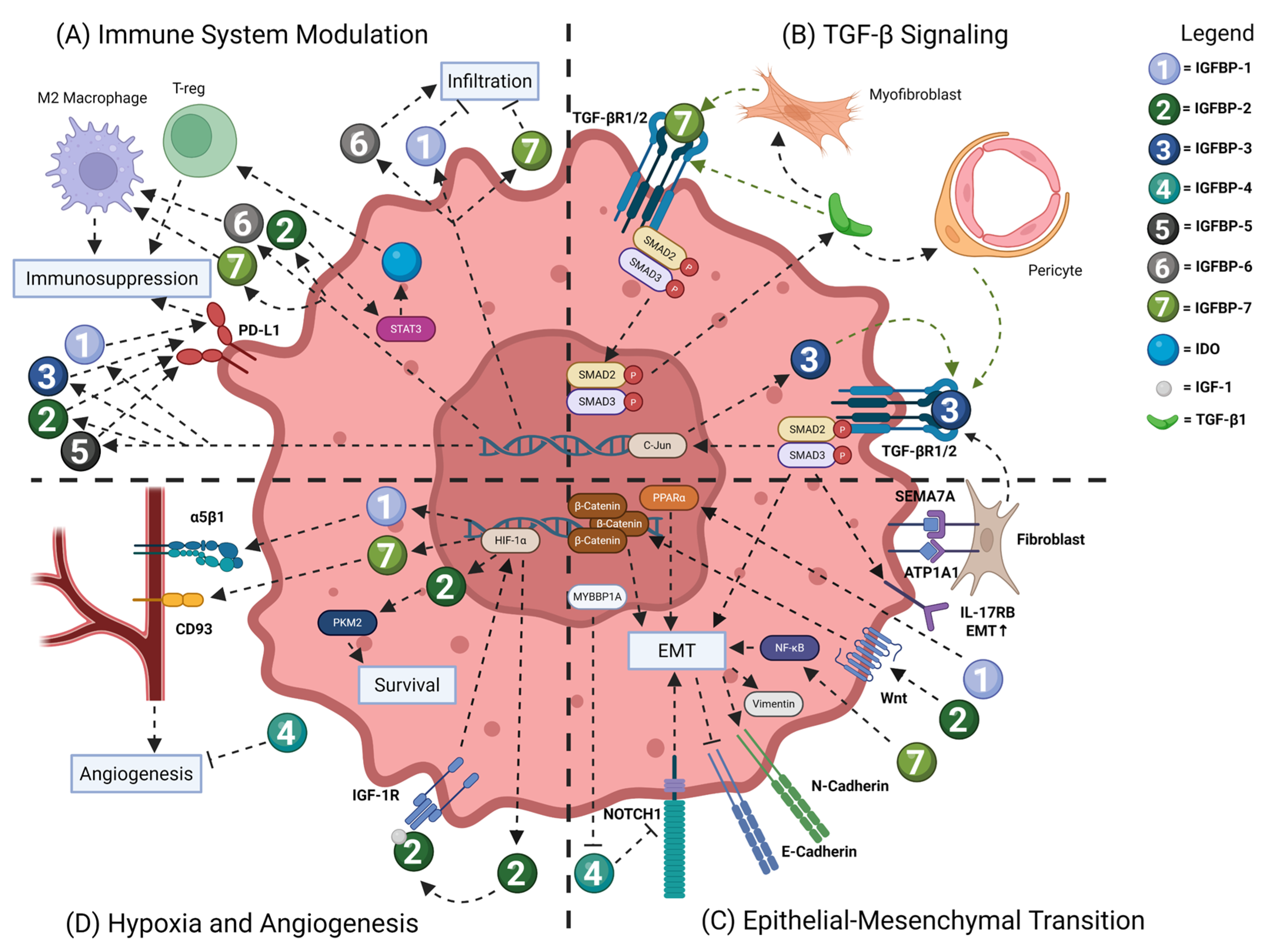
Figure 1. IGFBP family members play a complex role in regulating cancer cell growth, survival and migration. (A) Tumor-secreted IGFBP family members regulate the infiltration of pro-inflammatory immune cells and induce phenotype switching to a more immunosuppressive environment, promoting tumor growth. Moreover, IGFBP family members can elevate PD-L1 expression, exacerbating immunosuppressive phenotypes. (B) IGFBP-3 and 7 display a complex feed-forward relationship with TGF-β wherein TGF-β1 induces myofibroblast expression of IGFBP-7 and pericyte expression of IGFBP-3 that activates TGF-β receptors and downstream SMAD2/3 signaling. Note: green arrows represent direct pro-tumor growth signaling. (C) IGFBP family members regulate tumor cell migration and epithelial-to-mesenchymal transition (EMT) through a variety of signaling cascades. EMT potential is measured in the increase in mesenchymal markers N-Cadherin and Vimentin and the reduction in the epithelial marker E-Cadherin. (D) Hypoxic environments activate HIF-1α expression leading to IGFBP expression through direct transcription. IGFBPs promote tumor survival in a hypoxic environment. Lastly, IGFBPs in part regulate angiogenesis in tumors, further promoting tumor survival and growth. Note: dashed lines represent signaling interactions. Arrows mean positive regulation and blunt arrows mean inhibition. STAT3 = Signal Transducer and Activator of Transcription 3; PD-L1 = Programmed Death-Ligand 1; IDO = indoleamine 2; 3-dioxygenase; TGF-βR1/2 = Transforming Growth Factor Receptor 1/2; TGF-β1 = Transforming Growth Factor 1; SMAD2/3 = Mothers Against Decapentaplegic Homolog 2/3; SEMA7A = Semaphorin 7A; Peroxisome Proliferator-Activated Receptor alpha; MYBBP1A = MYB Binding Protein 1a; NOTCH1 = Neurogenic Locus Notch Homolog Protein 1; IGF-1R = Insulin-Like Growth Factor 1 Receptor; HIF-1α = Hypoxia-Inducible Factor 1-alpha; PKM2 = Pyruvate Kinase M2.
Ijms 26 10248 g001
Figure 2. IGFBPs’ role in vascular homeostasis and cardiovascular disease. (A) Angiogenesis—IGFBP-2 is pro-angiogenic and promotes VEGF transcription. IGFBP-4, 5, and 6 inhibit angiogenesis; however, IGFBP-4 promotes VEGF-mediated angiogenesis in myocardial infarction. IGFBP-3 and 5 inhibit VEGF signaling. IGFBP-7 binds to Activin A and inhibits angiogenesis. (B) Vasodilatation—IGFBP-3 contributes to the secretion of nitric oxide by binding to the scavenger receptor class B, type 1, thereby regulating vascular tone. (C) Cell differentiation—IGFBP-4 enhances cell differentiation by inhibiting β-catenin signaling. (D) Hypoxia—IGFBP-2 binds to IGF-1R and promotes HIF-1α transcription. HIF-1α regulates the expression of IGFBP-1, 2 and 6. IGBP-7 inhibits FOXO3a expression via the PI3K/AKT pathway, inducing senescence. (E) Apoptosis—IGFBP-1 is upregulated by HIF-1α, which in turn contributes to their apoptosis. IGFBP-3 is involved in ICAM1 suppression, preventing monocyte adhesion and limiting endothelial apoptosis. IGFBP-6 increases monocyte adhesion and apoptosis. (F) Inflammation—IGFBP-6 is pro-inflammatory. Via the JNK and NF-kB signaling, it is known to increase inflammation in the endothelial cells. Note: dashed lines represent signaling interactions. Arrows mean positive regulation and blunt arrows mean inhibition. STAT3 = Signal Transducer and Activator of Transcription 3; FOXO3a = Forkhead box O3; SMAD2/3 = Mothers Against Decapentaplegic Homolog 2/3; IGF-1R = Insulin-Like Growth Factor 1 Receptor; HIF-1α = Hypoxia-Inducible Factor 1-alpha; JNK = c-Jun NH-2 terminal kinase; MEKK = mitogen-activated protein kinase (MEK) kinase; VEGF = Vascular Endothelial Growth Factor; Sck = SH3 Domain-Containing Kinase-Binding Protein 1; VRAP = VEGF Receptor-Associated Protein; SRC = Sarcoma protein kinase; SR-B1 = scavenger receptor class B, type 1.
Ijms 26 10248 g002
Figure 3. IGFBPs’ involvement in fibrotic mechanisms in fibroblasts and mesenchymal cells. (A) Extracellular matrix regulation—IGFBP-3 and IGFBP-7 activate TGF-β receptors and downstream SMAD signaling, causing an increase in expression of Collagen, CTGF, and Tenascin C, enabling ECM deposition in fibrosis. TGF-β-induced myofibroblasts are involved in a self-amplifying loop of activating IGFBP-7 secretion and TGF-β receptors. IGFBP-5 promotes myofibroblast differentiation and fibrosis. It increases pro-fibrotic genes and ECM deposition. IGFBP-5’s ability to self-amplify its own expression adds to the fibrotic tissue microenvironment. IGFBP-4, however, blocks TGF-β-induced ECM production and deposition. It also reduces CTGF and CXCR4 levels. (B) Metabolism—IGF signaling via IGFBPs binding to IGFRs leads to downstream PI3K/AKT/mTOR, MEK/ERK, and JNK signaling. IGFBP-5 enables the MEK/ERK and JNK signaling that induces ROS production. This is also mediated by NADPH oxidase. (C) Immunomodulation and senescence—IGFBP-2 induces M2 polarization of macrophages, which contributes to a pro-inflammatory environment. Inflammatory cytokines from M2 macrophages stimulate myofibroblasts, which then create a pro-fibrotic condition. IGFBP-2 also inhibits SASP, including pro-inflammatory cytokines, chemokines, growth factors, proteases, and ECM components. IGFBP-6 induces myofibroblast differentiation by activating the sonic hedgehog pathway that, in turn, induces TLR4 signaling, whose downstream effects include the NF-kB pathway and activating inflammatory genes. Note: dashed lines represent signaling interactions. Arrows mean positive regulation and blunt arrows mean inhibition. ECM = extracellular matrix; CTGF = connective tissue growth factor; SASP = senescence-associated secretory phenotype; SMAD2/3 = Mothers Against Decapentaplegic Homolog 2/3; TLR4 = Toll-like receptor 4; IL = interleukins; TGF-β = Transforming Growth Factor 1; JNK = c-Jun NH-2 terminal kinase; MEKK = mitogen-activated protein kinase (MEK) kinase; ROS = reactive oxygen species.
Ijms 26 10248 g003
Table 1. IGFBP disease outcomes in cancer, cardiovascular disease and fibrosis.
DiseaseTissue TypeExpressionSample SizeResponse to Treatment/Longitudinal Study ResultsReferences
IGFBP-1Colorectal
Cancer
Serum and tumor tissueIncrease328Poor survival[142]
Esophageal
Cancer
SerumIncrease1064-[143]
Gastric
Cancer
Tumor tissueIncrease11,084Poor survival[144]
Lung
Cancer
Tumor TissueIncrease9736Poor survival[145]
Idiopathic pulmonary fibrosis (IPF)SerumIncrease72, 50Levels increased with time/severity, insensitive to anti-fibrotic treatments[107,108]
Non-alcoholic fatty liver disease (NAFLD)SerumIncrease52More advanced fibrosis[106]
Heart failure with reduced ejection fractionSerumIncrease250Presence of heart failure and likelihood of adverse event[146]
Peripheral arterial diseaseSerumIncrease465Increased risk of major adverse cardiovascular events (MACE)[84]
IGFBP-2Colorectal
Cancer
Tumor tissueIncrease5560Poor survival[147]
Esophageal
Cancer
Tumor tissueIncreaseNot reported-[19]
Gastric
Cancer
Serum and tumor tissueIncrease118Correlates with tumor stage and poor survival[19]
Lung
Cancer
Serum and tumor tissueStudy-dependent30, 20, 201Poor survival in lung adenocarcinoma and better survival in lung squamous cell carcinoma[145,147,148,149,150]
Breast
Cancer
Serum and tumor tissueIncrease412Poor survival and tamoxifen resistance[19]
Pancreatic
Cancer
SerumIncrease165Correlates with tumor stage and poor survival[151]
IPFSerumIncrease50, 15Elevated in patients and decreased with anti-fibrotic treatment[108,110]
Systemic Sclerosis (SSc)SerumIncrease102Negatively correlates with pulmonary function and disease progression[109]
Acute coronary syndromeSerumIncrease277Increased risk of MACE[152]
Heart failureSerumIncrease870Increased risk of cardiovascular mortality[86]
IGFBP-3Colorectal
Cancer
Tumor tissueIncrease202Poor survival[153]
Gastric
Cancer
Serum and tumor tissueStudy-dependent1541, 11,084-[144,154]
Lung
Cancer
Serum and tumor tissueStudy-dependent131Inverse relationship to tumor stage in non-small cell lung cancer, and disease risk in lung adenocarcinoma[145,150,155]
Breast
Cancer
SerumIncrease334,236Increased risk of developing disease[156,157]
Pancreatic
Cancer
Serum and tumor tissueStudy-dependent88, 478Serum negatively correlates and tumor expression positively correlates with survival[158,159]
SScSerumIncrease92-[160]
SSc-ILDLung tissueIncreaseNot reported-
IGFBP-4Gastric
Cancer
Tumor tissueIncrease11,084-[144]
Lung
Cancer
SerumIncrease83-[161]
Breast
Cancer
Tumor tissueDecrease162Improved survival[28]
Pulmonary Arterial Hypertension (PAH)SerumIncrease2579Shorter 6 min walk distance, worse NYHA functional classification, and decreased survival.[162]
Ischemic heart diseaseSerumIncrease1417Increased risk of MACE and mortality[163]
IGFBP-5Lung
Cancer
SerumDecrease100Improved survival[164]
Colorectal
Cancer
Tumor tissueIncrease56Correlates with tumor stage and poor survival[165]
IGFBP-6Breast
Cancer
Tumor tissueDecrease1091Improved survival[35]
Colorectal
Cancer
Tumor tissueDecrease130Improved survival[166]
Lung
Cancer
SerumDecrease31-[167]
Fibrosis in
NAFLD
Serum and liver tissueIncrease61Reduced IGFBP-6 expression on anti-fibrotic tesamorelin[124]
AtherosclerosisSerum and artery tissueDecrease3-[98]
IGFBP-7Gastric
Cancer
Tumor tissueIncrease16Poor survival[38]
Breast
Cancer
Tumor tissueIncrease878Increased disease risk and poor survival[168]
Lung
Cancer
SerumIncrease90Correlates with tumor stage and metastasis[169]
Heart
failure
SerumIncrease2250, 313Increased risk of all three main hospitalization and mortality causes[101,102]
Heart failure riskSerumIncrease13,900Risk of future CHF development in healthy individuals[170]
Pulmonary hypertensionSerumIncrease2582Decreased six-minute walk distance, higher mean right atrial pressure, decreased survival[162]
SScSerumIncrease37Higher modified Rodnan skin score (MRSS)[131]
Disclaimer/Publisher’s Note: The statements, opinions and data contained in all publications are solely those of the individual author(s) and contributor(s) and not of MDPI and/or the editor(s). MDPI and/or the editor(s) disclaim responsibility for any injury to people or property resulting from any ideas, methods, instructions or products referred to in the content.