Article

Genetic Diversity and Infection Prevalence of Biomphalaria pfeifferi (Krauss, 1848), the Intermediate Snail Host of Schistosoma mansoni in Gezira State, Sudan

1
State Key Laboratory of Bioreactor Engineering, East China University of Science and Technology, Shanghai 200237, China
2
Blue Nile National Institute for Communicable Diseases, University of Gezira, Wad Madani P.O. Box 20, Sudan
3
National Institute of Parasitic Diseases, Chinese Center for Disease Control and Prevention (Chinese Center for Tropical Diseases Research), Shanghai 200025, China
4
School of Global Health, Chinese Centre for Tropical Diseases Research, Shanghai Jiao Tong University School of Medicine, Shanghai 200025, China
5
Department of Immunology and Parasitology, Copenhagen University, 1172 København, Denmark
6
Department of Parasitology, Endemic Diseases Institute, University of Khartoum, Khartoum 11115, Sudan
7
Faculty of Medical Laboratories, University of Gezira, Wad Madani P.O. Box 20, Sudan
*
Author to whom correspondence should be addressed.
Int. J. Mol. Sci. 2025, 26(19), 9567; https://doi.org/10.3390/ijms26199567
Submission received: 13 August 2025 / Revised: 26 September 2025 / Accepted: 28 September 2025 / Published: 30 September 2025
(This article belongs to the Special Issue Molecular Insights into Zoology)

Abstract

Biomphalaria pfeifferi snails serve as the major intermediate host for intestinal schistosomiasis in Sudan. The genetic structure and infection status of 163 B. pfeifferi collected from six localities in Gezira State, Sudan (East Gezira, Greater Wadmedani, Hasahisa, North Umelgura, South Gezira, and Managil) were characterized. Cytochrome oxidase subunit I (COI) and 16S ribosomal RNA (16S rRNA) mitochondrial genes were used for B. pfeifferi molecular identification and genetic diversity investigation. Schistosoma mansoni infection was detected using the traditional cercarial shedding and molecular methods (SmF/R primers). Five COI haplotypes and ten 16S haplotypes were identified, with haplotype diversity of 0.50 for COI and 0.11 for 16S. High evolutionary divergence was observed between groups (Fst = 0.94) for the COI, and low genetic divergence (Fst = 0.04) for the 16S, indicating genetic divergence among Sudanese B. pfeifferi, with the 16S showing lower divergence than the COI, consistent with a post-bottleneck population expansion. Cercarial shedding detected an overall infection prevalence of 3.6% (8/219), with only two snails from Hasahisa shedding S. mansoni cercariae. The SmF/R primers revealed a higher infection prevalence of 7.4% (12/163), with all S. mansoni positive samples found at the Hasahisa site. Findings highlight the value of molecular diagnostic tools for accurate surveillance and emphasize the need for site-specific control strategies.

1. Introduction

The freshwater snail genus Biomphalaria (Gastropoda: Planorbidae) is widely distributed across sub-Saharan Africa, South and Central America, and the Middle East [1,2]. The disease disproportionately affects vulnerable groups, including preschool and school-aged children, certain occupational workers, women engaged in domestic activities involving contaminated water, and communities living in high-risk areas [3]. In Sudan, schistosomiasis has become a significant public health concern, and the disease is endemic in all states, except the Red Sea State, resulting in serious economic and social implications. The expansion of water resource development and increased population mobility have contributed to the disease spread and prevalence [4,5]. Gezira State, a major agricultural area in Sudan, has experienced a significant rise in schistosomiasis prevalence. This increase is partly linked to changes in crop rotation within the Gezira Irrigation Scheme and the development of the Managil Extension, which have limited access to sanitary services and safe water [5,6]. The interaction between schistosomiasis transmission and water resource development underscores the importance of understanding the dynamics of snail intermediate hosts and disease transmission [7].
All African Biomphalaria species are capable of transmitting Schistosoma mansoni, the primary causative agent of intestinal schistosomiasis in humans [1]. Among these, B. pfeifferi is considered the most important vector due to its high infection prevalence in field populations [1,8,9]. In addition to S. mansoni, African Biomphalaria species are also susceptible to a range of other trematode parasites [10]. For example, Biomphalaria snails in Sudan have been found infected with Amphistome, Echinostome, Furcocercous, and Xiphidiocercariae cercariae types [11,12].
Historically, snail identification relied on shell morphology and anatomical characteristics (e.g., radula and genitalia) to determine taxonomic differences between species [1,13]. To date, a total of 34 Biomphalaria species have been identified [1,2], with the African species being historically classified into four morphological groups: Alexandrina, Choanomphala, Pfeifferi, and Sudanica [14]. However, molecular phylogenies have since redefined these classifications, defining the African species as B. camerunensis, B. pfeifferi, and the Nilotic species complex (B. alexandrina, B. angulosa, B. choanomphala, B. smithi, B. stanleyi, and B. sudanica) [8,15,16,17]. Molecular techniques have been employed to improve the precision of species identification and to enhance our understanding of the population structure and genetic diversity of medically important snail species [18,19]. The application of molecular methods has greatly improved the accuracy of snail identification, thereby advancing our understanding of schistosomiasis epidemiology [20].
Numerous studies have investigated the phylogenetic structure of B. pfeifferi populations across sub-Saharan Africa using established genetic markers, such as the Cytochrome oxidase subunit I (COI) gene, 16S ribosomal RNA (16S rRNA), and the Internal Transcribed Spacer regions (ITS1 and ITS2) [2,8,21,22,23,24,25,26,27,28]. Similarly, several studies have investigated the prevalence of S. mansoni infection in B. pfeifferi populations, traditionally using the cercarial shedding method, which involves screening snails over a 35-day period [29]. However, this method detects only patent infections [30] and fails to detect pre-patent infections. To address this limitation, a variety of molecular xenomonitoring assays targeting different genes have been developed [23,28,31,32,33,34,35], enabling the detection of both patent and pre-patent infections. These molecular detection techniques offer high sensitivity for detecting low quantities of S. mansoni DNA in Biomphalaria spp. [32,35,36], enable simultaneous identification of multiple parasite species [37], and allow accurate species confirmation through genotyping [26].
In 2023, the prevalence of intestinal schistosomiasis in Gezira State was 8.6%, highlighting the ongoing public health burden and the urgent need for integrated control strategies. Accurate species-level identification and a thorough understanding of the genetic structure of Biomphalaria snails in endemic regions like Gezira State are essential for designing effective interventions, as both factors influence their susceptibility to Schistosoma infection and the dynamics of disease transmission. This study aims to investigate the genetic diversity and population structure of B. pfeifferi across multiple Gezira State localities using mitochondrial COI and 16S markers to infer phylogenetic relationships with other African populations. Infection prevalence was assessed through both cercarial shedding and PCR-based (SmF/R) methods. By integrating genetic and epidemiological data, this study provides insights into the diversity and infection dynamics of B. pfeifferi in Gezira State, with the aim of informing and strengthening evidence-based schistosomiasis control strategies.

2. Results

2.1. Species Identification and Phylogenetic Analysis

Of the six locations surveyed, Biomphalaria snails were found at all locations but were present at only seven of the 30 sites (23.3%; Table 1). In total, 219 Biomphalaria snails were collected from these seven sites across Gezira State, Sudan. Of those 219, 163 had their DNA extracted and were confirmed as B. pfeifferi through the phylogenetic analysis of the COI and 16S gene fragments (Figure 1). The remaining 56 snails were also identified as B. pfeifferi based on key morphological characteristics. The Maximum Likelihood (ML) and Neighbor-Joining consensus phylogenetic tree analysis of the concatenated COI and 16S dataset confirmed that all samples from Gezira clustered with reference B. pfeifferi sequences, supported by a 100% bootstrap value (Figure 1; Supplementary Figure S1). Among the Gezira populations, the Managil (MA) samples exhibited the highest genetic divergence, while moderate divergence was also observed in samples from Hasahisa (HA), East Gezira (EG), Greater Wadmedani (GW), South Gezira (SG), and both North Umelgura sites (NU1 and NU3; Figure 1).

2.2. Haplotype Analysis, Diversity Parameters, and Neutrality Tests

After alignment, the amplified fragments of the COI and 16S gene fragments were 527 bp and 387 bp in length, respectively. For the COI gene, five haplotypes were identified. The predominant haplotype, H1, was found across four localities, East Gezira, South Gezira, and North Umelgura (NU1 and NU3), and accounted for 59.5% of all samples (97/163). Haplotype H2 occurred at three localities: Hasahisa, East Gezira, and Greater Wadmedani, comprising 37.4% (61/163) of the samples. The remaining haplotypes were more localized, with H3 being unique to East Gezira, H4 to Hasahisa, and H5 to Managil. East Gezira exhibited the highest haplotype richness with three haplotypes, followed by Hasahisa with two, while the remaining sites only had single haplotype (Table 1). In contrast, the 16S gene revealed greater haplotype diversity, with ten haplotypes found across all sampled populations. Haplotype H1 was the most widespread, present in all seven localities, and constituted 93.8% of the total sequences (153/163). The remaining minor haplotypes were exclusive to Hasahisa (H2–H6), North Umelgura-1 (H7–H9), and North Umelgura-3 (H10). Hasahisa showed the highest haplotype diversity (with six haplotypes), followed by North Umelgura-1 (four haplotypes) and North Umelgura-3 (two haplotypes). The overall haplotype diversity (Hd) was 0.50 (±0.022) for COI and 0.11 (±0.035) for 16S. The average number of nucleotide differences (k) was 5.38 for COI and 2.05 for 16S. Lastly, nucleotide diversity (π) was estimated at 0.01 (±0.001) for COI and 0.008 (±0.003) for 16S (Table 1).
Neutrality analyses revealed distinct patterns between the COI and 16S gene datasets (Table 1). For the COI gene, Tajima’s D showed a negative but non-significant value (−0.867), suggesting weak evidence of population expansion or purifying selection at the population level. Similarly, Fu’s Fs was positive (13.62) and non-significant, which may indicate a recent population bottleneck or balancing selection, although the signal was weak. In contrast, the 16S gene exhibited a statistically significant negative Tajima’s D value (−2.71, p < 0.01), indicating stronger evidence for recent population expansion or purifying selection. At the population level, COI data from East Gezira showed a significantly negative Tajima’s D value (−2.36, p < 0.02), supporting expansion in this population. However, Fu’s Fs remained positive and non-significant in both the overall and East Gezira datasets. For the 16S gene, Hasahisa displayed the highest nucleotide diversity (0.035 ± 0.01) and haplotype diversity (Hd = 0.31 ± 0.11) among all sites, with a significantly negative Tajima’s D (−1.84, p < 0.05), indicating a recent expansion. It also showed a high, positive Fu’s F value (8.825). Though not statistically significant, this may suggest the presence of multiple divergent lineages at the site. Conversely, North Umelgura-1 (NU1) also showed relatively high haplotype diversity (0.25 ± 0.1), with a significantly negative Tajima’s D (−2.425, p < 0.01) and strongly negative Fu and Li’s D and F values, indicating a strong signal of recent expansion or purifying selection. Interestingly, North Umelgura-3 (NU3) showed limited diversity with only two haplotypes, and its Fu’s F value was negative (−1.21), but not statistically significant. Several other sites (Greater Wadmedani, South Gezira, and Managil) were monomorphic across both genes, consistent with low genetic variability, possibly due to recent colonization or population bottlenecks (Table 1).

2.3. Haplotypes Network Inference

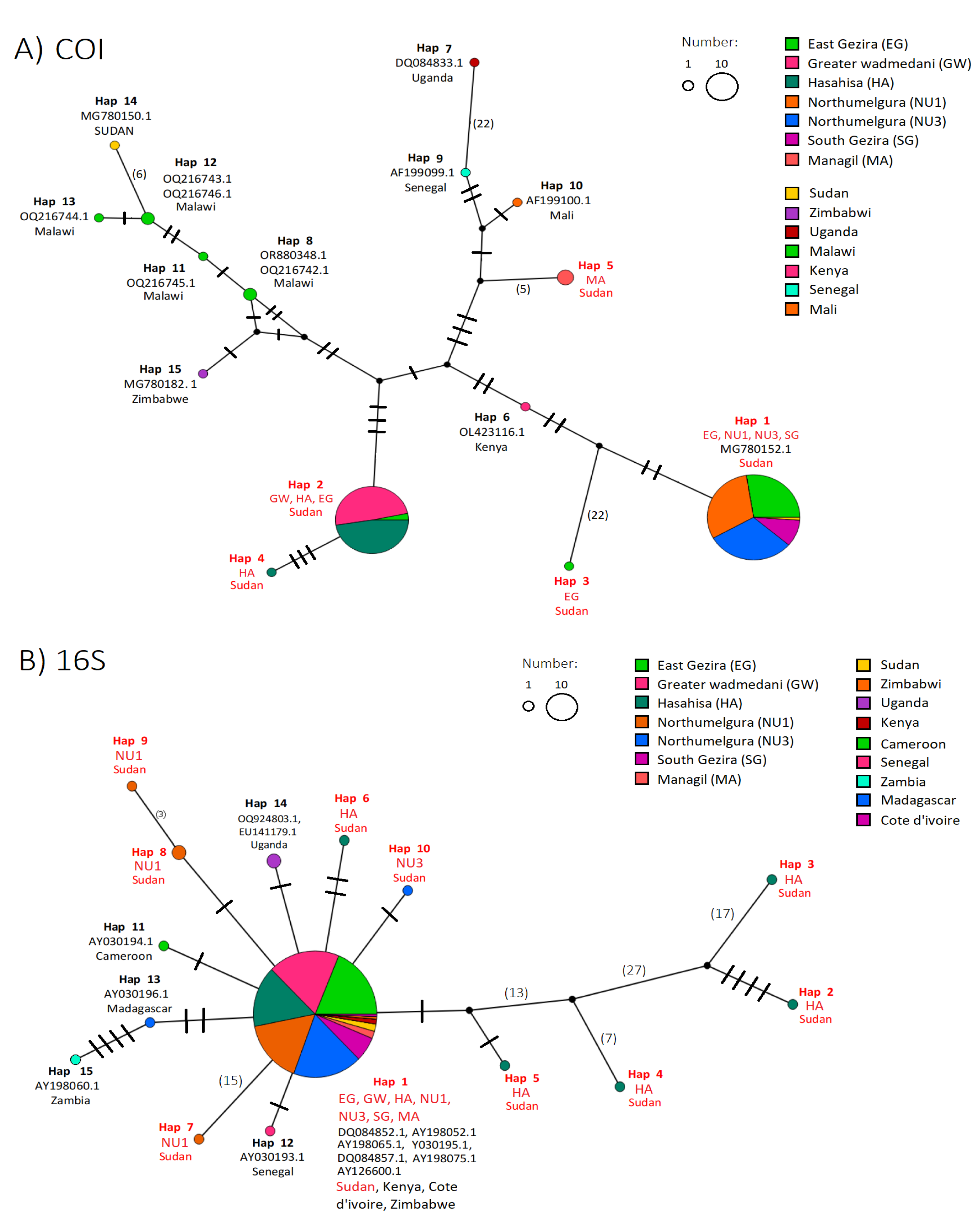
For the COI gene fragment, haplotype H1 was found across four localities in the Gezira state, and was distinct from all other African haplotypes, clustering only with a previously sequenced Sudanese B. pfeifferi sequence (MG78152; Figure 2A). Likewise, the remaining haplotypes (H2–H5) did not cluster with any other African haplotypes, suggesting local differentiation. For the 16S gene, haplotype H1, was distributed across all seven Gezira sites, and clustered with sequences from multiple African countries, including Kenya (DQ084852.1, AY198052.1), Côte d’Ivoire (AY198065), Sudan (Y030195, DQ084857, AY198075), and Zimbabwe (AY126600; Figure 2B). This widespread clustering indicates low divergence and the dominance of this haplotype across Africa. However, similar to the COI, the remaining 16S haplotypes (H2–H10) did not cluster with any other African haplotypes. These results highlight the higher resolution of the COI gene in detecting local genetic differentiation, while the 16S gene reveals broader haplotype conservation across African B. pfeifferi populations. Analysis of haplotype distribution across the seven sampling sites (Figure 2C; Supplementary Table S1) revealed pronounced spatial structuring. For the COI, Elgineid (S13) exhibited the greatest haplotype diversity, with Wadelfadni (S19), Atraa (S6), and Alnegeer village (S23) being made up of haplotypes H2, H4, and H5 respectively. For the 16S dataset, diversity was highest at Wadelfadni (S19) and Elhediba (S10, S12), where additional haplotypes (H2–H10) were detected at low frequencies (Figure 2C). Overall, these results indicate that haplotype H1 is highly conserved and dominant throughout Gezira, while localized differentiation is evident at specific sites, with COI offering finer resolution of site-specific variation than 16S.

2.4. Genetic Divergence and Population Differentiation

Estimates of evolutionary divergence among COI haplotypes, calculated using the Maximum Composite Likelihood model, revealed substantial genetic variation. The highest divergence values were found between H1 (the most abundant haplotype) and H3 (0.053), H2 and H3 (0.068), and between H3 and H4 (0.074) and H5 (0.075) (Supplementary Table S2). The 16S dataset exhibited even greater divergence, between H1 and H3 (0.192) and between H3 and H7 (0.220; Supplementary Table S2). The COI gene fragment revealed 41 polymorphic sites (Table 1), and pairwise population Fst values ranged from 0 to 1. The overall Fst was extremely high (0.94), indicating very strong genetic differentiation, and the Nm value was very low (0.03), suggesting minimal gene flow (Table 2A). Nearly all pairwise comparisons involving Managil (MA), Greater Wadmedani (GW), and Hasahisa (HA) exhibited very high differentiation (Fst > 0.85). Conversely, East Gezira (EG), North Umelgura sites (NU1 and NU3), and South Gezira (SG) shared low Fst values (0.02) among each other, indicating weak differentiation and relatively recent shared ancestry. The 16S gene fragment showed higher overall polymorphism (84 sites) but low overall Fst (0.04) and high gene flow (Nm = 5.72; Table 2B). Most pairwise comparisons fell within the low differentiation range (Fst < 0.05), particularly between East Gezira and all other sites. Notably, comparisons involving Hasahisa (HA) yielded moderate Fst values (0.04–0.05), suggesting slightly increased genetic structuring in that locality, which also exhibited the highest haplotype diversity for 16S.

2.5. Analyses of Molecular Variation (AMOVA)

The AMOVA results revealed strong genetic differentiation among the seven populations based on the COI gene fragment, with limited gene flow. The majority of genetic variation (84.6%) was attributed to differences among populations, while only 15.4% of the variation occurred within populations (Table 3). Although the fixation index (ΦST) was not statistically significant (p > 0.001), Wright’s criteria suggest that FST > 0.25 represents high divergence, while FST > 0.05 represents low divergence. Therefore, the COI gene has a very high fixation index (ΦST = 0.90), indicating significant evolutionary divergence and clear population structuring. FST values greater than 0.25 denote high genetic differentiation, which aligns well with the observed ΦST of 0.90 for COI. Although the term “FST” is often used interchangeably with “ΦST” in haplotype data, the distinction is important in this context. Here, ΦST clearly demonstrates strong population subdivision and restricted gene flow (Nm = 0.03). In contrast, the 16S gene fragment exhibited very low genetic differentiation among populations, with only 3.6% of the variation attributed to differences among populations and 96.4% occurring within populations (Table 3). The fixation index was correspondingly low (ΦST = 0.04), consistent with weak population structure and relatively high gene flow (Nm = 5.72). Together, these results suggest that while COI haplotypes are strongly structured among populations, 16S haplotypes show much less differentiation, reflecting differing evolutionary dynamics or rates of gene flow for the two markers.

2.6. Cercariae Shedding and PCR Infection

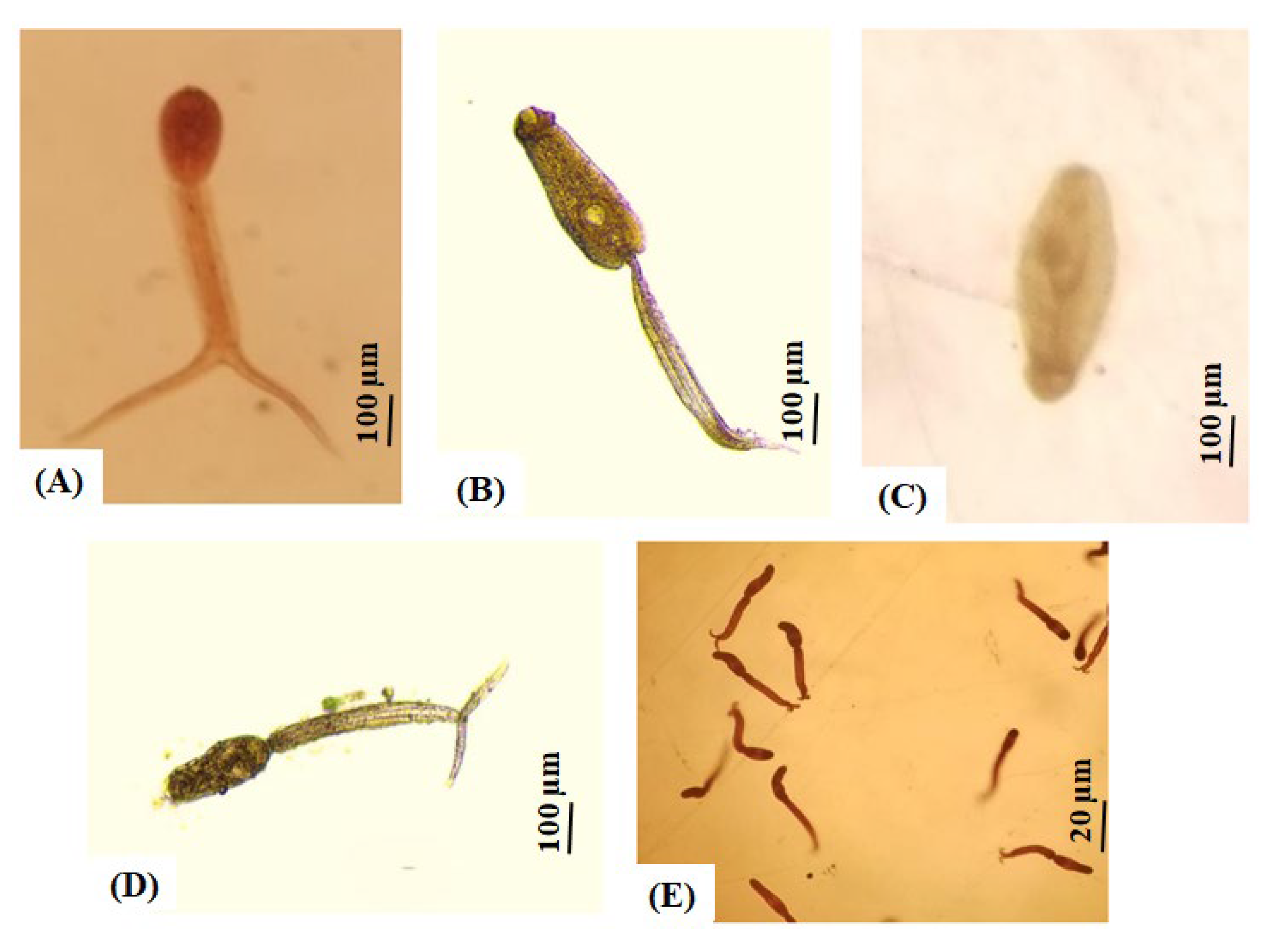
The overall prevalence of B. pfeifferi shedding cercariae across the seven surveyed sites was 3.6% (8/219), with North Umelgura showing the highest infection rate at 16.6% (Table 4). Among the eight shedding snails, four were co-infected with Echinostome spp. and Cotylomicrocercous spp. Cercariae (NU3), one was infected with Echinostome spp. (NU3), one was infected with Apharyngostrigea spp. (EG), and two were infected with S. mansoni (HA; Table 4; Figure 3). No snails were found shedding cercariae at the remaining sites (SG, GW, NU1, and MA), snails shedding S. mansoni cercariae were only found at Hasahisa (2/39; Table 4; Figure 3D,E).
To further assess S. mansoni infection, DNA extracts from B. pfeifferi snails were screened using SmF/R primers. The SmF/R primers detected S. mansoni DNA in 12 of 163 snail extracts tested (7.4%). All 12 of the SmF/R PCR-positive snails were from the Hasahisa site, and this included the two shedding snails and ten non-shedding (pre-patent) snails (Supplementary Figure S2). When sequenced, the SmF/R gene fragment matched (97% query coverage and 99.35% sequence identity) with multiple S. mansoni reference sequences in GenBank, including AY157173 [38], HE601625 [39], MK085970 [40], and OK310887, confirming the presence of S. mansoni DNA in Hasahisa.

3. Discussion

Mitochondrial genes, particularly COI and 16S rRNA, are well-established markers for detecting genetic patterns linked to demographic variation within a species [41]. The high value of haplotype diversity (Hd) compared to nucleotide diversity (π) suggests closely related haplotypes and recent population expansion after a bottleneck [42]. We used neutrality tests to assess the demographic expansion of B. pfeifferi populations. The results collectively support the expansion hypothesis, as Tajima’s D (COI) and the significantly negative Fu and Li’s D and F values (both COI and 16S) provide evidence of a past bottleneck. Additionally, Fuʼs Fs test also supports demographic expansion. However, some populations (e.g., Hasahisa, East Gezira, and North Umelgura-1) showed positive values, suggesting possible historical selection of alleles present in many individuals within these populations [43].
Next, Fst values were calculated to assess allele frequency variation among the studied populations, reflecting genetic differences and the degree of population substructure [44]. The results revealed strong divergence in COI, with highly significant Fst values (>0.5) and low gene flow among most populations, particularly in Managil (H5), which exhibited a unique SNP profile. However, the small sample size (n = 3) prevents a definitive conclusion about the isolation of Managil. The high divergence in COI among populations, combined with low gene flow, is consistent with previous studies on B. pfeifferi, B. glabrata, and B. choanomphala [2,8,43,45]. Conversely, the high homogeneity in the 16S gene (low Fst = 0.041) indicates minimal differentiation among populations but high variation within populations, consistent with a previous study in Uganda [46]. The levels of divergence for each genetic marker varied, although the collection sites were geographically distant (each from a different locality), except for North Umelgura (NU1 and NU3). Moreover, the presence of distinct individuals in the Hasahisa, East Gezira, and North Umelgura localities aligns with several studies reporting that Biomphalaria spp. and Bulinus spp. populations are typically more genetically similar at nearby sites, although distinct haplotypes may still occur at individual locations [45,47,48].
The genetic structure of Biomphalaria spp. populations is influenced by many factors, such as self-fertilization, environmental conditions, habitat fluctuations, host type, and parasitic infection [1,19,25,28,36,46,49]. Bottlenecks in these snails are often linked to drastic environmental changes, such as shifts in climate, drought, flooding, food scarcity, and the use of molluscicides [2,25,49,50,51]. All of our snail samples were collected from non-perennial canals within the Gezira Irrigation System, which are subject to seasonal fluctuations such as droughts and occasional flooding. These environmental disturbances likely lead to periodic local population extinctions followed by recolonization, creating repeated genetic bottlenecks. Such events can reduce genetic diversity within populations while simultaneously increasing genetic divergence among populations, as surviving or recolonizing snails may carry only a subset of the original alleles. In addition, B. pfeifferi populations typically show a preference for self-fertilization, resulting in limited genetic variation [52,53]. Moreover, limited dispersal further increases genetic divergence by restricting gene flow [2,54]. In addition, the observed low genetic diversity in mitochondrial genes may reflect the adaptation of oxidative systems to environmental stressors [55,56]. The low level of genetic variation in 16S is attributed to inbreeding or changes in habitat, leading to population subdivision [1,46,57]. It has been previously reported that Biomphalaria populations are subdivided based on variation in the 16S gene [28,49,58,59,60,61].
Assessing infection prevalence in snail populations through cercarial shedding remains a cornerstone of traditional field-based schistosomiasis epidemiology. However, its key limitation is that it only detects patent infections (snails actively shedding cercariae), while failing to identify pre-patent cases [62,63]. Consequently, molecular diagnostics provide a more accurate representation of snail-mediated transmission, particularly in areas where both patent and pre-patent infections occur [64,65]. In our study, only 2 of 219 (0.9%) B. pfeifferi snails were found to be infected with S. mansoni using the cercarial shedding method, both from Hasahisa. In contrast, PCR detection identified infection in 12 of 163 B. pfeifferi DNA extracts (7.4%), which were all from Hasahisa. A meta-analysis by Hailegebriel et al. (2020) estimated the pooled prevalence of S. mansoni infection in Biomphalaria snails across Africa at 5.6%, indicating that the infection prevalence in our samples is lower than generally expected in natural snail populations [66]. Also, our results are consistent with previous studies demonstrating that molecular methods are more sensitive than cercarial shedding in detecting both pre-patent and patent infections [32,67,68]. These results confirm that relying solely on cercarial shedding for schistosomiasis screening can underestimate the true prevalence of S. mansoni infection [69]. This highlights the greater sensitivity of molecular methods and the failure of the cercarial shedding method to accurately identify infection in field snails, which can give the false impression of low or even no transmission in an area [62].
Despite their simplicity and effectiveness, molecular methods are limited by high costs and logistical challenges, such as requiring specialized equipment and stable conditions. Moreover, molecular methods may overestimate transmission risk, as not all PCR-positive snails go onto shed cercariae. For example, Lu et al. (2016) [67] found only 60% of PCR-positive B. pfeifferi snails proceeded to shed cercariae, likely due to the failed encapsulated sporocysts still being detected. Molecular detection methods are also highly susceptible to contamination from DNA templates or reagents, exhibit limited efficiency in detecting extremely low DNA concentrations, and require stringent reaction conditions, particularly precise annealing temperature control, for optimal performance [70,71]. In resource-limited developing countries, DNA extraction and molecular xenomonitoring of snails poses significant challenges. Despite these constraints, molecular methods remain essential for accurate schistosomiasis diagnosis and for studying the genetic diversity of snail host populations in Sudan and across Africa.
In terms of genetic diversity, S. mansoni was only detected in one site, Hasahisa. This could impact the diversity of snails haplotypes in this area, as discussed previously [25,60]. However, we noticed that infected individuals were not limited to a specific haplotype, despite the genetically distinct individuals in this population (six haplotypes in the 16S gene and two in the COI), suggesting no clear genetic barrier to infection. Similar findings were noted in snails infected with different trematode cercariae at the two locations in the North Umelgura locality. This aligns with previous studies in Biomphalaria spp., where parasitism may shape population structure without affecting their susceptibility to schistosomes infection [45,49,60,72]. Notably, Hasahisa is one of the most genetically diverse sites, alongside East Gezira (EG) and the North Umelgura sites (NU1 and NU3). This pattern is consistent with Andrus et al. (2023, 2025), who found that Biomphalaria populations with higher amounts of COI and 16S haplotypes harbor more S. mansoni infections than those with fewer haplotypes [28,36]. This may result from greater migration of infected snails introducing new parasites, alongside a wider range of host genotypes that increase compatibility with S. mansoni. In contrast, populations with lower genetic diversity may have reduced parasite compatibility and less introduction of infection due to limited migration. Notably, non-S. mansoni infection was also found in East Gezira and North Umelgura, further supporting the role of genetic diversity in shaping trematode transmission dynamics among Biomphalaria populations.

4. Materials and Methods

4.1. Study Area, Site Selection, and Sample Collection

Gezira State, central Sudan, lies between the Blue and White Nile rivers (13–14.2° N, 32.5–33° E) and supports the continent’s largest gravity-fed irrigation scheme (~7 million acres) via canals supplied from Sennar Dam (Figure 4) [73]. The scheme’s network of open earth-lined canals and associated ditches provides ideal habitats for Biomphalaria snails and frequent human contact points that sustain schistosomiasis transmission [74,75,76]. Sites were surveyed between December 2022 and March 2023, with B. pfeifferi snails being collected from six locations: East Gezira (EG), South Gezira (SG), Hasahisa (HA), Greater Wadmedani (GW), Managil (MA), and North Umelgura (NU; Figure 4, Table 5). These sites were chosen based on water-body type (e.g., minor canal, stream, field canal), observed human and animal activity, and historical surveys of urinary and intestinal schistosomiasis [77,78]. At each location, five sampling sites (30 sites in total) were surveyed. However, B. pfeifferi snails were found at only seven of the 30 sites (Table 5). All seven sites were non-perennial canals, experiencing intermittent water levels that fluctuate according to the farming season. GPS coordinates were recorded on site and mapped in ArcGIS 10.5 (Esri, Redlands, CA, USA; Figure 4). Two trained collectors inspected each site for 15–20 min, scooping along the canal edge and vegetation with a metal mesh net. Biomphalaria snails were transported in canal water to the Medical Entomology and Vector Control Department Laboratory in the Blue Nile National Institute for Communicable Diseases, University of Gezira, Sudan for sorting and were then identified morphologically [79,80].

4.2. Snail Screening (Cercaria Shedding)

Field collected snails (n = 219) were individually placed in 3 mL of tap water within glass bottles (40 × 20 mm) and exposed to artificial light for 3 h (9:00 am–12:00 pm) to induce cercarial shedding. Cercariae were examined under a dissection microscope (Olympus, Tokyo, Japan) and identified as either non-mammalian or mammalian (either human-related or non-human-related) cercariae using morphological descriptions by Frandsen & Christensen [10] and Schell [81]. Snails were labeled as either shedding-positive or shedding-negative and then preserved in 95% ethanol and stored at −20 °C until processing for molecular analysis at the National Institute of Parasitic Diseases, China CDC, Shanghai, China.

4.3. DNA Extraction

Where possible, 30 snails were randomly selected from each site for DNA extraction. If fewer than 30 snails were available at a site, all snails were included (see Table 5). The snail shells were crushed individually, and the head–foot tissue were isolated and preserved in 95% ethanol and stored at −20 °C prior to molecular identification and infection detection. Prior to DNA extraction, tissue was soaked overnight in ddH2O to remove residual ethanol and then air-dried for 4 hrs. Genomic DNA was extracted using the Qiagene DNeasy Blood and Tissue Kit (LOT: 175021295, Cat No. 69506, QIAGEN GmbH, Hilden, Germany) following the manufacturer’s instructions. The extracted DNA samples were stored at −20 °C until further analysis.

4.4. Molecular Identification

Two mitochondrial gene fragments were used to identify the Biomphalaria DNA extracts, the cytochrome oxidase subunit I (COI), and the 16S ribosomal RNA (16S rRNA). The COI was amplified using the LCO1490 and HCO2198 primers developed by Folmer et al. [82], and the 16S was amplified using the 16Sar and 16Sbr primers developed by Palumbi et al. [83] (Table 6). All polymerase chain reactions (PCRs) were performed in a final volume of 25 μL, comprising 13 μL of TianGEN 2× Taq PCR Mix (0.1 U Taq polymerase/μL, 500 μM dNTPs, 20 mM Tris-HCl, 100 mM KCl, 3 mM MgCl2), 7.5 μL of nuclease-free water, 1 μL each of forward and reverse primers (10 μM), and 2.5 μL of genomic DNA template. PCR cycling conditions for the COI primers consisted of an initial denaturation at 94 °C for 2 min, followed by 35 cycles of 94 °C for 30 s, 45 °C for 30 s, and 72 °C for 1 min, with a final extension at 72 °C for 10 min. For the 16S primers, conditions were similar except the annealing temperature was 48 °C and each denaturation step was 94 °C for 1 min. Primer details and annealing temperatures followed those described by [8,24,84]. PCR products were visualized on a 1.5% agarose gel stained with GelStain C056 (1000× code #GS1014, Lot #P40910) and visualized under UV light. The PCR products were purified and sequenced using Beijing QingKe Biotechnology Company Ltd. (Shanghai, China).

4.4.1. Sequence Alignment and Phylogenetic Analysis

Nucleotide sequences were visually edited using Bioedit software v7.7.1 [85]. The sequences from each site were grouped together and aligned using Clustal W in Molecular Evolutionary Genetic Analysis software MEGA v11 [86]. To confirm the identification of B. pfeifferi snails, phylogenetic trees were constructed using both Maximum Likelihood (ML) and Neighbor-Joining (NJ) methods in MEGA v.11. The ML tree was inferred under the General Time Reversible model with a gamma distribution (GTR+Γ), which was selected as the best-fit model. The NJ tree was generated using the Maximum Composite Likelihood (MCL) method. For both approaches, node support was assessed with 1000 bootstrap replicates to evaluate the reliability of inferred phylogenetic relationships, with ML and NJ bootstrap values given in the following order: ML/NJ. This was performed using a combined COI and 16S data, and our samples were compared to different Biomphalaria spp. GenBank references (Supplementary Table S3).

4.4.2. Haplotype and Population Genetic Analysis

The COI and 16S rRNA datasets were analyzed separately, with all sequence alignments performed excluding gaps. Haplotype identification for each gene was conducted using DnaSP v5.10.01 [87], and genetic diversity parameters among B. pfeifferi populations were assessed using pairwise distances and diversity indices. These included the number of segregating sites (S), number of haplotypes (H), haplotype diversity (Hd), and nucleotide diversity (π). The average number of pairwise nucleotide differences within populations (K) was estimated using the Jukes–Cantor correction [88]. To assess signals of demographic history and population expansion, neutrality tests were performed separately for each population and the overall dataset. These included Tajima’s D [89], Fu and Li’s D and F tests [90], and Fu’s Fs neutrality test [91]. Genetic differentiation between populations was evaluated by calculating pairwise fixation indices (Fst), interpreted according to Wright’s criteria: low (Fst < 0.05), moderate (0.05–0.15), high (0.15–0.25), and very high (Fst > 0.25) [44,92]. Gene flow, expressed as the number of migrants per generation (Nm), was also estimated and categorized as low (Nm < 1), high (1–4), or very high (Nm > 4) [93]. Next, a TCS haplotype network was generated using PopArt v1.7 [94] to visualize reticulate relationships among COI and 16S haplotypes from Gezira State and to compare them with other African B. pfeifferi sequences obtained from GenBank (Supplementary Tables S2–S5). The B. pfeifferi sequences generated in this study were deposited in GenBank under the following accession numbers: COI (SAMN39626448-SAMN39626452) and 16S (SAMN39626453-SAMN39626457/SAMN40348294-SAMN40348298; Supplementary Tables S4 and S5).

4.5. PCR Infection Detection

All B. pfeifferi DNA extracts were tested for S. mansoni infection utilizing the SmF/R primer set designed by Sandoval et al. [33] (Table 6). All PCR reactions were performed using 11 μL of TianGEN 2× Taq PCR Mix buffer (0.1 U Taq polymerase/µL, 500 µM dNTPs, 20 mM Tris-HCl, 100 mM KCl, 3 mM MgCl2) with 5 μL of ddH2O, 1 μL of forward primer, 1 μL of reverse primer, and 2 μL of DNA template, giving a total volume of 20 µL. One negative (water) and positive control (S. mansoni DNA) were included. The PCR reaction mixture and cycling conditions followed precisely as described previously [33]. However, the SmF/R annealing temperature used in this study was 61 °C, and the DNA template used was 2 μL. In order to confirm the presence of a 350 bp band size for S. mansoni, PCR products were run on a 2% agarose gel containing GelStain C056 (1000× code #GS1014, Lot #P40910) and visualized under UV light. PCR products were purified and sequenced (Illumina Miseq) by Sangon Biotechnology Company Ltd. (Shanghai, China). After editing, SmF/R sequences were approximately 314–320 bp in length and were identified using the NCBI BLAST (the National Center for Biotechnology Information) tool (https://blast.ncbi.nlm.nih.gov) to find the most similar sequences on the GenBank Nucleotide Database. All SmF/R (28S rDNA) sequences generated in this study were added to GenBank under Bio-project (accession numbers: SAMN38735380-SAMN38735385).

5. Conclusions

This study provides the first combined assessment of genetic diversity and Schistosoma mansoni infection in Biomphalaria pfeifferi snails from Gezira State, Sudan. Mitochondrial markers indicated repeated bottlenecks and expansions driven by seasonal environmental fluctuations, with the COI showing strong population subdivision, and the 16S reflecting within-population homogeneity due to selfing and inbreeding. Molecular PCR detection proved far more sensitive than cercarial shedding, revealing a higher prevalence of infection. Infected snails were confined to Hasahisa and occurred across multiple haplotypes, suggesting no genetic barrier to parasite establishment. Our results suggest that higher haplotype diversity may facilitate greater compatibility with S. mansoni and enhance local transmission potential. The restriction of infections to a single site highlights the focal nature of transmission and the role of localized ecological drivers. Incorporating molecular diagnostics into routine surveys would prevent underestimation of prevalence and improve xenomonitoring accuracy. Future studies using multi-marker and genomic approaches, coupled with expanded sampling across Sudan, are needed to clarify host–parasite interactions and support more targeted schistosomiasis control strategies.

Supplementary Materials

The following supporting information can be downloaded at: https://www.mdpi.com/article/10.3390/ijms26199567/s1. References [2,15,21,23,25,26,28,45,95,96] are cited in Supplementary Materials.

Author Contributions

Conceptualization, A.O., X.Z., B.Y.M.N. and L.Z.; data curation, A.O.; formal analysis, A.O., P.S.A. and Y.F.; funding acquisition, I.E., X.Z. and L.Z.; investigation, A.O.; methodology, A.O. and B.Y.M.N.; project administration, X.Z., B.Y.M.N. and L.Z.; resources, X.Z. and L.Z.; supervision, X.Z., B.Y.M.N. and L.Z.; validation, P.S.A. and Y.F.; writing—original draft, A.O.; writing—review and editing, P.S.A., Y.F. and L.Z. All authors have read and agreed to the published version of the manuscript.

Funding

This study was funded by the Shanghai Frontiers Science Center of Optogenetic Techniques for Cell Metabolism, Shanghai Municipal Education Commission. This study was partially supported by the National Key Research and Development Program of the People’s Republic of China (Grant No. 2021YFC2300800 and 2021YFC2300804) and the Ministry of Higher Education, Sudan. This paper was published with the permission of the Director of the East China University of Science and Technology, China.

Institutional Review Board Statement

The animal study protocol was approved by the Health-Sector Ethical Review Committee, University of Ghezira (protocol number: 8–23; approved on: 1 March 2023).

Informed Consent Statement

Not applicable.

Data Availability Statement

All data is given within the manuscript itself. Sequence data is provided in GenBank accession numbers SAMN39626448-SAMN39626452 (COI), SAMN39626453-SAMN39626457/SAMN40348294-SAMN40348298 (16S), and SAMN38735380-SAMN38735385 (28S SmF/R).

Acknowledgments

The authors gratefully acknowledge the staff of the Medical Entomology and Vector Control Department at the Blue Nile National Institute for Communicable Diseases, University of Gezira, Sudan, for their assistance with fieldwork and snail sampling, especially Amel Hassan and Abuelgassim Mustafa. We also thank the staff of the National Institute of Parasitic Diseases, CDC, China, and Donghui Cheng, Zhiqiang Qin, and Yunhai Guo for their support.

Conflicts of Interest

The authors declare that they have no competing interests.

Abbreviations

COI: Cytochrome oxidase subunit I; 16S rRNA: 16S ribosomal RNA subunit; 28S rDNA: 28S ribosomal DNA subunit; ITS: Internal transcribed spacer; Fst: Pairwise fixation index; Nm: Gene flow/number of migrants; GPS: Global Positioning System; GIS: Geographic Information System; PCR: Polymerase chain reaction; mL: Milliliter; mM: Millimolar; mg: Milligram; μL: Microliter; bp: Base pair.

References

  1. Brown, D.S. Freshwater Snails of Africa and Their Medical Importance; CRC Press: Boca Raton, FL, USA, 1994; ISBN 0-429-09494-9. [Google Scholar]
  2. Dejong, R.J.; Morgan, J.A.; Wilson, W.D.; Al-Jaser, M.H.; Appleton, C.C.; Coulibaly, G.; D’Andrea, P.S.; Doenhoff, M.J.; Haas, W.; Idris, M.A.; et al. Phylogeography of Biomphalaria glabrata and B. pfeifferi, Important Intermediate Hosts of Schistosoma mansoni in the New and Old World Tropics. Mol. Ecol. 2003, 12, 3041–3056. [Google Scholar] [CrossRef]
  3. WHO. Fact Sheet 2024. Available online: https://www.who.int/news-room/fact-sheets/schistosomiasis (accessed on 1 July 2025).
  4. El-Tash, L.A. Inter Relationship of the Socio-Economic Status and Schistosomiasis Infection in the Gunaid Sugar Cane Scheme, Sudan. Faculty of Economics, University of Khartoum: Khartoum, Sudan, 2000. [Google Scholar]
  5. Amin, M.; Abubaker, H. Control of Schistosomiasis in the Gezira Irrigation Scheme, Sudan. J. Biosoc. Sci. 2017, 49, 83–98. [Google Scholar] [CrossRef] [PubMed]
  6. Fenwick, A. Irrigation in the Sudan and Schistosomiasis; Demography and Vector-Borne Disease; CRC Press: Boca Raton, FL, USA, 1989. [Google Scholar]
  7. Kloos, H.; Thompson, K. Schistosomiasis in Africa: An Ecological Perspective. J. Trop. Geogr. 1979, 48, 31–46. [Google Scholar]
  8. DeJong, R.J.; Morgan, J.A.; Paraense, W.L.; Pointier, J.P.; Amarista, M.; Ayeh-Kumi, P.F.; Babiker, A.; Barbosa, C.S.; Brémond, P.; Pedro Canese, A.; et al. Evolutionary Relationships and Biogeography of Biomphalaria (Gastropoda: Planorbidae) with Implications Regarding Its Role as Host of the Human Bloodfluke, Schistosoma mansoni. Mol. Biol. Evol. 2001, 18, 2225–2239. [Google Scholar] [CrossRef]
  9. Malek, E.A. Snail Hosts of Schistosomiasis and Other Snail-Transmitted Diseases in Tropical America: A Manual; Pan American Health Organization: Washington, DC, USA, 1985; ISBN 92-75-11478-1. [Google Scholar]
  10. Frandsen, F.; Christensen, N.O. An Introductory Guide to the Identification of Cercariae from African Freshwater Snails with Special Reference to Cercariae of Trematode Species of Medical and Veterinary Importance. Acta Trop. 1984, 41, 181–202. [Google Scholar]
  11. Mohammed, N.A.; Madsen, H.; Ahmed, A.A. Types of Trematodes Infecting Freshwater Snails Found in Irrigation Canals in the East Nile Locality, Khartoum, Sudan. Infect Dis. Poverty 2016, 5, 16. [Google Scholar] [CrossRef]
  12. Arwa Osman Yosif Ibrahim Snails’ Population Dynamics and Their Parasitic Infections with Trematode in Barakat Canal, Gezira Scheme) 2011; Blue Nile National Institute for Communicable Diseases, University of Gezira: Wad Madani, Sudan, 2012.
  13. Brown, D.S.; Kristensen, T.K. A Field Guide to African Freshwater Snails I. West African Species; Danish Bilharziasis Laboratory: Charlottenlund, Denmark, 1993. [Google Scholar]
  14. Mandahl-Barth, G. Intermediate Hosts of Schistosoma; African Biomphalaria and Bulinus. I. Bull. World Health Organ. 1957, 16, 1103–1163. [Google Scholar]
  15. Jorgensen, A.; Kristensen, T.K.; Stothard, J.R. Phylogeny and Biogeography of African Biomphalaria (Gastropoda: Planorbidae), with Emphasis on Endemic Species of the Great East African Lakes. Zool. J. Linn. Soc. 2007, 151, 337–349. [Google Scholar] [CrossRef]
  16. Vidigal, T.; Kissinger, J.C.; Caldeira, R.L.; Pires, E.C.R.; Monteiro, E.; Simpson, A.J.G.; Carvalho, O.S. Phylogenetic Relationships among Brazilian Biomphalaria Species (Mollusca: Planorbidae) Based upon Analysis of Ribosomal ITS2 Sequences. Parasitology 2000, 121, 611–620. [Google Scholar] [CrossRef]
  17. Andrus, P.S.; Stothard, J.R.; Kabatereine, N.B.; Wade, C.M. Comparing Shell Size and Shape with Canonical Variate Analysis of Sympatric Biomphalaria Species within Lake Albert and Lake Victoria, Uganda. Zool. J. Linn. Soc. 2023, 199, 713–722. [Google Scholar] [CrossRef]
  18. Allan, F.; Sousa-Figueiredo, J.C.; Paulo, R.; Emery, A.M.; Mirante, C.; Sebastiao, A.; Luciano, A.; Ficato, A.; Van-Dúnem, P.; Brito, M. Mapping Freshwater Snails in Angola: Distribution, Identity and Molecular Diversity of Medically Important Taxa. Trop. Med. Int. Health 2015, 20, 54. [Google Scholar] [CrossRef]
  19. Bu, L.; Lu, L.; Laidemitt, M.R.; Zhang, S.-M.; Mutuku, M.; Mkoji, G.; Steinauer, M.; Loker, E.S. A Genome Sequence for Biomphalaria pfeifferi, the Major Vector Snail for the Human-Infecting Parasite Schistosoma mansoni. PLoS Neglected Trop. Dis. 2023, 17, e0011208. [Google Scholar] [CrossRef]
  20. Kane, R.A.; Stothard, J.R.; Emery, A.M.; Rollinson, D. Molecular Characterization of Freshwater Snails in the Genus Bulinus: A Role for Barcodes? Parasites Vectors 2008, 1, 15. [Google Scholar] [CrossRef] [PubMed]
  21. Alharbi, M.H.; Condemine, C.; Hesketh, J.; Kayuni, S.A.; Arme, T.M.; Archer, J.; Jones, S.; LaCourse, E.J.; Makaula, P.; Musaya, J. Biomphalaria pfeifferi (Gastropoda: Planorbidae) in Lake Malawi and Upper Shire River, Mangochi District, Malawi: Distribution, Genetic Diversity and Pre-Patent Schistosome Infections. Trop. Med. Infect. Dis. 2023, 8, 126. [Google Scholar] [CrossRef] [PubMed]
  22. De Araújo, A.D.; Carvalho, O.d.S.; Gava, S.G.; Caldeira, R.L. DNA Barcoding as a Valuable Tool for Delimiting Mollusk Species of the Genus Biomphalaria Preston, 1910 (Gastropoda: Planorbidae). Front. Cell. Infect. Microbiol. 2023, 13, 388. [Google Scholar] [CrossRef] [PubMed]
  23. Archer, J.; Cunningham, L.J.; Juhász, A.; Jones, S.; O’Ferrall, A.M.; Rollason, S.; Mainga, B.; Chammudzi, P.; Kapira, D.R.; Lally, D.; et al. Molecular Epidemiology and Population Genetics of Schistosoma mansoni Infecting School-Aged Children Situated along the Southern Shoreline of Lake Malawi, Malawi. PLoS Neglected Trop. Dis. 2024, 18, e0012504. [Google Scholar] [CrossRef]
  24. Habib, M.R.; Lv, S.; Guo, Y.-H.; Gu, W.-B.; Standley, C.J.; Caldeira, R.L.; Zhou, X.-N. Morphological and Molecular Characterization of Invasive Biomphalaria straminea in Southern China. Infect. Dis. Poverty 2018, 7, 10–23. [Google Scholar] [CrossRef]
  25. Mutsaka-Makuvaza, M.J.; Zhou, X.-N.; Tshuma, C.; Abe, E.; Manasa, J.; Manyangadze, T.; Allan, F.; Chin’ombe, N.; Webster, B.; Midzi, N. Genetic Diversity of Biomphalaria pfeifferi, the Intermediate Host of Schistosoma mansoni in Shamva District, Zimbabwe: Role on Intestinal Schistosomiasis Transmission. Mol. Biol. Rep. 2020, 47, 4975–4987. [Google Scholar] [CrossRef]
  26. Plam, M.; Jørgensen, A.; Kristensen, T.; Madsen, H. Sympatric Biomphalaria Species (Gastropoda: Planorbidae) in Lake Albert, Uganda, Show Homoplasies in Shell Morphology. Afr. Zool. 2008, 43, 34–44. [Google Scholar] [CrossRef]
  27. Campbell, G.; Jones, C.S.; Lockyer, A.E.; Hughes, S.; Brown, D.; Noble, L.R.; Rollinson, D. Molecular Evidence Supports an African Affinity of the Neotropical Freshwater Gastropod, Biomphalaria glabrata, Say 1818, an Intermediate Host for Schistosoma mansoni. Proc. R. Soc. London Ser. B Biol. Sci. 2000, 267, 2351–2358. [Google Scholar] [CrossRef]
  28. Andrus, P.S.; Stothard, J.R.; Wade, C.M. Seasonal Patterns of Schistosoma mansoni Infection within Biomphalaria Snails at the Ugandan Shorelines of Lake Albert and Lake Victoria. PLoS Neglected Trop. Dis. 2023, 17, e0011506. [Google Scholar] [CrossRef]
  29. Wolmarans, C.T.; de Kock, K.N.; Strauss, H.D.; Bornman, M. Daily Emergence of Schistosoma mansoni and S. haematobium Cercariae from Naturally Infected Snails under Field Conditions. J. Helminthol. 2002, 76, 273–277. [Google Scholar] [CrossRef] [PubMed]
  30. Hamburger, J.; Weil, M.; Ouma, J.H.; Koech, D.; Sturrock, R.F. Identification of Schistosome-Infected Snails by Detecting Schistosomal Antigens and DNA Sequences. Mem. Inst. Oswaldo Cruz. 1992, 87 (Suppl. 4), 243–247. [Google Scholar] [CrossRef] [PubMed]
  31. Jannotti-Passos, L.K.; Vidigal, T.H.; Dias-Neto, E.; Pena, S.D.; Simpson, A.J.; Dutra, W.O.; Souza, C.P.; Carvalho-Parra, J.F. PCR Amplification of the Mitochondrial DNA Minisatellite Region to Detect Schistosoma Mansoni Infection in Biomphalaria glabrata Snails. J. Parasitol. 1997, 83, 395–399. [Google Scholar] [CrossRef] [PubMed]
  32. Bakuza, J.S.; Gillespie, R.; Nkwengulila, G.; Adam, A.; Kilbride, E.; Mable, B.K. Assessing S. mansoni Prevalence in Biomphalaria Snails in the Gombe Ecosystem of Western Tanzania: The Importance of DNA Sequence Data for Clarifying Species Identification. Parasites Vectors 2017, 10, 584. [Google Scholar] [CrossRef]
  33. Sandoval, N.; Siles-Lucas, M.; Pérez-Arellano, J.L.; Carranza, C.; Puente, S.; López-Abán, J.; Muro, A. A New PCR-Based Approach for the Specific Amplification of DNA from Different Schistosoma Species Applicable to Human Urine Samples. Parasitology 2006, 133, 581–587. [Google Scholar] [CrossRef]
  34. Joof, E.; Andrus, P.S.; Sowunmi, K.; Onyango, V.M.; Wade, C.M. Comparing PCR Techniques against Conventional Cercarial Shedding Methods for Detecting Schistosoma mansoni Infection in Biomphalaria Snails. Acta Trop. 2020, 212, 105716. [Google Scholar] [CrossRef]
  35. Pennance, T.; Lam, Y.; Bigot, N.; Trapp, J.; Spaan, J.M.; Ogara, G.; Rawago, F.; Andiego, K.; Mulonga, B.; Odhiambo, M.; et al. A Rapid Diagnostic PCR Assay for The Detection of Schistosoma mansoni In Their Snail Vectors. J. Parasitol. 2024, 110, 684–689. [Google Scholar] [CrossRef]
  36. Andrus, P.S.; Standley, C.J.; Stothard, J.R.; Wade, C.M. Molecular Xenomonitoring of Schistosoma mansoni Infections in Biomphalaria choanomphala at Lake Victoria, East Africa: Assessing Roles of Abiotic and Biotic Factors. PLoS Negl. Trop Dis. 2025, 19, e0012771. [Google Scholar] [CrossRef]
  37. Born-Torrijos, A.; Poulin, R.; Raga, J.A.; Holzer, A.S. Estimating Trematode Prevalence in Snail Hosts Using a Single-Step Duplex PCR: How Badly Does Cercarial Shedding Underestimate Infection Rates? Parasites Vectors 2014, 7, 243. [Google Scholar] [CrossRef]
  38. Lockyer, A.E.; Olson, P.D.; Littlewood, D.T.J. Utility of Complete Large and Small Subunit rRNA Genes in Resolving the Phylogeny of the Neodermata (Platyhelminthes): Implications and a Review of the Cercomer Theory. Biol. J. Linn. Soc. 2003, 78, 155–171. [Google Scholar] [CrossRef]
  39. Protasio, A.V.; Tsai, I.J.; Babbage, A.; Nichol, S.; Hunt, M.; Aslett, M.A.; De Silva, N.; Velarde, G.S.; Anderson, T.J.C.; Clark, R.C.; et al. A Systematically Improved High Quality Genome and Transcriptome of the Human Blood Fluke Schistosoma mansoni. PLoS Neglected Trop. Dis. 2012, 6, e1455. [Google Scholar] [CrossRef] [PubMed]
  40. Chibwana, F. Molecular Identification of Larval Schistosoma Species (Schistosomatidae: Digenea) in Intermediate Hosts (Biomphalaria) from Mwanza Gulf of Lake Victoria in Tanzania. East Afr. Med. J. 2018, 95, 2102–2112. [Google Scholar]
  41. Avise, J.C.; Neigel, J.E.; Arnold, J. Demographic Influences on Mitochondrial DNA Lineage Survivorship in Animal Populations. J. Mol. Evol. 1984, 20, 99–105. [Google Scholar] [CrossRef] [PubMed]
  42. Avise, J.C. Phylogeography: The History and Formation of Species; Harvard University Press: Cambridge, UK, 2000. [Google Scholar]
  43. Zhang, Y.; Manzoor, A.; Wang, X. Mitochondrial DNA Analysis Reveals Spatial Genetic Structure and High Genetic Diversity of Massicus raddei (Blessig) (Coleoptera: Cerambycidae) in China. Ecol. Evol. 2020, 10, 11657–11670. [Google Scholar] [CrossRef]
  44. Wright, S. Variability within and among Natural Populations. In Evolution and the Genetics of Populations; University of Chicago Press: Chicago, IL, USA, 1978; Volume 4. [Google Scholar]
  45. Standley, C.J.; Goodacre, S.L.; Wade, C.M.; Stothard, J.R. The Population Genetic Structure of Biomphalaria choanomphala in Lake Victoria, East Africa: Implications for Schistosomiasis Transmission. Parasites Vectors 2014, 7, 524. [Google Scholar] [CrossRef]
  46. Angers, B.; Charbonnel, N.; Galtier, N.; Jarne, P. The Influence of Demography, Population Structure and Selection on Molecular Diversity in the Selfing Freshwater Snail Biomphalaria pfeifferi. Genet. Res. 2003, 81, 193–204. [Google Scholar] [CrossRef]
  47. Palasio, R.G.S.; Chiaravalloti-Neto, F.; Tuan, R. Distribution of Genetic Diversity of Neotropical Biomphalaria (Preston 1910) (Basommatophora: Planorbidae) Intermediate Hosts for Schistosomiasis in Southeast Brazil. Front. Trop. Dis. 2023, 4, 1143186. [Google Scholar] [CrossRef]
  48. Zein-Eddine, R.; Djuikwo-Teukeng, F.F.; Dar, Y.; Dreyfuss, G.; Van den Broeck, F. Population Genetics of the Schistosoma Snail Host Bulinus truncatus in Egypt. Acta Trop. 2017, 172, 36–43. [Google Scholar] [CrossRef]
  49. Webster, J.P.; Davies, C.M.; Hoffman, J.I.; Ndamba, J.; Noble, L.R.; Woolhouse, M.E.J. Population Genetics of the Schistosome Intermediate Host Biomphalaria pfeifferi in the Zimbabwean Highveld: Implications for Co-Evolutionary Theory. Ann. Trop. Med. Parasitol. 2001, 95, 203–214. [Google Scholar] [CrossRef]
  50. Charbonnel, N.; Quesnoit, M.; Razatavonjizay, R.; Bremond, P.; Jarne, P. A Spatial and Temporal Approach to Microevolutionary Forces Affecting Population Biology in the Freshwater Snail Biomphalaria pfeifferi. Am. Nat. 2002, 160, 741–755. [Google Scholar] [CrossRef]
  51. Charbonnel, N.; Angers, B.; Rasatavonjizay, R.; Bremond, P.; Debain, C.; Jarne, P. The Influence of Mating System, Demography, Parasites and Colonization on the Population Structure of Biomphalaria pfeifferi in Madagascar. Mol. Ecol. 2002, 11, 2213–2228. [Google Scholar] [CrossRef] [PubMed]
  52. Jarne, P.; Charlesworth, D. The Evolution of the Selfing Rate in Functionally Hermaphrodite Plants and Animals. Annu. Rev. Ecol. Syst. 1993, 24, 441–466. [Google Scholar] [CrossRef]
  53. Tian-Bi, Y.-N.T.; Konan, J.-N.K.; Sangaré, A.; Ortega-Abboud, E.; Utzinger, J.; N’Goran, E.K.; Jarne, P. Spatio-Temporal Population Genetic Structure, Relative to Demographic and Ecological Characteristics, in the Freshwater Snail Biomphalaria pfeifferi in Man, Western Côte d’Ivoire. Genetica 2019, 147, 33–45. [Google Scholar] [CrossRef] [PubMed]
  54. Peterson, M.A.; Denno, R.F. The Influence of Dispersal and Diet Breadth on Patterns of Genetic Isolation by Distance in Phytophagous Insects. Am. Nat. 1998, 152, 428–446. [Google Scholar] [CrossRef]
  55. Coyne, V.E. The Importance of ATP in the Immune System of Molluscs. ISJ-Invertebr. Surviv. J. 2011, 8, 48–55. [Google Scholar]
  56. Matoo, O.B.; Neiman, M. Bringing Disciplines and People Together to Characterize the Plastic and Genetic Responses of Molluscs to Environmental Change. Integr. Comp. Biol. 2021, 61, 1689–1698. [Google Scholar] [CrossRef]
  57. Brown, D.S. Freshwater Snails of Africa and Their Medical Importance; Taylor & Francis Ltd.: London, UK, 1980. [Google Scholar]
  58. Webster, J.P.; Davies, C.M.; Ndamba, J.; Noble, L.R.; Jones, C.S.; Woolhouse, M.E.J. Spatio-Temporal Genetic Variability in the Schistosome Intermediate Host Biomphalaria pfeifferi. Ann. Trop. Med. Parasitol. 2001, 95, 515–527. [Google Scholar] [CrossRef]
  59. Bandoni, S.M.; Mulvey, M.; KOECH, D.K.; LOKER, E.S. Genetic Structure of Kenyan Populations of Biomphalaria pfeifferi (Gastropoda: Planorbidae). J. Molluscan Stud. 1990, 56, 383–391. [Google Scholar] [CrossRef]
  60. Hoffman, J.I.; Webster, J.P.; Ndamba, J.; Woolhouse, M.E. Extensive Genetic Variation Revealed in Adjacent Populations of the Schistosome Intermediate Host Biomphalaria pfeifferi from a Single River System. Ann. Trop. Med. Parasitol. 1998, 92, 693–698. [Google Scholar] [CrossRef]
  61. Tchami Mbagnia, M.C.; Melachio Tanekou, T.T.; Kengne Fokam, A.C.; Nguiffo Nguete, D.; Wondji, C.S.; Njiokou, F. PCR-Based Molecular Identification of Two Intermediate Snail Hosts of Schistosoma mansoni in Cameroon. Parasites Vectors 2020, 13, 158. [Google Scholar] [CrossRef]
  62. Hamburger, J. Molecular Tools and Schistosomiasis Transmission Elimination. Am. J. Trop. Med. Hyg. 2020, 103, 1376–1379. [Google Scholar] [CrossRef]
  63. Webbe, G. Transmission of Bilharziasis. 2. Production of Cercariae. Bull. World Health Organ. 1965, 33, 155–162. [Google Scholar]
  64. King, C.H.; Sturrock, R.F.; Kariuki, H.C.; Hamburger, J. Transmission Control for Schistosomiasis—Why It Matters Now. Trends Parasitol. 2006, 22, 575–582. [Google Scholar] [CrossRef] [PubMed]
  65. Hamburger, J.; Hoffman, O.; Kariuki, H.C.; Muchiri, E.M.; Ouma, J.H.; Koech, D.K.; Sturrock, R.F.; King, C.H. Large-Scale, Polymerase Chain Reaction-Based Surveillance of Schistosoma haematobium DNA in Snails from Transmission Sites in Coastal Kenya: A New Tool for Studying the Dynamics of Snail Infection. Am. J. Trop. Med. Hyg. 2004, 71, 765–773. [Google Scholar] [CrossRef] [PubMed]
  66. Hailegebriel, T.; Nibret, E.; Munshea, A. Prevalence of Schistosoma mansoni and S. haematobium in Snail Intermediate Hosts in Africa: A Systematic Review and Meta-Analysis. J. Trop. Med. 2020, 2020, 8850840. [Google Scholar] [CrossRef] [PubMed]
  67. Lu, L.; Zhang, S.M.; Mutuku, M.W.; Mkoji, G.M.; Loker, E.S. Relative Compatibility of Schistosoma mansoni with Biomphalaria sudanica and B. pfeifferi from Kenya as Assessed by PCR Amplification of the S. mansoni ND5 Gene in Conjunction with Traditional Methods. Parasites Vectors 2016, 9, 166. [Google Scholar] [CrossRef]
  68. Jannotti-Passos, L.K.; Magalhães, K.G.; Carvalho, O.S.; Vidigal, T.H. Multiplex PCR for Both Identification of Brazilian Biomphalaria Species (Gastropoda: Planorbidae) and Diagnosis of Infection by Schistosoma mansoni (Trematoda: Schistosomatidae). J. Parasitol. 2006, 92, 401–403. [Google Scholar] [CrossRef]
  69. Abbasi, I.; King, C.H.; Muchiri, E.M.; Hamburger, J. Detection of Schistosoma mansoni and Schistosoma haematobium DNA by Loop-Mediated Isothermal Amplification: Identification of Infected Snails from Early Prepatency. Am. J. Trop. Med. Hyg. 2010, 83, 427–432. [Google Scholar] [CrossRef]
  70. Mutuku, M.W.; Dweni, C.K.; Mwangi, M.; Kinuthia, J.M.; Mwangi, I.N.; Maina, G.M.; Agola, L.E.; Zhang, S.M.; Maranga, R.; Loker, E.S.; et al. Field-Derived Schistosoma mansoni and Biomphalaria pfeifferi in Kenya: A Compatible Association Characterized by Lack of Strong Local Adaptation, and Presence of Some Snails Able to Persistently Produce Cercariae for over a Year. Parasites Vectors 2014, 7, 533. [Google Scholar] [CrossRef]
  71. Kwok, P.-Y.; Chen, X. Detection of Single Nucleotide Polymorphisms. Curr. Issues Mol. Biol. 2003, 5, 43–60. [Google Scholar]
  72. Webster, J.P.; Shrivastava, J.; Johnson, P.J.; Blair, L. Is Host-Schistosome Coevolution Going Anywhere? BMC Evol. Biol. 2007, 7, 1–11. [Google Scholar] [CrossRef] [PubMed]
  73. Eltoum, M.A. Sudan Climate; Institute of Arab Research and Studies: Cairo, Egypt, 1974. [Google Scholar]
  74. Eltoum, I.A.; Sulaiman, S.M.; Elturabi, H.; Mahgoub, E.; Homeida, M.M. Infection with Schistosoma mansoni in Two Different Endemic Areas: A Comparative Population-Based Study in Elziedab and Gezira-Managil Irrigation Schemes, Sudan. J. Trop. Med. Hyg. 1993, 96, 100–106. [Google Scholar] [PubMed]
  75. Hilali, A.H.; Madsen, H.; Daffalla, A.A.; Wassila, M.; Christensen, N.O. Infection and Transmission Pattern of Schistosoma mansoni in the Managil Irrigation Scheme, Sudan. Ann. Trop. Med. Parasitol. 1995, 89, 279–286. [Google Scholar] [CrossRef] [PubMed]
  76. Oomen, J.M.V.; De Wolf, J.; Jobin, W.R. Health and Irrigation: Incorporation of Disease Control Measures in Irrigation: A Multi-Faceted Task in Design, Construction, and Operation: Volume 2; ILRI Publication: Wageningen, The Netherlands, 1988; ISBN 90-70754-17-7. [Google Scholar]
  77. Cha, S.; Elhag, M.S.; Lee, Y.-H.; Cho, D.-S.; Ismail, H.A.H.A.; Hong, S.-T. Epidemiological Findings and Policy Implications from the Nationwide Schistosomiasis and Intestinal Helminthiasis Survey in Sudan. Parasites Vectors 2019, 12, 429. [Google Scholar] [CrossRef]
  78. Albadawi, S.; Aldawabi Abdelbagi, T.; Mohamed, S.; Mohamed, A.; Ibrahim, E.; Ali, E.; Abakar, A.; Abdalla Elsharief, U.; Nour, B.Y.M. Prevalence of Intestinal and Urinary Schistosomaisis in Five Localities in Gezira State. Int. J. Med. Sci. Health Res. 2018, 2, 88–99. [Google Scholar]
  79. Frandsen, F.; McCullough, F.; Madsen, H.; WHO; Danish Bilharziasis, L. A Practical Guide to the Identification of African Freshwater Snails; Malacological review; World Health Organization, Danish Bilharziasis Laboratory: Geneva, Switzerland, 1980; Volume 13. [Google Scholar]
  80. WHO. A Field Guide to African Fresh Water Snails: Introduction, Danish Bilharziasis Laboratory WHO Collaborating Center for Applied Malacology; WHO: Geneva, Switzerland, 1998. [Google Scholar]
  81. Schell, S.C. Handbook of Trematodes of North America North of Mexico; University Press of Idaho: Moscow, ID, USA, 1985; ISBN 0-89301-095-2. [Google Scholar]
  82. Folmer, O.; Black, M.; Hoeh, W.; Lutz, R.; Vrijenhoek, R. DNA Primers for Amplification of Mitochondrial Cytochrome c Oxidase Subunit I from Diverse Metazoan Invertebrates. Mol. Mar. Biol. Biotechnol. 1994, 3, 294–299. [Google Scholar]
  83. Palumbi, S.R.; Martin, A.; Romano, S.; McMillan, W.O.; Stice, L.; Grabowski, G.; University of Hawaii at Manoa Department of Zoology; Kewalo Marine, L. The Simple Fool’s Guide to PCR; Version 2.0; Department of Zoology and Kewalo Marine Laboratory, University of Hawaii Honolulu : Honolulu, HI, USA, 2002. [Google Scholar]
  84. Vrijenhoek, R.C. Genetic Diversity and Fitness in Small Populations. In Conservation Genetics; Springer: Berlin/Heidelberg, Germany, 1994; pp. 37–53. [Google Scholar]
  85. Hall, T.A. BioEdit: A User-Friendly Biological Sequence Alignment Editor and Analysis Program for Windows 95/98/NT; Scientific Research: Oxford, UK, 1999; Volume 41, pp. 95–98. [Google Scholar]
  86. Tamura, K.; Stecher, G.; Kumar, S. MEGA11: Molecular Evolutionary Genetics Analysis Version 11. Mol. Biol. Evol. 2021, 38, 3022–3027. [Google Scholar] [CrossRef]
  87. Rozas, J.; Sánchez-DelBarrio, J.C.; Messeguer, X.; Rozas, R. DnaSP, DNA Polymorphism Analyses by the Coalescent and Other Methods. Bioinformatics 2003, 19, 2496–2497. [Google Scholar] [CrossRef]
  88. Jukes, T.H.; Cantor, C.R. Evolution of Protein Molecules; Munro, H.N., Ed.; Mammalian Protein Metabolism; Academic Press: New York, NY, USA, 1969. [Google Scholar]
  89. Tajima, F. Statistical Method for Testing the Neutral Mutation Hypothesis by DNA Polymorphism. Genetics 1989, 123, 585–595. [Google Scholar] [CrossRef]
  90. Fu, Y.X.; Li, W.H. Statistical Tests of Neutrality of Mutations. Genetics 1993, 133, 693–709. [Google Scholar] [CrossRef]
  91. Fu, Y.X. Statistical Tests of Neutrality of Mutations against Population Growth, Hitchhiking and Background Selection. Genetics 1997, 147, 915–925. [Google Scholar] [CrossRef]
  92. Govindaraju, D.R. Variation in Gene Flow Levels among Predominantly Self-pollinated Plants. J. Evol. Biol. 1989, 2, 173–181. [Google Scholar] [CrossRef]
  93. Boivin, T.; Bouvier, J.-C.; Beslay, D.; Sauphanor, B. Variability in Diapause Propensity within Populations of a Temperate Insect Species: Interactions between Insecticide Resistance Genes and Photoperiodism. Biol. J. Linn. Soc. 2004, 83, 341–351. [Google Scholar] [CrossRef]
  94. Clement, M.; Snell, Q.; Walker, P.; Posada, D.; Crandall, K. TCS: Estimating Gene Genealogies. Parallel Distrib. Process. Symp. Int. Proc. 2002, 2, 184. [Google Scholar]
  95. Zhang, S.M.; Bu, L.; Laidemitt, M.R.; Lu, L.; Mutuku, M.W.; Mkoji, G.M.; Loker, E.S. Complete mitochondrial and rDNA complex sequences of important vector species of Biomphalaria, obligatory hosts of the human-infecting blood fluke, Schistosoma mansoni. Sci. Rep. 2018, 8, 7341. [Google Scholar] [CrossRef]
  96. Laidemitt, M.R.; Gleichsner, A.M.; Ingram, C.D.; Gay, S.D.; Reinhart, E.M.; Mutuku, M.W.; Oraro, P.O.; Minchella, D.J.; Mkoji, G.M.; Loker, E.S.; et al. Host preference of field-derived Schistosoma mansoni is influenced by snail host compatibility and infection status. Ecosphere 2022, 13, e4004. [Google Scholar] [CrossRef]
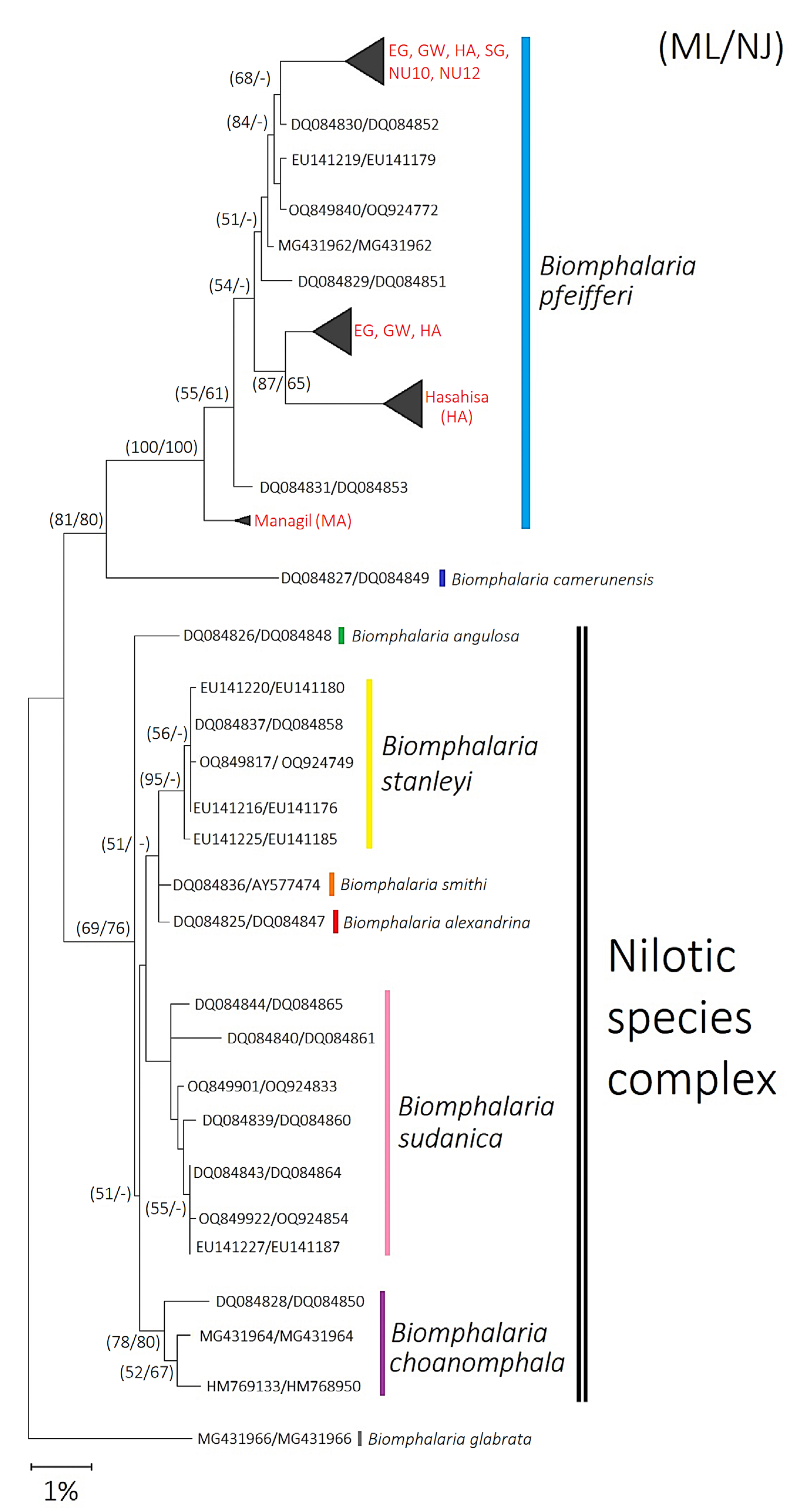
Figure 1. Maximum Likelihood and Neighbor-Joining consensus tree of African Biomphalaria species based on the concatenated COI (465 bp) and 16S rRNA (322 bp) gene fragments. Phylogenetic reconstruction was conducted under the GTR+Γ model and rooted with B. glabrata. Red labels indicate B. pfeifferi sequences generated in this study. Numbers on branches represent bootstrap support values (1000 replicates) from Maximum Likelihood and Neighbor-Joining (ML/NJ) analyses; values < 50% are not shown. The scale bar corresponds to 1% sequence divergence.
Ijms 26 09567 g001
Figure 2. TCS haplotype networks and haplotype distribution of Sudanese Biomphalaria pfeifferi based on partial COI (527 bp) and 16S (387 bp) gene fragments. (A) COI haplotype network and (B) 16S haplotype network generated using PopART. Circle sizes are proportional to haplotype frequency. Hatch marks and numbers on branches indicate mutational steps between haplotypes; branch lengths are not to scale. Each color represents a different geographic origin (Green = EG, Pink = GW, Dark green = HA, Orange = NU1, Blue = NU3, Purple = SG, Red = MA), and haplotypes identified in this study are written in red. (C) Haplotype distribution map showing the geographic composition of haplotypes for both markers (COI: top; 16S: bottom) across the seven Gezira State sites. Colors correspond to distinct haplotypes. Map created in ArcGIS v10.5 (Esri, CA, USA; https://www.arcgis.com/).
Ijms 26 09567 g002aIjms 26 09567 g002b
Figure 3. Types of cercariae shed by Biomphalaria pfeifferi across different localities in Gezira State: (A) Apharyngostrigea sp.; (B) Echinostome type; (C) Cotylomicrocercous type; (D,E) Schistosoma mansoni cercariae. All photos were taken under 10× magnification.
Ijms 26 09567 g003
Figure 4. Map of the six collection locations in Gezira State, Sudan, highlighting examples of the non-perennial canal sites: (A) Greater Wadmedani, Atraa canal; (B) Hasahisa, Wadelfadni canal; and (C) South Gezira, Barakat canal.
Ijms 26 09567 g004
Table 1. Haplotype diversity and neutrality parameters of Sudanese Biomphalaria pfeifferi snails using the COI (527 bp) and 16S (387 bp) gene fragments.
GeneSiteHSKHd ±SDHVπ (JC)± SDFu’ FsFu Li’ DFu Li’ FTajima’s DTheta Θ
COIOverall5415.3870.5 ± 0.0220.00050.01 ± 0.000713.62−5.01 **−3.91 **−0.8670.014
EG3312.800.191 ± 0.090.010.0055 ± 0.0035.57−2.84 *−3.17 *−2.36 **0.015
GW100--------
HA230.20.067 ± 0.060.0040.0003 ± 0.00030.135−2.68 *−2.79 *−1.7310.001
NU1100--------
NU3100--------
SG100--------
MA100--------
16SOverall10842.0560.11± 0.0350.00120.008 ± 0.003−0.043−2.57 ***−3.16 **−2.710 ***0.051
EG100--------
GW100--------
HA6729.400.31 ± 0.1090.0120.035 ± 0.0138.8250.031−0.69−1.84 *0.052
NU14181.320.25 ± 0.1020.010.004 ± 0.0031.38−4.24 **−4.3 ***−2.425 ***0.014
NU3210.060.067 ± 0.0610.0040.0002 ± 0.0002−1.21−1.68−1.76−1.1470.001
SG100--------
MA100--------
Note: Statistical significance: * p < 0.05; ** p < 0.02; *** p < 0.01. EG: East Gezira; GW: Greater Wadmedani; HA: Hasahisa; NU: North Umelgura; SG: South Gezira; MA: Managil; H: number of haplotypes; S: number of segregating (polymorphic/variable) sites; K: average number of pairwise nucleotide differences; Hd: haplotype diversity; HV: haplotype diversity variance; SD: standard deviation; π: nucleotide diversity; JC: Jukes–Cantor correction; Ɵ theta per site.
Table 2. Estimation of pairwise population fixation index (Fst) comparisons of genetic differentiation among Biomphalaria pfeifferi populations using the COI and 16S gene fragments.
(A) COI Gene Fragment
EGGWHANU1NU3SGMA
EG-
GW0.86-
HA0.850-
NU10.0210.99-
NU30.0210.990-
SG0.0210.9900-
MA0.910.99111-
Overall Fst = 0.94, Overall Nm = 0.03
(B) 16S Gene Fragment
EGGWHANU1NU3SGMA
EG-
GW0-
HA0.050.05-
NU10.010.010.04-
NU3000.050.01-
SG000.050.010-
MA000.050.0100-
Overall Fst = 0.04, Overall Nm = 5.72
Note: EG: East Gezira; GW: Greater Wadmedani; HA: Hasahisa; NU1: North Umelgura site 10; NU3: North Umelgura site 12; SG: South Gezira; MA: Managil; Nm: gene flow.
Table 3. Analysis of Molecular Variance (AMOVA) for Biomphalaria pfeifferi COI and 16S haplotypes.
Sum of VariationDegree of Freedom (df)Sigma Squared (σ2)%VariationFixation Index (ΦST) *
COI 0.9
(p > 0.001)
Among populations4203.261625.61684.6
Within populations724.8001564.64615.4
Total4928.06116230.262
16S 0.04
(p > 0.001)
Among populations556.69161.7273.6
Within populations7154.56715645.86396.4
Total7711.25816247.590
* (ΦST): Significance (1000 permutations).
Table 4. Infection prevalence of Biomphalaria pfeifferi snails determined by the cercarial shedding method and PCR detection using SmF/R primers.
Site (ID)Number of B. pfeifferi Tested
CollectedCercaria SheddingPCR (SmF/R)Type of Cercariae
SG (S3)1000None
GW (S6)3800None
NU1 (S10)3100None
NU3 (S12)3016.6% (5/30)0Echinostome (n:5) and Cotylomicrocercous spp. (n:4)
EG (S13)661.5% (1/66)0Apharyngostrigea sp.
HA (S19)395.1% (2/39)40% (12/30)S. mansoni
MA (S23)300None
Overall infection rate3.6% (8/219)7.4% (12/163) *
Note: * No. of extracted snails was 30 samples per area for East Gezira, North Umelgura, and Hasahisa; 10 for South Gezira; and 3 for Managil localities (total 163). S. mansoni: Schistosoma mansoni.
Table 5. Summary of non-perennial canal collection sites in Gezira State, Sudan, where Biomphalaria pfeifferi snails were found, including the number of DNA-extracted specimens and GPS coordinates.
Locality/Administrative UnitSite IDNo. DNA Extracted (n = 163)LatitudeLongitude
South Gezira/BarakatS3: Barakat(SG)1014.35733.526
Greater WadmedaniS6: Atraa(GW)3014.44533.487
North Umelgura/ElhedibaS10: Elhediba(NU1)3014.48433.658
S12: Elhediba(NU3)3014.48433.657
East Gezira/ElgineidS13: Elgineid(EG)3014.86633.277
Hasahisa/WadelfadniS19: Wadelfadni(HA)3014.67033.343
Managil/EboudS23: Eboud/Alnegeer village(MA)314.23033.173
Table 6. Primer set sequences used for the amplification of COI and 16S rRNA mitochondrial DNA for Biomphalaria pfeifferi molecular identification and the SmF/R primer set used for Schistosoma mansoni 28S rRNA amplification.
Primer NameSequence (5′→3′)Primer Length (bp)Annealing Temperature (°C)
LCO 1490GGT CAA CAA ATC ATA AAG ATA TTG G2545
HCO 2198TAA ACT TCA GGG TGA CCA AAA AAT CA26
16SarCTT CTC GAC TGT TTA TCA AAA ACA2448
16SbrGCC GGT CTG AAC TCA GAT CAT21
SmFGAG ATC AAG TGT GAC AGT TTT GC2361
SmRACA GTG CGC GCG TCG TAA GC20
Disclaimer/Publisher’s Note: The statements, opinions and data contained in all publications are solely those of the individual author(s) and contributor(s) and not of MDPI and/or the editor(s). MDPI and/or the editor(s) disclaim responsibility for any injury to people or property resulting from any ideas, methods, instructions or products referred to in the content.