Article

Chromosomal and Plasmid-Based CRISPRi Platforms for Conditional Gene Silencing in Lactococcus lactis

1
Key Laboratory of Combinatorial Biosynthesis and Drug Discovery (Ministry of Education), School of Pharmaceutical Sciences, Wuhan University, Wuhan 430071, China
2
Department of Molecular Genetics, Groningen Biomolecular Sciences and Biotechnology Institute, University of Groningen, 9747AG Groningen, The Netherlands
*
Authors to whom correspondence should be addressed.
The authors contributed equally to the work.
Int. J. Mol. Sci. 2025, 26(19), 9516; https://doi.org/10.3390/ijms26199516
Submission received: 17 August 2025 / Revised: 20 September 2025 / Accepted: 25 September 2025 / Published: 29 September 2025

Abstract

Inducible CRISPR interference (CRISPRi) systems were established in Lactococcus lactis using both plasmid and chromosomal approaches. Expression of nuclease-deficient Cas9 (dCas9) from Streptococcus pyogenes was placed under the control of the nisin-inducible promoter PnisA, while sgRNAs were transcribed from the constitutive Pusp45 promoter. To monitor expression, dCas9 was fused with superfolder GFP. Plasmid-based constructs successfully repressed a luciferase reporter gene and silenced the gene of the major autolysin, AcmA, leading to the expected morphological phenotype. However, plasmid systems showed leaky expression, producing mutant phenotypes even without induction. Chromosomal integration of dCas9 reduced its expression level by approximately 20-fold compared with plasmid-based expression, thereby preventing leaky activity and ensuring tight regulation. This chromosome-based (cbCRISPRi) platform enabled controlled repression of the essential gene ybeY, which resulted in severe growth defects. Restoration of wild-type phenotypes was achieved by introducing a synonymous codon substitution in the sgRNA target region. Transcriptome analysis of ybeY-silenced cells revealed downregulation of ribosomal protein genes and widespread effects on membrane-associated proteins, ATP synthase subunits, and various transporters. These inducible CRISPRi platforms provide robust and tunable tools for functional genomics in L. lactis, particularly for studying essential genes that cannot be deleted.

Graphical Abstract

1. Introduction

Lactococcus lactis is one of the most extensively studied lactic acid bacteria (LAB) and plays a central role in the dairy industry as a starter culture [1]. Beyond its traditional application, this species has been widely engineered as a microbial cell factory for producing metabolites such as mannitol and lactic acid [2], as a host for recombinant protein production [3], and as a delivery vehicle for vaccines and therapeutic proteins [4]. These applications have been facilitated by the development of a versatile set of genetic engineering tools. Several inducible systems have been explored in L. lactis, including zinc-inducible, low-pH-inducible, agmatine-inducible, and nisin-inducible promoters [5]. Among the available systems, the nisin-controlled expression (NICE) platform is the best-characterized and most commonly used strategy for inducible gene expression in L. lactis. [6]. NICE relies on the two-component regulatory system NisK–NisR, which responds to the lantibiotic peptide nisin, a natural product of some L. lactis strains [7].
For genome modification, methods such as double-crossover recombination using non-replicative vectors (e.g., pORI [8] or pCS1966 [9]) are available, but generating stable mutants typically requires several weeks, highlighting the need for faster and more flexible alternatives. The CRISPR-Cas system (clustered regularly interspaced short palindromic repeats and CRISPR-associated proteins) constitutes an RNA-guided adaptive immune defense in bacteria and archaea, where it protects cells from invading genetic elements such as phages and plasmids [10]. In the widely used Streptococcus pyogenes system, Cas9 functions as a DNA endonuclease directed by an RNA duplex that base-pairs with a complementary DNA sequence, generating a double-strand break (DSB) precisely at the target locus [11]. This duplex consists of a CRISPR RNA (crRNA), whose maturation requires RNase III cleavage, and a trans-activating RNA (tracrRNA). To simplify the system, a chimeric single-guide RNA (sgRNA) was designed to combine the roles of crRNA and tracrRNA, thereby avoiding the need for RNase III processing [12].
For gene repression, a catalytically inactive variant of Cas9 (dCas9) can be employed in CRISPR interference (CRISPRi). Guided by an sgRNA, dCas9 binds to a selected genomic locus—commonly near a promoter or within a coding sequence—where it obstructs RNA polymerase binding or transcript elongation, thereby preventing transcription without inducing DNA cleavage [13]. CRISPR-Cas9 technologies have been established in a variety of bacterial genera, including Bacillus, Escherichia, Clostridium, and Streptococcus [14,15,16]. In LAB, a CRISPR/Cas9-mediated ssDNA recombineering platform has been applied to Lactobacillus reuteri and L. lactis, enabling gene deletions with efficiencies ranging from 8% to 50% [17,18]. Furthermore, a nisin-inducible, single-plasmid CRISPRi system has been reported for L. lactis, achieving more than a 50-fold reduction in upp transcription in the presence of the inducer [19].
Because CRISPRi relies on controlled expression of dCas9, precise regulation at the protein level is particularly critical. Leaky expression can obscure phenotypes or cause artificial toxicity, whereas insufficient induction may fail to uncover essential gene functions. This is especially relevant when studying essential genes or genes linked to toxic phenotypes, where even minor changes in basal activity can have significant consequences.
In this study, both plasmid-based and chromosomally integrated CRISPRi systems were constructed for L. lactis. The optimized chromosome-based platform was further employed to probe the function of the essential gene ybeY. Transcriptome analysis revealed that silencing ybeY strongly affected the expression of ribosomal proteins as well as numerous small RNAs, suggesting roles in ribosome homeostasis and RNA regulation.

2. Results

2.1. Establishment of an Inducible CRISPRi System with Fluorescently Tagged dCas9 in L. lactis

To monitor expression levels, a fluorescent variant of dCas9 was generated by fusing its coding sequence at the 3′ end to the superfolder GFP (sfGFP) gene, connected through the flexible linker AGSGGEAEA [20,21]. For inducible expression, the dcas9 or dcas9-sfgfp gene was placed under the control of the nisin-responsive promoter PnisA in a pNZ8048-derived plasmid [7], while the sgRNA cassette was constitutively expressed from the Pusp45 promoter on a pTLR-based backbone (Figure 1A,B) [20,22]. sgRNA design followed previously established criteria to ensure efficient silencing: targeting of the non-template DNA strand, a minimum 12 bp base-pairing region, and complete sequence complementarity within the 7 nucleotides adjacent to the PAM site (5′-NGG-3′). To minimize off-target interactions, the 12 bp region immediately upstream of the PAM was selected [13]. The inducible host strain L. lactis NZ9000 was used [23]. To verify inducibility, fluorescence intensity was measured in NZ9000 harboring pNZ-PnisA-dcas9-sfgfp. Increasing concentrations of nisin (0–20 ng/mL) led to a dose-dependent rise in fluorescence, confirming controlled expression of dCas9-sfGFP (Figure 1C). Next, a luciferase reporter strain (NZ9000 pseudo10::Pusp45-luc) was constructed to evaluate CRISPRi function. The chromosomal locus pseudo10 is a commonly used silent region that allows stable chromosomal integration without affecting L. lactis growth. Cells carrying pNZ-PnisA-dcas9-sfgfp and pTLR-Pusp45-sgRNAluc were analyzed for luciferase activity. Upon induction with 5 ng/mL nisin, luciferase activity decreased markedly, demonstrating that the dCas9-sfGFP fusion retained silencing activity (Figure 1D). At the end of exponential growth, a 74% reduction in luciferase expression was observed without detectable effects on bacterial growth.

2.2. Leaky dCas9 Expression Causes Basal Gene Repression

To further evaluate the functionality of the CRISPRi system, its effects on cell growth and morphology were examined. The gene acmA, which encodes the major autolysin responsible for cell separation and autolysis in L. lactis NZ9000 was selected as a target. Deletion of acmA results in the formation of extremely long chains of cells [24].
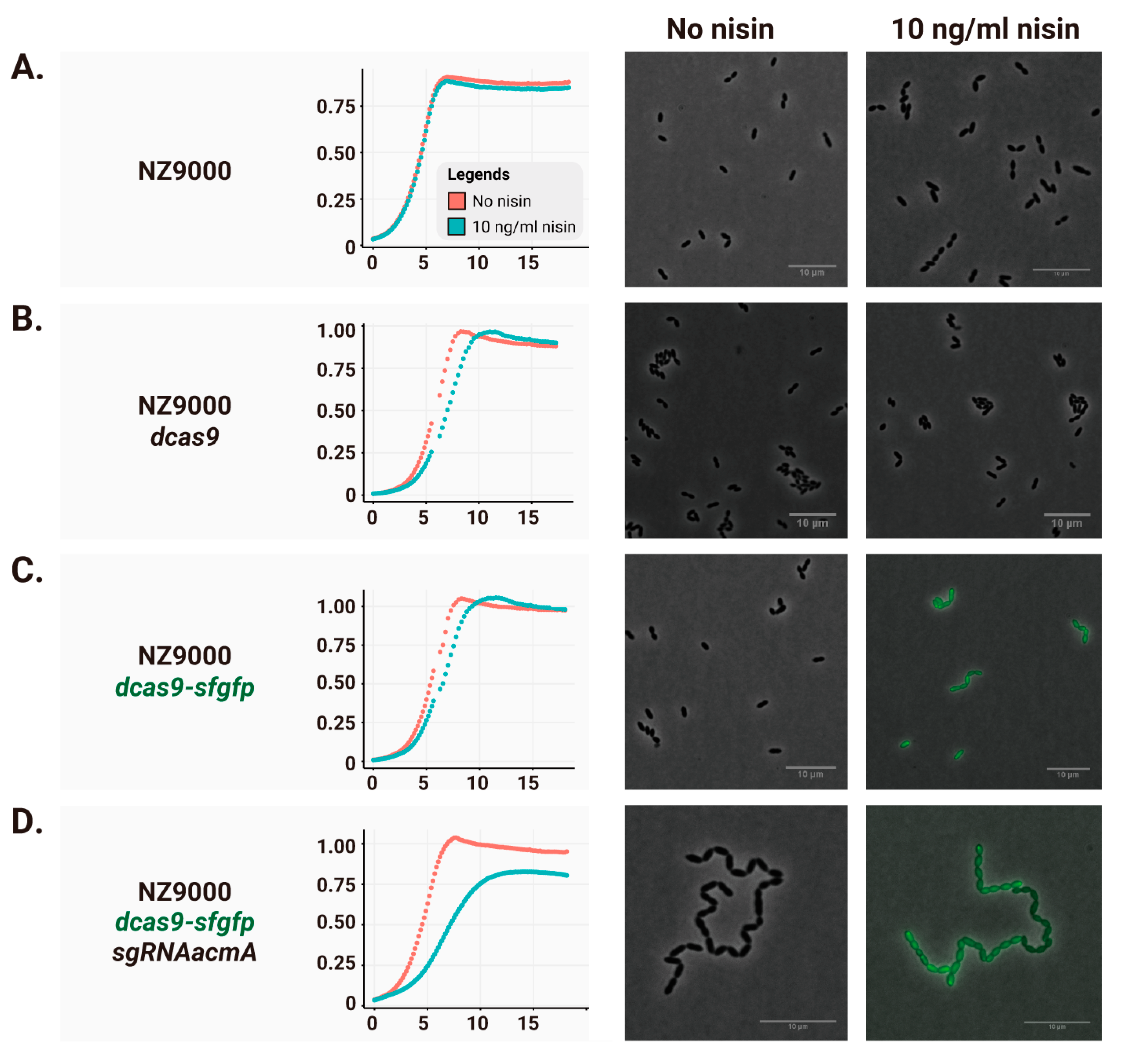
Adding nisin to strains expressing only dCas9 or dCas9-sfGFP had minimal impact on growth (Figure 2B,C, growth curves), whereas co-expression of the corresponding sgRNA targeting acmA caused a noticeable reduction in growth rate and final OD (Figure 2D). Microscopy analysis revealed that, as expected, long chains of cells were formed in the acmA knockdown strain (right micrographs in Figure 2A–D).
No detectable fluorescence from dCas9-sfGFP was observed in uninduced cells (left micrograph in Figure 2C). Nevertheless, long chains were present even without nisin induction (Figure 2D, left micrograph). These findings indicate that basal dCas9 expression resulting from PnisA leakiness, while not affecting growth rate or final OD, is sufficient to block transcription and elicit the corresponding mutant phenotype.

2.3. Reducing Leaky Expression Through Chromosomal dCas9-sfGFP Insertion

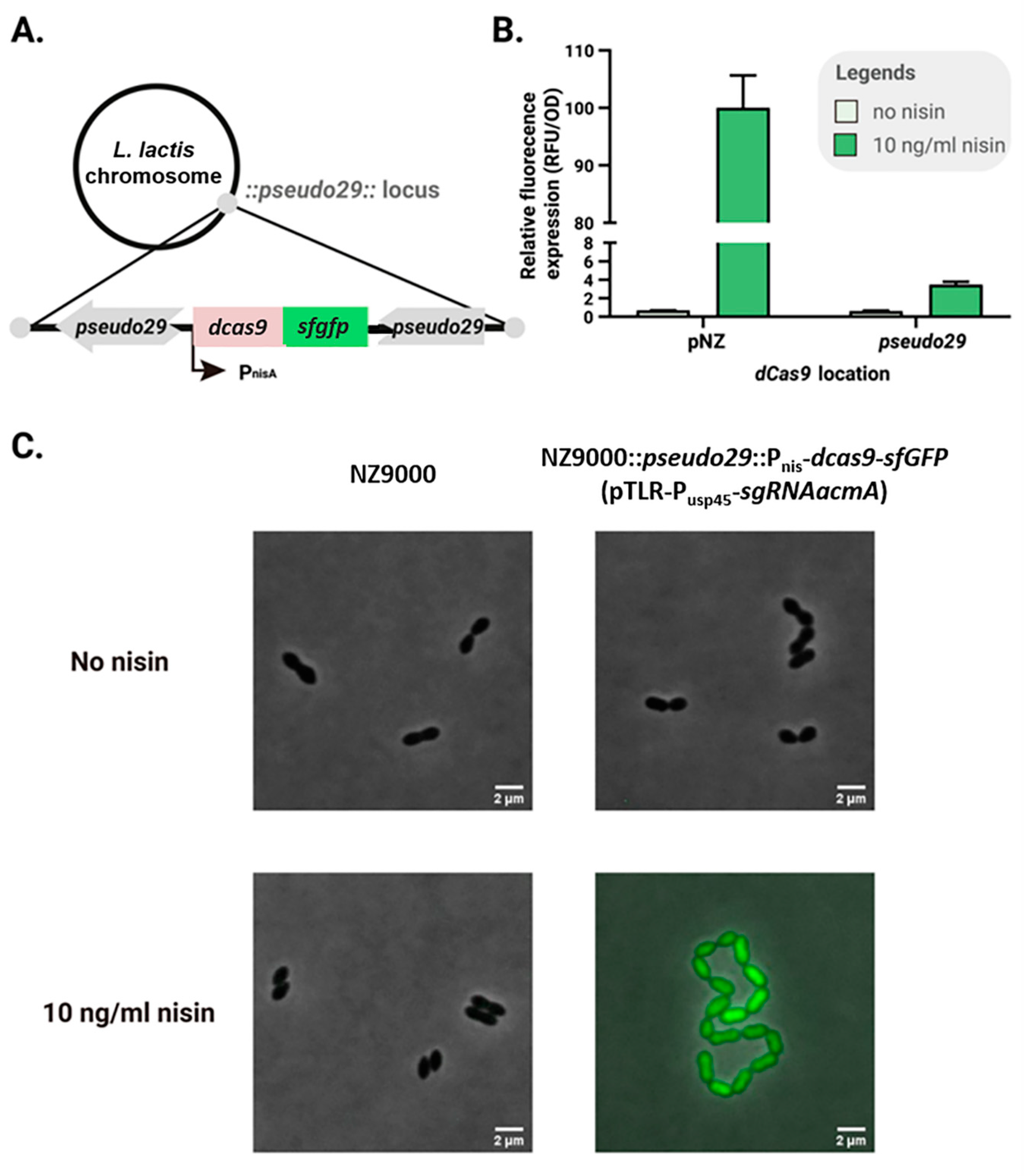
To minimize background activity of uninduced dCas9-sfGFP, the copy number of the dcas9-sfgfp cassette was reduced by chromosomal integration. The PnisA-dcas9-sfgfp construct was inserted into the transcriptionally silent pseudo29 locus of L. lactis NZ9000 [25], while the sgRNA continued to be expressed constitutively from Pusp45 on the plasmid pTLR. Expression levels of dCas9-sfGFP were compared between plasmid- and chromosome-based systems. Nisin (10 ng/mL) was added to cultures, and relative fluorescence was measured after 3 h of induction. As shown in Figure 3B, dCas9-sfGFP expression from the chromosome was approximately 20-fold lower than from the plasmid.
When targeting acmA with sgRNA in cells carrying the chromosomal dcas9-sfgfp cassette, strain NZ9000 pseudo29::PnisA-dcas9-sfgfp (pTLR-Pusp45-sgRNA_acmA) displayed normal morphology in the absence of nisin. In contrast, the plasmid-based system showed observable phenotypic changes under the same conditions (compare Figure 3C with Figure 2D). These results demonstrate that chromosomal insertion of dCas9-sfGFP significantly reduces basal expression, allowing a tighter, more controllable chromosome-basedCRISPRi system, cbCRISPRi.

2.4. Application of Chromosomal-Based CRISPRi to Study ybeY Function

YbeY is a highly conserved protein found across nearly all sequenced bacterial species. It is typically involved in ribosome maturation and quality control as an endoribonuclease, and implicated in biofilm formation, stress responses, and virulence [26,27,28,29,30]. In Sinorhizobium meliloti, YbeY is required for sRNA-mediated silencing of amino acid ABC transporters [28], and in Pseudomonas aeruginosa, it regulates RpoS via the sRNA ReaL [27]. Its role in L. lactis remains unexplored, though it may function as a candidate sRNA chaperone in a species lacking Hfq, CsrA, or ProQ homologs [31,32,33,34,35].
Although ybeY is essential in several bacteria [36,37,38], it can be deleted in P. aeruginosa, S. meliloti, and E. coli, with pleiotropic effects on growth, toxin production, or cell aggregation [39,40]. Attempts to delete ybeY in L. lactis using the temperature-sensitive pGhost system or the pCS1966 oroP-based selection/counterselection system failed, even after multiple attempts (Figure S1) [9,41]. Standard methods relying on double-crossover homologous recombination failed to delete ybeY from the chromosome, despite multiple alternative attempts (Figure S1).
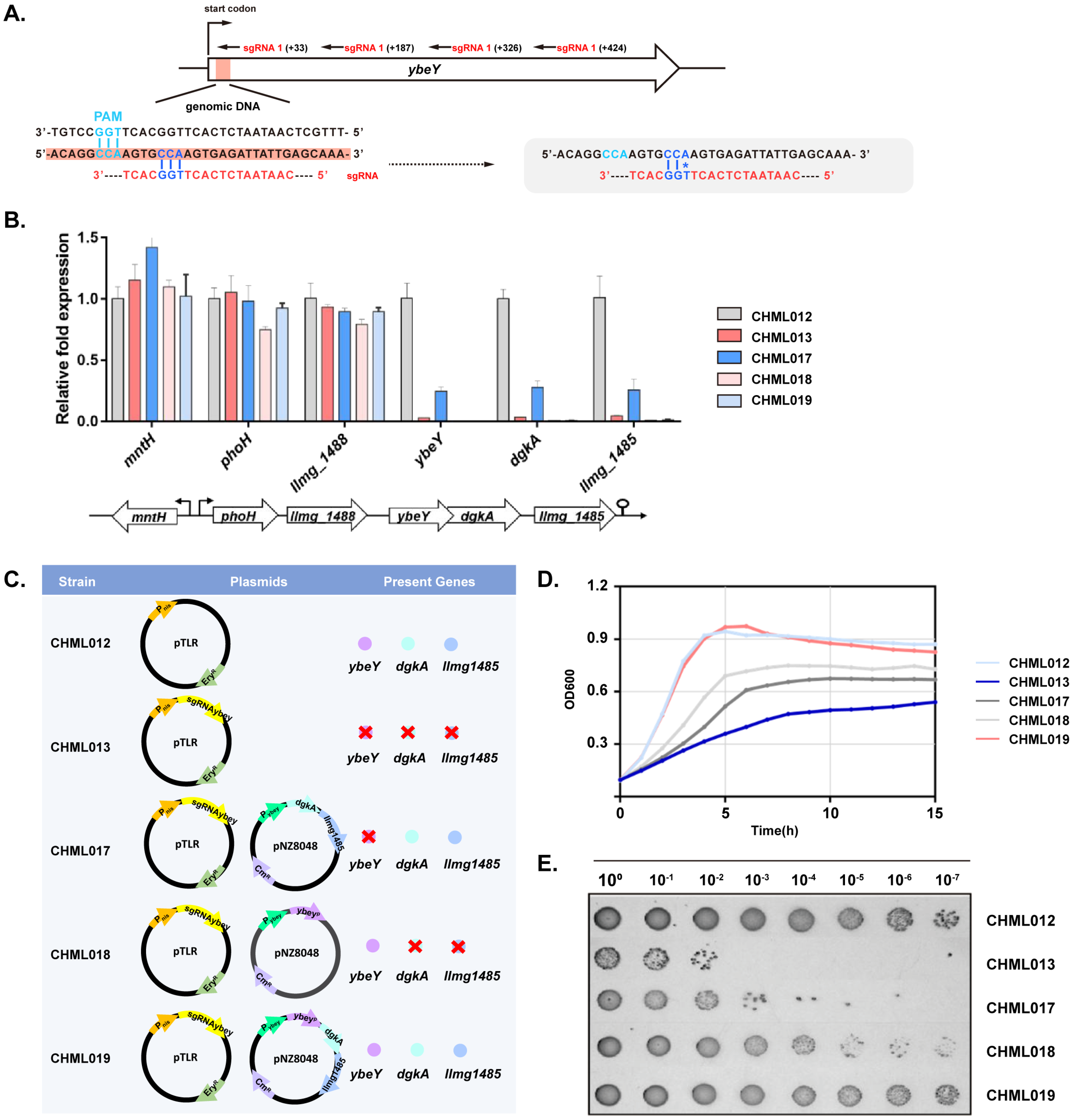
Four sgRNAs targeting ybeY were designed for cbCRISPRi, and qPCR confirmed that all four (strains CHML013–CHML016) efficiently silenced transcription (Figure 4A,B). Three sgRNAs decreased ybeY expression by roughly 200-fold, while sgRNA2 exhibited weaker silencing. This lower efficiency is likely due to its 12-base region adjacent to the PAM having a GC content of about 33%, compared to 42–58% for the other sgRNAs. Silencing ybeY also reduced expression of downstream genes dgkA-llmg1485 but did not affect upstream genes. Knockdown of the ybeY-dgkA-llmg1485 operon caused severe growth defects (Figure 4D,E), while overexpression of ybeY in NZ9000 had no growth impact (Figure S2). To dissect the roles of the three genes in the observed growth defects, we constructed plasmid-based complementation strains. The first expressed YbeYp with a synonymous mutation at the sgRNA target site to prevent silencing (Figure 4A). The second complemented dgkA and llmg_1485 under their native operon promoter, and the third co-expressed the mutated YbeYp together with dgkA and llmg_1485. As shown in Figure 4D,E, restoring only ybeY or dgkA–llmg_1485 partially alleviated the growth defect, while co-expression of all three genes fully rescued it. These findings indicate that loss of YbeY has the strongest impact, but reduced expression of dgkA and/or llmg_1485 also compromises growth.
Plasmid-based complementation showed partial recovery when either ybeY or the two downstream genes were complemented, and complete recovery when all three were expressed (Figure 4D,E). To prevent overexpression, dgkA-llmg1485 driven by their native promoter were integrated into the pseudo10 locus (strain CHML20). This restored the growth defects while ybeY remained silenced (Figure 5B). No differences in the recovery of growth defects were observed between CHML020 and the plasmid-based dgkA–llmg_1485 complementation strain CHML017. qPCR confirmed transcriptional restoration (Figure 5C). Light microscopy with live/dead staining indicated no viability difference in exponential phase, but stationary-phase cell death ranged from 15 to 54%, higher than 0.5–3% in the parent strain CHML012 (Figure 5D). These results confirm ybeY essentiality and the importance of downstream operon genes for normal growth of L. lactis.

2.5. Silencing of ybeY Alters Ribosomal and Other Gene Expression

Deletion of ybeY in several bacterial species results in the appearance of an additional band corresponding to 17S rRNA on agarose gels, representing the 16S rRNA precursor [29]. In L. lactis, however, knockdown of ybeY did not produce such a band (Figure S3). qPCR analysis also revealed no significant differences in the 5′ ends, middle regions, or 3′ ends of immature 16S rRNA and 23S rRNA (Figure S4). These findings suggest that L. lactis YbeY may not be primarily involved in 16S rRNA maturation, although a very low level of residual YbeY expression in the knockdown strain could suffice to complete this process.
To further investigate L. lactis YbeY function, RNA-seq was performed comparing the transcriptomes of the ybeY-silenced strain and the control strain lacking the sgRNA-expressing plasmid, L. lactis NZ9000-dCas9. All strains were grown at 30 °C in GM17 medium to early log phase (OD600 = 0.4), followed by induction with 10 ng/mL nisin for 1 h to produce sgRNA and/or dCas9. Total RNA was isolated, rRNA-depleted, sequenced, and analyzed. This induction ensured sufficient dCas9 for function without causing severe side effects. RNA-seq revealed that ybeY silencing broadly affected the transcriptome, with most highly differentially expressed genes (DEGs) being downregulated. Functional and gene set enrichment analyses using FUNAGE-Pro [42] annotated DEGs to Gene Ontology (GO) terms (Figure 6). Many downregulated genes encode proteins of the 50S and 30S ribosomal subunits. Although no accumulation of immature 16S and 23S rRNA ends was detected, as was the case in E. coli and P. aeruginosa [27,29], ribosomal protein genes were largely repressed, indicating a role of YbeY in ribosome processing in L. lactis. Additionally, genes encoding membrane proteins, ATP synthase subunits, sugar/amino acid permeases, other transporters, and components of the fab and acc operons were downregulated. Some changes may reflect secondary effects after the 1 h induction.
The cbCRISPRi system was also applied to the 66-nt noncoding sRNA ArgX. Deletion of ArgX elevates arc and arg operon expression [43], and qPCR confirmed effective knockdown by CRISPRi with expected transcriptomic effects (Figure S5). Due to PAM constraints, only 42 of 184 sRNAs in L. lactis are potentially targetable (Table S2). Since YbeY has been reported to degrade an sRNA involved in oxidative stress in P. aeruginosa [27], sRNA expression in the ybeY-silenced strain was examined. Ten of 186 trans-encoded sRNAs and 2 of 60 cis-encoded antisense RNAs identified in L. lactis MG1363 [44] were differentially expressed. Whether YbeY directly or indirectly affects these sRNAs requires further study.

3. Discussion

The CRISPRi system provides a precise method for gene silencing in both microbial and higher organisms [45,46,47]. In bacteria, a functional CRISPRi platform requires two essential components: (i) an sgRNA that binds to the genome, with specificity determined by approximately 20 bp of base pairing with the target DNA and the protospacer adjacent motif (PAM), which in S. pyogenes is NGG; (ii) a dCas9 protein that forms a dCas9-sgRNA complex guided to its target, where it blocks transcription initiation or elongation depending on the sgRNA binding site. Each target gene requires an adapted 20 bp sgRNA sequence. Distributing the dcas9 and sgRNA genes on separate small plasmids is technically simpler than combining them on a single plasmid, which could exceed 10 kb, as the PnisA-dcas9 cassette alone spans 3–5 kb, with or without sfgfp. Frequent replacement of the 20 bp sgRNA sequence on a single plasmid also risks backbone mutations, whereas separating the two cassettes reduces the likelihood of dCas9 mutations. Efficient cloning methods, such as quick-fusion cloning or golden-gate assembly, are preferred for rapidly changing sgRNA sequences; both strategies use a linearized sgRNA backbone, anneal commercial primers, and employ the product directly for subsequent cloning (Figure 1) [46,48].
These CRISPRi platforms are powerful tools for functional gene analysis in L. lactis and likely other LAB species. The cbCRISPRi system, in particular, provides a valuable addition to the genetic toolkit, enabling the study of essential genes, such as ybeY, which cannot be deleted in L. lactis. This platform allowed, for the first time, the investigation of ybeY function, despite challenges: incomplete depletion of YbeY and the substantial stress caused by its depletion, resulting in widespread transcriptomic effects. The absolute fluorescence values under uninduced conditions appear to be similar between the plasmid- and chromosome-based CRISPRi systems (Figure 3B). This is likely due to the detection limit of the plate reader, which may mask subtle differences at low fluorescence levels. Importantly, phenotypic assays (e.g., acmA silencing in Figure 2D) provide clear evidence that basal activity is reduced in the chromosomal system, confirming its functional advantage over the plasmid-based approach.
In this study, cbCRISPRi silencing of ybeY demonstrated that blocking transcript elongation with a single sgRNA can simultaneously suppress multiple genes within an operon. Complementation analysis is critical to confirm the contributions of downregulated genes. A synonymous codon mutation within the sgRNA target site can bypass CRISPRi; for instance, a single proline codon change fully counteracted sgRNA-mediated silencing of ybeY, whereas a single valine codon change partially restored growth (~70% of final OD). Generally, mutating two codons within the 12 bp core region adjacent to the PAM is recommended [13].
YbeY silencing in L. lactis did not reveal a clear role in 16S rRNA maturation, though residual protein expression from transcripts escaping silencing could suffice for this function. RNA-seq analysis suggests that YbeY may participate in rRNA maturation and/or ribosome quality control, consistent with homologous roles in other species. E. coli YbeY functions as an RNase that ensures 70S ribosome quality, working with RNase R to degrade rRNAs from defective 50S or 30S subunits [39]. In E. coli ΔybeY, defective 70S ribosomes slightly accumulate, with a notable rise in free defective subunits [29]. Impaired translation due to these defective ribosomes results in growth defects and stress responses.
YbeY has also been implicated in sRNA regulation in several species [26,28,30,40]. In E. coli, 28 of 54 detectable sRNAs show YbeY-dependent expression changes [40]. Prior to this study, L. lactis YbeY had not been linked to sRNA regulation. Here, YbeY appears to influence certain sRNAs, with three upregulated candidates in the ybeY knockdown strain—LLMGnc_060, LLMGnc_129, and LLMGnc_172 (ArgX)—potentially being direct targets. Downregulation of other sRNAs suggests indirect effects or involvement of additional regulatory factors.

4. Materials and Methods

4.1. Bacterial Strains, Media, and Culture Conditions

The bacterial strains used in this study are listed in Table 1. L. lactis NZ9000 and its derivatives were grown at 30 °C in Difco M17 medium (BD, Franklin Lakes, NJ, USA) containing 0.5% (wt/vol) glucose (GM17). For fluorescence and luminescence analyses, L. lactis strains were grown in a chemically defined medium supplemented with 1% (wt/vol) glucose (CDM) [16]. When needed, erythromycin or chloramphenicol was added at a final concentration of 5 μg/mL. Chemically defined SA medium supplemented with 0.5% (wt/vol) glucose and 20 μg/mL 5-fluoroorotic acid (5-FOA; Sigma-Aldrich, St. Louis, MO, USA) as the sole pyrimidine source was used for the selection of chromosomal insertions [9]. E. coli DH5α was used as a cloning host; it was grown aerobically at 37 °C in LB media (Formedium, Norfolk, UK) supplemented with, when needed, erythromycin at a final concentration of 200 μg/mL. All chemicals were obtained from Sigma-Aldrich.

4.2. Recombinant DNA Techniques and Oligonucleotides

Standard molecular cloning was performed essentially as described previously [50]. Chromosomal DNA from L. lactis was isolated using the GenElute Genomic DNA Kit (Sigma-Aldrich). Plasmids and PCR products were isolated and purified using a High Pure plasmid isolation and PCR purification kit (Roche Applied Science, Mannheim, Germany) and a NucleoSpin gel and PCR cleanup kit (Macherey-Nagel, Düren, Germany) according to the manufacturers’ instructions. PCR was performed with Phusion or DreamTaq polymerase (both from Fermentas, St. Leon Roth, Germany) according to the manufacturer’s protocol. The obtained PCR products were mixed and treated with a mixture of Quick-Fusion enzymes (BIO-Connect Services BV, Huissen, The Netherlands), yielding 15-nucleotide overhangs that were annealed to complementary overhangs. No ligation was needed; Quick-Fusion-treated mixtures were directly used to transform E. coli. The oligonucleotides used in this study are listed in Supplementary Table S1 and were purchased from Biolegio BV (Nijmegen, The Netherlands). Competent E. coli cells were transformed using heat shock [51], while electrocompetent L. lactis cells were transformed using electroporation [52] with a Bio-Rad gene pulser (Bio-Rad Laboratories, Richmond, CA, USA). All nucleotide sequencing was performed at Macrogen Europe (Amsterdam, The Netherlands).

4.3. Construction of Plasmid-Based CRISPRi Systems

Pertinent regions of all the plasmids constructed in this study were sequenced to confirm their proper nucleotide sequences. Streptococcus pyogenes dcas9 (dcas9) was obtained from plasmid pJWV102 [46] by PCR using the primers 0032-USER_Pnis_dCas9_F and 0220-dCas9-USER_R, while linearized pNZ8048 vector was obtained using the primers 0217-pNZ8048_USER_F/0221-Pnis_pNZ8048_R. The fragments were fused employing the USER cloning kit (New England Biolabs, Ipswich, MA, USA) according to the manufacturer’s instructions, with the exception of using only one half of the recommended volume per reaction. The reaction mixture was directly used to transform competent L. lactis NZ9000. The resulting vector was labeled pNZ-PnisA-dcas9. The plasmid pNZ-PnisA-dcas9-sfgfp, which expresses a Cas9 variant that couples C-terminally with sfGFP via the flexible linker sequence AGSGGEAEA, was generated as follows: dcas9 was amplified from pJWV102 using the primers 0032-USER_Pnis_dCas9_F/0218-dCas9_USER_R. The sfgfp gene with the polylinker-encoding sequence at its 5′ end was amplified from the plasmid pLG-MG1 [21] using the primers 0215-sfGFP_USER_F/0219-linker_sfGFP_USER_R. The fragments were fused through USER cloning, and the mixture was subsequently used to transform competent L. lactis NZ9000 to obtain the proper construct.
The DNA fragment encoding the single guide RNA targeting the luciferase gene (sgRNAluc) was amplified from the plasmid pPEPX-P3-sgRNAluc [46] by the primers 0149-sgRNA_F/0150-sgRNA_R. The constitutive L. lactis promoter Pusp45 was amplified from the plasmid pSEUDO::Pusp45-sfgfp(Bs) [20] with the primers 0147-Pusp45_F/0148-Pusp45_R, and the linearized pGHost vector was obtained after amplification using the primers 0145-pGHost_F/0146-pGHost_R. The three fragments were then fused with the USER cloning kit, and the reaction mixture was subsequently used to transform competent E. coli DH5α, resulting in the plasmid pGHost-Pusp45-sgRNAluc. The Pusp45-sgRNAluc cassette, obtained by double digestion of the latter plasmid with the restriction enzymes NcoI and XhoI, was ligated into pTLR [22] treated with the same enzymes. The ligation mixture was used to transform competent E. coli DH5α, yielding the plasmid pTLR-Pusp45-sgRNAluc. The sgRNAluc sequence is transcribed directly after +1 of the Pusp45 promoter and contains 20 nucleotides (nt) as the base-pairing region, which targets the luciferase gene and is followed by an optimized sgRNA [53]. The plasmid pTLR-Pusp45-sgRNAluc was used as the template for the generation of other sgRNA expression plasmids by quick-fusion cloning (see the next section).

4.4. Quick-Fusion Cloning of sgRNA Genes

The 20-nt guide sequences of sgRNAs targeting different genes were selected with the CRISPR Primer Designer [54]. Briefly, a search was performed within the coding sequence of each gene for a 14-nt specificity region consisting of the 12-nt “seed” region of the sgRNA and GG of the 3-nt PAM (GGN). sgRNAs with more than one binding site within the L. lactis genome, as determined by BLAST (v 2.12.0) searches, were discarded. The guide sequence was chosen to be as close as possible to the 5′ end of the coding sequence of the targeted gene [13]. Cloning of sgRNA sequences was performed by quick-fusion cloning (Figure S6). The primer pair 0153-sgRNA_backbone_FW/0154-sgRNA_backbone_RV was designed for linear amplification of the plasmid pTLR-Pusp45-sgRNAluc. The primers bind directly upstream and downstream of the 20 bp guide sequence to enable easy sgRNA sequence swapping. To fuse the new 20-nt guide sequence into the linearized vector, two 50-nt complementary primers were designed for each target gene. Each primer contained 15 nt at one end, overlapping with the sequence on the 5′ end of the linearized vector, followed by the 20-nt gene-specific guide sequence and then 15 nt overlapping with the sequence on the 3′ end of the linearized vector. The two 50-nt complementary primers were annealed in TEN buffer (10 mM Tris, 1 mM EDTA, 100 mM NaCl, pH 8) by heating at 95 °C for 5 min and slowly cooling to room temperature. The annealed product was fused with the linearized vector using the Quick-Fusion Cloning Kit (BIO-Connect Services BV) according to the manufacturer’s instructions, except for using only one half of the recommended volume per reaction. The reaction mixture was directly used to transform competent E. coli DH5α, and the designated plasmid was subsequently introduced into the strains CHML009 (NZ9000 pseudo29::PnisA-dcas9) or CHML010 (NZ9000 pseudo29::PnisA-dcas9-sfgfp).

4.5. Construction of the Luc Reporter Strain

The luc gene was amplified from the plasmid pPEP23 [55] using the primers 0189_luc_F/0190-luc_R, while the vector pSEUDO::Pusp45-sfgfp(Bs) [20] was linearized by amplification using the primers 0187-pseudo10_R/0188-pseudo10_F. The two fragments were fused by employing the Quick-Fusion Cloning Kit protocol. The reaction mixture was directly used to transform competent E. coli DH5α to pick up plasmid pSEUDO10-Pusp45-luc. This plasmid, which cannot replicate in L. lactis, was introduced into L. lactis NZ9000 via electroporation. Cells in which the plasmid had integrated into the genomic locus pseudo10 were obtained by selection on erythromycin. The proper integrant strain, NZ9000::pseudo10::Pusp45-luc, was subsequently selected by growing on selective SA media plates containing 20 μg/mL 5-FOA [9].

4.6. Construction of a Genome-Based CRISPRi System

The flanking regions of pseudo29 were amplified using 0247-pseudo29_UP_F/0248-pseudo29_UP_R and 0249-pseudo29_DOWN_F/0250-pseudo29_DOWN_R, while the linearized vector pCS1966 was obtained by PCR amplification using the primer pair pCS1966_1FW/pCS1966_1RV. The fragments were fused by Quick-Fusion Cloning as described above, after which the reaction mixture was used to transform competent E. coli DH5α. The resulting vector was designated pSEUDO29. The PnisA-dcas9-sfgfp cassette was amplified using the primers 03-dcas9-pseudo29-F/04-dcas9-pseudo29-R, while linearized pSEUDO29 was obtained by amplification employing the primers 01-pseudo29_F/02-pseudo29-R. The fragments were fused with a Quick-Fusion Cloning Kit. A transformant carrying pSEUDO29-PnisA-dcas9-sfgfp was selected in E. coli DH5α. This plasmid, which does not replicate in L. lactis, was introduced into L. lactis NZ9000 via electroporation; cells in which a two-step homologous recombination event occurred were selected on selective SA medium plates supplemented with 20 μg/mL 5-FOA [9]. The obtained strain was labeled NZ9000::pseudo29::PnisA-dcas9-sfgfp.

4.7. Optical Density, Fluorescence, and Luminescence Measurements

L. lactis was grown overnight in CDM at 30 °C and then diluted to an optical density (OD) at 600 nm (OD600) of 0.1 in fresh CDM. The mixture was incubated until the mid-log phase (OD600 = 0.5) was reached, after which the cells were diluted again to a starting OD600 of 0.05 in fresh CDM supplemented with different concentrations of nisin. Then, each culture was dispensed to 200 μL, in triplicates, in wells of a 96-well microtiter plate (Polystyrol, transparent, flat, and clear bottom; Corning, New York, NY, USA). The OD600 and fluorescence signal (GFP: excitation 485 nm/emission 535 nm) were measured every 10 min at 30 °C in an Infinite 200 Pro plate reader with I-control 1.10.4.0 software (Tecan Group Ltd., Männedorf, Switzerland). For luminescence measurements, the cells were grown as above, except that when the cells were diluted to OD600 = 0.05, D-luciferin sodium salt (SYNCHEM OHG, Altenburg, Germany) was added to a final concentration of 2.5 mg/mL. The cultures were subsequently divided in triplicate (200 μL each sample) in 96-well polystyrol microtiter plates (white, flat, and clear bottom; Corning). The OD600 and luminescence signals were measured every 10 min at 30 °C in an Infinite 200 Pro plate reader with I-control 1.10.4.0 software.

4.8. Microscopy

All micrographs were obtained with a DeltaVision Elite inverted epifluorescence microscope (Applied Precision, GE Healthcare, Issaquah, WA, USA) equipped with a stage holder, a climate chamber, a seven-color combined set InsightSSI solid-state illumination module, and a scientific complementary metal oxide semiconductor (sCMOS) camera (PCO AG, Kelheim, Germany). A 100× phase-contrast objective (numerical aperture 1.4, oil immersion, DV) was used for image capture in combination with SoftWorX 3.6.0 software (Applied Precision, GE Healthcare) to control the microscope setup. A fluorescence filter set with excitation at 475/28 nm and emission at 525/48 nm was used to visualize GFP. Fluorescence was imaged with 0.5 to 1 sec exposure times, while the maximum transmission of the light source was maintained for all exposure settings. A standard microscope slide was prepared with a layer of solidified agarose (1.5%, wt/vol in phosphate-buffered saline), and 1 μL of a bacterial cell culture was spotted onto the agar. The sample was covered with a standard microscope coverslip for microscopic observation.

4.9. Cell Viability Test

Cells were cultured overnight at 30 °C in Difco GM17 medium, transferred to fresh medium to an OD600 of 0.05 and grown to the early log phase (OD600 = 0.4). dCas9 protein expression was induced for 3 h with 10 ng/mL nisin, after which the cells were 10-fold serially diluted. Four microliters of solution were dropped on GM17 agar supplemented with the proper antibiotic, and the mixture was incubated at 30 °C for 24 h for observation.

4.10. RNA Isolation and Quality Control

All procedures were executed at 4 °C, and all solutions were treated with DEPC and subsequently sterilized. The cells were grown to the required OD600 via induction, if necessary, and pelleted by centrifugation. The cells were rapidly frozen in liquid nitrogen. For RNA isolation, the samples were resuspended in 400 μL of TE buffer (10 mM Tris-HCL, 1 mM EDTA, pH 7.4). SDS (to 1%), 0.5 g of glass beads (75–150 µm; Thermo Fischer Scientific, Rockford, IL, USA) and 500 μL of phenol/chloroform (1:1 v/v) were added, after which the cells were disrupted by shaking 2 times for 60 s, with a 1 min interval on ice, in a Biospec Mini-BeadBeater (Biospec Products, Bartlesville, OK, USA). The cell suspension was centrifuged at 14,000 rpm for 15 min, and the upper liquid containing nucleic acids was removed and treated with 250 μL of chloroform. The nucleic acids in the water phase were obtained after centrifugation at 400 μL and were precipitated by adding sodium acetate to 0.3 M and 2.5 volumes of 100% ethanol and incubating overnight at −80 °C. The nucleic acid pellet was resuspended in 100 μL DNase digestion buffer with 85 μL DEPC-Milli-Q water, 10 μL 10× DNase I buffer, and 5 μL DNase I (Roche Diagnostics GmbH, Mannheim, Germany) and incubated for 1 h at 37 °C. RNA was then purified using standard phenol/chloroform extraction and sodium acetate/ethanol precipitation. The RNA pellets were resuspended in 100 μL of DEPC-treated MiliQ water. The RNA concentration was measured with a Nanodrop ND-1000 (Thermo Fisher Scientific, Waltham, MA, USA). The integrity of the 16S/23S rRNA and the presence of any DNA contamination were assessed by both gel electrophoresis and analysis on an Agilent 2100 Bioanalyzer (Agilent Technologies, Waldbronn, Germany). The RNA samples were stored at −80 °C.

4.11. Real-Time PCR (RT-qPCR)

cDNAs were synthesized using the SuperScript™ III One-Step RT-PCR System and random hexamer primers (Thermo Fisher Scientific) in 20 μL reaction volumes. RT-PCR was performed using SYBR II Green Supermix and an iCycler Thermal Cycler with an iQ5 Multicolor Real-Time PCR Detection System (all three from Bio-Rad Laboratories). All the samples were analyzed in triplicate. Since ybeY might be involved in 16S maturation in L. lactis, the gyrA gene was used as an internal control instead of the 16S gene [56]. All the experiments were performed twice with 3 replicates.

4.12. RNA Sequencing, Data Analysis and Visualization

All RNA preparations for sequencing were made from 2 biological replicates. rRNA depletion and cDNA library preparation were performed using RiboCop rRNA Depletion and total RNA-Seq Library Prep Kits (Lexogen GmbH, Wien, Austria). cDNA libraries were sequenced using Illumina Next-Generation Sequencing technology. The raw sequencing reads were mapped to the L. lactis MG1363 reference genome (accession: NC_009004.1). Read alignment was performed on Bowtie [57]. The reads per kilobase of transcript and per million mapped reads (RPKM) values were used as inputs for the T-REx2 analysis pipeline [58] and the FUNctional Analysis and Gene Set Enrichment for Prokaryotes online tool (FUNAGE-Pro) [42] for global statistical analyses. As a large number of genes were widely affected, DEGs were visualized according to the following criterion: >3-fold change in expression (|Log2FC| ≥ 1.5) was considered to indicate statistical significance. The top hit genes were weighted-selected from gene set enrichment analysis. sRNAs with a 1.5-fold change in expression and statistical significance were selected (p-value < 0.01).

Supplementary Materials

The following supporting information can be downloaded at: https://www.mdpi.com/article/10.3390/ijms26199516/s1.

Author Contributions

Conceptualization, C.H., M.L. and J.K.; methodology, C.H. and M.L.; formal analysis, C.H. and M.L.; writing—original draft preparation, C.H. and M.L.; writing—review and editing, C.H., M.L. and J.K.; visualization, C.H. and M.L.; supervision, C.H. and J.K.; project administration, C.H. and J.K.; funding acquisition, C.H., M.L. and J.K. All authors have read and agreed to the published version of the manuscript.

Funding

This work was supported by China Scholarship Council No. 201706350298 and No. 201505990303.

Institutional Review Board Statement

Not applicable.

Informed Consent Statement

Not applicable.

Data Availability Statement

The data presented in this study are available on request from the corresponding author due to reasonable request.

Acknowledgments

We kindly thank Anne de Jong for support in RNA-seq data collection and analysis. We also thank Alex de Wit, Mark Hektor, Floor Schukking, Margot van den Beukel, and Marit Swierstra for their technical assistance.

Conflicts of Interest

The authors declare no conflicts of interest.

Abbreviations

The following abbreviations are used in this manuscript:
LABlactic acid bacteria
NICEnisin-controlled expression
CRISPR-Casclustered regularly interspaced short palindromic repeats and CRISPR-associated proteins
DSBdouble-strand break
crRNACRISPR RNA
tracrRNAtrans-activating RNA
dCas9catalytically inactive variant of Cas9
CRISPRiCRISPR interference
sfGFPsuperfolder GFP
DEGsdifferentially expressed genes
GOGene Ontology

References

  1. Wessels, S.; Axelsson, L.; Bech Hansen, E.; De Vuyst, L.; Laulund, S.; Lähteenmäki, L.; Lindgren, S.; Mollet, B.; Salminen, S.; von Wright, A. The Lactic Acid Bacteria, the Food Chain, and Their Regulation. Trends Food Sci. Technol. 2004, 15, 498–505. [Google Scholar] [CrossRef]
  2. Sauer, M.; Russmayer, H.; Grabherr, R.; Peterbauer, C.K.; Marx, H. The Efficient Clade: Lactic Acid Bacteria for Industrial Chemical Production. Trends Biotechnol. 2017, 35, 756–769. [Google Scholar] [CrossRef]
  3. Solopova, A.; van Tilburg, A.Y.; Foito, A.; Allwood, J.W.; Stewart, D.; Kulakauskas, S.; Kuipers, O.P. Engineering Lactococcus lactis for the Production of Unusual Anthocyanins Using Tea as Substrate. Metab. Eng. 2019, 54, 160–169. [Google Scholar] [CrossRef]
  4. Wang, M.; Gao, Z.; Zhang, Y.; Pan, L. Lactic Acid Bacteria as Mucosal Delivery Vehicles: A Realistic Therapeutic Option. Appl. Microbiol. Biotechnol. 2016, 100, 5691–5701. [Google Scholar] [CrossRef]
  5. Kok, J.; van Gijtenbeek, L.A.; de Jong, A.; van der Meulen, S.B.; Solopova, A.; Kuipers, O.P. The Evolution of Gene Regulation Research in Lactococcus lactis. FEMS Microbiol. Rev. 2017, 41, S220–S243. [Google Scholar] [CrossRef]
  6. Mierau, I.; Kleerebezem, M. 10 Years of the Nisin-Controlled Gene Expression System (NICE) in Lactococcus lactis. Appl. Microbiol. Biotechnol. 2005, 68, 705–717. [Google Scholar] [CrossRef] [PubMed]
  7. de Ruyter, P.G.; Kuipers, O.P.; Beerthuyzen, M.M.; van Alen-Boerrigter, I.; de Vos, W.M. Functional Analysis of Promoters in the Nisin Gene Cluster of Lactococcus lactis. J. Bacteriol. 1996, 178, 3434–3439. [Google Scholar] [CrossRef] [PubMed]
  8. Leenhouts, K.; Buist, G.; Bolhuis, A.; ten Berge, A.; Kiel, J.; Mierau, I.; Dabrowska, M.; Venema, G.; Kok, J. A General System for Generating Unlabelled Gene Replacements in Bacterial Chromosomes. Mol. Gen. Genet. 1996, 253, 217–224. [Google Scholar] [CrossRef] [PubMed]
  9. Solem, C.; Defoor, E.; Jensen, P.R.; Martinussen, J. Plasmid pCS1966, a New Selection/Counterselection Tool for Lactic Acid Bacterium Strain Construction Based on the oroP Gene, Encoding an Orotate Transporter from Lactococcus lactis. Appl. Environ. Microbiol. 2008, 74, 4772–4775. [Google Scholar] [CrossRef] [PubMed]
  10. Marraffini, L.A.; Sontheimer, E.J. CRISPR Interference: RNA-Directed Adaptive Immunity in Bacteria and Archaea. Nat. Rev. Genet. 2010, 11, 181–190. [Google Scholar] [CrossRef]
  11. Le Rhun, A.; Escalera-Maurer, A.; Bratovič, M.; Charpentier, E. CRISPR-Cas in Streptococcus Pyogenes. RNA Biol. 2019, 16, 380–389. [Google Scholar] [CrossRef]
  12. Jinek, M.; Chylinski, K.; Fonfara, I.; Hauer, M.; Doudna, J.A.; Charpentier, E. A Programmable Dual-RNA–Guided DNA Endonuclease in Adaptive Bacterial Immunity. Science 2012, 337, 816–821. [Google Scholar] [CrossRef] [PubMed]
  13. Qi, L.S.; Larson, M.H.; Gilbert, L.A.; Doudna, J.A.; Weissman, J.S.; Arkin, A.P.; Lim, W.A. Repurposing CRISPR as an RNA-Guided Platform for Sequence-Specific Control of Gene Expression. Cell 2013, 152, 1173–1183, Correction in Cell 2021, 184, 844. [Google Scholar] [CrossRef]
  14. Westbrook, A.W.; Moo-Young, M.; Chou, C.P. Development of a CRISPR-Cas9 Tool Kit for Comprehensive Engineering of Bacillus Subtilis. Appl. Environ. Microbiol. 2016, 82, 4876–4895. [Google Scholar] [CrossRef]
  15. Wang, Y.; Zhang, Z.-T.; Seo, S.-O.; Lynn, P.; Lu, T.; Jin, Y.-S.; Blaschek, H.P. Bacterial Genome Editing with CRISPR-Cas9: Deletion, Integration, Single Nucleotide Modification, and Desirable “Clean” Mutant Selection in Clostridium Beijerinckii as an Example. ACS Synth. Biol. 2016, 5, 721–732. [Google Scholar] [CrossRef]
  16. Schmitt, S.; Montalbán-López, M.; Peterhoff, D.; Deng, J.; Wagner, R.; Held, M.; Kuipers, O.P.; Panke, S. Analysis of Modular Bioengineered Antimicrobial Lanthipeptides at Nanoliter Scale. Nat. Chem. Biol. 2019, 15, 437–443. [Google Scholar] [CrossRef]
  17. Song, X.; Liu, L.; Liu, X.-X.; Xiong, Z.-Q.; Xie, C.-L.; Wang, S.-J.; Ai, L.-Z. Single-Plasmid Systems Based on CRISPR-Cas9 for Gene Editing in Lactococcus lactis. J. Dairy. Sci. 2021, 104, 10576–10585. [Google Scholar] [CrossRef]
  18. Oh, J.-H.; van Pijkeren, J.-P. CRISPR–Cas9-Assisted Recombineering in Lactobacillus Reuteri. Nucleic Acids Res. 2014, 42, e131. [Google Scholar] [CrossRef] [PubMed]
  19. Berlec, A.; Škrlec, K.; Kocjan, J.; Olenic, M.; Štrukelj, B. Single Plasmid Systems for Inducible Dual Protein Expression and for CRISPR-Cas9/CRISPRi Gene Regulation in Lactic Acid Bacterium Lactococcus lactis. Sci. Rep. 2018, 8, 1009. [Google Scholar] [CrossRef] [PubMed]
  20. Overkamp, W.; Beilharz, K.; Detert Oude Weme, R.; Solopova, A.; Karsens, H.; Kovács, Á.T.; Kok, J.; Kuipers, O.P.; Veening, J.-W. Benchmarking Various Green Fluorescent Protein Variants in Bacillus Subtilis, Streptococcus Pneumoniae, and Lactococcus lactis for Live Cell Imaging. Appl. Environ. Microbiol. 2013, 79, 6481–6490. [Google Scholar] [CrossRef] [PubMed]
  21. van Gijtenbeek, L.A.; Robinson, A.; van Oijen, A.M.; Poolman, B.; Kok, J. On the Spatial Organization of mRNA, Plasmids, and Ribosomes in a Bacterial Host Overexpressing Membrane Proteins. PLOS Genet. 2016, 12, e1006523. [Google Scholar] [CrossRef] [PubMed]
  22. García-Cayuela, T.; de Cadiñanos, L.P.G.; Mohedano, M.L.; de Palencia, P.F.; Boden, D.; Wells, J.; Peláez, C.; López, P.; Requena, T. Fluorescent Protein Vectors for Promoter Analysis in Lactic Acid Bacteria and Escherichia Coli. Appl. Microbiol. Biotechnol. 2012, 96, 171–181. [Google Scholar] [CrossRef]
  23. de Ruyter, P.G.; Kuipers, O.P.; de Vos, W.M. Controlled Gene Expression Systems for Lactococcus lactis with the Food-Grade Inducer Nisin. Appl. Environ. Microbiol. 1996, 62, 3662–3667. [Google Scholar] [CrossRef]
  24. Buist, G.; Venema, G.; Kok, J. Autolysis of Lactococcus lactis Is Influenced by Proteolysis. J. Bacteriol. 1998, 180, 22. [Google Scholar] [CrossRef]
  25. Wegmann, U.; O’Connell-Motherway, M.; Zomer, A.; Buist, G.; Shearman, C.; Canchaya, C.; Ventura, M.; Goesmann, A.; Gasson, M.J.; Kuipers, O.P.; et al. Complete Genome Sequence of the Prototype Lactic Acid Bacterium Lactococcus lactis Subsp. Cremoris MG1363. J. Bacteriol. 2007, 189, 3256–3270. [Google Scholar] [CrossRef]
  26. Xia, Y.; Xu, C.; Wang, D.; Weng, Y.; Jin, Y.; Bai, F.; Cheng, Z.; Kuipers, O.P.; Wu, W. YbeY Controls the Type III and Type VI Secretion Systems and Biofilm Formation through RetS in Pseudomonas Aeruginosa. Appl. Environ. Microbiol. 2020, 87, 5. [Google Scholar] [CrossRef] [PubMed]
  27. Xia, Y.; Weng, Y.; Xu, C.; Wang, D.; Pan, X.; Tian, Z.; Xia, B.; Li, H.; Chen, R.; Liu, C.; et al. Endoribonuclease YbeY Is Essential for RNA Processing and Virulence in Pseudomonas Aeruginosa. mBio 2020, 11, 3. [Google Scholar] [CrossRef]
  28. Saramago, M.; Peregrina, A.; Robledo, M.; Matos, R.G.; Hilker, R.; Serrania, J.; Becker, A.; Arraiano, C.M.; Jiménez-Zurdo, J.I. Sinorhizobium Meliloti YbeY Is an Endoribonuclease with Unprecedented Catalytic Features, Acting as Silencing Enzyme in Riboregulation. Nucleic Acids Res. 2017, 45, 1371–1391. [Google Scholar] [CrossRef]
  29. Jacob, A.I.; Köhrer, C.; Davies, B.W.; RajBhandary, U.L.; Walker, G.C. Conserved Bacterial RNase YbeY Plays Key Roles in 70S Ribosome Quality Control and 16S rRNA Maturation. Mol. Cell 2013, 49, 427–438. [Google Scholar] [CrossRef]
  30. Pandey, S.P.; Minesinger, B.K.; Kumar, J.; Walker, G.C. A Highly Conserved Protein of Unknown Function in Sinorhizobium Meliloti Affects sRNA Regulation Similar to Hfq. Nucleic Acids Res. 2011, 39, 4691–4708. [Google Scholar] [CrossRef] [PubMed]
  31. Hoekzema, M.; Romilly, C.; Holmqvist, E.; Wagner, E.G.H. Hfq-dependent mRNA Unfolding Promotes sRNA-based Inhibition of Translation. EMBO J. 2019, 38, e101199. [Google Scholar] [CrossRef] [PubMed]
  32. Müller, P.; Gimpel, M.; Wildenhain, T.; Brantl, S. A New Role for CsrA: Promotion of Complex Formation between an sRNA and Its mRNA Target in Bacillus Subtilis. RNA Biol. 2019, 16, 972–987. [Google Scholar] [CrossRef] [PubMed]
  33. Smirnov, A.; Wang, C.; Drewry, L.L.; Vogel, J. Molecular Mechanism of mRNA Repression in Trans by a ProQ-dependent Small RNA. EMBO J. 2017, 36, 1029–1045. [Google Scholar] [CrossRef]
  34. Hämmerle, H.; Amman, F.; Večerek, B.; Stülke, J.; Hofacker, I.; Bläsi, U. Impact of Hfq on the Bacillus Subtilis Transcriptome. PLoS ONE 2014, 9, e98661. [Google Scholar] [CrossRef]
  35. Gutiérrez, P.; Li, Y.; Osborne, M.J.; Pomerantseva, E.; Liu, Q.; Gehring, K. Solution Structure of the Carbon Storage Regulator Protein CsrA from Escherichia Coli. J. Bacteriol. 2005, 187, 3496–3501. [Google Scholar] [CrossRef]
  36. Vercruysse, M.; Köhrer, C.; Davies, B.W.; Arnold, M.F.F.; Mekalanos, J.J.; RajBhandary, U.L.; Walker, G.C. The Highly Conserved Bacterial RNase YbeY Is Essential in Vibrio Cholerae, Playing a Critical Role in Virulence, Stress Regulation, and RNA Processing. PLOS Pathog. 2014, 10, e1004175. [Google Scholar] [CrossRef]
  37. Kobayashi, K.; Ehrlich, S.D.; Albertini, A.; Amati, G.; Andersen, K.K.; Arnaud, M.; Asai, K.; Ashikaga, S.; Aymerich, S.; Bessieres, P.; et al. Essential Bacillus Subtilis Genes. Proc. Natl. Acad. Sci. USA 2003, 100, 4678–4683. [Google Scholar] [CrossRef]
  38. Akerley, B.J.; Rubin, E.J.; Novick, V.L.; Amaya, K.; Judson, N.; Mekalanos, J.J. A Genome-Scale Analysis for Identification of Genes Required for Growth or Survival of Haemophilus Influenzae. Proc. Natl. Acad. Sci. USA 2002, 99, 966–971. [Google Scholar] [CrossRef]
  39. Davies, B.W.; Köhrer, C.; Jacob, A.I.; Simmons, L.A.; Zhu, J.; Aleman, L.M.; RajBhandary, U.L.; Walker, G.C. Role of Escherichia Coli YbeY, a Highly Conserved Protein, in rRNA Processing. Mol. Microbiol. 2010, 78, 506–518. [Google Scholar] [CrossRef]
  40. Pandey, S.P.; Winkler, J.A.; Li, H.; Camacho, D.M.; Collins, J.J.; Walker, G.C. Central Role for RNase YbeY in Hfq-Dependent and Hfq-Independent Small-RNA Regulation in Bacteria. BMC Genom. 2014, 15, 121. [Google Scholar] [CrossRef] [PubMed]
  41. Biswas, I.; Gruss, A.; Ehrlich, S.D.; Maguin, E. High-Efficiency Gene Inactivation and Replacement System for Gram-Positive Bacteria. J. Bacteriol. 1993, 175, 3628–3635. [Google Scholar] [CrossRef]
  42. de Jong, A.; Kuipers, O.P.; Kok, J. FUNAGE-Pro: Comprehensive Web Server for Gene Set Enrichment Analysis of Prokaryotes. Nucleic Acids Res. 2022, 50, W330–W336. [Google Scholar] [CrossRef]
  43. van der Meulen, S.B.; Hesseling-Meinders, A.; de Jong, A.; Kok, J. The Protein Regulator ArgR and the sRNA Derived from the 3’-UTR Region of Its Gene, ArgX, Both Regulate the Arginine Deiminase Pathway in Lactococcus lactis. PLoS ONE 2019, 14, e0218508. [Google Scholar] [CrossRef] [PubMed]
  44. van der Meulen, S.B.; de Jong, A.; Kok, J. Transcriptome Landscape of Lactococcus lactis Reveals Many Novel RNAs Including a Small Regulatory RNA Involved in Carbon Uptake and Metabolism. Taylor Fr. 2016, 13, 353–366. [Google Scholar] [CrossRef] [PubMed]
  45. Kampmann, M. CRISPRi and CRISPRa Screens in Mammalian Cells for Precision Biology and Medicine. ACS Chem. Biol. 2018, 13, 406–416. [Google Scholar] [CrossRef] [PubMed]
  46. Liu, X.; Gallay, C.; Kjos, M.; Domenech, A.; Slager, J.; van Kessel, S.P.; Knoops, K.; Sorg, R.A.; Zhang, J.-R.; Veening, J.-W. High-throughput CRISPRi Phenotyping Identifies New Essential Genes in Streptococcus Pneumoniae. Mol. Syst. Biol. 2017, 13, 931. [Google Scholar] [CrossRef]
  47. Larson, M.H.; Gilbert, L.A.; Wang, X.; Lim, W.A.; Weissman, J.S.; Qi, L.S. CRISPR Interference (CRISPRi) for Sequence-Specific Control of Gene Expression. Nat. Protoc. 2013, 8, 2180–2196. [Google Scholar] [CrossRef]
  48. Zuckermann, M.; Hlevnjak, M.; Yazdanparast, H.; Zapatka, M.; Jones, D.T.W.; Lichter, P.; Gronych, J. A Novel Cloning Strategy for One-Step Assembly of Multiplex CRISPR Vectors. Sci. Rep. 2018, 8, 17499. [Google Scholar] [CrossRef]
  49. Taylor, R.G.; Walker, D.C.; Mclnnes, R.R. E. Coli Host Strains Significantly Affect the Quality of Small Scale Plasmid DNA Preparations Used for Sequencing. Nucleic Acids Res. 1993, 21, 1677–1678. [Google Scholar] [CrossRef]
  50. Sambrook, J.; Russell, D.W. Chapter 8. In Vitro Amplification of DNA by the Polymerase Chain Reaction. In Molecular Cloning; Cold Spring Harbor Laboratory Press: New York, NY, USA, 2001; Volume 2, ISBN 0-87969-576-5. [Google Scholar]
  51. Van Die, I.M.; Bergmans, H.E.N.; Hoekstra, W.P.M. Transformation in Escherichia coli: Studies On The Role Of The Heat Shock In Induction Of Competence. Microbiology 1983, 129, 663–670. [Google Scholar] [CrossRef]
  52. Holo, H.; Nes, I.F. High-Frequency Transformation, by Electroporation, of Lactococcus lactis subsp. Cremoris Grown with Glycine in Osmotically Stabilized Media. Appl. Environ. Microbiol. 1989, 55, 3119–3123. [Google Scholar] [CrossRef]
  53. Chen, B.; Gilbert, L.A.; Cimini, B.A.; Schnitzbauer, J.; Zhang, W.; Li, G.-W.; Park, J.; Blackburn, E.H.; Weissman, J.S.; Qi, L.S.; et al. Dynamic Imaging of Genomic Loci in Living Human Cells by an Optimized CRISPR/Cas System. Cell 2013, 155, 1479–1491. [Google Scholar] [CrossRef]
  54. Yan, M.; Zhou, S.-R.; Xue, H.-W. CRISPR Primer Designer: Design Primers for Knockout and Chromosome Imaging CRISPR-Cas System. J. Integr. Plant Biol. 2015, 57, 613–617. [Google Scholar] [CrossRef] [PubMed]
  55. Sorg, R.A.; Kuipers, O.P.; Veening, J.-W. Gene Expression Platform for Synthetic Biology in the Human Pathogen Streptococcus Pneumoniae. ACS Synth. Biol. 2015, 4, 228–239. [Google Scholar] [CrossRef] [PubMed]
  56. Rocha, D.J.P.; Santos, C.S.; Pacheco, L.G.C. Bacterial Reference Genes for Gene Expression Studies by RT-qPCR: Survey and Analysis. Antonie Van Leeuwenhoek 2015, 108, 685–693. [Google Scholar] [CrossRef]
  57. Langmead, B.; Trapnell, C.; Pop, M.; Salzberg, S.L. Ultrafast and Memory-Efficient Alignment of Short DNA Sequences to the Human Genome. Genome Biol. 2009, 10, R25. [Google Scholar] [CrossRef] [PubMed]
  58. de Jong, A.; van der Meulen, S.; Kuipers, O.P.; Kok, J. T-REx: Transcriptome Analysis Webserver for RNA-Seq Expression Data. BMC Genom. 2015, 16, 663. [Google Scholar] [CrossRef]
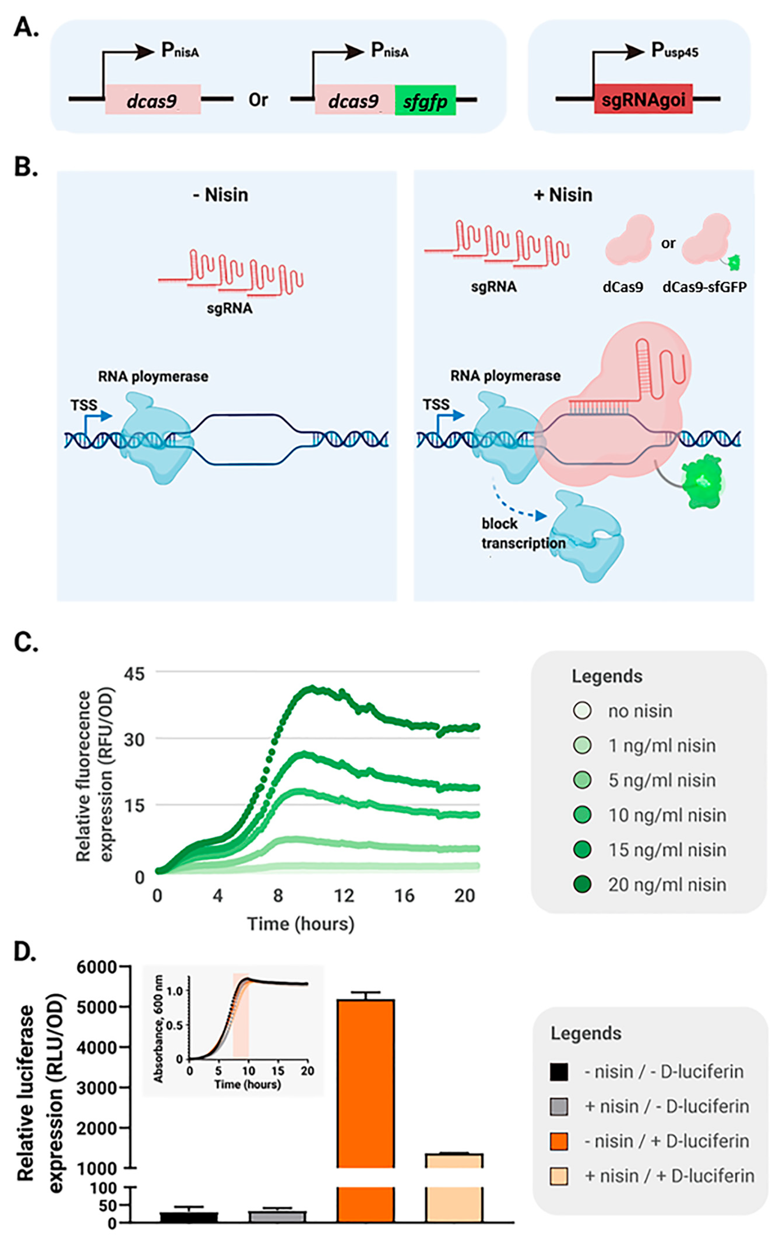
Figure 1. Nisin-inducible CRISPRi systems in L. lactis. (A) Two dCas9 expression modules were constructed: dCas9 alone or dCas9 fused with sfGFP (dCas9-sfGFP); each encoding gene was placed under the control of the nisin-inducible promoter PnisA. The sgRNA targeting a gene of interest (sgRNAgoi) was expressed from the constitutive Pusp45 promoter. (B) Without nisin, sgRNA is produced but dCas9-sfGFP is absent, allowing transcription of the target gene. Upon nisin addition, both sgRNA and dCas9-sfGFP are expressed; the sgRNA guides dCas9-sfGFP to its target, and the resulting complex blocks transcription. The same applies to the construct with dCas9 alone. (C) Different levels of dCas9-sfGFP were induced in L. lactis NZ9000 (pNZ-PnisA-dcas9-sfgfp) by varying nisin concentrations. Cell density and fluorescence were monitored every 10 min. Values represent the mean of three replicates. (D) The CRISPRi system was validated in the luciferase reporter strain L. lactis NZ9000 pseudo10::Pusp45-luc. Expression of dCas9-sfGFP was induced with 5 ng/mL nisin. OD600 and luciferase activity were measured every 10 min, and relative luciferase activity was calculated as luciferase/OD600. The maximum relative activity, observed at the end of exponential growth (highlighted by the orange bar in the inset growth curve), was used for the bar chart. Values are shown as the mean of triplicates.
Ijms 26 09516 g001
Figure 2. Plasmid-based CRISPRi-mediated silencing in L. lactis. Growth curves of strains cultured in CDM with (light blue) or without (red) 10 ng/mL nisin. Cell density was monitored every 10 min (mean of triplicates). Representative fluorescence images show corresponding morphological changes (scale bar = 10 μm). (A) NZ9000; (B) NZ9000 (pNZ-PnisA-dcas9); (C) NZ9000 (pNZ-PnisA-dcas9-sfgfp); (D) NZ9000 (pNZ-PnisA-dcas9-sfgfp; pTLR-Pusp45-sgRNAacmA).
Ijms 26 09516 g002
Figure 3. Chromosome-based CRISPRi provides tight control in L. lactis. (A) The PnisA-dcas9-sfgfp cassette was integrated into the pseudo29 locus of NZ9000. (B) Fluorescence and OD600 were measured after 3 h of induction with or without 10 ng/mL nisin. Relative fluorescence was calculated as fluorescence/OD600 (mean of triplicates, error bars represent standard deviations, SD). (C) Representative fluorescence images of NZ9000 and the acmA-silenced strain CHML011 [NZ9000 pseudo29::PnisA-dcas9-sfgfp (pTLR-Pusp45-sgRNA_acmA)]. Scale bar = 2 μm.
Ijms 26 09516 g003
Figure 4. CRISPRi-mediated silencing of L. lactis ybeY and downstream genes. (A) Four sgRNAs (20 nt each) were designed to target different positions within ybeY (489 nt). Numbers in brackets indicate distance (nt) from the AUG start codon. The proline codon is highlighted (dark blue); its mutated version in ybeYp is marked with *. The red-shaded areas indicate the relative positions of the sgRNA target sites within the ybeY gene. (B) qPCR analysis of ybeY and five adjacent genes in the parental strain CHML012 [NZ9000 pseudo29::PnisA-dcas9] and four ybeY-silencing strains (CHML013–CHML016, carrying sgRNAybeY1–4) (mean of triplicates, error bars represent standard deviations, SD). All strains retained the intact chromosomal ybeY locus. (C) Plasmid constructs of the five strains, showing chromosomal targets silenced (red cross) and plasmid-based complementation. (D) Morphology of CHML012, the ybeY-silenced strain CHML013, and complementation strains CHML017–CHML019 carrying the indicated plasmids after induction with 10 ng/mL nisin. ybeYp: ybeY allele containing the proline codon mutation shown in (A). (E) Growth of plasmid-carrying strains on GM17 agar supplemented with 10 ng/mL nisin. qPCR and growth assays were performed twice with three replicates each.
Ijms 26 09516 g004
Figure 5. Growth and morphological effects of ybeY silencing in L. lactis. (A) Chromosomal organization of the analyzed strains. Red crosses indicate gene silencing, green check marks indicate complementation. (B) Growth of the parental strain CHML012 and the complemented strain CHML020. The red bar marks the time point of cell sampling for microscopy. (C) qPCR analysis of operon gene expression. (D) Representative microscopy images of stationary-phase cells with ybeY silenced (bottom row) or unsilenced (top row). Left and middle panels show low- and high-density regions of the same culture, respectively. Right panels display live/dead staining of the middle panel; red fluorescence indicates dead cells. Scale bar = 5 μm.
Ijms 26 09516 g005
Figure 6. Transcriptomic impact of ybeY silencing in L. lactis. RNA-seq analysis of the ybeY-silenced strain at early stationary phase. Differentially expressed genes (DEGs) were subjected to functional annotation and gene set enrichment analysis to evaluate the global transcriptional response.
Ijms 26 09516 g006
Table 1. Strains used in this study.
StrainsRelevant Genotype and PlasmidsSource
DH5αDH5α[49]
NZ9000MG1363, pepN::nisRK[23]
CHML001NZ9000 (pNZ-PnisA-dcas9)This study
CHML003NZ9000 (pNZ-PnisA-dcas9-sfgfp)This study
CHML004NZ9000 (pNZ-PnisA-dcas9-sfgfp & pTLR-Pusp45-sgRNAluc)This study
CHML005NZ9000 (pNZ-PnisA-dcas9-sfgfp & pTLR-Pusp45-sgRNAacmA)This study
CHML007NZ9000 pseudo10::Pusp45-lucThis study
CHML008NZ9000 pseudo10::Pusp45-luc (pNZ-PnisA-dcas9-sfgfp & pTLR-Pusp45-sgRNAluc)This study
CHML009NZ9000 pseudo29::PnisA-dcas9This study
CHML010NZ9000 pseudo29::PnisA-dcas9-sfgfpThis study
CHML011NZ9000 pseudo29::PnisA-dcas9-sfgfp (pTLR-Pusp45-sgRNAacmA)This study
CHML012NZ9000 pseudo29::PnisA-dcas9 (pTLR)This study
CHML013NZ9000 pseudo29::PnisA-dcas9 (pTLR-Pusp45-sgRNAybey)This study
CHML014NZ9000 pseudo29::PnisA-dcas9 (pTLR-Pusp45-sgRNAybey2)This study
CHML015NZ9000 pseudo29::PnisA-dcas9 (pTLR-Pusp45-sgRNAybey3)This study
CHML016NZ9000 pseudo29::PnisA-dcas9(pTLR-Pusp45-sgRNAybey4)This study
CHML017NZ9000 pseudo29::PnisA-dcas9 (pTLR-Pusp45-sgRNAybey & pNZ-Pybey-dgkA-llmg1485)This study
CHML018NZ9000 pseudo29::PnisA-dcas9 (pTLR-Pusp45-sgRNAybey & pNZ-Pybey-ybeyp)This study
CHML019NZ9000 pseudo29::PnisA-dcas9 (pTLR-Pusp45-sgRNAybey & pNZ-Pybey- ybeyp-dgkA-llmg1485)This study
CHML020NZ9000 pseudo29::PnisA-dcas9, pseudo10::Pybey-dgkA-llmg1485 (pTLR & pNZ-Pusp45-sgRNAybey)This study
Disclaimer/Publisher’s Note: The statements, opinions and data contained in all publications are solely those of the individual author(s) and contributor(s) and not of MDPI and/or the editor(s). MDPI and/or the editor(s) disclaim responsibility for any injury to people or property resulting from any ideas, methods, instructions or products referred to in the content.