Review

The Role of GABA Pathway Components in Pathogenesis of Neurodevelopmental Disorders

by
Ekaterina V. Marilovtseva
*,
Amal Abdurazakov
,
Artemiy O. Kurishev
,
Vera A. Mikhailova
and
Vera E. Golimbet
Mental Health Research Center, Kashirskoe Sh., 34, Moscow 115522, Russia
*
Author to whom correspondence should be addressed.
Int. J. Mol. Sci. 2025, 26(19), 9492; https://doi.org/10.3390/ijms26199492
Submission received: 3 September 2025 / Revised: 25 September 2025 / Accepted: 26 September 2025 / Published: 28 September 2025
(This article belongs to the Special Issue Molecular Investigations in Neurodevelopmental Disorders)

Abstract

γ-aminobutyric acid (GABA), the primary inhibitory neurotransmitter in the central nervous system (CNS), regulates neuronal excitability, synaptic plasticity, and oscillatory activity essential for cognition, emotion, and behavior. Disruptions in GABAergic signaling are increasingly recognized as key contributors to a range of neurodevelopmental disorders (NDDs), including schizophrenia (SZ), autism spectrum disorder (ASD), major depressive disorder (MDD), bipolar disorder (BD), and intellectual disability (ID). In this review, we analyze the data available from the literature concerning the components of the GABA pathway. We describe the main steps of GABA metabolism, including GABA synthesis and release, GABA receptors neurotransmission, GABA reuptake and catabolism, and evaluate their involvement in the pathogenesis of neurodevelopmental disorders. We suggest the possibility of existence of so far undescribed mechanisms which maintain the concentrations of GABA at a relatively physiological level when the function of glutamic acid decarboxylases is compromised by mutations. Searching for these mechanisms could be important for better understanding neurodevelopment and could give a clue for future searches for new therapeutic approaches for treating or alleviating the symptoms of BD and SZ. We also argue that the metabolic stage of the GABA pathway has only a minor direct effect on GABA signaling and rather causes clinical effects due to accumulation of neurotoxic byproducts.

1. Introduction

As the primary inhibitory neurotransmitter in the central nervous system, GABA regulates neuronal excitability, synaptic plasticity, and oscillatory activity essential for cognition, emotion, and behavior [1]. Disruptions in GABAergic signaling are increasingly recognized as key contributors to a range of psychiatric disorders, including SZ, ASD, MDD, BD, as well as ID [2,3,4].
The aim of this review is to gather and analyze the data available from the literature, concerning the components of the GABA pathway, and to evaluate their contribution to pathogenesis of various NDDs.
To provide context, we start with the characterization of GABAergic neurons describing their complex classification based on different synaptic profiles underlying cognitive and emotional processes, whose disruption can result in neurodevelopmental conditions. We describe in detail the main steps of GABA metabolism, including (1) GABA synthesis and release, (2) GABA neurotransmission, (3) GABA reuptake, and (4) GABA catabolism. Firstly, we define a role of the main components, crucial for GABA synthesis, with a specific focus on glutamic acid decarboxylases (GADs). We summarize GADs’ functions in the brain and their association with NDDs such as SZ, ASD, epilepsy, etc. Based on the literature analysis, we suggest the possibility of the existence of mechanisms, so far undescribed, realized in early embryogenesis to compensate for heterozygous mutations within the GAD1 gene. Then, we present evidence implicating GABA receptors in the pathogenesis of MDD, ASD, BD, and SZ through the disbalance in expression rates of different types of GABAARs and GABABRs subunits. We further describe in detail a regulation role of the members of the SLC6 family responsible for GABA reuptake and their involvement in the pathogenesis of neurodevelopmental disorders. We especially emphasize the role of GAT1, the key GABA transporter in mammalian CNS, in maintaining the E/I balance and the spatiotemporal specificity of GABA signals in the cortex and other brain regions and provide evidence that GAT1 dysregulation caused by various mutations in the coding gene region SLC6A1 is implicated in a variety of neurodevelopmental disorders. Finally, we describe the components of the final stage of the GABA pathway and consider the therapeutic agents applicable at this stage.

1.1. GABAergic Neurons: Classification, Functions, and Implication in Neurodevelopmental Disorders

In embryogenesis, GABAergic neurons (Figure 1) originate from the medial ganglionic eminence (MGE), caudal ganglionic eminence (CGE), and lateral ganglionic eminence (LGE) [5]. Found in both the subcortical structures and prefrontal cortex (PFC), GABAergic neurons constitute up to 25% of all neurons [3]. Depending on the type of synaptic profiles they are associated with, GABAergic neurons may be divided into several subtypes. The first one is basket cells (BCs), associated with axo-somatic inhibitory synapses and providing strong inhibition to the somata and proximal dendrites of glutamatergic pyramidal cells (PyCs) [6], principal granule cells of the dentate gyrus [7], and principal neurons of the basolateral amygdala region [8], thus influencing their output and regulating working memory, cognitive flexibility, emotional responses, and fear processing. BCs are mostly found in cortical layers (L) 3/4 [3], as well as in the hippocampus [9], and the molecular layer of the cerebellar cortex near Purkinje cells [10]. Importantly, BCs are also known to come into contact directly with thalamic afferent fibers and, thus, to be directly engaged in shaping sensory inputs [3]. Based on their electrophysiological properties, BCs can be divided into two subpopulations, fast-spiking (or fast-firing) parvalbumin-positive (PV+) BCs and regular-spiking (or regular-firing) cholecystokinin-positive (CCK+) BCs [11]. Both PV+ BCs and CCK+ BCs contribute to the regulation of cortical rhythmogenesis and play a major role in synchronizing and coordinating the functions of the hippocampus [12,13]. According to the other classification, based on their morphological features, BCs are divided into three groups: mostly multipolar large BCs (LBCs), expressing PV, neuropeptide Y (NPY), and calbindin (CB) and are capable of inhibiting the neurons in neighboring columns and in upper and lower layers due to the expansive axonal arborization; vasoactive intestinal peptide (VIP) expressing small BCs (SBCs) with local multipolar (L4), bitufted or bipolar (L2/3) somato-dendritic morphology and frequently branching curvy axons innervating the cells within the same cortical layer; and VIP nest BCs (NBCs), which share particular morphological features with LBCs and SBCs [14].
The other subtype of GABAergic neurons is PV+/CB+ chandelier cells (ChCs), associated with axo-axonic synapses, providing inhibition to PyCs’ axon initial segments (AIS), where action potentials are generated. Unlike sparsely branched BCs’ axons, the axons of either multipolar or bitufted ChCs spread horizontally, their terminals arranged in vertical rows of 2 to 15 candlestick-like boutons, with each row innervating the postsynaptic AIS of a single PyC [15]. ChCs are found in the hippocampus, where they constitute up to 4% of all CA1 interneurons [15], PFC, and temporal and visual cortices, with the highest axon terminals’ density observed in L2–6 [16]. Recently, it has been established that in the visual cortex ChCs, receiving inputs from the L5 PyCs, provide the signal to PyCs in L2/3 in response to locomotion and visuomotor mismatch-related arousal [17]. Moreover, action potential discharge in ChCs present in the basolateral amygdala evokes local network activity, which initiates sharp waves (SWs), associated with learning, and memory consolidation and retrieval, by engaging other GABAergic interneurons and PyCs [18].
Axo-dendritic inhibition of PyCs is mostly mediated by somatostatin (SST)-expressing cells, such as double bouquet (DBCs) and Martinotti cells (MCs). The first ones are VIP+/calretinin (CR)+ regular-spiking interneurons located in L2/3 of the neocortex, characterized by their vertical bundled axons, termed horse-tails, which mostly innervate the shafts of small dendrites and form symmetric synapses with dendritic spines [19], providing disynaptic inhibition among PyCs, thus playing an important role in so-called cortical memory networks [20]. Of note, evidence suggests that DBCs may also target the dendrites of BCs and ChCs, providing a disinhibitory link to the GABAergic system, and selectively target L4. Interestingly, DBCs and PV+ GABAergic interneurons are differentially regulated by presynaptic group III metabolic glutamate receptors (mGluR), which increase the frequency of sIPSCs in the former, but suppress it in the latter [21]. With their axons, forming spiny boutons, MCs (whose somata are found in L2–6) reach L1 to target terminal tufts of PyCs, which differs them from DBCs. MCs, capable of inhibiting the dendrites, perisomatic dendrits, and somata of neighboring and even distant columns, are the only cells that mediate cross-columnar inhibition from L2–6 via L1. The other main types of GABAergic interneurons involved in axo-dendritic inhibition are the following: Axon tuft cells (ATCs), confined to L1, their axons following a horizontal trajectory and forming rich arborizations [3]; Bipolar cells (BPCs) with spindle or ovoid somata and vertically oriented dendritic arbors, which are significantly longer in L4–6 BPCs than in L2/3 BPCs; Small button-type neurogliaform cells (NGCs), forming a spherical and highly symmetrical dendritic field and ultra-thin axons with intertwined arborization [22].
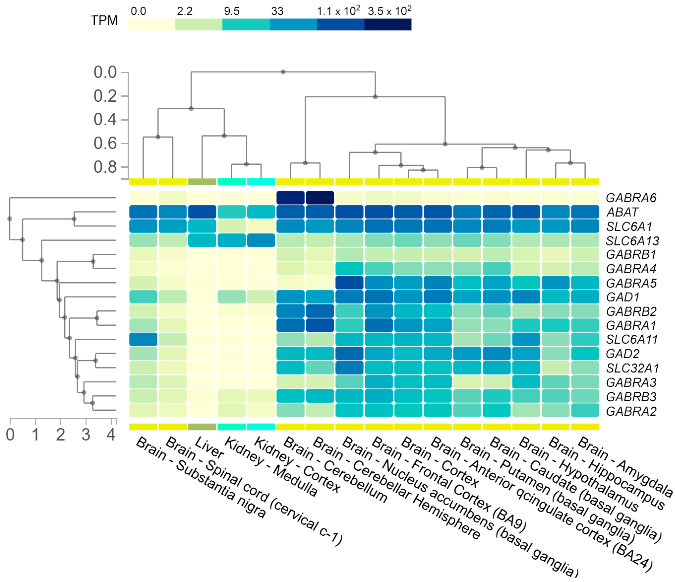
The maturation of GABAergic interneurons is one of the key elements initiating critical periods of neuroplasticity—the time-limited windows in brain development during which neural networks are particularly sensitive to sensory and behavioral experiences [23]. Thus, impaired development and maturation of GABAergic interneurons can result in alterations in the cortical and hippocampal circuits associated with severe NDDs, such as SZ and ASD, etc. Indeed, recent studies of clinical cases and animal models have revealed a number of physiological alterations (excitatory/inhibitory (E/I) neurotransmitter imbalance in the hippocampus [24], altered working memory and other functions associated with dorsolateral prefrontal cortex (DLPFC) [25], impaired sleep [26], etc.), appearing in response to a deficit of GABAergic interneuron-dependent inhibition [27], loss [28] or excessive production of GABAergic neurons [29], or compromised function of the main components of the GABA pathway (Figure 2), which are the focus of this paper.

1.2. GABA Synthesis and Release

In this section we describe the main components involved in GABA synthesis and release.

1.2.1. Sodium-Coupled Neutral Amino Acid Transporter 1

Mouse models demonstrate that at least in PV+ GABAergic neurons the main source of L-glutamate, the immediate precursor of GABA, is astroglia-derived glutamine imported into the cells by Snat1, encoded by SLC38A1 and detected in proximal dendrites and somata [30]. SNAT1-dependent import of glutamine is a voltage-dependent process driven by Na+ (or, alternatively, Li+) electrochemical gradient with 1:1 stoichiometry of Na+/amino acid cotransport [31]. Playing a pivotal role in regulating GABA replenishment and synaptic vesicular content in GABAergic neurons, SNAT1 is involved in inducing high-frequency oscillations and shaping cortical processing and plasticity [32]. Importantly, upregulation of SLC38A1 due to the lack of methylation at its promoter was observed in peripheral blood in MDD [33] and in microglia in Rett syndrome [34].
The deteriorating effect of SLC38A1 overexpression may be explained by a decrease in mitochondria viability and metabolism in response to glutamate overproduction, as well as by neurotoxic effect of elevated NMDAR activation. Logically, the inhibitors of SNAT1-dependent glutamine uptake, such as L-theanine, were proposed as potential therapeutic agents for preventing the onset of ASD [35] as well as for alleviating MDD symptoms [36].

1.2.2. Phosphate-Activated Mitochondrial Glutaminases

Prior to the initiation of GABA synthesis, glutamine is processed to glutamate by a phosphate-activated mitochondrial glutaminase (K-type GLS1 isoform or L-type GLS2 isoform), which generates NH4+ as a byproduct. Interestingly, GLSs are positively regulated by Ca2+ and inhibited by glutamate in a negative feedback loop [37]. Essential for neurogenesis and neuronal progenitor cell (NPC) survival in embryogenesis [38], the GLS1 gene is also capable of inducing microglia when overexpressed, thus contributing to neuroinflammation [39]. Examination of postmortem autistic brain samples and those of GLS1KO/KO mice demonstrated the link between the loss of GLS1 expression in PFC and ASD symptomatology, including synaptic E/I imbalance and abnormal expression patterns of genes associated with microglial synapse pruning, which were restored by low doses of lipopolysaccharide [40]. At the same time, overexpression of GLS1 (in particular, its GAC isoform) in the hippocampus, associated with severely impaired spatial and fear memory [41], is observed in MDD models [42], while GLS1 haploinsufficiency results in hippocampal hypometabolism and schizophrenia resilience phenotype [43], and, thus, has an antipsychotic-like effect [44]. Finally, overexpressed GLS2 alters mitophagy function in hippocampal neurons, reducing the occurrence of seizure-like events [45].
Altogether, these data indicate that GLS1-related pathogenesis of SZ and MDD involve mitochondria dysfunction and neuroinflammation rather than direct dysregulation of the GABA pathway, while in epilepsy and ASD GLS1 downregulation may indeed affect GABA synthesis and disrupt inhibitory processing in the brain.

1.2.3. Glutamic Acid Decarboxylases

Biosynthesis of GABA from L-glutamate is a one-step reaction catalyzed by paralogous pyridoxal 5′-phosphate (PLP)-dependent glutamic acid decarboxylases GAD65 (encoded by GAD2), GAD67 (encoded by GAD1) or the isoforms of the latter, such as enzymatically active GAD44, corresponding to the C-terminus of the enzyme [46]. In the human brain, GADs are found in the neocortex, hippocampus, basal ganglia, and cerebellum, with GAD65 being preferentially localized in neuropil granules (axonal boutons or terminals) and GAD67—in neuronal cell bodies and proximal dendrites and nerve terminals [47]. Interestingly, while GAD67 functions to maintain the basal pool of GABA, GAD65 is activated in response to short-term increases in the neurotransmitter demand in synaptic terminals [48]. GAD3, the third glutamic acid decarboxylase gene described, is absent in the genomes of higher primates, including humans [49]. Importantly, mouse models demonstrated that GAD2 complete knockout resulted in higher susceptibility to seizure induction but did not affect GABA concentrations [50], while GAD1 complete knockout caused neonatal death, severe cleft palate, and respiratory failure, accompanied by dramatic reduction in GABA concentration [51].
Highly homologous (~76% identity), GAD65 and GAD67 differ significantly in the N-terminal region (the first 95 amino acids) [52], which serves as a target for hydrophobic PTMs in case of GAD65, but not GAD67 [53]. Another PTM, known to regulate the activity of both GAD65 and GAD67, is PKA-dependent phosphorylation at Thr91, which activates the former and, vice versa, inhibits the latter [54]. Forming homo- and heterodimers, the two main GAD isoforms are further targeted to synaptic vesicles (SVs), the N-terminus of GAD67 (including Thr131 [55]), and the N-terminal palmitoyl groups of GAD65, playing a crucial role in this process [56]. Once GABA is released to synaptic cleft, GAD65 is clustered at the presynaptic membrane by Neuronal growth regulator 1 (NEGR1) and further removed with newly formed synaptic vesicles [57].
Both GAD1 and GAD2 have been found in association with multiple NDDs. Analysis of postmortem MDD specimens showed decreased GAD67/65 levels in the hypothalamic paraventricular nucleus (PVN) [58], which may, at least partly, explain impaired sleep and stress response characteristic to the patients with MDD. Moreover, a particular number of GAD1/2 SNPs have been identified in patients with NDDs. Recently, a single SZ-associated homozygous polymorphism c.391A>G (p.Thr131Ala) within GAD1 ORF has been described, disrupting the enzyme’s dimerization and function [55]. A comprehensive analysis of 235 Chinese Han family trios revealed 17 SZ-associated variants of GAD1, including ten SNPs in 5′-flanking regions, four SNPs, and one novel in-del in intronic regions, as well as two SNPs in the 3′-UTR. In silico research demonstrated that four of the identified 5′-flanking polymorphisms (rs3762556 (G>C), rs3762555 (G>C), rs6755102 (C>T), rs3791878 (G>T)) can change the affinity of the gene’s promoter for a number of transcription factors [59]. rs3749034 (C>T), another SZ-risk SNP located in the GAD1 5′-flanking region [60], was found to affect the methylation status of CpG sites within the gene’s putative promoter, downregulating GAD1 expression and increasing GAD25/GAD67 ratio in the hippocampus and DLPFC of patients with SZ [61,62]. On a clinical level, rs3749034 (C>T) is associated with negative symptoms and early-onset SZ, as well as with BD and MDD [63]. GAD2 expression rate was found to be reduced by 40% in the primary auditory cortex of SZ patients, indicating that the gene might contribute to auditory hallucinations occurrence [64], and GAD65 autoantibodies in serum and cerebrospinal fluid were attributed to catatonia in patients with prediagnosed chronic SZ and proposed as the condition’s biomarker [65]. GAD1/2 are also dysregulated in ASD; for instance, some patients exhibited a ~40% decrease in the level of GAD1 mRNA in Purkinje cells [66], which is, at least in part, due to the enrichment of 5hmC and MECP2 at its promoter [67]. In this regard, it is worth mentioning that prenatal immune activation due to maternal infections may result in increased levels of 5mC and 5hmC at the GAD1 promoter and that of 5mC at the GAD2 promoter in PFC. This, in turn, results in suppression of GAD1/2 promoters by elevated MECP2 binding and, on a global level, in compromised PFC function, manifesting in cognitive abnormalities, impaired working memory and social interaction, as well as other symptoms typical for ASD and SZ [68].
Finally, there are multiple lines of evidence confirming the role of GAD1/2 in epilepsy pathogenesis. Several bi-allelic GAD1 variants associated with early-infantile onset epilepsy and severe neurodevelopmental delay have been identified: homozygous c.87C>G (p.Tyr29*), c.568delC (p.Gln190Serfs ter11), c.971T>G (p.Phe325Cys), and c.1040C>T (p.Thr347Met) and heterozygous c.1591C>T/c.670delC (p.Arg531*/p.Leu224Serfs*5) among them [69]. Autoimmune-associated epilepsy (AAE), a drug-resistant disorder, has also been described in the literature. It manifests in refractory focal seizures and, on the histological level, in infiltration of the temporal lobe by CD8+ cells, and its pathogenesis is tightly linked to the GAD65 autoantibodies [70]. Recently, several polyphenols have been described as potential therapeutic agents in treating GABA deficiency-associated epilepsy. In particular, one of them, pterostilbene (PTE, 3,5-dimethoxy-4′-hydroxystilbene), was shown to upregulate GAD1 expression and, hence, to increase GABA synthesis in the hippocampal PV+ GABAergic neurons, as well as in the PFC in rats [71] and mice [72]. The other natural compound, honokiol, was found to stimulate GAD65 activity in the hippocampus of mice [73]. Also, intracerebral electroacupuncture in combination with pentobarbital significantly enhanced GAD1 expression in rats and was highly efficient in reducing seizures [74].
Based on the literature analysis and given the importance of GABA for neurogenesis and brain metabolism, we suggest that in early embryogenesis the cell might possess so far undescribed mechanisms to compensate for heterozygous mutations within GAD1 gene, encoding the key GABA synthesizing enzyme, in order to maintain GABA concentrations at the levels essential for surviving. These mechanisms may include GAD2 and/or fetal isoforms of GAD1, as well as upregulation of GAD1 expression by epigenetic mechanisms, such as H3K4 trimethylation, which was found to be reduced at GAD1 regulatory sequences in SZ [75]. This assumption is indirectly confirmed by the fact that, although associated with epilepsy and severe developmental delay, the bi-allelic GAD1 mutations (even those resulting in truncated GAD67 lacking the catalytic domain), described so far, are not lethal. At the same time, ubiquitous downregulation of GAD1 due to perturbations in promoter methylation results in complete allele LOF and significantly decreased transcripts; in turn, GABA concentration dramatically drops and so does the efficiency of inhibitory neurotransmission, inevitably leading to impaired neurodevelopment and subsequent disorders. Apparently, introducing large deletions into GAD1 may have a similar effect, making animal models with GAD1 haploinsufficiency less informative for studying the clinical effects of GAD67 mutations.

1.2.4. Vesicular GABA Transporter/Vesicular Inhibitory Amino Acid Transporter

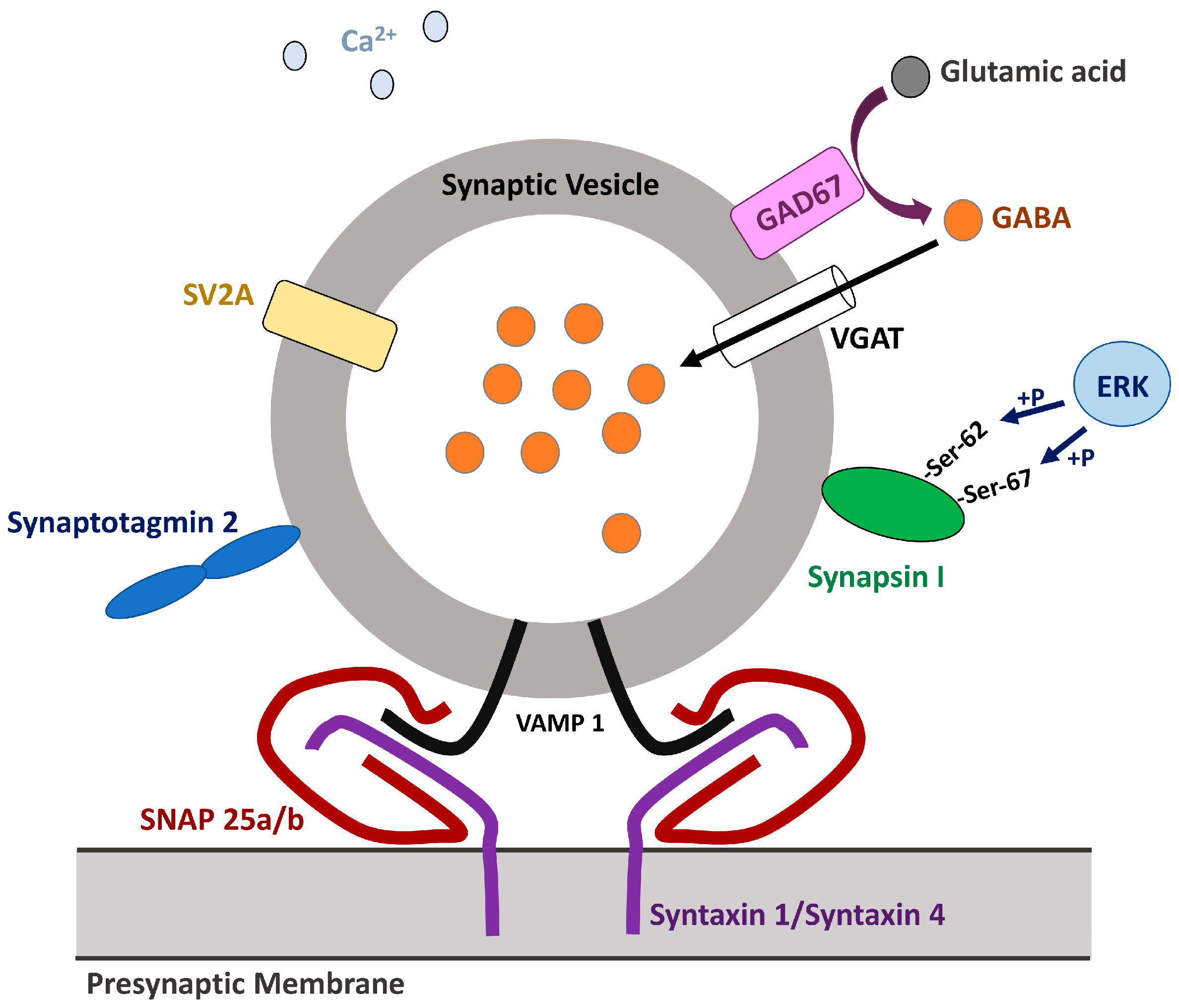
Once synthesized, GABA is packaged into synaptic vesicles (SVs) which are then trafficked to the presynaptic membrane to fuse with and to release the neurotransmitter to the synaptic cleft in response to the increase in intracellular Ca2+ concentration (Figure 3) [76,77]. The central role in both processes is played by vesicular GABA transporter/vesicular inhibitory amino acid transporter (VGAT/VIAAT), encoded by SLC32A1, the only known member of the SLC32 family; interestingly, VGAT-dependent loading of GABA into the SV requires for electrochemical luminal proton gradient [78,79]. The VGAT molecule consists of nine transmembrane domains (TMs), a long N-terminal domain facing the cytoplasm, and the C-terminus facing the lumen [80]. Interestingly, VGAT is also known to transport glycine, another inhibitory neurotransmitter [81], and to co-localize with glutamate transporters VGLUT1 (SLC17A7) [82] and VGLUT2 (SLC17A6) [83] on SVs. SLC32A1−/− mice exhibit complete stiffness by day E17.5 and die in utero after day E18, these observations confirming the indispensability of VGAT in development [79]. In humans, mutations in SLC32A1 have been identified in association with various epileptic disorders. The study of a large cohort of Australian patients with genetic epilepsy with febrile seizures plus (GEFS+) or idiopathic generalized epilepsy (IGE) revealed eight rare inherited heterozygous missense mutations in SLC32A1 (c.127G>T (p.Gly43Cys), c.788T>C (p.Val263Ala), c.989T>C (p.Met330Thr), c.1333C>T (p.Leu445Phe), c.1382G>A (p.Gly461Asp), c.1391C>G (p.Thr464Arg), c.1393G>A (p.Gly465Ser), c.1403T>C (p.Leu468Pro)), affecting the structure of the transporter’s TMs and N-terminus and apparently associated with reduced vesicular GABA transport [84]. A link between four SLC32A1 heterozygous de novo mutations (c.271G>A (p.Ala91Thr), c.787G>A (p.Val263Met), c.806T>C (p.Leu269Pro), c.965T>G (p.Phe322Cys)) and epileptic encephalopathy with infantile seizure onset and moderate-to-severe ID has also been described. Importantly, located at the N-terminus, the first mutation increases presynaptic GABA release probability and, hence, the stimuli frequency, while the other three found in the α-helices lining the GABA transport pathway affect the substrate uptake. Depending on the mutation, the patients predominantly showed either focal and focal impaired awareness seizures, or tonic–clonic seizures with myoclonic seizures and status epilepticus with suspicion of myoclonic atonic epilepsy in case of c.787G>A (p.Val263Met) [85]. Similar to GAD65/67, VGAT is implicated in ASD [86] and SZ [87], its expression being decreased in the PFC of adult BTBR mice and in postmortem specimens, respectively.
Based on clinical data, we speculate that, at least in embryogenesis, malfunctioning of VGAT is more deteriorating for the GABA pathway and neurodevelopment than that of GAD1/2, and that SLC32A1 polymorphisms severe enough to cause ASD or SZ might be lethal and that is why they have not yet been identified.
Interestingly, there are a number of other factors, including some signaling pathways, that govern GABA release. One of them is the RAS–ERK signaling cascade, which is directly involved in pathogenesis of various health conditions called “RASopathies”, some of which manifest in ID and learning disabilities [88]. Recently, it has been shown that while in excitatory neurons, hyperactivation of RAS–ERK signaling due to the SNPs (such as p.Gly12Val) within the KRAS is cytotoxic and leads to cell death, in GABAergic neurons it increases the risk of spontaneous GABA release [89]. The latter is, apparently, due to phosphorylation of synapsin I with its subsequent dissociation from SVs [90]. At a physiological level, this results in increased sIPSC frequency, as well as impaired long-term potentiation at CA3–CA1 synapses in the hippocampus that can be rescued by picrotoxin treatment [89]. This brings us to the conclusion that when designing new diagnostic protocols and therapeutic agents for treating NDDs associated with dysregulated GABA traffic and release, the whole spectrum of its regulators should be taken into account.

1.3. GABA Neurotransmission

In the neural networks of the brain, GABA functions as the primary inhibitory neurotransmitter, acting on both ionotropic (GABAAR, or GABRA) and metabotropic (GABABR, or GABRB) receptors, both of which are widely distributed in the cerebral cortex, hippocampus, basal ganglia, thalamus, cerebellum, and brainstem [91].

1.3.1. GABAA Receptors

GABAARs are heteropentameric ligand-gated Cl channels, detected in hippocampal and PFC PyCs, cerebellar granule cells, thalamic relay cells [92], and a subset of dopaminergic neurons in substantia nigra [93]; serotonergic and GABAergic neurons are also believed to possess distinct subtypes of GABAARs [94] and to be involved in the realization of the most prominent inhibitory pathway in the CNS. Despite a significant diversity of GABAAR’s subunits expressed in the human organism, mostly in a tissue-specific manner (six α (1-6), three β (1-3), three γ (1-3), three ρ (1-3), and one each of the δ, ε, π, and θ), the main adult isoform of the receptor is built up by 2 α1-, 2 β2-, and 1 γ2-subunits [95]. While the α1β2γ2 GABAAR subtype, accounting for ∼43% of all GABAARs, is expressed by GABAergic interneurons and pyramidal cells more or less ubiquitously within the brain [96], the other GABAAR subtypes are specific for particular brain structures: α1β2,3γ2 GABAAR is characteristic for aspiny cells and α2/α3β2,3γ2 for PyCs of the amygdala [97], α4βδ GABAAR [98] and α5β3γ2/γ3 [99] are found in hippocampal PyCs, and α6-containing GABAARs are restricted to differentiated cerebellar granule cells [100]. Interestingly, GABACRs, consisting solely of ρ-subunits, are also classified as one of the subgroups of GABAAR’s isoforms [101]. Depending on the class of the subunits it is made of, GABAAR provides either phasic inhibition (a short-term hyperpolarization of the postsynaptic membrane occurring after GABA release), or tonic inhibition (a long-term hyperpolarization of the postsynaptic membrane by low ambient levels of GABA) [95]. Also, an abundance of phosphorylation sites has been identified in various GABAAR subunits, governing the receptor’s activity [102]. Importantly, GABAAR is a target for benzodiazepines, which potentiate the effect of GABA [103] and are known to reduce the symptoms of anxiety and SZ when used in combination with particular AEDs [104]. Resveratrol is another agent known to potentiate GABAAR-mediated inhibitory neurotransmission [105,106] and suggested as a promising AED [107].
Imbalanced expression of different GABAAR subunits has been detected in BD, SZ, and MDD, e.g., postmortem analysis revealed upregulation of β3 subunit and downregulation of α1 and α2 in the SZ lateral cerebellum [108,109], and life [11C]Ro15-4513 position emission tomography (PET) imaging demonstrated significant reduction in the α5 subunit in the SZ hippocampus, but not in patients partaking in antipsychotics [110]. In addition, N-glycosylation of β1 and β2 subunits is altered in the SZ cortex, which may result in disrupted GABAAR assembly and function [111]. Furthermore, impaired transmission at α6-GABAARs correlates with dysregulation of dopaminergic system and is characteristic for SZ patients; furthermore, positive allosteric modulators highly selective for α6-GABAARs were shown to reduce positive and negative symptoms and cognitive impairment in animal models for SZ [112]. Also, the expression of β3 subunit of GABAAR was reduced in peripheral blood mononuclear cells in patients with BD, which may be used as a diagnostic marker [113]. Several GABAAR subunit polymorphisms were also reported in GEFS+ (e.g., GABRA1 c.274T>C (p.Phe92Leu)), MAE (e.g., GABRB3 c.851T>C (p.Leu284Pro)) [114], Lennox–Gastaut syndrome (e.g., GABRB3 c.358G>A (p.D120N)) [115], and MDD with the risk of suicide (GABRA6 3′-UTP SNP rs3219151 (C>T)) [116], but not in SZ [117]. Pathogenesis and clinical manifestation of a particular number of IDs, including those associated with chromosomal aberrations, are also known to be linked to impaired expression of GABAAR subunits. In Prader–Willi syndrome (PWS), a genetic disorder characterized by mild-to-moderate ID, impaired behavior, and depression [118], paternal alleles of GABRA5, GABRB3, and GABRG3 are missing due to the partial deletion of an imprinted region on paternal chromosome 15 (15q11–13) [119]. Given that all three GABAAR subunits are associated with the hippocampus [99], the loss of a half of their alleles may explain poor short-term memory, another known symptom of PWS [120]. Furthermore, according to the in vivo data obtained by single–voxel proton magnetic resonance spectroscopy (1H-MRS), GABA levels are also reduced in the PWS brain (at least, in the parieto-occipital lobe) [121], hinting at the possibility of existence of a feedback loop regulating either GAD1 expression or GAD67 enzymatic activity in GABAergic neurons in response to the fluctuations of GABAARs’ content in PyCs. Impaired GABA pathway is also typical for Fragile X syndrome (FXS), an ID caused by an expansion of the CGG repeat within the 5′-UTR of Fragile X messenger ribonucleoprotein 1 coding gene FMR1 [122]. FXS mice models demonstrated 35–50% decrease in expression rates of GABRA1, GABRA3, GABRA4, GABRB1, GABRB2, GABRG1, and GABRG2 in the cortex [123], as well as a four-fold decrease in Gabrd subunit in the hippocampus [124]. Interestingly, [11C]flumazenil PET performed on FXS patients identified a significant reduction in the non-displaceable binding potential (BPND) of GABAARs (reflecting their availability), throughout the brain, with the highest level of BPND reduction—up to 17%—detected in the thalamus [125]. Dysregulation of several other components of the GABA pathway have also been found in association with FXS, GAD67/65, GABA transporters GAT1 and GAT4, and GABA catabolizing enzymes among them [122]. Of note, in contrast to PWS and FXS, in Down syndrome (DS), GABA inhibition (mainly tonic) is enhanced. This phenomenon may be explained by the presence of an extra chromosome 21 and, hence, additional copies of OLIG1, OLIG2, and DYRK1A [126], well-known regulators of GABAergic interneurons production [127] and synaptic plasticity pathways [128]. Given the abovementioned data, it was suggested that treatment with basmisanil, a α5-GABAAR-specific negative allosteric modulator, could have a positive effect on cognition in patients with DS, but unfortunately, the clinical trials failed [129]. Nevertheless, therapeutic strategies targeting other components of the GABA pathway or the proteins modulating GABA neurotransmission may be effective in alleviating DS symptoms. One of the promising targets for such therapies is G-protein-activated inwardly rectifying potassium channel 2, GIRK2, whose gene is located on chromosome 21 and whose function is linked to that of GABAB receptors [130,131].

1.3.2. GABAB Receptors

GABABR is a heterodimeric G protein coupled class C receptor, expressed in glial cells [132] and somato-dendritic compartments of neocortical and hippocampal PyCs [133], serotonergic neurons in dorsal raphe nucleus (DRN) [134], dopaminergic neurons in substantia nigra [135], and other types of neurons. Each of the two receptor’s subunits, GABAB1 and GABAB2, contains an extracellular Venus Flytrap (VFT) domain, a heptahelical TM, and an intracellular C-terminal region which contributes to heterodimerization and serves as a binding site for several regulatory proteins. When activated by GABA or baclofen, GABABR undergoes conformational changes which result in G protein dissociation into Gαi/o and Gβγ subunits, the former inhibiting adenylyl cyclase (AC) and cAMP signaling, thus preventing vesicle fusion and neurotransmitter release; meanwhile, the latter directly binds to VGCC, inhibiting Ca2+ influx, and to GIRK(1-4) channels, triggering K+ efflux and contributing to the postsynaptic membrane hyperpolarization. Furthermore, in cellulo GABA–GABABR pathway was found to stimulate phosphorylation of ERK1/2 protein kinases, which subsequently activate a number of transcription factors, involved in neurogenesis and memory formation [136]. Hence, GABABRs play an essential role not only in regulating E/I balance in the adult brain, but also in coordinating the early stages of neurogenesis, and its dysregulation may have dramatic consequences for cognition and behavior. Indeed, genetic and postmortem studies have confirmed the association between GABABRs deficits and NDDs such as ASD, BD, SZ, and MDD. In lateral cerebella, for example, the levels of GABBR1 (which is located in SZ-associated loci) and GABBR2 are significantly reduced in cases of BD, SZ, and MDD [137], at least partly due to their attenuation by SZ-related microRNAs [138]. Additionally, exome sequencing identified the rs10985765 SNP (p.Thr611Ala/Pro, p.Thr771Ala/Pro, or Thr869Ala/Pro, depending on the isoform) within GABBR2 as a potential risk factor for treatment-resistant SZ [139]. Finally, the actin-binding protein SHROOM4 (SHRM4) (encoded by KIAA1202) has recently been shown to play a role in regulating the receptor’s activity by providing its synaptic localization [140]. This finding makes GABABRs a key player in pathogenesis of Stocco Dos Santos syndrome [141] and the NDD caused by Xp11.2 microduplication [142], both of which are associated with disrupted KIAA1202 and characterized by mild or mild-to-severe ID, seizures, anxiety, and impaired behavior.
Similar to GAD67/65, GABAARs and GABABRs subunit polymorphisms tend to correlate with epilepsy. Meanwhile, MDD, ASD, BD, and SZ seem to be associated with the disbalance in expression rates of different types of GABAARs and GABABRs subunits. Such disbalance may prevent different subtypes of the receptors from maturation, assembly, and incorporation into the synapses, and subsequently result in decreased inhibitory neurotransmission in one cell type of neurons and increased inhibitory neurotransmission in the others. This, in turn, may lead either to impaired neurogenesis, or to altered E/I balance in different structures of the adult brain. In the former case, dysregulated expression of the GABA receptor subunits may be either due to the presence of SNPs in their regulatory elements, altering their methylation status or affinity to transcription factors, or due to the lack of particular transcription factors; in the latter case, epigenetic regulation must play a central role.

1.4. GABA Reuptake

Once released, GABA does not undergo enzymatic breakdown within synaptic cleft, but is eliminated through reuptake via specialized transporters, belonging to the solute carrier 6 (SLC6) family of neurotransmitters:sodium symporters. Only four members of the SLC6 family, SLC6A1/GAT1, SLC6A11/GAT3, SLC6A13/GAT2, and SLC6A12/BGT1 [143], are known to effectively transport GABA by its symport with 2 Na+ ions and one Cl ion. Each of these four proteins consists of 12 helical TMs, connected by intra- and extracellular domains, with TMs 1, 3, 6, and 8 forming the orthosteric substrate binding pocket [144], which is a target for the majority of GAT-specific inhibitors, such as (S)-SNAP-5114 ((S)-1-[2-[Tris(4-methoxyphenyl)methoxy]ethyl]-3-piperidinecarboxylic acid) [145] and a highly selective and potent isatin derivative 1H indole 2,3-dione [146]; of note, the substrate binding pocket is capped from the extracellular side by two amino acid residues from TM3 and TM6, respectively (Tyr140 and Phe294 in case of GAT1; Tyr129 and Phe288 in case of GAT2; Tyr147 and Phe308 in case of GAT3), their mutations leading to dramatic reduction in GABA transport efficiency [147,148]. To note, similar to other SLC6 proteins, GABA transporters tend to oligomerize [149].
Below, we describe in detail the regulation role of the members of the SLC6 family and their involvement in the pathogenesis of neurodevelopmental disorders.

1.4.1. Sodium- and Chloride-Dependent GABA Transporter Type 1

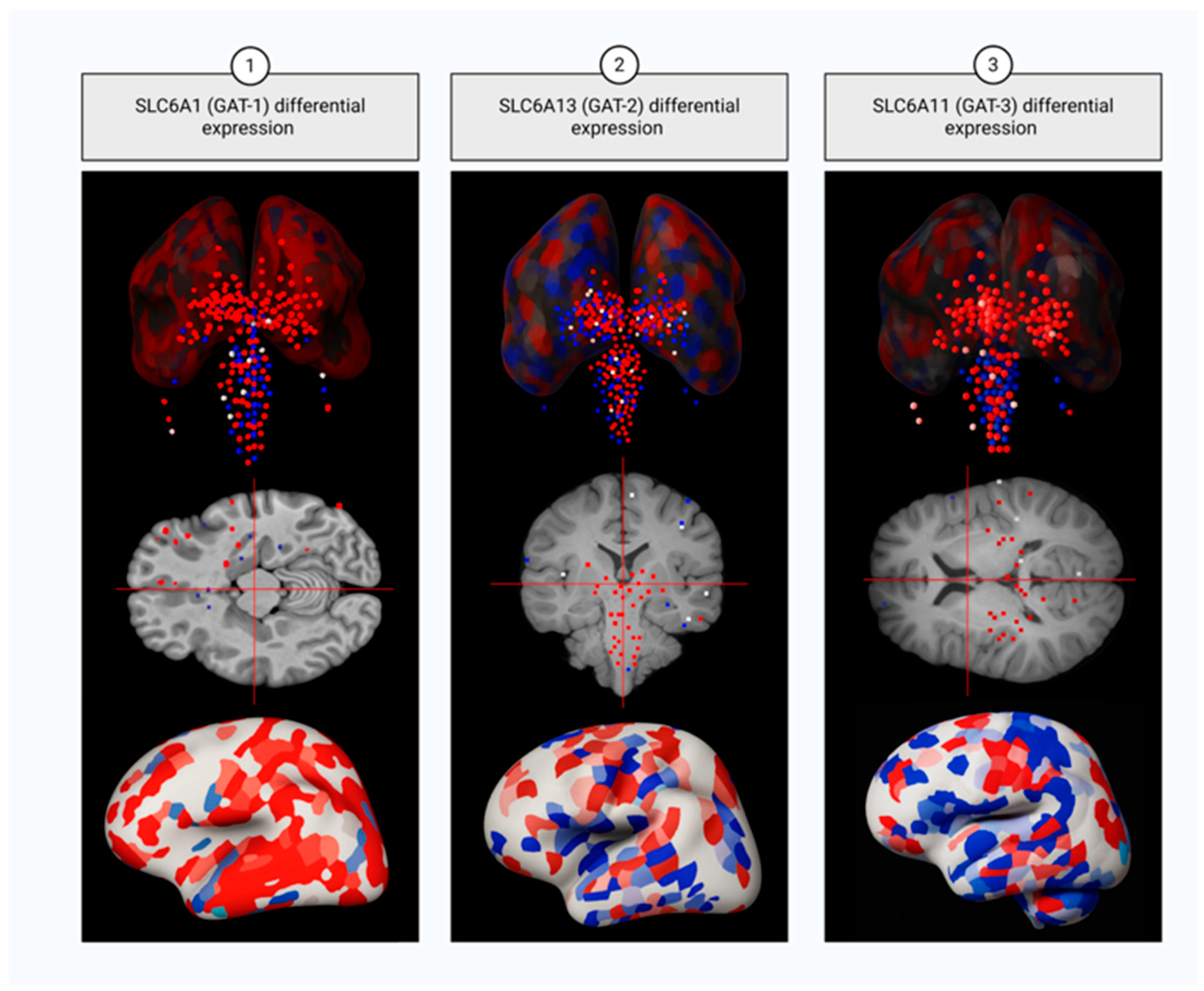
GAT1 is the key GABA transporter in mammalian CNS. Though mostly localized in astrocytes and presynaptic terminals of GABAergic neurons (Figure 4) [150], it is also found in striatal dopaminergic (DA) neurons [151]: taken up to DA neurons by GAT1, GABA is loaded into SVs by the monoamine transporter VMAT2 (the product of SLC18A2 gene) [152] and is further co-released with dopamine in order to tune striatal output [153] and prevent its hyperactivation, observed in SZ patients [154].
Notably, up to 50% of membrane-associated GAT1 molecules are immobile, anchored to the actin cytoskeleton due to the ability of GAT1 C-terminus to interact with scaffolding proteins [155], with disruption of these interactions increasing the lateral mobility and functional efficiency of GAT1. Given its participation in regulating both phasic and tonic inhibition [156], GAT1 serves as a multifunctional and dynamically regulated transporter, critical for maintaining the E/I balance and the spatiotemporal specificity of GABA signals in the cortex and other brain regions. Therefore, SLC6A1 dysregulation is implicated in a variety of neurodevelopmental disorders. In a large-scale functional screen of 213 unique SLC6A1 SNPs, including 67 de novo mutations, approximately 59% exhibited severe LOF (defined as <82.1% GABA uptake compared to wild-type) and 11.7% moderate LOF. Notably, 98.3% of individuals with the de novo variants were diagnosed with developmental delay (e.g., c.863C>T (p.Ala288Met), c.1070C>T (p.Ala357Val), c.1342A>T (p.Lys448*), etc.), 83.6% with epilepsy (predominantly, epilepsy with myoclonic-atonic seizures, EMAS) (e.g., c.881_883del (p.Phe294del*5), c.815_817delTCA (p.Ile272del*4), c.491G>A (p.Cys164Tyr), etc.), and 54.5% with ASD (e.g., c.896G>T (p.Gly299Val), c.1017C>G (p.Phe339Leu), c.1396_1379 delGG (p.Gly457Hisfs*10), etc.) [157,158]. ClinPred and other missense severity scores confirmed high sensitivity of GAT1 to amino acid substitutions (particularly within TMs 1–10, known to build the substrate binding pocket and maintain core structural architecture), which explains SLC6A1 sevenfold enrichment for pathogenic missense variants over truncating mutations in neurodevelopmental cohorts, despite the relatively small coding size of the gene (599 amino acids). These findings support a unifying haploinsufficiency model, wherein a single functional allele fails to maintain adequate GABAergic signaling, resulting in a broad spectrum of NDDs. Furthermore, surface expression assays revealed that the majority of LOF missense variants result from misfolding, ER retention, and defective membrane trafficking [159], while the remaining ones exhibit impaired transporter function despite GAT1 correct localization [157]. Importantly, three SLC6A1 de novo variants—c.277G>A (p.Ala93Thr), c.631C>T (p.Arg211Cys), and c.1484G>T (p.Trp495Leu)—were identified in association with both epilepsy and SZ [157,160]. The mechanism underlying SLC6A1-related SZ is still to be unveiled, although most likely it implicates the protein misfolding and subsequent degradation, as at least in DLPFC of SZ patients the levels of GAT1 were decreased [161]; it might also be speculated that SLC6A1-related SZ pathogenesis involves impaired GABA uptake by DA neurons.
The best-known inhibitor of GAT1 is an antiepileptic drug (AED) tiagabine (Gabitril™), approved by the FDA almost three decades ago [162]. Recently, low concentrations of betaine (which is transported through the blood–brain barrier by BGT1/SLC6A12) have also been shown to inhibit GAT1, the transporter’s activity being restored by increasing the concentrations of extracellular GABA or betaine itself [163]. Finally, recent studies have also demonstrated a moderate effect of 4-phenylbutyrate in reducing seizures (but not in mitigating cognitive impairment) in patients with SLC6A1 and SLC6A11 haploinsufficiency, caused by a microdeletion on 3p25.3 [164] and associated with ID, epilepsy, and multiple congenital abnormalities [165].

1.4.2. Sodium- and Chloride-Dependent GABA Transporter Type 2

GAT2, encoded by SLC6A13, exhibits low expression levels in the brain and is predominantly localized to leptomeningeal cells (pia and arachnoid mater), the ependymal lining of the ventricles, and the endothelium of select blood vessels (Figure 4); so, under physiological conditions, GAT2 does not participate in GABA reuptake from the synaptic cleft and has no influence on synaptic transmission, rather facilitating the movement of taurine from the brain into the bloodstream across the blood–brain barrier and participating in regulating GABA concentrations or osmotic pressure within the cerebrospinal fluid (CSF) [166]. Nevertheless, experiments in mice demonstrated possible association between depression-like states and decreased SLC6A13 expression [167].

1.4.3. Sodium- and Chloride-Dependent GABA Transporter Type 3

In contrast to rat cortexes, where GAT3 was found to be expressed exclusively in glial cells [168], in human cortexes it was detected both in astrocytes and perikaryal and dendrites of neurons (mostly, PyCs) (Figure 4) [169]. Importantly, later studies demonstrated that GAT1 and GAT3 co-express in thalamic astrocytes, where GAT1 is located closer to the synaptic cleft and is involved in GABA clearance during phasic transmission, whereas GAT3 is positioned more distally and participates in regulating tonic inhibition [170]; this spatial segregation of the two GABA transporters implicates the involvement of GAT3 in preventing GABA from diffusion at long distances, which makes it particularly relevant during prolonged or low-frequency neuronal activity when maintaining reduced perisynaptic GABA levels over extended periods of time is essential [171]. Impaired SLC6A11 function is mostly associated with epilepsy. For example, the polymorphism rs2272400 (c.1572C>T), potentially influencing the mRNA stability and translation kinetics, is associated with temporal lobe epilepsy in patients with a history of febrile seizures and is linked to resistance to AED therapy [172]. Moreover, GAT3 is also implicated in the pathogenesis of mood disorders: experiments in Sprague-Dawley rat models with congenital helpless behavior (cH) showed significant SLC6A11 downregulation [173]. Of note, another study performed in rats demonstrated the role of the transporter in maintaining astrocytic plasticity in the central nucleus of the amygdala and in responding to withdrawal from ethanol exposure, the latter manifesting in upregulation of GAT3 and changes in GAT3-sensitive tonic GABA currents [174]. Altogether these data indicate that it is dysregulation of tonic inhibition that underlies pathogenesis of GAT3-related disorders. Upregulation of GAT3 was also observed in SZ DLPFC and considered to be the result of a compensative mechanism activated in response to GAT1 deficit [161].

1.4.4. Sodium- and Chloride-Dependent Betaine/GABA Transporter

Despite its low expression rate in brain and extrasynaptic localization, BGT1 (SLC6A12) betaine-transporter is also involved in GABA reuptake in CNS and plays a role in pathogenesis of some NDDs [175]. For example, a study of a relatively large European cohort of patients revealed the association between SLC6A12 haplotype rs542736(G)–rs557881(A) (apparently, resulting in its overexpression) and mesial temporal lobe epilepsy with hippocampal sclerosis [176], which makes BGT1 a potential target for anti-seizure therapy [177]. Additionally, SLC6A12 SNP rs216250 was shown to correlate with the Scale for the Assessment of Negative Symptoms (SANS) scores in a Korean population, which allows us to presume that BGT1 might be genetically associated with SZ [178]. Hypothetically, the involvement of SLC6A12 in the pathogenesis of NDDs may be indirect and explained by the role of betaine in modulating GAT1 activity [163].

1.5. GABA Catabolism

GABA Transaminase

In the final stage of its pathway, GABA undergoes deamination and subsequent oxidation to form succinic acid. The first reaction, resulting in succinic acid semialdehyde (SSA), is catalyzed by GABA transaminase (GABA-T or GABA-AT, encoded by ABAT gene) that belongs to class-III of PLP-dependent aminotransferase family and exists in the form of a homodimer [179]. GABA-T acts to transfer the amino group from GABA to α-ketoglutarate, thus replenishing L-glutamate, which in neurons may further be used as a substrate for GAD1/2 in a new round of GABA synthesis [180]. Interestingly, located in the mitochondrial matrix, GABA-T also promotes dNDP conversion into dNTP, hence, providing the organelle with the material for mtDNA synthesis [181]. Of note, GABA-T is a well-known therapeutic target for 4-aminohex-5-enoate, γ-vinyl-GAB, or vigabatrin, an AED, whose action is based on irreversible inhibition of the enzyme [182]. Astrocytic GABA-T is vital for cognitive function, its suppression in the dentate gyrus leading to increased tonic inhibition, reduced neuronal excitability, and impaired long-term memory [183]. Inherited GABA-T deficiency is an ultra-rare autosomal recessive neurometabolic disorder, which causes infantile death in at least a half of the cases; the main symptoms of the disorder are hypotonia, epileptic encephalopathy, choreoathetosis, hyperreflexia, hypersomnolence [184], and dysmyelination of sub-cortical white matter [185], as well as depleted mitochondrial population [181]. With only a few cases of GABA-T deficiency described in the literature, the information available on the ABAT mutations associated with the disease is scarce. The short list of the identified pathogenic gene variants is the following: homozygous c.316G>A (p.Gly106Ser) [186], c.1490G>A (p.Arg497His) [187], c.888G>T (p.Gln296His) [188], c.631C>T (p.Leu211Phe), and c.1129C>T (p.Arg377Trp) and heterozygous c.454C>T (p.Pro152Ser)/c.1393G>C (p.Gly465Arg), c.862-2A>G/c.1505T>C (p.Leu502Pro), c.275G>A (p.Arg92Gln)/c.199-?_316+?del (p.Asn67ValfsTer8), and c.659G>A (p.Arg220Lys)/c.1433T>C (p.Leu478Pro) [184]. No treatment is available for this condition.

1.6. Succinate-Semialdehyde Dehydrogenase

The last step of GABA metabolism is SSA conversion into succinic acid by NAD+-dependent succinate-semialdehyde dehydrogenase (SSADH, encoded by ALDH5A1). The localization of the enzyme within the mitochondria allows the product of the reaction to immediately enter the Krebs cycle [189], which provides the cells with α-ketoglutarate, the precursor of glutamate and glutamine. A single monomeric SSADH consists of three domains—an N-terminal NAD+-binding domain (amino acid residues 48–173, 196–307, 509–524), a catalytic domain (residues 308–508), and an oligomerization domain (residues 174–195 and 525–535) built up by three β-sheets [190] and responsible for the formation of an active homotetrameric enzyme [191]. Under normal conditions in vivo, about 0.16% of GABA is used by NADP+-dependent SSA reductase (SSR) as a substrate for γ-hydroxybutyrate (GHB), which acts to inhibit dopamine release, indirectly increase serotonin turnover in the striatum and mesolimbic areas, and depress AMPA/kainite ePSP [192]. Therefore, elevated concentrations of GHB, detected in SSADH deficient neurons, are highly cytotoxic and are associated with a rare (1:460,000) autosomal recessive condition SSADHD (SSADH deficiency) [193]. SSADHD manifests in aciduria as well as varying degrees of intellectual disability, ASD, anxiety, speech deficits and, in some cases, seizures and/or psychosis in combination with auditory and visual hallucinations with an onset in adolescence or early adulthood [194]. SSADHD-associated SNPs described to date are relatively benign c.269T>C (p.Val90Ala) and c.538T>C (p.His180Tyr), exacerbated when the other allele contains a pathogenic mutation, e.g., c.763A>G (p.Asn255Asp) or c.278C>T (p.Cys93Phe), affecting NAD+ binding [195], heterozygous combination c.1226G>A (p.G409D)/c.1498G>C (p.V500L) resulting in the loss of enzymatic activity [196], protein destabilizing homozygous c.1321G>A (p.G441R) [197], and a double heterozygous combination c.526G>A (p.G176R);c.538C>T (p.H180Y)/c.709G>T (p.A237S);c.1267A>T (p.T423S), which both abolish SSADH enzymatic activity and destabilize its tertiary structure increasing degradation of the enzyme [198]. One of the proven effective anti-SSADHD therapeutic approaches implies long-term treatment of the patients with vigabatrin, which results in remarkable reduction in GHB concentrations in CSF and urine and significant improvement of communicative skills [199]. Among the most promising alternative anti-SSADHD therapeutic agents are NCS-382 [200] and PI3K/mTOR inhibitor XL-765 [201], which demonstrated high efficacy in vitro and in vivo in mouse models.

2. Conclusions and Future Perspectives

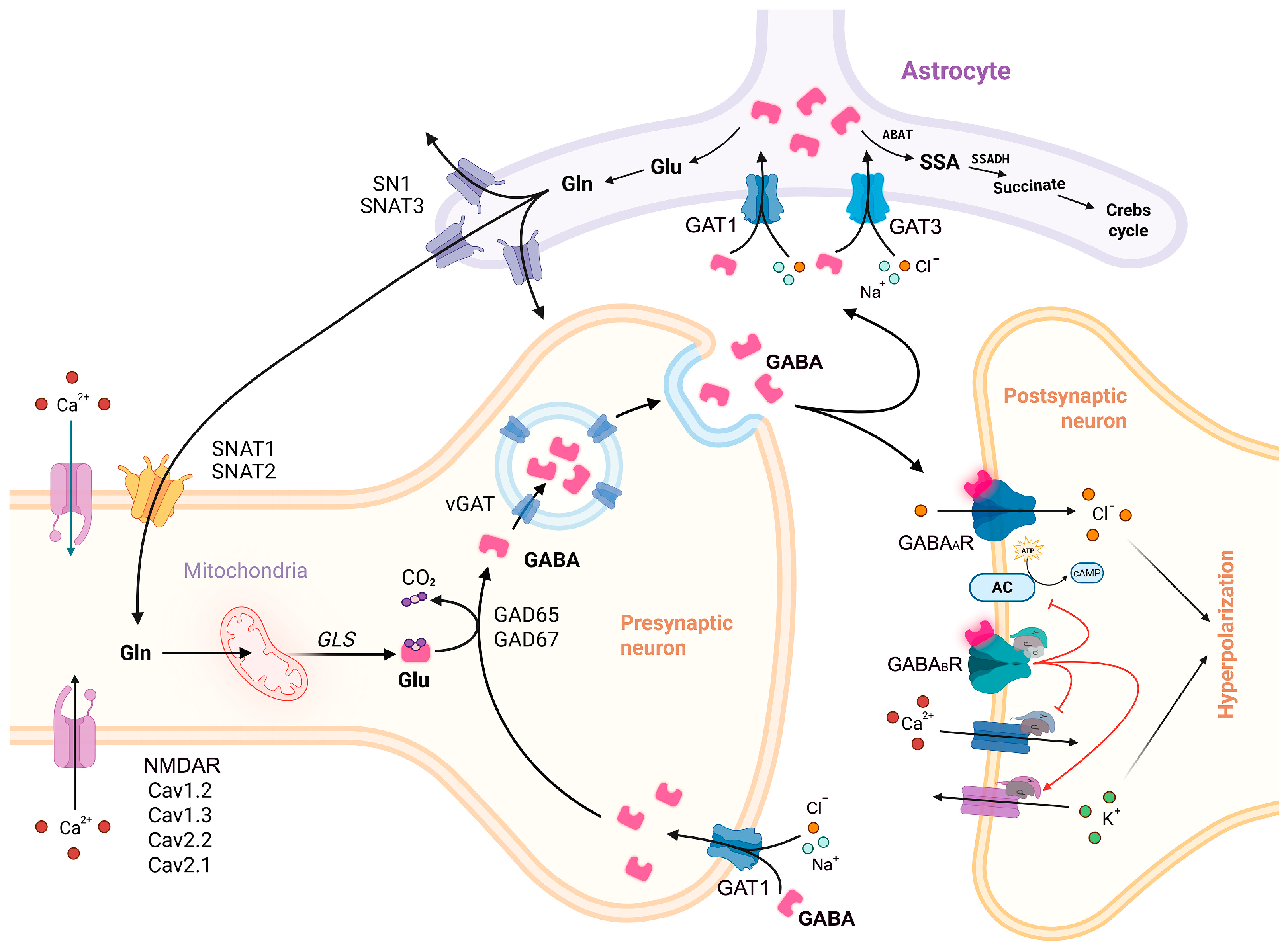
The GABA pathway (Figure 5) can be roughly divided into two main stages—the metabolic stage, also known as “GABA shunt” [202], involving glutamate and GABA synthesis, as well as GABA catabolism, and the signaling stage, involving GABA traffic, release, interaction with the receptors, and reuptake by GATs. Both dysregulation of the signaling stage and impairment of GAD67/65 function have a direct impact on the GABA pathway. Interestingly, involvement of the GABA receptors GABAARs and GABABRs and the key GABA synthesizing enzyme GAD67 in SZ and, apparently, ASD and MDD pathogenesis is unlikely due to the mutations in their ORFs, but rather due to dysregulation of their expression caused either by functional mutations in gene regulatory elements, or by epigenetic perturbations and/or disrupted function of particular transcription factors. In case of GABAARs and GABABRs, this may result in imbalanced production of different subtypes of the GABA receptors and impaired neurogenesis or altered E/I balance within the brain. As for GAD67, we find it plausible that in early embryogenesis additional mechanisms exist, compensating even for polymorphisms in the ORF of its gene, GAD1, and allowing for maintaining the concentrations of GABA at a relatively physiological level. These mechanisms may potentially involve GAD65, fetal GAD1 isoforms, or upregulation of GAD1 expression (which may be prevented by the presence of SNPs in the regulatory elements). Searching for these mechanisms could be important for better understanding neurodevelopment and could give a clue for future searches for new therapeutic approaches for treating or alleviating the symptoms of BD and SZ. Meanwhile, in infancy, when epilepsy usually begins, the apparent compensative mechanisms would weaken, with the pathogenic polymorphisms in GAD1 ORF coming into focus and leading to dramatic decrease in the levels of GABA. We also hypothesize that the GABA vesicular transporter VGAT may be the most vulnerable element of the GABA pathway, as in contrast to GAD67-related epilepsy, predominantly associated with bi-allelic mutations; in case of VGAT, a heterozygous SNP is enough to cause the condition. Intriguingly, some of the pathogenic VGAT variants are characterized by excessive transport activity, thus enhancing GABA release and inhibition. Interestingly, no SZ-associated polymorphisms in VGAT encoding gene SLC32A1 have yet been found, although VGAT level was decreased in both SZ and ASD. This decrease may result from both the existence of so far unidentified SNPs in SLC32A1 regulatory elements or disruption of transcription factors regulating its expression. If no SLC32A1 polymorphisms severe enough to contribute to SZ are ever found, this will most likely indicate that they are lethal. Regardless, additional research is needed to comprehend the role of VGAT in NDDs. Interestingly, a particular number of NDDs, mostly epileptic disorders, are associated with haploinsufficiencies of GABA transporter GATs—GAT1 regulating phasic inhibition, GAT3 regulating tonic inhibition, and BGT1 responsible for transporting betaine through the brain–blood barrier, thus indirectly modulating GAT1 function. The putative explanation for the clinical effect of single mutations in GAT1 implies misfolding and malfunctioning or subsequent degradation of half of the transporter’s molecules produced in the cell, the other half being insufficient for maintaining adequate levels of GABAergic inhibition. What needs to be clarified in this case is the mechanism of action of the three recently described SZ-associated GAT1 variants p.Ala93Thr, p.Arg211Cys, and p.Trp495Leu [203].
Speaking of the metabolic stage (except for the GABA synthesis step), it is reasonable to conclude that, in contrast to the signaling stage, its disruption has only a minor direct effect on GABA signaling and rather causes clinical effects due to accumulation of neurotoxic byproducts, such as excessive glutamate or GHB, with further mitochondrial depletion. Nevertheless, at least in the cerebellum, GLS1 is capable of indirectly enhancing the GABA pathway via the neuroinflammatory pathways TNFα–TNFR1–NF-κB [204]. Furthermore, neuroinflammation may be triggered by exposure to stress; for instance, Stattic, a drug known to downregulate IL-6 expression by inhibiting STAT3 phosphorylation at Tyr705, was shown to suppress epileptic activity in the hippocampal and limbic regions, accompanied by delayed and flat successive spike waves in a GEFS+ mouse model [205]. Moreover, in rats, chronic stress in adolescence, induced by the combination of restraint stress and footshock, and in adulthood caused SZ-like conditions including anxiety-like responses, disrupted cognitive function, and hyper-responsivity of DA neurons [206], the latter hinting at the possible role of stress in depleting the function of the GABA transporter GAT1 [151]. At the same time, acute stress (the threat-of-shock condition) resulted in approximately an 18% decrease in prefrontal GABA and presynaptic downregulation of GABAergic neurotransmission [207]. This indicates that acute and chronic stress affect the GABAergic neurotransmission via distinct mechanisms, but both may potentially accelerate the disease onset and/or aggravate its clinical symptoms in individuals with high genetic risk for psychiatric disorders. Hence, additional research on the interconnections between stress and the GABA pathway may be necessary for developing new therapeutic strategies for treating NDDs.
In sum, as research continues to refine the role of GABA pathway components in the pathogenesis of neurodevelopmental disorders, new therapeutic strategies may offer more effective and individualized interventions for patients with psychiatric disorders characterized by GABAergic dysfunction.

Author Contributions

Conceptualization: E.V.M.; writing—original draft preparation: E.V.M., A.A., A.O.K. and V.A.M.; writing—review and editing: E.V.M. and V.E.G.; visualization: A.A. and E.V.M. All authors have read and agreed to the published version of the manuscript.

Funding

This work was supported by the Russian Science Foundation, grant 25-25-00459 (https://rscf.ru/project/25-25-00459/ (accessed on 25 September 2025).

Institutional Review Board Statement

Not applicable.

Data Availability Statement

The datasets generated during and/or analyzed during the current study are available from the corresponding author on reasonable request.

Conflicts of Interest

The authors declare no conflicts of interest. The funders had no role in the design of the study; in the collection, analyses, or interpretation of data; in the writing of the manuscript; or in the decision to publish the results.

Abbreviations

The following abbreviations are used in this manuscript:
5hmC5-Hydroxymethylcytosine
5mC5-Methylcytosine
ASDAutism Spectrum Disorder
BC(s)Basket Cell(s)
BDBipolar Disorder
BPC(s)Bipolar Cell(s)
BPNDNon-Displaceable Binding Potential
ChC(s)Chandelier Cell(s)
CNSCentral Nervous System
CSFCerebrospinal Fluid
DBC(s)Double Bouquet Cell(s)
DLPFCDorsolateral Prefrontal Cortex
DSDown Syndrome
E/I balanceExcitatory/Inhibitory Neurotransmitter Balance
EMASEpilepsy with Myoclonic-Atonic Seizures
FXSFragile X Syndrome
GEFS+Febrile Seizures Plus
GABAγ-aminobutyric acid
GWASGenome-Wide Association Studies
ID(s)Intellectual Disability(-ies)
IGEIdiopathic Generalized Epilepsy
LOFLoss-of-Function
MC(s)Martinotti Cell(s)
MDDMajor Depressive Disorder
NDD(s)Neurodevelopmental Disorder(s)
NGC(s)Small Button-type Neurogliaform Cell(s)
PETPositron Emission Tomography
PFCPrefrontal Cortex
PLPPyridoxal 5′-Phosphate
PWSPrader–Willi Syndrome
PyC(s)Pyramidal Cell(s)
sIPSCsSpontaneous Inhibitory Postsynaptic Currents
SNPSingle Nucleotide Polymorphism
SSADHDSSADH Deficiency
SVSynaptic Vesicle
SZSchizophrenia

References

  1. Groth, C.L.; Singh, A.; Zhang, Q.; Berman, B.D.; Narayanan, N.S. Gabaergic Modulation in Movement Related Oscillatory Activity: A Review of the Effect Pharmacologically and with Aging. Tremor Other Hyperkinet. Mov. 2021, 11, 48. [Google Scholar] [CrossRef]
  2. Ali, D.N.; Ali, H.M.; Lopez, M.R.; Kang, S.; Choi, D.S. Astrocytic GABAergic Regulation in Alcohol Use and Major Depressive Disorders. Cells 2024, 13, 318. [Google Scholar] [CrossRef]
  3. Benes, F.M.; Berretta, S. GABAergic Interneurons: Implications for Understanding Schizophrenia and Bipolar Disorder. Neuropsychopharmacology 2001, 25, 1–27, Erratum in Neuropsychopharmacol 2001, 25, 453. [Google Scholar] [CrossRef]
  4. Chen, Q.; Deister, C.A.; Gao, X.; Guo, B.; Lynn-Jones, T.; Chen, N.; Wells, M.F.; Liu, R.; Goard, M.J.; Dimidschstein, J.; et al. Dysfunction of Cortical GABAergic Neurons Leads to Sensory Hyper-Reactivity in a Shank3 Mouse Model of ASD. Nat. Neurosci. 2020, 23, 520–532. [Google Scholar] [CrossRef]
  5. Bandler, R.C.; Mayer, C. Deciphering Inhibitory Neuron Development: The Paths to Diversity. Curr. Opin. Neurobiol. 2023, 79, 102691. [Google Scholar] [CrossRef]
  6. Fekete, Z.; Weisz, F.; Karlócai, M.R.; Veres, J.M.; Andrási, T.; Hájos, N. Synaptic Communication within the Microcircuits of Pyramidal Neurons and Basket Cells in the Mouse Prefrontal Cortex. J. Physiol. 2024, 1–22. [Google Scholar] [CrossRef]
  7. Pieraut, S.; Gounko, N.; Sando, R.; Dang, W.; Rebboah, E.; Panda, S.; Madisen, L.; Zeng, H.; Maximov, A. Experience-Dependent Remodeling of Basket Cell Networks in the Dentate Gyrus. Neuron 2014, 84, 107–122. [Google Scholar] [CrossRef] [PubMed]
  8. Veres, J.M.; Nagy, G.A.; Hájos, N. Perisomatic GABAergic Synapses of Basket Cells Effectively Control Principal Neuron Activity in Amygdala Networks. Elife 2017, 6, e20721. [Google Scholar] [CrossRef] [PubMed]
  9. Ribak, C.E.; Seress, L. Five Types of Basket Cell in the Hippocampal Dentate Gyrus: A Combined Golgi and Electron Microscopic Study. J. Neurocytol. 1983, 12, 577–597. [Google Scholar] [CrossRef] [PubMed]
  10. Zhou, J.; Brown, A.M.; Lackey, E.P.; Arancillo, M.; Lin, T.; Sillitoe, R.V. Purkinje Cell Neurotransmission Patterns Cerebellar Basket Cells into Zonal Modules Defined by Distinct Pinceau Sizes. Elife 2020, 9, e55569. [Google Scholar] [CrossRef]
  11. Senft, R.A.; Dymecki, S.M. Neuronal Pericellular Baskets: Neurotransmitter Convergence and Regulation of Network Excitability. Trends Neurosci. 2021, 44, 915–924. [Google Scholar] [CrossRef]
  12. Del Pino, I.; Brotons-Mas, J.R.; Marques-Smith, A.; Marighetto, A.; Frick, A.; Marín, O.; Rico, B. Abnormal Wiring of CCK+ Basket Cells Disrupts Spatial Information Coding. Nat. Neurosci. 2017, 20, 784–792. [Google Scholar] [CrossRef] [PubMed]
  13. Kawaguchi, Y.; Otsuka, T.; Morishima, M.; Ushimaru, M.; Kubota, Y. Control of Excitatory Hierarchical Circuits by Parvalbumin-FS Basket Cells in Layer 5 of the Frontal Cortex: Insights for Cortical Oscillations. J. Neurophysiol. 2019, 121, 2222–2236. [Google Scholar] [CrossRef] [PubMed]
  14. Markram, H.; Toledo-Rodriguez, M.; Wang, Y.; Gupta, A.; Silberberg, G.; Wu, C. Interneurons of the Neocortical Inhibitory System. Nat. Rev. Neurosci. 2004, 5, 793–807. [Google Scholar] [CrossRef]
  15. Pelkey, K.A.; Chittajallu, R.; Craig, M.T.; Tricoire, L.; Wester, J.C.; McBain, C.J. Hippocampal Gabaergic Inhibitory Interneurons. Physiol. Rev. 2017, 97, 1619–1747. [Google Scholar] [CrossRef]
  16. Druga, R.; Salaj, M.; Al-Redouan, A. Parvalbumin—Positive Neurons in the Neocortex: A Review. Physiol. Res. 2023, 72, S173–S191. [Google Scholar] [CrossRef]
  17. Seignette, K.; Jamann, N.; Papale, P.; Terra, H.; Porneso, R.O.; de Kraker, L.; van der Togt, C.; van der Aa, M.; Neering, P.; Ruimschotel, E.; et al. Experience Shapes Chandelier Cell Function and Structure in the Visual Cortex. Elife 2024, 12, RP91153. [Google Scholar] [CrossRef]
  18. Perumal, M.B.; Latimer, B.; Xu, L.; Stratton, P.; Nair, S.; Sah, P. Microcircuit Mechanisms for the Generation of Sharp-Wave Ripples in the Basolateral Amygdala: A Role for Chandelier Interneurons. Cell Rep. 2021, 35, 109106. [Google Scholar] [CrossRef] [PubMed]
  19. Peters, A.; Sethares, C. The Organization of Double Bouquet Cells in Monkey Striate Cortex. J. Neurocytol. 1997, 26, 779–797. [Google Scholar] [CrossRef]
  20. Chrysanthidis, N.; Fiebig, F.; Lansner, A. Introducing Double Bouquet Cells into a Modular Cortical Associative Memory Model. J. Comput. Neurosci. 2019, 47, 223–230. [Google Scholar] [CrossRef]
  21. Lukacs, I.P.; Francavilla, R.; Field, M.; Hunter, E.; Howarth, M.; Horie, S.; Plaha, P.; Stacey, R.; Livermore, L.; Ansorge, O.; et al. Differential Effects of Group III Metabotropic Glutamate Receptors on Spontaneous Inhibitory Synaptic Currents in Spine-Innervating Double Bouquet and Parvalbumin-Expressing Dendrite-Targeting GABAergic Interneurons in Human Neocortex. Cereb. Cortex 2023, 33, 2101–2142. [Google Scholar] [CrossRef]
  22. Tremblay, R.; Lee, S.; Rudy, B. GABAergic Interneurons in the Neocortex: From Cellular Properties to Circuits. Neuron 2016, 91, 260–292. [Google Scholar] [CrossRef]
  23. Warm, D.; Schroer, J.; Sinning, A. Gabaergic Interneurons in Early Brain Development: Conducting and Orchestrated by Cortical Network Activity. Front. Mol. Neurosci. 2022, 14, 807969. [Google Scholar] [CrossRef]
  24. Mancini, V.; Saleh, M.G.; Delavari, F.; Bagautdinova, J.; Eliez, S. Excitatory/Inhibitory Imbalance Underlies Hippocampal Atrophy in Individuals with 22q11.2 Deletion Syndrome with Psychotic Symptoms. Biol. Psychiatry 2023, 94, 569–579. [Google Scholar] [CrossRef] [PubMed]
  25. Ragland, J.D.; Maddock, R.J.; Hurtado, M.Y.; Tanase, C.; Lesh, T.A.; Niendam, T.A.; Carter, C.S.; Ranganath, C. Disrupted GABAergic Facilitation of Working Memory Performance in People with Schizophrenia. Neuroimage Clin. 2020, 25, 102127. [Google Scholar] [CrossRef] [PubMed]
  26. Park, S.; Kang, I.; Edden, R.A.E.; Namgung, E.; Kim, J.; Kim, J. Shorter Sleep Duration Is Associated with Lower GABA Levels in the Anterior Cingulate Cortex. Sleep Med. 2020, 71, 1–7. [Google Scholar] [CrossRef] [PubMed]
  27. Filice, F.; Janickova, L.; Henzi, T.; Bilella, A.; Schwaller, B. The Parvalbumin Hypothesis of Autism Spectrum Disorder. Front. Cell. Neurosci. 2020, 14, 577525. [Google Scholar] [CrossRef]
  28. Gaus, R.; Popal, M.; Heinsen, H.; Schmitt, A.; Falkai, P.; Hof, P.R.; Schmitz, C.; Vollhardt, A. Reduced Cortical Neuron Number and Neuron Density in Schizophrenia with Focus on Area 24: A Post-Mortem Case–Control Study. Eur. Arch. Psychiatry Clin. Neurosci. 2023, 273, 1209–1223. [Google Scholar] [CrossRef]
  29. Rabinovitch, A.; Braunstein, D.; Rabinovitch, R.; Biton, Y. Possible Mechanism of Schizophrenia Origin by Excess GABA and Synaptic Pruning. IBRO Neurosci. Rep. 2023, 15, 126–130. [Google Scholar] [CrossRef]
  30. Qureshi, T.; Bjørkmo, M.; Nordengen, K.; Gundersen, V.; Utheim, T.P.; Watne, L.O.; Storm-Mathisen, J.; Hassel, B.; Chaudhry, F.A. SLC38A1 Conveys Astroglia-Derived Glutamine into GABAergic Interneurons for Neurotransmitter GABA Synthesis. Cells 2020, 9, 1686. [Google Scholar] [CrossRef]
  31. Mackenzie, B.; Schäfer, M.K.H.; Erickson, J.D.; Hedige, M.A.; Weihe, E.; Varoqui, H. Functional Properties and Cellular Distribution of the System A Glutamine Transporter SNAT1 Support Specialized Roles in Central Neurons. J. Biol. Chem. 2003, 278, 23720–23730. [Google Scholar] [CrossRef] [PubMed]
  32. Qureshi, T.; Sørensen, C.; Berghuis, P.; Jensen, V.; Dobszay, M.B.; Farkas, T.; Dalen, K.T.; Guo, C.; Hassel, B.; Utheim, T.P.; et al. The Glutamine Transporter SLC38A1 Regulates GABAergic Neurotransmission and Synaptic Plasticity. Cereb. Cortex 2019, 29, 5166–5179. [Google Scholar] [CrossRef] [PubMed]
  33. Sun, Y.; Lin, Y.; Liang, N.; Xue, Z.; Xu, J.; Lin, L.; Shen, Y.; Li, H.; Liu, J.; Lu, J. Methylome-Wide Association Study of Adolescent Depressive Episode with Psychotic Symptoms and Childhood Trauma. J. Affect Disord. 2025, 370, 439–448. [Google Scholar] [CrossRef]
  34. Jin, L.W.; Horiuchi, M.; Wulff, H.; Liu, X.B.; Cortopassi, G.A.; Erickson, J.D.; Maezawa, I. Dysregulation of Glutamine Transporter SNAT1 in Rett Syndrome Microglia: A Mechanism for Mitochondrial Dysfunction and Neurotoxicity. J. Neurosci. 2015, 35, 2516–2529. [Google Scholar] [CrossRef] [PubMed]
  35. Kawada, K.; Kuramoto, N.; Mimori, S. Possibility That the Onset of Autism Spectrum Disorder Is Induced by Failure of the Glutamine-Glutamate Cycle. Curr. Mol. Pharmacol. 2020, 14, 170–174. [Google Scholar] [CrossRef]
  36. Hidese, S.; Ota, M.; Wakabayashi, C.; Noda, T.; Ozawa, H.; Okubo, T.; Kunugi, H. Effects of Chronic L-Theanine Administration in Patients with Major Depressive Disorder: An Open-Label Study. Acta Neuropsychiatr. 2017, 29, 72–79. [Google Scholar] [CrossRef]
  37. Márquez, J.; Martín-Rufián, M.; Segura, J.A.; Matés, J.M.; Campos-Sandoval, J.A.; Alonso, F.J. Brain Glutaminases. Biomol. Concepts 2010, 1, 3–15. [Google Scholar] [CrossRef]
  38. Wang, Y.; Huang, Y.; Zhao, L.; Li, Y.; Zheng, J. Glutaminase 1 Is Essential for the Differentiation, Proliferation, and Survival of Human Neural Progenitor Cells. Stem Cells Dev. 2014, 23, 2782–2790. [Google Scholar] [CrossRef]
  39. Ji, C.; Tang, Y.; Zhang, Y.; Li, C.; Liang, H.; Ding, L.; Xia, X.; Xiong, L.; Qi, X.R.; Zheng, J.C. Microglial Glutaminase 1 Deficiency Mitigates Neuroinflammation Associated Depression. Brain Behav. Immun. 2022, 99, 231–245. [Google Scholar] [CrossRef]
  40. Ji, C.; Tang, Y.; Zhang, Y.; Huang, X.; Li, C.; Yang, Y.; Wu, Q.; Xia, X.; Cai, Q.; Qi, X.R.; et al. Glutaminase 1 Deficiency Confined in Forebrain Neurons Causes Autism Spectrum Disorder-like Behaviors. Cell Rep. 2023, 42, 112712. [Google Scholar] [CrossRef]
  41. Wang, Y.; Li, Y.; Zhao, R.; Wu, B.; Lanoha, B.; Tong, Z.; Peer, J.; Liu, J.; Xiong, H.; Huang, Y.; et al. Glutaminase C Overexpression in the Brain Induces Learning Deficits, Synaptic Dysfunctions, and Neuroinflammation in Mice. Brain Behav. Immun. 2017, 66, 135–145. [Google Scholar] [CrossRef]
  42. Chen, H.; Fu, S.; Li, X.; Shi, M.; Qian, J.; Zhao, S.; Yuan, P.; Ding, L.; Xia, X.; Zheng, J.C. Microglial Glutaminase 1 Mediates Chronic Restraint Stress-Induced Depression-like Behaviors and Synaptic Damages. Signal Transduct. Target Ther. 2023, 8, 452. [Google Scholar] [CrossRef]
  43. Mingote, S.; Masson, J.; Gellman, C.; Thomsen, G.M.; Lin, C.S.; Merker, R.J.; Gaisler-Salomon, I.; Wang, Y.; Ernst, R.; Hen, R.; et al. Genetic Pharmacotherapy as an Early CNS Drug Development Strategy: Testing Glutaminase Inhibition for Schizophrenia Treatment in Adult Mice. Front. Syst. Neurosci. 2016, 9, 165. [Google Scholar] [CrossRef]
  44. Gaisler-Salomon, I.; Miller, G.M.; Chuhma, N.; Lee, S.; Zhang, H.; Ghoddoussi, F.; Lewandowski, N.; Fairhurst, S.; Wang, Y.; Conjard-Duplany, A.; et al. Glutaminase-Deficient Mice Display Hippocampal Hypoactivity, Insensitivity to Pro-Psychotic Drugs and Potentiated Latent Inhibition: Relevance to Schizophrenia. Neuropsychopharmacology 2009, 34, 2305–2322. [Google Scholar] [CrossRef] [PubMed]
  45. Gao, Y.; Ma, L.; Yuan, J.; Huang, Y.; Ban, Y.; Zhang, P.; Tan, D.; Liang, M.; Li, Z.; Gong, C.; et al. GLS2 Reduces the Occurrence of Epilepsy by Affecting Mitophagy Function in Mouse Hippocampal Neurons. CNS Neurosci. Ther. 2024, 30, e70036. [Google Scholar] [CrossRef] [PubMed]
  46. Trifonov, S.; Yamashita, Y.; Kase, M.; Maruyama, M.; Sugimoto, T. Glutamic Acid Decarboxylase 1 Alternative Splicing Isoforms: Characterization, Expression and Quantification in the Mouse Brain. BMC Neurosci. 2014, 15, 114. [Google Scholar] [CrossRef]
  47. Schwab, C.; Yu, S.; Wong, W.; McGeer, E.G.; McGeer, P.L. GAD65, GAD67, and GABAT Immunostaining in Human Brain and Apparent GAD65 Loss in Alzheimer’s Disease. J. Alzheimers Dis. 2013, 33, 1073–1088. [Google Scholar] [CrossRef]
  48. Mower, G.D.; Guo, Y. Comparison of the Expression of Two Forms of Glutamic Acid Decarboxylase (GAD67 and GAD65) in the Visual Cortex of Normal and Dark-Reared Cats. Dev. Brain Res. 2001, 126, 65–74. [Google Scholar] [CrossRef]
  49. Grone, B.P.; Maruska, K.P. Three Distinct Glutamate Decarboxylase Genes in Vertebrates. Sci. Rep. 2016, 6, 30507. [Google Scholar] [CrossRef] [PubMed]
  50. Asada, H.; Kawamura, Y.; Maruyama, K.; Kume, H.; Ding, R.G.; Ji, F.Y.; Kanbara, N.; Kuzume, H.; Sanbo, M.; Yagi, T.; et al. Mice Lacking the 65 KDa Isoform of Glutamic Acid Decarboxylase (GAD65) Maintain Normal Levels of GAD67 and GABA in Their Brains but Are Susceptible to Seizures. Biochem. Biophys. Res. Commun. 1996, 229, 891–895. [Google Scholar] [CrossRef]
  51. Asada, H.; Kawamura, Y.; Maruyama, K.; Kume, H.; Ding, R.G.; Kanbara, N.; Kuzume, H.; Sanbo, M.; Yagi, T.; Obata, K. Cleft Palate and Decreased Brain γ-Aminobutyric Acid in Mice Lacking the 67-KDa Isoform of Glutamic Acid Decarboxylase. Proc. Natl. Acad. Sci. USA 1997, 94, 6496–6499. [Google Scholar] [CrossRef]
  52. Schwartz, H.L.; Chandonia, J.M.; Kash, S.F.; Kanaani, J.; Tunnell, E.; Domingo, A.; Cohen, F.E.; Banga, J.P.; Madec, A.M.; Richter, W.; et al. High-Resolution Autoreactive Epitope Mapping and Structural Modeling of the 65 KDa Form of Human Glutamic Acid Decarboxylase. J. Mol. Biol. 1999, 287, 983–999. [Google Scholar] [CrossRef]
  53. Shi, Y.; Veit, B.; Bækkeskov, S. Amino Acid Residues 24-31 but Not Palmitoylation of Cysteines 30 and 45 Are Required for Membrane Anchoring of Glutamic Acid Decarboxylase, GAD65. J. Cell Biol. 1994, 124, 927–934. [Google Scholar] [CrossRef]
  54. Wei, J.; Davis, K.M.; Wu, H.; Wu, J.Y. Protein Phosphorylation of Human Brain Glutamic Acid Decarboxylase (GAD)65 and GAD67 and Its Physiological Implications. Biochemistry 2004, 43, 6182–6189. [Google Scholar] [CrossRef] [PubMed]
  55. Magri, C.; Giacopuzzi, E.; La Via, L.; Bonini, D.; Ravasio, V.; Elhussiny, M.E.A.; Orizio, F.; Gangemi, F.; Valsecchi, P.; Bresciani, R.; et al. A Novel Homozygous Mutation in GAD1 Gene Described in a Schizophrenic Patient Impairs Activity and Dimerization of GAD67 Enzyme. Sci. Rep. 2018, 8, 15470, Erratum in Sci. Rep. 2020, 10, 4725. [Google Scholar] [CrossRef] [PubMed]
  56. Kanaani, J.; Diacovo, M.J.; El-Husseini, A.E.D.; Bredt, D.S.; Baekkeskov, S. Palmitoylation Controls Trafficking of GAD65 from Golgi Membranes to Axon-Specific Endosomes and a Rab5a-Dependent Pathway to Presynaptic Clusters. J. Cell Sci. 2004, 117, 2001–2013. [Google Scholar] [CrossRef]
  57. Su, F.; Pfundstein, G.; Sah, S.; Zhang, S.; Keable, R.; Hagan, D.W.; Sharpe, L.J.; Clemens, K.J.; Begg, D.; Phelps, E.A.; et al. Neuronal Growth Regulator 1 (NEGR1) Promotes the Synaptic Targeting of Glutamic Acid Decarboxylase 65 (GAD65). J. Neurochem. 2025, 169, e16279. [Google Scholar] [CrossRef]
  58. Gao, S.F.; Klomp, A.; Wu, J.L.; Swaab, D.F.; Bao, A.M. Reduced GAD65/67 Immunoreactivity in the Hypothalamic Paraventricular Nucleus in Depression: A Postmortem Study. J. Affect Disord. 2013, 149, 422–425. [Google Scholar] [CrossRef] [PubMed]
  59. Du, J.; Duan, S.; Wang, H.; Chen, W.; Zhao, X.; Zhang, A.; Wang, L.; Xuan, J.; Yu, L.; Wu, S.; et al. Comprehensive Analysis of Polymorphisms throughout GAD1 Gene: A Family-Based Association Study in Schizophrenia. J. Neural Transm. 2008, 115, 513–519. [Google Scholar] [CrossRef]
  60. Straub, R.E.; Lipska, B.K.; Egan, M.F.; Goldberg, T.E.; Callicott, J.H.; Mayhew, M.B.; Vakkalanka, R.K.; Kolachana, B.S.; Kleinman, J.E.; Weinberger, D.R. Allelic Variation in GAD1 (GAD67) Is Associated with Schizophrenia and Influences Cortical Function and Gene Expression. Mol. Psychiatry 2007, 12, 854–869. [Google Scholar] [CrossRef]
  61. Hyde, T.M.; Lipska, B.K.; Ali, T.; Mathew, S.V.; Law, A.J.; Metitiri, O.E.; Straub, R.E.; Ye, T.; Colantuoni, C.; Herman, M.M.; et al. Expression of GABA Signaling Molecules KCC2, NKCC1, and GAD1 in Cortical Development and Schizophrenia. J. Neurosci. 2011, 31, 11088–11095. [Google Scholar] [CrossRef]
  62. Tao, R.; Davis, K.N.; Li, C.; Shin, J.H.; Gao, Y.; Jaffe, A.E.; Gondré-Lewis, M.C.; Weinberger, D.R.; Kleinman, J.E.; Hyde, T.M. GAD1 Alternative Transcripts and DNA Methylation in Human Prefrontal Cortex and Hippocampus in Brain Development, Schizophrenia. Mol. Psychiatry 2018, 23, 1496–1505. [Google Scholar] [CrossRef]
  63. Storozheva, Z.I.; Kirenskaya, A.V.; Samylkin, D.V.; Gruden, M.A.; Sewell, R.D.E. Association of GAD1 Gene Polymorphism Rs3749034 with the Information Processing and Cognitive Event-Related Potentials in Schizophrenia Patients and Healthy Subjects. Clin. Neurophysiol. 2024, 166, 142–151. [Google Scholar] [CrossRef]
  64. Moyer, C.E.; Delevich, K.M.; Fish, K.N.; Asafu-Adjei, J.K.; Sampson, A.R.; Dorph-Petersen, K.A.; Lewis, D.A.; Sweet, R.A. Reduced Glutamate Decarboxylase 65 Protein within Primary Auditory Cortex Inhibitory Boutons in Schizophrenia. Biol. Psychiatry 2012, 72, 734–743. [Google Scholar] [CrossRef]
  65. Hansen, N.; Bartels, C.; Teegen, B.; Wiltfang, J.; Malchow, B. Catatonic Schizophrenia Associated with Cerebrospinal GAD65 Autoantibodies: Case Report and Literature Review. Front. Immunol. 2022, 13, 829058. [Google Scholar] [CrossRef]
  66. Yip, J.; Soghomonian, J.J.; Blatt, G.J. Decreased GAD67 MRNA Levels in Cerebellar Purkinje Cells in Autism: Pathophysiological Implications. Acta Neuropathol. 2007, 113, 559–568. [Google Scholar] [CrossRef] [PubMed]
  67. Zhubi, A.; Chen, Y.; Dong, E.; Cook, E.H.; Guidotti, A.; Grayson, D.R. Increased Binding of MeCP2 to the GAD1 and RELN Promoters May Be Mediated by an Enrichment of 5-HmC in Autism Spectrum Disorder (ASD) Cerebellum. Transl. Psychiatry 2014, 4, e349. [Google Scholar] [CrossRef]
  68. Labouesse, M.A.; Dong, E.; Grayson, D.R.; Guidotti, A.; Meyer, U. Maternal Immune Activation Induces GAD1 and GAD2 Promoter Remodeling in the Offspring Prefrontal Cortex. Epigenetics 2015, 10, 1143–1155. [Google Scholar] [CrossRef]
  69. Neuray, C.; Maroofian, R.; Scala, M.; Sultan, T.; Pai, G.S.; Mojarrad, M.; Khashab, H.E.; Deholl, L.; Yue, W.; Alsaif, H.S.; et al. Early-Infantile Onset Epilepsy and Developmental Delay Caused by Bi-Allelic GAD1 Variants. Brain 2020, 143, 2388–2397. [Google Scholar] [CrossRef] [PubMed]
  70. Tröscher, A.R.; Mair, K.M.; Verdú De Juan, L.; Köck, U.; Steinmaurer, A.; Baier, H.; Becker, A.; Blümcke, I.; Finzel, M.; Geis, C.; et al. Temporal Lobe Epilepsy with GAD Antibodies: Neurons Killed by T Cells Not by Complement Membrane Attack Complex. Brain 2023, 146, 1436–1452. [Google Scholar] [CrossRef] [PubMed]
  71. Kajita, Y.; Ono, K.; Kaneda, S.; Mushiake, H. Pterostilben Upregulates GAD67-Mediated GABA Synthesis in Hippocampal Parvalbumin-Positive Cells. Neuroscience 2025, 573, 482–490. [Google Scholar] [CrossRef] [PubMed]
  72. Nieoczym, D.; Socała, K.; Zelek-Molik, A.; Pieróg, M.; Przejczowska-Pomierny, K.; Szafarz, M.; Wyska, E.; Nalepa, I.; Wlaź, P. Anticonvulsant Effect of Pterostilbene and Its Influence on the Anxiety- and Depression-like Behavior in the Pentetrazol-Kindled Mice: Behavioral, Biochemical, and Molecular Studies. Psychopharmacology 2021, 238, 3167–3181. [Google Scholar] [CrossRef]
  73. Ku, T.H.; Lee, Y.J.; Wang, S.J.; Fan, C.H.; Tien, L.T. Effect of honokiol on activity of GAD(65) and GAD(67) in the cortex and hippocampus of mice. Phytomedicine 2011, 18, 1126–1129. [Google Scholar] [CrossRef]
  74. Vega-García, A.; Neri-Gómez, T.; Buzoianu-Anguiano, V.; Guerra-Araiza, C.; Segura-Uribe, J.; Feria-Romero, I.; Orozco-Suarez, S. Electroacupuncture Reduces Seizure Activity and Enhances GAD 67 and Glutamate Transporter Expression in Kainic Acid Induced Status Epilepticus in Infant Rats. Behav. Sci. 2019, 9, 68. [Google Scholar] [CrossRef]
  75. Huang, H.S.; Matevossian, A.; Whittle, C.; Se, Y.K.; Schumacher, A.; Baker, S.P.; Akbarian, S. Prefrontal Dysfunction in Schizophrenia Involves Mixed-Lineage Leukemia 1-Regulated Histone Methylation at GABAergic Gene Promoters. J. Neurosci. 2007, 27, 11254–11262. [Google Scholar] [CrossRef]
  76. Zaitsev, A.V.; Povysheva, N.V.; Lewis, D.A.; Krimer, L.S. P/Q-Type, but Not N-Type, Calcium Channels Mediate GABA Release from Fast-Spiking Interneurons to Pyramidal Cells in Rat Prefrontal Cortex. J. Neurophysiol. 2007, 97, 3567–3573. [Google Scholar] [CrossRef]
  77. Gopal, N.; Leitz, J.; Wang, C.; Esquivies, L.; Pfuetzner, R.A.; Brunger, A.T. A New Method for Isolation and Purification of Fusion-Competent Inhibitory Synaptic Vesicles. Curr. Res. Physiol. 2024, 7, 100121. [Google Scholar] [CrossRef]
  78. McIntire, S.L.; Reimer, R.J.; Schuske, K.; Edwards, R.H.; Jorgensen, E.M. Identification and Characterization of the Vesicular GABA Transporter. Nature 1997, 389, 870–876. [Google Scholar] [CrossRef] [PubMed]
  79. Wojcik, S.M.; Katsurabayashi, S.; Guillemin, I.; Friauf, E.; Rosenmund, C.; Brose, N.; Rhee, J.S. A Shared Vesicular Carrier Allows Synaptic Corelease of GABA and Glycine. Neuron 2006, 50, 575–587. [Google Scholar] [CrossRef] [PubMed]
  80. Martens, H.; Weston, M.C.; Boulland, J.L.; Grønborg, M.; Grosche, J.; Kacza, J.; Hoffmann, A.; Matteoli, M.; Takamori, S.; Harkany, T.; et al. Unique Luminal Localization of VGAT-C Terminus Allows for Selective Labeling of Active Cortical GABAergic Synapses. J. Neurosci. 2008, 28, 13125–13131. [Google Scholar] [CrossRef]
  81. Chaudhry, F.A.; Reimer, R.J.; Bellocchio, E.E.; Danbolt, N.C.; Osen, K.K.; Edwards, R.H.; Storm-Mathisen, J. The Vesicular GABA Transporter, VGAT, Localizes to Synaptic Vesicles in Sets of Glycinergic as Well as GABAergic Neurons. J. Neurosci. 1998, 18, 9733–9750. [Google Scholar] [CrossRef] [PubMed]
  82. Fattorini, G.; Antonucci, F.; Menna, E.; Matteoli, M.; Conti, F. Co-Expression of VGLUT1 and VGAT Sustains Glutamate and GABA Co-Release and Is Regulated by Activity in Cortical Neurons. J. Cell Sci. 2015, 128, 1669–1673. [Google Scholar] [CrossRef]
  83. Xu, J.; Jo, A.; DeVries, R.P.; Deniz, S.; Cherian, S.; Sunmola, I.; Song, X.; Marshall, J.J.; Gruner, K.A.; Daigle, T.L.; et al. Intersectional Mapping of Multi-Transmitter Neurons and Other Cell Types in the Brain. Cell Rep. 2022, 40, 111036. [Google Scholar] [CrossRef] [PubMed]
  84. Heron, S.E.; Regan, B.M.; Harris, R.V.; Gardner, A.E.; Coleman, M.J.; Bennett, M.F.; Grinton, B.E.; Helbig, K.L.; Sperling, M.R.; Haut, S.; et al. Association of SLC32A1 Missense Variants with Genetic Epilepsy with Febrile Seizures Plus. Neurology 2021, 96, e2251–e2260. [Google Scholar] [CrossRef]
  85. Platzer, K.; Sticht, H.; Bupp, C.; Ganapathi, M.; Pereira, E.M.; Le Guyader, G.; Bilan, F.; Henderson, L.B.; Lemke, J.R.; Taschenberger, H.; et al. De Novo Missense Variants in SLC32A1 Cause a Developmental and Epileptic Encephalopathy Due to Impaired GABAergic Neurotransmission. Ann. Neurol. 2022, 92, 958–973. [Google Scholar] [CrossRef] [PubMed]
  86. Chen, M.; Shi, J.; Liu, T.; Liu, J.; Liu, Y.; Li, J.; Luo, Y.; Luo, J.; Li, X.; Gong, H.; et al. Astragaloside IV Ameliorates Autism-like Behaviors in BTBR Mice by Modulating Camk2n2-Dependent OXPHOS and Neurotransmission in the MPFC. J. Adv. Res. 2025. [Google Scholar] [CrossRef]
  87. Alshammari, T.K.; Alshammari, M.A.; Nenov, M.N.; Hoxha, E.; Cambiaghi, M.; Marcinno, A.; James, T.F.; Singh, P.; Labate, D.; Li, J.; et al. Genetic Deletion of Fibroblast Growth Factor 14 Recapitulates Phenotypic Alterations Underlying Cognitive Impairment Associated with Schizophrenia. Transl. Psychiatry 2016, 6, e806. [Google Scholar] [CrossRef]
  88. San Martín, A.; Pagani, M.R. Understanding Intellectual Disability through RASopathies. J. Physiol. Paris 2014, 108, 232–239. [Google Scholar] [CrossRef]
  89. Ryu, H.H.; Kang, M.; Hwang, K.D.; Jang, H.B.; Kim, S.J.; Lee, Y.S. Neuron Type-Specific Expression of a Mutant KRAS Impairs Hippocampal-Dependent Learning and Memory. Sci. Rep. 2020, 10, 17730. [Google Scholar] [CrossRef]
  90. Cui, Y.; Costa, R.M.; Murphy, G.G.; Elgersma, Y.; Zhu, Y.; Gutmann, D.H.; Parada, L.F.; Mody, I.; Silva, A.J. Neurofibromin Regulation of ERK Signaling Modulates GABA Release and Learning. Cell 2008, 135, 549–560. [Google Scholar] [CrossRef]
  91. Young, A.B.; Chu, D. Distribution of GABAA and GABAB Receptors in Mammalian Brain: Potential Targets for Drug Development. Drug Dev. Res. 1990, 21, 161–167. [Google Scholar] [CrossRef]
  92. Mortensen, M.; Patel, B.; Smart, T.G. GABA Potency at GABA A Receptors Found in Synaptic and Extrasynaptic Zones. Front. Cell. Neurosci. 2012, 6, 1. [Google Scholar] [CrossRef]
  93. Petri, S.; Krampfl, K.; Dengler, R.; Bufler, J.; Weindl, A.; Arzberger, T. Human GABAA Receptors on Dopaminergic Neurons in the Pars Compacta of the Substantia Nigra. J. Comp. Neurol. 2002, 452, 360–366. [Google Scholar] [CrossRef]
  94. Gao, B.; Fritschy, J.M.; Benke, D.; Mohler, H. Neuron-Specific Expression of GABAA-Receptor Subtypes: Differential Association of the A1- and A3-Subunits with Serotonergic and Gabaergic Neurons. Neuroscience 1993, 54, 881–892. [Google Scholar] [CrossRef] [PubMed]
  95. Ghit, A.; Assal, D.; Al-Shami, A.S.; Hussein, D.E.E. GABAA Receptors: Structure, Function, Pharmacology, and Related Disorders. J. Genet. Eng. Biotechnol. 2021, 19, 123. [Google Scholar] [CrossRef]
  96. McKernan, R.M.; Whiting, P.J. Which GABAA-Receptor Subtypes Really Occur in the Brain? Trends Neurosci. 1996, 19, 139–143. [Google Scholar] [CrossRef] [PubMed]
  97. Song, J.J.; Curtis, M.A.; Faull, R.L.M.; Waldvogel, H.J. The Regional and Cellular Distribution of GABAA Receptor Subunits in the Human Amygdala. J. Chem. Neuroanat. 2022, 126, 102185. [Google Scholar] [CrossRef]
  98. Yang, L.; Shen, H.; Merlin, L.R.; Smith, S.S. Pubertal Expression of A4βδ GABAA Receptors Reduces Seizure-Like Discharges in CA1 Hippocampus. Sci. Rep. 2016, 6, 31928. [Google Scholar] [CrossRef]
  99. Sur, C.; Quirk, K.; Dewar, D.; Atack, J.; Mckernan, R. Rat and Human Hippocampal A5 Subunit-Containing γ-Aminobutyric Acid(A) Receptors Have A5β3γ2 Pharmacological Characteristics. Mol. Pharmacol. 1998, 54, 928–933. [Google Scholar] [CrossRef] [PubMed]
  100. Bahn, S.; Jones, A.; Wisden, W. Directing Gene Expression to Cerebellar Granule Cells Using γ-Aminobutyric Acid Type A Receptor A6 Subunit Transgenes. Proc. Natl. Acad. Sci. USA 1997, 94, 9417–9421. [Google Scholar] [CrossRef]
  101. Enz, R. GABAc Receptor p Subunits Are Heterogeneously Expressed in the Human CMS and Form Homo- And Heterooligomers with Distinct Physical Properties. Europ. J. Neurosci. 1999, 11, 41–50. [Google Scholar] [CrossRef]
  102. Nakamura, Y.; Darnieder, L.M.; Deeb, T.Z.; Moss, S.J. Regulation of GABAARs by Phosphorylation. Adv. Pharmacol. 2015, 72, 97–146. [Google Scholar] [CrossRef]
  103. Goldschen-Ohm, M.P. Benzodiazepine Modulation of GABAA Receptors: A Mechanistic Perspective. Biomolecules 2022, 12, 1784. [Google Scholar] [CrossRef]
  104. Ochoa-de la Paz, L.D.; Gulias-Cañizo, R.; D’Abril Ruíz-Leyja, E.; Sánchez-Castillo, H.; Parodí, J. The Role of GABA Neurotransmitter in the Human Central Nervous System, Physiology, and Pathophysiology. Rev. Mex. Neuroci. 2021, 22, 67–76. [Google Scholar] [CrossRef]
  105. Li, Y.; Yu, L.; Zhao, L.; Zeng, F.; Liu, Q.S. Resveratrol Modulates Cocaine-Induced Inhibitory Synaptic Plasticity in VTA Dopamine Neurons by Inhibiting Phosphodiesterases (PDEs). Sci. Rep. 2017, 7, 15657. [Google Scholar] [CrossRef]
  106. Jang, S.H.; Park, S.J.; Kim, K.A.; Han, S.K. Resveratrol Activates GABAA and/or Glycine Receptors on Substantia Gelatinosa Neurons of the Subnucleus Caudalis in Mice. Nat. Prod. Res. 2022, 36, 5788–5792. [Google Scholar] [CrossRef] [PubMed]
  107. Decui, L.; Garbinato, C.L.L.; Schneider, S.E.; Mazon, S.C.; Almeida, E.R.; Aguiar, G.P.S.; Müller, L.G.; Oliveira, J.V.; Siebel, A.M. Micronized Resveratrol Shows Promising Effects in a Seizure Model in Zebrafish and Signalizes an Important Advance in Epilepsy Treatment. Epilepsy Res. 2020, 159, 106243. [Google Scholar] [CrossRef] [PubMed]
  108. Fatemi, S.H.; Folsom, T.D.; Rooney, R.J.; Thuras, P.D. Expression of GABAa A2-, B1-and e-Receptors Are Altered Significantly in the Lateral Cerebellum of Subjects with Schizophrenia, Major Depression and Bipolar Disorder. Transl. Psychiatry 2013, 3, e303. [Google Scholar] [CrossRef] [PubMed]
  109. Fatemi, S.H.; Folsom, T.D.; Thuras, P.D. GABAA and GABAB Receptor Dysregulation in Superior Frontal Cortex of Subjects with Schizophrenia and Bipolar Disorder. Synapse 2017, 71. [Google Scholar] [CrossRef]
  110. Marques, T.R.; Ashok, A.H.; Angelescu, I.; Borgan, F.; Myers, J.; Lingford-Hughes, A.; Nutt, D.J.; Veronese, M.; Turkheimer, F.E.; Howes, O.D. GABA-A Receptor Differences in Schizophrenia: A Positron Emission Tomography Study Using [11C]Ro154513. Mol. Psychiatry 2021, 26, 2616–2625. [Google Scholar] [CrossRef]
  111. Mueller, T.M.; Haroutunian, V.; Meador-Woodruff, J.H. N-Glycosylation of GABAA Receptor Subunits Is Altered in Schizophrenia. Neuropsychopharmacology 2014, 39, 528–537. [Google Scholar] [CrossRef]
  112. Chiou, L.C.; Sieghart, W. IUPHAR Review: Alpha6-Containing GABAA Receptors—Novel Targets for the Treatment of Schizophrenia. Pharmacol. Res. 2025, 213, 107613. [Google Scholar] [CrossRef]
  113. Moradi, M.; Saidijam, M.; Yadegarazari, R.; Jahangard, L.; Seifi, M.; Matinnia, N.; Ghaleiha, A. Genes Encoding GABA-β and HT1D Receptors in Bipolar I (Manic Phase) Patients. Basic Clin. Neurosci. 2018, 9, 129–134. [Google Scholar] [CrossRef] [PubMed]
  114. Maillard, P.Y.; Baer, S.; Schaefer, É.; Desnous, B.; Villeneuve, N.; Lépine, A.; Fabre, A.; Lacoste, C.; El Chehadeh, S.; Piton, A.; et al. Molecular and Clinical Descriptions of Patients with GABAA Receptor Gene Variants (GABRA1, GABRB2, GABRB3, GABRG2): A Cohort Study, Review of Literature, and Genotype–Phenotype Correlation. Epilepsia 2022, 63, 2519–2533. [Google Scholar] [CrossRef] [PubMed]
  115. Qu, S.; Catron, M.; Zhou, C.; Janve, V.; Shen, W.; Howe, R.K.; Macdonald, R.L. GABAAreceptor Β3 Subunit Mutation D120N Causes Lennox-Gastaut Syndrome in Knock-in Mice. Brain Commun. 2020, 2, fcaa028. [Google Scholar] [CrossRef]
  116. Gonda, X.; Sarginson, J.; Eszlari, N.; Petschner, P.; Toth, Z.G.; Baksa, D.; Hullam, G.; Anderson, I.M.; Deakin, J.F.W.; Juhasz, G.; et al. A New Stress Sensor and Risk Factor for Suicide: The T Allele of the Functional Genetic Variant in the GABRA6 Gene. Sci. Rep. 2017, 7, 12887. [Google Scholar] [CrossRef]
  117. Zhang, T.; Li, J.; Yu, H.; Shi, Y.; Li, Z.; Wang, L.; Wang, Z.; Lu, T.; Wang, L.; Yue, W.; et al. Meta-Analysis of GABRB2 Polymorphisms and the Risk of Schizophrenia Combined with GWAS Data of the Han Chinese Population and Psychiatric Genomics Consortium. PLoS ONE 2018, 13, e0198690. [Google Scholar] [CrossRef] [PubMed]
  118. Roof, E.; Stone, W.; MacLean, W.; Feurer, I.D.; Thompson, T.; Butler, M.G. Intellectual Characteristics of Prader-Willi Syndrome: Comparison of Genetic Subtypes. J. Intellect. Disabil. Res. 2000, 44 Pt 1, 25–30. [Google Scholar] [CrossRef]
  119. Lucignani, G.; Panzacchi, A.; Bosio, L.; Moresco, R.M.; Ravasi, L.; Coppa, I.; Chiumello, G.; Frey, K.; Koeppe, R.; Fazio, F. GABAA Receptor Abnormalities in Prader-Willi Syndrome Assessed with Positron Emission Tomography and [11C]Flumazenil. Neuroimage 2004, 22, 22–28. [Google Scholar] [CrossRef]
  120. Ho, A.Y.; Dimitropoulos, A. Clinical Management of Behavioral Characteristics of Prader-Willi Syndrome. Neuropsychiatr. Dis. Treat. 2010, 6, 107–118. [Google Scholar] [CrossRef]
  121. Rice, L.J.; Lagopoulos, J.; Brammer, M.; Einfeld, S.L. Reduced Gamma-Aminobutyric Acid Is Associated with Emotional and Behavioral Problems in Prader-Willi Syndrome. Am. J. Med. Genet. B Neuropsychiatr. Genet. 2016, 171, 1041–1048. [Google Scholar] [CrossRef]
  122. Paluszkiewicz, S.M.; Martin, B.S.; Huntsman, M.M. Fragile X Syndrome: The GABAergic System and Circuit Dysfunction. Dev. Neurosci. 2011, 33, 349–364. [Google Scholar] [CrossRef]
  123. D’Hulst, C.; De Geest, N.; Reeve, S.P.; Van Dam, D.; De Deyn, P.P.; Hassan, B.A.; Kooy, R.F. Decreased Expression of the GABAA Receptor in Fragile X Syndrome. Brain Res. 2006, 1121, 238–245. [Google Scholar] [CrossRef] [PubMed]
  124. Zhang, N.; Peng, Z.; Tong, X.; Lindemeyer, A.K.; Cetina, Y.; Huang, C.S.; Olsen, R.W.; Otis, T.S.; Houser, C.R. Decreased Surface Expression of the δ Subunit of the GABAA Receptor Contributes to Reduced Tonic Inhibition in Dentate Granule Cells in a Mouse Model of Fragile X Syndrome. Exp. Neurol. 2017, 297, 168–178. [Google Scholar] [CrossRef]
  125. D’Hulst, C.; Heulens, I.; Van Der Aa, N.; Goffin, K.; Koole, M.; Porke, K.; Van De Velde, M.; Rooms, L.; Van Paesschen, W.; Van Esch, H.; et al. Positron Emission Tomography (PET) Quantification of GABAA Receptors in the Brain of Fragile X Patients. PLoS ONE 2015, 10, e0131486. [Google Scholar] [CrossRef]
  126. Hannan, S.B.; Lana-Elola, E.; Watson-Scales, S.; Fisher, E.M.C.; Tybulewicz, V.L.J.; Smart, T.G. Hippocampal Circuit-Specific Enhancement of GABA-Inhibition Caused by Discrete Gene Regions in a Down Syndrome Model. bioRxiv 2025. [Google Scholar] [CrossRef]
  127. Silbereis, J.C.; Nobuta, H.; Tsai, H.H.; Heine, V.M.; McKinsey, G.L.; Meijer, D.H.; Howard, M.A.; Petryniak, M.A.; Potter, G.B.; Alberta, J.A.; et al. Olig1 Function Is Required to Repress Dlx1/2 and Interneuron Production in Mammalian Brain. Neuron 2014, 81, 574–587. [Google Scholar] [CrossRef]
  128. Souchet, B.; Guedj, F.; Sahún, I.; Duchon, A.; Daubigney, F.; Badel, A.; Yanagawa, Y.; Barallobre, M.J.; Dierssen, M.; Yu, E.; et al. Excitation/Inhibition Balance and Learning Are Modified by Dyrk1a Gene Dosage. Neurobiol. Dis. 2014, 69, 65–75. [Google Scholar] [CrossRef]
  129. Goeldner, C.; Kishnani, P.S.; Skotko, B.G.; Casero, J.L.; Hipp, J.F.; Derks, M.; Hernandez, M.C.; Khwaja, O.; Lennon-Chrimes, S.; Noeldeke, J.; et al. A Randomized, Double-Blind, Placebo-Controlled Phase II Trial to Explore the Effects of a GABAA-A5 NAM (Basmisanil) on Intellectual Disability Associated with Down Syndrome. J. Neurodev. Disord. 2022, 14, 10. [Google Scholar] [CrossRef] [PubMed]
  130. Harashima, C.; Jacobowitz, D.M.; Witta, J.; Borke, R.C.; Best, T.K.; Siarey, R.J.; Galdzicki, Z. Abnormal Expression of the G-Protein-Activated Inwardly Rectifying Potassium Channel 2 (GIRK2) in Hippocampus, Frontal Cortex, and Substantia Nigra of Ts65Dn Mouse: A Model of Down Syndrome. J. Comp. Neurol. 2006, 494, 815–833. [Google Scholar] [CrossRef] [PubMed]
  131. Cramer, N.P.; Best, T.K.; Stoffel, M.; Siarey, R.J.; Galdzicki, Z. GABA B -GIRK2-Mediated Signaling in down Syndrome. Adv. Pharmacol. 2010, 58, 397–426. [Google Scholar] [CrossRef]
  132. Charles, K.J.; Deuchars, J.; Davies, C.H.; Pangalos, M.N. GABAB Receptor Subunit Expression in Glia. Mol. Cell. Neurosci. 2003, 24, 214–223. [Google Scholar] [CrossRef]
  133. Breton, J.D.; Stuart, G.J. GABAB Receptors in Neocortical and Hippocampal Pyramidal Neurons Are Coupled to Different Potassium Channels. Eur. J. Neurosci. 2017, 46, 2859–2866. [Google Scholar] [CrossRef]
  134. Takahashi, A.; Shimamoto, A.; Boyson, C.O.; DeBold, J.F.; Miczek, K.A. GABAB Receptor Modulation of Serotonin Neurons in the Dorsal Raphé Nucleus and Escalation of Aggression in Mice. J. Neurosci. 2010, 30, 11771–11780. [Google Scholar] [CrossRef] [PubMed]
  135. Waldvogel, H.J.; Baer, K.; Faull, R.L.M. The Localization of Inhibitory Neurotransmitter Receptors on Dopaminergic Neurons of the Human Substantia Nigra. J. Neural. Transm. Suppl. 2009, 73, 59–70. [Google Scholar] [CrossRef]
  136. Evenseth, L.S.M.; Gabrielsen, M.; Sylte, I. The GABAB Receptor—Structure, Ligand Binding and Drug Development. Molecules 2020, 25, 3093. [Google Scholar] [CrossRef]
  137. Fatemi, S.H.; Folsom, T.D.; Thuras, P.D. Deficits in GABAB Receptor System in Schizophrenia and Mood Disorders: A Postmortem Study. Schizophr. Res. 2011, 128, 37–43. [Google Scholar] [CrossRef]
  138. Gumerov, V.; Hegyi, H. MicroRNA-Derived Network Analysis of Differentially Methylated Genes in Schizophrenia, Implicating GABA Receptor B1 [GABBR1] and Protein Kinase B [AKT1]. Biol. Direct. 2015, 10, 59. [Google Scholar] [CrossRef]
  139. Miyazawa, A.; Kanahara, N.; Kogure, M.; Otsuka, I.; Okazaki, S.; Watanabe, Y.; Yamasaki, F.; Nakata, Y.; Oda, Y.; Hishimoto, A.; et al. A Preliminary Genetic Association Study of GAD1 and GABAB Receptor Genes in Patients with Treatment-Resistant Schizophrenia. Mol. Biol. Rep. 2022, 49, 2015–2024. [Google Scholar] [CrossRef] [PubMed]
  140. Zapata, J.; Moretto, E.; Hannan, S.; Murru, L.; Longatti, A.; Mazza, D.; Benedetti, L.; Fossati, M.; Heise, C.; Ponzoni, L.; et al. Epilepsy and Intellectual Disability Linked Protein Shrm4 Interaction with GABA B Rs Shapes Inhibitory Neurotransmission. Nat. Commun. 2017, 8, 14536. [Google Scholar] [CrossRef] [PubMed]
  141. Kolvenbach, C.M.; Felger, T.; Schierbaum, L.; Thiffault, I.; Pastinen, T.; Szczepańska, M.; Zaniew, M.; Adamczyk, P.; Bayat, A.; Yilmaz, Ö.; et al. X-Linked Variations in SHROOM4 Are Implicated in Congenital Anomalies of the Urinary Tract and the Anorectal, Cardiovascular and Central Nervous Systems. J. Med. Genet. 2023, 60, 587–596. [Google Scholar] [CrossRef]
  142. Kokkonen, H.; Siren, A.; Määttä, T.; Kamila Kadlubowska, M.; Acharya, A.; Nouel-Saied, L.M.; Leal, S.M.; Järvelä, I.; Schrauwen, I. Identification of Microduplications at Xp21.2 and Xq13.1 in Neurodevelopmental Disorders. Mol. Genet. Genomic. Med. 2021, 9, e1703. [Google Scholar] [CrossRef]
  143. Jomura, R.; Akanuma, S.I.; Tachikawa, M.; Hosoya, K.I. SLC6A and SLC16A Family of Transporters: Contribution to Transport of Creatine and Creatine Precursors in Creatine Biosynthesis and Distribution. Biochim. Biophys. Acta Biomembr. 2022, 1864, 183840. [Google Scholar] [CrossRef]
  144. Pramod, A.B.; Foster, J.; Carvelli, L.; Henry, L.K. SLC6 Transporters: Structure, Function, Regulation, Disease Association and Therapeutics. Mol. Aspects Med. 2013, 34, 197–219. [Google Scholar] [CrossRef]
  145. Yadav, R.; Han, G.W.; Gati, C. Molecular Basis of Human GABA Transporter 3 Inhibition. Nat. Commun. 2025, 16, 3830. [Google Scholar] [CrossRef]
  146. Damgaard, M.; Al-Khawaja, A.; Vogensen, S.B.; Jurik, A.; Sijm, M.; Lie, M.E.K.; Bæk, M.I.; Rosenthal, E.; Jensen, A.A.; Ecker, G.F.; et al. Identification of the First Highly Subtype-Selective Inhibitor of Human GABA Transporter GAT3. ACS Chem. Neurosci. 2015, 6, 1591–1599. [Google Scholar] [CrossRef]
  147. Motiwala, Z.; Aduri, N.G.; Shaye, H.; Han, G.W.; Lam, J.H.; Katritch, V.; Cherezov, V.; Gati, C. Structural Basis of GABA Reuptake Inhibition. Nature 2022, 606, 820–826, Erratum in Nature 2022, 608, E15. [Google Scholar] [CrossRef] [PubMed]
  148. Łątka, K.; Jończyk, J.; Bajda, M. Structure Modeling of γ-Aminobutyric Acid Transporters—Molecular Basics of Ligand Selectivity. Int. J. Biol. Macromol. 2020, 158, 1380–1389. [Google Scholar] [CrossRef] [PubMed]
  149. Shah, N.; Kasture, A.S.; Fischer, F.P.; Sitte, H.H.; Hummel, T.; Sucic, S. A Transporter’s Doom or Destiny: SLC6A1 in Health and Disease, Novel Molecular Targets and Emerging Therapeutic Prospects. Front. Mol. Neurosci. 2024, 17, 1466694. [Google Scholar] [CrossRef] [PubMed]
  150. Fattorini, G.; Melone, M.; Conti, F. A Reappraisal of GAT-1 Localization in Neocortex. Front. Cell. Neurosci. 2020, 14, 9. [Google Scholar] [CrossRef]
  151. Melani, R.; Tritsch, N.X. Inhibitory Co-Transmission from Midbrain Dopamine Neurons Relies on Presynaptic GABA Uptake. Cell. Rep. 2022, 39, 110716. [Google Scholar] [CrossRef]
  152. Limani, F.; Srinivasan, S.; Hanzlova, M.; La Batide-Alanore, S.; Klotz, S.; Hnaskoxs, T.S.; Steinkellner, T. Evidence for Low Affinity of GABA at the Vesicular Monoamine Transporter VMAT2. Implications for Transmitter Co-Release from Dopamine Neurons. bioRxiv 2024. [Google Scholar] [CrossRef]
  153. Tritsch, N.X.; Ding, J.B.; Sabatini, B.L. Dopaminergic Neurons Inhibit Striatal Output through Non-Canonical Release of GABA. Nature 2012, 490, 262–266. [Google Scholar] [CrossRef]
  154. Zhang, L.; Wang, W.; Ruan, Y.; Li, Z.; Yanjun; Ji, G.J.; Tian, Y.; Wang, K. Hyperactivity and Altered Functional Connectivity of the Ventral Striatum in Schizophrenia Compared with Bipolar Disorder: A Resting State FMRI Study. Psychiatry Res. Neuroimaging 2024, 345, 111881. [Google Scholar] [CrossRef] [PubMed]
  155. Imoukhuede, P.I.; Moss, F.J.; Michael, D.J.; Chow, R.H.; Lester, H.A. Ezrin Mediates Tethering of the γ-Aminobutyric Acid Transporter GAT1 to Actin Filaments via a C-Terminal PDZ-Interacting Domain. Biophys. J. 2009, 96, 2949–2960. [Google Scholar] [CrossRef] [PubMed]
  156. Bragina, L.; Marchionni, I.; Omrani, A.; Cozzi, A.; Pellegrini-Giampietro, D.E.; Cherubini, E.; Conti, F. GAT-1 Regulates Both Tonic and Phasic GABAA Receptor-Mediated Inhibition in the Cerebral Cortex. J. Neurochem. 2008, 105, 1781–1793. [Google Scholar] [CrossRef] [PubMed]
  157. Silva, D.B.; Trinidad, M.; Ljungdahl, A.; Revalde, J.L.; Berguig, G.Y.; Wallace, W.; Patrick, C.S.; Bomba, L.; Arkin, M.; Dong, S.; et al. Haploinsufficiency Underlies the Neurodevelopmental Consequences of SLC6A1 Variants. Am. J. Hum. Genet. 2024, 111, 1222–1238. [Google Scholar] [CrossRef]
  158. Fischer, F.P.; Kasture, A.S.; Hummel, T.; Sucic, S. Molecular and Clinical Repercussions of GABA Transporter 1 Variants Gone Amiss: Links to Epilepsy and Developmental Spectrum Disorders. Front. Mol. Biosci. 2022, 9, 834498. [Google Scholar] [CrossRef]
  159. Mermer, F.; Poliquin, S.; Rigsby, K.; Rastogi, A.; Shen, W.; Romero-Morales, A.; Nwosu, G.; McGrath, P.; Demerast, S.; Aoto, J.; et al. Common Molecular Mechanisms of SLC6A1 Variant-Mediated Neurodevelopmental Disorders in Astrocytes and Neurons. Brain 2021, 144, 2499–2512. [Google Scholar] [CrossRef]
  160. Mikhailova, V.; Kondratyev, N.; Alfimova, M.; Kaleda, V.; Lezheiko, T.; Ublinsky, M.; Ushakov, V.; Lebedeva, I.; Galiakberova, A.; Artyuhov, A.; et al. The SLC6A1 Mutation Schizophrenia Case—A Comprehensive Case Study with IPSC Generation. Eur. Psychiatry 2024, 67, S764–S765. [Google Scholar] [CrossRef]
  161. Schleimer, S.B.; Hinton, T.; Dixon, G.; Johnston, G.A.R. GABA Transporters GAT-1 and GAT-3 in the Human Dorsolateral Prefrontal Cortex in Schizophrenia. Neuropsychobiology 2004, 50, 226–230. [Google Scholar] [CrossRef]
  162. Patel, A.B.; de Graaf, R.A.; Rothman, D.L.; Behar, K.L. Effects of γ-Aminobutyric Acid Transporter 1 Inhibition by Tiagabine on Brain Glutamate and γ-Aminobutyric Acid Metabolism in the Anesthetized Rat In Vivo. J. Neurosci. Res. 2015, 93, 1101–1108. [Google Scholar] [CrossRef]
  163. Bhatt, M.; Lazzarin, E.; Alberto-Silva, A.S.; Domingo, G.; Zerlotti, R.; Gradisch, R.; Bazzone, A.; Sitte, H.H.; Stockner, T.; Bossi, E. Unveiling the Crucial Role of Betaine: Modulation of GABA Homeostasis via SLC6A1 Transporter (GAT1). Cell. Mol. Life Sci. 2024, 81, 269. [Google Scholar] [CrossRef]
  164. DeLeeuw, M.B.; Shen, W.; Tian, X.; Ding, C.; Randhave, K.; Kang, J.Q. 4-Phenylbutyrate Restored GABA Uptake, Mitigated Seizures in SLC6A1 and SLC6A11 Microdeletions/3p- Syndrome: From Cellular Models to Human Patients. Epilepsy Res. 2025, 210, 107514. [Google Scholar] [CrossRef]
  165. Dikow, N.; Maas, B.; Karch, S.; Granzow, M.; Janssen, J.W.G.; Jauch, A.; Hinderhofer, K.; Sutter, C.; Schubert-Bast, S.; Anderlid, B.M.; et al. 3p25.3 Microdeletion of GABA Transporters SLC6A1 and SLC6A11 Results in Intellectual Disability, Epilepsy and Stereotypic Behavior. Am. J. Med. Genet. A 2014, 164A, 3061–3068. [Google Scholar] [CrossRef]
  166. Zhou, Y.; Holmseth, S.; Guo, C.; Hassel, B.; Höfner, G.; Huitfeldt, H.S.; Wanner, K.T.; Danbolt, N.C. Deletion of the γ-Aminobutyric Acid Transporter 2 (GAT2 and SLC6A13) Gene in Mice Leads to Changes in Liver and Brain Taurine Contents. J. Biol. Chem. 2012, 287, 35733–35746. [Google Scholar] [CrossRef] [PubMed]
  167. Kovalenko, I.L.; Galyamina, A.G.; Smagin, D.A.; Kudryavtseva, N.N. Influence of Chronic Social Stress on the Expression of Genes Associated with Neurotransmitter Systems in the Hypothalamus of Male Mice. Genetika 2024, 60, 44–54. [Google Scholar] [CrossRef]
  168. Minelli, A.; DeBiasi, S.; Brecha, N.C.; Zuccarello, L.V.; Conti, F. GAT-3, a High-Affinity GABA Plasma Membrane Transporter, Is Localized to Astrocytic Processes, and It Is Not Confined to the Vicinity of GABAergic Synapses in the Cerebral Cortex. J. Neurosci. 1996, 16, 6255–6264. [Google Scholar] [CrossRef]
  169. Melone, M.; Barbaresi, P.; Fattorini, G.; Conti, F. Neuronal Localization of the GABA Transporter GAT-3 in Human Cerebral Cortex: A Procedural Artifact? J. Chem. Neuroanat. 2005, 30, 45–54. [Google Scholar] [CrossRef]
  170. Beenhakker, M.P.; Huguenard, J.R. Astrocytes as Gatekeepers of GABAB Receptor Function. J. Neurosci. 2010, 30, 15262–15276. [Google Scholar] [CrossRef] [PubMed]
  171. Kersanté, F.; Rowley, S.C.S.; Pavlov, I.; Gutièrrez-Mecinas, M.; Semyanov, A.; Reul, J.M.H.M.; Walker, M.C.; Linthorst, A.C.E. A Functional Role for Both γ-Aminobutyric Acid (GABA) Transporter-1 and GABA Transporter-3 in the Modulation of Extracellular GABA and GABAergic Tonic Conductances in the Rat Hippocampus. J. Physiol. 2013, 591, 2429–2441. [Google Scholar] [CrossRef]
  172. Schijns, O.E.M.G.; Bisschop, J.; Rijkers, K.; Dings, J.; Vanherle, S.; Lindsey, P.; Smeets, H.J.M.; Hoogland, G. GAT-1 (Rs2697153) and GAT-3 (Rs2272400) Polymorphisms Are Associated with Febrile Seizures and Temporal Lobe Epilepsy. Epileptic Disord. 2020, 22, 176–182. [Google Scholar] [CrossRef]
  173. Zink, M.; Vollmayr, B.; Gebicke-Haerter, P.J.; Henn, F.A. Reduced Expression of GABA Transporter GAT3 in Helpless Rats, an Animal Model of Depression. Neurochem. Res. 2009, 34, 1584–1593. [Google Scholar] [CrossRef]
  174. Nentwig, T.B.; Obray, J.D.; Kruyer, A.; Wilkes, E.T.; Vaughan, D.T.; Scofield, M.D.; Chandler, L.J. Central Amygdala Astrocyte Plasticity Underlies GABAergic Dysregulation in Ethanol Dependence. Transl. Psychiatry 2025, 15, 132. [Google Scholar] [CrossRef]
  175. Kickinger, S.; Hellsberg, E.; Frølund, B.; Schousboe, A.; Ecker, G.F.; Wellendorph, P. Structural and Molecular Aspects of Betaine-GABA Transporter 1 (BGT1) and Its Relation to Brain Function. Neuropharmacology 2019, 161, 107644. [Google Scholar] [CrossRef]
  176. Li, J.; Lin, H.; Niu, F.; Zhu, X.; Shen, N.; Wang, X.; Li, L.; Liu, A.; Wu, X.; Sun, W.; et al. Combined Effect between Two Functional Polymorphisms of SLC6A12 Gene Is Associated with Temporal Lobe Epilepsy. J. Genet. 2015, 94, 637–642. [Google Scholar] [CrossRef]
  177. Ying, Y.; Liu, W.; Wang, H.; Shi, J.; Wang, Z.; Fei, J. GABA Transporter MGat4 Is Involved in Multiple Neural Functions in Mice. Biochim. Biophys. Acta Mol. Cell Res. 2024, 1871, 119740. [Google Scholar] [CrossRef] [PubMed]
  178. Park, H.J.; Kim, J.W.; Lee, S.K.; Kim, S.K.; Park, J.K.; Cho, A.R.; Chung, J.H.; Song, J.Y. Association between the SLC6A12 Gene and Negative Symptoms of Schizophrenia in a Korean Population. Psychiatry Res. 2011, 189, 478–479. [Google Scholar] [CrossRef]
  179. Storici, P.; Capitani, G.; De Biase, D.; Moser, M.; John, R.A.; Jansonius, J.N.; Schirmer, T. Crystal Structure of GABA-Aminotransferase, a Target for Antiepileptic Drug Therapy. Biochemistry 1999, 38, 8628–8634. [Google Scholar] [CrossRef] [PubMed]
  180. Clift, M.D.; Ji, H.; Deniau, G.P.; O’Hagan, D.; Silverman, R.B. Enantiomers of 4-Amino-3-Fluorobutanoic Acid as Substrates for γ-Aminobutyric Acid Aminotransferase. Conformational Probes for GABA Binding. Biochemistry 2007, 46, 13819–13828. [Google Scholar] [CrossRef] [PubMed]
  181. Besse, A.; Wu, P.; Bruni, F.; Donti, T.; Graham, B.H.; Craigen, W.J.; McFarland, R.; Moretti, P.; Lalani, S.; Scott, K.L.; et al. The GABA Transaminase, ABAT, Is Essential for Mitochondrial Nucleoside Metabolism. Cell Metab. 2015, 21, 417–427. [Google Scholar] [CrossRef] [PubMed]
  182. Jeon, S.G.; Bahn, J.H.; Jang, J.S.; Park, J.; Kwon, O.S.; Cho, S.W.; Choi, S.Y. Human Brain GABA Transaminase Tissue Distribution and Molecular Expression. Eur. J. Biochem. 2000, 267, 5601–5607. [Google Scholar] [CrossRef]
  183. Park, M.G.; Lim, J.; Kim, D.; Lee, W.S.; Yoon, B.E.; Lee, C.J. Suppressing Astrocytic GABA Transaminase Enhances Tonic Inhibition and Weakens Hippocampal Spatial Memory. Exp. Mol. Med. 2025, 57, 379–389. [Google Scholar] [CrossRef]
  184. Koenig, M.K.; Hodgeman, R.; Riviello, J.J.; Chung, W.; Bain, J.; Chiriboga, C.A.; Ichikawa, K.; Osaka, H.; Tsuji, M.; Gibson, K.M.; et al. Phenotype of GABA-Transaminase Deficiency. Neurology 2017, 88, 1919–1924. [Google Scholar] [CrossRef]
  185. Nagappa, M.; Bindu, P.S.; Chiplunkar, S.; Govindaraj, P.; Narayanappa, G.; Krishnan, A.; Bharath, M.M.S.; Swaminathan, A.; Saini, J.; Arvinda, H.R.; et al. Hypersomnolence-Hyperkinetic Movement Disorder in a Child with Compound Heterozygous Mutation in 4-Aminobutyrate Aminotransferase (ABAT) Gene. Brain Dev. 2017, 39, 161–165. [Google Scholar] [CrossRef] [PubMed]
  186. Oshi, A.; Alfaifi, A.; Seidahmed, M.Z.; Al Hussein, K.; Miqdad, A.; Samadi, A.; Abdelbasit, O. GABA Transaminase Deficiency. Case Report and Literature Review. Clin. Case Rep. 2021, 9, 1295–1298. [Google Scholar] [CrossRef]
  187. Hegde, A.U.; Karnavat, P.K.; Vyas, R.; DiBacco, M.L.; Grant, P.E.; Pearl, P.L. GABA Transaminase Deficiency with Survival Into Adulthood. J. Child Neurol. 2019, 34, 216–220. [Google Scholar] [CrossRef]
  188. Louro, P.; Ramos, L.; Robalo, C.; Cancelinha, C.; Dinis, A.; Veiga, R.; Pina, R.; Rebelo, O.; Pop, A.; Diogo, L.; et al. Phenotyping GABA Transaminase Deficiency: A Case Description and Literature Review. J. Inherit. Metab. Dis. 2016, 39, 743–747. [Google Scholar] [CrossRef]
  189. Chambliss, K.L.; Caudle, D.L.; Hinson, D.D.; Moomaw, C.R.; Slaughter, C.A.; Jakobs, C.; Gibson, K.M. Molecular Cloning of the Mature NAD+-Dependent Succinic Semialdehyde Dehydrogenase from Rat and Human: CDNA Isolation, Evolutionary Homology, and Tissue Expression. J. Biol. Chem. 1995, 270, 461–467. [Google Scholar] [CrossRef]
  190. Kim, Y.G.; Lee, S.; Kwon, O.S.; Park, S.Y.; Lee, S.J.; Park, B.J.; Kim, K.J. Redox-Switch Modulation of Human SSADH by Dynamic Catalytic Loop. EMBO J. 2009, 28, 959–968. [Google Scholar] [CrossRef] [PubMed]
  191. Ryzlak, M.T.; Pietruszko, R. Human Brain “High Km” Aldehyde Dehydrogenase: Purification, Characterization, and Identification as NAD+-Dependent Succinic Semialdehyde Dehydrogenase. Arch. Biochem. Biophys. 1988, 266, 386–396. [Google Scholar] [CrossRef] [PubMed]
  192. Pardi, D.J. Exploring the Relationships of Gamma-Hydroxybutyrate and Sleep on Metabolism, Physiology, and Behavior in Humans. Doctoral Thesis, Universiteit Leiden, Leiden, The Netherlands, 24 January 2019. [Google Scholar]
  193. Hoffman, P.L.; Wermuth, B.; Wartburg, J.V. Human Brain Aldehyde Reductases: Relationship to Succinic Semialdehyde Reductase and Aldose Reductase. J. Neurochem. 1980, 35, 354–366. [Google Scholar] [CrossRef]
  194. Colijn, M.A. The Characterization of Psychotic Symptoms in Succinic Semialdehyde Dehydrogenase Deficiency: A Review. Psychiatr. Genet. 2020, 30, 153–161. [Google Scholar] [CrossRef]
  195. Didiasova, M.; Cesaro, S.; Feldhoff, S.; Bettin, I.; Tiegel, N.; Füssgen, V.; Bertoldi, M.; Tikkanen, R. Functional Characterization of a Spectrum of Genetic Variants in a Family with Succinic Semialdehyde Dehydrogenase Deficiency. Int. J. Mol. Sci. 2024, 25, 5237. [Google Scholar] [CrossRef]
  196. Leo, S.; Capo, C.; Ciminelli, B.M.; Iacovelli, F.; Menduti, G.; Funghini, S.; Donati, M.A.; Falconi, M.; Rossi, L.; Malaspina, P. SSADH Deficiency in an Italian Family: A Novel ALDH5A1 Gene Mutation Affecting the Succinic Semialdehyde Substrate Binding Site. Metab. Brain Dis. 2017, 32, 1383–1388. [Google Scholar] [CrossRef]
  197. Fattahi, M.; Bushehri, A.; Alavi, A.; Asghariazar, V.; Nozari, A.; Ghasemi Firouzabadi, S.; Motamedian Dehkordi, P.; Javid, M.; Farajzadeh Valiliou, S.; Karimian, J.; et al. Bi-Allelic Mutations in ALDH5A1 Is Associated with Succinic Semialdehyde Dehydrogenase Deficiency and Severe Intellectual Disability. Gene 2020, 144918. [Google Scholar] [CrossRef]
  198. Menduti, G.; Biamino, E.; Vittorini, R.; Vesco, S.; Puccinelli, M.P.; Porta, F.; Capo, C.; Leo, S.; Ciminelli, B.M.; Iacovelli, F.; et al. Succinic Semialdehyde Dehydrogenase Deficiency: The Combination of a Novel ALDH5A1 Gene Mutation and a Missense SNP Strongly Affects SSADH Enzyme Activity and Stability. Mol. Genet. Metab. 2018, 124, 210–215. [Google Scholar] [CrossRef] [PubMed]
  199. Casarano, M.; Alessandrì, M.G.; Salomons, G.S.; Moretti, E.; Jakobs, C.; Gibson, K.M.; Cioni, G.; Battini, R. Efficacy of Vigabatrin Intervention in a Mild Phenotypic Expression of Succinic Semialdehyde Dehydrogenase Deficiency. JIMD Rep. 2012, 2, 119–123. [Google Scholar] [CrossRef] [PubMed]
  200. Vogel, K.R.; Ainslie, G.R.; McConnell, A.; Roullet, J.B.; Gibson, K.M. Toxicologic/Transport Properties of NCS-382, a γ-Hydroxybutyrate (GHB) Receptor Ligand, in Neuronal and Epithelial Cells: Therapeutic Implications for SSADH Deficiency, a GABA Metabolic Disorder. Toxicol. Vitr. 2018, 46, 203–212. [Google Scholar] [CrossRef]
  201. Vogel, K.R.; Ainslie, G.R.; Jansen, E.E.W.; Salomons, G.S.; Gibson, K.M. Therapeutic Relevance of MTOR Inhibition in Murine Succinate Semialdehyde Dehydrogenase Deficiency (SSADHD), a Disorder of GABA Metabolism. Biochim. Biophys. Acta Mol. Basis Dis. 2017, 1863, 33–42. [Google Scholar] [CrossRef]
  202. Siegel, G.J. Basic Neurochemistry: Molecular, Cellular and Medical Aspects, 6th ed.; Agranoff, B.W., Albers, R.W., Fisher, S.K., Uhler, M.D., Eds.; Lippincott-Raven: Philadelphia, PA, USA, 1999. [Google Scholar]
  203. Chick, S.L.; Holmans, P.; Cameron, D.; Grozeva, D.; Sims, R.; Williams, J.; Bray, N.J.; Owen, M.J.; O’Donovan, M.C.; Walters, J.T.R.; et al. Whole-Exome Sequencing Analysis Identifies Risk Genes for Schizophrenia. Nat. Commun. 2025, 16, 7102. [Google Scholar] [CrossRef]
  204. Izquierdo-Altarejos, P.; Martínez-García, M.; Felipo, V. Extracellular Vesicles From Hyperammonemic Rats Induce Neuroinflammation in Cerebellum of Normal Rats: Role of Increased TNFα Content. Front. Immunol. 2022, 13, 921947. [Google Scholar] [CrossRef] [PubMed]
  205. Ling, Y.; Wang, Y.; Jiang, X.; Yuan, C. Mechanism of the Promotion of GEFS+ by the STAT3-Mediated Expression of Interleukin-6. Transl. Pediatr. 2022, 11, 1491–1501. [Google Scholar] [CrossRef] [PubMed]
  206. Gomes, F.V.; Grace, A.A. Adolescent Stress as a Driving Factor for Schizophrenia Development—A Basic Science Perspective. Schizophr. Bull. 2017, 43, 486–489. [Google Scholar] [CrossRef] [PubMed]
  207. Hasler, G.; Van Der Veen, J.W.; Grillon, C.; Drevets, W.C.; Shen, J. Effect of Acute Psychological Stress on Prefrontal GABA Concentration Determined by Proton Magnetic Resonance Spectroscopy. Am. J. Psychiatry 2010, 167, 1226–1231. [Google Scholar] [CrossRef]
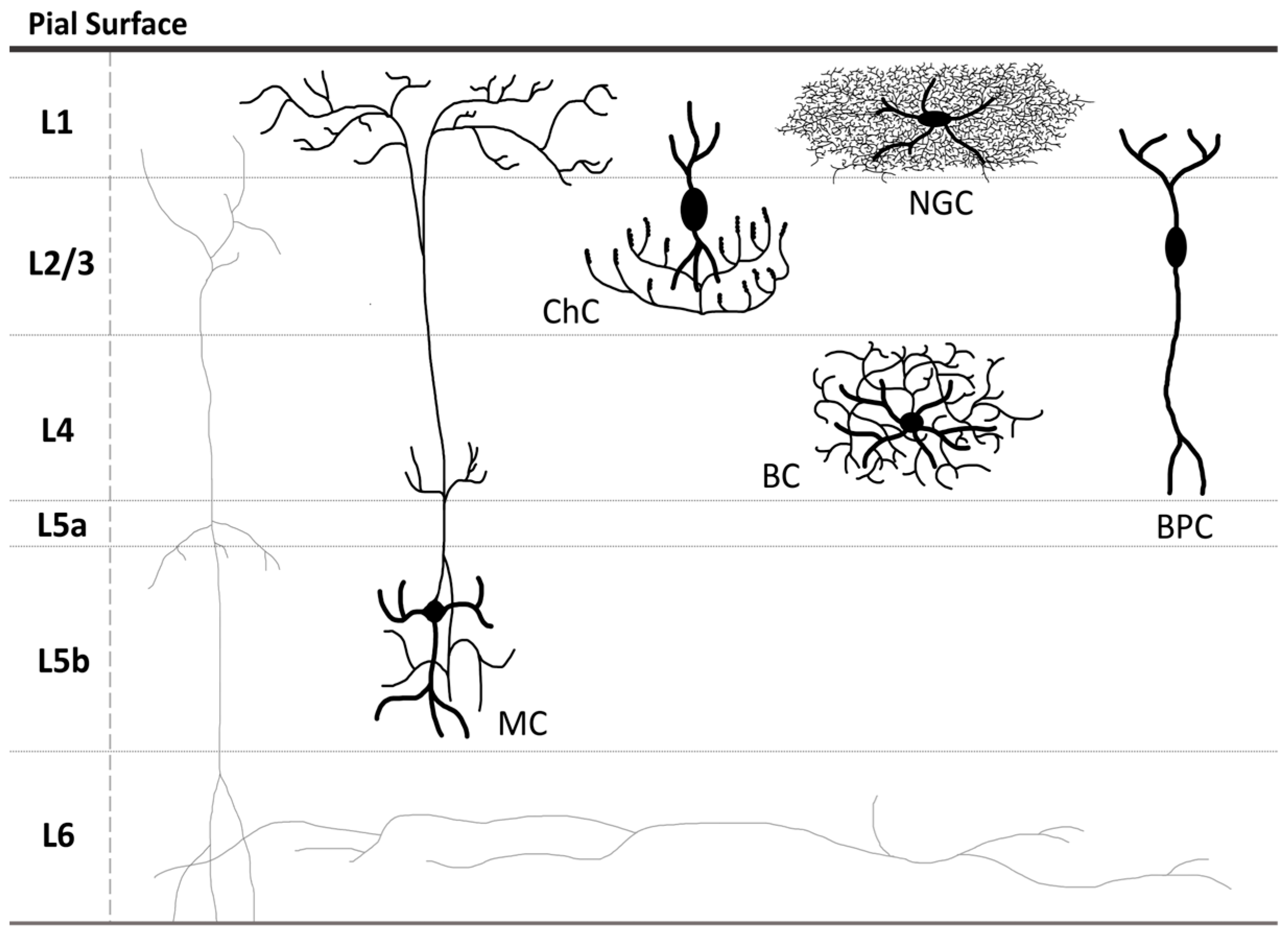
Figure 1. Localization of some types of inhibitory GABAergic neurons within the neocortex. MC—Martinotti cell; ChC—chandelier cell; BC—basket cell; NGC—neurogliaform cell; BPC—bipolar cell; L1, L2/3, L4, L5a, L5b, and L6—the neocortex layers.
Ijms 26 09492 g001
Figure 2. Expression of GABAergic system genes in various human brain regions and peripheral organs based on GTEx data (transcripts per million, TPM). The heatmap shows expression levels of key genes involved in the function of the GABAergic system, including genes encoding GABAA and GABAB receptor subunits (e.g., GABRA1–6, GABRB1–3, etc.), enzymes of GABA metabolism (ABAT, GAD1, GAD2), and GABA transporters (SLC6A1, SLC6A11, SLC6A13, SLC32A1). Tissues with negligible expression levels or not relevant to the focus of this study were not included in the analysis.
Ijms 26 09492 g002
Figure 3. Interaction of the synaptic vesicle loaded with GABA with presynaptic membrane.
Ijms 26 09492 g003
Figure 4. Expression of GABA transporter GAT1 (column 1), GAT2 (column 2), and GAT3 (column 3) in neocortex and subcortical structures. Top panels: regional expression values projected onto anatomical brain surfaces; middle panels: axial MRI slices illustrating spatial distribution; bottom panels: cortical surface renderings. Red denotes regions with upregulated expression, blue—regions with downregulated expression.
Ijms 26 09492 g004
Figure 5. The GABA pathway. Glutamine (Gln) is released by the astrocyte via SNAT3 transporter and is further imported by SNAT1/2 into the GABAergic neuron, where it is metabolized to glutamic acid (Glu) by glutaminases GLS1/2. GAD67/65 converts Glu into GABA (pink polygons), which is then loaded into the SV by VGAT, trafficked to the presynaptic membrane, and released into the synaptic cleft. Once released, GABA interacts with its receptors GABAAR and GABABR, activating Cl (orange circles) influx by the former, and modulating the activity of the potassium and calcium channels associated with the latter, increasing K+ (green circles) efflux and decreasing Ca2+ (red circles) influx; this, in turn, results in the postsynaptic membrane hyperpolarization. The excess of GABA is taken up by the astrocyte via GAT1 (or GAT3), which simultaneously translocates 2 Na+ ions (light-blue circles) and one Cl ion. In the astrocyte, GABA is sequentially catabolized by GABA transaminase ABAT and SSA dehydrogenase SSADH into succinic acid, which further enters the Krebs cycle, where it is processed into α-ketoglutarate, the precursor of Glu and Gln.
Ijms 26 09492 g005
Disclaimer/Publisher’s Note: The statements, opinions and data contained in all publications are solely those of the individual author(s) and contributor(s) and not of MDPI and/or the editor(s). MDPI and/or the editor(s) disclaim responsibility for any injury to people or property resulting from any ideas, methods, instructions or products referred to in the content.