Review

The Role of CRABS CLAW Transcription Factor in Floral Organ Development in Plants

1
Department of Biology and Pharmaceutical Botany, Medical University of Łódź, Muszyńskiego 1, 90-151 Łódz, Poland
2
Department of Pharmacology and Therapeutics, School of Biomedical Sciences, College of Health Sciences, Makerere University, Kampala P.O. Box 7062, Uganda
3
Department of Oncobiology and Epigenetics, Faculty of Biology and Environmental Protection, University of Lodz, Pomorska 141/143, 90-236 Łódź, Poland
*
Author to whom correspondence should be addressed.
Int. J. Mol. Sci. 2025, 26(19), 9377; https://doi.org/10.3390/ijms26199377
Submission received: 31 July 2025 / Revised: 10 September 2025 / Accepted: 22 September 2025 / Published: 25 September 2025

Abstract

CRABS CLAW (CRC) is a member of the plant-specific YABBY transcription factor family, defined by the presence of a C2C2 zinc-finger domain and a C-terminal YABBY domain. CRC is essential for proper floral development, functioning in the termination of the floral meristem, maintenance of adaxial–abaxial polarity within the gynoecium, and regulation of nectary and leaf morphogenesis. CRC orchestrates its diverse regulatory functions through interaction networks comprising other transcription factors and plant developmental regulators, including chromatin-modifying enzymes and proteins involved in auxin biosynthesis, transport, and signaling. The roles of genes and proteins interacting with CRC or CRC have been characterized in several model plant species, and the number of identified CRC/CRC-associated interactions continues to expand, revealing both species-specific and conserved functional roles across angiosperms. Many functions of CRC and its interacting partners have been elucidated through the analysis of anatomical and physiological phenotypes associated with specific gene mutations. The functional roles of CRC in plant development appear to have been acquired progressively through evolutionary diversification. These evolutionary changes have been associated with the relative conservation of CRC gene copy number and a predominant role of mutations occurring in non-coding regulatory regions. These properties are attributed to the relatively limited number of genes comprising the CRC regulatory network and the capacity to induce dosage-dependent effects via the emergence of novel proteins with overlapping or analogous functions. The identification and functional characterization of CRC transcription factors across diverse plant species has advanced rapidly in recent years, yet a comprehensive synthesis of these findings has not been presented in a dedicated article. Therefore, this study reviews the current knowledge on CRC transcription factors, with a focus on their identification, expression patterns, and functional roles in plant development.

1. Introduction

The CRC gene belongs to the YABBY transcription factor family and plays a pivotal role in meristem termination, carpel development, and nectary formation in plants [1,2,3,4,5,6]. Its ortholog in the monocot Oryza sativa, known as DROOPING LEAF (DL), regulates not only carpel identity but also the development of the leaf midrib [7,8,9]. Given the critical role of CRC in carpel development and floral meristem (FM) termination, its expression is subject to strict regulation, primarily mediated by other transcription factors [10,11,12].
The termination of plant meristem activity requires repression of the WUSCHEL (WUS) gene, which is achieved through two feedback loop systems [13,14]. In one of these loops, WUS is initially repressed either directly by AGAMOUS (AG) or indirectly via the AG-dependent activation of KNUCKLES (KNU) during early stages of floral development [13,14]. This repression is subsequently enhanced by the action of additional regulatory proteins, including CRC and TORNADO2 (TRN2), which contribute to the maintenance of auxin maxima necessary for proper meristem termination and gynoecium development [15,16,17,18]. Simultaneously, auxin represses cytokinin biosynthesis and signaling, a regulatory interaction essential for proper FM determinacy [18].
CRC and AG regulate the expression of YUCCA4 (YUC4), a gene involved in the two-step conversion of tryptophan to indole-3-acetic acid (IAA), a key process in auxin biosynthesis [15,16]. Elevated local auxin concentrations resulting from YUC4 activation promote cell wall loosening and cell proliferation, particularly within the medial domain of the developing gynoecium [12,13,14,15].
Analysis of phylogenetic studies on CRC genes suggests their numerical stability across angiosperms [19]. This feature is relatively uncommon among evolutionarily younger genes and may be attributed to the compact size of the gene regulatory network involved in carpel development [20,21]. The introduction of novel genes with overlapping functions may disrupt the delicate equilibrium, of this regulatory network, potentially causing dosage imbalances—particularly detrimental in processes such as the termination of reproductive meristems [20].
Ancestral CRC genes likely contributed to the polarity of the carpel—an organ unique to angiosperms—through subfunctionalization of the broader role of YABBY genes in establishing lateral organ polarity [1,22,23,24]. During angiosperm evolution, CRC expression and function underwent significant diversification, including the acquisition of roles in specifying carpel identity, the loss of polarity-regulating function in grass carpels, and the evolution of novel functions such as midrib development in grass leaves and nectary formation in core eudicots [1,7,8,9,11,24,25].
This review aims to synthesize current knowledge on the role of the CRC transcription factor in FM termination and the development of carpels and nectaries. Particular emphasis is placed on the identification and characterization of CRC-associated gene and protein partners, highlighting their contributions to the regulatory networks that coordinate plant growth, organ specification, and reproductive development.

2. Structural Features of the CRC Protein

The Arabidopsis thaliana CRC transcription factor belongs to the YABBY family and exhibits characteristic structural elements, including a C2C2-type zinc finger motif spanning amino acid residues 26–53, which confers DNA, RNA, and protein-binding capabilities [4,26] (Figure 1). The YABBY family is plant-specific and classified within the fourth superclass of all-α-helical DNA-binding domains, namely the High-mobility Group (HMG) domain factors. This class is conserved between plants and mammals [27]. The A. thaliana CRC protein (GenBank: KAL9311171.1) consists of 181 amino acids, with an intermediate segment located between residues 54–108 that likely functions as a flexible linker (Figure 1). This region contains a serine- and proline-rich domain at its center—a typical feature of transcription factor activation domains [28,29].
The YABBY domain of CRC is located between amino acid residues 109–155 and shares structural similarity with helix–loop–helix motifs found in HMG-box-containing proteins and plays a critical role in DNA binding [4,26] (Figure 1). However, in CRC, only the first two of the three helices characteristic of A. thaliana HMG1-like proteins are present [4,26,30].
Proteins exhibiting this domain organization were classified in the late 1990s as members of a newly defined YABBY transcription factor family. The family was named after the Australian freshwater crayfish “yabby”, in reference to its founding member, CRABS CLAW (CRC) [4,31,32].
Subcellular localization studies using CRC deletion mutants suggest the presence of a putative nuclear localization signal (NLS) within the YABBY domain, specifically spanning amino acid residues 110–117 (Lys-Pro-Pro-Lys-Glu-Lys-Lys-Gln-Arg; KPPEKKQR) (Figure 1). Despite the basic residue composition characteristic of classical NLSs, site-directed mutagenesis of this sequence does not abolish nuclear import, indicating that the NLS may exhibit non-canonical features [26,33]. This behavior is consistent with NLS motifs that interact with the minor binding site of importin α, as described for other atypical nuclear import signals [33,34,35].
Results from P-BLAST analysis using the A. thaliana amino acid sequence, followed by multiple sequence alignment, enabled the identification of highly conserved and less conserved amino acid positions based on the relative entropy threshold of each residue [36]. The data indicate that the strongest conservation occurs within the C2C2-type zinc finger motif, the N-terminal half of the Ser/Pro-rich domain, the NLS, and the N-terminal half of Helix 1 (Figure S1).

3. CRC in Floral Meristem Termination

Meristematic cells of the FM originate from the shoot apical meristem (SAM), which remains active throughout the plant’s life cycle and maintains a population of stem cells in its apex, referred to as the central zone (CZ) [37,38]. Cells in the CZ undergo slow divisions and are subsequently displaced laterally into the peripheral zone, developing further into lateral organs such as flowers or leaves [37,38].
In A. thaliana, the timing of FM termination is primarily regulated by AG, which controls the expression of WUS, KNU, and CRC through different feedback loop systems [39,40,41,42,43,44].
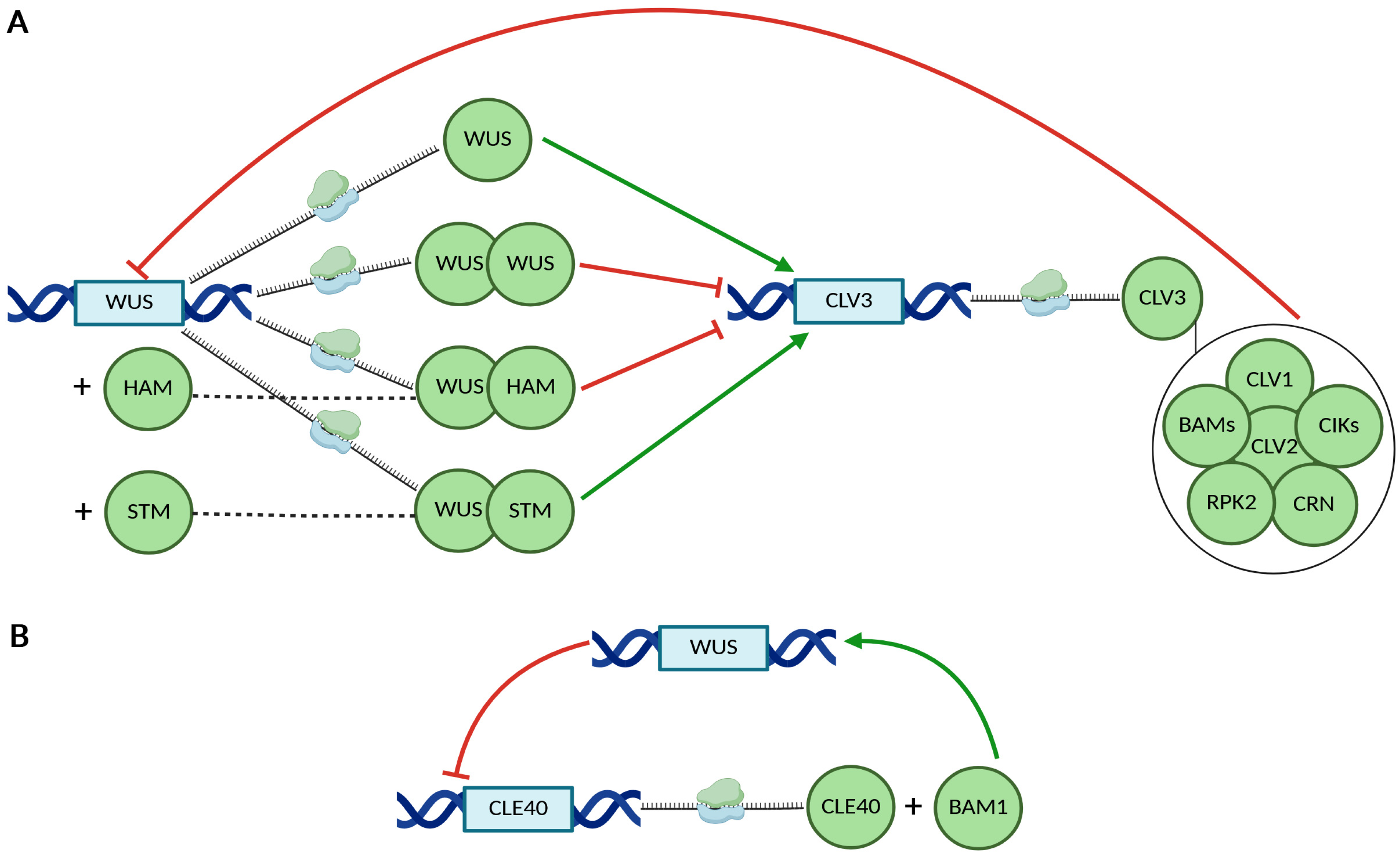
The first regulatory feedback loop involves WUS, which activates the expression of CLAVATA3 (CLV3) in the CZ [43,44,45]. The activation of CLV3 transcription requires the physical interaction between WUS and members of the HAIRY MERISTEM (HAM) protein family [46]. Additionally, WUS binds to the CLV3 promoter as a heterodimer in association with SHOOT MERISTEMLESS (STM) [47]. In turn, CLV3 is perceived by receptor complexes composed of CLAVATA1 (CLV1), CLAVATA2 (CLV2), RECEPTOR-LIKE PROTEIN KINASE 2 (RPK2), CLAVATA3 INSENSITIVE RECEPTOR KINASES (CIKs), CORYNE (CRN), and BARELY ANY MERISTEMS (BAMs) [48]. These complexes mediate the repression of WUS expression, thereby regulating stem cell maintenance in the shoot apical meristem [37,48,49,50] (Figure 2A).
A related but independent feedback loop involves the CLE40 peptide, which is closely related to CLV3 and promotes WUS expression through the CLV1-family receptor BAM1 [51]. In turn, CLE40 expression is repressed in a WUS-dependent manner, establishing a regulatory circuit that contributes to meristem homeostasis [51] (Figure 2B).
AG plays a central role in the second feedback loop that regulates the termination of WUS expression and is overlaid on the CLV-WUS pathway [44,52]. Mutations in AG disrupt this regulation, leading to the failure of WUS repression and resulting in the formation of supernumerary floral whorls from the floral center. This produces a characteristic ‘flower-within-a-flower’ phenotype, also referred to as the Russian doll phenotype [4,53].
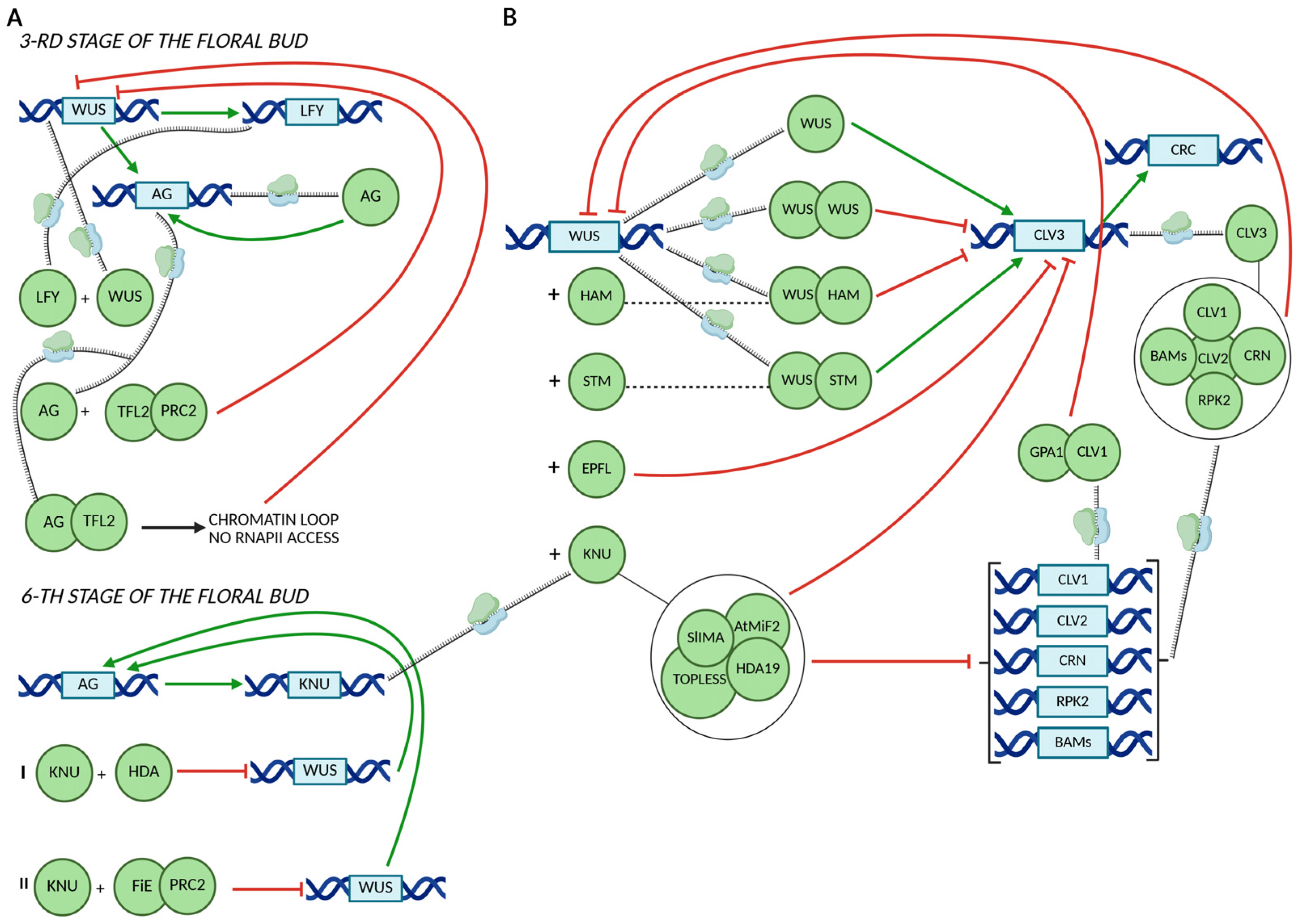
The mechanisms by which AG regulates WUS expression are dependent on the developmental stage of the floral bud (Figure 3A). During floral stage 3, WUS promotes the expression of the C-class gene AG and LEAFY (LFY) [54]. The floral identity protein LEAFY (LFY) cooperates with WUS to activate AG in the center of flowers [54]. Subsequently, AG represses WUS by recruiting the TERMINAL FLOWER 2 (TFL2) protein, a component of the Polycomb Repressive Complex 1 (PRC1) [13,55]. In a later developmental stage (stage 6), AG directly enhances the expression of the C2H2-type zinc finger protein KNU, initiating a two-step repression mechanism of WUS expression [14,56,57]. Initially, WUS expression is repressed through the association of KNU with a histone deacetylase complex [56]. This initial repression is subsequently stabilized by KNU-mediated recruitment of the Polycomb Repressive Complex 2 (PRC2), which catalyzes the trimethylation of lysine 27 on histone H3 (H3K27me3), a hallmark of stable gene silencing (Figure 3A) [14,58]. The influence of KNU, EPFL and GPA1 on the WUS-CLV3 regulatory route and CRC expression was presented in Figure 3B [3,13,43,56,59,60,61,62,63,64,65,66].
Representative genes that are either directly regulated by AG or integrated within the AG regulatory network are listed in Table S1 [2,42,67,68,69,70,71,72].
It is plausible that CRC alone is not sufficient to modulate WUS activity, as suggested by analyses of CRC mutants across multiple species, including A. thaliana, pomegranate, and rice, which exhibit no overt FM defects [7,25,73].
In A. thaliana, CRC appears to play an ancillary role to AG in FM development [11,25,68,74]. The heterotetramer formed by SEP3 and the MADS protein AGAMOUS is necessary to activate two target genes, KNU and CRC, which are required for meristem determinacy [75,76]. However, more direct and independent roles of CRC have been observed in tomato, where it contributes to high temperature-induced meristem termination [3,39]. In tomato, SlCRCa and SlCRCb directly bind to the SlWUS promoter, with SlCRCb also interacting with the second intron of SlWUS [39]. Interestingly, AtCRC does not bind to the AtWUS promoter (pAtWUS) or its genomic locus (gAtWUS), suggesting a species-specific regulatory mechanism [39].
Under heat shock conditions, a reduction in brassinosteroid concentrations within the floral meristem results in the persistent downregulation of SlCRCa [39]. This failure to terminate SlWUS expression ultimately leads to the development of malformed fruits [39]. Although previous studies have demonstrated the critical role of tomato AG1 (TAG1) in activating SlKNU and SlIMA, leading to repression of WUS expression, overexpression of TAG1 alone is insufficient to activate the promoter activity of SlKNU and SlIMA. Therefore, in tomato, SlCRCa is necessary for TAG1-mediated activation of SlIMA and SlKNU [56].
Dual-luciferase assays suggest that this missing transcriptional activation may be provided by SlCRCa, but not by SlCRCb, indicating functional divergence between the two CRC homologs in tomato [26,56]. Moreover, SlCRCa directly binds to the SlWUS locus, where it recruits the SlIMA complex, leading to complete repression of SlWUS expression during the establishment of FM determinacy [3]. More specifically, SlCRCa and SlCRCb proteins can interact with one another, and each can also interact with SlKNU, SlIMA, and SlHDA1. Notably, only SlCRCb, and not SlCRCa, interacts with SlTPL1 [3].

4. CRC in Auxin/Cytokinin Crosstalk

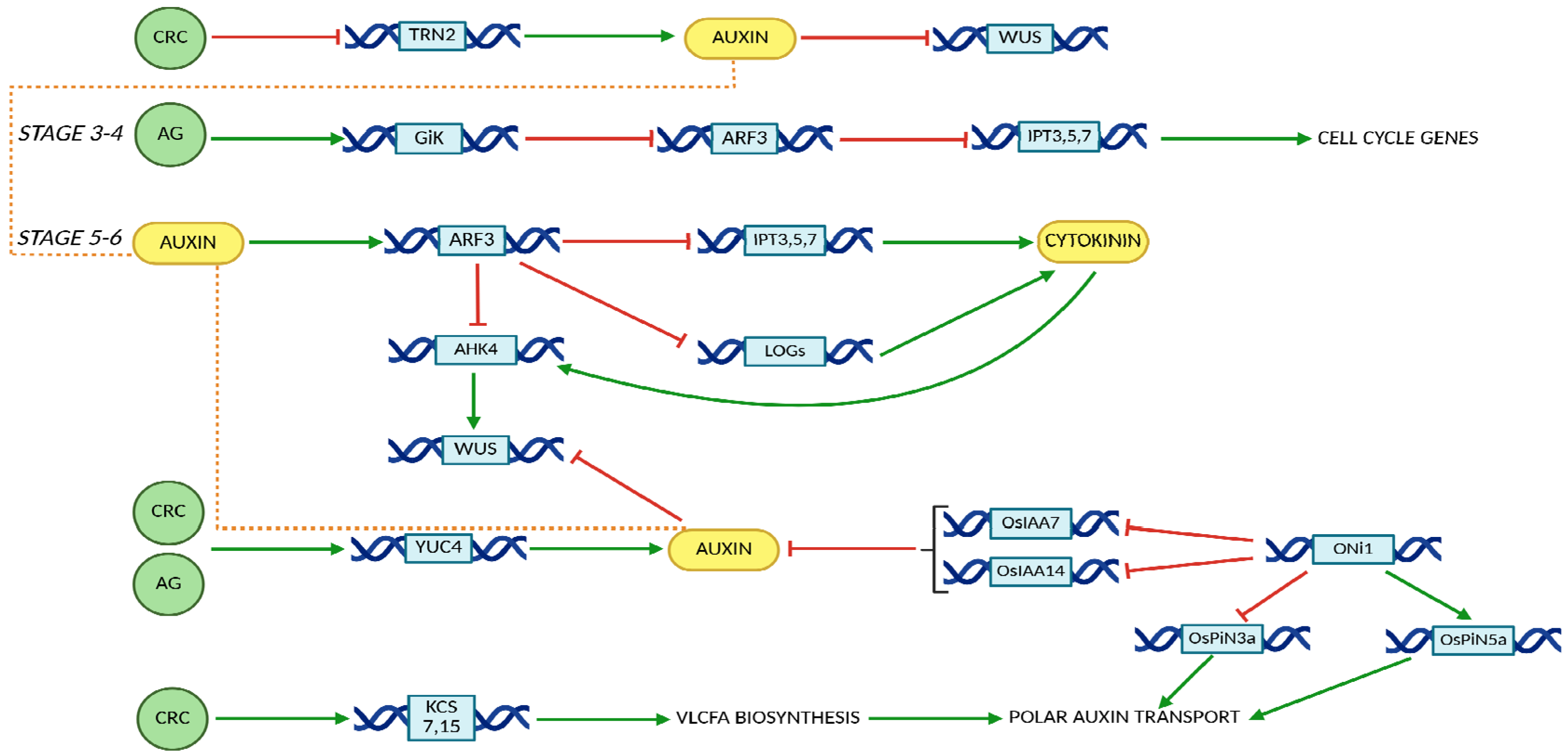
Further studies have demonstrated that the pivotal role of AG in FM termination and gynoecium development is mediated through its regulation of the crosstalk between cytokinin, miRNA and auxin signaling pathways [16,77,78,79,80]. Similarly, findings by Yamaguchi et al. (2017,2018) highlight the essential function of CRC in FM termination and gynoecium formation via the fine-tuning of auxin homeostasis [12,13,15,16,81]. Moreover, the transcriptional repressor KNU integrates and modulates the activities of auxin and cytokinin, thus securing timed FM termination [82]. The roles of CRC and AG in regulating auxin and cytokinin activity are presented in Figure 4 [80,83,84,85,86,87,88,89,90,91,92,93,94].
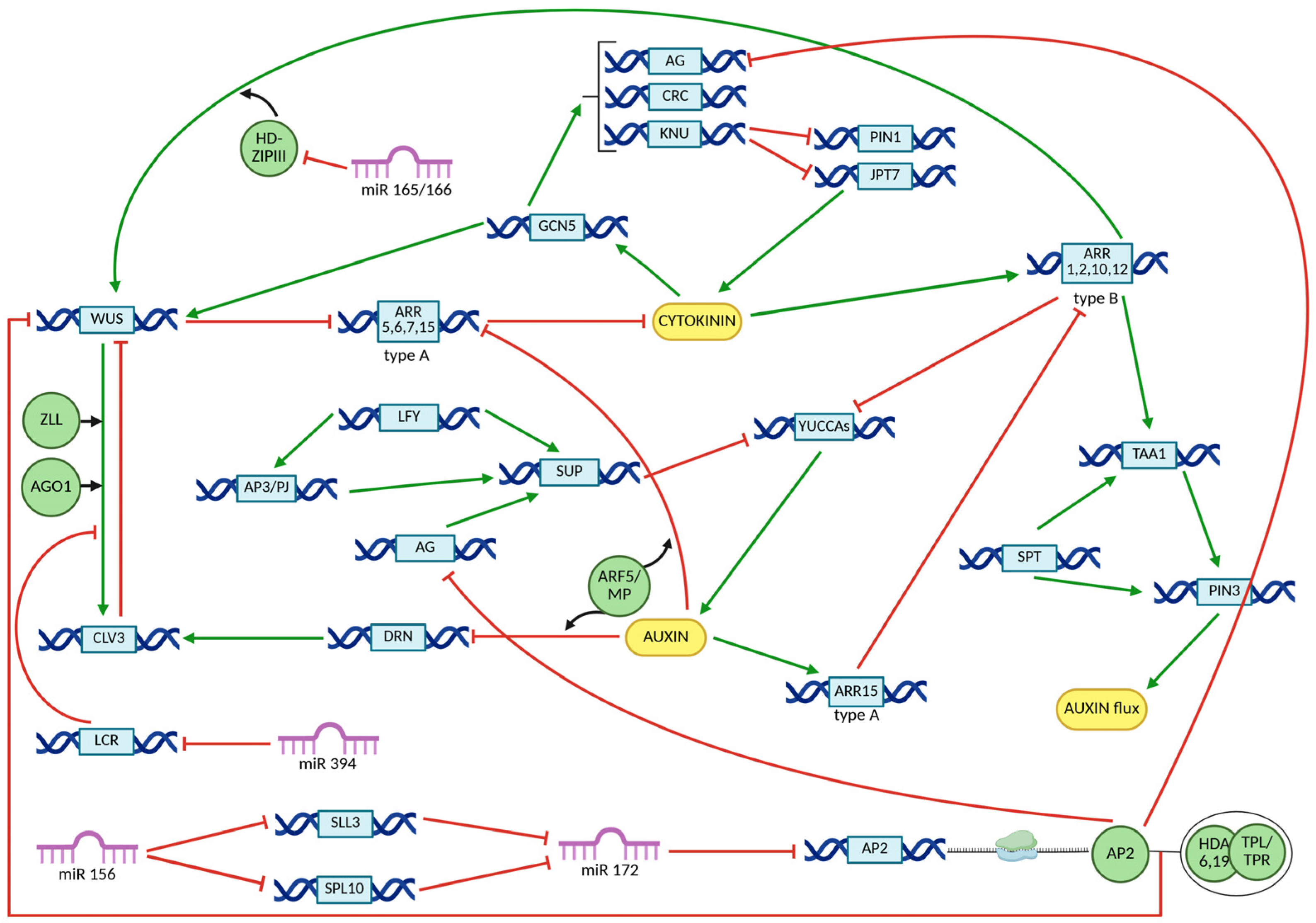
Although AG represses WUS directly or via KNU during floral stages 3–5, this repression is further reinforced at stage 6 by additional regulatory factors, including CRC and TRN2. CRC represses TRN2, leading to the formation of local auxin maxima (Figure 4) [15,16]. Concurrently, AG and auxin synergistically activate ARF3, which in turn downregulates the expression of ISOPENTENYL TRANSFERASES (IPTs), LONELY GUY (LOG), and ARABIDOPSIS HISTIDINE KINASE 4 (AHK4), thereby attenuating cytokinin signaling activity (Figure 4) [77]. The increase in auxin activity, combined with the decrease in cytokinin motion, results in the indirect inhibition of WUS expression, or WUS destabilization, leading to floral meristem termination (Figure 5) [15,16,77,81,95,96,97,98,99,100,101].
The pivotal role of auxin in the regulatory functions of CRC and AG during FM termination has been elucidated through transcriptomic analyses of relevant mutants, including crc-1, knu-1 versus knu-1, and ag-12 versus wild-type plants. These comparisons led to the identification of a core set of 53 genes that are co-regulated by both CRC and AG [15]. Among the 53 genes co-regulated by AG and CRC, a subset of nine genes were identified as direct AG targets based on publicly available AG ChIP-seq datasets [15]. Among these, YUC4 is an auxin-related target of CRC, acting downstream and synergistically with TRN2 to promote FM termination through the indirect repression of WUS (Figure 4) [12,15].
YUC4, in conjunction with TRYPTOPHAN AMINOTRANSFERASE of ARABIDOPSIS (TAA) family enzymes, catalyzes a two-step conversion of tryptophan to indole-3-acetic acid (IAA), representing a critical phase in the auxin biosynthesis pathway [17,99,102,103].
Putative synergistic and positive regulation of YUC4 by CRC and AG was supported by qRT-PCR analysis in crc-1 and ag-1-/+ crc-1 double mutants. Notably, YUC4 reporter (GUS) expression in the abaxial carpels at floral stage 6 was markedly reduced in the ag-1-/+ crc-1 double mutants compared to the crc-1 single mutant [15]. Induced expression of AG and CRC, either individually or in combination, supported a synergistic role in the activation of YUC4 transcription [15].
The regulation of YUC4 by CRC involves its binding to YABBY-binding motifs (GA[A/G]AGAAA) located within conserved regulatory modules 1–4 (CRMs 1–4) of the YUC4 promoter [12,104].
Upregulated expression of YUC4 enhances local auxin accumulation, leading to cell wall loosening and increased cell proliferation, potentially facilitated by heightened cell division activity in the medial domain of the developing gynoecium [16,105]. The dynamic distribution of the plant morphogen auxin is pivotal for the bilateral-to-radial symmetry transition, which is required for proper formation of the apical style in the Arabidopsis gynoecium [106].
AtCRC and AtAP1 may participate in the regulation of very long-chain fatty acid (VLCFA) biosynthesis. In Arabidopsis, VLCFAs regulate polar auxin transport and lateral root organogenesis (Figure 4) [99,100].

5. CRC Interaction Networks

Mutations in the CRC gene in A. thaliana lead to a distinct floral phenotype, characterized by a wider and shorter gynoecium, unfused carpels at the apex, reduced ovule number, and a complete absence of nectary development at all stages of floral maturation [4]. The gene was mapped between CLV2 and CLV1 on chromosome 1, subsequently positionally cloned, and effectively used to complement the crc mutation, thereby suppressing early radial expansion of the gynoecium and promoting its elongation at later developmental stages [4,25,43].
The inhibition of radial growth in the developing gynoecium by CRC begins in the lateral regions of the gynoecial primordium and is later reinforced within the epidermal layer. Although CRC also functions in the early stages of nectary development, its regulatory interactions differ between the gynoecium and nectary tissues. In the gynoecium, CRC expression is negatively regulated by A-class genes, including APETALA2 (AP2) and LEUNIG (LUG) [4]. The same holds true for the B-class genes in Arabidopsis, PISTILLATA (PI) and APETALA3 (AP3), which negatively regulate CRC expression in the third whorl [4]. Since mutations in the C-class gene AGAMOUS (AG) disrupt carpel development, the effect of AG loss-of-function was assessed in the first whorl. These studies showed that CRC expression persists in the absence of AG, but is significantly reduced and displays altered spatial distribution. Notably, CRC expression in nectaries remains unaffected by mutations in A-, B-, or C-class genes [4]. Genes and proteins associated with CRC regulation and the biological outcomes of their interactions are presented in Table S2 [4,12,17,39,62,73,77,83,100,107,108,109,110,111,112,113,114].
In A. thaliana, the gynoecium is composed of two congenitally fused carpels, forming a single-chambered ovary (Figure 6) [115,116,117].
Abaxial–adaxial polarity within the gynoecium is evident from distinct epidermal cell morphologies on the abaxial (outer) and adaxial (inner) surfaces of the ovary valves, which contribute to proper organ development (Figure 6) [107].
A novel mutant phenotype in Arabidopsis carpels, characterized by the duplication of adaxial tissues—such as placentae and ovules—at abaxial positions, in addition to their normal localization on the adaxial (internal) side, was observed in double mutants where the function of CRC and either GYMNOS (GYM) or KANADI (KAN) is compromised [106,107]. The ectopic development of adaxial tissues is normally suppressed by CRC and KAN, which act through independent pathways and provide positional information to specify abaxial cell fate in the developing carpel (Table S2). The patterning events in the developing gynoecium depend on the activity of SEUSS and AINTEGUMENTA genes [108]. Phenotypic analyses of mutants for both genes suggest their synergistic interaction in the development of ovules and the medial domain of the gynoecium. Identified downstream targets of SEUSS and AINTEGUMENTA include PHABULOSA (PHB), REVOLUTA (REV), and CRC genes (Table S2) [107]. Both SEU and ANT promote CRC expression in the internal domains of the gynoecial ovary. However, in the apical region of the gynoecium, SEU and ANT act to repress CRC expression within the medial ridge, which correlates with an increased incidence of carpel separation spanning approximately 90% of the apical–basal axis of the gynoecium (Table S2) [107,108].
JAIBA (HAT1), a member of the class II HD-ZIP transcription factor subfamily, is implicated in both male and female reproductive development. In jab homozygous mutants, defects include a reduced number of mature pollen grains and ovules lacking a functional embryo sac [108,109,118]. Double jaiba crc-1 mutants exhibit severe defects in FM determinacy and development of the gynoecium medial domain, indicating a functional relationship with CRC (Table S2) [108,109]. The jab crc-1 double mutant produced flowers with up to six sepals, up to six petals, and between five and seven stamens (Table S2) [108,109]. Additionally, the gynoecia of these flowers were composed of two to four carpels, which at later developmental stages transformed into carpeloid structures within the fruit.
Gynoecia lacking functional STYLISH1 (STY1) expression due to a transposon insertion exhibit aberrant style morphology. These defects are further enhanced in sty1 sty2 double mutants, which show a marked reduction in stylar and stigmatic tissues as well as decreased proliferation of stylar xylem [110]. CRC is proposed to interact with STY1, as sty1-1 crc-1 double mutants display a more severe reduction in stylar, stigmatic, septal, and medial xylem tissues compared to either single mutant (Table S2) [110]. STY1 functions as a transcriptional activator of the gene encoding the flavin-containing monooxygenase THREAD/YUCCA4, a key enzyme in the auxin biosynthesis pathway, ultimately resulting in altered auxin homeostasis [111,119].
The functional activity of transcription factors is frequently mediated not by monomeric forms but through the formation of homo- or heterodimers, which may assemble in the cytoplasm prior to nuclear translocation or upon binding to closely spaced cis-regulatory motifs within promoter regions [52,53,75,76,120,121]. Dimerization is a well-documented feature among members of the YABBY transcription factor family [26,32,44,48]. In the case of CRC, bimolecular fluorescence complementation (BiFC) assays suggest that dimer formation is dependent on the presence of the YABBY domain [26,44].
However, these CRC-CRC or CRC-YABBY protein interactions appear to be relatively weak and could not be confirmed using yeast two-hybrid (Y2H) assays [26,44]. Interestingly, Y2H analyses have indicated a weak interaction between the transactivation domain of CRC and the YABBY domain of the INNER NO OUTER (INO) protein, suggesting possible functional crosstalk between these two transcription factors (Table S2). CRC-interacting partners may exert either activating or repressive effects on CRC-mediated transcriptional regulation [26,44]. Further evidence from Y2H and BiFC assays in Punica granatum (pomegranate) demonstrated that both CRC and INO interact with a common partner, PgBEL1, implicated in the maintenance of ovule identity (Table S2) [73].
The cooperative function of the YABBY domain and the zinc finger motif in DNA binding is supported by Y1H assays. However, Y1H screens using isolated or mutated CRC domains revealed that the YABBY domain plays a predominant role in DNA binding, while the zinc finger contributes a weaker, auxiliary interaction with the target DNA sequence [26].
In silico analysis of the regulatory mechanisms governing CRC expression suggests a predominant role for transcription factors, as indicated by the presence of only two CHH-context DNA methylation sites located approximately 0.3 to 3 kb upstream of the transcription start site (TSS). Furthermore, the CRC genomic locus appears to be largely unaffected by microRNA-mediated regulation and exhibits only moderate epigenetic control. This is supported by the presence of the repressive histone mark H3K27me3, which spans most of the CRC locus, including the promoter region, and by H2AK121ub enrichment within the transcribed region [12].
To identify putative transcription factors interacting with the CRC promoter, yeast Y1H screening was performed, yielding 140 candidate proteins. Of these, only 48 were annotated in the PlantPAN3 database and exhibited predicted DNA-binding motifs matching sequences within the CRC promoter region (Table S2) [12].
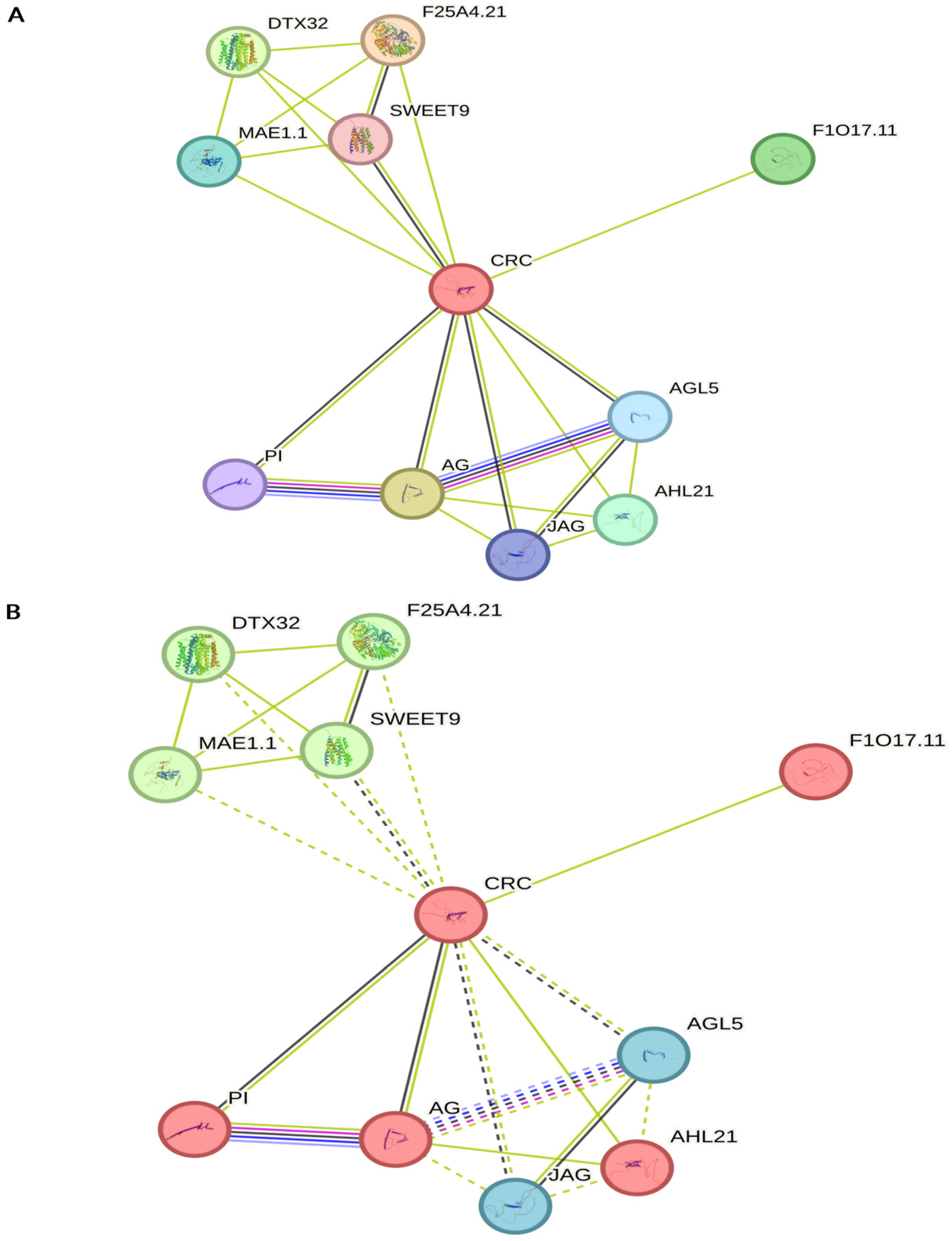
Functional partners of CRC predicted by the STRING database are presented in Figure 7 [122]. Most of them are related to the known CRC functions in floral organ development, FM determinacy, or fatty acid homeostasis [3,4,100,102]. However, novel functionalities associated with plant defense or myo-inositol hexakisphosphate and phytic acid metabolism were observed (Figure 7). Detailed information concerning CRC functional partners is provided in File S1.

6. Evolutionary Diversification of CRC Gene

The origin of CRC coincided with the emergence of angiosperms, as CRC plays a pivotal role in the development of the carpel—an organ unique to flowering plants [20,123]. Although closely related CRC paralogs are present in gymnosperm genomes, it remains unclear how CRC was integrated into gene regulatory networks to control carpel-specific functions during the evolution of angiosperms [20,124].
Phylogenetic analyses indicate that CRC genes involved in FM termination, carpel organ identity, and abaxial-adaxial polarity in A. thaliana have remained numerically stable and have not undergone significant expansion across plant lineages [20,124].
The CRC gene is maintained as a single-copy gene in most angiosperms, which is atypical for evolutionarily recent genes [20]. It is hypothesized that the evolution of a relatively small and tightly regulated carpel gene regulatory network (GRN)—comprising genes such as ALCATRAZ (ALC), CRC, HALF FILLED (HAF), HECATE (HEC), INDEHISCENT (IND), NGATHA (NGA), and SPATULA (SPT)—does not tolerate dosage imbalances or the expansion of gene families encoding proteins with redundant functions [20].
The termination of reproductive meristems appears particularly sensitive to dosage imbalance, as demonstrated by the effects of increased CRC expression. Artificial elevation of CRC levels can lead to premature termination of reproductive meristems—an outcome that is strongly selected against in natural populations [1,125].
Although CRC is typically maintained as a single-copy gene in most plant species, an exception is observed in the Solanaceae family, where it occurs as paralogous pairs—CRCa and CRCb in Solanum lycopersicum, and CRC1 and CRC2 in Petunia hybrida [126]. These paralogs are believed to have arisen from a large segmental duplication event that likely occurred in a common ancestor of the Solanaceae [126].
The CRC ortholog in the monocot Oryza sativa, known as DL, exhibits differences in both spatial expression and function compared to CRC [7,8,9]. In A. thaliana, CRC is expressed in the abaxial region of carpel primordia and in floral nectaries, where it plays a key role in regulating carpel morphology and nectary development [1,3,5,6,7,12]. In Oryza sativa, DL is expressed throughout the entire carpel primordium and in the central undifferentiated cells of developing leaves, where it regulates both carpel identity and midrib development [7].
Analysis of CRC orthologs in two Fabaceae species, Pisum sativum and Medicago truncatula, revealed not only an atypical absence of abaxial expression in the carpel but also an unusual expression pattern associated with the medial vein of the ovary. This suggests a potential role in vascular development—previously thought to be specific to DL in monocotyledons [127].
The temporal and spatial expression patterns of DL orthologs in three grass species—maize, wheat, and sorghum—closely mirror those of O. sativa DL during both floral and leaf development. These findings suggest a high degree of functional conservation of DL-related genes within the Poaceae (grass) family [128].
Analysis of the spatial expression patterns of CRC/DL orthologs in eudicots and basal eudicots—such as Petunia hybrida, Gossypium hirsutum, Eschscholzia californica (basal eudicot), and Aquilegia formosa (basal eudicot)—reveals that the abaxial specificity and absence of expression in leaves, characteristic of A. thaliana CRC, are highly conserved among these species. This conservation suggests a stable and ancestral regulatory role for CRC/DL orthologs in gynoecial development across eudicot lineages [1,11].
The ancestral role of CRC in carpel development was confirmed in the basal angiosperm Amborella trichopoda, whose CRC orthologue partially complemented the crc-1 mutant phenotype in carpels but not in nectaries [129]. This partial complementation suggests that novel functions of CRC orthologues, such as nectary development, may be mediated by evolutionary changes in non-coding regulatory sequences [130].
CRC also regulates the formation of nectaries, which are present not only in the floral nectaries of Arabidopsis but also in the extrafloral nectaries of Gossypium hirsutum and Capparis flexuosa [4,131]. However, in basal eudicots such as Aquilegia formosa, the CRC orthologue is not expressed in the nectary, suggesting that CRC orthologues were recruited for nectary development at the base of the core eudicots lineage [130,131,132].
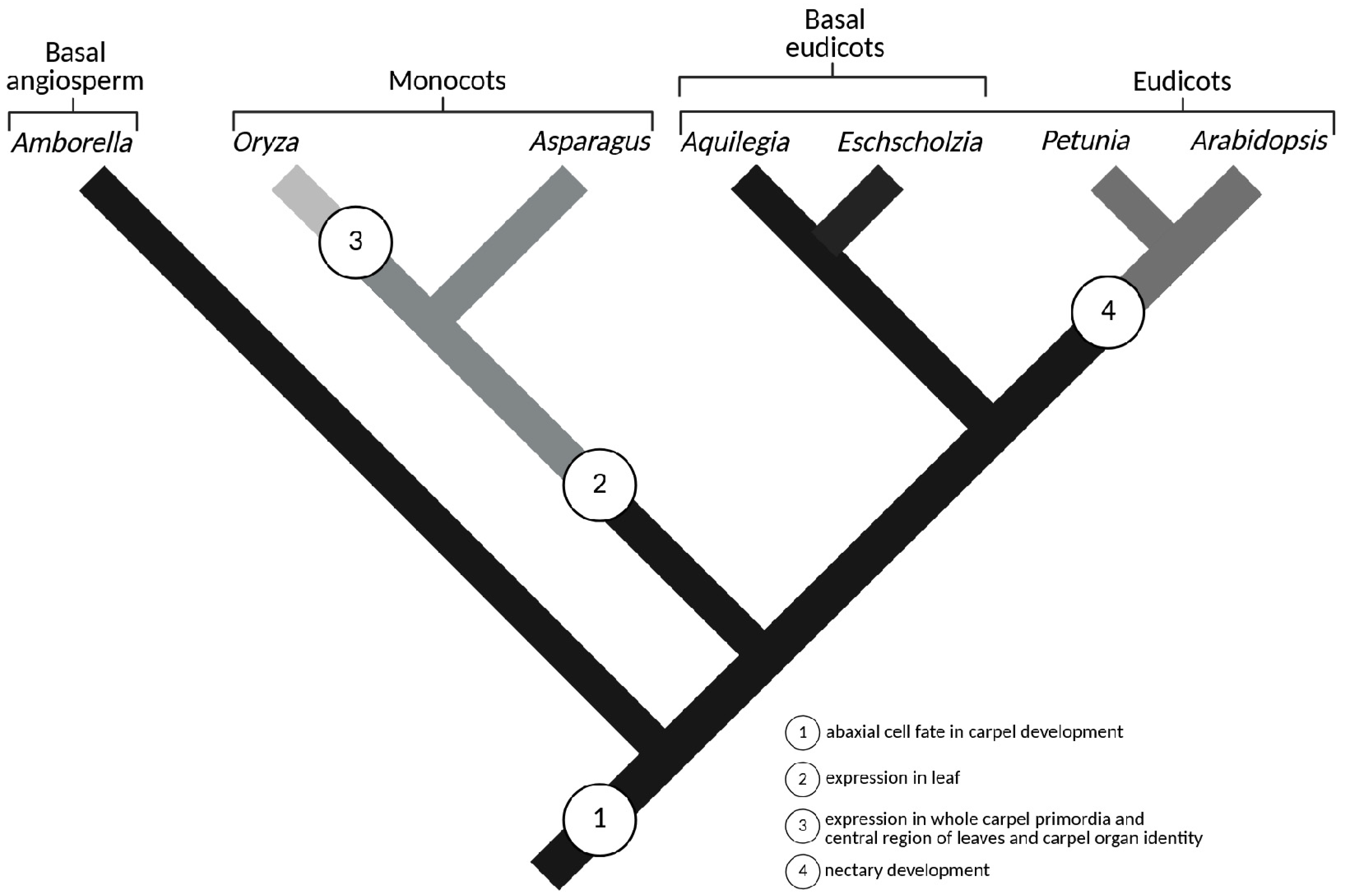
Studies of CRC/DL expression across major angiosperm clades demonstrate the stepwise acquisition of novel functions without gene duplication, exemplifying the role of regulatory elements—such as promoters and enhancers—in broadening gene function (Figure 8) [1,24,130,131]. Alternatively, the novel function of CRC in nectary development may have arisen through one or more changes affecting the composition of downstream target genes, the availability or identity of protein interaction partners, and/or the spatiotemporal expression of CRC in nectary tissues [130].
Analysis of the functional diversification of DL in Asparagus asparagoides revealed that the evolution of CRC/DL genes from their ancestral role in specifying abaxial cell fate during carpel development to acquiring expression in leaves occurred prior to the divergence of the order Asparagales. However, traits such as expression throughout the entire carpel primordium, in the central region of leaves, and involvement in carpel organ identity were gained after the divergence of Asparagales (Figure 8) [24].
In the orchid Phalaenopsis equestris, two DL/CRC paralogs, PeDL1 and PeDL2, were identified as products of a whole-genome duplication (WGD) event in the last common ancestor of orchids. Both genes are expressed in the FM and carpel tissue, supporting the ancestral role of DL/CRC genes in FM determinacy and carpel specification [23].
Moreover, expression of both PeDL genes was observed in the placenta and ovule primordia during early stages of ovary development in Phalaenopsis equestris [133]. Similarly, in California poppy (Eschscholzia californica), the CRC ortholog EcCRC has also been co-opted for additional roles in ovule initiation [1].

7. Polymorphisms and Natural Variation of the CRC Gene

T-DNA and ethyl methanesulfonate (EMS) mutagenesis screens performed in the crc-1 background led to the identification of novel genetic modifiers of the crc-1 phenotype. These were cloned and characterized as rebelote-1 (rbl-1, meaning “once again” in French), squint-4 (sqn-4), and ultrapetala1-4 (ult1-4). In all three resulting double mutants, the FM remains indeterminate, with ectopic stamens and carpels arising from the central region of the FM, between the primary carpels, and along an elongated floral axis [125].
RBL, SQN, and ULT1 function redundantly in the regulation of FM termination, likely through modulation of WUS activity [125].
Moreover, the rbl-1, sqn-4, and ult1-4 mutations enhance the ag-4 phenotype, suggesting that RBL, SQN, and ULT1 play partially redundant roles in supporting AG function [33]. Additionally, genetic crosses with the ag-6 mutant indicate that RBL, SQN, and, to a lesser extent, ULT1 contribute to FM determinacy via an alternative pathway, potentially involving SUPERMAN (SUP), as the phenotypes of ag-6 rbl-1 and ag-6 sqn-4 resemble those of the ag-1 sup-1 double mutant [125,131].
Mutations in the tomato (Solanum lycopersicum) CRCa gene (SlCRCa) lead to the development of abnormal carpels that grow concentrically, one inside another, resulting in iterative fruit morphologies characteristic of the fig mutant phenotype [3]. In addition, affected fruits frequently develop secondary fruit structures that initiate internally and progressively expand to emerge on the fruit surface [3]. The molecular basis of the fig mutation is a 365-bp insertion of a putative transposable element within the intron between exons 4 and 5 of the SlCRCa gene [3]. The regulation of fruit size changes induced by SlCRCa may also involve YABBY5a and genes of the gibberellin biosynthesis pathway, both of which are generally downregulated in response to SlCRCa overexpression [132].
Although SlWUS expression is terminated from stage 6 onward in wild-type plants, SlCRCa and SlCRCb mutants maintain SlWUS expression beyond stage 6, resulting in defects in floral determinacy [3]. Similar to A. thaliana, the SlWUS locus in tomato is associated with a chromatin remodeling complex exhibiting histone deacetylase activity, as demonstrated by BiFC and co-immunoprecipitation assays.
Analysis of quantitative trait loci (QTL), in combination with map-based cloning, led to the identification of a nonsynonymous polymorphism (G to A) in the CRC gene of cucumber (Cucumis sativus, CsCRC) as the causal factor underlying variations in fruit size and shape. RT-PCR analyses revealed that CsCRC expression is predominantly confined to reproductive organs, including flowers and fruits, with no detectable expression in vegetative tissues such as stems and leaves. Among reproductive organs, the highest CsCRC expression was observed in the nectary, followed by female flower buds and young ovaries [133].
The presence of a rare CsCRC^A allele is associated with reduced fruit length in Cucumis sativus, whereas the CsCRC^G allele shows a positive correlation with increased fruit size. The causal variant is a nonsynonymous SNP (G-to-A) in CsGy5G023910, a homolog of Arabidopsis CRC, which leads to an amino acid substitution from arginine (R) to glutamine (Q) within the conserved YABBY DNA-binding domain [133]. The arginine-to-glutamine (R→Q) substitution within the YABBY DNA-binding domain of CsCRC markedly diminishes its binding affinity to [A/T]ATCAT[A/T] and [T/A]ATGAT[T/A] cis-regulatory motifs in the CsARP1 promoter, thereby reducing CsARP1 expression [133].
Putatively, the upregulated CsARP1 transcription mediated by CsCRC wild type enhances cell expansion in the pulp cells of cucumber fruit mesocarp, potentially via transmembrane electron transport and cell wall modification, as implied by the presence of a conserved cytochrome b561 domain and an additional DOMON domain in the encoded protein [133].
These findings support a critical link between CRC function and auxin-regulated pathways, consistent with earlier studies [15,16,81].

8. Outlook

Studies on the function of CRC in meristem termination and gynoecium or nectary development were initially conducted in the model plant A. thaliana [4,11,12,26,37,109,131,134]. The phenotypic consequences of CRC gene mutations, together with the availability of the A. thaliana genome sequence and transcriptomic datasets, provided a foundation for the identification of novel genes that are either co-expressed with CRC or modulate its role in meristem termination and gynoecium development [12,21,100,107,125,135].
Furthermore, Y2H and BiFC assays have yielded critical insights into proteins that interact with CRC or AG and directly contribute to the termination of WUS expression [17,26,73].
Beyond A. thaliana, CRC paralogs have been relatively well studied in tomato, providing insights into species- or developmental stage-specific variations in the composition of protein complexes involved in WUS repression [3,39,56]. However, studies investigating CRC function in other plant species remain relatively limited [1,10,24,111,136,137,138].
Consequently, future research should focus on the isolation of CRC orthologs and paralogs in a broader range of plant species. Generating transgenic lines with CRC overexpression, gene silencing, or targeted mutations would provide a basis for analyzing phenotypic and physiological alterations associated with CRC function.
These effects could be compared with observations from A. thaliana, rice and tomato to elucidate both conserved and novel roles of the CRC gene. In newly studied plant species, CRC gene mutations could be combined with chemical mutagenesis or transcriptomic analyses to identify previously uncharacterized genes that are co-expressed with CRC and modulate phenotypic or physiological traits associated with meristem termination and gynoecium development. In this way, the already known and novel functionalities of CRC could be revealed.
The application of Y1H assays or chromatin immunoprecipitation (ChIP) techniques could facilitate the identification of trans-acting factors regulating CRC gene expression in novel, often non-model, plant species. Similarly, Y2H, Y3H, and BiFC assays could be employed to identify components of protein complexes centered around CRC that are directly involved in executing its function. The functional relevance of these interactions could be validated by analyzing phenotypic alterations in plants harboring mutations in CRC-associated proteins.
Y2H screening often yields a substantial number of putative interacting proteins, frequently exceeding one hundred, many of which are false positives or are partially annotated in current databases [139,140,141,142,143,144,145]. Moreover, future progress in identifying cis-elements or trans-acting factors regulating CRC-dependent gene expression will rely upon advancements in the functionality and comprehensiveness of plant genomic and proteomic databases and improvements in Y1H methodology [140,141,142,145,146].

Supplementary Materials

The following supporting information can be downloaded at: https://www.mdpi.com/article/10.3390/ijms26199377/s1.

Author Contributions

Conceptualization, writing—original draft preparation, P.S.; English correction, writing—review and editing, J.N.; visualization, M.M. All authors have read and agreed to the published version of the manuscript.

Funding

This research was supported by statutory funds of the Department of Biology and Pharmaceutical Botany at the Medical University of Łódź (503/3-012-01/503-31-001).

Conflicts of Interest

The authors declare no conflicts of interest.

References

  1. Orashakova, S.; Lange, M.; Lange, S.; Wege, S.; Becker, A. The CRABS CLAW ortholog from California poppy (Eschscholzia californica, Papaveraceae), EcCRC, is involved in floral meristem termination, gynoecium differentiation and ovule initiation. Plant J. 2009, 58, 682–693. [Google Scholar] [CrossRef] [PubMed]
  2. Liu, C.; Thong, Z.; Yu, H. Coming into bloom: The specification of floral meristems. Development 2009, 136, 3379–3391. [Google Scholar] [CrossRef] [PubMed]
  3. Castañeda, L.; Giménez, E.; Pineda, B.; García-Sogo, B.; Ortiz-Atienza, A.; Micol-Ponce, R.; Angosto, T.; Capel, J.; Moreno, V.; Yuste-Lisbona, F.J.; et al. Tomato CRABS CLAW paralogues interact with chromatin remodelling factors to mediate carpel development and floral determinacy. New Phytol. 2022, 234, 1059–1074. [Google Scholar] [CrossRef]
  4. Bowman, J.L.; Smyth, D.R. CRABS CLAW, a gene that regulates carpel and nectary development in Arabidopsis, encodes a novel protein with zinc finger and helix-loop-helix domains. Development 1999, 126, 2387–2396. [Google Scholar] [CrossRef] [PubMed]
  5. Golz, J.F.; Hudson, A. Plant development: YABBYs claw to the fore. Curr. Biol. 1999, 9, R861–R863. [Google Scholar] [CrossRef][Green Version]
  6. Han, K.; Lai, M.; Zhao, T.; Yang, X.; An, X.; Chen, Z. Plant YABBY transcription factors: A review of gene expression, biological functions, and prospects. Crit. Rev. Biotechnol. 2025, 45, 214–235. [Google Scholar] [CrossRef]
  7. Yamaguchi, T.; Nagasawa, N.; Kawasaki, S.; Matsuoka, M.; Nagato, Y.; Hirano, H.Y. The YABBY gene DROOPING LEAF regulates carpel specification and midrib development in Oryza sativa. Plant Cell 2004, 16, 500–509. [Google Scholar] [CrossRef]
  8. Ohmori, Y.; Toriba, T.; Nakamura, H.; Ichikawa, H.; Hirano, H.Y. Temporal and spatial regulation of DROOPING LEAF gene expression that promotes midrib formation in rice. Plant J. 2011, 65, 77–86. [Google Scholar] [CrossRef] [PubMed]
  9. Abiko, M.; Ohmori, Y.; Hirano, H.Y. Genome-wide expression profiling and identification of genes under the control of the DROOPING LEAF gene during midrib development in rice. Genes Genet. Syst. 2008, 83, 237–244. [Google Scholar] [CrossRef]
  10. Duan, X.; Xie, W.; Chen, X.; Zhang, H.; Zhao, T.; Huang, J.; Zhang, R.; Li, X. Morphological and molecular mechanisms of floral nectary development in Chinese Jujube. BMC Plant Biol. 2024, 24, 1041. [Google Scholar] [CrossRef]
  11. Lee, J.Y.; Baum, S.F.; Alvarez, J.; Patel, A.; Chitwood, D.H.; Bowman, J.L. Activation of CRABS CLAW in the Nectaries and Carpels of Arabidopsis. Plant Cell 2005, 17, 25–36. [Google Scholar] [CrossRef]
  12. Gross, T.; Becker, A. Transcription Factor Action Orchestrates the Complex Expression Pattern of CRABS CLAW in Arabidopsis. Genes 2021, 12, 1663. [Google Scholar] [CrossRef] [PubMed]
  13. Shang, E.; Wang, X.; Li, T.; Guo, F.; Ito, T.; Sun, B. Robust control of floral meristem determinacy by position-specific multifunctions of KNUCKLES. Proc. Natl. Acad. Sci. USA 2021, 118, e2102826118. [Google Scholar] [CrossRef] [PubMed]
  14. Sun, B.; Zhou, Y.; Cai, J.; Shang, E.; Yamaguchi, N.; Xiao, J.; Looi, L.S.; Wee, W.Y.; Gao, X.; Wagner, D.; et al. Integration of Transcriptional Repression and Polycomb-Mediated Silencing of WUSCHEL in Floral Meristems. Plant Cell 2019, 31, 1488–1505. [Google Scholar] [CrossRef] [PubMed]
  15. Yamaguchi, N.; Huang, J.; Tatsumi, Y.; Abe, M.; Sugano, S.S.; Kojima, M.; Takebayashi, Y.; Kiba, T.; Yokoyama, R.; Nishitani, K.; et al. Chromatin-mediated feed-forward auxin biosynthesis in floral meristem determinacy. Nat. Commun. 2018, 9, 5290. [Google Scholar] [CrossRef]
  16. Yamaguchi, N.; Huang, J.; Xu, Y.; Tanoi, K.; Ito, T. Fine-tuning of auxin homeostasis governs the transition from floral stem cell maintenance to gynoecium formation. Nat. Commun. 2017, 8, 1125. [Google Scholar] [CrossRef]
  17. Herrera-Ubaldo, H.; Campos, S.E.; López-Gómez, P.; Luna-García, V.; Zúñiga-Mayo, V.M.; Armas-Caballero, G.E.; González-Aguilera, K.L.; DeLuna, A.; Marsch-Martínez, N.; Espinosa-Soto, C.; et al. The protein-protein interaction landscape of transcription factors during gynoecium development in Arabidopsis. Mol. Plant 2023, 16, 260–278. [Google Scholar] [CrossRef]
  18. Müller, C.J.; Larsson, E.; Spíchal, L.; Sundberg, E. Cytokinin-Auxin Crosstalk in the Gynoecial Primordium Ensures Correct Domain Patterning. Plant Physiol. 2017, 175, 1144–1157. [Google Scholar] [CrossRef]
  19. Kidokoro, S.; Konoura, I.; Soma, F.; Suzuki, T.; Miyakawa, T.; Tanokura, M.; Shinozaki, K.; Yamaguchi-Shinozaki, K. Clock-regulated coactivators selectively control gene expression in response to different temperature stress conditions in Arabidopsis. Proc. Natl. Acad. Sci. USA 2023, 120, e2216183120. [Google Scholar] [CrossRef]
  20. Pfannebecker, K.C.; Lange, M.; Rupp, O.; Becker, A. Seed Plant-Specific Gene Lineages Involved in Carpel Development. Mol. Biol. Evol. 2017, 34, 925–942. [Google Scholar] [CrossRef]
  21. Zúñiga-Mayo, V.M.; Gómez-Felipe, A.; Herrera-Ubaldo, H.; de Folter, S. Gynoecium development: Networks in Arabidopsis and beyond. J. Exp. Bot. 2019, 70, 1447–1460. [Google Scholar] [CrossRef] [PubMed]
  22. Scutt, C.P.; Vinauger-Douard, M.; Fourquin, C.; Finet, C.; Dumas, C. An evolutionary perspective on the regulation of carpel development. J. Exp. Bot. 2006, 57, 2143–2152. [Google Scholar] [CrossRef]
  23. Yamada, T.; Yokota, S.; Hirayama, Y.; Imaichi, R.; Kato, M.; Gasser, C.S. Ancestral expression patterns and evolutionary diversification of YABBY genes in angiosperms. Plant J. 2011, 67, 26–36. [Google Scholar] [CrossRef] [PubMed]
  24. Nakayama, H.; Yamaguchi, T.; Tsukaya, H. Expression patterns of AaDL, a CRABS CLAW ortholog in Asparagus asparagoides (Asparagaceae), demonstrate a stepwise evolution of CRC/DL subfamily of YABBY genes. Am. J. Bot. 2010, 97, 591–600. [Google Scholar] [CrossRef]
  25. Alvarez, J.; Smyth, D.R. CRABS CLAW and SPATULA, two Arabidopsis genes that control carpel development in parallel with AGAMOUS. Development 1999, 126, 2377–2386. [Google Scholar] [CrossRef]
  26. Gross, T.; Broholm, S.; Becker, A. CRABS CLAW Acts as a Bifunctional Transcription Factor in Flower Development. Front. Plant Sci. 2018, 9, 835. [Google Scholar] [CrossRef]
  27. Blanc-Mathieu, R.; Dumas, R.; Turchi, L.; Lucas, J.; Parcy, F. Plant-TFClass: A structural classification for plant transcription factors. Trends Plant Sci. 2024, 29, 40–51. [Google Scholar] [CrossRef]
  28. Bobb, A.J.; Eiben, H.G.; Bustos, M.M. PvAlf, an embryo-specific acidic transcriptional activator enhances gene expression from phaseolin and phytohemagglutinin promoters. Plant J. 1995, 8, 331–343. [Google Scholar] [CrossRef]
  29. Mitchell, P.J.; Tjian, R. Transcriptional regulation in mammalian cells by sequence-specific DNA binding proteins. Science 1989, 245, 371–378. [Google Scholar] [CrossRef]
  30. Stemmer, C.; Ritt, C.; Igloi, G.L.; Grimm, R.; Grasser, K.D. Variability in Arabidopsis thaliana chromosomal high-mobility-group-1-like proteins. Eur. J. Biochem. 1997, 250, 646–652. [Google Scholar] [CrossRef] [PubMed]
  31. Romanova, M.A.; Maksimova, A.I.; Pawlowski, K.; Voitsekhovskaja, O.V. YABBY Genes in the Development and Evolution of Land Plants. Int. J. Mol. Sci. 2021, 22, 4139. [Google Scholar] [CrossRef]
  32. Zhang, T.; Li, C.; Li, D.; Liu, Y.; Yang, X. Roles of YABBY transcription factors in the modulation of morphogenesis, development, and phytohormone and stress responses in plants. J. Plant Res. 2020, 133, 751–763. [Google Scholar] [CrossRef]
  33. Kosugi, S.; Hasebe, M.; Matsumura, N.; Takashima, H.; Miyamoto-Sato, E.; Tomita, M.; Yanagawa, H. Six classes of nuclear localization signals specific to different binding grooves of importin alpha. J. Biol. Chem. 2009, 284, 478–485. [Google Scholar] [CrossRef]
  34. Cross, E.M.; Akbari, N.; Ghassabian, H.; Hoad, M.; Pavan, S.; Ariawan, D.; Donnelly, C.M.; Lavezzo, E.; Petersen, G.F.; Forwood, J.K.; et al. A functional and structural comparative analysis of large tumor antigens reveals evolution of different importin α-dependent nuclear localization signals. Protein Sci. 2024, 33, e4876. [Google Scholar] [CrossRef] [PubMed]
  35. Nakada, R.; Hirano, H.; Matsuura, Y. Structure of importin-α bound to a non-classical nuclear localization signal of the influenza A virus nucleoprotein. Sci. Rep. 2015, 5, 15055. [Google Scholar] [CrossRef]
  36. Altschul, S.F.; Madden, T.L.; Schäffer, A.A.; Zhang, J.; Zhang, Z.; Miller, W.; Lipman, D.J. Gapped BLAST and PSI-BLAST: A new generation of protein database search programs. Nucleic Acids Res. 1997, 25, 3389–3402. [Google Scholar] [CrossRef]
  37. Shang, E.; Ito, T.; Sun, B. Control of floral stem cell activity in Arabidopsis. Plant Signal Behav. 2019, 14, 1659706. [Google Scholar] [CrossRef] [PubMed]
  38. Yadav, S.; Kumar, H.; Mahajan, M.; Sahu, S.K.; Singh, S.K.; Yadav, R.K. Local auxin biosynthesis promotes shoot patterning and stem cell differentiation in Arabidopsis shoot apex. Development 2023, 150, dev202014. [Google Scholar] [CrossRef] [PubMed]
  39. Wu, J.; Li, P.; Li, M.; Zhu, D.; Ma, H.; Xu, H.; Li, S.; Wei, J.; Bian, X.; Wang, M.; et al. Heat stress impairs floral meristem termination and fruit development by affecting the BR-SlCRCa cascade in tomato. Plant Commun. 2024, 5, 100790. [Google Scholar] [CrossRef]
  40. Kwaśniewska, K.; Breathnach, C.; Fitzsimons, C.; Goslin, K.; Thomson, B.; Beegan, J.; Finocchio, A.; Prunet, N.; Ó’Maoiléidigh, D.S.; Wellmer, F. Expression of KNUCKLES in the Stem Cell Domain Is Required for Its Function in the Control of Floral Meristem Activity in Arabidopsis. Front. Plant Sci. 2021, 12, 704351. [Google Scholar] [CrossRef]
  41. Guo, L.; Cao, X.; Liu, Y.; Li, J.; Li, Y.; Li, D.; Zhang, K.; Gao, C.; Dong, A.; Liu, X. A chromatin loop represses WUSCHEL expression in Arabidopsis. Plant J. 2018, 94, 1083–1097. [Google Scholar] [CrossRef]
  42. ÓMaoiléidigh, D.S.; Wuest, S.E.; Rae, L.; Raganelli, A.; Ryan, P.T.; Kwasniewska, K.; Das, P.; Lohan, A.J.; Loftus, B.; Graciet, E.; et al. Control of reproductive floral organ identity specification in Arabidopsis by the C function regulator AGAMOUS. Plant Cell 2013, 25, 2482–2503. [Google Scholar] [CrossRef]
  43. Liu, Z.; Shpak, E.D.; Hong, T. A mathematical model for understanding synergistic regulations and paradoxical feedbacks in the shoot apical meristem. Comput. Struct. Biotechnol. J. 2020, 18, 3877–3889. [Google Scholar] [CrossRef] [PubMed]
  44. Han, H.; Liu, X.; Zhou, Y. Transcriptional circuits in control of shoot stem cell homeostasis. Curr. Opin. Plant Biol. 2020, 53, 50–56. [Google Scholar] [CrossRef] [PubMed]
  45. Perales, M.; Rodriguez, K.; Snipes, S.; Yadav, R.K.; Diaz-Mendoza, M.; Reddy, G.V. Threshold-dependent transcriptional discrimination underlies stem cell homeostasis. Proc. Natl. Acad. Sci. USA 2016, 113, E6298–E6306. [Google Scholar] [CrossRef]
  46. Rodriguez, K.; Kao, L.; Cerbantez-Bueno, V.E.; Delgadillo, C.; Nguyen, D.; Ullah, S.; Delgadillo, C.; Reddy, G.V. HAIRY MERISTEM proteins regulate the WUSCHEL protein levels in mediating CLAVATA3 expression. Physiol. Plant. 2024, 176, e14505. [Google Scholar] [CrossRef]
  47. Su, Y.H.; Zhou, C.; Li, Y.J.; Yu, Y.; Tang, L.P.; Zhang, W.J.; Yao, W.J.; Huang, R.; Laux, T.; Zhang, X.S. Integration of pluripotency pathways regulates stem cell maintenance in the Arabidopsis shoot meristem. Proc. Natl. Acad. Sci. USA 2020, 117, 22561–22571. [Google Scholar] [CrossRef]
  48. Hu, C.; Zhu, Y.; Cui, Y.; Cheng, K.; Liang, W.; Wei, Z.; Zhu, M.; Yin, H.; Zeng, L.; Xiao, Y.; et al. A group of receptor kinases are essential for CLAVATA signalling to maintain stem cell homeostasis. Nat. Plants. 2018, 4, 205–211. [Google Scholar] [CrossRef]
  49. Wang, Y.; Jiao, Y. Cell signaling in the shoot apical meristem. Plant Physiol. 2023, 193, 70–82. [Google Scholar] [CrossRef] [PubMed]
  50. Shpak, E.D.; Uzair, M. WUSCHEL: The essential regulator of the Arabidopsis shoot Apical Meristem. Curr. Opin. Plant Biol. 2025, 85, 102739. [Google Scholar] [CrossRef]
  51. Schlegel, J.; Denay, G.; Wink, R.; Pinto, K.G.; Stahl, Y.; Schmid, J.; Blümke, P.; Simon, R.G. Control of Arabidopsis shoot stem cell homeostasis by two antagonistic CLE peptide signalling pathways. eLife 2021, 10, 70934. [Google Scholar] [CrossRef] [PubMed]
  52. Lenhard, M.; Bohnert, A.; Jürgens, G.; Laux, T. Termination of stem cell maintenance in Arabidopsis floral meristems by interactions between WUSCHEL and AGAMOUS. Cell 2001, 105, 805–814. [Google Scholar] [CrossRef] [PubMed]
  53. Sieburth, L.E.; Running, M.P.; Meyerowitz, E.M. Genetic separation of third and fourth whorl functions of AGAMOUS. Plant Cell 1995, 7, 1249–1258. [Google Scholar] [CrossRef]
  54. Lohmann, J.U.; Hong, R.L.; Hobe, M.; Busch, M.A.; Parcy, F.; Simon, R.; Weigel, D. A molecular link between stem cell regulation and floral patterning in Arabidopsis. Cell 2001, 105, 793–803. [Google Scholar] [CrossRef]
  55. Liu, X.; Kim, Y.J.; Müller, R.; Yumul, R.E.; Liu, C.; Pan, Y.; Cao, X.; Goodrich, J.; Chen, X. AGAMOUS terminates floral stem cell maintenance in Arabidopsis by directly repressing WUSCHEL through recruitment of Polycomb Group proteins. Plant Cell 2011, 23, 3654–3670. [Google Scholar] [CrossRef]
  56. Bollier, N.; Sicard, A.; Leblond, J.; Latrasse, D.; Gonzalez, N.; Gévaudant, F.; Benhamed, M.; Raynaud, C.; Lenhard, M.; Chevalier, C.; et al. At-MINI ZINC FINGER2 and Sl-INHIBITOR OF MERISTEM ACTIVITY, a Conserved Missing Link in the Regulation of Floral Meristem Termination in Arabidopsis and Tomato. Plant Cell 2018, 30, 83–100. [Google Scholar] [CrossRef]
  57. Sun, B.; Xu, Y.; Ng, K.H.; Ito, T. A timing mechanism for stem cell maintenance and differentiation in the Arabidopsis floral meristem. Genes Dev. 2009, 23, 1791–1804. [Google Scholar] [CrossRef]
  58. Zhou, S.; Liu, X.; Zhou, C.; Zhou, Q.; Zhao, Y.; Li, G.; Zhou, D.X. Cooperation between the H3K27me3 Chromatin Mark and Non-CG Methylation in Epigenetic Regulation. Plant Physiol. 2016, 172, 1131–1141. [Google Scholar] [CrossRef]
  59. Schoof, H.; Lenhard, M.; Haecker, A.; Mayer, K.F.; Jürgens, G.; Laux, T. The stem cell population of Arabidopsis shoot meristems in maintained by a regulatory loop between the CLAVATA and WUSCHEL genes. Cell 2000, 100, 635–644. [Google Scholar] [CrossRef]
  60. Causier, B.; Ashworth, M.; Guo, W.; Davies, B. The TOPLESS interactome: A framework for gene repression in Arabidopsis. Plant Physiol. 2012, 158, 423–438. [Google Scholar] [CrossRef] [PubMed]
  61. Kagale, S.; Rozwadowski, K. EAR motif-mediated transcriptional repression in plants: An underlying mechanism for epigenetic regulation of gene expression. Epigenetics 2011, 6, 141–146. [Google Scholar] [CrossRef]
  62. Han, L.; Huang, Y.; Li, C.; Tian, D.; She, D.; Li, M.; Wang, Z.; Chen, J.; Liu, L.; Wang, S.; et al. Heterotrimeric Gα-subunit regulates flower and fruit development in CLAVATA signaling pathway in cucumber. Hortic. Res. 2024, 11, uhae110. [Google Scholar] [CrossRef]
  63. Uzair, M.; Urquidi Camacho, R.A.; Liu, Z.; Overholt, A.M.; DeGennaro, D.; Zhang, L.; Herron, B.S.; Hong, T.; Shpak, E.D. An updated model of shoot apical meristem regulation by ERECTA family and CLAVATA3 signaling pathways in Arabidopsis. Development 2024, 151, dev202870. [Google Scholar] [CrossRef]
  64. Zhang, L.; DeGennaro, D.; Lin, G.; Chai, J.; Shpak, E.D. ERECTA family signaling constrains CLAVATA3 and WUSCHEL to the center of the shoot apical meristem. Development 2021, 148, dev189753. [Google Scholar] [CrossRef]
  65. Clark, S.E.; Running, M.P.; Meyerowitz, E.M. CLAVATA1, a regulator of meristem and flower development in Arabidopsis. Development 1993, 119, 397–418. [Google Scholar] [CrossRef] [PubMed]
  66. Clark, S.E.; Running, M.P.; Meyerowitz, E.M. CLAVATA3 is a specific regulator of shoot and floral meristem development affecting the same processes as CLAVATA1. Development 1995, 121, 2057–2067. [Google Scholar] [CrossRef]
  67. Zhou, L.; Iqbal, A.; Yang, M.; Yang, Y. Research Progress on Gene Regulation of Plant Floral Organogenesis. Genes 2025, 16, 79. [Google Scholar] [CrossRef] [PubMed]
  68. Gómez-Mena, C.; de Folter, S.; Costa, M.M.; Angenent, G.C.; Sablowski, R. Transcriptional program controlled by the floral homeotic gene AGAMOUS during early organogenesis. Development 2005, 132, 429–438. [Google Scholar] [CrossRef] [PubMed]
  69. Engelhorn, J.; Moreau, F.; Fletcher, J.C.; Carles, C.C. ULTRAPETALA1 and LEAFY pathways function independently in specifying identity and determinacy at the Arabidopsis floral meristem. Ann. Bot. 2014, 114, 1497–1505. [Google Scholar] [CrossRef]
  70. Carles, C.C.; Choffnes-Inada, D.; Reville, K.; Lertpiriyapong, K.; Fletcher, J.C. ULTRAPETALA1 encodes a SAND domain putative transcriptional regulator that controls shoot and floral meristem activity in Arabidopsis. Development 2005, 132, 897–911. [Google Scholar] [CrossRef]
  71. Uemura, A.; Yamaguchi, N.; Xu, Y.; Wee, W.; Ichihashi, Y.; Suzuki, T.; Shibata, A.; Shirasu, K.; Ito, T. Regulation of floral meristem activity through the interaction of AGAMOUS, SUPERMAN, and CLAVATA3 in Arabidopsis. Plant Reprod. 2018, 31, 89–105. [Google Scholar] [CrossRef] [PubMed]
  72. Bao, X.; Franks, R.G.; Levin, J.Z.; Liu, Z. Repression of AGAMOUS by BELLRINGER in floral and inflorescence meristems. Plant Cell 2004, 16, 1478–1489. [Google Scholar] [CrossRef] [PubMed]
  73. Zhao, Y.; Wang, Y.; Yan, M.; Liu, C.; Yuan, Z. BELL1 interacts with CRABS CLAW and INNER NO OUTER to regulate ovule and seed development in pomegranate. Plant Physiol. 2023, 191, 1066–1083. [Google Scholar] [CrossRef]
  74. Pelayo, M.A.; Yamaguchi, N.; Ito, T. One factor, many systems: The floral homeotic protein AGAMOUS and its epigenetic regulatory mechanisms. Curr. Opin. Plant Biol. 2021, 61, 102009. [Google Scholar] [CrossRef]
  75. Hugouvieux, V.; Silva, C.S.; Jourdain, A.; Stigliani, A.; Charras, Q.; Conn, V.; Conn, S.J.; Carles, C.C.; Parcy, F.; Zubieta, C. Tetramerization of MADS family transcription factors SEPALLATA3 and AGAMOUS is required for floral meristem determinacy in Arabidopsis. Nucleic Acids Res. 2018, 46, 4966–4977. [Google Scholar] [CrossRef]
  76. Lai, X.; Stigliani, A.; Lucas, J.; Hugouvieux, V.; Parcy, F.; Zubieta, C. Genome-wide binding of SEPALLATA3 and AGAMOUS complexes determined by sequential DNA-affinity purification sequencing. Nucleic Acids Res. 2020, 48, 9637–9648. [Google Scholar] [CrossRef]
  77. Zhang, K.; Wang, R.; Zi, H.; Li, Y.; Cao, X.; Li, D.; Guo, L.; Tong, J.; Pan, Y.; Jiao, Y.; et al. AUXIN RESPONSE FACTOR3 Regulates Floral Meristem Determinacy by Repressing Cytokinin Biosynthesis and Signaling. Plant Cell 2018, 30, 324–346. [Google Scholar] [CrossRef]
  78. Zúñiga-Mayo, V.M.; Reyes-Olalde, J.I.; Marsch-Martinez, N.; de Folter, S. Cytokinin treatments affect the apical-basal patterning of the Arabidopsis gynoecium and resemble the effects of polar auxin transport inhibition. Front. Plant Sci. 2014, 5, 191. [Google Scholar] [CrossRef] [PubMed]
  79. Cerbantez-Bueno, V.E.; Zúñiga-Mayo, V.M.; Reyes-Olalde, J.I.; Lozano-Sotomayor, P.; Herrera-Ubaldo, H.; Marsch-Martinez, N.; de Folter, S. Redundant and Non-redundant Functions of the AHK Cytokinin Receptors During Gynoecium Development. Front. Plant Sci. 2020, 11, 568277. [Google Scholar] [CrossRef]
  80. Knauer, S.; Holt, A.L.; Rubio-Somoza, I.; Tucker, E.J.; Hinze, A.; Pisch, M.; Javelle, M.; Timmermans, M.C.; Tucker, M.R.; Laux, T. A protodermal miR394 signal defines a region of stem cell competence in the Arabidopsis shoot meristem. Dev. Cell 2013, 24, 125–132. [Google Scholar] [CrossRef]
  81. Han, N.; Zhu, H.; Li, F.; Wang, M.; Tian, Z.; Wei, J.; Zhang, Z. Genome-wide identification of YABBY genes and functional characterization of CRABS CLAW (AktCRC) in flower development of Akebia trifoliata. Int. J. Biol. Macromol. 2025, 311, 143892. [Google Scholar] [CrossRef]
  82. Wang, G.; Wu, Z.; Sun, B. KNUCKLES regulates floral meristem termination by controlling auxin distribution and cytokinin activity. Plant Cell 2024, 37, koae312. [Google Scholar] [CrossRef] [PubMed]
  83. Hawar, A.; Chen, W.; Zhu, T.; Wang, X.; Liu, J.; Xiong, S.; Ito, T.; Chen, D.; Sun, B. The histone acetyltransferase GCN5 regulates floral meristem activity and flower development in Arabidopsis. Plant Cell 2025, 37, koaf135. [Google Scholar] [CrossRef]
  84. Luo, L.; Zeng, J.; Wu, H.; Tian, Z.; Zhao, Z. A Molecular Framework for Auxin-Controlled Homeostasis of Shoot Stem Cells in Arabidopsis. Mol. Plant 2018, 11, 899–913. [Google Scholar] [CrossRef]
  85. Leibfried, A.; To, J.P.; Busch, W.; Stehling, S.; Kehle, A.; Demar, M.; Kieber, J.J.; Lohmann, J.U. WUSCHEL controls meristem function by direct regulation of cytokinin-inducible response regulators. Nature 2005, 438, 1172–1175. [Google Scholar] [CrossRef] [PubMed]
  86. Zhang, T.Q.; Lian, H.; Zhou, C.M.; Xu, L.; Jiao, Y.; Wang, J.W. A Two-Step Model for de Novo Activation of WUSCHEL during Plant Shoot Regeneration. Plant Cell 2017, 29, 1073–1087. [Google Scholar] [CrossRef]
  87. Bomblies, K.; Dagenais, N.; Weigel, D. Redundant enhancers mediate transcriptional repression of AGAMOUS by APETALA2. Dev. Biol. 1999, 216, 260–264. [Google Scholar] [CrossRef]
  88. Fouracre, J.P.; Poethig, R.S. Role for the shoot apical meristem in the specification of juvenile leaf identity in Arabidopsis. Proc. Natl. Acad. Sci. USA 2019, 116, 10168–10177. [Google Scholar] [CrossRef] [PubMed]
  89. Huang, Z.; Shi, T.; Zheng, B.; Yumul, R.E.; Liu, X.; You, C.; Gao, Z.; Xiao, L.; Chen, X. APETALA2 antagonizes the transcriptional activity of AGAMOUS in regulating floral stem cells in Arabidopsis thaliana. New Phytol. 2017, 215, 1197–1209. [Google Scholar] [CrossRef]
  90. Landau, U.; Asis, L.; Eshed Williams, L. The ERECTA, CLAVATA and class III HD-ZIP Pathways Display Synergistic Interactions in Regulating Floral Meristem Activities. PLoS ONE 2015, 10, e0125408. [Google Scholar] [CrossRef]
  91. Meng, W.J.; Cheng, Z.J.; Sang, Y.L.; Zhang, M.M.; Rong, X.F.; Wang, Z.W.; Tang, Y.Y.; Zhang, X.S. Type-B ARABIDOPSIS RESPONSE REGULATORs Specify the Shoot Stem Cell Niche by Dual Regulation of WUSCHEL. Plant Cell 2017, 29, 1357–1372. [Google Scholar] [CrossRef]
  92. Nowak, K.; Morończyk, J.; Grzyb, M.; Szczygieł-Sommer, A.; Gaj, M.D. miR172 Regulates WUS during Somatic Embryogenesis in Arabidopsis via AP2. Cells 2022, 11, 718. [Google Scholar] [CrossRef]
  93. Sakai, H.; Krizek, B.A.; Jacobsen, S.E.; Meyerowitz, E.M. Regulation of SUP expression identifies multiple regulators involved in arabidopsis floral meristem development. Plant Cell 2000, 12, 1607–1618. [Google Scholar] [CrossRef][Green Version]
  94. Tucker, M.R.; Hinze, A.; Tucker, E.J.; Takada, S.; Jürgens, G.; Laux, T. Vascular signalling mediated by ZWILLE potentiates WUSCHEL function during shoot meristem stem cell development in the Arabidopsis embryo. Development 2008, 135, 2839–2843. [Google Scholar] [CrossRef]
  95. Jain, M.; Kaur, N.; Garg, R.; Thakur, J.K.; Tyagi, A.K.; Khurana, J.P. Structure and expression analysis of early auxin-responsive Aux/IAA gene family in rice (Oryza sativa). Funct. Integr. Genom. 2006, 6, 47–59. [Google Scholar] [CrossRef]
  96. Takasugi, T.; Ito, Y. Altered expression of auxin-related genes in the fatty acid elongase mutant oni1 of rice. Plant Signal Behav. 2011, 6, 887–888. [Google Scholar] [CrossRef] [PubMed]
  97. Ng, K.H.; Yu, H.; Ito, T. AGAMOUS controls GIANT KILLER, a multifunctional chromatin modifier in reproductive organ patterning and differentiation. PLoS Biol. 2009, 7, e1000251. [Google Scholar] [CrossRef] [PubMed]
  98. Snipes, S.A.; Rodriguez, K.; DeVries, A.E.; Miyawaki, K.N.; Perales, M.; Xie, M.; Reddy, G.V. Cytokinin stabilizes WUSCHEL by acting on the protein domains required for nuclear enrichment and transcription. PLoS Genet. 2018, 14, e1007351. [Google Scholar] [CrossRef] [PubMed]
  99. Roudier, F.; Gissot, L.; Beaudoin, F.; Haslam, R.; Michaelson, L.; Marion, J.; Molino, D.; Lima, A.; Bach, L.; Morin, H.; et al. Very-long-chain fatty acids are involved in polar auxin transport and developmental patterning in Arabidopsis. Plant Cell 2010, 22, 364–375. [Google Scholar] [CrossRef]
  100. Han, X.; Yin, L.; Xue, H. Co-expression analysis identifies CRC and AP1 the regulator of Arabidopsis fatty acid biosynthesis. J. Integr. Plant Biol. 2012, 54, 486–499. [Google Scholar] [CrossRef]
  101. Chen, X.; Zhang, A.; Liu, C.; Saeed, M.; Li, J.; Wu, Y.; Wu, Y.; Gu, H.; Yuan, J.; Wang, B.; et al. A Comprehensive Analysis In Silico of KCS Genes in Maize Revealed Their Potential Role in Response to Abiotic Stress. Plants 2024, 13, 3507. [Google Scholar] [CrossRef]
  102. Cheng, Y.; Dai, X.; Zhao, Y. Auxin biosynthesis by the YUCCA flavin monooxygenases controls the formation of floral organs and vascular tissues in Arabidopsis. Genes Dev. 2006, 20, 1790–1799. [Google Scholar] [CrossRef]
  103. Cao, X.; Yang, H.; Shang, C.; Ma, S.; Liu, L.; Cheng, J. The Roles of Auxin Biosynthesis YUCCA Gene Family in Plants. Int. J. Mol. Sci. 2019, 20, 6343. [Google Scholar] [CrossRef]
  104. Shamimuzzaman, M.; Vodkin, L. Genome-wide identification of binding sites for NAC and YABBY transcription factors and co-regulated genes during soybean seedling development by ChIP-Seq and RNA-Seq. BMC Genom. 2013, 14, 477. [Google Scholar] [CrossRef] [PubMed]
  105. Majda, M.; Robert, S. The Role of Auxin in Cell Wall Expansion. Int. J. Mol. Sci. 2018, 19, 951. [Google Scholar] [CrossRef]
  106. Carabelli, M.; Turchi, L.; Morelli, G.; Østergaard, L.; Ruberti, I.; Moubayidin, L. Coordination of biradial-to-radial symmetry and tissue polarity by HD-ZIP II proteins. Nat. Commun. 2021, 12, 4321. [Google Scholar] [CrossRef]
  107. Eshed, Y.; Baum, S.F.; Bowman, J.L. Distinct mechanisms promote polarity establishment in carpels of Arabidopsis. Cell 1999, 99, 199–209. [Google Scholar] [CrossRef]
  108. Azhakanandam, S.; Nole-Wilson, S.; Bao, F.; Franks, R.G. SEUSS and AINTEGUMENTA mediate patterning and ovule initiation during gynoecium medial domain development. Plant Physiol. 2008, 146, 1165–1181. [Google Scholar] [CrossRef] [PubMed]
  109. Zúñiga-Mayo, V.M.; Marsch-Martínez, N.; de Folter, S. JAIBA, a class-II HD-ZIP transcription factor involved in the regulation of meristematic activity, and important for correct gynoecium and fruit development in Arabidopsis. Plant J. 2012, 71, 314–326. [Google Scholar] [CrossRef] [PubMed]
  110. Kuusk, S.; Sohlberg, J.J.; Long, J.A.; Fridborg, I.; Sundberg, E. STY1 and STY2 promote the formation of apical tissues during Arabidopsis gynoecium development. Development. 2002, 129, 4707–4717. [Google Scholar] [CrossRef]
  111. Gong, P.; Song, C.; Liu, H.; Li, P.; Zhang, M.; Zhang, J.; Zhang, S.; He, C. Physalis floridana CRABS CLAW mediates neofunctionalization of GLOBOSA genes in carpel development. J. Exp. Bot. 2021, 72, 6882–6903. [Google Scholar] [CrossRef]
  112. Wang, H.; Liu, C.; Zhou, X.; Wan, Y.; Song, X.; Li, W.; Guo, W. Suppressing a β-1,3-glucanase gene expression increases the seed and fibre yield in cotton. Plant J. 2024, 120, 289–301. [Google Scholar] [CrossRef] [PubMed]
  113. Meister, R.J.; Oldenhof, H.; Bowman, J.L.; Gasser, C.S. Multiple protein regions contribute to differential activities of YABBY proteins in reproductive development. Plant Physiol. 2005, 137, 651–662. [Google Scholar] [CrossRef] [PubMed]
  114. Wang, Y.; Wang, N.; Lan, J.; Pan, Y.; Jiang, Y.; Wu, Y.; Chen, X.; Feng, X.; Qin, G. Arabidopsis transcription factor TCP4 controls the identity of the apical gynoecium. Plant Cell 2024, 36, 2668–2688. [Google Scholar] [CrossRef]
  115. Reyes-Olalde, J.I.; Zúñiga-Mayo, V.M.; Serwatowska, J.; Chavez Montes, R.A.; Lozano-Sotomayor, P.; Herrera-Ubaldo, H.; Gonzalez-Aguilera, K.L.; Ballester, P.; Ripoll, J.J.; Ezquer, I.; et al. The bHLH transcription factor SPATULA enables cytokinin signaling, and both activate auxin biosynthesis and transport genes at the medial domain of the gynoecium. PLoS Genet. 2017, 13, e1006726. [Google Scholar] [CrossRef]
  116. Jamil, I.; Giri, A.; Moubayidin, L. Organ symmetry establishment during gynoecium development. Curr. Opin. Plant Biol. 2025, 85, 102732. [Google Scholar] [CrossRef]
  117. Reyes-Olalde, J.I.; Zuñiga-Mayo, V.M.; Chávez Montes, R.A.; Marsch-Martínez, N.; de Folter, S. Inside the gynoecium: At the carpel margin. Trends Plant Sci. 2013, 18, 644–655. [Google Scholar] [CrossRef]
  118. Zúñiga-Mayo, V.M.; Marsch-Martínez, N.; de Folter, S. The class II HD-ZIP JAIBA gene is involved in meristematic activity and important for gynoecium and fruit development in Arabidopsis. Plant Signal Behav. 2012, 11, 1501–1503. [Google Scholar] [CrossRef] [PubMed][Green Version]
  119. Sohlberg, J.J.; Myrenås, M.; Kuusk, S.; Lagercrantz, U.; Kowalczyk, M.; Sandberg, G.; Sundberg, E. STY1 regulates auxin homeostasis and affects apical-basal patterning of the Arabidopsis gynoecium. Plant J. 2006, 47, 112–123. [Google Scholar] [CrossRef] [PubMed]
  120. Strader, L.; Weijers, D.; Wagner, D. Plant transcription factors—Being in the right place with the right company. Curr. Opin. Plant Biol. 2022, 65, 102136. [Google Scholar] [CrossRef] [PubMed]
  121. Amoutzias, G.D.; Robertson, D.L.; Van de Peer, Y.; Oliver, S.G. Choose your partners: Dimerization in eukaryotic transcription factors. Trends Biochem. Sci. 2008, 33, 220–229. [Google Scholar] [CrossRef]
  122. Szklarczyk, D.; Nastou, K.; Koutrouli, M.; Kirsch, R.; Mehryary, F.; Hachilif, R.; Hu, D.; Peluso, M.E.; Huang, Q.; Fang, T.; et al. The STRING database in 2025: Protein networks with directionality of regulation. Nucleic Acids Res. 2025, 53, D730–D737. [Google Scholar] [CrossRef]
  123. Pfannebecker, K.C.; Lange, M.; Rupp, O.; Becker, A. An Evolutionary Framework for Carpel Developmental Control Genes. Mol. Biol. Evol. 2017, 34, 330–348. [Google Scholar] [CrossRef]
  124. Dreni, L.; Kater, M.M. MADS reloaded: Evolution of the AGAMOUS subfamily genes. New Phytol. 2014, 201, 717–732. [Google Scholar] [CrossRef]
  125. Prunet, N.; Morel, P.; Thierry, A.M.; Eshed, Y.; Bowman, J.L.; Negrutiu, I.; Trehin, C. REBELOTE, SQUINT, and ULTRAPETALA1 function redundantly in the temporal regulation of floral meristem termination in Arabidopsis thaliana. Plant Cell 2008, 20, 901–919. [Google Scholar] [CrossRef][Green Version]
  126. Phukela, B.; Geeta, R.; Das, S.; Tandon, R. Ancestral segmental duplication in Solanaceae is responsible for the origin of CRCa-CRCb paralogues in the family. Mol. Genet. Genom. 2020, 295, 563–577. [Google Scholar] [CrossRef] [PubMed]
  127. Fourquin, C.; Primo, A.; Martínez-Fernández, I.; Huet-Trujillo, E.; Ferrándiz, C. The CRC orthologue from Pisum sativum shows conserved functions in carpel morphogenesis and vascular development. Ann. Bot. 2014, 114, 1535–1544. [Google Scholar] [CrossRef] [PubMed]
  128. Ishikawa, M.; Ohmori, Y.; Tanaka, W.; Hirabayashi, C.; Murai, K.; Ogihara, Y.; Yamaguchi, T.; Hirano, H.Y. The spatial expression patterns of DROOPING LEAF orthologs suggest a conserved function in grasses. Genes. Genet. Syst. 2009, 84, 137–146. [Google Scholar] [CrossRef]
  129. Fourquin, C.; Vinauger-Douard, M.; Fogliani, B.; Dumas, C.; Scutt, C.P. Evidence that CRABS CLAW and TOUSLED have conserved their roles in carpel development since the ancestor of the extant angiosperms. Proc. Natl. Acad. Sci. USA 2005, 102, 4649–4654. [Google Scholar] [CrossRef] [PubMed]
  130. Fourquin, C.; Vinauger-Douard, M.; Chambrier, P.; Berne-Dedieu, A.; Scutt, C.P. Functional conservation between CRABS CLAW orthologues from widely diverged angiosperms. Ann. Bot. 2007, 100, 651–657. [Google Scholar] [CrossRef]
  131. Lee, J.Y.; Baum, S.F.; Oh, S.H.; Jiang, C.Z.; Chen, J.C.; Bowman, J.L. Recruitment of CRABS CLAW to promote nectary development within the eudicot clade. Development 2005, 132, 5021–5032. [Google Scholar] [CrossRef]
  132. Endress, P.K. The flowers in extant basal angiosperms and inferences on ancestral flowers. Int. J. Plant Sci. 2001, 162, 1111–1140. [Google Scholar] [CrossRef]
  133. Chen, Y.Y.; Hsiao, Y.Y.; Li, C.I.; Yeh, C.M.; Mitsuda, N.; Yang, H.X.; Chiu, C.C.; Chang, S.B.; Liu, Z.J.; Tsai, W.C. The ancestral duplicated DL/CRC orthologs, PeDL1 and PeDL2, function in orchid reproductive organ innovation. J. Exp. Bot. 2021, 72, 5442–5461. [Google Scholar] [CrossRef]
  134. Morel, P.; Heijmans, K.; Ament, K.; Chopy, M.; Trehin, C.; Chambrier, P.; Rodrigues Bento, S.; Bimbo, A.; Vandenbussche, M. The Floral C-Lineage Genes Trigger Nectary Development in Petunia and Arabidopsis. Plant Cell 2018, 30, 2020–2037. [Google Scholar] [CrossRef]
  135. Deng, M.H.; Zhao, K.; Lv, J.H.; Huo, J.L.; Zhang, Z.Q.; Zhu, H.S.; Zou, X.X.; Wen, J.F. Flower transcriptome dynamics during nectary development in pepper (Capsicum annuum L.). Genet. Mol. Biol. 2020, 43, e20180267. [Google Scholar] [CrossRef]
  136. Katzer, A.M.; Wessinger, C.A.; Anaya, B.M.; Hileman, L.C. CRABS CLAW-independent floral nectary development in Penstemon barbatus. Am. J. Bot. 2025, 112, e70058. [Google Scholar] [CrossRef]
  137. Wei, L.; Li, C.; Duan, Y.; Qu, W.; Wang, H.; Miao, H.; Zhang, H. A SNP Mutation of SiCRC Regulates Seed Number Per Capsule and Capsule Length of cs1 Mutant in Sesame. Int. J. Mol. Sci. 2019, 20, 4056. [Google Scholar] [CrossRef] [PubMed]
  138. Qin, P.; Gao, J.; Shen, W.; Wu, Z.; Dai, C.; Wen, J.; Yi, B.; Ma, C.; Shen, J.; Fu, T.; et al. BnaCRCs with domestication preference positively correlate with the seed-setting rate of canola. Plant J. 2022, 111, 1717–1731. [Google Scholar] [CrossRef] [PubMed]
  139. Shivhare, D.; Musialak-Lange, M.; Julca, I.; Gluza, P.; Mutwil, M. Removing auto-activators from yeast-two-hybrid assays by conditional negative selection. Sci. Rep. 2021, 11, 5477. [Google Scholar] [CrossRef] [PubMed]
  140. Strickler, S.R.; Bombarely, A.; Mueller, L.A. Designing a transcriptome next-generation sequencing project for a nonmodel plant species. Am. J. Bot. 2012, 99, 257–266. [Google Scholar] [CrossRef]
  141. Klepikova, A.V.; Penin, A.A. Gene Expression Maps in Plants: Current State and Prospects. Plants 2019, 8, 309. [Google Scholar] [CrossRef] [PubMed]
  142. Jiang, M.; Qian, Q.; Lu, M.; Chen, M.; Fan, Z.; Shang, Y.; Bu, C.; Du, Z.; Song, S.; Zeng, J.; et al. PlantPan: A comprehensive multi-species plant pan-genome database. Plant J. 2025, 122, e70144. [Google Scholar] [CrossRef] [PubMed]
  143. Elmore, J.M.; Velásquez-Zapata, V.; Wise, R.P. Next-Generation Yeast Two-Hybrid Screening to Discover Protein-Protein Interactions. Methods Mol. Biol. 2023, 2690, 205–222. [Google Scholar] [CrossRef] [PubMed]
  144. Velásquez-Zapata, V.; Elmore, J.M.; Banerjee, S.; Dorman, K.S.; Wise, R.P. Next-generation yeast-two-hybrid analysis with Y2H-SCORES identifies novel interactors of the MLA immune receptor. PLoS Comput. Biol. 2021, 17, e1008890. [Google Scholar] [CrossRef]
  145. Wang, P.; Nolan, T.M.; Yin, Y.; Bassham, D.C. Identification of transcription factors that regulate ATG8 expression and autophagy in Arabidopsis. Autophagy 2020, 16, 123–139. [Google Scholar] [CrossRef]
  146. Wang, J.W.; Wang, J.X.; Zhu, Y.; Zhu, Y.; Liu, C.Z.; Chen, Y.Y.; Zeng, F.L.; Chen, S.; Wang, Y.C. Building an improved transcription factor-centered yeast one hybrid system to identify DNA motifs bound by protein comprehensively. BMC Plant Biol. 2023, 23, 236. [Google Scholar] [CrossRef]
Figure 1. The domain organization of A. thaliana CRC protein (GenBank: KAL9311171.1). The following domains are presented and their span is marked: C2C2 zinc finger-like domain (26–53; blue); Flexible Linker (FL) (54–108; underlined); Ser/Pro-rich domain (87–106; orange); NLS (110–117; green), YABBY (109–155; italics), Helix 1 (121–138; red) and Helix 2 (142–156; purple). Some domains such as FL and Ser/Pro-rich domain, YABBY and Helix 1, YABBY and Helix 2 or NLS and YABBY are overlapping.
Ijms 26 09377 g001
Figure 2. Basic regulatory mechanisms of WUS and CLV3 in a feedback loop. (A) The role of WUS monomers and homo- or heterodimers in regulating CLV3 expression. (B) Reciprocal regulation of WUS and CLE40 mediated via the BAM1 receptor. Green arrows indicate activation of gene expression, whereas red half-lines represent transcriptional repression. Protein names are shown within green, geometric circle shapes. Interacting proteins are shown as partially overlapping green circles or thin black lines. Protein complexes are presented inside larger circles. Gene names are shown within blue rectangles.
Ijms 26 09377 g002
Figure 3. Mechanisms regulating WUS expression at the 3rd and 6th stages of floral bud development. At stage 3, AG plays a pivotal role either by recruiting PRC2 to the WUS promoter through TFL2, or by forming a chromatin loop with TFL2 that prevents RNAPII access. At stage 6, AG activates KNU expression, which, together with HAD and PRC2, represses WUS expression (A). The effects of KNU, EPFL, and GPA1 on the WUS–CLV3 regulatory circuit and CRC expression are shown in (B). Green arrows indicate gene expression activation, whereas red half-lines represent transcriptional repression. Protein names are displayed within green, geometric circle shapes. Interacting proteins are shown as partially overlapping green circles or thin gray lines. Protein complexes are presented inside larger circles. Gene names are shown within blue rectangles.
Ijms 26 09377 g003
Figure 4. Roles of CRC and AG in regulating auxin and cytokinin activity. Green arrows indicate gene expression activation, whereas red half-lines represent transcriptional repression. Protein names are displayed within green, geometric circle shapes. VLCFA; Very Long Chain Fatty Acids. Orange dotted lines indicate the shared auxin pool, modulated by distinct transport and regulatory pathways. Gene names are shown within blue rectangles.
Ijms 26 09377 g004
Figure 5. Influence of auxin, cytokinin, and miRNAs on WUS and CLV3 gene expression. Green arrows indicate activation of gene expression, while red half-lines represent transcriptional repression. Protein names are displayed within green, geometric circle shapes. Interacting proteins are shown as partially overlapping green circles or thin gray lines. Protein complexes are presented inside larger elipses. Gene names are shown within blue rectangles.
Ijms 26 09377 g005
Figure 6. Structure and developmental stages of the gynoecium in A. thaliana. Schematic models and transverse sections at stages 5, 8 and 13. Dashed lines indicate the positions of the cross sections. A—apical; B—basal; Ad—adaxial; Ab—abaxial [116].
Ijms 26 09377 g006
Figure 7. Functional partners of CRC predicted by the STRING database (A) and k-means clustering of obtained results (B) [122]. Among the three clusters, the largest (five members, marked in red) is associated with fatty acid homeostasis and floral meristem determinacy; AGAMOUS (AG), CRABS CLAW (CRC), PISTILLATA (PI), Homeobox-like protein F1O17.11, and AT-hook motif nuclear-localized protein 21 (AHL21). The second cluster comprises four proteins (marked in green), linked to nectar secretion, Myo-inositol hexakisphosphate and phytic acid metabolism as well as plant defence; DETOXIFICATION 32 (DTX32), Germin-like protein subfamily T member 3 (F25A4.21), Bidirectional sugar transporter SWEET9, and P-loop NTPase domain-containing protein LPA1 homolog 1 (MAE1.1). The third cluster (marked in blue) includes two proteins; Agamous-Like 5 (AGL5) and JAGGED (JAG), related to floral and lateral organ development. Basic properties of CRCs functional partners are presented in File S1. Solid and dotted lines represent different types of functional associations: blue () known interactions from curated databases, purple () experimentally validated interactions, green () predicted interactions based on gene neighborhood, red () predicted interactions inferred from gene fusions, dark blue () predicted interactions based or gene co-occurrence, grey () associations based on text mining, black () co-expression based associations, pale purple () interactions inferred from protein homology.
Ijms 26 09377 g007
Figure 8. Evolutionary stages of CRC/DL subfamily genes across major angiosperm clades [24]. Line colors indicate the evolutionary state of orthologs: black lines represent the ancestral state (1), while grey lines in varying shades correspond to derived states (2–4).
Ijms 26 09377 g008
Disclaimer/Publisher’s Note: The statements, opinions and data contained in all publications are solely those of the individual author(s) and contributor(s) and not of MDPI and/or the editor(s). MDPI and/or the editor(s) disclaim responsibility for any injury to people or property resulting from any ideas, methods, instructions or products referred to in the content.