Article

Structural and Dynamic Insights into Acyl Carrier Protein upon Metal Binding and Acylation Revealed by NMR Spectroscopy and MD Simulations

Department of Bioscience and Biotechnology, Konkuk University, Seoul 05029, Republic of Korea
*
Author to whom correspondence should be addressed.
These authors contributed equally to this work.
Int. J. Mol. Sci. 2025, 26(18), 9005; https://doi.org/10.3390/ijms26189005
Submission received: 20 August 2025 / Revised: 11 September 2025 / Accepted: 13 September 2025 / Published: 16 September 2025
(This article belongs to the Special Issue Advanced Research on Protein Structure and Protein Dynamics)

Abstract

Protein dynamics are crucial for the acyl carrier protein (ACP) acting as a cofactor, communicating with various fatty acid synthesis (FAS) enzymes. Using a combination of NMR spectroscopy and molecular dynamics (MD) simulations, we demonstrate how the conformational flexibility of Escherichia coli ACP (EcACP) modulates metal binding and facilitates its molecular switches, thereby determining the pathway for different acyl chains. Our results show that Ca2+ binding greatly stabilizes the protein—boosting thermal stability by over 13 °C—and modulates its dynamic properties, affecting two acidic metal binding sites and the conformation of the hydrophobic cavity. Hydrogen–deuterium exchange and chemical denaturation experiments revealed that Ile11 and Ile72 are the key residues for the global folding of EcACP, stabilizing hydrophobic cavity. Backbone dynamics and MD simulation results indicate that longer acyl chains induce conformational adjustments, increasing flexibility in α3-helix and hydrophobic motifs, including Phe28 and Ile54. Furthermore, our findings highlight the conformational plasticity of EcACP, with key molecular switches, Leu42 and Leu46, adapting to accommodate various acyl chains and directing their pathway. These insights deepen our understanding of ACP flexibility and its functional role in FAS, offering a new strategy for designing inhibitors that target the dynamic nature of bacterial FAS pathways.

Graphical Abstract

1. Introduction

Acyl carrier proteins (ACPs) are essential cofactors that play a central role in a wide range of vital biochemical pathways, including fatty acid synthesis (FAS) [1], polyketide synthesis [2], and lipopolysaccharide biosynthesis [3]. Among these, FAS stands out as a fundamental process, not only forming the essential components of cellular membranes but also serving as a pivotal mediator in intracellular signaling and primary energy production [4]. ACPs are central to these pathways, serving as dynamic molecular shuttles that deliver growing acyl chains between enzymatic active sites. In type I FAS of fungi and mammalians, ACP is intricately integrated into a large, multifunctional megasynthase complex, where it orchestrates substrate transfer through covalent interactions, ensuring efficient chain elongation [5,6]. In contrast, in bacterial type II FAS, ACP exists as an independent protein that precisely delivers intermediates to individual enzymes, thereby enabling modular and adaptable fatty acid biosynthesis [7,8].
ACPs are small acidic proteins typically composed of four α-helices, with a molecular weight of approximately 10 kDa. The structures of type II FAS ACPs have been extensively studied [9,10,11,12,13,14], with Escherichia coli ACP (EcACP) serving as the most widely used model system [15,16,17]. A key feature of ACPs is the covalent attachment of fatty acid intermediates. Inactive apo-ACP is converted into the active holo form by holo-ACP synthase (ACPS), which attaches a 4′-phosphopantetheine (4′-PP) prosthetic group to the conserved serine residue within the DSL motif. The growing acyl chain is then covalently linked via a thioester bond to the 4′-PP arm of holo-ACP [18].
With the rising prevalence of multi-drug-resistant (MDR) Gram-negative bacteria, there is an urgent need for novel antibacterial targets. E. coli is a Gram-negative bacterium commonly found in the human lower intestine, where it exists as a versatile and mostly harmless commensal. However, the rapid spread of MDR strains within E. coli populations has made infections increasingly difficult and costly to treat. Especially, the emergence of MDR uropathogenic E. coli presents a serious threat that accelerates the demand for new antibiotic development. One promising strategy involves targeting proteins involved in FAS pathways, as these are essential for bacterial survival [19,20,21,22]. Recent research has therefore focused on investigating the structures and functions of FAS enzymes and ACPs [10,12,13,23,24,25,26,27,28]. Notably, ACP is highly dynamic, interacting with various partner proteins throughout the FAS process. Therefore, detailed studies of its structure, dynamics, and specific interactions with partner proteins are of significant interest for developing innovative antibacterial agents.
During communication with enzymes involved in fatty acid synthesis, the acyl chain flips back into the hydrophobic cavity of ACP for chain sequestration. This process, known as “chain-flipping”, has been described in previous studies [29,30]. When the elongating substrate is sequestered in the hydrophobic pocket, the highly dynamic and flexible α3-helix moves in the opposite direction to the α2-helix—its degree of movement depending on the acyl-chain length—to expand the pocket and accommodate the acyl chain substrate [1,29,31]. As the acyl-chain length increases, the cavity entrance—comprising the N-terminal region of the α2-helix, and the α3α4-loop—widens due to outward movement of the α3-helix, while the cavity bottom, including the C-terminal region of the α2-helix, the α2α3-loop, and the α1-helix, narrows to facilitate substrate sequestration [32]. Furthermore, the α3-helix, recognized as the most dynamic and structurally plastic region, serves as a gatekeeper in the “chain-flipping” process, undergoing conformational rearrangements that open a pathway to the partner enzyme’s active site, thereby enabling the acyl chain to flip out for transfer [29,33,34].
Bacterial ACPs possess numerous negatively charged residues, and fewer positively charged residues, which can lead to electrostatic repulsion. The “recognition α2-helix”, composed of highly conserved negatively charged residues, plays a crucial role in mediating interactions with other FAS enzymes. Previous studies have reported that EcACP contains two metal-binding sites: site A (comprising Glu30, Asp35, and Asp38) and site B (comprising Glu47, Asp51, Glu53, and Asp56) [35,36]. Furthermore, the addition of monovalent and divalent cations has been shown to increase the melting temperatures (Tm) of ACPs [10,12,14,37,38], likely by neutralizing the electrostatic repulsion between negatively charged clusters on the surface of the protein. FAS enzymes often require divalent cation for their enzymatic activity. For instance, ACPS catalyzes the transfer of a 4′-PP group from coenzyme A to a serine residue on the apo-ACP, where Mg2+ plays an essential role in stabilizing the transition state and facilitating catalysis to create the active holo-ACP [39]. Therefore, investigating the interactions between FAS proteins and divalent cations is crucial for understanding the function and regulation of the FAS pathway.
The relationship between protein structure, dynamics, and function is highly complex and challenging to elucidate, primarily due to the extensive conformational space available to proteins and the wide range of timescales over which conformational motions occur. To effectively investigate these dynamic processes, combining solution NMR spectroscopy with molecular dynamics (MD) simulations has proven to be an invaluable approach. This synergistic methodology allows for a comprehensive exploration of protein motions at atomic resolution over various timescales, providing deeper insights into the structural flexibility and functional mechanisms of proteins [40,41,42].
Understanding the fundamental mechanisms underlying ACP stability and dynamics is critical for advancing the development of targeted antibacterial therapies. EcACP serves as a representative model system for bacterial ACP, and ongoing research into its structure and function remains highly relevant. In this study, we investigated how cationic metal ions can thermally stabilize EcACP and examined its chemical stability upon metal addition using guanidine hydrochloride (Gdn-HCl). Using NMR spectroscopy, we determined the high-resolution solution structure of holo-EcACP by incorporating residual dipolar coupling (RDC) data in the presence of Ca2+ ions and identified its Ca2+ binding sites. Hydrogen–deuterium (H/D) exchange and chemical denaturation experiments were employed to understand the key residues contributing to the global folding of the protein. Additionally, we explored the motional properties, molecular switching, and metal-binding characteristics through NMR spectroscopy and MD simulations of both holo- and various acylated forms, as well as differential scanning calorimetry (DSC) and circular dichroism (CD) experiments. Together, complementary techniques—NMR spectroscopy and MD simulations—serve as powerful tools that synergistically enhance our understanding of ACP’s structural and dynamic properties, particularly their interactions with metal ions and the effects of acylation. Our study provides valuable insights into the molecular mechanisms governing fatty acid synthesis regulation in bacteria.

2. Results

2.1. Comparative Sequence Analysis of EcACP with Mesophilic, Thermophilic, and Psychrophilic ACPs

We compared the sequences of EcACP with mesophilic and thermophilic ACPs, and psychrophilic ACPs, highlighting key differences related to environmental adaptation. Sequence alignment reveals that the acidic residues in metal-binding sites A and B are highly conserved among all mesophilic ACPs (Figure 1). The DSL motif within metal-binding site A, including the 4′-PP attachment residue serine (Ser36 in EcACP), indicated by a red square, is fully conserved in all ACPs. Interestingly, Glu53 residue in site B of EcACP is replaced by lysine in the thermophilic Thermotoga maritima ACP (TmACP), which forms electrostatic interactions with nearby glutamate residues, contributing to greater structural stability and a higher Tm (Table 1) [13]. Comparison of the numbers of acidic and basic residues showed that thermophilic ACPs possess more basic residues (9–10), whereas mesophilic ACPs, including EcACP, have 5–6, and psychrophilic Colwellia psychrerythraea ACP (CpACP) contains only 2. This variation suggests adaptation mechanisms: increased basic residues in thermophiles stabilize the structure at high temperatures via salt bridges, while fewer basic residues in psychrophiles maintain flexibility at low temperatures for efficient enzyme interactions. Stacking interactions between side chains of histidine and tyrosine located at the α4-helix stabilize the helix, and this histidine may contribute to mitigating electrostatic repulsion, as the side chain maintains a positive charge at neutral pH [14]. Thermophilic ACPs, such as TmACP and Thermus aquaticus ACP (TaACP), have lysine residues instead of histidine residues at that position and often contain two lysines at their C-terminus. Comparison of Tm values among mesophilic ACPs showed that Acinetobacter baumannii ACP (AbACP), containing two lysines in its C-terminal region, exhibits a slightly higher Tm of 68.0 °C compared to EcACP (67.4 °C) and Vibrio harveyi ACP (VhACP) (66.4 °C), each with only one histidine. These findings suggest that the positive charges contributed by C-terminal histidine or lysine residues—located near the highly flexible N- and C-terminal regions—play a role in enhancing the structural stability of ACPs.

2.2. Thermal Stabilization of EcACP by Different Metal Ions

It is well-established that metal ion binding can mitigate the electrostatic repulsion in ACPs, thereby enhancing structural stability [14,45,46]. However, further investigation was necessary to examine how different metal ions influence the degree of stabilization. To this end, the thermal stability of EcACP was assessed under conditions without metal ions and with K+, Mg2+, or Ca2+ added, using circular dichroism (CD) experiments (Figure 2A). In the absence of metal ions, the Tm was 54.5 °C, which was the lowest among all tested conditions. The addition of K+ resulted in a slight increase in Tm to 55.9 °C, but this was significantly lower compared to divalent cations such as Mg2+ and Ca2+. When Mg2+ and Ca2+ were introduced, the Tm values increased substantially, reaching 65.6 °C with Mg2+ and 67.4 °C with Ca2+. Both conditions showed an increase of over 10 °C, conclusively demonstrating that divalent cations more effectively mitigate electrostatic repulsion and stabilize EcACP. The α-helix structure exhibits double minima characteristics at 208 nm and 222 nm, indicative of well-preserved α-helical secondary structure. Notably, when comparing these CD spectra, the intensity of these minima increased in the presence of Ca2+ (Figure 2B), suggesting an increase in ellipticity and implying a more stable, folded conformation of the protein in the presence of this divalent cation.
DSC experiments were conducted to assess the contribution of divalent (Ca2+) and monovalent (K+) cation to the structural stability, by comparing its thermal stability in the presence of these ions (Figure 2C). The Tm measured by DSC was 66.4 °C with Ca2+ and 57.5 °C with K+ (Figure 2C). The transition occurred at a significantly higher temperature with Ca2+, a finding consistent with the CD results (Figure 2A). Additionally, the maximum specific heat capacity (Cp) in the presence of Ca2+ was 4.47 kcal mol−1 K−1, compared to 3.21 kcal mol−1 K−1 with K+, indicating a difference of 1.26 kcal mol−1 K−1 (Figure 2C). The calorimetric enthalpy (ΔHcal) obtained from the total area under the transition curve, was 71.2 kcal mol−1 with Ca2+, which is 16.7 kcal mol−1 higher than the 54.5 kcal mol−1 measured with K+. These results suggest that divalent cations, like Ca2+, help maintain a more compact hydrophobic core, thereby further stabilizing the structure.

2.3. NMR Structure of EcACP in the Presence of Ca2+

Although several structures EcACP and related studies have significantly advanced our understanding of its structural features and conformational changes due to metal ion binding, as well as acylation and interactions with other partner enzymes, high-resolution information on the solution structure of the holo form of EcACP interacting with divalent cation remains limited. To date, the crystal structure of holo-EcACP has only been obtained in complex with ACPS (PDB ID 5VCB), and stand-alone X-ray model of holo-EcACP has not been reported. Solution NMR studies have provided valuable insights into the solution structure of holo-EcACP (e.g., PDB IDs 1ACP and 2K93) in potassium phosphate buffer. In this study, we determined the high-resolution solution structure of holo-EcACP in the presence of Ca2+, incorporating RDC data, to achieve a well-defined structure of the protein bound to a divalent metal ion. The backbone and heavy-atom root-mean-square deviations (RMSDs) for the 20 lowest-energy models are 0.20 Å and 0.50 Å, respectively, indicating good convergence (Figure 3A and Table S1). The overall solution structure of EcACP consists of four α-helices—α1 (3–15), α2 (36–50), α3 (56–61), and α4 (65–74)—connected by three loops.
To understand how Ca2+ ions stabilize EcACP, we examined its structure focusing on previously identified metal-binding sites [35]. At metal binding site A, composed of Glu30, Asp35, and Asp38, Ca2+ binding stabilizes the long α1α2-loop, promoting an inward conformation compared with crystal structure (PDB ID: 1L0I). The phenyl ring of conserved Phe28 to be deeply embedded within the hydrophobic cavity, enhancing interactions with other hydrophobic residues (Figure 3B). As Phe28 is located near the cavity entrance, where it likely interacts with the incoming acyl chain and may facilitate its proper accommodation via subtle reorientation of its phenyl ring plane [47].
In the bottom region of the cavity, a conserved hydrophobic triad, composed of Ile3, Phe50, and Ile72, along with Val7, creates a hydrophobic patch that can interact with the tail of longer acyl chains (Figure 3C). Additionally, Leu42 and Leu46 in the α2-helix function as switch residues, as their side chain rotations regulate the path that the acyl chain follows within the cavity (Figure 3C) [47].

2.4. Structural Stability Analysis Through Hydrogen–Deuterium Exchange and Chemical Denaturation Experiments

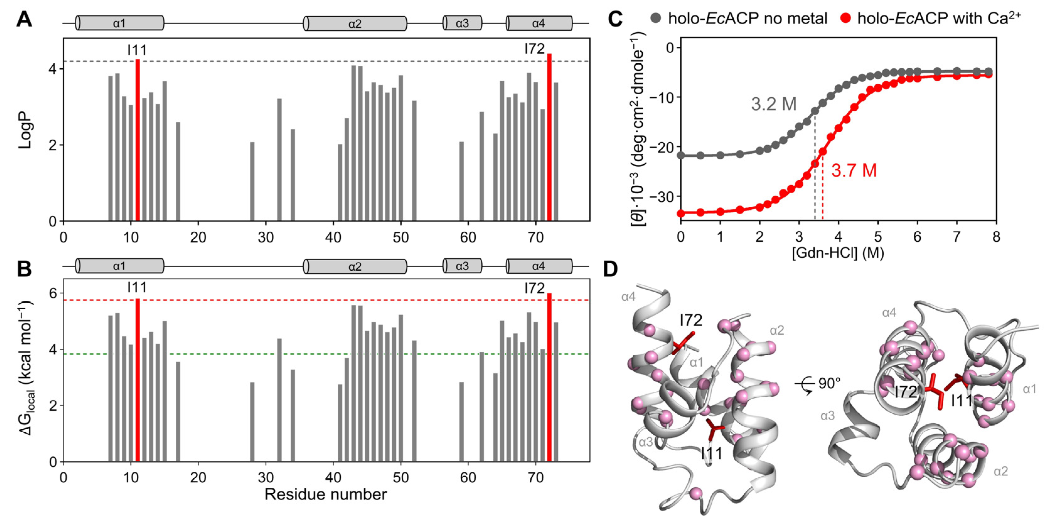
To further investigate the stability of structural packing of holo-EcACP in its stable state in the presence of Ca2+, we performed an H/D exchange experiment on the backbone amide protons (Figure 4A,B). This technique analyzes the exchange rate of amide hydrogen within the protein structure, enabling assessment of structural stability and flexibility on timescales ranging from milliseconds to seconds or even longer. Protection factors (LogP), which are calculated through comparison of experimentally measured H/D exchange rate (kprot) with the random coil exchange rate (krc), were employed for each residue to quantitatively evaluate the degree to which the amide protons were protected from exchanging with deuterium in solution (Figure 4A).
In holo-EcACP, all α-helical regions except α3-helix exhibited high LogP values, indicating that the α1-, α2-, and α4-helices are relatively stable. Within the α3-helix, only Ala59 showed slow amide proton exchange with deuterium, whereas in apo-EcACP, slow amide proton exchange was observed for all residues of α3-helix [48]. This suggests that the transition from the apo to the holo form specifically affects the stability of the α3-helix, likely due to the movement of the prosthetic group. Ala59 maintains a high amide proton protection because its side chain faces the hydrophobic pocket rather than the solvent, thus minimizing external interactions and stabilizing its backbone amide proton. Notably, even after 1000 min, amide protons of residues such as Ile11, Val43, Ile46, Glu47, Ile69, and Ile72 remained, reflecting persistent structural stability in these regions over time. Phe28 in the α1α2-loop, responsible for the proper accommodation of acyl chain at the entrance of cavity, also exhibited a high LogP value, likely because its phenyl ring interacts with other hydrophobic residues within the cavity, thereby contributing to a more compact structural packing. Leu42 and Leu46, which function to determine the path of the acyl chain, also showed protection of the amide proton. In addition, to analyze the local unfolding, we calculated the free energies of local unfolding (ΔGlocal) for each residue based on H/D exchange data (Figure 4B). A consistent trend with the LogP values was observed: residues in helical regions, except for the α3-helix, exhibited greater stability.
To assess how the presence or absence of Ca2+ affects the chemical stability of EcACP and to compare the free energies of global unfolding (ΔGglobal) with ΔGlocal, we conducted chemical denaturation experiments using Gdn-HCl. The half-denaturation concentration ([Gdn-HCl]1/2)—the concentration at which half of the protein structure begins to denature—was measured for EcACP (Figure 4C). In the presence of Ca2+, [Gdn-HCl]1/2 was 3.7 M whereas in the absence of metal ions, it decreased to 3.2 M. This indicates that metal ions enhance not only the thermal stability but also the chemical stability of EcACP, promoting a more stable folded conformation.
Using the denaturation curves of chemical denaturation experiments, we calculated the ΔGglobal. The ΔGglobal of EcACP in the presence of Ca2+ was determined to be 3.83 kcal mol−1 at [Gdn-HCl]1/2. Comparing the ΔGlocal from H/D exchange experiments with the ΔGglobal, we identified residues with a higher ΔGlocal than ΔGglobal: Val7, Lys8, Lys9, Ile10, Ile11, Gly12, Glu13, Gln14, Leu15, Leu32, Val43, Met44, Ala45, Leu46, Glu47, Glu48, Glu49, Phe50, Thr52, Ile62, Val65, Gln66, Ala67, Ala68, Ile69, Asp70, Tyr71, Ile72, and Asn73. These residues are predominantly located in the α-helix, and they are indicated by pink spheres in Figure 4D.
The Ile11 in the α1-helix, and the Ile72 in the α4-helix showed the highest ΔGlocal values (5.82 kcal mol−1 for Ile11 and 6.01 kcal mol−1 for Ile72), which were more than 1.5 times higher than that of ΔGglobal, indicating that these residues are the critical for protein folding. These residues are conserved across multiple ACP homologs, underscoring their structural and functional importance (Figure 1). The Ile11 of the α1-helix is a key residue contributing to the formation of a hydrophobic core, and it also maintains the tight packing of the structure. Also, Ile72 stabilizes the structure of EcACP by interacting with other hydrophobic residues, such as Ile3 and Phe50, at the top end of the hydrophobic cavity. As these two residues dominate hydrophobic interactions, residues such as Val7, Leu46, Phe50, Ile62, and Ala68 also exhibited higher ΔGlocal values than the ΔGglobal value. Ile11 and Ile72 are also represented in the structure of EcACP in Figure 4D.

2.5. Analysis of Metal-Binding Sites by Metal Titration HSQC Spectra

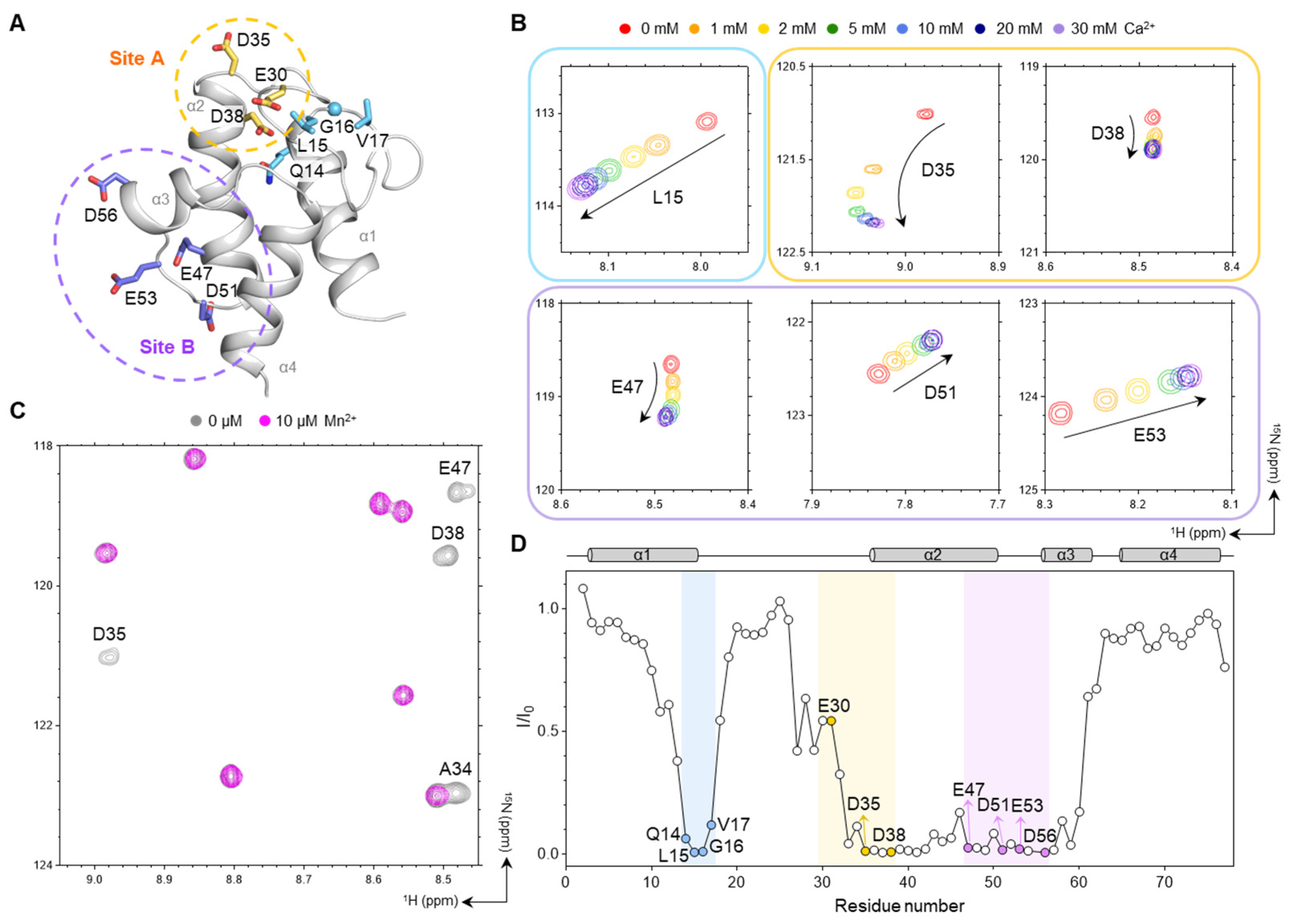
Previous studies have identified two metal-binding sites in EcACP: Site A, comprising Glu30, Asp35, and Asp38; and Site B, composed of Glu47, Asp51, Glu53, and Asp56 (Figure 5A) [35,36]. In our Ca2+ titration experiments with concentrations ranging from 0 to 30 mM, both sites showed chemical-shift perturbations (CSPs), with the magnitude increasing at higher Ca2+ concentrations (Figure 5B and Figure S1A). As Ca2+ is a diamagnetic ion, it perturbs the local structure showing CSPs in the 1H–15N heteronuclear single-quantum coherence (HSQC) spectra. As shown in Figure 5B, these residues near the metal-binding sites exhibited significant CSPs during titration. In addition to the residues directly involved in metal binding, residues located near site A, such as Leu15, exhibited significant CSPs (Figure 5B). Interestingly, Ile54, located between the site B residues, exhibited the largest CSP, despite not directly coordinating the Ca2+. This suggests that Ca2+ binding to site B induces substantial structural rearrangement of the loop region, especially Ile54, thereby stabilizing its conformation (Figure S1B,C).
To assess the proximity of residues to these metal-binding sites, additional titration experiments using Mn2+—a strongly paramagnetic ion—were performed (Figure 5C and Figure S2). Even at low concentrations (10 μM), Mn2+ induced severe line broadening, resulting in significant intensity reduction for residues such as Glu30 or rapid disappearance of peaks for Asp35, Asp38, Glu47, Asp51, Glu53, and Asp56 from sites A and B (Figure 5C,D), confirming their close proximity to bound metal ions. Residues near site A (Gln14, Leu15, Gly16, and Val17), which also showed CSPs in the Ca2+ titration, displayed marked signal loss, indicating that even hydrophobic residues not directly coordinating the metal can undergo structural rearrangement when located adjacent to site A, as the region is stabilized upon metal binding (Figure 5A,D).

2.6. Chemical Shift Perturbation by Acylation of EcACP

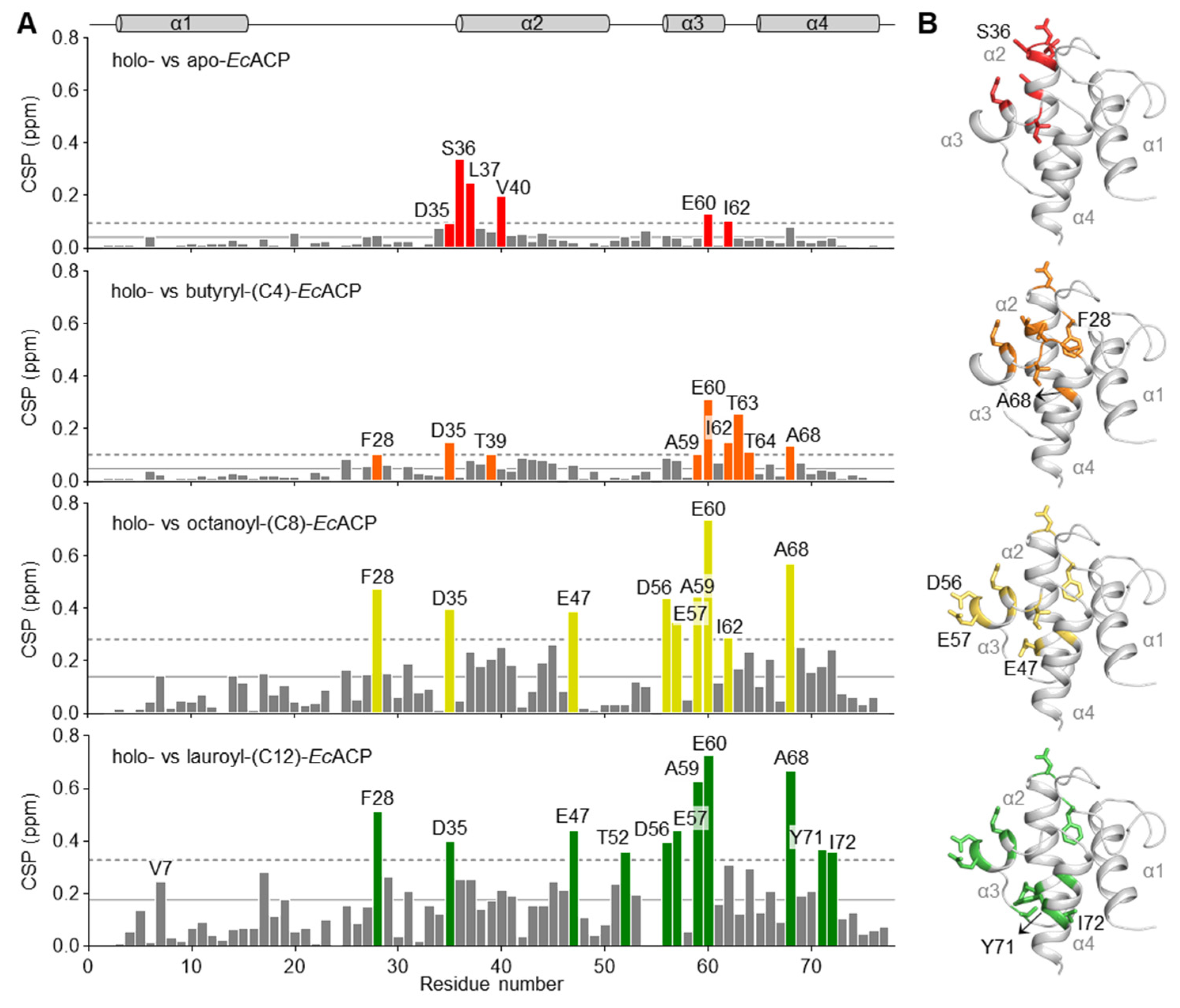
To assess structural perturbations caused by different acyl-chain lengths, we acquired and compared 1H–15N HSQC spectra for various acylated forms and analyzed the corresponding CSPs (Figure 6). The transition from the apo to holo form, with the 4′-PP attachment to Ser in the DSL motif, resulted in large CSPs in residues nearby this motif. Glu60 and Ile62, located at the cavity entrance, also exhibited significant CSPs, suggesting that the α3-helix and α3α4-loop not only participate in cavity expansion but are also affected by interactions between the 4′-PP arm and this region [31,49].
A significant CSP between the holo and butyryl form was newly observed for a key residue, Phe28, which is located near the cavity entrance. Phe28 is likely perturbed upon insertion of the butyryl group, indicating a direct interaction. Additionally, a notable CSP was observed for Ala68, located at the N-terminal region of the α4-helix and oriented toward the cavity interior, whereas residues at the bottom of the cavity exhibited minimal CSPs. These findings suggest that the short butyryl group can be accommodated only up to the midpoint of the cavity. In addition, residues Glu57, Ile54, and Asp56 at α3-helix exhibited minimal CSPs. These residues are located within or near metal-binding site B, and the reduced CSP values are presumably caused by structural stabilization upon Ca2+ binding, leading to comparatively decreased flexibility of this region in the short butyryl form.
Upon transition to the octanoyl form, residues in the α3-helix, including Asp56 and Glu57, exhibited significantly increased CSPs, indicating conformational rearrangement in the α3-helix associated with the accommodation of the longer acyl chain-like octanoyl group. In addition, increased CSP values were observed for residues in the α2-helix, particularly around Glu47, when comparing CSPs between the holo and butyryl forms. These observations suggest that the cavity undergoes a more pronounced expansion in the octanoyl form than in the butyryl form, primarily involving the rearrangement of α3-helix, with the α2-helix likely affected by structural adjustments.
In the lauroyl form, CSPs of Tyr71 and Ile72 in the α4-helix increased significantly. The large CSP values observed for these residues at the cavity bottom are consistent with previous studies showing hydrophobic contacts between the longer acyl chain and these residues [17,50]. Overall, these findings highlight that the extent and pattern of CSPs are dependent on acyl-chain length, with longer chains inducing more pronounced structural rearrangements in the α3-helix and additional perturbations within the cavity, reflecting progressive cavity adaptation. In the case of longer chains, such as the palmitoyl form, hydrolysis occurs so rapidly that the percentage of the holo form far exceeds that of the acylated form, making it impossible to analyze the CSPs.

2.7. Backbone Dynamics of Holo-, Butyryl-, and Octanoyl-EcACP

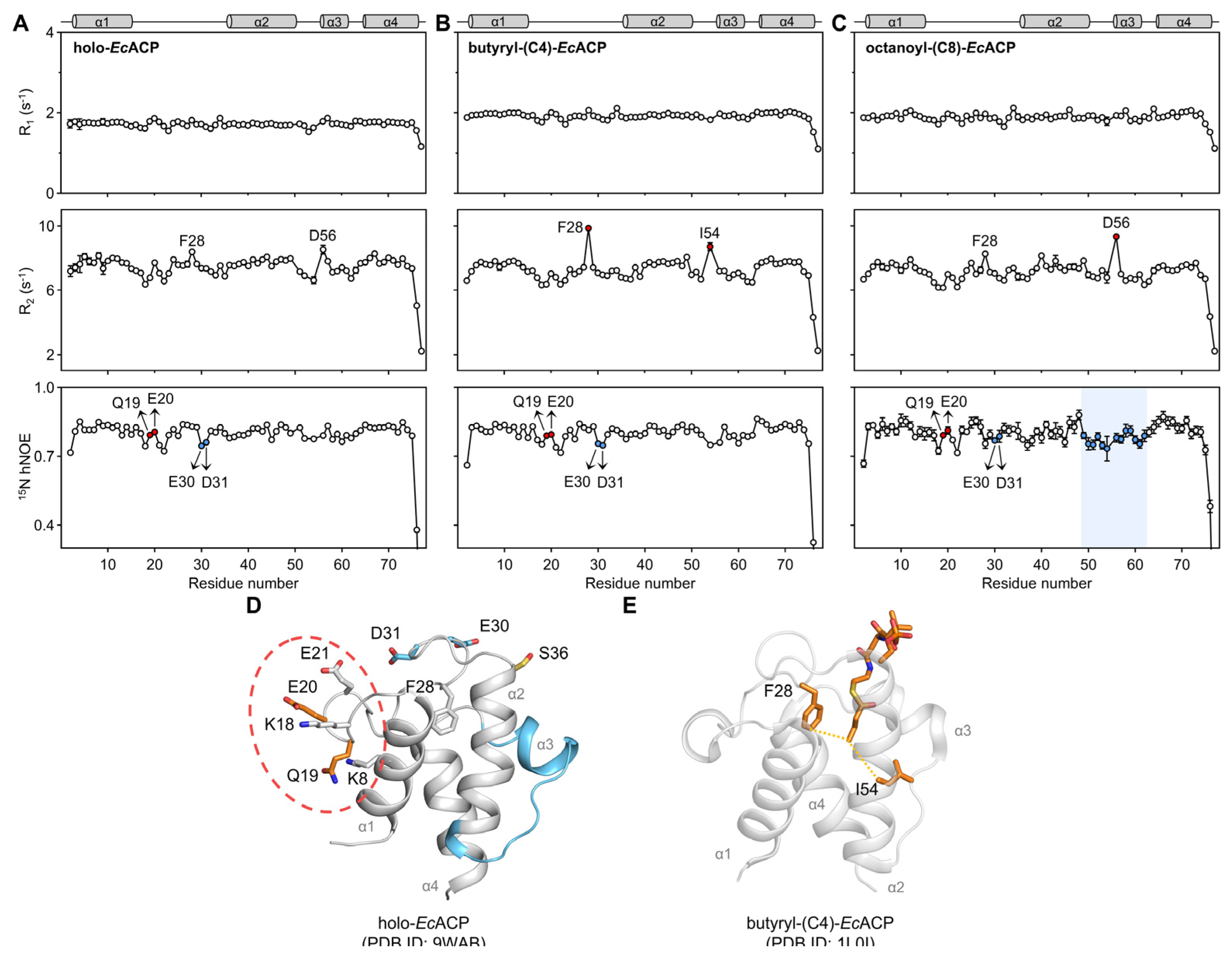
To characterize and compare the backbone dynamics of EcACP according to various chain length on a ps-ns timescale, we measured the spin-relaxation rates of longitudinal (R1) and transverse (R2) and heteronuclear NOEs (hNOEs) for the holo, butyryl, and octanoyl forms of EcACP (Figure 7A–C).
Across all three forms, Gln19 and Glu20 consistently displayed relatively high hNOE values despite being located in the α1α2-loop. This may reflect enhanced structural stability resulting from the formation of salt bridges with nearby polar residues (Figure 7D).
In the butyryl form, Phe28 in the α1α2-loop, and Ile54 in the α2-helix exhibited significantly higher R2 rates (9.86 and 8.72 s−1, respectively), likely reflecting interactions between the terminal end of the butyryl chain and the side chains of Phe28 and Ile54 with close spatial proximity, both oriented toward the cavity interior (Figure 7E). In contrast, in the holo form, the 4′-PP arm is too short to influence these residues much, while in the octanoyl form, the longer chain remains stably accommodated within the cavity. Due to these differences in chain length, significant exchange was observed only for butyryl form at these residues. The previously reported crystal structure of butyryl-EcACP (PDB ID 1L0I) is consistent with this interaction pattern [51] (Figure 7E). In contrast, these residues exhibited relatively low R2 rates in the holo and octanoyl forms. In the holo form, the acyl chain is too short to interact with these residues, while in the octanoyl form, the chain is more stably embedded within the cavity, resulting in less pronounced exchange compared to the butyryl form.
In the octanoyl form, Asp56, located in the N-terminal region of the α3-helix, exhibited a significantly higher R2 rate compared to the holo and butyryl forms. This suggests that accommodation of the octanoyl chain involves movement of the α3-helix, which is supported by low hNOE values, reflecting increased flexibility in this region (Figure 7C). Moreover, from the holo to the butyryl and octanoyl forms, hNOE values in the α3-helix and adjacent loop regions progressively decrease, indicating that longer acyl chains are accompanied by greater outward movement of α3-helix to accommodate them.
Glu30 and Asp31, located in the α1α2-loop, exhibited notably lower hNOE values compared to near residues in the loop region (Figure 7D). This increased flexibility is likely due to their position at the cavity entrance, where they are solvent-exposed, as well as the dynamic motion of Phe28, which forms transient interactions with short acyl chains, such as the butyryl chain. Although Ca2+ binding may enhance the overall stability of the loop, Glu30 and Asp31 at the cavity entrance are functionally required to retain conformational flexibility. Furthermore, hNOE values of Glu30 and Asp31 slightly increased in the octanoyl form compared to holo and butyryl forms, suggesting that the longer acyl chain stabilizes the cavity entrance, reflecting reduced dynamic fluctuations at the cavity entrance due to tighter acyl-chain packing.

2.8. Molecular Dynamics Simulation of EcACP

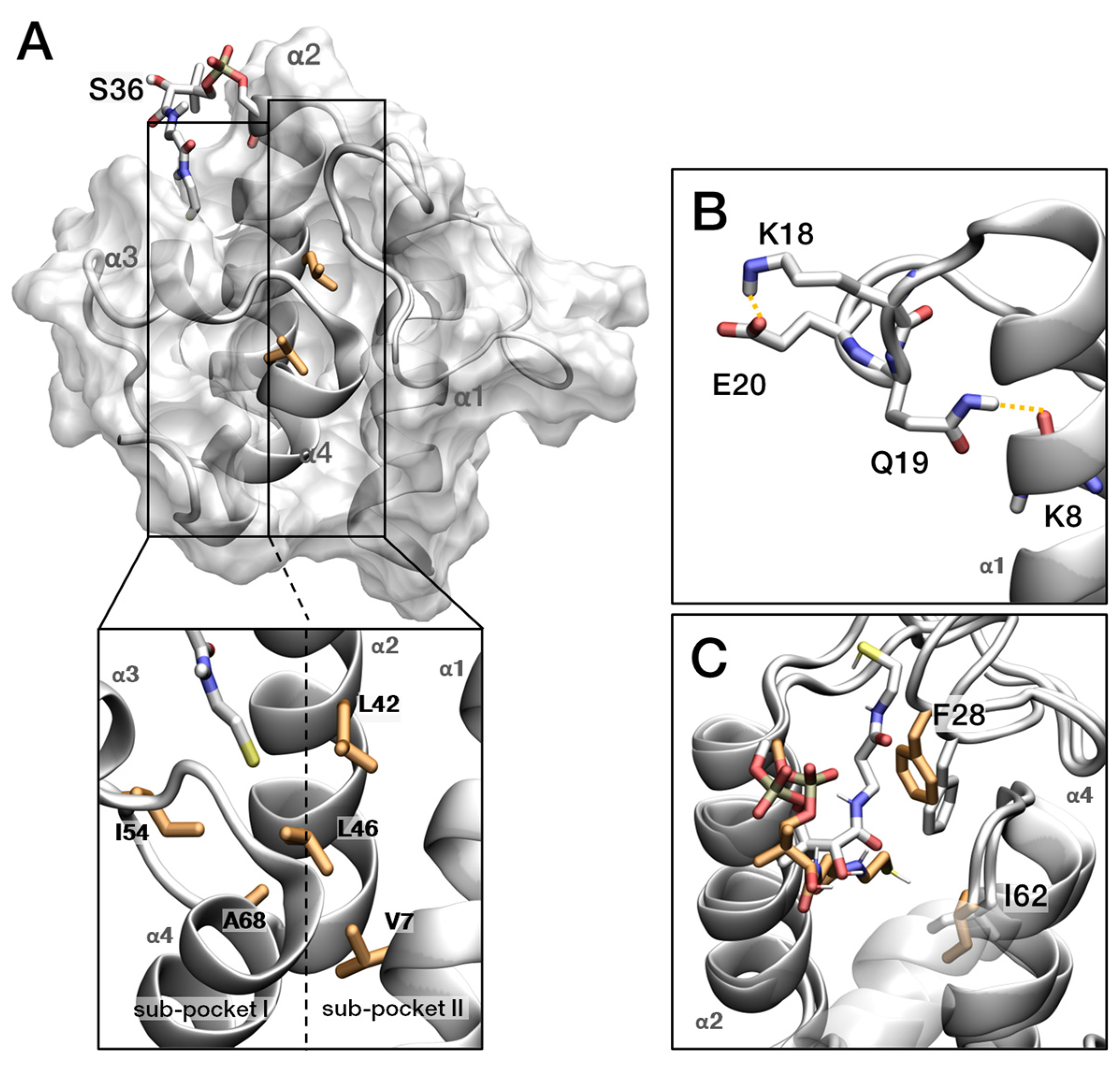
To investigate structural movements of EcACP during acyl-chain accommodation, we performed 1000 ns MD simulation on holo-, butyryl-, octanoyl-, lauroyl-, and palmitoyl-EcACP. The acyl chain and prosthetic group were initially solvent-exposed, allowing us to observe their entry into the hydrophobic cavity. First, the residues that exhibited significant structural changes are examined within the holo-EcACP structure. EcACP features a 4′-PP arm that binds acyl intermediates from the Type II-FAS pathway, with its internal cavity divided into two separate sub-pockets designed to accommodate a wide range of acyl-chain lengths (Figure 8A). Additionally, hydrophobic residues such as Ile54, Ala68, and Val7 line the interior, forming a hydrophobic environment facing inward. Leu42 and Leu46 are aligned along the α2-helix and positioned between sub-pocket I and sub-pocket II, contributing to the structural framework necessary for substrate binding and accommodation.
The residue from 19 to 21 forms hydrogen bonds, despite residing in the α1α2-loop, and these bonds display relatively high hNOE values (Figure 8B). During the 1 μs MD simulations, we observed the sidechain of Glu19 and backbone of Lys8 retain stable hydrogen bonding within 3.5Å about 40% occupancy, and this proximity is further supported by observed bending and fluctuations in this loop region. In addition, loop bending enables Lys18 to access the side chain of Glu20, forming an extra hydrogen bond occasionally (~5% occupancy). Two types of hydrogen bonds strengthen the interaction between the α1-helix and the α1α2-loop, influencing the backbone dynamics of EcACP.
Ile62 changes its torsion angle corresponding to the entry of the 4′-PP arm, suggesting that Ile62 may mirror structural changes associated with variations in acyl-chain length (Figure 8C). Since Ile62 has a significant value of CSP in holo- and octanoyl-EcACP, we compared it with the distance data, which are the extent to which the acyl chain enters the cavity, and the torsion angle value of the Ile62 side chain in octanoyl-EcACP (Figure S3A). We observed that when the side chain rotates by approximately −70 degrees, the eight carbon molecules became deeply sequestered, suggesting that Ile62 is a residue that responds to acyl-chain entry.
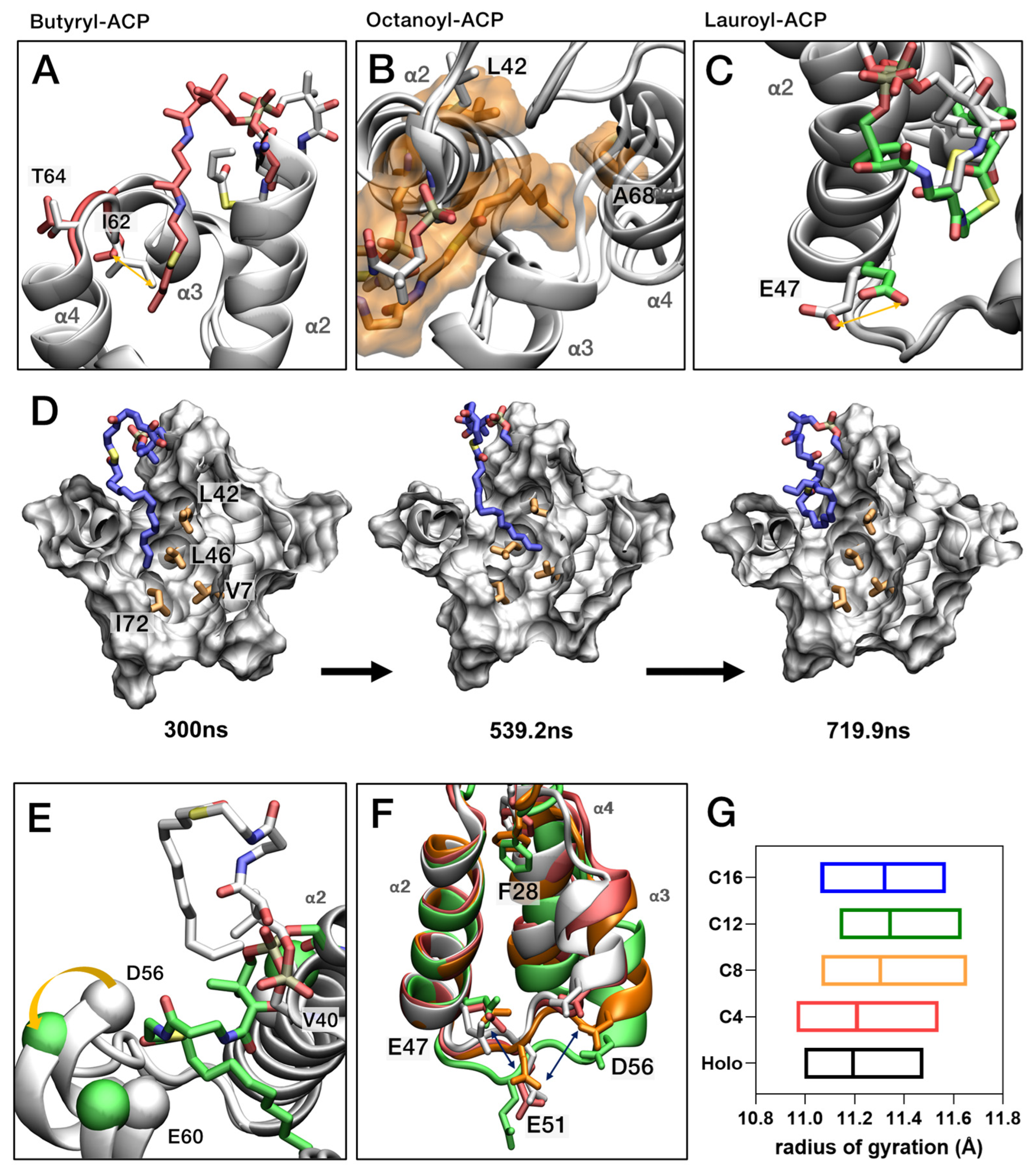
To understand the fatty acids biosynthesis mechanism, we investigated how the structure of EcACP adapts to varying substrate lengths by modeling butyryl-, octanoyl-, lauroyl-, and palmitoyl-EcACP, each extending the acyl chain by four carbon units. Butyryl-EcACP serves as the initial comparison with holo-EcACP, representing the effect of a short acyl chain. Due to its short acyl chain, it was insufficient to sample a stable conformation or sustain long-term (around 200 ns) sequestration. Similar dynamics were observed between the Ile62 switch pattern and the substrate entry, where the acyl chain was blocked underneath the side chain of Ile62 (Figure 9A). Although Val43 occasionally exhibited torsional changes in its side chain, these did not correlate with acyl-chain entry. Leu37 and Met44 displayed behaviors similar to holo-EcACP, with their side chains oriented outward, and both experienced significant fluctuations due to solvent exposure.
In the case of octanoyl-EcACP, the acyl-chain length efficiently occupies sub-pocket I, remaining there for extended periods. The chain approaches closer to Leu42 of the α2-helix and Ala68 of the α4-helix, enhancing the potential for hydrophobic interactions (Figure 9B). This observation is consistent with chemical-shift perturbation data, which show significant increases. Additionally, residues such as Phe28 and Glu47 underwent notable structural changes when the acyl chain exceeds eight carbons, indicating increased flexibility and interaction potential (Figure 9C). For example, Glu47 tended to rotate about −180° when the acyl-chain length reached more than 12 carbons, compared to around −70° (Figure S3B). Asp56 similarly exhibited a χ1 torsion angle around 60° in the octanoyl form. This change is thought to be due to an increase in the contact area with the residues as a result of the expansion of the loop.
Lauroyl-EcACP with a 12-carbon chain marks the point at which the acyl chain begins to invade sub-pocket II. Gating effects caused by Leu42 are evident, with the acyl chain approaching Val7, located at the bottom of sub-pocket II. Corresponding changes in NMR data support this movement for chains with 12 or more carbons (Figure 9D). To study movement dynamics from sub-pocket I to II, palmitoyl-EcACP with a 16-carbon chain was modeled, revealing that Leu42 and Leu46 act as gates. Each leucine bends in opposite directions, opening a pathway for the acyl chain to orient toward sub-pocket II and thereby approach Val7. Subsequently, the palmitoyl chain exhibits an exit pathway from the cavity, exposing the long chain to the solvent, as shown in snapshots at 300 ns, 539.2 ns, and 719.9 ns, respectively. Monitoring the side-chain torsion angle (χ1) during the simulations showed that, upon opening toward sub-pocket II, Leu42 and Leu46 adopted dihedrals of approximately −70° and −180°, respectively (Figure S3B). This gating behavior becomes prominent starting with lauroyl-EcACP, and we suggest that it is consistent with the CSP for lauroyl-EcACP in Figure 7A, in which Val7 rises above the average.
As the acyl-chain length increased, structural changes became more pronounced. Using Glu60 and Asp56 on the α3 helix as a reference, we observed substantial positional shifts associated with acyl-chain ingress and egress; concomitantly, the distance to Val40 on the α2 helix increased, indicating deeper penetration of the acyl chain (Figure 9E). Asp56 on the α3 helix exhibits high R2 rate, indicating outward movement of the α3 helix to accommodate longer acyl chains, as observed in MD simulation, consistence with backbone dynamics data. The α2α3 loop region, which clusters negatively charged metal-binding sites, also expanded and exhibited greater fluctuation as the chain lengthened (Figure 9F). Phe28, located near the cavity entrance, tended to tilt in a consistent direction as larger chains were accommodated, further supporting their prominent R2 rate observed in our backbone dynamics analyses. This expansion of helices and loops ultimately led to an increase in the radius of gyration, indicating overall structural swelling, with the α2α3 loop exhibiting notable enlargement (Figure 9G).
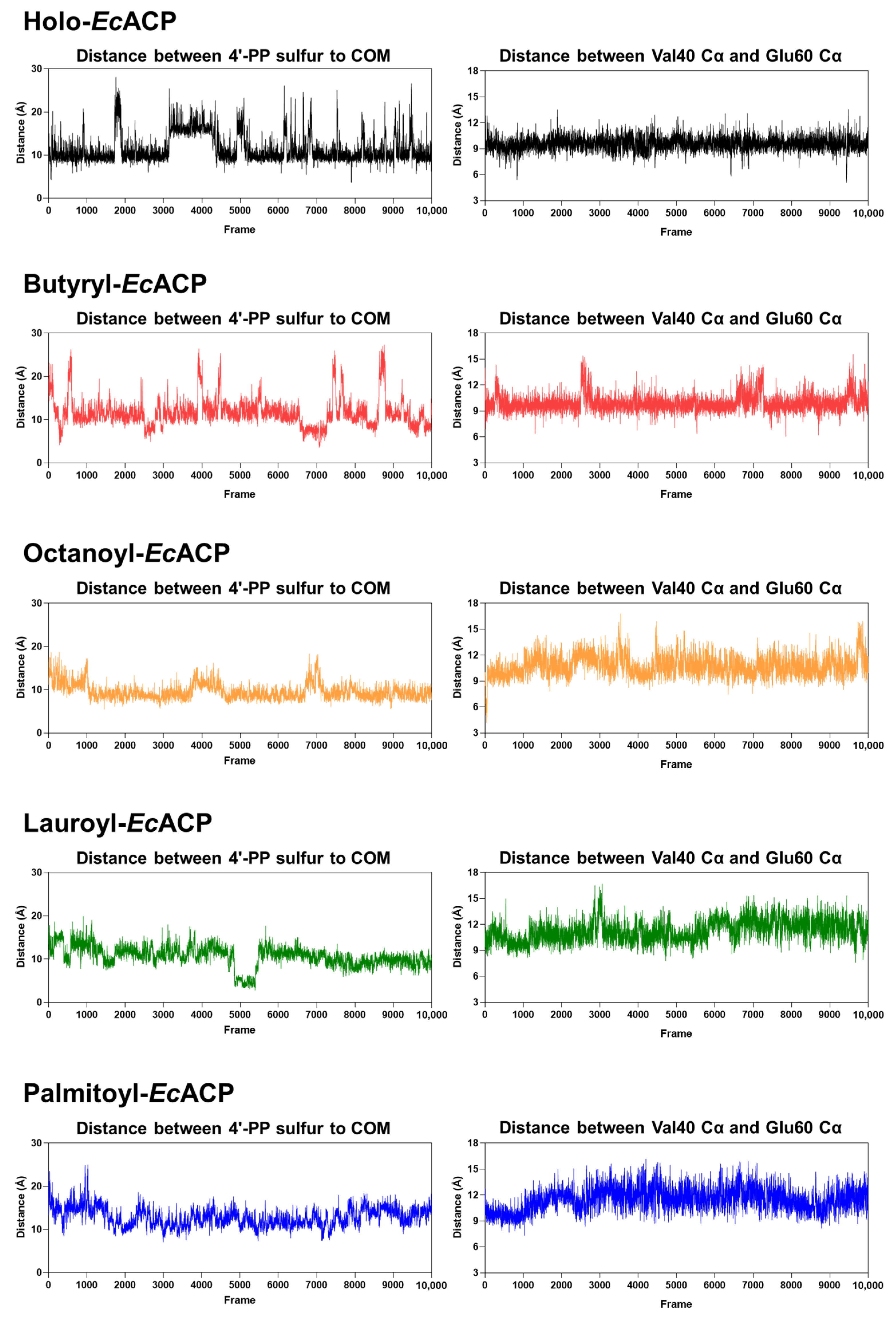
The decrease in the distance between the center of mass (COM)—reflecting the extent of acyl-chain insertion—and the sulfur atom of the 4′-PP arm shows a tendency to correspond to the increase in the distance between the α2-helix (Val40) and the α3-helix (Glu60) (Figure 10). This trend is observed from holo- to octanoyl-EcACP but diverges starting from lauroyl-EcACP. Considering the similar pattern observed in the radius of gyration analysis (Figure 9G), it suggests that EcACP can optimally accommodate an acyl chain of up to eight carbons within its cavity [52].

3. Discussion

This study provides profound insights into the structural dynamics and stabilization mechanisms of EcACP, which is the most extensively studied bacterial ACP and a critical player in fatty acid synthesis. Combining NMR spectroscopy and MD simulations, we elucidate the crucial role of divalent metal ions, particularly Ca2+, in stabilizing EcACP, while also revealing how its conformational landscape adapts to varying acyl-chain lengths. This is the first to present an integration study of R1, R2, and hNOE backbone relaxation data with MD simulations on various acylate forms of EcACP, demonstrating that ACP dynamics are precisely regulated by acyl-chain length, closely associated with chain-flipping mechanisms governed by the movement of the α3-helix. These findings significantly advance our understanding of ACP’s functional plasticity and open new ways of targeting fatty acid synthesis in bacterial pathogens.
Our comprehensive investigation revealed the critical role of Ca2+ in stabilizing EcACP, and, notably, we report the first high-resolution solution structure of EcACP determined in the presence of Ca2+—an essential ion for fatty acid synthesis. This structural elucidation may provide fundamental insights into how calcium ions regulate EcACP’s stability and function within the context of FAS. NMR spectroscopy offered detailed insights into two Ca2+ binding sites, showing specific CSPs indicative of direct coordination and local conformational stabilization. The solution structure in the presence of Ca2+ further revealed a notable inward conformation of metal-binding site A, with Phe28 deeply embedded within the hydrophobic cavity, likely stabilized through Ca2+-mediated interactions.
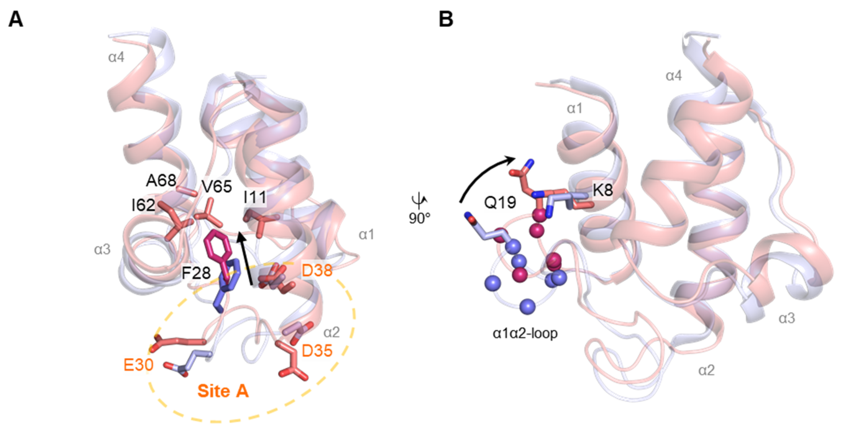
We compared our NMR structure of EcACP with Ca2+ to a previously reported holo-EcACP solution structure (PDB ID: 2K93) determined in potassium phosphate buffer. In the Ca2+-bound structure, coordination at metal-binding site A appears to shift Phe28 more inward into the hydrophobic cavity, enhancing interactions with surrounding residues such as Ile12, Ile62, Val65, and Ala68, leading to a more tightly packed cavity (Figure 11A). Notably, Gln19 near metal-binding site A exhibits a high hNOE value within the α1α2-loop, indicating enhanced local stability. In the Ca2+-bound structure, Gln19 (Val17–Val22) is positioned closer to Lys8 compared to the structure with K+ monovalent ion, and a hydrogen bond between Gln19 and Lys8 was observed in MD simulations, suggesting a metal-induced structural rearrangement.
Complementing these findings, CD experiments demonstrated a remarkable increase in structural stability upon calcium addition, with melting temperatures rising by more than 10 °C. The DSC data further supported this, revealing a higher Tm and a substantial increase in calorimetric enthalpy (16.7 kcal mol−1 increase in ΔHcal), indicating a more thermodynamically stable folded state facilitated by Ca2+ binding compared to K+ binding. These observations are consistent with the observed enhanced thermal stability under Ca2+ conditions compared to K+, as evidenced by the increased Tm values from both DSC and CD analyses.
Through structural and dynamics analysis, these findings highlight that Ca2+ not only mitigates electrostatic repulsion—given the highly negatively charged surface of EcACP—but also prompts specific conformational changes that reinforce the hydrophobic core and stabilize critical structural regions, such as the α1α2-loop and α3-helix. Notably, Gln19, situated near metal-binding site A, displayed a high hNOE value within the α1α2-loop, further corroborated by MD simulations, which suggest that Lys8 forms a hydrogen bond with Gln19 in the Ca2+-bound structure, contributing to enhanced local stability.
The Ca2+ titration experiments monitored by NMR CSPs confirmed binding at known metal-binding sites, with the most significant chemical shift change observed for Ile54—located at the cavity bottom—suggesting structural rearrangement rather than direct metal coordination for Ile54 (Figure S1). Additional Mn2+ titration revealed conformational changes in the nearby α1α2-loop, indicating that Ca2+ binding not only stabilizes the coordination site but also promotes conformational tightening in adjacent flexible regions. Overall, divalent cations like Ca2+ may play a key role in enhancing the structural stability and functional integrity of EcACP in bacterial FAS.
In the H/D exchange experiments, residues Ile11 and Ile72 stand out as key residues critical to the global folding stability of EcACP. Their high protection factors indicate their important role in maintaining a stable hydrophobic core, which is essential for the protein’s structural integrity. Ile11, located within the α1-helix at the middle of the cavity, and Ile72, situated at the bottom of the α4-helix, occupy strategic positions that are highly conserved among bacterial ACPs (Figure 1 and Figure 3B,C). The hydrophobic packing mediated by Ile11 appears vital for stability; notably, in thermophilic ACPs, such as TmACP, the equivalent residue, Ile15, demonstrates even higher amide proton protection, with ΔGlocal exceeding 8 kcal mol−1 and surpassing the global stability (ΔGglobal), highlighting its crucial contribution to structural robustness [13]. The increased stability of TmACP can partly be attributed to minimized electrostatic repulsion due to additional basic residues, which enhance hydrophobic interactions. Supporting this, mutation of Ile15 to alanine in TmACP weakens packing and reduces protection, emphasizing the importance of hydrophobic contacts—a result likely paralleled if Ile11 can be mutated in EcACP, a hypothesis warranting future validation. Additionally, Ile72, positioned at the α4-helix, forms part of the hydrophobic triad at the bottom of the cavity, contributing similarly to the highest ΔGlocal value; its role in preserving the hydrophobic core further underscores its significance in stabilizing EcACP’s folded state.
Spin-relaxation experiments provide precise insights into EcACP backbone dynamics, and MD simulations deepen these findings by offering atomistic views of dynamic behavior. In this study, combining both approaches revealed how EcACP accommodates different acyl-chain lengths (Figure 8, Figure 9 and Figure 10) and identified key residues involved in acyl-chain binding. Notably, Phe28 and Ile54 showed high R2 rates specifically in the butyryl form, which MD simulations confirmed by showing that the short chain cannot be stably sequestered and frequently exchanges between sequestered and solvent-exposed states. This dynamic exchange explains the elevated R2 rates, demonstrating the synergy of experimental and computational approaches in elucidating EcACP’s flexibility and substrate interactions.
Furthermore, our NMR and MD analyses demonstrated that the α3-helix (Asp56–Lys61) undergoes notable conformational expansion and outward movement as the acyl chain lengthens. Increased CSPs, especially in Glu60, highlight this structural change. In the octanoyl form, low hNOE values across the α3-helix and nearby loops, along with a high R2 rate for Asp56, indicate greater flexibility to accommodate longer chains. These experimental results are corroborated by MD simulations, which show notable fluctuations in Asp56 and Glu60, highlighting the dynamic adjustment of the α3-helix with increasing acyl-chain size (Figure 9E). This flexibility likely plays a key role in acyl-chain transfer to partner enzymes via the chain-flipping mechanism.
These findings offer insight into the structural adjustments necessary for EcACP to reach its typical biological endpoint of synthesizing C16 or C18 fatty acids. The CSP (Figure 6) and backbone dynamics data (Figure 7) from NMR experiments of acyl-EcACP consistently demonstrated chain-length-dependent conformational changes, most notably the outward displacement of the α3-helix. This is further corroborated by MD simulations, which show conformational switching of Leu42 and Leu46 to open sub-pocket II, thereby facilitating the accommodation of longer chains. Moreover, previous studies have shown that as the acyl cavity approaches its maximum volume, incomplete sequestration of longer acyl chains leads to elevated hydrolysis rates [32,53]. This trend is consistent with our MD-based analysis, including radius of gyration (Figure 9G) and distance analysis (Figure 10). Collectively, these findings align well with the biological necessity for EcACP to accommodate long-chain fatty acids.
One limitation of our study is the challenge in obtaining reliable NMR data for longer acyl chains exceeding C12, due to rapid hydrolysis and the long acquisition times required for backbone dynamics experiments. Consequently, we relied on MD simulations to investigate their conformational behavior, focusing on the motions of Leu42 and Leu46, which serve as switch pivots in the gating mechanism for sub-pocket II (Figure 9D). These residues rotate in opposite directions to create a pathway for longer acyl chains. Additionally, fluctuations in negatively charged residues such as Glu47, Glu51, and Asp56, shown in MD simulations, suggest that electrostatic repulsion drives cavity expansion and conformational looseness, facilitating longer-chain accommodation (Figure 9F). Meanwhile, residues like Ala68 remain oriented inward to maintain hydrophobic stability. To overcome the limitations caused by rapid hydrolysis during solution NMR experiments, future studies may incorporate acyl–acyl carrier protein synthetase, which attaches fatty acids to holo-ACP in an ATP-dependent manner [54], to suppress hydrolysis and stabilize long-chain acyl-EcACP samples for further investigation. Future studies involving targeted mutational analyses and advanced experimental techniques to investigate ACP-enzyme interactions are essential for directly capturing these dynamic processes. Such efforts could significantly enhance our understanding of substrate specificity and the mechanistic functions of ACPs in their interactions with enzymes during fatty acid biosynthesis.
Taken together, this study demonstrates how divalent cation Ca2+ coordination stabilizes EcACP and reveals how acyl-chain length modulates its conformational dynamics. By integrating advanced NMR techniques with MD simulations, we have uncovered the fundamental mechanisms driving EcACP’s structural adaptability and functional flexibility. This combined approach has elucidated the dynamic behavior of key regions and residues, revealing how structural flexibility regulates its function in fatty acid biosynthesis. These insights significantly advance our knowledge of EcACP’s conformational adaptability, highlighting the importance of developing strategies to inhibit its dynamics, which could be crucial for effectively targeting fatty acid biosynthesis.

4. Materials and Methods

4.1. Cloning, Expression, and Purification of EcACP

The acpP gene was amplified from E. coli K12 genomic DNA and inserted into the multiple cloning site of the pET-21a expression vector via Nde I and Xho I restriction enzymes. Recombinant plasmids were introduced into E. coli BL21(DE3) competent cells and used for protein expression. To produce the isotope-labelled proteins required for NMR analysis, the cells were cultured in an M9 minimal medium that was supplemented with either 0.5 g of 15NH4Cl (Cambridge Isotope Laboratories, Andover, MA, USA) for two-dimensional spectra, or with 0.5 g of 15NH4Cl and 1.0 g of 13C-glucose (Cambridge Isotope Laboratories, Andover, MA, USA) for three-dimensional experiments. For other experiments, LB medium was used instead. Protein overexpression was induced via supplementation with 0.5 Mm isopropyl β-D-thiogalactopyranoside (IPTG) when the optical density value of the culture medium reached between 0.8 and 1.0. Then, the culture was incubated at 25 °C for 16 h.
Purification of EcACP was achieved through sequential FPLC steps exploiting its physicochemical properties: ion-exchange chromatography (HiTrap™ QFF and Resource™ Q, GE Healthcare Bio-Sciences, Uppsala, Sweden) and size-exclusion chromatography (Superdex 75 16/600, GE Healthcare Bio-Science, Uppsala, Sweden). The apo-protein was subsequently converted into the holo- or acylated forms by incubations with recombinant EcACPS at 25 °C for 16 h in 25 mM of Tris-HCl buffer (pH 8.0) containing 20 mM MgCl2.

4.2. Circular Dichroism Experiments

The secondary structure of EcACP under varying temperatures was analyzed using far-UV CD with a J-810 spectropolarimeter (Jasco, Tokyo, Japan). Protein samples were prepared at a final concentration of 30 μM in 25 mM of MES buffer (pH 6.1) containing 5 mM of dithiothreitol (DTT). Four conditions were tested: buffer supplemented with 5 mM CaCl2, 5 mM MgCl2, 5 mM KCl, and a sample without added metal ions. The 1 mm path-length quartz cuvette was used for measurements. CD spectra were acquired from 200 to 250 nm, with a data pitch of 0.1 nm. The sample temperature was gradually raised from 25 to 100 °C, and thermal unfolding was tracked by monitoring changes in mean residue ellipticity (θ) at 222 nm, which reflects α-helical content.

4.3. Differential Scanning Calorimetry

Thermal properties were analyzed using DSC experiments on a MicroCal PEAQ-DSC system (Malvern Instruments, Malvern, UK) at the Korea Basic Science Institute (KBSI) (Ochang, Cheongju, Republic of Korea). Then, 5 mg/mL of protein samples was prepared in 50 mM sodium acetate buffer (pH 5.6) with 5 mM CaCl2 or 5 mM MgCl2. The DSC thermograms were collected over a temperature range of 20–100 °C, at a constant rate 1 °C/min. Data acquisition and baseline subtraction were carried out with PEAQ-DSC measurement software (v1.64), and thermodynamic parameters, including the Tm, ΔHcal, and Cp, were extracted by nonlinear least-squares fitting, assuming a two-state transition model.

4.4. NMR Experiments and Assignment

All NMR experiments in this study were conducted on Bruker Avance 700 MHz and 900 MHz spectrometers at the KBSI (Ochang, Cheongju, Republic of Korea). EcACP samples, either single- or double-isotope-labeled, were prepared at concentrations of 0.4–0.5 mM in a buffer consisting of 25 mM MES (pH 6.1), supplemented with 5 mM CaCl2 and 5 mM DTT, in a 9:1 (v/v) of H2O/D2O mixture. 2,2-Dimethyl-2-silapentane-5-sulfonate (DSS) served as the internal chemical shift reference, and 0.02% NaN3 was added for the purpose of preventing microbial growth. All spectra were processed with NMRPipe [55] and analyzed using NMRFAM-Sparky [56].
Backbone resonance assignments were obtained from HNCO, HNCACB, and CBCA(CO)NH experiments, while side-chain resonances were assigned using CC(CO)NH, HBHA(CO)NH, H(CCO)NH, and HCCH-TOCSY. Assignments were further validated by 1H-15N-1H NOESY-HSQC and 1H-13C-1H NOESY-HSQC spectra.
The RDCs of the backbone amide N-H bonds were determined by comparing the isotropic and anisotropic conditions in the IPAP-HSQC spectra. For the anisotropic sample, EcACP was dissolved in a radially compressed polyacrylamide gel, enabling partial alignment [57,58,59].

4.5. Solution Structure Calculation

NOE cross-peaks were assigned using NMRFAM-Sparky [56]. The three-dimensional structure of holo-EcACP was calculated using the PONDEROSA-C/S package with Xplor-NIH-based calculations [60]. From the ensemble generated, the twenty structures with the lowest energy levels were selected. Constrain violations for bond angles and interproton distances within these 20 models were further checked and refined using PONDEROSA-Analyzer [61]. The structural ensemble was validated with Protein Structure Validation Software [62]. All protein structure images were prepared using PyMOL (version 3.1.3.1) [63]. The final coordinates and NOE constraints were submitted to the Protein Data Bank (PDB) (PDB ID 9WAB).

4.6. Hydrogen/Deuterium Exchange Experiment

For the H/D exchange analysis, a 15N-labeled EcACP sample was prepared at a concentration of 0.5 mM in 25 mM of MES buffer (pH 6.1) containing 5 mM DTT and 0.02% NaN3 with 5 mM CaCl2. A set of 1H-15N HSQC spectra was acquired every 10 min at 25 °C for a total period of 1000 min. The decay of amide proton signals over time was quantified from the decrease in peak intensities. The exchange rate constants (kex) were obtained by fitting the intensity decay to a single-exponential function:
I = I 0 exp k e x t + C
where I represents the NH resonance intensity at time t, and I0 is the initial intensity. The experimentally determined exchange rate constant, kprot, was treated as equivalent to kex; the protection factor (P) was calculated as the ratio of the krc to kprot; and ΔGlocal was subsequently determined using the following relationship [64]:
Δ G l o c a l = R T   l n ( k p r o t k r c )

4.7. Chemical Denaturation Experiments

The global unfolding of EcACP under chemical denaturant was evaluated by far-UV CD experiment. Protein samples (30 μM) were prepared in 25 mM of MES buffer (pH 6.1) containing 5 mM of DTT, either with or without 5 mM CaCl2. Various Gdn-HCl concentrations were added to the protein samples, followed by 25 °C for 12 h to ensure that equilibrium was reached. CD spectra were recorded over the wavelength range of 200–250 nm, in steps of 0.1 nm. Ther denaturation curves were analyzed, assuming a reversible two-state equilibrium between the native (N) and unfolded (U) states. Data fitting was performed using the linear extrapolation model [65,66,67,68]. The global free energy of unfolding (ΔGglobal) was calculated following the procedure in a previous study [13].

4.8. Metal Titration Experiments

Successive 1H-15N HSQC spectra were acquired following the stepwise addition of Ca2+ and Mn2+ to final concentrations of 0 mM, 0.5 mM, 1 mM, 2 mM, 5 mM, 10 mM, 20 mM, 30 mM for Ca2+; and 0 mM, 0.001 mM, 0.005 mM, 0.01 mM, 0.02 mM, and 0.05 mM, 0.1 mM for Mn2+ to 0.3 mM of EcACP, respectively. For the Ca2+ titration, CSP analysis was carried out to identify residues exhibiting significant chemical shift changes. In the Mn2+ titration, peak intensities at each titration data point were normalized against the initial spectrum to monitor residue-specific decrease in signal intensity.

4.9. Chemical Shift Perturbations

CSP analysis was also performed to assess the effect of acylation on the structural properties of EcACP. 1H-15N HSQC spectra for 0.5 mM of apo-, holo-, butyryl-, octanoyl-, lauroyl-EcACP were obtained from NMR experiments. The CSPs between holo-EcACP and apo- or acylated forms were calculated using the following formula:
δ = 0.5 ( δ ( 1 H ) 2 ) + ( α δ ( 15 N ) 2
α = 0.2 for most residues, and 0.14 for glycine

4.10. Backbone Dynamics

Backbone dynamics of holo-, butyryl-, octanoyl-EcACP (0.5 mM) were characterized using NMR spin-relaxation measurements at 25 °C with a Bruker Avance 700 MHz spectrometer. A series of 1H–15N HSQC spectra of R1, R2, and hNOE were acquired. For R1 experiments, the applied relaxation delays were 0.01 s, 0.01 s, 0.05 s, 0.1 s, 0.2 s, 0.3 s, 0.3 s, 0.5 s, 0.8 s, and 1.2 s. For R2 experiments, delays of 0 s, 0 s, 0.01696 s, 0.03392 s, 0.05088 s, 0.05088 s, 0.0848 s, 0.013568 s, 0.022048 s, and 0.32224 s were used. The recycle delays were 2.3 and 2.0 s for R1 and R2 experiments, respectively. The hNOE experiments were performed with a recycle delay of 4.0 s and proton saturation of 3.0 s. Relaxation rates were obtained by fitting the exponential decay of peak intensities across the respective delay series.

4.11. Molecular Dynamics Simulations

All MD simulations were performed from the NMR structure of EcACP determined in this study (PDB ID: 9WAB). Acyl chains were covalently attached to the serine in the DSL motif using CHAARMM-GUI’s Ligand Reader & Modeler [69], with parameters generated by CGenFF (force field 3.0) [70,71,72]. Systems were prepared in CHARMM-GUI’s Solution Builder, employing a cubic water box size that maintained a 10.0 Å edge distance from the protein [73,74,75]. K+ and Cl were added to reach 0.15 M, neutralizing all models with TIP3P water. Simulations were performed in OpenMM 8.0.0 with the CHARMM36 m force field [76,77], ensuring consistent treatment of water models and nonbonded settings in our study. Long-range electrostatics were treated with particle-mesh Ewald (PME) under periodic boundary conditions, using a 1.2 nm real-space cutoff and an Ewald error tolerance of 5 × 10⁻⁴; and short-range Coulomb interactions were computed directly within the cutoff, and reciprocal-space contributions were evaluated on a mesh. Lennard–Jones interactions employed a force-switch between 1.0 and 1.2 nm, going smoothly to zero at 1.2 nm. During equilibration and production, we simulated in the NVT and NPT ensemble, respectively, with temperature held at 310.15 K, using a Langevin thermostat (friction coefficient 1 ps−1), as implemented in OpenMM 8.0.0. Pressure control (1 bar, isotropic Monte Carlo barostat) was applied during production only after equilibration had converged. For both equilibration and production, the lengths of all bonds that involve a hydrogen atom are constrained to use a larger integration time step. With HBonds constraints, the time step increased to about 2 fs in production and 1 fs in equilibration for Langevin dynamics. Each system underwent energy minimization for 0.125 ns, followed by the standard six-step CHARMM-GUI equilibration protocol with a 1 fs timestep. Production runs were then carried out for 1000 ns under constant temperature (310.15 K) and pressure (1 bar, isotropic), using Langevin dynamics. Production trajectories were collected for 1000 ns per replica, with three independent replicas per system. Trajectories and snapshots were analyzed and visualized using VMD 1.9.3 [78].

5. Conclusions

This study highlights how Ca2+ stabilizes EcACP and how acyl-chain length influences its structural flexibility and dynamics. CD, DSC, and H/D exchange experiments revealed that Ca2+ enhances the stability of EcACP, and hydrophobic interactions mediated by Ile11 and Ile72 are crucial for maintaining EcACP folding. By combining NMR and MD approaches, we gained detailed insights into how chain-length-dependent conformational changes—particularly the motion of the α3-helix acting as a molecular gatekeeper—facilitate substrate accommodation and transfer. These findings deepen our understanding of EcACP’s mechanistic flexibility and identify potential targets for designing antibacterial agents that disrupt FAS.

Supplementary Materials

The following supporting information can be downloaded at https://www.mdpi.com/article/10.3390/ijms26189005/s1.

Author Contributions

Conceptualization, Y.K.; resources, Y.K.; data curation, C.Y.L., S.J., H.C., M.-C.J. and Y.O.; writing—original draft preparation, C.Y.L., S.J., H.C. and Y.K.; writing—review and editing, C.Y.L., S.J., H.C. and Y.K. visualization, C.Y.L., S.J. and H.C.; supervision, Y.K.; funding acquisition, Y.K. All authors have read and agreed to the published version of the manuscript.

Funding

This work was supported by Konkuk University in 2024.

Data Availability Statement

The data presented in this study are available upon request from the corresponding author.

Conflicts of Interest

The authors declare no conflicts of interest.

Abbreviations

The following abbreviations are used in this manuscript:
ACPAcyl carrier protein
FASFatty acid synthesis
MDMolecular dynamics
EcEscherichia coli
ACPSHolo-ACP synthase
4′-PP4′-phosphopantetheine
MDRMulti-drug-resistant
Gdn-HClGuanidine hydrochloride
RDCResidual dipolar coupling
H/DHydrogen–deuterium
DSCDifferential scanning calorimetry
pIIsoelectric point
TmThermotoga maritima
CpColwellia psychrerythraea
TaThermus aquaticus
AbAcinetobacter baumannii
VhVibrio harveyi
TmMelting temperature
CDCircular dichroism
CpSpecific heat capacity
ΔHcalCalorimetric enthalpy
RMSDRoot-mean-square deviation
LogPProtection factor
kexExchange rate
kprotH/D exchange rate
krcRandom coil exchange rate
ΔGlocalLocal unfolding free energy
ΔGglobalGlobal unfolding free energy
[Gdn-HCl]1/2Half-denaturation concentration of Gdn-HCl
CSPChemical shift perturbation
HSQCHeteronuclear single quantum coherence
R1Longitudinal spin relaxation
R2Transverse spin relaxation
hNOEHeteronuclear NOE
χ1Side-chain torsion angle
COMCenter of mass
IPTGIsopropyl β-D-thiogalactopyranoside
DTTDithiothreitol
DSS2,2-Dimethyl-2-silapentane-5-sulfonate
PDBProtein Data Bank
KBSIKorea Basic Science Institute

References

  1. Chan, D.I.; Vogel, H.J. Current understanding of fatty acid biosynthesis and the acyl carrier protein. Biochem. J. 2010, 430, 1–19. [Google Scholar] [CrossRef]
  2. Shen, B.; Summers, R.G.; Gramajo, H.; Bibb, M.J.; Hutchinson, C.R. Purification and characterization of the acyl carrier protein of the Streptomyces glaucescens tetracenomycin C polyketide synthase. J. Bacteriol. 1992, 174, 3818–3821. [Google Scholar] [CrossRef][Green Version]
  3. Masoudi, A.; Raetz, C.R.; Zhou, P.; Pemble, C.W.T. Chasing acyl carrier protein through a catalytic cycle of lipid A production. Nature 2014, 505, 422–426. [Google Scholar] [CrossRef]
  4. Wakil, S.J.; Stoops, J.K.; Joshi, V.C. Fatty acid synthesis and its regulation. Annu. Rev. Biochem. 1983, 52, 537–579. [Google Scholar] [CrossRef] [PubMed]
  5. Jenni, S.; Leibundgut, M.; Boehringer, D.; Frick, C.; Mikolasek, B.; Ban, N. Structure of fungal fatty acid synthase and implications for iterative substrate shuttling. Science 2007, 316, 254–261. [Google Scholar] [CrossRef] [PubMed]
  6. Smith, S.; Tsai, S.C. The type I fatty acid and polyketide synthases: A tale of two megasynthases. Nat. Prod. Rep. 2007, 24, 1041–1072. [Google Scholar] [CrossRef] [PubMed]
  7. White, S.W.; Zheng, J.; Zhang, Y.M.; Rock, C.O. The structural biology of type II fatty acid biosynthesis. Annu. Rev. Biochem. 2005, 74, 791–831. [Google Scholar] [CrossRef]
  8. Lu, Y.J.; Zhang, Y.M.; Rock, C.O. Product diversity and regulation of type II fatty acid synthases. Biochem. Cell Biol. 2004, 82, 145–155. [Google Scholar] [CrossRef]
  9. Park, J.; Lee, Y.; Cheon, D.; Kim, Y. Structure and dynamics of human and bacterial acyl carrier proteins and their interactions with fatty acid synthesis proteins. Biochem. Biophys. Res. Commun. 2019, 516, 1183–1189. [Google Scholar] [CrossRef] [PubMed]
  10. Choi, S.; Park, J.; Yeon, J.; Jang, A.; Lee, W.C.; Kim, Y. Deciphering the Binding Interactions between Acinetobacter baumannii ACP and β-ketoacyl ACP Synthase III to Improve Antibiotic Targeting Using NMR Spectroscopy. Int. J. Mol. Sci. 2021, 22, 3317. [Google Scholar] [CrossRef]
  11. Son, M.; Oh, S.; Oh, Y.; Cheon, D.; Jang, A.; Kim, E.; Kim, N.K.; Kim, Y. Structural and dynamic insights into acyl carrier protein in Cutibacterium acnes reveal mechanisms for fatty acid synthesis and transport. Biochem. Biophys. Res. Commun. 2024, 741, 151090. [Google Scholar] [CrossRef]
  12. Park, Y.G.; Jung, M.C.; Song, H.; Jeong, K.W.; Bang, E.; Hwang, G.S.; Kim, Y. Novel Structural Components Contribute to the High Thermal Stability of Acyl Carrier Protein from Enterococcus faecalis. J. Biol. Chem. 2016, 291, 1692–1702. [Google Scholar] [CrossRef]
  13. Lee, Y.; Jang, A.; Jeong, M.C.; Park, N.; Park, J.; Lee, W.C.; Cheong, C.; Kim, Y. Structural Characterization of an ACP from Thermotoga maritima: Insights into Hyperthermal Adaptation. Int. J. Mol. Sci. 2020, 21, 2600. [Google Scholar] [CrossRef]
  14. Chan, D.I.; Chu, B.C.; Lau, C.K.; Hunter, H.N.; Byers, D.M.; Vogel, H.J. NMR solution structure and biophysical characterization of Vibrio harveyi acyl carrier protein A75H: Effects of divalent metal ions. J. Biol. Chem. 2010, 285, 30558–30566. [Google Scholar] [CrossRef] [PubMed]
  15. Kim, Y.; Prestegard, J.H. Refinement of the NMR structures for acyl carrier protein with scalar coupling data. Proteins 1990, 8, 377–385. [Google Scholar] [CrossRef]
  16. Wu, B.N.; Zhang, Y.M.; Rock, C.O.; Zheng, J.J. Structural modification of acyl carrier protein by butyryl group. Protein Sci. 2009, 18, 240–246. [Google Scholar] [CrossRef]
  17. Roujeinikova, A.; Simon, W.J.; Gilroy, J.; Rice, D.W.; Rafferty, J.B.; Slabas, A.R. Structural studies of fatty acyl-(acyl carrier protein) thioesters reveal a hydrophobic binding cavity that can expand to fit longer substrates. J. Mol. Biol. 2007, 365, 135–145. [Google Scholar] [CrossRef]
  18. Sharma, A.K.; Sharma, S.K.; Surolia, A.; Surolia, N.; Sarma, S.P. Solution structures of conformationally equilibrium forms of holo-acyl carrier protein (PfACP) from Plasmodium falciparum provides insight into the mechanism of activation of ACPs. Biochemistry 2006, 45, 6904–6916. [Google Scholar] [CrossRef]
  19. Lee, J.Y.; Jeong, K.W.; Lee, J.U.; Kang, D.I.; Kim, Y. Novel E. coli β-ketoacyl-acyl carrier protein synthase III inhibitors as targeted antibiotics. Bioorg. Med. Chem. 2009, 17, 1506–1513. [Google Scholar] [CrossRef] [PubMed]
  20. Jeong, K.W.; Lee, J.; Kang, D.I.; Lee, J.U.; Shin, S.Y.; Kim, Y. Screening of Flavonoids as Candidate Antibiotics against Enterococcus faecalis. J. Nat. Prod. 2009, 72, 719–724. [Google Scholar] [CrossRef] [PubMed]
  21. Lee, J.Y.; Jeong, K.W.; Shin, S.; Lee, J.U.; Kim, Y. Discovery of novel selective inhibitors of Staphylococcus aureus β-ketoacyl acyl carrier protein synthase III. Eur. J. Med. Chem. 2012, 47, 261–269. [Google Scholar] [CrossRef] [PubMed]
  22. Lee, J.Y.; Jeong, M.C.; Jeon, D.; Lee, Y.; Lee, W.C.; Kim, Y. Structure-activity relationship-based screening of antibiotics against Gram-negative Acinetobacter baumannii. Bioorg. Med. Chem. 2017, 25, 372–380. [Google Scholar] [CrossRef] [PubMed]
  23. Lee, W.C.; Park, J.; Balasubramanian, P.K.; Kim, Y. Elucidation of the crystal structure of FabD from the multidrug-resistant bacterium Acinetobacter baumannii. Biochem. Biophys. Res. Commun. 2018, 505, 208–214. [Google Scholar] [CrossRef]
  24. Cheon, D.; Lee, W.C.; Lee, Y.; Lee, J.Y.; Kim, Y. Structural basis of branched-chain fatty acid synthesis by Propionibacterium acnes β-ketoacyl acyl Carrier protein synthase. Biochem. Biophys. Res. Commun. 2019, 509, 322–328. [Google Scholar] [CrossRef] [PubMed]
  25. Ha, Y.; Jang, M.; Lee, S.; Lee, J.Y.; Lee, W.C.; Bae, S.; Kang, J.; Han, M.; Kim, Y. Identification of inhibitor binding hotspots in Acinetobacter baumannii β-ketoacyl acyl carrier protein synthase III using molecular dynamics simulation. J. Mol. Graph. Model. 2020, 100, 107669. [Google Scholar] [CrossRef]
  26. Mindrebo, J.T.; Chen, A.; Kim, W.E.; Re, R.N.; Davis, T.D.; Noel, J.P.; Burkart, M.D. Structure and Mechanistic Analyses of the Gating Mechanism of Elongating Ketosynthases. Acs Catal. 2021, 11, 6787–6799. [Google Scholar] [CrossRef] [PubMed]
  27. Misson, L.E.; Mindrebo, J.T.; Davis, T.D.; Patel, A.; McCammon, J.A.; Noel, J.P.; Burkart, M.D. Interfacial plasticity facilitates high reaction rate of E. coli FAS malonyl-CoA:ACP transacylase, FabD. Proc. Natl. Acad. Sci. USA 2020, 117, 24224–24233. [Google Scholar] [CrossRef]
  28. Mindrebo, J.T.; Patel, A.; Kim, W.E.; Davis, T.D.; Chen, A.; Bartholow, T.G.; La Clair, J.J.; McCammon, J.A.; Noel, J.P.; Burkart, M.D. Gating mechanism of elongating beta-ketoacyl-ACP synthases. Nat. Commun. 2020, 11, 1727. [Google Scholar] [CrossRef]
  29. Cronan, J.E. The chain-flipping mechanism of ACP (acyl carrier protein)-dependent enzymes appears universal. Biochem. J. 2014, 460, 157–163. [Google Scholar] [CrossRef]
  30. Beld, J.; Cang, H.; Burkart, M.D. Visualizing the Chain-Flipping Mechanism in Fatty-Acid Biosynthesis. Angew. Chem. Int. Ed. 2014, 53, 14456–14461. [Google Scholar] [CrossRef]
  31. Arya, R.; Sharma, B.; Dhembla, C.; Pal, R.K.; Patel, A.K.; Sundd, M.; Ghosh, B.; Makde, R.D.; Kundu, S. A conformational switch from a closed apo- to an open holo-form equips the acyl carrier protein for acyl chain accommodation. Biochim. Biophys. Acta Proteins Proteom. 2019, 1867, 163–174. [Google Scholar] [CrossRef]
  32. Sztain, T.; Bartholow, T.G.; Lee, D.J.; Casalino, L.; Mitchell, A.; Young, M.A.; Wang, J.; McCammon, J.A.; Burkart, M.D. Decoding allosteric regulation by the acyl carrier protein. Proc. Natl. Acad. Sci. USA 2021, 118, e2025597118. [Google Scholar] [CrossRef]
  33. Colizzi, F.; Masetti, M.; Recanatini, M.; Cavalli, A. Atomic-Level Characterization of the Chain-Flipping Mechanism in Fatty-Acids Biosynthesis. J. Phys. Chem. Lett. 2016, 7, 2899–2904. [Google Scholar] [CrossRef]
  34. Colizzi, F.; Recanatini, M.; Cavalli, A. Mechanical Features of Plasmodium falciparum Acyl Carrier Protein in the Delivery of Substrates. J. Chem. Inf. Model. 2008, 48, 2289–2293. [Google Scholar] [CrossRef]
  35. Frederick, A.F.; Kay, L.E.; Prestegard, J.H. Location of divalent ion sites in acyl carrier protein using relaxation perturbed 2D NMR. FEBS Lett. 1988, 238, 43–48. [Google Scholar] [CrossRef]
  36. Gong, H.; Murphy, A.; McMaster, C.R.; Byers, D.M. Neutralization of acidic residues in helix II stabilizes the folded conformation of acyl carrier protein and variably alters its function with different enzymes. J. Biol. Chem. 2007, 282, 4494–4503. [Google Scholar] [CrossRef] [PubMed]
  37. Horvath, L.A.; Sturtevant, J.M.; Prestegard, J.H. Kinetics and thermodynamics of thermal denaturation in acyl carrier protein. Protein Sci. 1994, 3, 103–108. [Google Scholar] [CrossRef]
  38. Oh, S.; Lee, C.; Son, M.; Yeon, J.; Kim, Y. Dynamics of acyl carrier protein in de novo fatty acid synthesis by Enterococcus faecalis based on NMR spectroscopy and molecular dynamics simulation. J. Anal. Sci. Technol. 2024, 15, 31. [Google Scholar] [CrossRef]
  39. Beld, J.; Sonnenschein, E.C.; Vickery, C.R.; Noel, J.P.; Burkart, M.D. The phosphopantetheinyl transferases: Catalysis of a post-translational modification crucial for life. Nat. Prod. Rep. 2014, 31, 61–108. [Google Scholar] [CrossRef] [PubMed]
  40. Stenstroem, O.; Champion, C.; Lehner, M.; Bouvignies, G.; Riniker, S.; Ferrage, F. How does it really move? Recent progress in the investigation of protein nanosecond dynamics by NMR and simulation. Curr. Opin. Struct. Biol. 2022, 77, 102459. [Google Scholar] [CrossRef] [PubMed]
  41. Kümmerer, F.; Orioli, S.; Harding-Larsen, D.; Hoffmann, F.; Gavrilov, Y.; Teilum, K.; Lindorff-Larsen, K. Fitting Side-Chain NMR Relaxation Data Using Molecular Simulations. J. Chem. Theory Comput. 2021, 17, 5262–5275. [Google Scholar] [CrossRef] [PubMed]
  42. Champion, C.; Lehner, M.; Smith, A.A.; Ferrage, F.; Bolik-Coulon, N.; Riniker, S. Unraveling motion in proteins by combining NMR relaxometry and molecular dynamics simulations: A case study on ubiquitin. J. Chem. Phys. 2024, 160, 104105. [Google Scholar] [CrossRef] [PubMed]
  43. Wilkins, M.R.; Gasteiger, E.; Bairoch, A.; Sanchez, J.C.; Williams, K.L.; Appel, R.D.; Hochstrasser, D.F. Protein identification and analysis tools in the ExPASy server. Methods Mol. Biol. 1999, 112, 531–552. [Google Scholar] [CrossRef]
  44. Barnwal, R.P.; Kaur, M.; Heckert, A.; Gartia, J.; Varani, G. Comparative structure, dynamics and evolution of acyl-carrier proteins from Borrelia burgdorferi, Brucella melitensis and Rickettsia prowazekii. Biochem. J. 2020, 477, 491–508. [Google Scholar] [CrossRef]
  45. Paul, S.; Ishida, H.; Nguyen, L.T.; Liu, Z.; Vogel, H.J. Structural and dynamic characterization of a freestanding acyl carrier protein involved in the biosynthesis of cyclic lipopeptide antibiotics. Protein Sci. 2017, 26, 946–959. [Google Scholar] [CrossRef]
  46. Rivas, M.; Courouble, V.C.; Baker, M.C.; Cookmeyer, D.L.; Fiore, K.E.; Frost, A.J.; Godbe, K.N.; Jordan, M.R.; Krasnow, E.N.; Mollo, A.; et al. The Effect of Divalent Cations on the Thermostability of Type II Polyketide Synthase Acyl Carrier Proteins. AIChE J. 2018, 64, 4308–4318. [Google Scholar] [CrossRef]
  47. Chan, D.I.; Stockner, T.; Tieleman, D.P.; Vogel, H.J. Molecular Dynamics Simulations of the Apo-, Holo-, and Acyl-forms of Escherichia coli Acyl Carrier Protein. J. Biol. Chem. 2008, 283, 33620–33629. [Google Scholar] [CrossRef]
  48. Andrec, M.; Hill, R.B.; Prestegard, J.H. Amide exchange rates in Escherichia coli acyl carrier protein: Correlation with protein structure and dynamics. Protein Sci. 1995, 4, 983–993. [Google Scholar] [CrossRef] [PubMed]
  49. Xie, Y.Z.; Liu, Z.Y.; Qin, H.W.; Zhao, H.; Zhang, W.Q.; Xiao, C.L.; Wang, X.; Yang, X.M.; Wang, F.J. Ultraviolet Photodissociation Mass Spectrometry Captures the Acyl Chain Length-Dependent Conformation Dynamics of Acyl Carrier Protein. J. Am. Chem. Soc. 2025, 147, 16760–16765. [Google Scholar] [CrossRef]
  50. Mayo, K.H.; Prestegard, J.H. Acyl Carrier Protein from Escherichia Coli—Structural Characterization of Short-Chain Acylated Acyl Carrier Proteins by NMR. Biochemistry 1985, 24, 7834–7838. [Google Scholar] [CrossRef]
  51. Roujeinikova, A.; Baldock, C.; Simon, W.J.; Gilroy, J.; Baker, P.J.; Stuitje, A.R.; Rice, D.W.; Slabas, A.R.; Rafferty, J.B. X-ray crystallographic studies on butyryl-ACP reveal flexibility of the structure around a putative acyl chain binding site. Structure 2002, 10, 825–835. [Google Scholar] [CrossRef] [PubMed]
  52. Cronan, J.E., Jr. Molecular properties of short chain acyl thioesters of acyl carrier protein. J. Biol. Chem. 1982, 257, 5013–5017. [Google Scholar] [CrossRef]
  53. Zornetzer, G.A.; Tanem, J.; Fox, B.G.; Markley, J.L. The length of the bound fatty acid influences the dynamics of the acyl carrier protein and the stability of the thioester bond. Biochemistry 2010, 49, 470–477. [Google Scholar] [CrossRef] [PubMed]
  54. Beld, J.; Finzel, K.; Burkart, M.D. Versatility of acyl-acyl carrier protein synthetases. Chem. Biol. 2014, 21, 1293–1299. [Google Scholar] [CrossRef]
  55. Delaglio, F.; Grzesiek, S.; Vuister, G.W.; Zhu, G.; Pfeifer, J.; Bax, A. NMRPipe: A multidimensional spectral processing system based on UNIX pipes. J. Biomol. NMR 1995, 6, 277–293. [Google Scholar] [CrossRef]
  56. Lee, W.; Tonelli, M.; Markley, J.L. NMRFAM-SPARKY: Enhanced software for biomolecular NMR spectroscopy. Bioinformatics 2015, 31, 1325–1327. [Google Scholar] [CrossRef]
  57. Chou, J.J.; Gaemers, S.; Howder, B.; Louis, J.M.; Bax, A. A simple apparatus for generating stretched polyacrylamide gels, yielding uniform alignment of proteins and detergent micelles. J. Biomol. NMR 2001, 21, 377–382. [Google Scholar] [CrossRef]
  58. Sass, H.J.; Musco, G.; Stahl, S.J.; Wingfield, P.T.; Grzesiek, S. Solution NMR of proteins within polyacrylamide gels: Diffusional properties and residual alignment by mechanical stress or embedding of oriented purple membranes. J. Biomol. NMR 2000, 18, 303–309. [Google Scholar] [CrossRef]
  59. Cordier, F.; Dingley, A.J.; Grzesiek, S. A doublet-separated sensitivity-enhanced HSQC for the determination of scalar and dipolar one-bond J-couplings. J. Biomol. NMR 1999, 13, 175–180. [Google Scholar] [CrossRef]
  60. Lee, W.; Stark, J.L.; Markley, J.L. PONDEROSA-C/S: Client-server based software package for automated protein 3D structure determination. J. Biomol. NMR 2014, 60, 73–75. [Google Scholar] [CrossRef] [PubMed]
  61. Lee, W.; Cornilescu, G.; Dashti, H.; Eghbalnia, H.R.; Tonelli, M.; Westler, W.M.; Butcher, S.E.; Henzler-Wildman, K.A.; Markley, J.L. Integrative NMR for biomolecular research. J. Biomol. NMR 2016, 64, 307–332. [Google Scholar] [CrossRef]
  62. Bhattacharya, A.; Tejero, R.; Montelione, G.T. Evaluating protein structures determined by structural genomics consortia. Proteins 2007, 66, 778–795. [Google Scholar] [CrossRef] [PubMed]
  63. Schrodinger, LLC. The PyMOL Molecular Graphics System, version 1.8; Schrodinger, LLC: New York, NY, USA, 2015. [Google Scholar]
  64. Bai, Y.; Milne, J.S.; Mayne, L.; Englander, S.W. Primary structure effects on peptide group hydrogen exchange. Proteins 1993, 17, 75–86. [Google Scholar] [CrossRef] [PubMed]
  65. Santoro, M.M.; Bolen, D.W. A test of the linear extrapolation of unfolding free energy changes over an extended denaturant concentration range. Biochemistry 1992, 31, 4901–4907. [Google Scholar] [CrossRef]
  66. Pace, C.N. Determination and analysis of urea and guanidine hydrochloride denaturation curves. Methods Enzymol. 1986, 131, 266–280. [Google Scholar] [CrossRef] [PubMed]
  67. Schellman, J.A. The thermodynamic stability of proteins. Annu. Rev. Biophys. Biophys. Chem. 1987, 16, 115–137. [Google Scholar] [CrossRef]
  68. Padmanabhan, S.; Laurents, D.V.; Fernandez, A.M.; Elias-Arnanz, M.; Ruiz-Sanz, J.; Mateo, P.L.; Rico, M.; Filimonov, V.V. Thermodynamic analysis of the structural stability of phage 434 Cro protein. Biochemistry 1999, 38, 15536–15547. [Google Scholar] [CrossRef][Green Version]
  69. Kim, S.; Lee, J.; Jo, S.; Brooks, C.L., 3rd; Lee, H.S.; Im, W. CHARMM-GUI ligand reader and modeler for CHARMM force field generation of small molecules. J. Comput. Chem. 2017, 38, 1879–1886. [Google Scholar] [CrossRef]
  70. Vanommeslaeghe, K.; Hatcher, E.; Acharya, C.; Kundu, S.; Zhong, S.; Shim, J.; Darian, E.; Guvench, O.; Lopes, P.; Vorobyov, I.; et al. CHARMM general force field: A force field for drug-like molecules compatible with the CHARMM all-atom additive biological force fields. J. Comput. Chem. 2010, 31, 671–690. [Google Scholar] [CrossRef]
  71. Vanommeslaeghe, K.; MacKerell, A.D., Jr. Automation of the CHARMM General Force Field (CGenFF) I: Bond perception and atom typing. J. Chem. Inf. Model. 2012, 52, 3144–3154. [Google Scholar] [CrossRef]
  72. Vanommeslaeghe, K.; Raman, E.P.; MacKerell, A.D., Jr. Automation of the CHARMM General Force Field (CGenFF) II: Assignment of bonded parameters and partial atomic charges. J. Chem. Inf. Model. 2012, 52, 3155–3168. [Google Scholar] [CrossRef]
  73. Brooks, B.R.; Brooks, C.L., 3rd; Mackerell, A.D., Jr.; Nilsson, L.; Petrella, R.J.; Roux, B.; Won, Y.; Archontis, G.; Bartels, C.; Boresch, S.; et al. CHARMM: The biomolecular simulation program. J. Comput. Chem. 2009, 30, 1545–1614. [Google Scholar] [CrossRef]
  74. Jo, S.; Kim, T.; Iyer, V.G.; Im, W. CHARMM-GUI: A web-based graphical user interface for CHARMM. J. Comput. Chem. 2008, 29, 1859–1865. [Google Scholar] [CrossRef]
  75. Lee, J.; Cheng, X.; Swails, J.M.; Yeom, M.S.; Eastman, P.K.; Lemkul, J.A.; Wei, S.; Buckner, J.; Jeong, J.C.; Qi, Y.; et al. CHARMM-GUI Input Generator for NAMD, GROMACS, AMBER, OpenMM, and CHARMM/OpenMM Simulations Using the CHARMM36 Additive Force Field. J. Chem. Theory Comput. 2016, 12, 405–413. [Google Scholar] [CrossRef] [PubMed]
  76. Eastman, P.; Galvelis, R.; Pelaez, R.P.; Abreu, C.R.A.; Farr, S.E.; Gallicchio, E.; Gorenko, A.; Henry, M.M.; Hu, F.; Huang, J.; et al. OpenMM 8: Molecular Dynamics Simulation with Machine Learning Potentials. arXiv 2023, arXiv:2310.03121. [Google Scholar] [CrossRef] [PubMed]
  77. Huang, J.; Rauscher, S.; Nawrocki, G.; Ran, T.; Feig, M.; de Groot, B.L.; Grubmuller, H.; MacKerell, A.D., Jr. CHARMM36m: An improved force field for folded and intrinsically disordered proteins. Nat. Methods 2017, 14, 71–73. [Google Scholar] [CrossRef] [PubMed]
  78. Humphrey, W.; Dalke, A.; Schulten, K. VMD: Visual molecular dynamics. J. Mol. Graph. 1996, 14, 33–38. [Google Scholar] [CrossRef]
Figure 1. Sequence alignment of EcACP with mesophilic, thermophilic, and psychrophilic ACPs. Mesophilic ACPs are indicated with a green box, thermophilic ACPs with a red box, and psychrophilic ACPs with a blue box. The conserved DSL motif is highlighted in red, with the serine residue that covalently binds the 4′-phosphopantetheine (4′-PP) arm marked by a red square. The histidine and lysine residues are highlighted in green and blue, respectively. Two metal-binding sites are outlined in orange (site A) and purple (site B), with coordinating residues indicated by circles. AbACP refers to Acinetobacter baumannii ACP; VhACP to Vibrio harveyi ACP; BrACP to Brucella melitensis ACP; TmACP to Thermotoga maritima ACP; TaACP to Thermus aquaticus ACP; and CpACP to Colwellia psychrerythraea ACP.
Ijms 26 09005 g001
Figure 2. Thermal stability of EcACP as assessed by circular dichroism (CD) and differential scanning calorimetry (DSC) experiments in the presence of different metal ions. (A) Melting temperatures determined with no metal added (gray) and following the addition of Ca2+ (red), Mg2+ (green), and K+ (purple). (B) The effect of Ca2+ addition on the secondary structure of EcACP, compared between conditions without metal (gray) and with Ca2+ (red), as observed in CD experiments. (C) DSC transition curves showing the thermal profile in the presence of Ca2+ (red) and K+ (purple) with each maximum specific heat capacity (Cp) and enthalpy values.
Ijms 26 09005 g002
Figure 3. Solution structure of holo-EcACP in the presence of Ca2+ (PDB ID 9WAB). (A) Superimposition of the backbone atoms from the 20 lowest-energy structures of holo-EcACP, illustrating structural convergence. (B) The 4′-PP attachment residue, Ser36, is colored yellow. Position of Phe28 at the entrance of the cavity, highlighted in pink, is deeply embedded within the hydrophobic cavity, where it strengthens interactions with hydrophobic residues Ile11, Ile62, Val65, and Ala68, indicated by sky-blue dashed circle. (C) The hydrophobic triad, formed by Ile3, Phe50, and Ile72, along with Val7 at the top end of the cavity, is highlighted in sky-blue. Switch resides, Leu42 and Leu46, are colored in pink.
Ijms 26 09005 g003
Figure 4. Hydrogen–deuterium exchange and chemical denaturation experiments on EcACP. (A) LogP value for each residue of EcACP in presence of Ca2+. The gray dashed line denotes LogP = 4.2. (B) The local unfolding free energies (ΔGlocal) for each residue of EcACP in presence of Ca2+. The green dashed line indicates the global unfolding free energy (ΔGglobal) of EcACP. The red dashed line represents 1.5 × ΔGglobal, which was used as a reference threshold. (C) Denaturation curves of EcACP measured with guanidine hydrochloride (Gdn-HCl) comparing conditions without metal (gray) and with Ca2+ (red). (D) Residues with ΔGlocal values higher than ΔGglobal values are represented as pink spheres in the solution structure of EcACP. Ile11 and Ile72 are highlighted in red.
Ijms 26 09005 g004
Figure 5. Metal titration experiments of EcACP. (A) Structural representation of metal binding sites, with site A highlighted in yellow, and site B in purple. Residues near site A are highlighted in sky-blue. (B) 1H-15N HSQC spectra showing peak traces of residues within the metal-binding sites during titration with Ca2+ at various concentrations: 0 mM (red), 1 mM (orange), 2 mM (yellow), 5 mM (green), 10 mM (blue), 20 mM (navy), and 30 mM (purple). (C) Peak disappearance or broadening in the spectra in the absence (gray) and presence of 10 µM Mn2+ (magenta). (D) Quantitative plot illustrating the ratio of peak intensities of residues in the presence of 10 µM Mn2+ relative to no Mn2+, indicating the proximity of these residues to the paramagnetic Mn2+. Metal-binding sites A and B and residues near site A (Gln14, Leu15, Gly16, and Val17) are highlighted in yellow, purple, and sky-blue, respectively.
Ijms 26 09005 g005
Figure 6. Chemical-shift perturbation analysis of acylated EcACP. (A) Bar graph showing the chemical shift perturbation (CSP) changes between the acylated forms and holo form of EcACP. The solid lines represent the average value, and the dotted lines above represent the average value plus one standard deviation. Residues with CSP values exceeding the average plus-one standard deviation are highlighted in color. (B) Structural representation illustrating these residues with CSPs highlighted in their respective colors, as shown in (A).
Ijms 26 09005 g006
Figure 7. Backbone dynamics of EcACP. 15N-longitudinal relaxation (R1), transverse relaxation (R2), and 1H-15N heteronuclear NOE (hNOE) values for (A) holo-, (B) butyryl-, and (C) octanoyl-EcACP. Residues with high R2 or hNOE values are marked in red, and those with low hNOE values in sky blue. (D) Residues Glu30 and Asp31, located at the cavity entrance and regions with low hNOE values in the octanoyl form, are highlighted in sky blue. In contrast, Gln19 and Glu20, which show high hNOE values and are positioned to potentially form salt bridges with nearby polar residues, are marked with a red dashed circle. (E) Residues exhibiting high R2 rates, such as Phe28 and Ile54—resulting from their contact with the butyryl chain—are identified in the crystal structure of butyryl-EcACP (PDB ID: 1L0I). All experiments were performed at 700 MHz and 25 °C.
Ijms 26 09005 g007
Figure 8. MD simulations on holo-EcACP showing the movement of the key residues. (A) Detailed illustration of holo-EcACP. Inside the cavity, sub-pocket I and sub-pocket II are separated by two leucine residues of α2 helix (dashed line). Hydrophobic residues constituting the pocket are depicted in orange. (B) Hydrogen bond distance between Gln19 side chain (NE) and the backbone carbonyl oxygen of Lys8. Additional hydrogen bonds between Lys18 and Glu20 are also depicted. (C) Phe28 and Ile62 tilting related to the entry of the prosthetic linker in holo-EcACP.
Ijms 26 09005 g008
Figure 9. Molecular dynamics simulation results of EcACP with various acyl chains. (AC) Structural changes in EcACP with increasing acyl-chain length from holo-EcACP: (A) butyryl-EcACP, (B) octanoyl–EcACP, and (C) lauroyl-EcACP. The transparent shading illustrates the molecular surface using the VMD Surf drawing method. (D) Cavity of palmitoyl-EcACP, illustrating the entry of the acyl chain from sub-pocket I to sub-pocket II at 300 ns, 539.2 ns, and 719.9 ns. (E) Outward movement with the increasing distance from Val40, indicating cavity expansion for acyl-chain entry. (F) Fluctuations in residues with negative charges near the α2α3-loop, reflecting shifts in Glu60 that suggest electrostatic adjustments in response to longer acyl chains. (G) Radius of gyration from each model, suggesting overall swelling of the protein structure with increasing acyl-chain length.
Ijms 26 09005 g009
Figure 10. Distance analysis of EcACP extending its acyl-chain length. The left panel shows the distance between the sulfur atom on the phosphopantetheine group and the center of mass (COM), reflecting the level of acyl-chain sequestration—smaller values indicate tighter binding. The right panel displays the distance between the Cα atoms of Val40 and Glu60, representing the spatial relationship between the α2 and α3 helices.
Ijms 26 09005 g010
Figure 11. Structural comparison between holo-EcACP solution structures under different metal conditions. Holo-EcACP structures in the presence of Ca2+ (PDB ID 9WAB, this study) and obtained with K+ ion (PDB ID 2K93) are shown in pink and purple, respectively [16]. (A) Comparison of positioning of Phe28. Phe28 (magenta) is oriented deeper into the cavity (indicated by a black arrow), forming hydrophobic contacts with Ile11, Ile62, Val65, and Ala68. Metal-binding site A is highlighted with a yellow dashed circle. (B) Residues Val17–Val22 within the α1α2-loop, shown as spheres, shift toward the α1-helix, positioning Gln19 closer to Lys8 (indicated by a black arrow).
Ijms 26 09005 g011
Table 1. Comparison of the number of charged residues, isoelectric points and melting temperature among mesophilic, thermophilic, and psychrophilic ACPs.
EcACPAbACP aVhACP bBrACP cTmACP dTaACP eCpACP f
Acidic19192220212118
Basic56569102
pI g3.983.923.793.974.134.293.70
Tm (°C) h67.468.066.460.8100.4--
a Acinetobacter baumannii ACP; b Vibrio harveyi ACP; c Brucella melitensis ACP; d Thermotoga maritima ACP; e Thermus aquaticus ACP; f Colwellia psychrerythraea ACP. g The isoelectric points (pI) of ACPs were calculated using the ProtParam tool [43]. h Melting temperatures (Tm) of EcACP, AbACP, VhACP, and TmACP were determined in the presence of Ca2+, while the information regarding metal addition is not reported for BrACP [10,13,14,44]. A dash indicates that the Tm value was not reported.
Disclaimer/Publisher’s Note: The statements, opinions and data contained in all publications are solely those of the individual author(s) and contributor(s) and not of MDPI and/or the editor(s). MDPI and/or the editor(s) disclaim responsibility for any injury to people or property resulting from any ideas, methods, instructions or products referred to in the content.