Article

Monothiooxalamide–Benzothiazole Hybrids: Predictive Docking on HDAC6, Synthesis, Molecular Structure, and Antiproliferative Activity on Breast Cancer Cells

by
Carlos Eduardo Macías-Hernández
1,
Irving Balbuena-Rebolledo
1,
Efrén V. García-Báez
1,
Laura C. Cabrera-Pérez
1,
Marycarmen Godínez-Victoria
2,
Martha C. Rosales-Hernández
3 and
Itzia I. Padilla-Martínez
1,*
1
Laboratorio de Química Supramolecular y Nanociencias, Unidad Profesional Interdisciplinaria de Biotecnología, Instituto Politécnico Nacional, Avenida Acueducto s/n, Barrio la Laguna Ticomán, Mexico City 07340, Mexico
2
Laboratorio de Citometría de Flujo e Inmunología Clínica, Sección de Estudios de Posgrado e Investigación, Escuela Superior de Medicina, Instituto Politécnico Nacional, Plan de San Luis y Salvador Díaz Mirón s/n Casco de Santo Tomas, Mexico City 11340, Mexico
3
Laboratorio de Biofísica y Biocatálisis, Sección de Estudios de Posgrado e Investigación, Escuela Superior de Medicina, Instituto Politécnico Nacional, Plan de San Luis y Salvador Díaz Mirón s/n, Casco de Santo Tomas, Mexico City 11340, Mexico
*
Author to whom correspondence should be addressed.
Int. J. Mol. Sci. 2025, 26(17), 8684; https://doi.org/10.3390/ijms26178684
Submission received: 6 August 2025 / Revised: 25 August 2025 / Accepted: 1 September 2025 / Published: 5 September 2025

Abstract

A new family of monothiooxalamide derived from 2-aminobenzothiazole was synthesized with the purpose of investigating its anticancer activity. The design of the compounds was focused on targeting the HDAC6 enzyme, a target for antineoplastic drugs. The in silico affinity of compounds to HDAC6 was performed and confirmed by docking simulation. The structures of monothiooxalamide–benzothiazole hybrids were characterized by 1D and 2D NMR experiments, as well as through mass spectrometry and IR spectroscopy. In addition, the antiproliferative activity of compounds was assessed in human breast cancer cell lines (MCF-7 and MDA-MB231) and non-malignant cells (MCF-10A and NIH/3T3). The most active compound was N-(benzo[d]thiazol-2-yl)-2-((4-methoxybenzyl)amino)-2-thioxoacetamide (1c), which inhibited breast cancer cell growth and invasiveness in vitro and induced late apoptosis in the MCF-7 cell line. The molecular structure of 1c was solved by single-crystal X-ray diffraction. The supramolecular arrangement of benzothiazole and 4-methoxy-benzylamine moieties, present in the crystal structure of 1c, was consistent with the interactions on the docked DD2-HDAC6 catalytic site.

1. Introduction

Monothiooxalamides (MTAs) are a group of oxalic acid derivatives, analogous to compounds named as oxalamides and thiooxalamides, Figure 1. The ability of these compounds to form intramolecular hydrogen bonds has been extensively studied [1,2]. It is well known that intramolecular and intermolecular hydrogen bonding exerts a strong influence on the physicochemical properties of compounds, such as their crystalline structure and solubility. Moreover, it can also be critical for the interaction between small molecules and many biological targets [2,3]. The stability provided by intramolecular hydrogen bonds in MTA and oxalamides has been previously reported, allowing these compounds to be stored for extended periods, a desirable property in the pharmaceutical industry [2].
Nitrogen-sulfur-containing heterocyclic compounds such as thiazole and benzothiazole (BT) are biologically important building blocks in drug discovery and development [4,5,6,7]. They are widely distributed in several commercially available drugs and natural products [8,9]. The BT fragment is present in compounds that exhibit a wide range of biological activities, including anticancer [10,11,12,13,14,15], antimicrobial [13,16], and antifungal [13] effects. Particularly, 2-aminobezothiazole (2ABT) derivatives have deserved great interest due to its antitumor activity [17]. Many biological targets have been identified for this heterocycle, such as tyrosine-kinase receptors (RTKs), which are involved in crucial processes of cell proliferation, cell cycle, metabolism, and cell migration [8,18]. Other targets of interest in cancer therapy are Histone Deacetylase enzymes (HDACs), a group of cancer-related proteins involved in tumorigenesis and cancer progression, particularly the isoforms 4, 6, and 8. These HDACs are highly expressed in MCF-7 and MDA-MB-231 cells and play a critical role during cancer cell proliferation [19,20,21,22,23]. Numerous 2ABT derivatives exhibit HDAC inhibitory activity, demonstrating that this heterocycle could be a suitable alternative for the development of anticancer drugs [8]. On the other hand, MTAs have a planar conformation due to their inherent intramolecular hydrogen bonding capabilities [2]. It is known that planar structures are effective DNA intercalating agents, as they can induce changes in DNA conformation, ultimately leading to the death of cancer cells. In continuation with our studies on the antiproliferative activity of MTAs [24], this work reports the synthesis of novel MTAs with a 2ABT moiety, named MTA-BTs, whose antiproliferative capacity was assessed, targeting the HDAC6 enzyme.

2. Results and Discussion

2.1. Rational Design of Compounds

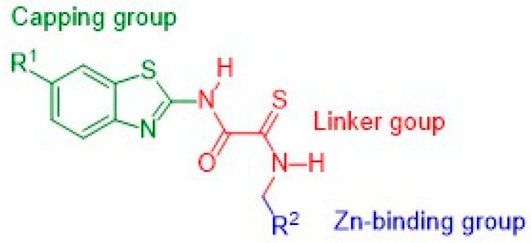
The classical HDAC6 inhibitors consist of a tripartite structure. It contains a capping group that usually occupies the rim of the pocket (L1 or L2 domains), followed by a linker that interacts with the hydrophobic tunnel of the catalytic domain, and a zinc-binding group that interacts with the metal ion [25,26,27,28,29,30,31]. Typically, the zinc-binding group is a hydroxamate that efficiently interacts with zinc in the catalytic pocket. However, it has been demonstrated that hydroxamate is susceptible to glucuronidation reactions by metabolism, considerably reducing the bioavailability of the drug [32,33,34]. Furthermore, the hydroxamate group has also been associated with mutagenicity in belinostat, vorinostat, and panobinostat [35,36,37]. The importance of improving these characteristics of classical HDAC6 inhibitors could be crucial to achieving better results in antineoplastic pharmacological strategies and reducing their side effects. In addition, it is well known that the hydrophobic tunnel is wider in HDAC6 than in the other isoforms. This leads to the hypothesis that the addition of bulky aromatic groups to the structure could enhance the interaction with the hydrophobic channel, thereby improving the affinity with the enzyme. In this context, we proposed the design of an MTA family of compounds containing a BT group, which has also been associated with exhibiting a wide range of biological activities, such as anticancer and antimicrobial [8,38,39,40]. The MTA group was proposed as a linker to improve the interaction of compounds with the hydrophobic tunnel. Additionally, an aromatic ring was incorporated into the design to enhance interaction with the wide hydrophobic channel of HDAC6, Figure 2.

2.2. Synthesis and NMR Assignments

MTA-BTs 1ad and 2ad were obtained by the inexpensive method previously established by our research group [2,24,41] (Scheme 1), using a chloroacetamide as an intermediate (1 and 2), the appropriate amine (ad), and S8 as a sulfur source. Compounds 1a,b, and 2a were purified by recrystallization from ethanol, in medium to good yields (63–76%). Whereas 1c,d, and 2bd were purified by column chromatography in medium yields (38–51%).
Both families of MTA-BTs were characterized by 1H, 13C, and 2D NMR experiments. The results confirmed the structures of the compounds. The characteristic high-frequency chemical shifts of the amide (δNOH) and thioamide (δNSH) protons were observed in the 1H NMR spectra of compounds 1ad and 2ad. δNOH is in the 11.22–11.31 ppm range, whereas the δNSH is in the 9.38–9.77 ppm range. The COSY experiment confirmed the correct assignments of both signals, Figure 3. The NOH appears as the most deshielded signal, probably due to intramolecular hydrogen bonding engagement with the thiocarbonyl sulfur (CS), as previously reported by our research group [2,41]. Additionally, the spin-spin splitting between NSH (H13) and the neighboring H14 protons was observed in all compounds (3J13-14 = 5.6–6.4 Hz). This result suggests the stabilization of the NSH by intramolecular hydrogen bonding with the amide carbonyl (CO). It is worth highlighting that the thioamide proton is usually engaged in fast acidic hydrogen exchange [2,41].
In the 13C-spectra, the characteristic CS group appears in the 182.3–184.5 ppm range, and the CO signal appears in the 156.1–160.8 ppm range. The deshielding effect observed on the CS is stronger than on CO; this effect is explained because of the preferred delocalization of the lone pair of thioamide nitrogen (NCS) to the CS group, in contrast to the shared delocalization of the lone pair of amide nitrogen (NCO) to both the CO group and the BT ring [42].

2.3. Molecular Structure and Intramolecular Hydrogen Bonding of Compound 1c

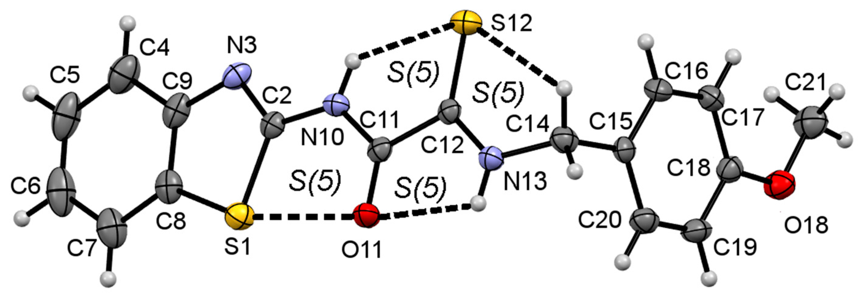
NMR is a widely used technique to elucidate the structure of organic compounds. However, single-crystal X-ray diffraction is one of the most accurate methods for determining the molecular structure of compounds, as well as their supramolecular arrangement. A detailed description of the three-dimensional structure of compounds is essential in drug design because it leads to a better understanding of the kind of supramolecular interactions that compounds can establish in the crystal structure, which can be correlated with those interactions that could be established between compounds and biological targets [43,44,45,46]. Compound 1c crystallized as a triclinic crystal system in the space group P-1, with two molecules in the unit cell. Details of the crystal data, data collection, and structure refinement are summarized in Table S1. The molecular structure of 1c is displayed in Figure 4. The experimental bond lengths values of 1c are in agreement with aromatic C—X (X = C, N, S), C=O and C=S reported values [47], and therefore with two isolated CO and CS groups (C=O = 1.234(12) Å, C=S = 1.681(20) Å), as has been found in phenylene bis-monothiooxalamides [2]. The complete list of parameters inherent to the geometry of compound 1c is given in the Supplementary Materials Tables S2 and S3.
NOH and NSH are anti-positioned between each other, with YCNH torsion angle values of 179(2)° (Y = O11) and 179(2)° (Y = S12), and O11C11C12S12 torsion angle of 178.7(2)°. The thiooxamide group is almost coplanar with the BT heterocycle, forming an angle of only 4(2)° between the two planes; however, it is tilted away from the (4-methoxy)benzylamine residue by 77(2)°. Nevertheless, the lone pair of the NCS is more delocalized to the CS (N13—C12 = 1.316(4) Å) than the lone pair of the NCO to the CO (N10—C11 = 1.351(4) Å).
Thiooxamide group is intramolecularly hydrogen bonded, displaying the typical pattern of adjacent S(5)S(5)S(5) set of rings formed by NH···Y (X = O11, S12) and CH···S12 interactions. The geometric parameters associated with intramolecular hydrogen bonding are listed in Table 1.
In addition to coplanarity between BT-heterocycle and thiooxamide group, the heterocyclic sulfur S1 atom and the carbonyl oxygen O11 are in syn conformation at a distance of 2.808(3) Å, below the sum of the van der Waals radii (rvdW = 3.32 Å) [48], and a C8—S1⋯O11 angle of 161.6(1)°, in agreement with chalcogen bonding (nO → σ*C–S) [49], forming an S(5) ring motif. The interaction distance O11⋯S1 and C8―S1⋯O11 angle values agree with reported values [50].

2.4. Supramolecular Structure of Compound 1c

In the Hirshfeld surface (HS) of compound 1c, the strongest interaction (O⋯H/H⋯O) is given by N—H13⋯O11 Hydrogen Bond (HB), Figure 5a, which is shown as bright red spots. Among all the close interactions, H⋯H contacts represent the most extensive set of interactions contributing to the surface, accounting for 39.6%. In addition, the H⋯A/A⋯H (A = C, N, O, S) interactions, represent the 45.6% as a whole, whereas the remaining 14.8% is given by C⋯X/X⋯C and N⋯X/X⋯N (X = C, N, O, S) contacts, Figure 5b.
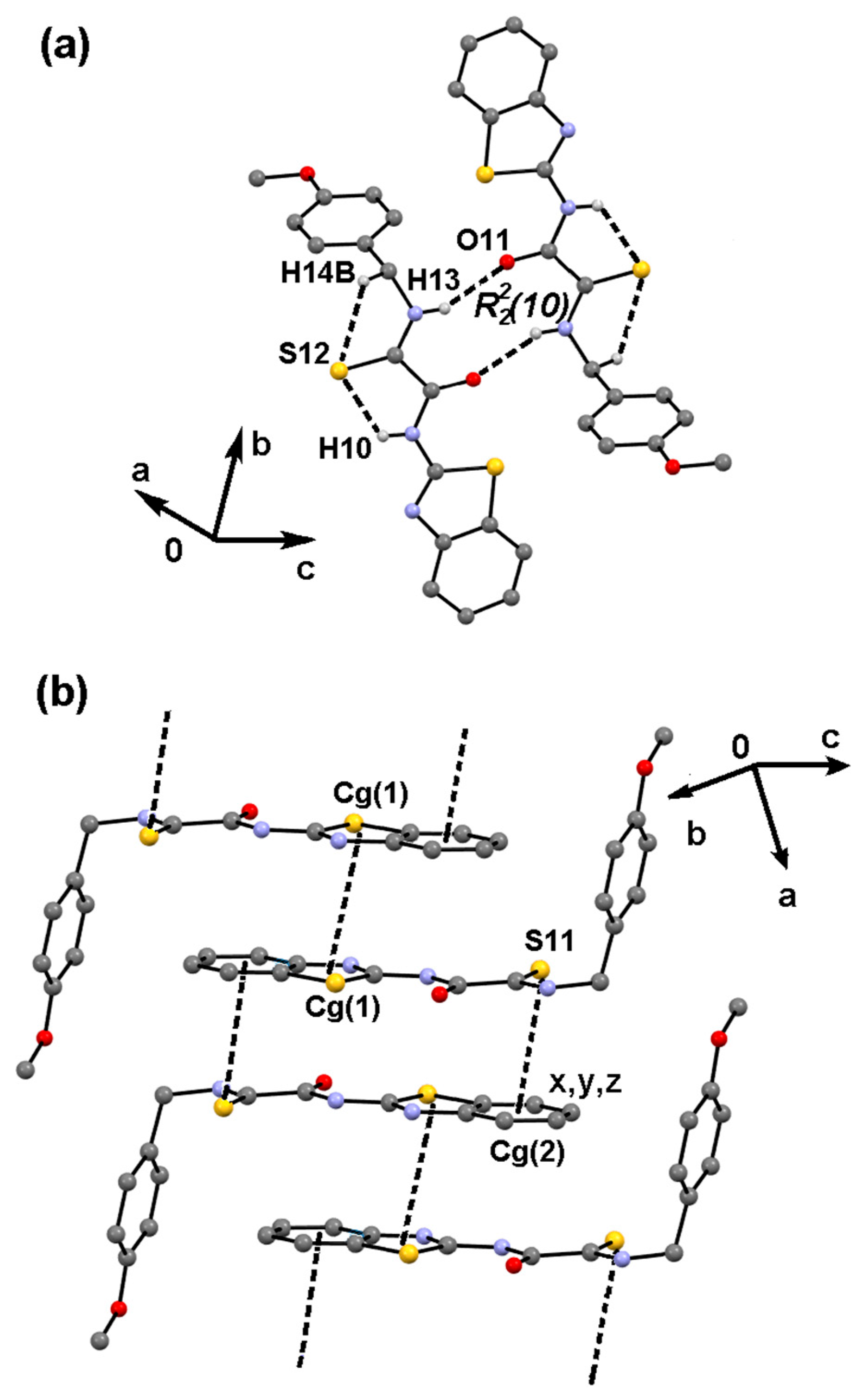
The geometric parameters associated with intermolecular interactions are listed in Table 1. Selected results of quantitative interaction energy analysis of 1c, performed at the B3LYP/6-311G(d,p) level of theory, are tabulated in Table 2. Interaction energy (Etot) values smaller than −20.0 kJ mol−1 were neglected. The most energetic interaction, N—H13⋯O11 hydrogen bonding, assembles as R22(10) intermolecular ring motif, whose geometric parameters agree with a strong interaction. In fact, this motif contributes with −63.3 kJ mol−1 to the energy in the crystal and shapes a centrosymmetric dimer, Figure 6a. The nature of the attractive energy involved in N—H13⋯O11 hydrogen bonding (HB) is primarily electrostatic, with a percent contribution to the stabilization energy (%Eele) of 60.4%.
The second largest energy contributor to the crystal energy, with −59.0 kJ mol−1, is the π-assembly between two thiazole rings, with distances of 3.6317(17) Å and 3.5301(12) Å between Cg1Cg1 centroids and Cg1 perpendicular planes, respectively (Cg1 is the centroid of the S1C2N3C9C8 ring; symmetry code = 1-x, 1-y, 1-z) (Figure 6b). This π-stacking interaction is mainly dispersive, with a %Edis value of 80.0%.
At last, C12=S12⋯Cg2 interaction, of nS → π* type, contributes with −40.1 kJ mol−1, being mostly dispersive, with %Edis value of 89.0% of its attractive energy, Table 2. Both Cg1Cg1 and S12⋯Cg2 interactions between centrosymmetric π-stacked dimers develop the second dimension (2D) in the (0 4 7) crystal plane, Figure 6b. Thus, the contribution of soft π-stacking interactions (Etot = −99.1 kJ mol−1) to the stability of the crystal network of 1c is the largest and mostly dispersive in nature.
The visual representation of the energy-framework diagrams for Eele (red), Edis (green) and Etot (blue) for a cluster of nearest-neighbor molecules is shown in Figure 7. The crystal network of 1c is mainly dominated by the Edis component, showing zig-zagging energy framework, Figure 7b, that overcome the Eelec component given by the most energetic N—H13⋯O11 interaction, Figure 7a, to provide the final shape to the Etot energy framework, Figure 7c.

2.5. Molecular Docking to DD2-HDAC6 Catalytic Site

The affinity of MTA-BT derivatives 1ad and 2ad for the DD2-HDAC6 catalytic site was evaluated by performing a docking simulation. The results are expressed in terms of free energy of ligand-receptor binding (ΔGob) and dissociative equilibrium constant (Kd), Table 3. According to the docking calculations, all compounds interact with the DD2-HDAC6 catalytic site, showing ΔGob values ranging from −7.3 to −8.5 kcal/mol. The values were close to Nexturastat A (NA) (ΔGob = −7.8 kcal/mol), the reference drug and the crystallized ligand used for docking validation. The binding modes of MTA-BTs and NA revealed that the compounds reached the catalytic DD2 domain of HDAC6, as shown in Figure 8. The tested compounds interact with the surface binding domain of the catalytic site through the BT system and the thiooxalamide group, while the aromatic ring, from the amine residue, is introduced into the hydrophobic channel, efficiently blocking the catalytic site. The presence of the phenethyl group in the R2 position of compounds 1d and 2d improves the interaction with the hydrophobic channel by π-dispersive forces with Phe-544, Phe-604, and His-575. For compounds 1c and 2c, the para-methoxy group on the aromatic ring (R2) appears to reinforce the interaction with the hydrophobic channel by binding to His-535 and His-575 through π-alkyl interactions, thereby blocking access to the catalytic site of DD2-HDAC6. On the other hand, for compounds 1b and 2b, with an ortho-methoxy group, the effect of the substituent makes the interaction with the hydrophobic channel slightly less favorable. Regarding the reference drug, the binding mode of the NA with the DD2-HDAC6 catalytic site shows that the ligand reaches the catalytic site domain by blocking the surface (His-424 and Pro-425) through phenyl and butyl groups (Phe-604). A strong binding with the hydrophobic channel is also observed. To achieve a good blocking of the hydrophobic channel, it is crucial to bind the aromatic rings with Phe-604, Phe-544, and His-575 by π-interactions. In addition, the molecular structure of compound 1c, which was determined by single-crystal X-ray diffraction, has an almost 80° angle in which the (4-methoxy)benzylamine residue is involved, as shown in Figure 4. This arrangement is induced by hydrogen bonds between H10, S12, and H14 atoms and π-interactions between the BT rings, Figure 6. Taking into account the tridimensional structure of the DD2-HDAC6 catalytic domain, in which the pocket rim and hydrophobic channel are involved on HDAC6 inhibition, the planar structure of BT moiety on the crystal structure seems to contribute with the affinity against the rim of the pocket by π-cation interactions with His-424 and Asn-418, similarly with occurs on crystal structure between BT rings. Additionally, the planar structure of the (4-methoxy)benzylamine moiety reinforces the interaction with the hydrophobic tunnel through π-π interactions with Phe-544 and Phe-604, resulting in a good cytotoxic effect exerted by 1c on human cancer cell lines [26,51,52]. NA can also interact with the zinc-binding domain by the hydroxamate group. This interaction involves hydrogen bonding with His-534 and His-535. It is worth noting that this kind of interaction, which involves the surface, the hydrophobic channel, and the zinc-binding domains, was also previously described as crucial to efficiently inhibit the catalytic activity of HDAC6 [23,53,54,55,56]. The complete list of interactions of NA, 1ad, and 2ad with DD2-HDAC6 is in the Supplementary Materials Table S4.

2.6. Antiproliferative Activity

The antiproliferative effect of monothiooxalamides 1ad and 2ad was assessed in MCF-7 (human breast cancer cell line with estrogen, progesterone, and glucocorticoid receptors), MDA-MB-231 (triple-negative human breast cancer), MCF-10A (non-tumorigenic human mammary epithelial cell line), and NIH/3T3 (mouse fibroblast). The cytotoxicity is expressed as IC50 values in micromolar concentrations, Table 4. The most active compound was 1c for both carcinogenic cell lines, with higher activity observed in MCF-7 cells. On the other hand, MDA-MB-231 cells were less sensitive to 1c (1.6-fold more active on MCF-7 cells). Regarding non-tumorigenic MCF-10A cells, compound 1c showed moderate selectivity toward malignant cells (1.7-fold more active in MCF-7 cells). In contrast, the cytotoxic effect on triple-negative cells MDA-MB-231 was similar to that observed in MCF-10A. In the other non-tumorigenic cell line, NIH/3T3, all compounds and MTX showed considerably more toxicity than both tumorigenic cell lines (MCF-7 and MDA-MB-231) and non-tumorigenic MCF-10A cells. Nevertheless, it is worth considering that NIH/3T3 are mouse fibroblasts, while the other three cell lines are human cells; therefore, the physiological differences between both species could play an essential role in the exerted effect. Several studies have evaluated 2-aminobenzothiazole derivatives against various cell lines. The targets of this kind of compounds are wide, including HDAC6, tyrosine kinase receptors family (CSF1R kinase, EGFR kinase, VEGFR-2 kinase, FAK kinase), serine/threonine kinases, lysine-specific demethylase (LSD1), phosphoinositide 3-kinase (PI3K), topoisomerases (I and II), Heat shock protein (HSP90), p53, nuclear receptor binding set (NSD1), microsomal prostaglandin E synthase-1 (mPGES-1), and also as chelators of metal ions, which are necessaries for critical biological processes such as gene expression, oxygen transport, and cell respiration [8]. In all cases, the IC50 values were reported in a wide range, from 1 nM to 50 µM. On the other hand, our research group reported a family of MTAs containing 2-aminopyridine, with an activity range of 0.19–18 µM against several human cancer cell lines (A2780-wt, A2780-cis-MSTO-211H, and HT-29) [24]. The tested compounds 1ad and 2ad fall within a medium range, between those reported previously for this kind of compounds (32–95 µM), with 1c being the most active against MCF-7 and MDA-MB-231cancer cell lines. Although the tested compounds did not show high cytotoxicity against human cancer cell lines, the values are in the reported average for other BT compounds [17,57], and reported ReI-MTA complexes [58]. This study led to the identification of the most promising compound, 1c, in which the presence of the 2-methoxyparaminoethyl structural feature appears to enhance antineoplastic activity significantly. Whereas 1c could be a feasible candidate to obtain better results compared with 1a, 2ac, which were highly cytotoxic to non-tumorigenic cells. As a perspective, the synergic activity of 1c, in combination with antioxidants like curcumin, resveratrol, and quercetin, could also be explored [59,60,61], with the aim to sensitize cancer cells against cytotoxic agents and diminish their toxicity in non-tumorigenic cells. In contrast, MTX, which is an antineoplastic drug, is widely used for many kinds of cancer treatments, including breast cancer. Their mechanism of action is related to the inhibition of dihydrofolate reductase (DHFR). However, MTX shares some structural similarities with HDAC inhibitors, and some studies have demonstrated the ability of this drug to inhibit HDAC6 enzyme [62,63]. This drug was used as a reference and proved to be the most active against both tumorigenic cell lines (MCF-7 and MDA-MB-231). Nevertheless, MTX exerted an undesirable effect on NIH/3T3 cells. Although the compounds were not better than MTX, their antiproliferative effect was similar to 2ABT, thioureas, 3-carboxy-coumarin sulfonamides, pyrimethamine conjugates, and N-hydroxycinnamide analogs [38,64,65,66]. Highlighting its low toxicity to non-tumor cells, 1c might be a better option than methotrexate to reduce adverse side effects associated with chemotherapy.

2.7. Cell Migration Assay

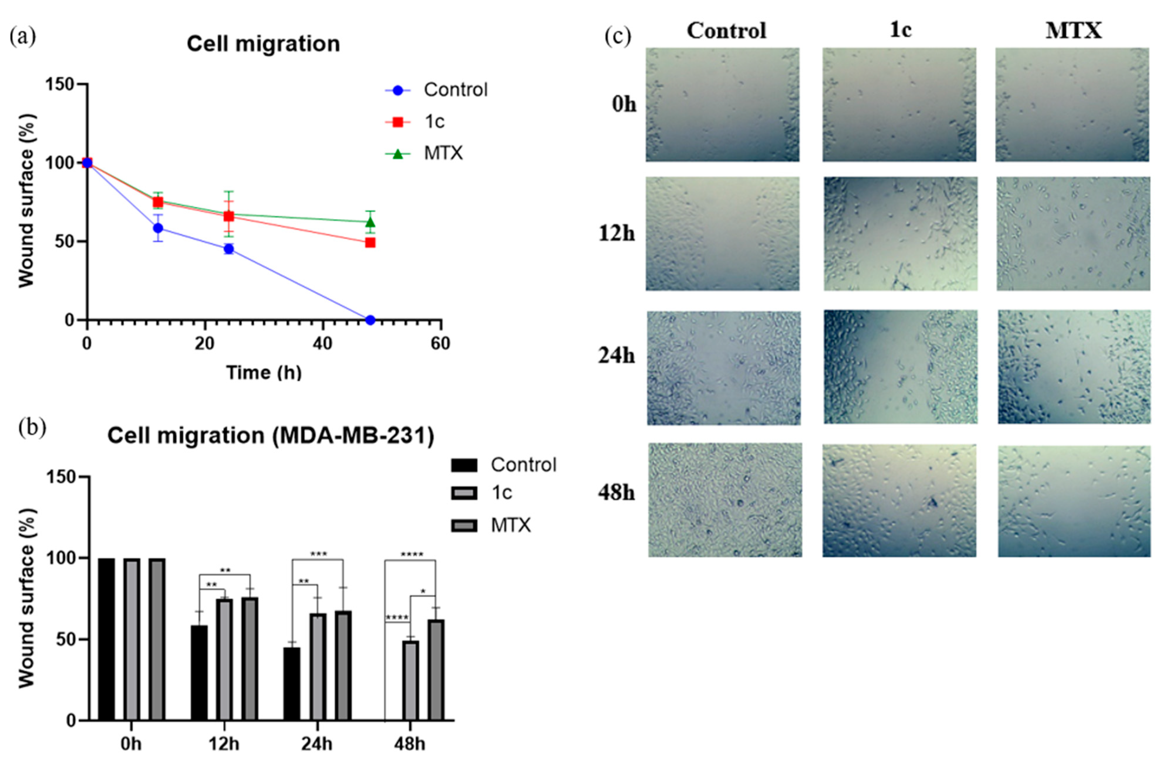
The triple-negative cancer, produced by MDA-MB-231 cells, is highly invasive and metastatic, capable of migrating to other tissues. To investigate the effect of MTA-BTs on inhibiting cell migration, a wound-closure assay was performed using MDA-MB-231 cells with the most active compound, 1c. Cells were treated at the IC50 concentration (52 μM), and the wound closure was monitored at 0 h, 12 h, 24 h, and 48 h. The results are expressed as a percentage of the remaining wound surface after the wound, as shown in Figure 9. The wound closure begins at 12 h, at this time, there is a significant difference between cells treated with compound 1c and MTX compared with non-treated cells. Nevertheless, the maximum effect is observed at 48 h, when wound closure is complete for non-treated cells. For cells treated with compound 1c, the maximum wound closure was 51%, while for the reference drug, MTX, it was 38% at 48 h. In both cases, there is a significant difference from the control, with wound closure rates of −2.0 ± 0.1 (R = 0.98), −1.0 ± 0.1 (R = 0.94), and −0.7 ± 0.1 (0.88) in %area/h for control, 1c, and MTX, respectively. The results confirmed the capability of 1c to prevent cell migration in MDA-MB-231 cells, similar to the reference drug MTX, and other reported BTs [67,68].

2.8. Apoptosis Induction

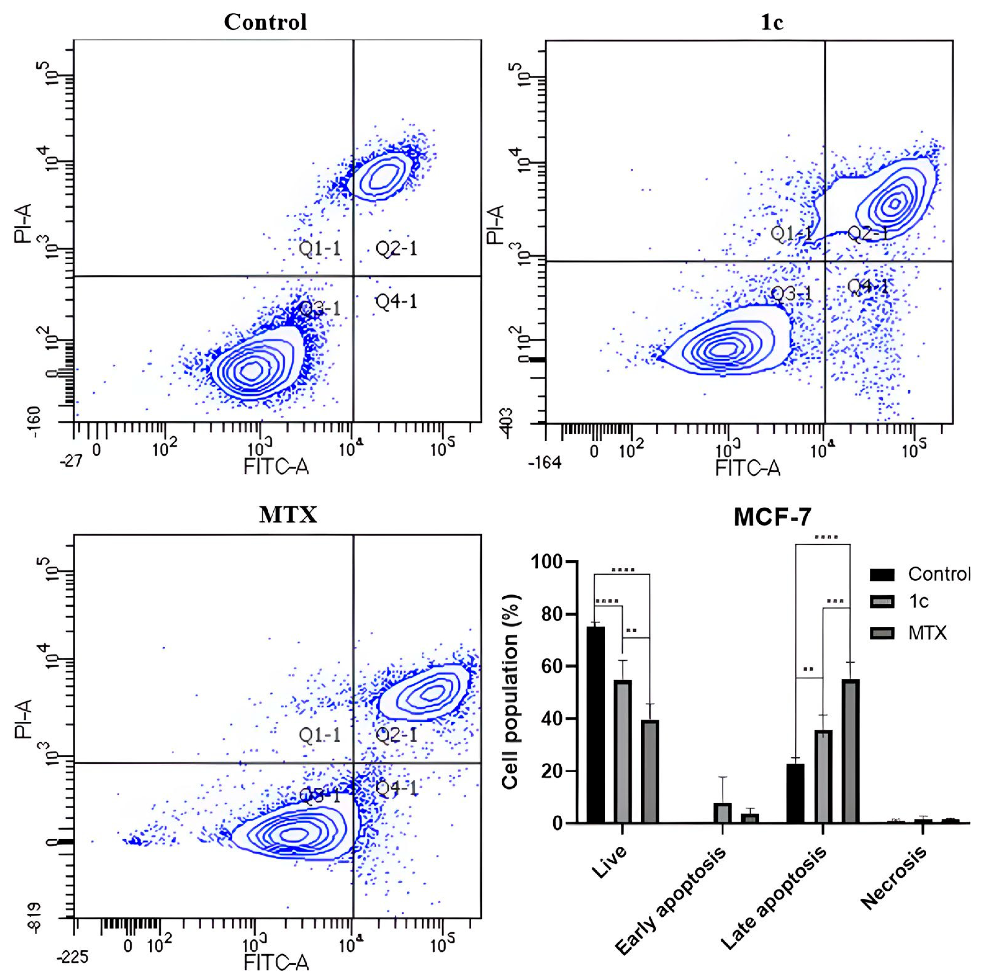
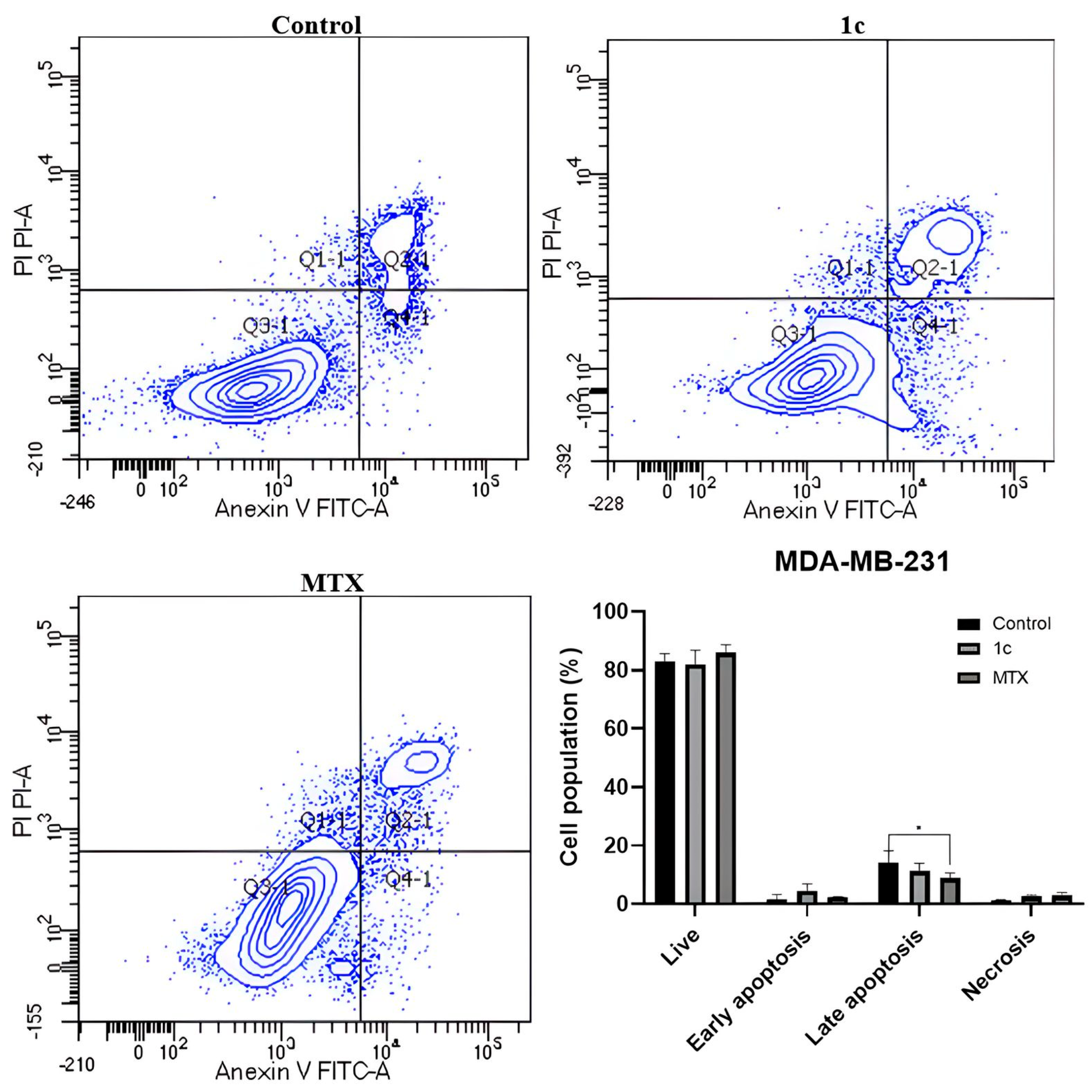
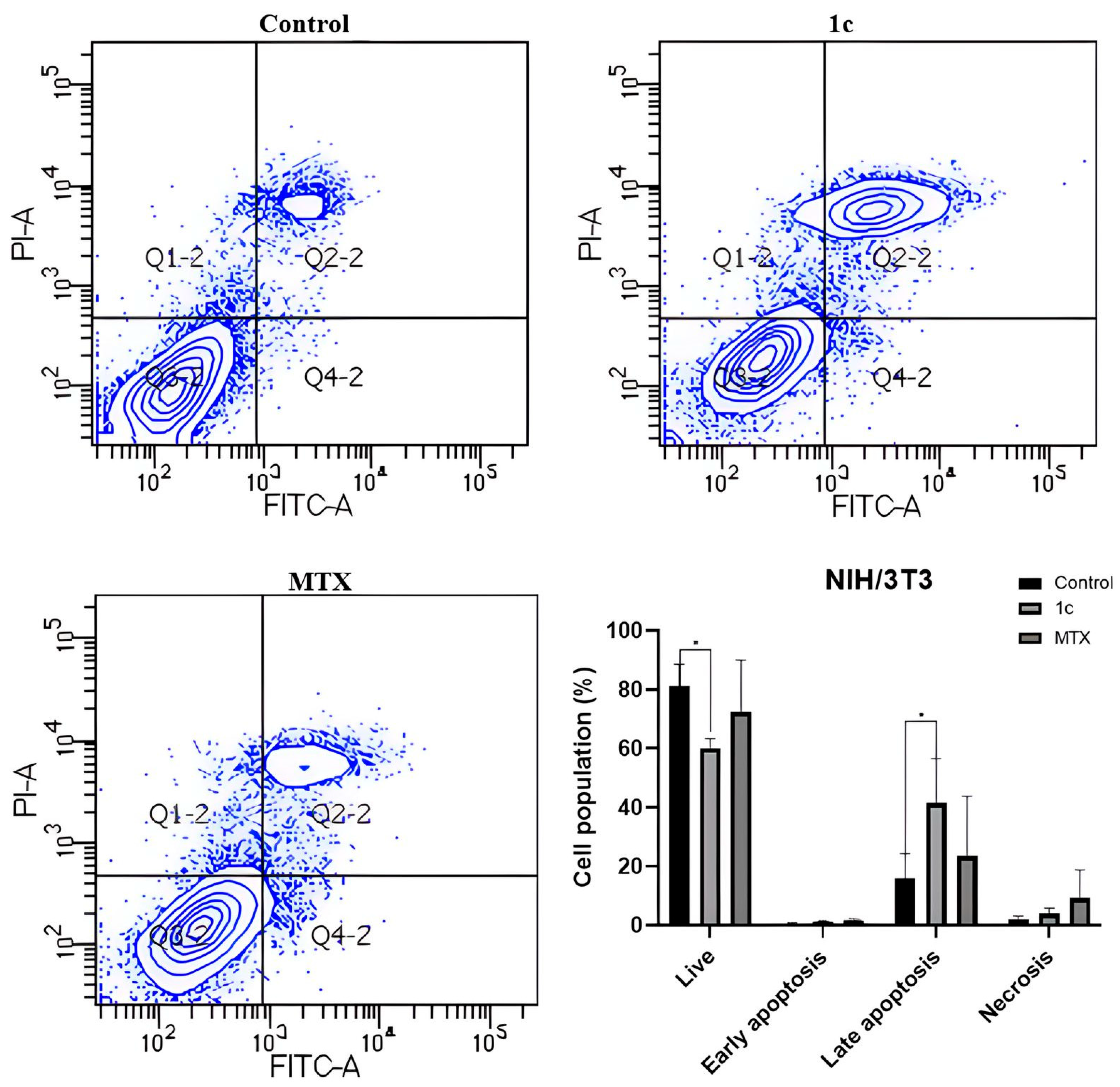
Apoptosis induction is one of the most common mechanisms of cell death exerted by antineoplastic drugs. This is because cancer cells can evade this cell death mechanism, allowing malignant cells to survive and leading to tumor progression [69]. Apoptosis involves several physiological cell modifications, including the destabilization of the phospholipid bilayer and the subsequent exposure of phosphatidylserine. This phospholipid can be easily recognized by annexin V, a protein that is useful for identifying apoptotic cells [70]. Therefore, apoptosis was determined for both breast cancer human cell lines, MCF-7 and MDA-MB-231, and the NIH/3T3 non-tumoral cell line. The cells were treated with the most active compound 1c and MTX, this last as a death-positive control, for 48 h of exposure. The viability of MCF-7 cells significantly decreased when treated with compound 1c (55%) and MTX (39%); consequently, a significant increase in the late apoptosis cell population was observed for both treatments, as shown in Figure 10. This result was expected because MCF-7 cells were the most sensitive to 1c. On triple-negative breast cancer cells (MDA-MB-231), methotrexate and 1c did not show a significant effect on cell viability; nevertheless, MTX, the reference drug, appears to decrease the percentage of late apoptotic cells (9%) compared with non-treated cells (14%), as shown in Figure 11. Regarding non-malignant fibroblast cells (NIH/3T3), a slight decrease in live cells was observed only for compound 1c (60%) compared to the control (81%) and MTX (72%), as shown in Figure 12.

2.9. Physicochemical and Toxicological Properties Prediction

The physicochemical and toxicological properties of MTA-BTs were predicted with OSIRIS Property Explorer; the results are listed in Table S5. The n-octanol/water partition coefficient (cLogp) describes the hydrophobicity of compounds, which is crucial to cross biological membranes. cLogp values must be less than 5 to achieve a good absorption process in living organisms [41,71]. All compounds have cLogp values between 2.66 and 3.51. LogS is a parameter that expresses the solubility of the drugs. This physicochemical property helps predict drug absorption [41,72]. The LogS values must be greater than −4 to achieve a good absorption, distribution, and elimination process. In contrast, all compounds have values below −4, suggesting that aqueous solubility could be restricted. Nevertheless, it is worth noting that numerous drugs on the market have poor aqueous solubility. Regarding LogS, the best compounds are 1a, 1b, and 1c. Moreover, the toxicity risks of MTA-BTs 1ad and 2ad were also estimated, showing a low risk of toxicological effects; thus, both families of compounds are suitable in terms of toxicity.
The drug score expresses the potential of compounds to be feasible as a drug. The value combines physicochemical parameters and toxicity risk to obtain a global score (between 0 and 1), in which 1 is the best score and, 0 is the worst. Osiris has been widely used to select suitable compounds [73,74,75,76]. In a previous study, the toxicity of a series of benzimidazole derivatives on red blood cells was reported, and the prediction by OSIRIS was consistent with the in vitro evaluation [41]. In this study, compounds 1a, 1b, 1c, and 2d have the best drug-score values. Considering that compound 1c is the most active compound against human cancer cell lines, particularly MCF-7 cells, it suggests that 1c could be a promising drug candidate.

3. Materials and Methods

3.1. Materials and Equipment

All reagents were purchased from commercial suppliers and used without further purification. Chloroacetyl chloride, 2-aminobenzothiazole, phenethylamine, benzylamine, 2-methoxybenzylamine, 4-methoxybenzylamine, deuterated chloroform (CDCl3), deuterated DMSO (DMSO-d6), hexane, ethyl acetate, dichloromethane, sodium carbonate, potassium carbonate, and triethylamine. The solvents for column chromatography were distilled before use (hexane and ethyl acetate). Chloroacetamides 1 and 2 were obtained by the previously described method by our research group [2,24].
Melting points were measured using an Electrothermal Mel-Temp melting point apparatus (Cole-Parmer) and are uncorrected. 1H and 13C NMR spectra, were acquired on Varian NMR spectrometer Mercury-300 using CDCl3 or DMSO-d6 as solvent. All chemical shift values (δ) are reported in parts per million (ppm), using TMS as internal reference, and coupling constants (nJ) are in Hz. The following abbreviations are used to indicate the multiplicity of the signals: s for a singlet, d for a doublet, t for a triplet, and q for a quadruplet. The numbering of compounds is shown in Figure S1 and the NMR spectra are in Figures S2–S32. Mass spectra were acquired using a micrOTOF-Q Bruker Daltonics instrument from acetonitrile solutions, Figures S33–S40. FT-IR spectra were recorded neat at 25 °C using a PerkinElmer FT-IR Spectrum Two, equipped with a Miracle ATR Single Reflection Diamond (PIKE Technologies) device, and are reported in terms of wavenumber of absorption (cm−1), Figures S41–S48. To indicate the intensity of the signals, the following abbreviations are used: w for weak, m for medium, and s for strong. The purity of the compounds was determined using an Agilent Technologies 1260 Infinity HPLC instrument (Santa Clara, CA, USA), using a ZORBAX XDC-C18 column, at 25 °C; HPLC chromatograms are shown in Figures S49–S56.

3.2. General Procedure for the Synthesis of MTA-BTs 1a–d and 2a–d

In a 100 mL ball flask, 2.50 mmol of the appropriate amine ad, 10 mL of THF, 1 mL of triethylamine, and 1.20 mmol of elemental sulfur (0.311 g) were placed. The mixture was stirred at room temperature for 30 min. Subsequently, 2.30 mmol of chloroacetamide 1 (0.515 g) or 2 (0.547 g) was slowly added, and the reaction solution was stirred for 72 h at RT. Compounds 1a and 1b were purified by recrystallization from ethanol, and MTA-BTs by column chromatography using a hexane/ethyl acetate mixture of suitable polarity as eluent. All compounds were obtained as yellow solids and were spectroscopically characterized by 1D and 2D NMR (1H and 13C NMR), and IR.
N-(benzo[d]thiazol-2-yl)-2-(benzylamino)-2-thioxoacetamide (1a). Obtained from 0.268 g of amine a and chloroacetamide 1 in 76% yield (0.562 g), m.p. 163–164 °C after recrystallization from ethanol. IR (cm−1) ν: 3267 (m, N-H amide I), N-H 3250 (br, thioamide I), 1683 (s, C=O), 1530 (m, N-H + C-N amide II), 1269 (s, C=S). 1H NMR (300 MHz, DMSO-d6) δ: 7.90 (H7, d, 3J(7-6) = 6.9, 1H), 7.72 (H4, d, 1H), 7.40 (H6, t, 3J(6-7) = 6.9, 1H), 7.27 (H5, H16, H17, m, 5H), 7.20 (H18, s, 1H), 4.76 (H14, s, 2H). 13C NMR (75 MHz, DMSO-d6) δ: 186.9 (C12), 160.8 (C11), 158.1 (C2), 148.71 (C9), 136.6 (C15), 132.2 (C8), 129.3 (C17), 128.6 (C16), 128.4 (C18), 127.5 (C6), 125.3 (C5), 122.7 (C7), 121.7 (C4), 49.0 (C14). Mass analysis ESI [M-H]+ (m/z): 328.0634 found, 328.05 calculated. HPLC purity: 97.6%.
N-(benzo[d]thiazol-2-yl)-2-((2-methoxybenzyl)amino)-2-thioxoacetamide (1b). Obtained from 0.343 g of amine b and chloroacetamide 1 in 64% yield (0.519 g), m.p. 167–168 °C after recrystallization from ethanol. IR (cm −1) ν: 3336 (m, N-H amide I), 3248 (br, N-H thioamide I), 1692 (s, C=O), 1530 (m, N-H + C-N amide II), 1240 (s, C=S). 1H NMR (300 MHz, CDCl3) δ: 11.31 (H10, s, 1H), 9.77 (H13, s, 1H), 7.85 (H4, H7, m, 3J(4-5) = 7.2, 4J(7-5)= 1.2, 2H), 7.48 (H5, ddd, 3J(5-4) = 7.2, 4J(5-7) 1.2, 1H), 7.35 (H6, H17, H20, m, 3H), 6.95 (H18, H19, m, 2H), 4.89 (H14, d, 2H), 3.91 (H21, s, 3H). 13C NMR (75 MHz, CDCl3) δ: 182.3 (C12), 157.8 (C16), 156.4 (C2), 156.3 (C11), 149.0 (C9), 132.5 (C8), 130.9 (C15), 130.3 (C20), 126.7 (C5), 124.6 (C6), 122.9 (C18), 121.8 (C7), 121.6 (C4), 120.8 (C19), 110.7 (C17), 55.7 (C21), 46.9 (C14). Mass analysis ESI [M-H]+ (m/z): 358.0779 found, 358.06 calculated. HPLC purity: 99.6%.
N-(benzo[d]thiazol-2-yl)-2-((4-methoxybenzyl)amino)-2-thioxoacetamide (1c). Obtained from 0.343 g of amine c and chloroacetamide 1 in 43% yield (0.348 g), m.p. 168–170 °C. IR (cm −1) ν: 3301 (m, N-H amide I), 3209 (br, N-H thioamide I), 1676 (s, C=O), 1508 (m, N-H + C-N amide II), 1507 (s, N-H + C-N thioamide II), 1240 (s, C=S). 1H NMR (300 MHz, CDCl3) δ: 11.27 (H10, s, 1H), 9.48 (H13, s, 1H), 7.85 (H7, H4, dddd, 3J(4-5) = 8.5, 3J(7-6) = 7.8, 4J(4-6) = 1.3, 2H), 7.48 (H5, ddd, 3J(5-6) = 8.3, 4J(5-7) = 1.3, 1H), 7.35 (H6, ddd, 3J(6-5) = 8.3, 3J(6-7) = 7.2, 4J(6.4) = 1.3, 1H), 7.27 (H16, d, 3J(16-17) = 8.3, 2H), 6.89 (H17, d, 3J(17-16) = 8.3, 2H), 4.79 (H14, d, 3J(14-13) = 5.6, 2H), 3.81 (H19, s, 3H). 13C NMR (75 MHz, CDCl3) δ: 182.8 (C12), 159.9 (C18), 156.3 (C11), 156.2 (C2), 149.0 (C9), 132.6 (C8), 130.0 (C16), 126.7 (C5), 124.8 (C6), 121.9 (C7), 121.6 (C4), 114.6 (C17), 55.6 (C19), 50.3 (C14). Mass analysis ESI [M-H]+ (m/z): 358.0763 found, 358.06 calculated. HPLC purity: 97.7%. Suitable crystals for X-ray diffraction analysis were obtained from a saturated chloroform solution at R.T.
N-(benzo[d]thiazol-2-yl)-2-(phenethylamino)-2-thioxoacetamide (1d). Obtained from 0.303 g of amine d and chloroacetamide 1 in 40% yield (0.314 g), m.p. 186–187 °C. IR (cm −1) ν: 3283 (s, N-H amide I), 3215 (w, N-H thioamide I), 1680 (s, C=O), 1508 (m, N-H + C-N amide II), 1526 (s, N-H + C-N thioamide II), 1271 (s, C=S). 1H NMR (300 MHz, CDCl3) δ: 11.27 (H10, s, 1H), 9.38 (H13, s, 1H), 7.86 (H7, H4, t, 3J(4-5) = 6.6, 2H), 7.49 (H5, t, 3J(5-6) = 7.6, 3J(5-4) = 6.6, 1H), 7.36 (H6, H17, t, 3J(6-5) = 7.6, 3H), 7.27 (H18, H19, m, 3H), 4.00 (H14, dt, 3J(14-15) = 7.0, 3J(14-13) = 6.5, 2H), 3.06 (H15, t, 3J(15-14) = 7.0, 2H). 13C NMR (75 MHz, CDCl3) δ: 183.3 (C12), 156.2 (C11), 156.2 (C2), 149.0 (C9), 137.5 (C16), 132.6 (C8), 129.2 (C17), 128.8 (C18), 127.3 (C19), 126.7 (C5), 124.8 (C6), 121.9 (C7), 121.6 (C4), 47.6 (C14), 33.6 (C15). Mass analysis ESI [M-H]+ (m/z): 342.0813 found, 342.07 calculated. HPLC purity: 97.6%.
2-(benzylamino)-N-(6-methylbenzo[d]thiazol-2-yl)-2-thioxoacetamide (2a). It was obtained from 0.268 g of amine a and chloroacetamide 2 in 63% yield (0.492 g), m.p. 158–160 °C. IR (cm −1) ν: 3332 (m, N-H amide I), 3270 (m, N-H thioamide I), 1682 (s, C=O), 1547 (s, N-H + C-N amide II), 1512 (s, N-H + C-N thioamide II), 1256 (s, C=S). 1H NMR (300 MHz, CDCl3) δ: 11.23 (H11, s, 1H), 9.56 (H14, s, 1H), 7.74 (H4, d, 3J(4-5) = 8.3, 1H), 7.62 (H7, dt, 4J(7-5) = 1.8, 1H), 7.37 (H17, H18, H19, m, 5H), 7.31 (H5, dd, 3J(5-4) = 8.3, 4J(5-7) = 1.8, 1H), 4.87 (H15, d, 3J(15-14) = 5.7, 2H), 2.48 (H10, s, 3H). 13C NMR (75 MHz, CDCl3) δ: 183.2 (C13), 156.1 (C12), 155.4 (C2), 146.9 (C9), 134.9 (C8), 134.7 (C6), 132.7 (C16), 129.2 (C18)), 128.7 (C19), 128.5 (C17), 128.2 (C5), 121.4 (C4), 121.3 (C7), 50.7 (C15), 21.7 (C10). Mass analysis ESI [M-H]+ (m/z): 342.0810 found, 342.07 calculated. HPLC purity: 97.0%.
2-((2-methoxybenzyl)amino)-N-(6-methylbenzo[d]thiazol-2-yl)-2-thioxoacetamide (2b). Obtained from 0.343 g of amine b and chloroacetamide 2 in 46% yield (0.387 g), m.p. 161–163 °C. IR (cm −1) ν: 3281 (m, N-H amide I), 3247 (w, N-H thioamide I), 1676 (s, C=O), 1519 (s, N-H + C-N amide II and thioamide II), 1243 (s, C=S). 1H NMR (300 MHz, CDCl3) δ: 11.25 (H11, s, 1H), 9.76 (H14, t, 3J(14-15) = 6.2, 1H), 7.72 (H4, d, 3J(4-5) = 8.3, 1H), 7.60 (H7, dt, 4J(7-5) = 1.7, 1H), 7.33 (H5, H21, dtd, 3J(5-4) = 8.3, 4J(5-7) = 1.7, 2H), 7.25 (H20, dt, 1H), 6.93 (H18, H19, m, 2H), 4.88 (H15, d, 3J(15-14) = 6.2, 2H), 3.89 (H22, s, 3H), 2.46 (H10, s, 3H). 13C NMR (75 MHz, CDCl3) δ: 182.41 (C13), 157.79 (C17), 156.22 (C12), 155.42 (C2), 146.96 (C9), 134.75 (C6), 132.65 (C8), 130.87 (C21), 130.19 (C20), 128.12 (C5), 122.67 (C16), 121.33 (C4), 121.28 (C7), 120.83 (C19), 110.63 (C18), 55.60 (C22), 46.77 (C15), 21.64 (C10). Mass analysis ESI [M-H]+ (m/z): 372.0914 found, 372.08 calculated. HPLC purity: 98.3%.
2-((4-methoxybenzyl)amino)-N-(6-methylbenzo[d]thiazol-2-yl)-2-thioxoacetamide (2c). Obtained from 0.343 g of amine c and chloroacetamide 2 in 38% yield (0.322 g), m.p. 167–169 °C. IR (cm −1) ν: 3297 (m, N-H amide I), 3222 (w, N-H thioamide I), 1672 (s, C=O), 1529 (s, N-H + C-N amide II), 1509 (s, N-H + C-N thioamide II), 1249 (s, C=S). 1H NMR (300 MHz, CDCl3) δ: 11.23 (H11, s, 1H), 9.52 (H14, s, 1H), 7.73 (H4, d, 3J(4-5) = 8.3 Hz, 1H), 7.60 (H7, dt 4J(7-5) = 1.4 Hz, 1H), 7.28 (H5, H17, m, 3J(17-18) = 8.8 Hz, 3J(5-4) = 8.3 Hz, 4J(5-7) = 1.4 Hz, 3H), 6.88 (H18, m, 3J(18-17) = 8.8 Hz, 2H), 4.77 (H15, d, 3J(15-14) = 5.6 Hz, 2H), 3.80 (H20, s, 3H), 2.47 (H10, s, 3H). 13C NMR (75 MHz, CDCl3) δ: 182.8 (C13), 159.8 (C12), 156.1 (C19), 155.4 (C2), 146.9 (C9), 134.8 (C6), 132.7 (C8), 129.9 (C17), 128.2 (C5), 126.7 (C16), 121.4 (C4), 121.3 (C7), 114.5 (C18), 55.4 (C20), 50.2 (C15), 21.7 (C10). Mass analysis ESI [M-H]+ (m/z): 371.1327 found, 371.08 calculated. HPLC purity: 99.3%.
N-(6-methylbenzo[d]thiazol-2-yl)-2-(phenethylamino)-2-thioxoacetamide (2d). Obtained from 0.343 g of amine d and chloroacetamide 2 in 51% yield (0.412 g), m.p. 202–203 °C. IR (cm −1) ν: 3290 (m, N-H amide I), 3205 (w, N-H thioamide I), 1676 (m, C=O), 1526 (s, N-H + C-N amide II and thioamide II), 1256 (s, C=S). 1H NMR (300 MHz, CDCl3) δ: 11.22 (H11, s, 1H), 9.44 (H14, s, 1H), 7.74 (H4, d, 3J(4-5) = 8.3, 1H), 7.60 (H7, dt, 4J(7-5) = 1.6, 1H), 7.35 (H18, m, 2H), 7.31 (H5, m, , 3J(5-4) = 8.3, 4J(5-7) = 1.6, 1H), 7.26 (H19, m, 2H), 7.23 (H20, t, 1H), 3.99 (H15 ,td, 3J(15-16) = 7.4, 3J(15-14) = 6.0, 2H), 3.04 (H16, t, 3J(16-15) = 7.4, 2H), 2.47 (H10, s, 3H). 13C NMR (75 MHz, CDCl3) δ: 183.4 (C13), 156.1 (C12), 155.4 (C2), 147.0 (C9), 137.5 (C17), 134.9 (C6), 132.7 (C8), 129.1 (C18), 128.7 (C19), 128.2 (C5), 127.2 (C20), 121.4 (C4), 121.3 (C7), 47.5 (C15), 33.5 (C16), 21.7 (C10). Mass analysis ESI [M-H]+ (m/z): 356.0977 found, 356.08 calculated. HPLC purity: 98.7%.

3.3. Monocrystal X-Ray Diffraction

Single crystal X-ray diffraction data were collected on an Agilent SuperNova diffractometer equipped with graphite-monochromatic Mo Kα radiation (λ = 0.71073 Å) for compound 1c. The data collection, cell refinement, and data reduction were accomplished using CrysAlisPro software (v 171.36.20) [77] at 293(2) K. Refinement and structure solution were performed using the SHELXL-2018/3 program [78] of the WINGX (2021.3) package [79] (See Supplementary Materials Table S1). Material for publication was prepared with Platon (2023) [80] and Mercury (3.8) [81]. Graph set notation was used for the description of non-covalent interactions [82]. CCDC 2418428 contains the supplementary crystallographic data for this paper.
The crystal structure of compound 1c was analyzed through the Hirshfeld surface [83] approach. The intermolecular interactions and their reciprocal fingerprints [84,85] were calculated in the CrystalExplorer 21.5 software as previously reported [86,87]. The individual energetic components were obtained by linking the software to the Gaussian 09 package [88] using the B3LYP/6-311G(d,p) theory. Scale factors [89] were included in the calculation, and the percentage contribution of individual components to the stabilization energy was also determined. The results were depicted as frameworks [89,90] in which the Eelect (electrostatic), Edisp (dispersion), and Etot (total energy) were included in a 33-unit cell with a 3.8 Å radius, a tube size of 180 kJ mol−1, and a 10 kJ mol−1 cut-off value.

3.4. Docking Simulation

Both families of MTA-BTs 1ab, 1d, and 2ad were drawn using ChemSketch (2020-1.2), and the correct connectivity and geometry were confirmed using Gauss View 6.0. For compound 1c, the crystalline structure solved by X-ray diffraction was employed. The geometry optimization of compounds was performed using Gaussian 09W with a semiempirical method at the AM1 level. The catalytic domain-2 of HDAC6 enzyme, DD2-HDAC6 (PDB: 5G0J), was taken from a previous work [23], and the protein structure was prepared using Autodock Tools (1.5.7). The partial charges for protein and ligand (Kollman and Gasteiger, respectively) were added. The grid box was centered at x, y and z (−55.3669, 62.3515, −1.0206) of 60 Å3 with a grid spacing of 0.375 Å3. Autodock Vina with BFGS (Broyden-Fletcher-Goldfarb-Shanno) algorithm and hybrid score function (X-Score) was used for a randomized 100-individual population [91,92]. The validation was performed by redocking with Nexturastat A (NA), the crystalized ligand of DD2-HDAC6, and the results were expressed as the free energy of binding (ΔGbo) (See Supplementary Materials Figure S57). Docking results were analyzed and processed with Discovery Studio 20.

3.5. Antiproliferative Activity Assays

MDA-MB-231 adenocarcinoma cell line was procured from ATCC (CRM-HTB-26TM). MCF-7 (adenocarcinoma), MCF-10A (non tumorigenic breast), and NIH/3T3 (fibroblast) cell lines were generously provided by Dr. José Ruben García-Sánchez (Instituto Politécnico Nacional, Escuela Superior de Medicina). Cell culture was performed in the appropriate medium for each cell line (MCF-7, MDA-MB-231, MCF-10A, and NIH/3T3). MCF-7, MDA-MB-231, and NIH/3T3 cells were grown in Dulbecco’s modified Eagle Medium (DMEM) high-glucose with phenol red, supplemented with fetal bovine serum inactivated by heat (10%) (BioWest, Miami, FL, USA) and antibiotic (100 U/mL penicillin and 100 mg/mL streptomycin). MCF-10A were grown in DMEM-F12 medium, supplemented with 5% of horse serum inactivated by heat (Biowest, Miami, FL, USA), 10 mg/mL of insulin, 20 ng/mL of epidermal growth factor, 500 ng/mL hydrocortisone, and antibiotic (100 U/mL penicillin and 100 mg/mL streptomycin). All cell lines were incubated at 37 °C with 5% CO2 after reaching 80% confluence at 24 h. The cells were seeded in a 96-well cell culture plate (10,000 cells/well). After incubation, the cells were treated with compounds (1a1d and 2a2d) at varying concentrations (100, 50, 25, 12.5, and 6.25 µM) and then incubated for 42 h under standard conditions. Cell viability was determined using the MTT [3-(4,5-dimethylthiazol-2-yl)-2,5-diphenyl-tetrazolium bromide (Sigma, St. Louis, MO, USA) assay following reported methods [23], Figures S58–S62.

3.6. Wound Closure Assay

The wound closure assay was performed according to the reported methods [23] on MDA-MB-231 cell line. For this purpose, cells (2 × 10⁵) were seeded in complete medium on a 24-well plate and incubated for 24 h under standard conditions. After that, the wound gap was made by scratching the cell monolayer with a 200 µL pipette tip. Cell debris and exhausted medium were discarded, washed with PBS, and the medium was replaced. This was followed by treatment with compound 1c and methotrexate (MTX) at their IC50 concentration (52 µM for 1c and 28 µM for MTX). The wound closure was monitored by microscopy at 12, 24, and 48 h, and the wound area was measured in duplicate in three independent experiments using ImageJ (1.54k) software. The results were expressed as a percentage of the control (cell culture medium with 0.1% DMSO).

3.7. Apoptosis Induction Assays

The annexin V cytometry assay was performed on malignant cells (MCF-7 and MDA-MB-231) and non-malignant cells (NIH/3T3) using the Annexin V-FITC Apoptosis Detection kit (Santa Cruz Biotechnology). The corresponding cells were seeded in a 12-well plate as previously described and incubated under standard conditions (1.5 × 10⁵ cells/well). After 24 h of incubation, the exhausted medium was replaced, and cells were treated at the IC50 concentration for compound 1c and MTX. After 48 h, the exhausted medium was recovered for cytometric measurements, and the cells were trypsinized, inactivated with DMEM, centrifuged, washed with PBS and centrifuged (1500 rpm, 5 min, 4 °C). The PBS was removed, and the remainder was drained on gauze. The formed pellet was shaken with 300 µL of the buffer from the flow cytometry kit (Miltenyi Biotec, Bergisch Gladbach, Germany), which had previously been prepared at a concentration of 1%. The sample was left to incubate for 15 min at room temperature, protected from light, and then 100 µL aliquots were taken to the flow cytometry equipment (BD FACSAria Fusion, Franklin Lakes, NJ, USA). Five microliters of annexin and two microliters of propidium iodide (100 µg/mL) were added to the reaction with the cells. The samples were stored for 15 min at room temperature in the dark, and analysis was performed using cytometry [70].

4. Conclusions

In this work, two families of MTA-BT derivatives, 1ad and 2ad, were efficiently synthesized using elemental sulfur for thiocarbonyl formation, in good to moderate yields (76–38%). Both families of MTA-BTs were structurally characterized using 1H, 13C, and 2D NMR, as well as mass spectrometry. The molecular structure of 1c was confirmed by monocrystal X-ray diffraction. According to OSIRIS, the synthesized compounds have favorable physicochemical, ADME, and toxicological properties. It was demonstrated that the proposed structures are suitable as a drug candidate and have a low risk of presenting toxicological effects. Regarding the biological activity of this group of compounds, MTA-BTs 1a, 1c, 2a, and 2c exhibited antiproliferative effects at concentrations ranging from 32 to 95 µM. The results were similar to those reported in the literature for HDAC6 inhibitors. The most promising compound was 1c, which even demonstrated moderate selectivity (1.7) between the tumorigenic MCF-7 and non-tumorigenic human MCF-10A cells. In addition, 1c can also significantly reduce the cell migration in triple-negative breast cancer cells, which represents a big challenge in the treatment of this aggressive and metastatic cancer. Compound 1c also increased the number of late apoptotic cells in MCF-7 cells, indicating that the antiproliferative effect is mediated through apoptosis, which is the programmed mechanism of cell death. These results are consistent with in silico docking calculations, which predicted the best affinity of 1c to the DD2-HDAC6 catalytic site. The X-ray diffraction confirmed the conformation adopted by 1c in the catalytic site of DD2-HDAC6. Although MTA-BTs do not have superior performance compared to MTX, their activity is similar to that of other HDAC6 inhibitors. Therefore, the results herein reported will undoubtedly contribute to the pursuit of better alternatives in breast cancer treatments.

Supplementary Materials

The following supporting information can be downloaded at: https://www.mdpi.com/article/10.3390/ijms26178684/s1.

Author Contributions

Conceptualization, funding acquisition, and supervision, I.I.P.-M.; methodology, formal analysis, C.E.M.-H., I.B.-R., E.V.G.-B. and L.C.C.-P.; resources, validation, supervision, M.G.-V. and M.C.R.-H.; conceptualization, data curation, and writing. C.E.M.-H.; writing—review and editing, I.I.P.-M. All authors have read and agreed to the published version of the manuscript.

Funding

This research was funded by Secretaría de Posgrado e Investigación del Instituto Politécnico Nacional, grant 20250238.

Institutional Review Board Statement

Not applicable.

Informed Consent Statement

Not applicable.

Data Availability Statement

The data supporting this article have been included as Supplementary Information.

Acknowledgments

C.E.M.-H. thanks CONAHCYT for the postdoctoral scholarship. Authors thanks to Unidad Profesional Interdisciplinaria de Biotecnología and Escuela Superior de Medicina of Instituto Politécnico Nacional for the used infrastructure and equipment. Authors thank Centro de Nanociencias y Micro-Nanotecnologías del Instituto Politécnico Nacional for mass spectrometry, and Escuela Superior de Medicina for HPLC analysis. E.V.G.-B thanks SIP-IPN-20250681.

Conflicts of Interest

The authors declare no conflicts of interest.

References

  1. Nguyen, T.L.; Fowler, F.W.; Lauher, J.W. Commensurate and Incommensurate Hydrogen Bonds. An Exercise in Crystal Engineering. J. Am. Chem. Soc. 2001, 123, 11057–11064. [Google Scholar] [CrossRef]
  2. Ramírez-Milanés, E.G.; Martínez-Martínez, F.J.; Magaña-Vergara, N.E.; Rojas-Lima, S.; Avendano-Jiménez, Y.A.; García-Báez, E.V.; Morín-Sánchez, L.M.; Padilla-Martínez, I.I. Positional Isomerism and Steric Effects in the Self-Assemblies of Phenylene Bis-Monothiooxalamides. Cryst. Growth Des. 2017, 17, 2513–2528. [Google Scholar] [CrossRef]
  3. Savchenko, R.G.; Mescheryakova, E.S.; Bikmukhametov, K.S.; Tulyabaev, A.R.; Parfenova, L.V.; Khalilov, L.M. Hydroxy Derivatives of Poststerone and Its Nontrivial 13(14→8)-Abeo-analogues: Synthesis, Crystal Packing, and Intermolecular Hydrogen Bonds. J. Mol. Struct. 2021, 1227, 129509. [Google Scholar] [CrossRef]
  4. Colorado-Peralta, R.; Olivares-Romero, J.L.; Rosete-Luna, S.; García-Barradas, O.; Reyes-Márquez, V.; Hernández-Romero, D.; Morales-Morales, D. Copper-Coordinated Thiazoles and Benzothiazoles: A Perfect Alliance in the Search for Compounds with Antibacterial and Antifungal Activity. Inorganics 2023, 11, 185. [Google Scholar] [CrossRef]
  5. Bhat, M.; Belagali, S.L. Structural Activity Relationship and Importance of Benzothiazole Derivatives in Medicinal Chemistry: A Comprehensive Review. Mini-Rev. Org. Chem. 2023, 17, 323–350. [Google Scholar] [CrossRef]
  6. Law, C.S.W.; Yeong, K.Y. Current trends of benzothiazoles in drug discovery: A patent review (2015–2020). Expert Opin. Ther. Pat. 2022, 32, 323–350. [Google Scholar] [CrossRef]
  7. Dawood, D.H.; Anwar, M.M. Recent advances in the therapeutic insights of thiazole scaffolds as acetylcholinesterase inhibitors. Eur. J. Med. Chem. 2025, 287, 117331. [Google Scholar] [CrossRef]
  8. Huang, G.; Cierpicki, T.; Grembecka, J. 2-Aminobenzothiazoles in anticancer drug design and discovery. Bioorg. Chem. 2023, 135, 106477. [Google Scholar] [CrossRef]
  9. Rouf, A.; Tanyeli, C. Bioactive thiazole and benzothiazole derivatives. Eur. J. Med. Chem. 2015, 97, 911–927. [Google Scholar] [CrossRef]
  10. Irfan, A.; Batool, F.; Naqvi, S.A.Z.; Islam, A.; Osman, S.M.; Nocentini, A.; Alissa, S.A.; Supuran, C.T. Benzothiazole derivatives as anticancer agents. J. Enzym. Inhib. Med. Chem. 2020, 35, 265–279. [Google Scholar] [CrossRef]
  11. Dhadda, S.; Raigar, A.K.; Saini, K.; Manju; Guleria, A. Benzothiazoles: From recent advances in green synthesis to anti-cancer potential. Sustain. Chem. Pharm. 2021, 24, 100521. [Google Scholar] [CrossRef]
  12. Paoletti, N.; Supuran, C.T. Benzothiazole derivatives in the design of antitumor agents. Arch. Pharm. 2024, 357, e2400259. [Google Scholar] [CrossRef] [PubMed]
  13. Tratrat, C. Benzothiazole as a Promising Scaffold for the Development of Antifungal Agents. Curr. Top. Med. Chem. 2023, 23, 491–519. [Google Scholar] [CrossRef] [PubMed]
  14. Vemuluri, S.P.; Somarapu, V.L.; Eppakayala, L. Design, synthesis and anticancer evaluation of various aryl amide derivatives of thiazole-benzothiazole-pyrimidines. Results Chem. 2024, 7, 101403. [Google Scholar] [CrossRef]
  15. Raju, V.K.; Jha, A. New benzothiazole and benzoxazole picolinamide conjugates as potential anti-cancer agents: Design, synthesis, molecular docking and anticancer studies. J. Mol. Struct. 2024, 1309, 138153. [Google Scholar] [CrossRef]
  16. Padalkar, V.S.; Gupta, V.D.; Phatangare, K.R.; Patil, V.S.; Umape, P.G.; Sekar, N. Synthesis of novel dipodal-benzimidazole, benzoxazole and benzothiazole from cyanuric chloride: Structural, photophysical and antimicrobial studies. J. Saudi Chem. Soc. 2014, 18, 262–268. [Google Scholar] [CrossRef]
  17. Haider, K.; Shrivastava, N.; Pathak, A.; Dewangan, R.P.; Yahya, S.; Yar, M.S. Recent advances and SAR study of 2-substituted benzothiazole scaffold based potent chemotherapeutic agents. Results Chem. 2022, 4, 100258. [Google Scholar] [CrossRef]
  18. Gschwind, A.; Fischer, O.M.; Ullrich, A. The discovery of receptor tyrosine kinases: Targets for cancer therapy. Nat. Rev. Cancer 2004, 4, 361–370. [Google Scholar] [CrossRef]
  19. Ropero, S.; Esteller, M. The role of histone deacetylases (HDACs) in human cancer. Mol. Oncol. 2007, 1, 19–25. [Google Scholar] [CrossRef]
  20. Aldana-Masangkay, G.I.; Sakamoto, K.M.; Matthias, P. The Role of HDAC6 in cancer. J. Biomed. Biotechnol. 2011, 2011, 875824. [Google Scholar] [CrossRef]
  21. Cheng, B.; Pan, W.; Xiao, Y.; Ding, Z.; Zhou, Y.; Fei, X.; Liu, J.; Su, Z.; Peng, X.; Chen, J. HDAC-targeting epigenetic modulators for cancer immunotherapy. Eur. J. Med. Chem. 2024, 265, 116129. [Google Scholar] [CrossRef]
  22. Park, C.G.; Park, S.Y.; Jun, J.A.; Jeong, K.J.; Heo, H.J.; Sohn, J.S.; Lee, H.Y.; Park, C.G.; Kang, J. Histone deacetylases 1, 6 and 8 are critical for invasion in breast cancer. Oncol. Rep. 2011, 25, 1677–1681. [Google Scholar] [CrossRef]
  23. Balbuena-Rebolledo, I.; Rivera-Antonio, A.M.; Sixto-López, Y.; Correa-Basurto, J.; Rosales-Hernández, M.C.; Mendieta-Wejebe, J.E.; Martínez-Martínez, F.J.; Olivares-Corichi, I.M.; García-Sánchez, J.R.; Guevara-Salazar, J.A.; et al. Dihydropyrazole-Carbohydrazide Derivatives with Dual Activity as Antioxidant and Anti-Proliferative Drugs on Breast Cancer Targeting the HDAC6. Pharmaceuticals 2022, 15, 690. [Google Scholar] [CrossRef] [PubMed]
  24. Macías-Hernández, C.E.; Romero-Chávez, M.M.; Mojica-Sánchez, J.P.; Pineda-Urbina, K.; Martínez, M.T.S.; Jimenez-Ruiz, E.I.; Via, L.D.; Ramos-Organillo, A. Synthesis and characterization of new monothiooxalamides containing pyridine nuclei with promising antiproliferative and antioxidant activity. J. Mol. Struct. 2022, 1265, 13336. [Google Scholar] [CrossRef]
  25. Osko, J.D.; Christianson, D.W. Structural determinants of affinity and selectivity in the binding of inhibitors to histone deacetylase 6. Bioorg. Med. Chem. Lett. 2020, 30, 127023. [Google Scholar] [CrossRef] [PubMed]
  26. Li, S.; Zhao, C.; Zhang, G.; Xu, Q.; Liu, Q.; Zhao, W.; Chou, C.J.; Zhang, Y. Development of selective HDAC6 inhibitors with in vitro and in vivo anti-multiple myeloma activity. Bioorg. Chem. 2021, 116, 105278. [Google Scholar] [CrossRef]
  27. Estiu, G.; Greenberg, E.; Harrison, C.B.; Kwiatkowski, N.P.; Mazitschek, R.; Bradner, J.E.; Wiest, O. Structural Origin of Selectivity in Class II-Selective Histone Deacetylase Inhibitors Guillermina. J. Med. Chem. 2008, 51, 2898–2906. [Google Scholar] [CrossRef]
  28. Butler, K.V.; Kalin, J.; Brochier, C.; Vistoli, G.; Langley, B.; Kozikowski, A.P. Rational Design and Simple Chemistry Yield a Superior, Neuroprotective HDAC6 Inhibitor, Tubastatin A. J. Am. Chem. Soc. 2010, 132, 10842–10846. [Google Scholar] [CrossRef]
  29. Sixto-López, Y.; Gómez-Vidal, J.A.; de Pedro, N.; Bello, M.; Rosales-Hernández, M.C.; Correa-Basurto, J. Hydroxamic acid derivatives as HDAC1, HDAC6 and HDAC8 inhibitors with antiproliferative activity in cancer cell lines. Sci. Rep. 2020, 10, 10462. [Google Scholar] [CrossRef]
  30. Mottamal, M.; Zheng, S.; Huang, T.L.; Wang, G. Histone Deacetylase Inhibitors in Clinical Studies as Templates for New Anticancer Agents. Molecules 2015, 20, 3898–3941. [Google Scholar] [CrossRef]
  31. Yousefian, M.; Hashemi, M.; Eskandarpour, V.; Hadizadeh, F.; Zarghi, A.; Ghodsi, R. Design, synthesis, biological evaluation and molecular docking study of novel chalcone-based hydroxamic acids possessing a central 2, 4-dimethy pyrrole linker as potential HDAC (Histone Deacetylase) inhibitors and anticancer agents. J. Mol. Struct. 2024, 1305, 137749. [Google Scholar] [CrossRef]
  32. Wang, L.Z.; Ramírez, J.; Yeo, W.; Chan, M.Y.M.; Thuya, W.L.; Lau, J.Y.A.; Wan, S.C.; Wong, A.L.A.; Zee, Y.K.; Lim, R.; et al. Glucuronidation by UGT1A1 Is the Dominant Pathway of the Metabolic Disposition of Belinostat in Liver Cancer Patients. PLoS ONE 2013, 8, 354522. [Google Scholar] [CrossRef]
  33. Cheng, M.H.; Weng, J.Y.; Chuang, C.H.; Liao, W.T.; Lai, Y.F.; Liu, J.Y.; Fang, Y.P. Prolonging the Half-Life of Histone Deacetylase Inhibitor Belinostat via 50 nm Scale Liposomal Subcutaneous Delivery System for Peripheral T-Cell Lymphoma. Cancers 2020, 12, 2558. [Google Scholar] [CrossRef]
  34. Finnegan, E.; Ding, W.; Ude, Z.; Terer, S.; McGivern, T.; Blümel, A.M.; Kirwan, G.; Shao, X.; Genua, F.; Yin, X.; et al. Complexation of histone deacetylase inhibitor belinostat to Cu(II) prevents premature metabolic inactivation in vitro and demonstrates potent anti-cancer activity in vitro and ex vivo in colon cancer. Cell. Oncol. 2024, 47, 533–553. [Google Scholar] [CrossRef]
  35. Shen, S.; Kozikowski, A.P. Why Hydroxamates May Not Be the Best Histone Deacetylase Inhibitors—What Some May Have Forgotten or Would Rather Forget? ChemMedChem 2016, 11, 15–21. [Google Scholar] [CrossRef]
  36. Friedrich, A.; Assmann, A.S.; Schumacher, L.; Stuijvenberg, J.V.; Kassack, M.U.; Schulz, W.A.; Roos, W.P.; Hansen, F.K.; Pflieger, M.; Kurz, T.; et al. In Vitro Assessment of the Genotoxic Hazard of Novel Hydroxamic Acid- and Benzamide-Type Histone Deacetylase Inhibitors (HDACi). Int. J. Mol. Sci. 2020, 21, 4747. [Google Scholar] [CrossRef]
  37. Citarella, A.; Moi, D.; Pinzi, L.; Bonanni, D.; Rastelli, G. Hydroxamic Acid Derivatives: From Synthetic Strategies to Medicinal Chemistry Applications. ACS Omega 2021, 6, 21843–21849. [Google Scholar] [CrossRef]
  38. Manjula, S.N.; Noolvi, N.M.; Parihar, K.V.; Reddy, S.A.M.; Ramani, V.; Gadad, A.K.; Singh, G.; Kutty, N.G.; Rao, C.M. Synthesis and antitumor acticity of optically active thiourea and their 2-aminobenzothiazole derivatives: A novel class of anticancer agents. Eur. J. Med. Chem. 2009, 44, 2923–2929. [Google Scholar] [CrossRef] [PubMed]
  39. Kassem, A.F.; Althomali, R.H.; Anwar, M.M.; El-Sofany, W.I. Thiazole moiety: A promising scaffold for anticancer drug discovery. J. Mol. Struct. 2024, 1303, 137510. [Google Scholar] [CrossRef]
  40. Oanh, D.T.K.; Hai, H.V.; Park, S.H.; Kim, H.J.; Han, B.-W.; Kim, H.-S.; Hong, J.-T.; Han, S.-B.; Hue, V.T.M.; Nam, N.-H. Benzothiazole-containing hydroxamic acids as histone deacetylase inhibitors and antitumor agents Dao. Bioorg. Med. Chem. Lett. 2011, 21, 7509–7512. [Google Scholar] [CrossRef] [PubMed]
  41. Romero-Chávez, M.M.; Macías-Hernández, C.E.; Ramos-Organillo, A.; Jiménez-Ruiz, E.I.; Robles-Machuca, M.; Ocaño-Higuera, V.M.; Sumaya-Martínez, M.T. Synthesis and toxicity of monothiooxalamides against human red blood cells, brine shrimp (Artemia salina), and fruit fly (Drosophila melanoganster). Heliyon 2024, 10, e36182. [Google Scholar] [CrossRef] [PubMed]
  42. Wiberg, K.B.; Wang, Y. A comparison of some properties of C=O and C=S bonds Kenneth. ARKIVOC 2011, 2011, 45–56. [Google Scholar] [CrossRef]
  43. Uhlenheuer, D.A.; Petkau, K.; Brunsveld, L. Combining supramolecular chemistry with biology. Chem. Soc. Rev. 2010, 39, 2817–2826. [Google Scholar] [CrossRef] [PubMed]
  44. Ma, X.; Zhao, Y. Biomedical Applications of Supramolecular Systems Based on Host–Guest Interactions. Chem. Rev. 2015, 115, 7794–7839. [Google Scholar] [CrossRef]
  45. Lu, R.; Zhang, X.; Cheng, X.; Zhang, Y.; Zan, X.; Zhang, L. Medical Applications Based on Supramolecular Self-Assembled Materials from Tannic Acid. Front. Chem. 2020, 8, 583484. [Google Scholar] [CrossRef]
  46. Regen, S.L. Drug Discovery and the Supramolecular Factor. ACS Med. Chem. Lett. 2023, 14, 687–688. [Google Scholar] [CrossRef]
  47. Allen, F.H.; Kennard, O.; Watson, D.G.; Brammer, L.; Orpen, A.G.; Taylor, R. Tables of Bond Lengths determined by X-Ray and Neutron Diffraction. Part I. Bond Lengths in Organic Compounds. J. Chem. Soc. Perkin Trans. 2 1987, 1, 1987. [Google Scholar] [CrossRef]
  48. Bondi, A. Van der Waals Volumes and Radii. J. Phys. Chem. 1964, 68, 441–451. [Google Scholar] [CrossRef]
  49. Ho, P.C.; Wang, J.Z.; Meloni, F.; Vargas-Baca, I. Chalcogen bonding in materials chemistry. Coord. Chem. Rev. 2020, 422, 213464. [Google Scholar] [CrossRef]
  50. Mahmudov, K.T.; Gurbanov, A.V.; Aliyeva, V.A.; da Silva, M.F.C.G.; Resnati, G.; Pombeiro, A.J.L. Chalcogen bonding in coordination chemistry. Coord. Chem. Rev. 2022, 464, 214556. [Google Scholar] [CrossRef]
  51. Sixto-López, Y.; Bello, M.; Rodríguez-Fonseca, R.A.; Rosales-Hernández, M.C.; Martínez-Archundia, M.; Gómez-Vidal, J.A.; Correa-Basurto, J. Searching the Conformational complexity and binding properties of HDAC6 through docking and Molecular Dynamic simulations. J. Biomol. Struct. Dyn. 2017, 35, 2794–2814. [Google Scholar] [CrossRef]
  52. Huang, Z.; Li, L.; Cheng, B.; Li, D. Small molecules targeting HDAC6 for cancer treatment: Current progress and novel strategies. Biomed. Pharmacother. 2024, 178, 117218. [Google Scholar] [CrossRef]
  53. Singh, A.; Patel, V.K.; Rajak, H. Appraisal of pyrrole as connecting unit in hydroxamic acid based histone deacetylase inhibitors: Synthesis, anticancer evaluation and molecular docking studies Avineesh. J. Mol. Struct. 2021, 1240, 130590. [Google Scholar] [CrossRef]
  54. Kumar, K.; Das, R.; Thapa, B.; Rakhecha, B.; Srivastava, S.; Savita, K.; Israr, M.; Chanda, D.; Banerjee, D.; Shanker, K.; et al. Dual targeted 2-Benzylideneindanone pendant hydroxamic acid group exhibits selective HDAC6 inhibition along with tubulin stabilization effect. Bioorg. Med. Chem. 2023, 86, 117300. [Google Scholar] [CrossRef] [PubMed]
  55. Porter, N.J.; Mahendran, A.; Breslow, R.; Christianson, D.W. Unusual zinc-binding mode of HDAC6-selective hydroxamate inhibitors. Proc. Natl. Acad. Sci. USA 2017, 114, 13459–13464. [Google Scholar] [CrossRef] [PubMed]
  56. Pflieger, M.; Sönnichsen, M.; Horstick-Muche, N.; Yang, J.; Schliehe-Diecks, J.; Schöler, A.; Borkhardt, A.; Hamacher, A.; Kassack, M.U.; Hansen, F.K.; et al. Oxa Analogues of Nexturastat A Demonstrate Improved HDAC6 Selectivity and Superior Antileukaemia Activity. ChemMedChem 2021, 16, 1798–1803. [Google Scholar] [CrossRef]
  57. Kotte, R.; Vedula, G.S. Design, synthesis and anticancer activity of benzothiazole hybrids: Insights from molecular docking studies. Asian J. Chem. 2025, 37, 521–528. [Google Scholar] [CrossRef]
  58. Aleksanyan, D.V.; Churusova, S.G.; Brunova, V.V.; Rybalkina, E.Y.; Susova, O.Y.; Peregudov, A.S.; Klemenkova, Z.S.; Denisov, G.L.; Kozlov, V.A. Synthesis, characterization, and cytotoxic activity of N-metallated rhenium(I) pincer complexes with (thio)phosphoryl pendant arms. J. Organomet. Chem. 2020, 926, 121498. [Google Scholar] [CrossRef]
  59. Aljbr, B.A.; Zihlif, M.; Abu-Dagab, R.; Zalloum, H. Effect of quercetin on doxorubicin cytotoxicity in sensitive and resistant human MCF7 breast cancer cell lines. Biomed. Rep. 2024, 20, 53. [Google Scholar] [CrossRef]
  60. Zou, J.; Zhu, L.; Jiang, X.; Wang, Y.; Wang, Y.; Wang, X.; Chen, B. Curcumin increases breast cancer cell sensitivity to cisplatin by decreasing FEN1 expression. Oncotarget 2018, 9, 11268–11278. [Google Scholar] [CrossRef]
  61. Leon-Galicia, I.; Diaz-Chavez, J.; Albino-Sanchez, M.E.; Garcia-Villa, E.; Bermudez-Cruz, R.; Garcia-Mena, J.; Herrera, L.A.; García-Carrancá, A.; Gariglio, P. Resveratrol decreases Rad51 expression and sensitizes cisplatin-resistant MCF-7 breast cancer cells. Oncol. Rep. 2018, 39, 3025–3033. [Google Scholar] [CrossRef]
  62. Yang, P.-M.; Lin, J.-H.; Huang, W.-Y.; Lin, Y.-C.; Yeh, S.-H.; Chen, C.-C. Inhibition of histone deacetylase activity is a novel function of the antifolate drug methotrexate. Biochem. Biophys. Res. Commun. 2010, 391, 1396–1399. [Google Scholar] [CrossRef]
  63. Huang, W.-Y.; Yang, P.-M.; Chang, Y.-F.; Marquez, V.E.; Chen, C.-C. Methotrexate induces apoptosis through p53/p21-dependent pathway and increases E-cadherin expression through downregulation of HDAC/EZH2. Biochem. Pharmacol. 2011, 81, 510–517. [Google Scholar] [CrossRef]
  64. Wu, B.; Fathi, S.; Mortley, M.; Mohiuddin, M.; Jang, Y.C.; Oyelere, A.K. Pyrimethamine conjugated histone deacetylase inhibitors: Design, synthesis and evidence for triple negative breast cancer selective cytotoxicity. Bioorg. Med. Chem. 2020, 28, 115345. [Google Scholar] [CrossRef]
  65. Shukla, A.K.; Hamidullah; Shrivash, M.K.; Tripathi, V.D.; Konwar, R.; Pandey, J. Identification of N-Hydroxycinnamamide analogues and their bio-evaluation against breast cancer cell lines. Biomed. Pharmacother. 2018, 107, 475–483. [Google Scholar] [CrossRef]
  66. Madrigal-Angulo, J.L.; Hernández-Fuentes, G.A.; Parra-Delgado, H.; Olvera-Valdéz, M.; Padilla-Martínez, I.I.; Cabrera-Licona, A.; Espinosa-Gil, A.S.; Delgado-Enciso, I.; Martínez-Martínez, F.J. Design, synthesis, biological and in silico evaluation of 3-carboxy-coumarin sulfonamides as potential antiproliferative agents targeting HDAC6. Biomed. Rep. 2025, 22, 6. [Google Scholar] [CrossRef]
  67. Hiyoshi, H.; Goto, N.; Tsuchiya, M.; Iida, K.; Nakajima, Y.; Hirata, N.; Kanda, Y.; Nagasawa, K.; Yanagisawa, J. 2-(4-Hydroxy-3-methoxyphenyl)-benzothiazole suppresses tumor progression and metastatic potential of breast cancer cells by inducing ubiquitin ligase CHIP. Sci. Rep. 2014, 4, 7095. [Google Scholar] [CrossRef] [PubMed]
  68. Uremis, M.M.; Ceylan, M.; Turkoz, Y. Investigation of Apoptotic and Anticancer Effects of 2-substituted Benzothiazoles in Breast Cancer Cell Lines: EGFR Modulation and Mechanistic Insights. Anti-Cancer Agents Med. Chem. 2025, 25, 433–445. [Google Scholar] [CrossRef] [PubMed]
  69. Geng, S.; Zuo, D.; Song, A.; Huang, B.; Wang, H.; Yu, S. RNAs associated with oxidative stress and apoptosis show anticancer effects of Flos Sophorae flavonoids extract on human hepatoma cells. Int. J. Biol. Macromol. 2024, 282, 136750. [Google Scholar] [CrossRef]
  70. Salvador, D.; Bastos, V.; Oliveira, H. Hyperthermia Enhances Doxorubicin Therapeutic Efficacy against A375 and MNT-1 Melanoma Cells. Int. J. Mol. Sci. 2022, 23, 35. [Google Scholar] [CrossRef] [PubMed]
  71. Escobedo-González, R.; Vargas-Requena, C.L.; Moyers-Montoya, E.; Aceves-Hernández, J.M.; Nicolás-Vázquez, M.I.; Miranda-Ruvalcaba, R. In silico Study of the Pharmacologic Properties and Cytotoxicity Pathways in Cancer Cells of Various Indolylquinone Analogues of Perezone. Molecules 2017, 22, 1060. [Google Scholar] [CrossRef] [PubMed]
  72. Ayati, A.; Falahati, M.; Irannejad, H.; Emami, S. Synthesis, in vitro antifungal evaluation and in silico study of 3-azolyl-4-chromanone phenylhydrazones. DARU J. Pharm. Sci. 2012, 20, 46. [Google Scholar] [CrossRef]
  73. Uysal, U.D.; Ercengiz, D.; Karaosmanoğlu, O.; Berber, B.; Sivas, H.; Berber, H. Theoretical and experimental electronic transition behaviour study of 2-((4-(dimethylamino)benzylidene) amino)-4-methylphenol and its cytotoxicity. J. Mol. Struct. 2021, 1227, 129370. [Google Scholar] [CrossRef]
  74. Li, W.; Si, H.; Li, Y.; Ge, C.; Song, F.; Ma, X.; Duan, Y.; Zhai, H. 3D-QSAR and molecular docking studies on designing inhibitors of the hepatitis C virus NS5B polymerase. J. Mol. Struct. 2016, 1117, 227–239. [Google Scholar] [CrossRef]
  75. Bechlem, K.; Aissaoui, M.; Belhani, B.; Rachedi, K.O.; Bouacida, S.; Bahadi, R.; Djouad, S.-E.; Ben Mansour, R.; Bouaziz, M.; Almalki, F.; et al. Synthesis, X-ray crystallographic study and molecular docking of new α-sulfamidophosphonates: POM analyses of their cytotoxic activity. J. Mol. Struct. 2020, 1210, 127990. [Google Scholar] [CrossRef]
  76. Grib, I.; Berredjem, M.; Rachedi, K.O.; Djouad, S.-E.; Bouacida, S.; Bahadi, R.; Ouk, T.-S.; Kadri, M.; Ben Hadda, T.; Belhani, B. Novel N-sulfonylphthalimides: Efficient synthesis, X-ray characterization, spectral investigations, POM analyses, DFT computations and antibacterial activity. J. Mol. Struct. 2020, 1217, 128423. [Google Scholar] [CrossRef]
  77. CrysAlis PRO, (v 17136.20); Agilent Technologies Ltd.: Oxfordshire, UK, 2012.
  78. Sheldrick, G.M. Crystal structure refinement with SHELXL. Cryst. Struct. Commun. 2015, 71, 3–8. [Google Scholar] [CrossRef]
  79. Farrugia, L.J. WinGX and ORTEP for Windows: An update. J. Appl. Crystallogr. 2012, 45, 849–854. [Google Scholar] [CrossRef]
  80. Spek, A.L. Structure validation in chemical crystallography. Biol. Crystallogr. 2009, 65, 148–155. [Google Scholar] [CrossRef] [PubMed]
  81. Macrae, C.F.; Bruno, I.J.; Chisholm, J.A.; Edgington, P.R.; Mccabe, P.; Pidcock, E.; Rodriguez-monge, L.; Taylor, R.; Van De Streek, J.; Wood, P.A. Mercury CSD 2.0—New features for the visualization and investigation of crystal structures. J. Appl. Crystallogr. 2008, 41, 466–470. [Google Scholar] [CrossRef]
  82. Bernstein, J.; Davis, R.E.; Shimoni, L.; Chang, N. Patterns in Hydrogen Bonding: Functionality and Graph Set Analysis in Crystals. Angew. Chem. Int. Ed. Engl. 1995, 34, 1555–1573. [Google Scholar] [CrossRef]
  83. Spackman, M.A.; Jayatilaka, D. Hirshfeld surface analysis. CrystEngComm 2009, 11, 19–32. [Google Scholar] [CrossRef]
  84. Mckinnon, J.J.; Jayatilaka, D.; Spackman, M.A. Towards quantitative analysis of intermolecular interactions with Hirshfeld surfaces. Chem. Commun. 2007, 7, 3814–3816. [Google Scholar] [CrossRef]
  85. Spackman, M.A.; Mckinnon, J.J. Fingerprinting intermolecular interactions in molecular crystals. CrystEngComm 2002, 4, 378–392. [Google Scholar] [CrossRef]
  86. Morales-Santana, M.; Chong-Canto, S.; Santiago-Quintana, J.M.; Martínez-Martínez, F.J.; García-Báez, E.V.; Cruz, A.; Rojas-Lima, S.; Padilla-Martínez, I.I. Microcrystalline solid–solid transformations of conformationally-responsive solvates, desolvates and a salt of N,N′-(1,4-phenylene)dioxalamic acid: The energetics of hydrogen bonding and n/π → π* interactions. CrystEngComm 2022, 24, 1017–1034. [Google Scholar] [CrossRef]
  87. Luna-Martínez, M.M.; Morales-Santana, M.; Santiago-Quintana, J.M.; García-Báez, E.V.; Narayanan, J.; Rosales-Hoz, M.J.; Padilla-Martínez, I.I. Crystal Structure, Supramolecular Organization, Hirshfeld Analysis, Interaction Energy, and Spectroscopy of Two Tris(4-aminophenyl)amine-Based Derivatives Mayra. Crystals 2024, 14, 855. [Google Scholar] [CrossRef]
  88. Frisch, M.J.; Trucks, G.W.; Schlegel, H.B.; Scuseria, G.E.; Robb, M.A.; Cheeseman, J.R.; Scalmani, G.; Barone, V.; Mennucci, B.; Petersson, G.A.; et al. Gaussian 09; Revision A.02; Gaussian, Inc.: Wallingford, CT, USA, 2009. [Google Scholar]
  89. Mackenzie, C.F.; Spackman, P.R.; Jayatilaka, D.; Spackman, M.A. CrystalExplorer model energies and energy frame- works: Extension to metal coordination compounds, organic salts, solvates and open-shell systems. IUCrJ 2017, 4, 575–587. [Google Scholar] [CrossRef] [PubMed]
  90. Turner, M.J.; Thomas, S.P.; Shi, M.W.; Jayatilaka, D.; Spackman, M.A. Energy frameworks: Insights into interaction anisotropy and the mechanical properties of molecular crystals. Chem. Commun. 2015, 51, 3735–3738. [Google Scholar] [CrossRef]
  91. Eberhardt, J.; Santos-Martins, D.; Tillack, A.F.; Forli, S. AutoDock Vina 1.2.0: New Docking Methods, Expanded Force Field, and Python Bindings. J. Chem. Inf. Model. 2021, 61, 3891–3898. [Google Scholar] [CrossRef]
  92. Allouche, A. Software News and Updates Gabedit—A Graphical User Interface for Computational Chemistry Softwares. J. Comput. Chem. 2012, 32, 174–182. [Google Scholar] [CrossRef]
Figure 1. Oxalic acid derivatives, also known as oxamide, monothiooxamide and thiooxamide.
Ijms 26 08684 g001
Figure 2. Rational design of MTA-BT compounds. Three elements of design were considered to enhance the affinity of MTA-BTs with DD2-HDAC6. The BT scaffold is the target for the pocket rim (capping group). The presence of the MTA group, as the linker group, was expected to enhance the interaction with the hydrophobic tunnel, and the R2 was added to interact with the zinc-binding domain.
Ijms 26 08684 g002
Scheme 1. General procedure for the synthesis of 1ad and 2ad.
Ijms 26 08684 sch001
Figure 3. (a) Spin-spin splitting of H14 protons of compounds 1a (3J(13-14) = 5.6 Hz) and 1d (3J(14-15) = 7.0 Hz, 3J(13-14) = 6.5 Hz), respectively. (b) Correlation between the thioamide (NSH) and the neighboring protons (H14). (c) Structures of compounds 1a and 1d.
Ijms 26 08684 g003
Figure 4. Molecular structure of compound 1c, ORTEP plot at 30% probability level. Intramolecular hydrogen bonding and chalcogen bonding are shown with dashed lines.
Ijms 26 08684 g004
Figure 5. (a) Hirshfeld surface (HS) of 1c showing the N—H13⋯O11 interaction involved in an R22(10) ring motif. (b) Comparative percentage of contributions to the HS of 1c.
Ijms 26 08684 g005
Figure 6. Supramolecular architecture of 1c. (a) Intermolecular N—H13⋯O11 hydrogen bonding (0D). (b) π-stacking interactions develop the 2D structure.
Ijms 26 08684 g006
Figure 7. Energy frameworks of 1c. (a) Electrostatic, (b) dispersive, (c) total.
Ijms 26 08684 g007
Figure 8. Docking binding modes for 1a (blue), 1c (pink), 2a (green), 2c (magenta), and NA (nexturastat A in orange) with DD2-HDAC6. Zn2+ ion is shown in yellow.
Ijms 26 08684 g008
Figure 9. The wound closure assay of compound 1c and MTX on MDA-MB-231 cells. (a) Rate of wound closure. (b) Multiple comparisons for each cell population at 0 h, 12 h, 24 h, and 48 h. (c) Remaining wound surface at different times for non-treated cells using MTX and compound 1c, observed under optic microscope at 10X magnification. The symbols **** p ≤ 0.0001, *** p ≤ 0.001, ** p ≤ 0.01, and * p ≤ 0.05 refer to the multiple comparisons between each treatment, indicating significant differences according to the Tukey test.
Ijms 26 08684 g009
Figure 10. Effect of compound 1c and MTX on malignant MCF-7 cells expressed in percentage of cell populations. The symbols **** p ≤ 0.0001, *** p ≤ 0.001, and ** p ≤ 0.01 refer to the multiple comparisons between cell populations, indicating a significant difference according to the Tukey test.
Ijms 26 08684 g010
Figure 11. Effect of compound 1c and MTX on malignant MDA-MB-231 cells expressed in percentage of cell populations. The symbol * p ≤ 0.05 refers to the multiple comparisons between cell populations, indicating a significant difference according to the Tukey test.
Ijms 26 08684 g011
Figure 12. Effect of compound 1c and MTX on non-malignant NIH/3T3 cells expressed in percentage of cell populations. The symbol * p ≤ 0.05 refers to the multiple comparisons between cell populations, indicating a significant difference according to the Tukey test.
Ijms 26 08684 g012
Table 1. Geometric parameters of inter- and intramolecular interactions in compound 1c.
Interaction
Y—X⋯Z a
Symmetry CodeY—XX⋯Z/ÅY⋯Z/ÅY—X⋯Z/°
N10—H10⋯S12 0.84(3)2.42(3)2.955(3)122(2)
N13—H13⋯O11 0.85(3)2.31(3)2.668(3)106(2)
C14—H14B⋯S12 0.972.763.113(3)102
N13—H13⋯O11-x,2-y,1-z0.85(3)2.15(3)2.914(3)150(3)
C12—S12⋯Cg2 b-x,1-y,1-z1.645(3)3.582(3)3.6996(18)72.98(11)
a Y = D, X = H, Z = A in D—H⋯A HB interactions; b Cg2 is the centroid of the C4–C9 ring, Cg4 is the centroid of the C4–C9 ring.
Table 2. Calculated interaction energies (kJ mol−1), %Ecomp contributions to stabilization energy for selected HB and close contacts in compound 1c.
MotifInteractionEeleEpolEdisErepEtot%Eele a%Edis aR b
R22(10)N—H13⋯O1164.39.532.743.263.360.430.86.32
DCg1Cg1 c17.21.976.236.359.018.180.08.36
DC8 —S12⋯Cg2 d5.61.960.628.140.18.289.08.48
a The % contribution of Ecomp (%Ecomp) is calculated through (Ecomp/Estab) × 100 where Estab = Eele + Epol + Edisp. b R = distance between centroids in Å. c Cg1 is the centroid of the S1C2N3C9C8 ring. d Cg2 is the centroid of the C4–C9 ring.
Table 3. Molecular docking results expressed in ΔGob (kcal/mol) and Kd (µM) for MTA-BTs 1ad and 2ad.
CompoundΔGob (kcal/mol)Kd (µM)
1a−7.43.76
1b−7.34.45
1c−8.30.82
1d−8.11.15
2a−8.20.98
2b−7.81.91
2c−8.60.50
2d−8.01.37
NA−7.81.91
Table 4. IC50 of MTA-BTs and MTX on human cancer cell lines (MCF-7 and MDA-MB-231) and non-tumor cells (MCF-10A and NIH/3T3). The data are expressed as the mean ± standard deviation of at least three independent experiments.
CompoundIC50 (μM)
MCF-7MDA-MB-231MCF-10ANIH/3T3
1a95 ± 1372 ± 744 ± 629 ± 6
1b>100>100ND *ND *
1c32 ± 552 ± 855 ± 641 ± 5
1d>100>100ND *ND *
2a72 ± 9>10043 ± 622 ± 4
2b>100>10042 ± 5ND
2c88 ± 6>10074 ± 1231 ± 6
2d>100>100ND *ND *
MTX0.8 ± 0.128 ± 314 ± 326 ± 5
* ND, non-determined.
Disclaimer/Publisher’s Note: The statements, opinions and data contained in all publications are solely those of the individual author(s) and contributor(s) and not of MDPI and/or the editor(s). MDPI and/or the editor(s) disclaim responsibility for any injury to people or property resulting from any ideas, methods, instructions or products referred to in the content.