Article

Decoding Thalamic Glial Interplay in Multiple Sclerosis Through Proton Magnetic Resonance Spectroscopy and Positron Emission Tomography

1
Department of Radiology, Mayo Clinic, Rochester, MN 55905, USA
2
Department of Information Technology, Mayo Clinic, Rochester, MN 55905, USA
3
Mayo Clinic Center for MS and Autoimmune Neurology, Rochester, MN 55905, USA
4
Department of Quantitative Health Sciences, Mayo Clinic, Rochester, MN 55905, USA
5
Center for Magnetic Resonance Research, Department of Radiology, University of Minnesota, Minneapolis, MN 55455, USA
6
Mayo Clinic Women’s Health Research Center, Rochester, MN 55905, USA
7
Department of Neurology, Mayo Clinic-Minnesota, Rochester, MN 55905, USA
*
Authors to whom correspondence should be addressed.
Int. J. Mol. Sci. 2025, 26(17), 8656; https://doi.org/10.3390/ijms26178656
Submission received: 23 July 2025 / Revised: 26 August 2025 / Accepted: 29 August 2025 / Published: 5 September 2025
(This article belongs to the Special Issue Glial Cells in Neurodegenerative Disorders)

Abstract

The study assesses the relationship between thalamic proton-MR spectroscopy (1H-MRS) metabolites and thalamic 11C-ER176 translocator-protein positron emission tomography (TSPO-PET) standardized uptake value ratios (SUVR) to advance our understanding of thalamic involvement in multiple sclerosis (MS)-associated neurodegeneration and disability. In this prospective cross-sectional study, patients with MS (pwMS) and controls underwent 3T-MRI, 1H-MRS, and 11C-ER176-PET targeting the thalamus. MRI-derived thalamic volume was normalized by intracranial volume. 1H-MRS metabolites—N-acetylaspartate (NAA), glutamate (Glu), glutamine (Gln), total choline (tCho), and myo-inositol (mIns)—were normalized to total creatine (tCr). Clinical disability was evaluated using MS-specific tests of Expanded Disability Status Scale-EDSS and MS-functional composite-MSFC (including Paced Auditory Serial Addition Test-PASAT). Compared to controls (n = 30), pwMS (n = 21) exhibited smaller thalamic volume, higher thalamic 1H-MRS mIns/tCr (putative gliosis marker), and higher thalamic 11C-ER176-PET SUVR (glial density marker). In pwMS, higher thalamic mIns/tCr (r = −0.67) and tCho/tCr (r = −0.52) correlated with smaller thalamic volume. In pwMS, higher thalamic mIns/tCr correlated with higher thalamic 11C-ER176-PET SUVR (r = 0.48) and decreased cognitive function (PASAT, rho = −0.48). In controls, decreased thalamic NAA/tCr correlated with increased thalamic 11C-ER176-PET SUVR (r = −0.41). Thalamus, a core central nervous system relay, is affected early in MS disease course. Glial-mediated innate immune activation in the thalamus, evaluated by increased 1H-MRS mIns/tCr and 11C-ER176-PET SUVR, is associated with loss of thalamic volume and increased disability in pwMS. The multimodal imaging approach with 1H-MRS mIns/tCr and 11C-ER176-PET SUVR emerges as potential glial biomarkers, to better understand disease mechanisms and evaluate therapeutic interventions targeting glial activity in MS.

Graphical Abstract

1. Introduction

Atrophy in the thalamus, a key central nervous system relay center, is among the earliest structural brain changes observed in patients with multiple sclerosis (MS) [1,2]. Given the pivotal role of the thalamus in sensory processing, motor coordination, and cognitive function, thalamic atrophy is clinically significant and strongly associated with disability in MS [1]. Recent research has focused on multimodal imaging of thalamic innate immune activation to elucidate its role in thalamic atrophy, associated with neurodegenerative features, and disability in MS.
Proton Magnetic Resonance Spectroscopy (1H-MRS) allows measurement of a range of brain metabolites, including myo-inositol (mIns), a putative marker of gliosis; total creatine (tCr), reflecting energy metabolism; glutamate (Glu) and glutamine (Gln), central to excitatory neurotransmission; N-acetylaspartate (NAA), a marker of neuronal integrity; and total choline (tCho), associated with membrane turnover [3]. 1H-MRS-mIns levels have been associated with gliosis, reflecting astrocytic and/or microglial activity, which are key components of the innate immune system [4,5]. Only a few studies have investigated mIns levels specifically in the thalamus of patients with MS (pwMS) [6,7,8,9], with two reporting increased mIns (mM) levels in pwMS [6,7].
Interpreting increased thalamic mIns levels is complex. While 1H-MRS mIns serves as a putative marker of gliosis, it also functions as an osmolyte [10]. Validating with independent imaging methods sensitive to glial activity or density is crucial to better understand the role of thalamic glial activation in pwMS and its link to neurodegeneration and disability. Therefore, in the current study, we coupled 1H-MRS metabolite alterations with 18 kDa translocator-protein (TSPO) positron emission tomography (PET), an advanced imaging method sensitive to innate immune activity. Coupling mIns levels with 11C-ER176 PET standardized uptake value ratio (SUVR) offers a unique opportunity to cross-validate mIns as a reliable in vivo marker of gliosis. Demonstrating a robust association between these two imaging modalities would provide converging evidence for the role of thalamic glial activation in MS and enhance the interpretability and utility of 1H-MRS in both research and clinical settings in MS.
A previous study using an older-generation TSPO radioligand (11C-PBR28) demonstrated that TSPO SUVR within the entire 1H-MRS voxel was not associated with 1H-MRS (mIns or mIns/tCr) in pwMS [11]. However, a subgroup with a high TSPO PET SUVR did show a positive relationship between the mIns/tCr ratio and TSPO SUVR [11]. Notably, that study neither examined the thalamic 1H-MRS metabolites nor investigated the correlations between mIns levels, thalamic atrophy, and MS disability, which are the specific foci of our study. To our knowledge, the association between thalamic 1H-MRS and thalamic TSPO PET SUVR using a third-generation radioligand 11C-ER176 to study thalamic glial activity/density, and its role in MS has not been investigated before.
The objective of this study was to compare thalamic 1H-MRS metabolite profiles between pwMS and controls, and to elucidate the correlation of thalamic 1H-MRS metabolites, particularly mIns/tCr, with thalamic 11C-ER176 PET SUVR, and thalamic volume in pwMS and controls. This molecular multimodal imaging approach aims to enhance our understanding of the interplay between elevated thalamic innate immune activation in MS-related neurodegeneration and disability, which may ultimately help uncover critical disease mechanisms and guide future therapeutic interventions in MS.

2. Results

2.1. Demographics

Twenty-one pwMS and 30 controls were prospectively enrolled in this study. The mean age (±SD) was 39.0 (±11.1) years for controls and 47.3 (±12.1) years for pwMS, with controls being significantly younger (p = 0.017). The sex distribution (pwMS women: 71% versus control women: 63%) did not differ between groups (p = 0.76). Among pwMS, 9 (43%) had no thalamic lesions, while 12 (57%) had thalamic lesions. Detailed cohort characteristics are summarized in Table 1 and Table 2.

2.2. Comparison of Imaging Parameters Between pwMS and Controls

No statistically significant interactions between groups and age were observed in any linear regression model (Supplementary Table S3), indicating that age did not influence the association between group (MS vs. control) and imaging parameters. Table 2 demonstrates a comparison of MRI, 11C-ER176 PET and 1H-MRS parameters between pwMS and controls after adjusting for age. Normalized thalamic volume (mean ± SD) was significantly smaller in pwMS (3.95 × 10⁻³ ± 0.50 × 10⁻³) than in controls (4.64 × 10⁻³ ± 0.46 × 10⁻³) (p < 0.001). On 1H-MRS, thalamic mIns/tCr ratios were higher in pwMS than in controls (p = 0.001). Additionally, thalamic 11C-ER176 PET SUVR was significantly higher in pwMS than in controls (p < 0.001).

2.3. Correlation of Thalamic 1H-MRS with Normalized Thalamic Volume and Thalamic ER176 PET SUVR

Table 3 demonstrates the age-adjusted correlation analysis between normalized thalamic volume and 1H-MRS metabolites in pwMS and controls. Smaller normalized thalamic volume correlated with higher thalamic mIns/tCr (p = 0.001) and higher thalamic tCho/tCr (p = 0.019) in pwMS (Table 3).
Table 4 demonstrates the age-adjusted correlation analysis between 1H-MRS metabolites and 11C-ER176 PET SUVR in pwMS and controls. In pwMS, higher thalamic mIns/tCr correlated with higher thalamic 11C-ER176 PET SUVR (p = 0.034) (Table 4). In controls, lower thalamic NAA/tCr correlated with higher thalamic 11C-ER176 PET SUVR (p = 0.031) (Table 4).
Figure 1 presents scatter plots showing the relationship between 1H-MRS metabolites, normalized thalamic volume and 11C-ER176 PET SUVR without age-adjustment.

2.4. Correlation of 1H-MRS Metabolites with MS-Specific Disability Metrics

In pwMS, increased thalamic mIns/tCr correlated with decreased Paced Auditory Serial Addition Test (PASAT) z-scores (p = 0.036) (Table 5). Neither the Expanded Disability Status Scale (EDSS) nor the Multiple Sclerosis Functional Composite (MSFC) demonstrated statistically significant correlations with any thalamic 1H-MRS metabolites.
The main associations, specifically the correlation between increased mIns/tCr and higher thalamic 11C-ER176 PET SUVR, as well as the correlation between increased mIns/tCr and smaller normalized thalamic volume, were consistent when using estimated mIns (millimolar, mM) as well (Supplementary Table S4, Table S5 and Figure S1). However, the results on estimated metabolite concentrations (mM) and their associations with normalized thalamic volume, thalamic 11C-ER176 PET SUVR, and MS-specific disability metrics are provided in Supplementary Tables S2–S6 and Figures S1 and S2 to keep the main manuscript focused and concise. The results of estimated metabolite concentrations and the potential bias related to these findings are discussed in the Supplementary Discussion Section.

3. Discussion

This study uniquely implemented advanced multimodal cellular and molecular neuroimaging tools to investigate the relationship between thalamic 1H-MRS metabolites, 11C-ER176 PET and normalized thalamic volume, thereby expanding our understanding of thalamic 1H-MRS metabolite associations with thalamic glial density, neurodegeneration and disability in MS. The results suggest that increased thalamic mIns/tCr ratios were associated with smaller normalized thalamic volume, greater thalamic 11C-ER176 PET SUVR, and ultimately decreased cognitive function in pwMS.

3.1. Smaller Normalized Thalamic Volume in pwMS

MRI-derived measures of brain volume have established thalamic volume loss as a promising structural biomarker in MS [12]. Consistent with prior research [13,14], we observed significantly smaller normalized thalamic volume in pwMS than in controls. Thalamic atrophy is a clinically relevant biomarker of neurodegeneration in MS, which may be exacerbated by the damage to connected white matter tracts passing through the thalamus [15,16]. Several proposed mechanisms underlie thalamic atrophy, including increased innate immune activity, gliosis, glutamate excitotoxicity, oxidative stress, and axonal damage, all of which may lead to neural injury or loss [17].

3.2. Increased Thalamic 11C-ER176 PET SUVR in PwMS

TSPO PET SUVR is commonly used as a marker of glial cell density and/or activity [18,19,20]. Our PET results, using a third-generation radioligand 11C-ER176, which improves specificity and eliminates genotype-based variability [21,22], confirmed earlier reports showing increased thalamic TSPO PET SUVR in pwMS compared to controls, as demonstrated with first- and second-generation ligands [13,23]. We recently reported that 11C-ER176 PET signal was higher in multiple gray matter regions, particularly in the deep gray matter regions, with the highest 11C-ER176 uptake displayed in the thalamus in pwMS compared to controls [24]. Moreover, we showed that higher 11C-ER176 uptake in the thalamus correlated with higher clinical disability as well as worse imaging biomarkers of neurodegeneration including thalamus atrophy [24]. Elevated thalamic 11C-ER176 PET SUVR in pwMS likely indicates ongoing innate immune responses to demyelination and neurodegeneration occurring diffusely throughout the brain, as well as specifically within the thalamus [13].

3.3. Increased Thalamic 1H-MRS [mIns/tCr] in PwMS

The current study results extend earlier 1H-MRS studies in pwMS by employing an advanced modified sLASER MRS protocol at 3T, which enhances spectral quality, voxel localization, and signal-to-noise ratio [25]. We observed an increased mIns/tCr ratio in the thalamus in pwMS compared to controls, consistent with prior research [6,7]. mIns/tCr is a putative marker of gliosis but also functions as an osmolyte. Moreover, TSPO expression is most often associated with microglial activation, but it can also reflect the activity of astrocytes [26,27]. Therefore, 11C-ER176 PET SUVR is a promising molecular biomarker for glial cell density and/or activity [18,19,20]. When our findings of increased thalamic mIns/tCr and 11C-ER176 PET SUVR were considered together, these results suggest increased glial cell density and/or activity in the thalamus in pwMS.
There were no significant differences in the thalamic tCho/tCr ratio between pwMS and controls, aligning with earlier reports on thalamic metabolite alterations [7,8,28]. While a previous study linked choline-containing compounds (tCho) to glial cells [29], our results suggest that mIns/tCr may serve as a more sensitive marker of gliosis in the thalamus.
In our study, NAA/tCr did not differ significantly between pwMS and controls, consistent with prior findings [7], though some studies report decreased thalamic NAA in pwMS [6,8,14,28]. Discrepancies may arise from differences in 1H-MRS protocols, and clinical characteristics. Additionally, MS-related innate immune activity and increased glial cell density may play roles in promoting not only injury but also repair and recovery in axons and neurons. NAA/tCr alterations in pwMS or mouse model of MS have been reported to be reversible, reflecting the dynamic interplay between injury and repair mechanisms in the central nervous system [30,31].
In the current study, metabolite levels were expressed relative to tCr (metabolite/tCr ratio) to reduce the variability due to scanner settings, voxel composition, and tissue relaxation time [32]. Brain tCr may vary in pwMS due to metabolic or glial changes, potentially confounding results [32]. However, we found no significant differences in partial volume-corrected tCr (mM) between pwMS and controls (Supplementary Table S2), aligning with previous studies.

3.4. Increased 1H-MRS (mIns/tCr) Correlates with Higher Thalamic 11C-ER176 PET SUVR and Smaller Normalized Thalamic Volume in pwMS

Attributing the increase in mIns/tCr to a single glial population is challenging due to the common interaction between astrocytes and microglia. Previous findings suggest that mIns/tCr is a putative marker of gliosis, with its association to specific cell types potentially varying depending on the underlying pathology [5,33,34]. The significant positive correlations between thalamic 1H-MRS (mIns/tCr) and thalamic 11C-ER176 PET SUVR in pwMS successfully reflect the complex interplay of glial populations in the context of innate immune activation, neuroinflammation and neurodegeneration in MS. To the best of our knowledge, this is the first study to describe the correlation between thalamic 11C-ER176 PET SUVR and 1H-MRS in pwMS simultaneously using two advanced cellular/molecular imaging modalities.
In the current study, decreased NAA/tCr correlated with increased 11C-ER176 PET SUVR in controls, potentially reflecting a direct link between glial activity and neuronal integrity. In pwMS, adaptive plasticity mechanisms may preserve NAA/tCr levels despite glial activation, which contributes to both axonal injury and repair processes, including remyelination [35]. This decoupling may be due to compensatory or adaptive plasticity mechanisms unique to MS pathology, where glial cells participate not only in injury but also in repair. Thus, the absence of a significant correlation in pwMS may reflect this more complex pathophysiological environment related to neural integrity, in contrast to the more stable and linear processes observed in controls. Furthermore, decreased normalized thalamic volume was associated with increased tCho/tCr ratios in pwMS but not in controls. These findings suggest that decreased normalized thalamic volume may be associated with increased membrane turnover and/or gliosis.

3.5. Increased Thalamic mIns/tCr on 1H-MRS Correlates with Decreased Cognitive Function in PwMS

To our knowledge, this study is the first to demonstrate a significant correlation between PASAT z-scores (a measure of cognitive function, particularly of information processing speed, working memory, and attention) and thalamic mIns/tCr in pwMS, highlighting the close connection between gliosis and cognitive impairment. While we focused on the thalamus as the critical relay for most white matter tracks in the central nervous system, a previous study reported a negative association between cognitive function (PASAT) and elevated mIns/tCr in the normal-appearing white matter [36]. These findings align with the broader understanding of MS pathology, where gliosis, glial cell density and innate immune activity are not confined to visible lesions but extend more diffusely to white matter and deep gray matter regions as key regions relevant to cognitive impairment in MS. Overall, these correlations underscore the critical role of thalamus in the disease’s pathophysiology and suggests that mIns/tCr could serve as a biomarker sensitive to cognitive worsening in MS if confirmed in longitudinal studies.
In the current study, none of the metabolite ratios statistically significantly correlated with EDSS or MSFC scores, possibly due to variability in pwMS characteristics, consistent with earlier studies [14]. While, the EDSS score showed a negative correlation with NAA/tCr supporting previous findings [37], this association did not reach statistical significance.
These results underscore the complex and multifactorial nature of pathological processes related to MS-related disability, where everything from overall lesional density, critical lesion location (e.g., spinal cord or thalamic lesions), amount of lesion level recovery, and overall and critically localized (e.g., thalamus) glial activity may play significant roles in driving clinical outcomes such as cognitive function.

3.6. Limitations and Future Directions

The cross-sectional design of the study precludes causal inferences. Longitudinal studies are needed to elucidate the temporal dynamics of neurodegeneration and gliosis in MS. The small sample size limits generalizability, and future research should include diverse cohorts across MS subtypes and stages. In this study, due to the limited sample size, our analysis adjusting the associations between 1H-MRS, other imaging variables, and clinical metrics was restricted to including age as the only covariate. Future studies with larger sample sizes are encouraged to investigate the impact of additional relevant covariates to further clarify these associations. 1H-MRS metabolite levels can be influenced by relaxation times of tissue water or metabolites (See Supplementary for more detailed discussion [28,38,39]). However, accounting for relaxation time weighting in 1H-MRS is challenging in pathological tissues due to time constraints during data acquisition. In our study, these effects were minimized by using a high repetition time (TR) and low echo time (TE) for 1H-MRS data acquisition. Furthermore, metabolite concentrations were normalized to tCr to reduce variability and account for potential differences in partial volume and relaxation time effects. However, concerns remain regarding potential disease-related changes in tCr levels, as these could influence its reliability as a reference. Importantly, estimated mIns (mM) showed the same directional associations as mIns/tCr with 11C-ER176 PET SUVR (Supplementary Table S5, Figure S1) and normalized thalamic volume (Supplementary Table S6). This consistency strengthens the conclusion that the observed associations between mIns/tCr with 11C-ER176 PET SUVR and normalized thalamic volume were independent of the use of tCr as a reference, adding robustness to our main findings.

4. Methods

4.1. Study Design and Participants

This cross-sectional single-center study utilized data collected from prospective MRI and 11C-ER176 PET imaging (Mayo Clinic Advanced Imaging in MS—MCAIMS) scans obtained between October 2023 and June 2024, involving pwMS and controls. The diagnosis of MS was established using the MS diagnostic criteria [40,41]. The control dataset excluded individuals with any form of central nervous system involvement or disease. Only participants with both 1H-MRS and 11C-PET data were included in our study and analyses presented here were focused on thalamic 1H-MRS and its association with thalamic volume, 11C-ER176 PET and clinical assessments.

4.2. MRI Acquisition and Processing

All MRI and 1H-MRS were performed on a 3.0 Tesla scanner (Prisma, Siemens Healthcare, Erlangen, Germany, XA30 software and 64-channel head coil) at the Mayo Clinic Rochester, as previously described [42]. Three-dimensional (3D) T1-weighted magnetization-prepared rapid acquisition with gradient echo (MPRAGE) image volumes were obtained using the following parameters: TR/TE = 2530/3.65 ms, slice thickness = 1.0 mm, field-of-view 256 × 256 mm2, matrix size = 256 × 256, flip angle = 70°, spatial resolution = 1 × 1 × 1 mm3.
Thalamic volume (mL) was quantified using an in-house atlas-based region of interests on T1-weighted-3D-MPRAGE MRI and the Mayo Clinic Adult Lifespan Template [43]. The left and right thalamic volumes were summed to calculate total thalamic volume, which was then normalized to total intracranial volume (TIV).
3D high resolution FLAIR (spatial resolution = 1 mm3 ) was utilized for segmentation of lesions using a semi-automated segmentation algorithm as described earlier [44]. Lesion masks were inspected and edited by a radiologist.

4.3. 1H-MRS

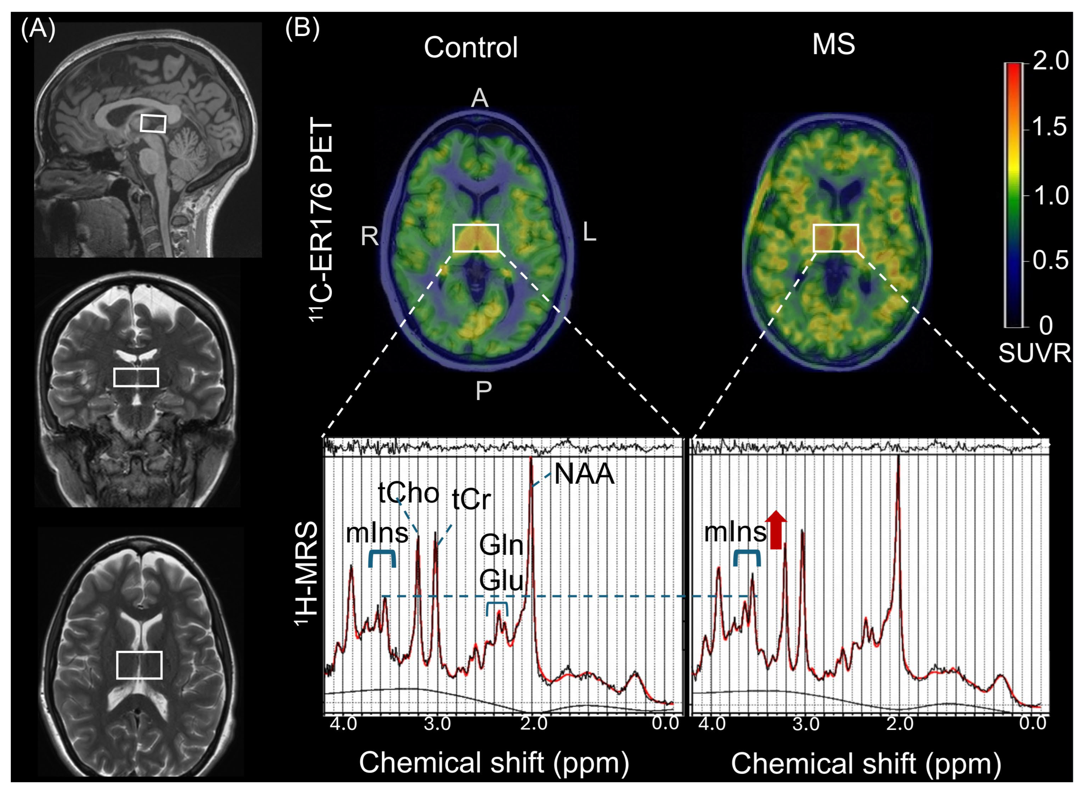
Single-voxel 1H-MRS data for each participant were acquired by a modified semi-localized adiabatic selective refocusing (sLASER) sequence with the following acquisition parameters (repetition time = 5000 ms; echo time = 30 ms; and 64 averages) as described previously [45,46,47,48,49,50]. The volume of interest (21 × 34 × 14 mm3) was manually positioned on a mid-sagittal T1 weighted image, including right and left thalamus regions by an experienced MRI technician (Figure 2). Thalamic voxels were consistently placed bilaterally and centered to ensure reproducibility across participants. Voxel positioning in thalamus was evaluated with mid-sagittal anterior landmark of mammillary body and posterior landmark of mesencephalic aqueduct to place the inferior edge of the voxel and superior landmark of fornix for the upper edge limit. The thalamic MRS voxel was composed of white matter (WM), gray matter (GM), and cerebrospinal fluid (CSF) partial volume fractions (Supplementary Table S1).
The processed sLASER spectra were quantified using LCModel 6.3 (Stephen Provencher, Oakville, ON, Canada). Metabolite levels were calculated in as (1) estimated metabolite concentrations (mM) using tissue water as an internal reference; and (2) as metabolite/tCr ratios by normalizing the estimated metabolite concentrations to total creatine (tCr) [47,51,52,53,54,55]. The acquisition and processing techniques of 1H-MRS were described earlier [46,47]. A detailed description is available in the Supplementary Methods (1H-MRS). Briefly, estimated metabolite concentration were corrected for partial volume effect using GM, WM, and CSF tissue partial volume fractions in thalamic 1H-MRS voxel (Supplementary Table S1) and T2 relaxation time of thalamus (96 ms) was taken into account in LCModel fitting for concentration calculations [8,56] (Supplementary). The tissue classification images were created using Mayo Clinic Adult Lifespan Template [57] (see Supplementary Methods [1H-MRS] for more details).
For 1H-MRS data quality control, we followed the most recent 1H-MRS expert consensus recommendations [52]. Spectra were evaluated for lipid contaminations and insufficient water suppression, which may lead to sidebands and unwanted coherences. In addition, LCModel residuals, and baseline distortions were evaluated. Spectra were excluded when these artifacts hampered the quantification of the metabolites of interest. For quantitative quality control, we assessed the Cramér-Rao Lower Bounds (CRLB) of metabolites and signal-to-noise (SNR) (i.e., the ratio of the maximum in the spectrum minus baseline over the window to twice root mean square error residual) ratio of a single spectrum, and the full width at the half maximum amplitude (FWHM) of the water-unsuppressed signal and LCModel FWHM (i.e., a rough estimate of the linewidth in the in vivo spectrum). The group mean CRLB was checked separately for pwMS and controls. Concentrations with CRLB values of 999%, indicating failed fitting, were not included for mean CRLB calculations. The reliably quantified metabolites (CRLB < 20; FWHM ≤ 0.1 ppm [≤13 Hz] at 3T) were NAA, Glu, Gln, tCho, and mIns and tCr. Mean CRLBs (±standard deviation) were within acceptable limits in both groups (controls vs. pwMS: tCho: 4.23 ± 0.68 vs. 4.57 ± 1.03; NAA: 3.60 ± 0.72 vs. 3.71 ± 0.72; Glu: 6.10 ± 1.35 vs. 5.90 ± 1.41; Gln: 18.5 ± 7.9 vs. 16 ± 3.64; mIns: 4.80 ± 0.96 vs. 4.52 ± 1.03; tCr: 2.90 ± 0.48 vs. 3.10 ± 0.54). Spectral quality was adequate and comparable across groups, with NAA SNR >3 in all individual spectra prior to averaging (i.e., before summing the 64 transients) (group-averaged NAA SNR: 149.39 ± 23.78 in controls; 140.40 ± 28.11 in pwMS). The partial volume-corrected estimated concentration (mM) of thalamic tCr did not differ between pwMS and controls (Table S2). Therefore, tCr was used as the denominator for metabolite ratio calculations. The partial volume-corrected estimated 1H-MRS concentrations are presented in the Supplementary Table S2.

4.4. 11C-ER176 PET

PET imaging was performed using a PET/CT scanner (Siemens Healthineers, Erlangen, Germany or GE Healthcare Milwaukee, WI, USA), operating in 3D mode using the third-generation radioligand, 11C-ER176 (IND# 149229) at the Mayo Clinic in Rochester. 11C-ER176 PET images were acquired at 60–90 min after injection of 11C-ER176 (518 MBq, 14 mCi; range, 370–666 MBq, 10–18 mCi) using four 5 min dynamic frames. Standardized uptake value ratios (SUVR) were normalized to the cerebellar crus [58]. In this cohort, an age-adjusted linear model showed no significant difference in cerebellar crus SUVR between pwMS and controls (p = 0.114), consistent with a prior report of no group differences in this region [24]. Based on these results, the cerebellar crus was selected as the reference region for SUVR normalization. Quantification was performed using an automated imaging pipeline [59]. No additional correction was applied for the low-affinity TSPO binding genotype, as this genotype is relatively rare [26,60,61,62,63]. Further details are provided in the Supplementary Methods.

4.5. Clinical Assessments

For pwMS, clinical assessments included the EDSS, the MS Functional Composite (MSFC), and its three components: the 25-foot timed walk (25FTW), the 9-hole peg test (9HPT), and the PASAT [64,65,66,67]. The z-scores of PASAT, 9HPT (dominant hand) and 25FTW were averaged to calculate MSFC z-scores (see Supplementary Methods) [65,68,69]. All clinical assessments were performed by a trained examiner blinded to the imaging results.

4.6. Statistical Analysis

Group differences were assessed using the Mann–Whitney U test (Wilcoxon Rank-Sum test) or linear regression. Fisher’s exact test was used to analyze categorical variables. Available data were used without imputation of missing values in the analysis.
Multiple linear regression models were fitted with and without interaction term to estimate whether age influenced the association between the group (pwMS and controls) and imaging variables (11C-ER176 PET SUVR, 1H-MRS and MRI). The age × group as an interaction term was not statistically significant and was excluded from the final models [70]. To evaluate group differences in imaging measures, multiple linear regression analyses were conducted with imaging metrics as the dependent variables, group (pwMS vs. controls) as the primary independent variable. Age was included as a covariate in all models. Linear regression models were used to evaluate whether the mean values of the dependent imaging variables differed between pwMS and controls after adjusting for age.
Correlations between imaging variables were assessed using partial Pearson correlation coefficients (r), while associations between MS clinical disability scores and imaging variables were evaluated using age-adjusted Spearman rank correlation coefficients (rho). Given pwMS group comprises 21 participants, only age, the key confounder for imaging studies, was used as a covariate for partial Spearman and partial Pearson correlations. Normality assumptions for linear regression and Pearson correlation were assessed using Q-Q plots and histograms of residuals. Linearity in linear regression was evaluated using residuals vs. fitted values plots, confirming the assumption of linearity and normality. Multicollinearity in the multiple linear regression models was tested using the variance inflation factor (VIF) for each independent variable [71]. Multicollinearity was defined as a VIF value ≥ 5. All independent variables had a VIF value < 5.
No corrections for Type 1 errors were applied. Although adjustments for multiple comparisons reduce the risk of type I errors (false positives), they increase the risk of type II errors (false negatives), potentially leading to missed significant findings [72]. However, we reported all exact p-values enabling readers to evaluate the robustness of our results. Statistical significance was determined by p < 0.05. Statistical analyses were conducted using the R software (version 4.4.1) within the RStudio environment (version 2024.09.1 + 394).

5. Conclusions

This study highlights the unique and significant association of thalamic 1H-MRS (mIns/tCr) with thalamic 11C-ER176 PET SUVR as well as with thalamic atrophy and cognitive decline in pwMS. As reliable markers of glial-mediated innate immune activation, our 1H-MRS (mIns/tCr) and 11C-ER176 PET findings in pwMS emphasize the interplay between glial activity, glial density and neurodegeneration, and the potential contribution of innate immune activity to the clinical disability, including cognitive impairment. These findings further enhance our understanding of the cellular and molecular pathological mechanisms in the thalamus associated with MS, positioning 1H-MRS and 11C-ER176 PET detected glial activity and density as reliable and sensitive biomarkers of pathological alterations in the thalamus. Furthermore, thalamic 1H-MRS and 11C-ER176 PET hold promise as potential biomarkers in current and future MS therapeutic intervention trials targeting glial cells.

Supplementary Materials

The following supporting information can be downloaded at: https://www.mdpi.com/article/10.3390/ijms26178656/s1.

Author Contributions

F.K., B.Z., K.K. and O.H.K. contributed to the conception and design of the study as well as critical review and drafting of the manuscript; F.K., N.N., M.G.K., C.G.S., J.K.-T., H.A.M., M.L.S., S.A.P., A.J.F., J.D.P., D.K.D., V.J.L. and G.Ö. contributed to the acquisition and analysis of data or drafting the text or preparing the figures. All authors have read and agreed to the published version of the manuscript.

Funding

This work was supported by National Institutes of Health (NIH) K12 AR084222; P41 EB027061, R01 NS080816 and K12 HD065987.

Institutional Review Board Statement

This study was approved by the Mayo Clinic Institutional Review Board in Rochester, MN, USA (#22-009149, 2023-03-21). All procedures performed in studies involving human participants were in accordance with the ethical standards of the institutional and/or national research committee and with the 1964 Helsinki declaration and its later amendments or comparable ethical standards.

Informed Consent Statement

This study was approved by the Institutional Review Board, and all participants provided written informed consent.

Data Availability Statement

The data are not publicly available due to privacy or ethical restrictions. The data is stored in the Medidata repository. Qualified academic and industry researchers can request data, from the Mayo Clinic Advanced Imaging Research Center. Once a request is submitted, the committee sends the indicated principal investigator an email confirming that the request was received and giving a timeline for committee review.

Conflicts of Interest

K. Kantarci has served on the data safety monitoring board for Pfizer Inc. and Takeda Global Research & Devel-opment Center, Inc. She received research support from Avid. MM-A received consulting fees from the Biomedical Research Alliance of New York Radiopharmaceuticals, Eli Lilly. She consults for Biogen; C. Schwarz has received research funding from the NIH; B. Zeydan has received research funding from NIH. She received the Eugene and Marcia Applebaum Award; D. K. Deelchand has received funding from NIH (NIH R01 EB030000; NIH P41 EB027061); G. Oz has received funding from NIH (R01 NS080816, P41 EB027061, P30 NS076408, S10 OD017974, R01 NS124065, R01 NS035192, U01 NS104326, R61 AT009995). She has received grants from National Ataxia Foundation and Biogen, Inc. She has received consulting fees from “Servier, Inc”, “UCB Biopharma SRL / Lacerta Therapeutics Inc”, “IXICO Technologies Limited”. She has received payment for Honorarium for lecture (Purdue University, UCLA Brain Research Institute, Society for Neuroscience Louisville Chapter). She is in the scientific ad-visory board of “BrainSpec Inc” and “Medical Research Advisory Board, National Ataxia Foundation”; V. J. Lowe has had grants from “Siemens Medical Imaging”, and “AVID Radiopharmaceuticals” and NIH. He received pay-ment for “Peer View Institute for Medical Education”. He received equipment from AVID Radiopharmaceuticals; All other authors report no competing interest.

Abbreviations

CSFCerebrospinal Fluid
CIConfidence Interval
EDSSExpanded Disability Status Scale
GMGray Matter
GluGlutamate
GlnGlutamine
MSMultiple Sclerosis
mInsMyo-inositol
MRIMagnetic Resonance Imaging
MPRAGEMagnetization Prepared Rapid Acquisition Gradient Echo
mM:Millimolar
MSFCMultiple Sclerosis Functional Composite
NAAN-acetylaspartate
pwMSPatients with Multiple Sclerosis
PETPositron Emission Tomography
PASATPaced Auditory Serial Addition Test
sLASERSemi-Localized Adiabatic Selective Refocusing
SUVRStandardized Uptake Value Ratio
SNRSignal-to-Noise Ratio
tCrTotal Creatine
tChoTotal Choline
TSPOTranslocator Protein
TIVTotal Intracranial Volume
TRRepetition Time
TEEcho Time
VIFVariance Inflation Factor
9HPT9-Hole Peg Test
25FTW25-Foot Timed Walk
11C-ER176Third-generation TSPO radioligand
1H-MRSProton Magnetic Resonance Spectroscopy
WMWhite Matter

References

  1. Azevedo, C.J.; Overton, E.; Khadka, S.; Buckley, J.; Liu, S.; Sampat, M.; Kantarci, O.; Frenay, C.L.; Siva, A.; Okuda, D.T.; et al. Early CNS neurodegeneration in radiologically isolated syndrome. Neurol. Neuroimmunol. Neuroinflamm. 2015, 2, e102. [Google Scholar] [CrossRef] [PubMed]
  2. Eshaghi, A.; Marinescu, R.V.; Young, A.L.; Firth, N.C.; Prados, F.; Cardoso, M.J.; Tur, C.; De Angelis, F.; Cawley, N.; Brownlee, W.J.; et al. Progression of regional grey matter atrophy in multiple sclerosis. Brain 2018, 141, 1665–1677. [Google Scholar] [CrossRef] [PubMed]
  3. Öz, G.; Alger, J.R.; Barker, P.B.; Bartha, R.; Bizzi, A.; Boesch, C.; Bolan, P.J.; Brindle, K.M.; Cudalbu, C.; Dinçer, A.; et al. Clinical Proton MR Spectroscopy in Central Nervous System Disorders. Radiology 2014, 270, 658–679. [Google Scholar] [CrossRef]
  4. Ligneul, C.; Palombo, M.; Hernández-Garzón, E.; Sauvage, M.-A.C.-D.; Flament, J.; Hantraye, P.; Brouillet, E.; Bonvento, G.; Escartin, C.; Valette, J. Diffusion-weighted magnetic resonance spectroscopy enables cell-specific monitoring of astrocyte reactivity in vivo. NeuroImage 2019, 191, 457–469. [Google Scholar] [CrossRef] [PubMed]
  5. Lopez, M.Y.; Pardon, M.-C.; Baiker, K.; Prior, M.; Yuchun, D.; Agostini, A.; Bai, L.; Auer, D.P.; Faas, H.M.; Walczak, P. Myoinositol CEST signal in animals with increased Iba-1 levels in response to an inflammatory challenge—Preliminary findings. PLoS ONE 2019, 14, e0212002. [Google Scholar] [CrossRef]
  6. Geurts, J.J.G.; Reuling, I.E.W.; Vrenken, H.; Uitdehaag, B.M.J.; Polman, C.H.; Castelijns, J.A.; Barkhof, F.; Pouwels, P.J.W. MR spectroscopic evidence for thalamic and hippocampal, but not cortical, damage in multiple sclerosis. Magn. Reson. Med. 2006, 55, 478–483. [Google Scholar] [CrossRef]
  7. Tisell, A.; Leinhard, O.D.; Warntjes, J.B.M.; Aalto, A.; Smedby, Ö.; Landtblom, A.-M.; Lundberg, P.; Smeyne, R.J. Increased Concentrations of Glutamate and Glutamine in Normal-Appearing White Matter of Patients with Multiple Sclerosis and Normal MR Imaging Brain Scans. PLoS ONE 2013, 8, e61817. [Google Scholar] [CrossRef]
  8. Muhlert, N.; Atzori, M.; De Vita, E.; Thomas, D.L.; Samson, R.S.; Wheeler-Kingshott, C.A.M.; Geurts, J.J.G.; Miller, D.H.; Thompson, A.J.; Ciccarelli, O. Memory in multiple sclerosis is linked to glutamate concentration in grey matter regions. J. Neurol. Neurosurg. Psychiatry 2014, 85, 833–839. [Google Scholar] [CrossRef]
  9. Tzanetakos, D.; Kyrozis, A.; Karavasilis, E.; Velonakis, G.; Tzartos, J.; Toulas, P.; Sotirli, S.A.; Evdokimidis, I.; Tsivgoulis, G.; Potagas, C.; et al. Early metabolic alterations in the normal-appearing grey and white matter of patients with clinically isolated syndrome suggestive of multiple sclerosis: A proton MR spectroscopic study. Exp. Ther. Med. 2023, 26, 349. [Google Scholar] [CrossRef]
  10. Rae, C.D. A Guide to the Metabolic Pathways and Function of Metabolites Observed in Human Brain 1H Magnetic Resonance Spectra. Neurochem. Res. 2013, 39, 1–36. [Google Scholar] [CrossRef]
  11. Datta, G.; Violante, I.R.; Scott, G.; Zimmerman, K.; Santos-Ribeiro, A.; A Rabiner, E.; Gunn, R.N.; Malik, O.; Ciccarelli, O.; Nicholas, R.; et al. Translocator positron-emission tomography and magnetic resonance spectroscopic imaging of brain glial cell activation in multiple sclerosis. Mult. Scler. J. 2016, 23, 1469–1478. [Google Scholar] [CrossRef]
  12. Sotirchos, E.S.; Gonzalez-Caldito, N.; E Dewey, B.; Fitzgerald, K.C.; Glaister, J.; Filippatou, A.; Ogbuokiri, E.; Feldman, S.; Kwakyi, O.; Risher, H.; et al. Effect of disease-modifying therapies on subcortical gray matter atrophy in multiple sclerosis. Mult. Scler. J. 2019, 26, 312–321. [Google Scholar] [CrossRef]
  13. Misin, O.; Matilainen, M.; Nylund, M.; Honkonen, E.; Rissanen, E.; Sucksdorff, M.; Airas, L. Innate Immune Cell–Related Pathology in the Thalamus Signals a Risk for Disability Progression in Multiple Sclerosis. Neurol. Neuroimmunol. Neuroinflamm. 2022, 9, e1182. [Google Scholar] [CrossRef]
  14. Inglese, M.; Liu, S.; Babb, J.S.; Mannon, L.J.; Grossman, R.I.; Gonen, O. Three-dimensional proton spectroscopy of deep gray matter nuclei in relapsing–remitting MS. Neurology 2004, 63, 170–172. [Google Scholar] [CrossRef]
  15. Henry, R.G.; Shieh, M.; Amirbekian, B.; Chung, S.; Okuda, D.T.; Pelletier, D. Connecting white matter injury and thalamic atrophy in clinically isolated syndromes. J. Neurol. Sci. 2009, 282, 61–66. [Google Scholar] [CrossRef]
  16. Steenwijk, M.D.; Daams, M.; Pouwels, P.J.; Balk, L.J.; Tewarie, P.K.; Geurts, J.J.G.; Barkhof, F.; Vrenken, H. Unraveling the relationship between regional gray matter atrophy and pathology in connected white matter tracts in long-standing multiple sclerosis. Hum. Brain Mapp. 2015, 36, 1796–1807. [Google Scholar] [CrossRef]
  17. Cifelli, A.; Arridge, M.; Jezzard, P.; Esiri, M.M.; Palace, J.; Matthews, P.M. Thalamic neurodegeneration in multiple sclerosis. Ann. Neurol. 2002, 52, 650–653. [Google Scholar] [CrossRef]
  18. Cosenza-Nashat, M.; Zhao, M.; Suh, H.; Morgan, J.; Natividad, R.; Morgello, S.; Lee, S.C. Expression of the translocator protein of 18 kDa by microglia, macrophages and astrocytes based on immunohistochemical localization in abnormal human brain. Neuropathol. Appl. Neurobiol. 2009, 35, 306–328. [Google Scholar] [CrossRef]
  19. Venneti, S.; Lopresti, B.J.; Wang, G.; Hamilton, R.L.; Mathis, C.A.; Klunk, W.E.; Apte, U.M.; Wiley, C.A. PK11195 labels activated microglia in Alzheimer’s disease and in vivo in a mouse model using PET. Neurobiol. Aging 2009, 30, 1217–1226. [Google Scholar] [CrossRef]
  20. Nutma, E.; Fancy, N.; Weinert, M.; Tsartsalis, S.; Marzin, M.C.; Muirhead, R.C.J.; Falk, I.; Breur, M.; de Bruin, J.; Hollaus, D.; et al. Translocator protein is a marker of activated microglia in rodent models but not human neurodegenerative diseases. Nat. Commun. 2023, 14, 1–25. [Google Scholar] [CrossRef]
  21. Papadopoulos, V.; Baraldi, M.; Guilarte, T.R.; Knudsen, T.B.; Lacapère, J.-J.; Lindemann, P.; Norenberg, M.D.; Nutt, D.; Weizman, A.; Zhang, M.-R.; et al. Translocator protein (18kDa): New nomenclature for the peripheral-type benzodiazepine receptor based on its structure and molecular function. Trends Pharmacol. Sci. 2006, 27, 402–409. [Google Scholar] [CrossRef]
  22. Werry, E.L.; Bright, F.M.; Piguet, O.; Ittner, L.M.; Halliday, G.M.; Hodges, J.R.; Kiernan, M.C.; Loy, C.T.; Kril, J.J.; Kassiou, M. Recent Developments in TSPO PET Imaging as A Biomarker of Neuroinflammation in Neurodegenerative Disorders. Int. J. Mol. Sci. 2019, 20, 3161. [Google Scholar] [CrossRef]
  23. Datta, G.; Colasanti, A.; Kalk, N.; Owen, D.; Scott, G.; Rabiner, E.A.; Gunn, R.N.; Lingford-Hughes, A.; Malik, O.; Ciccarelli, O.; et al. 11C-PBR28 and 18F-PBR111 Detect White Matter Inflammatory Heterogeneity in Multiple Sclerosis. J. Nucl. Med. 2017, 58, 1477–1482. [Google Scholar] [CrossRef]
  24. Zeydan, B.; Neyal, N.; Son, J.; Schwarz, C.G.; Thomas, J.C.K.; A Morrison, H.; Bush, M.L.; I Reid, R.; A Przybelski, S.; Fought, A.J.; et al. Microglia positron emission tomography and progression in multiple sclerosis: Thalamus on fire. Brain Commun. 2025, 7, fcaf141. [Google Scholar] [CrossRef]
  25. Deelchand, D.K.; Kantarci, K.; Öz, G. Improved localization, spectral quality, and repeatability with advanced MRS methodology in the clinical setting. Magn. Reson. Med. 2017, 79, 1241–1250. [Google Scholar] [CrossRef]
  26. Cumbers, G.A.; Harvey-Latham, E.D.; Kassiou, M.; Werry, E.L.; Danon, J.J. Emerging TSPO-PET Radiotracers for Imaging Neuroinflammation: A Critical Analysis. Semin. Nucl. Med. 2024, 54, 856–874. [Google Scholar] [CrossRef]
  27. Guilarte, T.R. TSPO in diverse CNS pathologies and psychiatric disease: A critical review and a way forward. Pharmacol. Ther. 2019, 194, 44–58. [Google Scholar] [CrossRef]
  28. Wylezinska, M.; Cifelli, A.; Jezzard, P.; Palace, J.; Alecci, M.; Matthews, P.M. Thalamic neurodegeneration in relapsing-remitting multiple sclerosis. Neurology 2003, 60, 1949–1954. [Google Scholar] [CrossRef]
  29. Urenjak, J.; Williams, S.; Gadian, D.; Noble, M. Proton nuclear magnetic resonance spectroscopy unambiguously identifies different neural cell types. J. Neurosci. 1993, 13, 981–989. [Google Scholar] [CrossRef]
  30. Davie, C.A.; Hawkins, C.P.; Barker, G.J.; Brennan, A.; Tofts, P.S.; Miller, D.H.; McDonald, W.I. Serial proton magnetic resonance spectroscopy in acute multiple sclerosis lesions. Brain 1994, 117, 49–58. [Google Scholar] [CrossRef]
  31. Orije, J.; Kara, F.; Guglielmetti, C.; Praet, J.; Van der Linden, A.; Ponsaerts, P.; Verhoye, M. Longitudinal monitoring of metabolic alterations in cuprizone mouse model of multiple sclerosis using 1H-magnetic resonance spectroscopy. NeuroImage 2015, 114, 128–135. [Google Scholar] [CrossRef]
  32. Caramanos, Z.; Narayanan, S.; Arnold, D.L. 1H-MRS quantification of tNA and tCr in patients with multiple sclerosis: A meta-analytic review. Brain 2005, 128, 2483–2506. [Google Scholar] [CrossRef] [PubMed]
  33. Haris, M.; Singh, A.; Cai, K.; Nath, K.; Crescenzi, R.; Kogan, F.; Hariharan, H.; Reddy, R. MICEST: A potential tool for non-invasive detection of molecular changes in Alzheimer’s disease. J. Neurosci. Methods 2013, 212, 87–93. [Google Scholar] [CrossRef] [PubMed]
  34. Genovese, G.; Palombo, M.; Santin, M.D.; Valette, J.; Ligneul, C.; Aigrot, M.; Abdoulkader, N.; Langui, D.; Millecamps, A.; Evercooren, A.B.; et al. Inflammation-driven glial alterations in the cuprizone mouse model probed with diffusion-weighted magnetic resonance spectroscopy at 11.7 T. NMR Biomed. 2021, 34, e4480. [Google Scholar] [CrossRef]
  35. Sun, Y.; Yu, H.; Guan, Y. Glia Connect Inflammation and Neurodegeneration in Multiple Sclerosis. Neurosci. Bull. 2023, 39, 466–478. [Google Scholar] [CrossRef] [PubMed]
  36. Solanky, B.; John, N.; DeAngelis, F.; Stutters, J.; Prados, F.; Schneider, T.; Parker, R.; Weir, C.; Monteverdi, A.; Plantone, D.; et al. NAA is a Marker of Disability in Secondary-Progressive MS: A Proton MR Spectroscopic Imaging Study. Am. J. Neuroradiol. 2020, 41, 2209–2218. [Google Scholar] [CrossRef]
  37. Niess, E.; Dal-Bianco, A.; Strasser, B.; Niess, F.; Hingerl, L.; Bachrata, B.; Motyka, S.; Rommer, P.; Trattnig, S.; Bogner, W. Topographical mapping of metabolic abnormalities in multiple sclerosis using rapid echo-less 3D-MR spectroscopic imaging at 7T. NeuroImage 2025, 308, 121043. [Google Scholar] [CrossRef]
  38. Gracien, R.M.; Jurcoane, A.; Wagner, M.; Reitz, S.C.; Mayer, C.; Volz, S.; Hof, S.M.; Fleischer, V.; Droby, A.; Steinmetz, H.; et al. The Relationship between Gray Matter Quantitative MRI and Disability in Secondary Progressive Multiple Sclerosis. PLoS One 2016, 11, e0161036. [Google Scholar] [CrossRef] [PubMed]
  39. Srinivasan, R.; Sailasuta, N.; Hurd, R.; Nelson, S.; Pelletier, D. Evidence of elevated glutamate in multiple sclerosis using magnetic resonance spectroscopy at 3 T. Brain 2005, 128, 1016–1025. [Google Scholar] [CrossRef] [PubMed]
  40. Montalban, X. Revisions of the McDonald criteria ECTRIMS: 40th Congress of the European Committee for Treatment and Research in Multiple Sclerosis. 2024. Available online: https://ectrims.eu/mcdonald-diagnostic-criteria (accessed on 19 June 2025).
  41. Brownlee, W.J.; Vidal-Jordana, A.; Shatila, M.; Strijbis, E.; Schoof, L.; Killestein, J.; Barkhof, F.; Bollo, L.; Rovira, A.; Sastre-Garriga, J.; et al. Towards a Unified Set of Diagnostic Criteria for Multiple Sclerosis. Ann. Neurol. 2024, 97, 571–582. [Google Scholar] [CrossRef] [PubMed]
  42. Kara, F.; Joers, J.M.; Deelchand, D.K.; Park, Y.W.; Przybelski, S.A.; Lesnick, T.G.; Senjem, M.L.; Zeydan, B.; Knopman, D.S.; Lowe, V.J.; et al. 1H MR spectroscopy biomarkers of neuronal and synaptic function are associated with tau deposition in cognitively unimpaired older adults. Neurobiol. Aging 2022, 112, 16–26. [Google Scholar] [CrossRef] [PubMed]
  43. Schwarz, C.G.; Gunter, J.L.; Wiste, H.J.; Przybelski, S.A.; Weigand, S.D.; Ward, C.P.; Senjem, M.L.; Vemuri, P.; Murray, M.E.; Dickson, D.W.; et al. A large-scale comparison of cortical thickness and volume methods for measuring Alzheimer’s disease severity. NeuroImage Clin. 2016, 11, 802–812. [Google Scholar] [CrossRef]
  44. Raz, L.; Jayachandran, M.; Tosakulwong, N.; Lesnick, T.G.; Wille, S.M.; Murphy, M.C.; Senjem, M.L.; Gunter, J.L.; Vemuri, P.; Jr, C.R.J.; et al. Thrombogenic microvesicles and white matter hyperintensities in postmenopausal women. Neurology 2013, 80, 911–918. [Google Scholar] [CrossRef]
  45. Öz, G.; Tkáč, I. Short-echo, single-shot, full-intensity proton magnetic resonance spectroscopy for neurochemical profiling at 4 T: Validation in the cerebellum and brainstem. Magn. Reson. Med. 2010, 65, 901–910. [Google Scholar] [CrossRef]
  46. Tkáč, I.; Starčuk, Z.; Choi, I.; Gruetter, R. In vivo1H NMR spectroscopy of rat brain at 1 ms echo time. Magn. Reson. Med. 1999, 41, 649–656. [Google Scholar] [CrossRef]
  47. Deelchand, D.K.; Adanyeguh, I.M.; Emir, U.E.; Nguyen, T.-M.; Valabregue, R.; Henry, P.-G.; Mochel, F.; Öz, G. Two-site reproducibility of cerebellar and brainstem neurochemical profiles with short-echo, single-voxel MRS at 3T. Magn. Reson. Med. 2014, 73, 1718–1725. [Google Scholar] [CrossRef]
  48. Gruetter, R.; Tkáč, I. Field mapping without reference scan using asymmetric echo-planar techniques. Magn. Reson. Med. 2000, 43, 319–323. [Google Scholar] [CrossRef]
  49. Provencher, S.W. Automatic quantitation of localized in vivo1H spectra with LCModel. NMR Biomed. 2001, 14, 260–264. [Google Scholar] [CrossRef]
  50. Deelchand, D.K.; Henry, P.; Uǧurbil, K.; Marjańska, M. Measurement of transverse relaxation times of J-coupled metabolites in the human visual cortex at 4 T. Magn. Reson. Med. 2011, 67, 891–897. [Google Scholar] [CrossRef] [PubMed]
  51. Quadrelli, S.; Mountford, C.; Ramadan, S. Hitchhiker’S Guide to Voxel Segmentation for Partial Volume Correction of in Vivo Magnetic Resonance Spectroscopy. Magn. Reson. Insights 2016, 9, 1–8. [Google Scholar] [CrossRef] [PubMed]
  52. Near, J.; Harris, A.D.; Juchem, C.; Kreis, R.; Marjańska, M.; Öz, G.; Slotboom, J.; Wilson, M.; Gasparovic, C. Preprocessing, analysis and quantification in single-voxel magnetic resonance spectroscopy: Experts’ consensus recommendations. NMR Biomed. 2020, 34, e4257. [Google Scholar] [CrossRef] [PubMed]
  53. Randall, L.O. CHEMICAL TOPOGRAPHY OF THE BRAIN. J. Biol. Chem. 1938, 124, 481–488. [Google Scholar] [CrossRef]
  54. Norton, W.T.; Poduslo, S.E.; Suzuki, K. Subacute Sclerosing Leukoencephalitis*†‡. J. Neuropathol. Exp. Neurol. 1966, 25, 582–597. [Google Scholar] [CrossRef]
  55. Bednařík, P.; Henry, P.-G.; Khowaja, A.; Rubin, N.; Kumar, A.; Deelchand, D.; E Eberly, L.; Seaquist, E.; Öz, G.; Moheet, A. Hippocampal Neurochemical Profile and Glucose Transport Kinetics in Patients With Type 1 Diabetes. J. Clin. Endocrinol. Metab. 2019, 105, 479–491, Erratum in J. Clin. Endocrinol. Metab. 2020, 105, e1935.. [Google Scholar] [CrossRef]
  56. Sedlacik, J.; Boelmans, K.; Löbel, U.; Holst, B.; Siemonsen, S.; Fiehler, J. Reversible, irreversible and effective transverse relaxation rates in normal aging brain at 3T. NeuroImage 2014, 84, 1032–1041. [Google Scholar] [CrossRef]
  57. Schwarz, C.G.; Gunter, J.L.; Ward, C.P.; Kantarci, K.; Senjem, M.L.; Petersen, R.C.; Knopman, D.S.; Jack, C.R., Jr. P3-382: Methods to improve SPM12 tissue segmentations of older adults brains. Alzheimer’s & Dementia 2018, 14, 1240–1241. [Google Scholar] [CrossRef]
  58. Rossano, S.; Talmasov, D.; Johnson, A.S.; Smith, A.C.; Guzmán, D.S.; Okafor, A.; Kreisl, W.C.; De Jager, P.L.; Lao, P.J. Validating noninvasive techniques for 11C-ER176 PET quantification in controls and Alzheimer’s disease patients. Alzheimer’s Dement. 2023, 19, e081801. [Google Scholar] [CrossRef]
  59. Schwarz, C.G.; Senjem, M.L.; Gunter, J.L.; Tosakulwong, N.; Weigand, S.D.; Kemp, B.J.; Spychalla, A.J.; Vemuri, P.; Petersen, R.C.; Lowe, V.J.; et al. Optimizing PiB-PET SUVR change-over-time measurement by a large-scale analysis of longitudinal reliability, plausibility, separability, and correlation with MMSE. NeuroImage 2017, 144, 113–127. [Google Scholar] [CrossRef]
  60. Kreisl, W.C.; Fujita, M.; Fujimura, Y.; Kimura, N.; Jenko, K.J.; Kannan, P.; Hong, J.; Morse, C.L.; Zoghbi, S.S.; Gladding, R.L.; et al. Comparison of [11C]-(R)-PK 11195 and [11C]PBR28, two radioligands for translocator protein (18 kDa) in human and monkey: Implications for positron emission tomographic imaging of this inflammation biomarker. NeuroImage 2010, 49, 2924–2932. [Google Scholar] [CrossRef] [PubMed]
  61. Ikawa, M.; Lohith, T.G.; Shrestha, S.; Telu, S.; Zoghbi, S.S.; Castellano, S.; Taliani, S.; Da Settimo, F.; Fujita, M.; Pike, V.W.; et al. 11C-ER176, a Radioligand for 18-kDa Translocator Protein, Has Adequate Sensitivity to Robustly Image All Three Affinity Genotypes in Human Brain. J. Nucl. Med. 2017, 58, 320–325. [Google Scholar] [CrossRef]
  62. Kreisl, W.C.; Kim, M.-J.; Coughlin, J.M.; Henter, I.D.; Owen, D.R.; Innis, R.B. PET imaging of neuroinflammation in neurological disorders. Lancet Neurol. 2020, 19, 940–950. [Google Scholar] [CrossRef]
  63. Viviano, M.; Barresi, E.; Siméon, F.G.; Costa, B.; Taliani, S.; Da Settimo, F.; Pike, V.W.; Castellano, S. Essential Principles and Recent Progress in the Development of TSPO PET Ligands for Neuroinflammation Imaging. Curr. Med. Chem. 2022, 29, 4862–4890. [Google Scholar] [CrossRef]
  64. Kurtzke, J.F. Rating neurologic impairment in multiple sclerosis: An expanded disability status scale (EDSS). Neurology 1983, 33, 1444–1452. [Google Scholar] [CrossRef]
  65. Cutter, G.R.; Baier, M.L.; Rudick, R.A.; Cookfair, D.L.; Fischer, J.S.; Petkau, J.; Syndulko, K.; Weinshenker, B.G.; Antel, J.P.; Confavreux, C.; et al. Development of a multiple sclerosis functional composite as a clinical trial outcome measure. Brain 1999, 122, 871–882. [Google Scholar] [CrossRef] [PubMed]
  66. Goodkin, D.E.; Hertsgaard, D.; Seminary, J. Upper extremity function in multiple sclerosis: Improving assessment sensitivity with box-and-block and nine-hole peg tests. Arch. Phys. Med. Rehabil. 1988, 69, 850–854. [Google Scholar]
  67. Gronwall, D.M.A. Paced Auditory Serial-Addition Task: A Measure of Recovery from Concussion. Percept. Mot. Ski. 1977, 44, 367–373. [Google Scholar] [CrossRef] [PubMed]
  68. Fischer, J.S.; Rudick, R.A.; Cutter, G.R.; Reingold, S.C. The Multiple Sclerosis Functional Composite measure (MSFC): An integrated approach to MS clinical outcome assessment. Mult. Scler. J. 1999, 5, 244–250. [Google Scholar] [CrossRef]
  69. Fischer, J.S.; Jak, A.; Kniker, J.; Rudick, R.; Cutter, G.; Multiple Sclerosis Functional Composite (MSFC): Administration and Scoring Manual. New York: National Multiple Sclerosis Society 2001; pp. 1–2. Available online: http://main.nationalmssociety.org/docs/HOM/MSFC_Manual_and_Forms.pdf (accessed on 26 August 2025).
  70. Engqvist, L. The mistreatment of covariate interaction terms in linear model analyses of behavioural and evolutionary ecology studies. Anim. Behav. 2005, 70, 967–971. [Google Scholar] [CrossRef]
  71. Cohen, J.; Cohen, P.; West, S.G.; Aiken, L.S. Applied Multiple Regression/Correlation Analysis for the Behavioral Sciences, 3rd ed.; Routledge: New York, NY, USA, 2003; ISBN 9780805822236. [Google Scholar]
  72. Rothman, K.J. No adjustments are needed for multiple comparisons. Epidemiology 1990, 1, 43–46. [Google Scholar] [CrossRef] [PubMed]
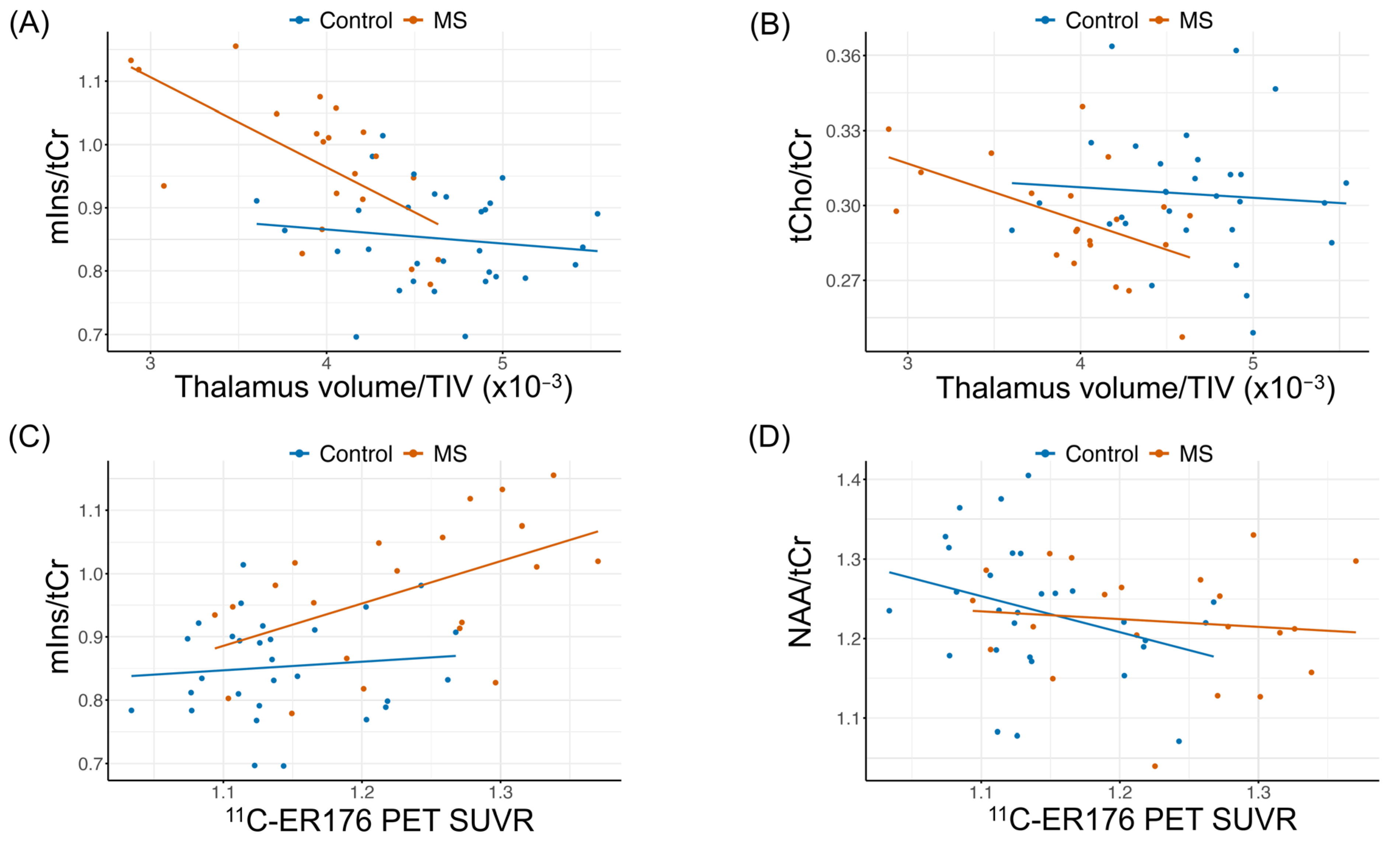
Figure 1. Scatter plots showing the relationships between (A) myo-inositol/total creatine (mIns/tCr) and normalized thalamic volume (thalamic volume/total intracranial volume, TIV); (B) total choline/tCr (tCho/tCr) and normalized thalamic volume; (C) mIns/tCr and 11C-ER176 PET standardized uptake value ratios (SUVR); and (D) N-acetylaspartate (NAA)/tCr and 11C-ER176 PET SUVR. The red dots = patients with multiple sclerosis (MS); blue dots = controls. The graphs display the original data points, focusing on original values rather than residuals. Trend lines show unadjusted linear regressions to visualize the overall direction of the relationships. Pearson correlation coefficients and p-values (r, p) of raw data from controls vs. pwMS are: (A): r = −0.13, p = 0.489 vs. r = −0.65, p = 0.002; (B): r = −0.08, p = 0.690 vs. r = −0.52, p = 0.016; (C): r = 0.10, p = 0.599 vs. r = 0.51, p = 0.017; (D): r = −0.32, p = 0.088 vs. r = −0.11, p = 0.62. Age-adjusted correlations and p-values are demonstrated in Table 3 and Table 4.
Ijms 26 08656 g001
Figure 2. Sagittal, coronal, and axial T1–weighted magnetic resonance images with superimposed thalamus proton magnetic resonance spectroscopy volume of interest (21 × 34 × 14 mm3) (A). The figures show representative 11C-ER176 positron emission tomography (PET) scans from a healthy control without Multiple Sclerosis (MS) and a patient with MS (pwMS) (B). Below is representative single-voxel proton (1H) magnetic resonance (MR) spectrum acquired at 3T using the sLASER sequence from the same participants who underwent 11C-ER176 PET. The healthy control had an mIns/tCr ratio of 0.69 and a thalamus 11C-ER176 PET standardized uptake value ratio (SUVR) of 1.14. (B) The pwMS had an mIns/tCr ratio of 0.91 and a thalamus 11C-ER176 PET SUVR of 1.27. The color bar scale indicates SUVR. The thick red curve on the MR spectrum represents the LCModel fit to the data, while the thin curve beneath it shows the fitted baseline. The residuals (data minus fit) are displayed at the top of the spectrum. A white rectangular box on the PET image indicates the thalamus region. The blue dashed line marks the mIns level in a healthy control, while the red arrow highlights the elevated mIns signal in a pwMS. Abbreviations: NAA = N-acetylaspartate; Glu = glutamate; Gln = glutamine; tCr = phosphocreatine + creatine; tCho = phosphocholine + glycerophosphocholine; mIns = myo-inositol; ppm, parts per million (a unit to express the frequency of chemical shift of metabolites in the magnetic field); A: Anterior; P: Posterior; L: Left; R: Right.
Ijms 26 08656 g002
Table 1. Characteristics of Patients with MS.
MS
(N = 21)
Sex
F15 (71%)
M6 (29%)
MS phase
Relapsing14 (67%)
Progressive7 (33%)
Disease duration14.8 [6.3, 20.3]/14.9 (±9.9)
Age at MS onset (years)31.9 [26.7, 34.6]/33.1 (±10.3)
Age at progressive MS onset (years)46.0 [34.3, 51.5]/43.7 (±10.6)
Age at imaging (years)48.0 [40.0, 54.0]/47.3 (±12.1)
DMT
No7 (33%)
Yes14 (67%)
EDSS score3 [1, 4]/2.81 (±2.12)
EDSS status
Mild10 (48%)
Moderate7 (33%)
Severe4 (19%)
MSFC z-score−0.19 [−0.32, 0.31]/−0.04 (±0.56)
PASAT score45.00 [38.75, 52.00]/44.25 (±9.66)
9HPT score (s)22.35 [20.04, 26.98]/28.74 (±19.43)
25FTW (s)4.90 [4.25, 6.40]/8.04 (±8.66)
Data are presented as median [IQR: 1st to 3rd quartile range]/mean (±SD), or count (%), depending on the variable type. Abbreviations: DMT, Disease-Modifying Therapy; EDSS, Expanded Disability Status Scale; EDSS status, mild (EDSS, 0.0–2.5), moderate (EDSS 3.0–5.5), and severe (EDSS ≥ 6.0); F, female; M, male; MS, Multiple Sclerosis; PASAT, Paced Auditory Serial Addition Test; 9HPT, 9-Hole Peg Test from dominant hand; 25FTW, Timed 25-Foot Walk Test; MSFC, Multiple Sclerosis Functional Composite (see Supplementary Methods MSFC Section). This table demonstrates all the available data, but there are some missing characteristics: disease duration, age at progressive MS onset, and MSFC, PASAT, 9HPT, 25FTW (N = 1).
Table 2. Characteristics and Imaging Variables of Controls and Patients with MS.
Control
(N = 30)
MS
(N = 21)
p-Value
(Age-Adjusted)
Sex 0.76 a
F19 (63%)15 (71%)
M11 (37%)6 (29%)
Age at imaging (years) 0.017 b
IQR (Q1, Q3)38.5 [29.2, 47.5]48.0 [40.0, 54.0]
Mean (±SD)39.0 (±11.1)47.3 (±12.1)
Thalamus volume (mL) 1 × 10⁻⁵
IQR (Q1, Q3)6.70 [6.34, 7.17]5.60 [5.35, 6.05]
Mean (±SD)6.72 (±0.72)5.58 (±0.76)
Total intracranial volume (mL) 0.52
IQR (Q1, Q3)1463.15 [1348.13, 1558.50]1383.83 [1339.77, 1492.54]
Mean (±SD)1454.57 (±144.92)1413.36 (±110.85)
Thalamus volume/TIV ×10−3 1 × 10⁻⁵
IQR (Q1, Q3)4.64 [4.34, 4.92]4.01 [3.86, 4.21]
Mean (±SD)4.64 (±0.46)3.95 (±0.50)
Thalamus 11C-ER176 PET SUVR 2.7 × 10⁻⁴
IQR (Q1, Q3)1.13 [1.11, 1.17]1.23 [1.15, 1.30]
Mean (±SD)1.14 (±0.06)1.23 (±0.08)
Thalamus 1H-MRS metabolites
tCho/tCr 0.20
IQR (Q1, Q3)0.3 [0.29, 0.32]0.29 [0.28, 0.3]
Mean (±SD)0.30 (±0.03)0.29 (±0.02)
NAA/tCr 0.95
IQR (Q1, Q3)1.24 [1.19, 1.30]1.22 [1.19, 1.27]
Mean (±SD)1.24 (±0.09)1.22 (±0.07)
Glu/tCr 0.20
IQR (Q1, Q3)1.14 [1.05, 1.24]1.15 [1.11, 1.22]
Mean (±SD)1.14 (±0.14)1.18 (±0.09)
Gln/tCr 0.48
IQR (Q1, Q3)0.41 [0.36, 0.49]0.45 [0.42, 0.52]
Mean (±SD)0.42 (±0.10)0.45 (±0.08)
mIns/tCr 5.5 × 10⁻⁴
IQR (Q1, Q3)0.84 [0.79, 0.91]0.98 [0.91, 1.05]
Mean (±SD)0.85 (±0.08)0.97 (±0.11)
Data are shown as n (%) or median [IQR: 1st to 3rd quartile range, Q1–Q3] and mean (±SD). For continuous variables, all group comparisons (except for “age at imaging” and sex) were adjusted for “age” using linear regression model. Separate models were run for each MR spectroscopy, volume, or PET SUVR measure as the dependent variable, with “age” and “group” as independent variables. The p-values reported are the p-values of the estimates for the “group” variable, indicating whether the group differences are statistically significant after adjusting for “age at imaging”. a For sex, the p-value is from Fisher’s Exact Test. b p-value for “age at imaging” group comparison is from the Mann–Whitney U test. Abbreviations: 11C-ER176 PET SUVR, ER176 positron emission tomography standardized uptake value ratio; thalamic 1H-MRS metabolite ratio indicates metabolite/tCr ratio; tCr, total creatine; tCho, total choline; NAA, N-acetylaspartate; Glu, glutamate; Gln, glutamine; mIns, myo-inositol; TIV, total intracranial volume. This table demonstrates all the available data, but there are some missing characteristics: 11C-ER176 PET SUVR (n = 1). Bold p-values denote statistical significance (p < 0.05).
Table 3. Partial Pearson Correlation (age-adjusted) Between Thalamic 1H-MRS and Normalized Thalamic Volume.
MS
(N = 21)
Control
(N = 30)
1H-MRS Metabolitesr95% CI p-Valuer95% CIp-Value
tCho/tCr−0.52[−0.9, −0.14]0.019−0.08[−0.45, 0.29]0.678
NAA/tCr0.32[−0.11, 0.75]0.175−0.11[−0.48, 0.26]0.564
Glu/tCr0.22[−0.22, 0.66]0.347−0.10[−0.47, 0.27]0.617
Gln/tCr−0.07[−0.52, 0.38]0.7820.13[−0.24, 0.5]0.498
mIns/tCr−0.67[−1, −0.34]0.001−0.19[−0.55, 0.17]0.32
See Table 2 for abbreviations of metabolites: r, correlation coefficient; MS (number of participants = 21); Control (number of participants = 30); Bold p-values denote statistical significance (p < 0.05). Associations were assessed using age-adjusted Pearson correlation.
Table 4. Partial Pearson Correlation (age-adjusted) Between Thalamic 1H-MRS and 11C-ER176 PET SUVR.
MSControl
1H-MRS Metabolitesr95% CIp-Valuer95% CIp-Value
tCho/tCr0.24[−0.2, 0.68]0.319−0.12[−0.49, 0.25]0.528
NAA/tCr−0.04[−0.49, 0.41]0.876−0.41[−0.75, −0.07]0.031
Glu/tCr−0.13[−0.58, 0.32]0.578−0.28[−0.64, 0.08]0.151
Gln/tCr0.01[−0.44, 0.46]0.972−0.19[−0.56, 0.18]0.327
mIns/tCr0.48[0.09, 0.87]0.0340.19[−0.18, 0.56]0.338
See Table 2 for abbreviations of metabolites: r, correlation coefficient; MS (number of participants = 21); Control (number of participants = 29); Bold p-values denote statistical significance (p < 0.05). Associations were assessed using age-adjusted Pearson correlation.
Table 5. Associations Between Thalamic 1H-MRS with Clinical Metrics in Patients with MS.
MSFC z-Score
(N = 21)
PASAT z-Score
(N = 21)
EDSS Score
(N = 21)
1H-MRS Metabolites Rho95% CIp-ValueRho95% CIp-ValueRho95% CIp-Value
tCho/tCr0.26[−0.19, 0.71]0.2750.13[−0.33, 0.59]0.6040.13[−0.32, 0.58]0.580
NAA/tCr0.15[−0.31, 0.61]0.5460.03[−0.43, 0.49]0.907−0.4[−0.81, 0.01]0.084
Glu/tCr0.18[−0.27, 0.63]0.4660.31[−0.13, 0.75]0.198−0.12[−0.57, 0.33]0.627
Gln/tCr0.12[−0.34, 0.58]0.6280.23[−0.22, 0.68]0.3400.04[−0.41, 0.49]0.868
mIns/tCr−0.22[−0.67, 0.23]0.363−0.48[−0.89, −0.07]0.0360.13[−0.32, 0.58]0.588
Age-adjusted Partial Spearman correlation coefficient (rho) is presented with confidence interval [95% CI]. This non-parametric method is appropriate given the non-normal distribution of clinical disability measures. Bold p-values indicate statistically significant associations (p < 0.05). For abbreviations of the variables see Table 1 and Table 2.
Disclaimer/Publisher’s Note: The statements, opinions and data contained in all publications are solely those of the individual author(s) and contributor(s) and not of MDPI and/or the editor(s). MDPI and/or the editor(s) disclaim responsibility for any injury to people or property resulting from any ideas, methods, instructions or products referred to in the content.