Review

WBSCR Locus: At the Crossroads of Human Behavioral Disorders and Domestication of Animals

by
Mikhail V. Shepelev
1,*,
Olga I. Skobel
2,
Tatiana T. Glazko
2,
Dmitry V. Popov
2,
Denis E. Vysotskii
2,
Pavel G. Georgiev
3,
Oksana G. Maksimenko
1,
Gleb Y. Kosovsky
2 and
Yuliya Y. Silaeva
4,*
1
Center for Genome Research, Institute of Gene Biology, Russian Academy of Sciences, 34/5 Vavilova Str., 119334 Moscow, Russia
2
Afanas’ev Institute of Fur-Bearing Animal Breeding and Rabbit Breeding, 6 Trudovaya Str., Rodniki, 140143 Moscow Region, Russia
3
Department of the Control of Genetic Processes, Institute of Gene Biology, Russian Academy of Sciences, 34/5 Vavilova Str., 119334 Moscow, Russia
4
Core Facility Center, Institute of Gene Biology, Russian Academy of Sciences, 34/5 Vavilova Str., 119334 Moscow, Russia
*
Authors to whom correspondence should be addressed.
Int. J. Mol. Sci. 2025, 26(17), 8549; https://doi.org/10.3390/ijms26178549
Submission received: 29 July 2025 / Revised: 28 August 2025 / Accepted: 2 September 2025 / Published: 3 September 2025
(This article belongs to the Special Issue Molecular Investigations in Neurodevelopmental Disorders)

Abstract

Social interaction between the domesticated animal and the domesticator is one of the key features of the “domestication syndrome”. Recent research has identified genes in the WBSCR (Williams–Beuren syndrome control region) locus as significant contributors to social behavior in dogs. Large chromosomal deletions and duplications in the human WBSCR locus lead to the development of WBS (Williams–Beuren syndrome) and WBSCR duplication syndrome, respectively. Hypersociability is one of the key symptoms of WBS, while the duplication syndrome is manifested as an autism spectrum disorder (ASD). The data from both humans and dogs highlight the WBSCR locus as one of the key genetic determinants of social behavior in mammals. Several genes in the WBSCR are candidates for the regulation of social behavior in mammals including GTF2I, GTF2IRD, AUTS2 and GALNT17. Here, we discuss the role of WBSCR locus in the regulation of social behavior in mammals including the recent data that highlight the importance of 3D genome alterations in this genomic region for both domestication of animals and development of neurobehavioral disorders in humans. In addition, we bring attention to the role of the poorly characterized GALNT17 gene as a putative player in the development of ASD symptoms and in the regulation of social behavior in animals. We provide a brief summary of its known functions and propose the future research directions aimed at the elucidation of Galnt17 involvement in the regulation of central nervous system (CNS) functions.

Graphical Abstract

1. Genetic Basis of Domestication

Charles Darwin noted that domesticated animals have a distinctive and unusual set of hereditary traits that are absent in their wild ancestors [1]. The combination of these traits is called the “domestication syndrome” (a term previously used for domesticated crop plants) [2]. While many hypotheses focus on the mechanisms underlying specific economic traits (e.g., milk production in cows, wool quality in sheep, etc.), a recent explanation addresses traits common to all domesticated animals that allow them to live in captivity and interact with humans [3]. Namely, it was hypothesized that changes in activity of certain upstream genes in a genetic regulatory network lead to changes in the regulation of downstream genetic modules [4].
Research has shown that during the initial stages of domestication, reduction in animals’ stress during interactions with humans typically occurs through selection of animals with adrenal hypofunction and reduced stress hormone production [5]. However, the diverse phenotypic traits comprising the domestication syndrome cannot be explained solely by decreased adrenal function. Development of all traits of domestication syndrome is closely associated with neural crest cells (NCCs). These cells first appear during early embryogenesis at the dorsal edge (“crest”) of the neural tube and then migrate, giving rise to precursors of many cell types and tissues, such as cranial bones, adrenal medulla, pigment melanoblasts, odontoblasts [6,7,8]. Although NCCs are not direct precursors to any part of the central nervous system (CNS) or the adrenal cortex, they play an important role in the development of these tissues through post-migratory embryological interactions [9]. The hypothesis on the role of NCCs in domestication has received experimental support: accumulating evidence suggests that domesticated species have higher polymorphism of genes involved in the control of neural crest and its derivatives than closely related wild species. In particular, a study of 15 pairs of such species found that the number of codons with a relatively high ratio of nonsynonymous to synonymous substitutions was higher in domesticated animals compared to closely related wild species [10].
One of the recent studies proposes that the phenomenon of domestication is based on human intervention in the reproductive functions of both males and females during the process of domestication: limited interaction between males of the same species, limited pool of males participating in reproduction, selection of the most productive females in conditions of unlimited access to resources for feeding offspring and selection of females experiencing minimal stress when interacting with humans [11]. These interventions result in changes in the neural crest, since neural crest cells are involved in many processes during ontogenesis. This is consistent with earlier studies, which, in particular, showed that when backcrossing a domestic rabbit with a wild one, a seasonality of reproduction appeared in the second generation. This effect was not observed in the domestic rabbit [12].
Thus, despite the fact that the phenomenon of domestication has been known for thousands of years, its mechanisms have been studied rather poorly and there is no consensus on the genetic basis of this phenomenon. Meanwhile, the study of the genetic basis of domestication has recently acquired particular importance in connection with the pace and biospheric consequences of the development of agrarian civilization, the rapid reduction in biodiversity and growth of biomass of domesticated animals [13]. Therefore, the problem of clarifying the mechanisms of domestication is becoming more and more practically important, since the need for its management is consistently intensifying.
It is obvious that the genetic basis of domestication is polygenic and will differ depending on the tasks faced by the domesticator. The process of domestication involves taxonomic, ecological and cultural components that differ for each specific case. In general, in the case of successful domestication, the mutual benefit and adaptability of both the domesticated species and the domesticator increases [14]. In particular, it is possible to trace the parallel evolution of a number of genes in humans and dogs, which is most obvious in the genes responsible for food digestion, metabolism, neurological processes and the control of cell division, apoptosis and oncogenesis [15]. Based on the above, it becomes obvious that the process of domestication is regulated by incredibly complex genetic mechanisms. As a result, a variety of phenotypic characteristics of animals are changed and they are not limited to those selected by the domesticator. In the first direct multi-year experiment on the domestication of foxes, Dmitry K. Belyaev chose a decrease in aggression towards humans in a series of generations as a universal sign of domestication. However, after a number of generations, in addition to a decrease in the aggression, some other signs of domestication began to be detected in the offspring, such as a tail shape typical for dogs, drooping ears and a dog-like bark [16]. This means that, using classical selection methods, a domesticator can change only the complex of traits included in the domestication syndrome, but not its individual elements. Considering the modern possibilities of genome editing, the task of altering individual elements of the gene network regulating the entire complex of domestication syndrome is very attractive. It allows, on the one hand, to achieve a reduction in aggressiveness of domesticated mammals, and, on the other hand, to retain valuable economically significant phenotypic traits of wild animals (color and thickness of fur, tail shape, meat quality, etc.).
In this review, we will focus on the social behavior of animals as one of the crucial traits of the domestication syndrome. Recent genetic analyses of dogs and wolves—two related species that represent a domesticated species and its wild counterpart—have revealed an important role of the Williams–Beuren syndrome critical region (WBSCR) genomic locus in regulating social behavior in dogs [17]. Remarkably, the same genomic region is disrupted in Williams–Beuren syndrome (WBS), a human neurodevelopmental disorder characterized by hypersociability [18]. In addition, we will thoroughly discuss the emerging role of a GALNT17 gene from the WBSCR locus in the regulation of social behavior in mammals. On top of that, we will highlight the effects of 3D genome alterations in the WBSCR locus on both domestication of animals and development of neurobehavioral disorders in humans. The genetic basis of animal domestication, particularly selection for enhanced sociability toward humans, provides a compelling evolutionary framework for understanding certain human neurodevelopmental diseases. We propose that in order to reduce the aggressiveness of domesticated animals towards humans, genes that are involved both in the process of domestication in mammals and various neurological or neurodevelopmental disorders in humans can be considered as possible targets for genome editing [19].

2. Genetic Basis of Behavioral Disorders in Human

2.1. The Genetics of Behavioral Disorders in Human

Social behavior in mammals is a complex phenotype regulated by multiple genetic pathways that have been conserved throughout evolution [20]. Social behavior of every individual in the population fits into a certain area of a sociability spectrum that could be presented as a normal distribution of behavioral phenotypes [21]. Behavioral disorders manifest with either hyposociability or hypersociability which are found at the extremities of the sociability spectrum [21]. Recent advances in molecular genetics, genome-wide association studies (GWAS) and functional genomics have identified numerous genetic loci that play critical roles in regulating various aspects of social behavior, from basic social recognition to complex behaviors [21,22]. Of particular importance for the regulation of social behavior are genes from oxytocin–vasopressin neuropeptide system: oxytocin receptor gene (OXTR, located on human chromosome 3p25.3) and arginine vasopressin receptor genes (AVPR1A on 12q14.2 and AVPR1B on 1q32.1) [23]. Other examples of functional gene groups include, but are not limited to, the following: neurotransmitter receptors (dopamine D2 receptor gene (DRD2, 11q23.2)) [24]; synaptic adhesion and scaffolding proteins (postsynaptic density protein PSD95 (DLG4 gene on 17p13.1)) [25,26]; neuroligin gene family (e.g., NLGN3 on Xq13.1) [27]; SHANK gene family (SHANK1 on 19q13.33, SHANK2 on 11q13.3, SHANK3 on 22q13.33) [28]; and many others. Besides individual genes, there are multiple genomic loci affected by chromosomal aberrations (deletions, inversion, duplication) that lead to behavioral disorders in human. These include 1q21.1, 7q11.23, 15q11-q13, 16p11.2 and many others [21,22].
Autism spectrum disorder (ASD) refers to a group of heterogenous neurobehavioral disorders which are characterized by impaired social communication (hyposociability) and stereotypic behavior. Genetic defects in ASD include chromosomal aberrations (deletions, duplication) or point mutations in individual genes. Currently, over 800 individual genes and dozens of genomic loci with chromosomal rearrangements are associated with the development of ASD in human [22,29,30]. One of these loci is the WBSCR on chromosome 7 in human that is implicated both in the development of several ASD syndromes as well as the neurodevelopmental hypersociability disorder called WBS.

2.2. The Williams–Beuren Syndrome Critical Region (WBSCR) and Behavioral Disorders

WBS (OMIM: #194250), that is sometimes called “hypersociability syndrome”, is caused by a 1.55–1.83 Mb deletion in the WBSCR located in 7q11.23 [18,31]. In 95% of patients with WBS, a 1.5 Mb hemizygous deletion encompasses 24 genes from TRIM50 up to GTF2I (Figure 1); 4% of patients carry a 1.8 Mb deletion that includes two additional genes (NCF1 and GTF2IRD2) and 1% of patients carry other deletions of variable size [32]. In populations, WBS occurs with a frequency from 1:7000 to 1:20,000 of newborns, according to different estimations [33]. It is believed that a high rate of chromosomal rearrangements in this locus is due to the presence of region-specific repeats (low-copy repeat elements, LCRs), that flank the deleted region [34] and mediate non-allelic homologous recombination during meiosis [35,36,37].
Patients with WBS are characterized by a developmental delay, intellectual disability, hypersociability, hyperacusis, anxiety, defects in visuospatial constructive cognition, speech delay, craniofacial anomalies and some other somatic symptoms [18] and these defects are accompanied by the morphological changes in the brain structure [38].
Genetic alterations in the WBSCR are not limited to deletions but also include duplications and triplications. The Williams–Beuren region duplication syndrome (WBDS; OMIM: #609757) is caused by a hemizygous duplication of the region that is usually deleted in patients with WBS and leads to the development of ASD [39,40] that is manifested with intellectual disabilities and speech delay [21,41]. Triplication syndrome is a rare event and is characterized by a phenotype that is similar to duplication syndrome but with more severe symptoms [42]. Notably, mice bearing the WBS deletion replicate most of the human symptoms, including hypersociability, thus indicating the evolutionary conserved features of the WBSCR locus [43].
Hypersociability phenotype of patients with WBS indicate that some genes in this locus are associated with social behavior and hence might also be involved in the evolution of social behavior in animals. Analysis of individual genes in the deleted region revealed that GTF2I and its paralogue GTF2IRD1 might be the primary candidates responsible for the hypersocial phenotype of WBS patients. Loss of these genes is associated with intellectual disability, anxiety and impaired social behavior [18,41,44,45]. It has been shown that changes in the GTF2I expression level can affect the balance of excitation/inhibition of cortical neurons [46], that is, an agreement with multiple evidences pointing out such balance as the basis of the socialization network [47,48].
GTF2I gene encodes transcriptional factor II-I (TFII-I). In mice the peak expression of Gtf2i is detected during prenatal and early postnatal development [49]. Gtf2I regulates cell cycle [50], embryonic development [51], coordinates activity of multiple transcription factors [52]. Reduced levels of Gtf2I lead to mitochondrial dysfunction as manifested by impaired fission/fusion, autophagy and mitophagy [53].
Experiments using cortical organoids obtained from induced pluripotent stem cell (iPSC) lines have shown that chromosomal rearrangements in the WBSCR locus lead to disturbances in the proliferation and maturation of neurons, and such disturbances are reproduced if the expression level of only one gene, GTF2I, is changed [41]. The idea of the leading role of the GTF2I gene in the formation of the pathological phenotype was confirmed using mouse model: an increase in the expression level of Gtf2i led to a disruption of social interactions [54].
In addition to neurobehavioral disturbances, deletions and duplications in the WBSCR lead to multiple somatic pathologies. The best studied example of genotype–phenotype relationship in the WBSCR locus is the deletion of ELN gene. ELN encodes elastin, a protein that forms polymers in the extracellular matrix. Deletion of ELN in WBS patients leads mostly to a stenosis of large arteria and hypertension due to the loss of vessel elasticity [55]. These data are supported by the mouse model of heterozygous Eln knockout [56].
The contribution of the majority of other deleted/duplicated genes to the development of WBS phenotype is largely unknown. There are limited data on the genotype–phenotype relationship for several commonly deleted genes. The WBSCR locus contains the BAZ1B gene (also known as Williams syndrome transcription factor, WSTF), which is involved in chromatin remodeling and is necessary for the correct migration of neural crest cells in vitro and in vivo [57,58]. It is believed that loss of BAZ1B is associated with the presence of craniofacial defects in patients with WBS [18].
Another gene in the deleted region, LIMK1, regulates the assembly and disassembly of the actin cytoskeleton and is associated with impaired visual–spatial cognitive abilities in people with WBS [59]. In Limk1 knockout mice, a decrease in long-term memory is observed [60]. Input of several genes (e.g., STX1A, MLXIPL, DNAJC30) to WBS phenotype and other aspects of WBS are discussed in the excellent review by Kozel et al. [18].
Two other genes from the WBSCR locus, AUTS2 and GALNT17, are not affected by common deletions/duplications in WBS/WBDS. These genes together with CALN1 gene flank the commonly deleted region in WBS on the centromeric side (Figure 1). Notably, these three genes span 2.9 Mb in the genome that is longer than the 1.5 Mb deletion in WBS patients (Figure 1). Pathogenic structural variants in AUTS2 gene include translocations, duplications, deletions or single-nucleotide polymorphisms (SNPs) and lead to the development of AUTS2 syndrome [61]. Core symptoms in patients with AUTS2 syndrome include developmental delay and intellectual disability as well as microcephalia, autistic symptoms, attention deficit hyperactivity disorder, craniofacial abnormalities and certain somatic pathologies [61,62]. These symptoms at least in part are replicated in Auts2 heterozygous knockout mice [63].
GALNT17 (WBSCR17) gene encodes N-acetylgalactosaminyltransferase enzyme of which cellular functions remain largely elusive. Several SNPs in GALN17 gene are linked to the development of Parkinson’s disease (PD) [64], but no structural variants in GALNT17 have been definitely associated with the development of neurobehavioral disorders in human. Loss of Galnt17 in mouse leads to ASD symptoms, developmental delay and anomalies of cerebellum [65] (see below), thus placing GALNT17 as an attractive candidate for further dissecting its role in the development of neurobehavioral disorders and domestication of animals.
The putative role of Galnt17 as a driver of ASD symptoms was uncovered using a mouse model with reciprocal translocation between the fifth and eighth chromosomes [62]. The breakage point on the fifth chromosome is located between Auts2 and Galnt17 genes and translocation results in decreased expression of both genes. The phenotype of the animals is characterized by growth abnormalities (lower body weight and length), facial skeleton anomalies, behavioral, memory and learning disturbances, increased anxiety, decreased exploratory activity and decreased righting reflex [62]. Animals with a homozygous translocation were found to have an altered structure of the hippocampus and cerebellum. Some of the phenotypic manifestations are similar to Sox1 gene knockout (KO) that is located on the eighth chromosome and is disrupted by the translocation, but most of the phenotypic features are likely associated with a decrease in the levels of Auts2 and Galnt17 transcripts. Many phenotypic features are characteristic of Auts2 gene KO mice [66] and patients with AUTS2 syndrome [61,67]. Notably, co-expression of Auts2 и Galnt17 was detected in most cell types in the brain thus allowing us to predict that these two genes are located within one TAD (topology-associated domain) and therefore their expression is also co-regulated.
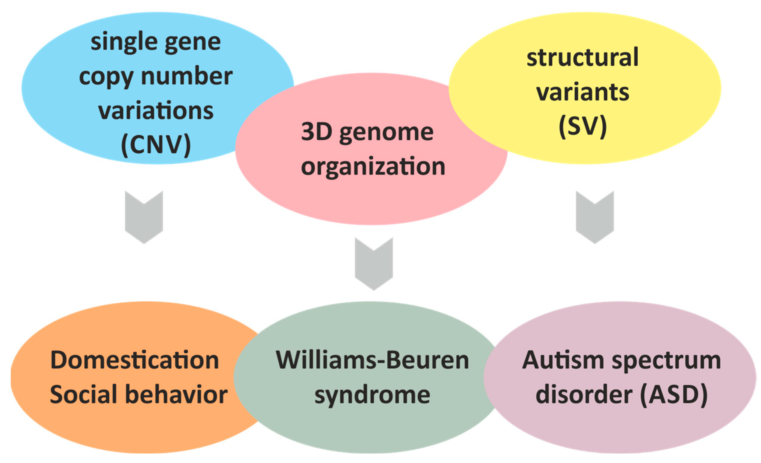
Taken together, this growing body of evidence suggests neurobehavioral and somatic phenotypes that linked to genes in the WBSCR locus are attributed not only to changes in copy number of deleted/amplified genes but also to changes in the expression levels of flanking genes due altered 3D genome organization as a result of large chromosomal rearrangements in this locus. Recent findings both in mouse and dog models (see below) indicate that the input of the altered 3D genome in the WBSCR locus is indispensable for the social behavior of animals as well.

3. WBSCR Locus and Dog Domestication

Evidence for the possible involvement of the WBSCR locus in the domestication process was obtained by GWAS analyzing more than 48,000 SNPs in the genomes of dogs of various breeds and in the gray wolf genome. As a result, one of the SNPs associated with positive selection in domesticated dogs was identified near the WBSCR17 gene [68]. Subsequent targeted analysis of more than 25,000 SNPs in a 5 Mb region of the WBSCR locus on chromosome 6 in dogs revealed 89 structural variants (SVs) in GTF2I, GTF2IRD1, AUTS2, WBSCR17 (GALNT17), GALNT9, CBX3, BAZ1B, NSUN5, POM121 and STYXL1 genes. These SVs are retrotransposons and four of them are reliably associated in dogs with attention bias to social stimuli: two in the WBSCR17 gene, and one in GTF2I and GTF2IRD1 genes. Thus, for the first time, a relationship was established between structural variants in the WBSCR17, GTF2I, GTF2IRD1 genes and hypersociability in dogs in relation to humans, which is one of the critical features of domestication syndrome [69].
Subsequent analysis of the molecular mechanisms that may underlie the changes in social behavior in dogs showed that retrotransposons near and in the WBSCR17 (Cfa6.6 and Cfa6.7), GTF2I (Cfa6.66) and POM121 (Cfa6.83) genes are hypermethylated, which leads to disruption of the expression of the WBSCR17, GTF21I, LIMK1, WBSCR27, BAZ1B and BCL7B genes [17]. Thus, positive selection for hypersociability towards the host in dogs is based on the presence of mobile genetic elements which are methylated to affect the expression profile of several genes in the WBSCR locus. In particular, the GTF21I and WBSCR17 genes, which are associated with the regulation of social behavior. Importantly, it was uncovered that the presence of one transposon element (TE) affects the expression of several genes in the locus, indirectly indicating the involvement of distant cis-regulatory interactions between genetic elements in the WBSCR locus. This was confirmed in further work using the example of a TE insertion in 17th intron of the GTF2I gene as discussed below.
It was found that in dogs, the insertion of TEs into the GALNT17 gene leads to increased learning ability and social activity, which is accompanied by a decrease in the transcription of this gene due to an increased methylation of the transposon insertion region [17,70,71]. Moreover, it was shown that the higher the number of TE copies, the higher the degree of hypersociability in the dog. Genotyping of dogs for the presence/absence of a retrotransposon insertion (Cfa6.6) in the GALNT17 gene is proposed for use in selecting puppies for raising service dogs (guide dogs) [70]. Although these genes in dogs are not damaged by the deletion/duplication observed in humans with WBS, their altered expression can lead to related social phenotypes. This suggests that positive selection in this region of the genome contributed to the evolution of behavior during dog domestication by reducing fear and increasing tolerance to humans, thereby facilitating interspecies sociality [72].
At first glance there is a contradiction between the role of Galnt17 gene in socialization in mouse and dogs. Mice with Galnt17 KO show symptoms of ASD, including developmental delay, anxiety, etc. [65], while in dogs the TE-mediated downregulation of GALNT17 expression is associated with increased sociability towards humans [17]. But it should be noted that in mice, researchers evaluated the socialization towards other animals of the same species, while in dogs the socialization towards humans was evaluated. Therefore, these two types of behaviors could differ significantly in their neurological and molecular basis, but this assumption requires further investigation. In addition, in mice it was gene KO while in dogs it was only reduced expression level. We cannot exclude that a complete loss of Galnt17 leads to a different behavioral phenotype as compared to reduced expression level. Also, it is possible that the role of Galnt17 in social behavior could be species-specific. Obviously additional research is required to fully clarify the contribution of Galnt17 to social behavior in dogs and other mammals. Generation of Galnt17 gene KO animals of other species would help to clarify the role of Galnt17 in the regulation of social behavior in mammals. We propose that rat and rabbit models will be of particular interest since CRISPR/Cas9-mediated KO technology is well established in these species, these animals are small and easy to propagate, there are many established behavioral tests especially for rats and, finally, there is a possibility to compare the genetic variants in behavioral genes between domesticated and wild animals of these species.

4. Three-Dimensional Genome Alterations in the WBSCR and Social Behavior
Accumulating evidence suggests that alterations in 3D genome organization contribute to the development of human diseases [73]. TADs (0.1–1 Mb in size) and chromatin loops (0.05–0.5 Mb in size) are the basic units of the 3D genome architecture. They function at the level of genomic loci or individual genes by regulating contacts between genomic regions and their regulatory elements [73]. TAD boundaries harbor CTCF-binding sites and cohesin complexes, thereby insulating TADs [74]. The human genome contains both ultraconserved TADs that are found across multiple species- and human-specific TADs [74]. Importantly, TAD boundaries are enriched with retrotransposons that contribute to species-specific gene expression patterns. In addition, transposons as well as SNPs can disrupt 3D genome organization by deleting or inserting CTCF-binding sites, consequently altering TAD boundaries and chromatin looping [75].
Chromosomal aberrations (deletion, duplications, insertions) are the most frequent SVs that affect TAD and chromatin loop borders in the mammalian genome [73]. The predominant deletions/duplications in patients with WBS span approximately 1.5 Mb (Figure 1). Such large-scale alterations inevitably disrupt TAD boundaries and short-range chromatin interactions. Consequently, evaluating 3D genome organization within this locus in WBS and WBDS patients, compared to healthy individuals, is of paramount importance. Substantial evidence indicates that these extensive genome rearrangements perturb proper interactions between gene clusters and dysregulate the expression of neighboring genes, including AUTS2 and GALNT17, which play the role in the regulation of behavioral phenotype.
The first evidence demonstrating the impact of large-scale chromosomal rearrangements in the WBSCR locus on activity of flanking genes were obtained by Merla et al. who showed significantly reduced expression levels of genes flanking the WBS deletion in skin fibroblasts and lymphoblastoid cells from WBS patients (e.g., ASL, KCTD7 genes on centromeric side; HIP1, POR and MDH2—on telomeric side) [76]. Notably, some affected genes are located up to 6.5 Mb away from the deletion boundary (e.g., KCTD7, Figure 1). AUTS2 expression was also reduced (though not statically significant), but GALNT17 expression is not analyzed in this work [76]. Taken together these findings suggest that large deletion affects long-range cis-regulatory interaction in the WBSCR.
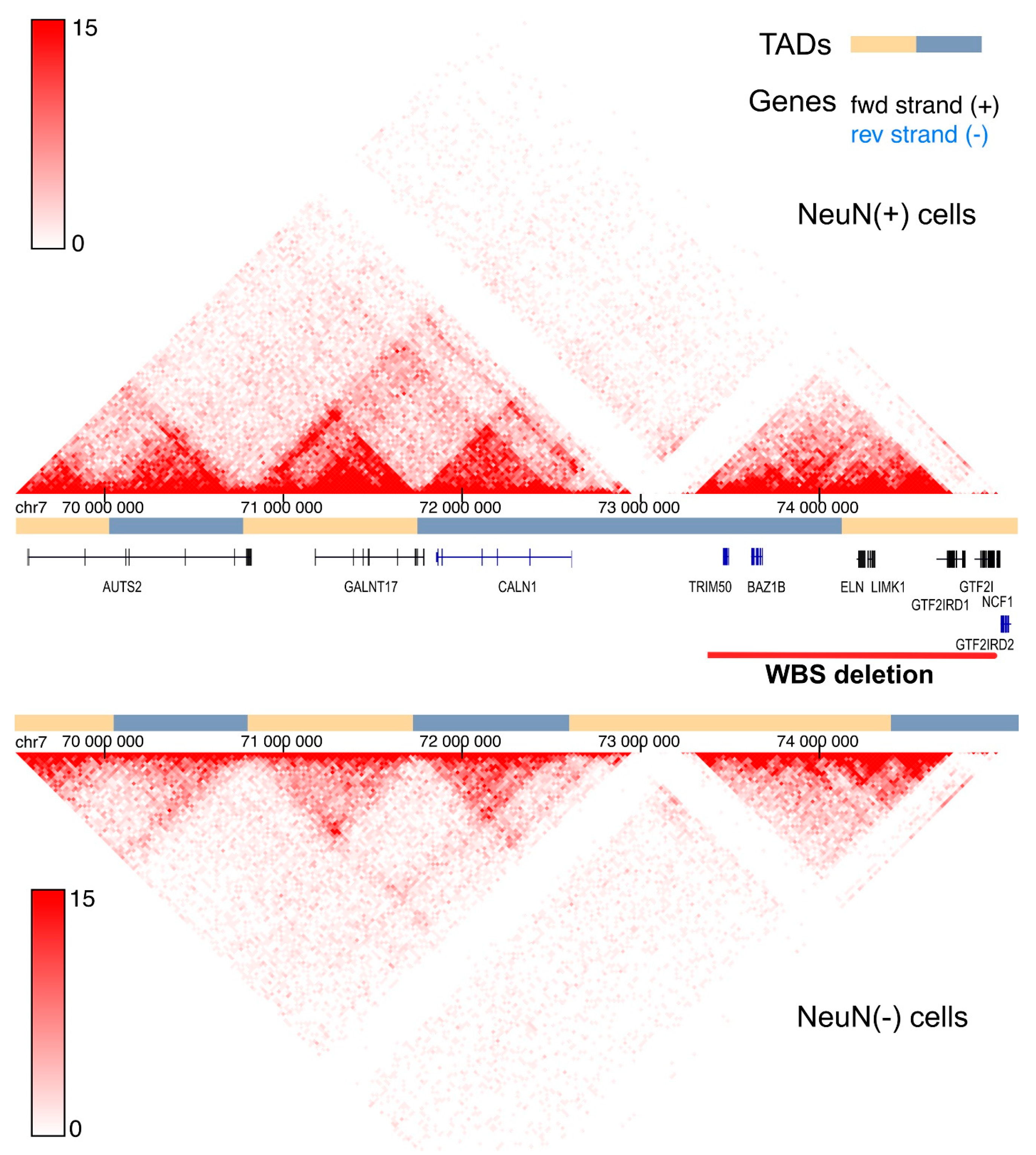
Subsequent studies employing 3D genome analysis directly corroborated these initial observations. Using 4C-seq on lymphoblastoid cells from WBS patients versus healthy donors, interactions between the WBSCR locus and its flanking regions were dissected [77]. The interactions of WBSCR with genes flanking WBSCR on both sides were detected. Genes on the telomeric side (HIP1, POR and MDH2) primarily interacted with the region that included ELN, LIMK1, EIF4H and CLIP2 genes. The closest gene at the telomeric side (HIP1) of the WBSCR was located 1 Mb away from the commonly deleted region (Figure 1). Genes from centromeric flank (ASL, KCTD7 and ZNF107) which are located farther from the WBSCR as compared to HIP1, POR and MDH2 showed weaker interaction with the WBSCR locus. In cells from WBS patients the interactions between WBSCR and other regions were diminished. Interestingly, AUTS2 and CALN1 were identified as interacting with both centromeric and telomeric genes [77]. AUTS2 expression was significantly reduced in WBS cells as compared to healthy donor cells but GALNT17 expression level was not assessed [77]. Importantly, GALNT17 gene is located between AUTS2 and CALN1, and these three genes span around 3 Mb in the human genome. Therefore, it is likely that these genes reside within distinct TADs (Figure 1 and Figure 2). Furthermore, transposon insertions in this region in dogs might affect TAD boundaries thus leading to perturbed expression of these genes, as exemplified by reduced GALNT17 expression as a result of TE insertion [17].
Figure 2. Schematic representation of the human Williams–Beuren Syndrome Critical Region (WBSCR) genomic locus integrated with the 3D Genome Browser [78] for Hi-C data exploration. The figure depicts the chromatin organization of the WBSCR genomic region in neuronal (NeuN(+)) and non-neuronal (NeuN(−)) cells. Hi-C data derived from cells of the left posterior superior temporal gyrus [79] are shown. The Hi-C interaction map displays a 6 Mb region on chromosome 7 (chr7: 69,500,000–75,500,000) at 25 kb resolution. Alternating yellow and blue bars demarcate predicted TADs.
Ijms 26 08549 g002
To further support the importance of the 3D genome organization for proper expression of the genes in the WBSCR, Engmann et al. demonstrated a chromatin loop-mediated interaction between the AUTS2 and CALN1 genes [80]. Cocaine administration in rats disrupted CTCF binding and looping in this region thus inducing the higher expression of both genes [80].
Analysis of copy number variants (CNVs) in a large cohort of children with developmental delay identified highly recurrent deletions of human-specific TAD boundaries [74]. One of these boundaries is located within the WBSCR locus, 200 kb upstream AUTS2 gene, between GALNT17 and AUTS2 [74]. CRISPR/Cas9-mediated deletion of this TAD boundary resulted in elevated AUTS2 expression. Unfortunately, expression levels of GALNT17 and genes typically deleted in WBS were not assessed in this study. It would be of great interest to determine if these genes are also affected by the deletion of human-specific TAD boundary in the WBSCR locus.
Relevant to this context, chromatin in neuronal cells forms more compact domains that constrain gene expression. Hi-C map of the WBSCR genomic region reveals stronger long-range interactions in neurons compared to other brain cells, with TADs also appearing more prominent (Figure 2). These interacting regions likely contain developmental transcription factors correlating with repressed status of many genes in neurons [79].
Taken together, recent findings utilizing 3D chromatin structure analysis provide crucial insights into the complex relationship between structural variants in the WBSCR locus and regulation of gene activity. This will have an impact on our understanding of the genetic basis of human neurobehavioral disorders. At the same time, a thorough and systematic analysis of the 3D genome regulation in the WBSCR—including the delineation of all TADs, loops and their boundaries across different cell lines, accompanied by the analysis of gene expression and histone modification—would be invaluable for understanding of many human pathologies, including ASD and drug addiction. Several symptoms of AUTS2 syndrome and features of Galnt17 KO mice are strikingly similar to those of WBS patients. These data unequivocally indicate that the phenotype of WBS patients, namely developmental delay, behavioral disturbances and ASD symptoms, at least in part are caused by dysregulated expression of AUTS2 and GALNT17.
In addition to the effect on human neurobehavioral disorders recent work from vonHoldt’s lab for the first time showed that 3D genome changes are involved in the evolution of social behavior in dogs. This links animal domestication and human neurobehavioral disorders through the WBSCR genomic locus. As stated above, TEs are enriched at TAD boundaries [75], and there are several TE in the canine WBSCR locus that are associated with social behavior towards humans [68,69]. In particular these TEs were identified in the first intron of GALNT17, the fifth intron of POM121 and the seventeenth intron of GTF2I [17]. The TE insertion in the intron of GTF2I represents an ancestral genotype of the gray wolf genome, and its presence is associated with diminished human-directed sociability [81].
Tandon et al. found that a SINE (Short-Interspersed Nucleotide Element) retrotransposon in intron 17 of canine GTF2I gene facilitates a chromatin loop formation, creating contact between introns 17 and 1 [81]. In the absence of the retrotransposon, the contact between the introns is not detected. At the molecular level the TE insertion does not affect the overall GTF2I transcript level but instead promotes the alternative splicing and increased usage of exon 18. This intronic interaction is likely mediated by the E2F1 transcription factor which binds to sequences within the TE in intron 17, facilitating contact with the first intron [81]. However, molecular details of this model require further experimental delineation. At the tissue level, transposon insertion in intron 17 induced differential expression of the extracellular matrix-related genes. The significance of these changes on social behavior remains unclear.
While the transposon insertion in GTF2I intron 17 is characteristic of the gray wolf genome, evolutionary selection in dogs appears to have favored absence of this TE. This provides the first evidence for chromatin structure influencing the evolution of social behavior in dogs [81]. As shown earlier by the same group, retrotransposons in the first intron of the GALNT17 gene decrease its expression and increase sociability in dogs [17]. It will be interesting to find out how the presence of this retrotransposon in GALNT17 gene affects the boundaries of TADs and loops and overall 3D chromatin structure.
Taken together, these results clearly indicated that the complex phenotype of neurodevelopmental disorders, such as WBS or WBDS, might arise not only from changes in copy number of gene but also from perturbed 3D genome organization in the locus. Therefore, investigating the relationship between structural variants of genes in the WBSCR locus and social behavior/domestication in mammalian species beyond dogs holds significant interest. We propose rats and rabbits as highly suitable model species for the evaluation of social behavior and generation of genome-edited animal models. These animal models will aim in the dissection of molecular basis of evolution of social behavior in animals and molecular mechanisms underlying human neurobehavioral disorders.

5. GALNT17 (WBSCR17) Gene

Recent data from mouse models and analysis of genetic basis of domestication of dogs placed the GALNT17 gene central to the regulation of social behavior in mammals. But our current knowledge on functions of Galnt17 is limited. In the following section we will summarize known cellular functions of Galnt17 as well as data from mouse models and genetic studies in humans that highlight putative functions of Galnt17 in different species.

5.1. Cellular Functions of Galnt17

WBSCR17 gene was identified as one of the transcripts in the WBSCR locus (7q11.23) with high expression in brain and heart [82]. Later, on the basis of homology to human and rat GALNT9 gene a novel gene named GALNT17 was cloned and it was found identical to WBSCR17 [83]. Detailed analysis of GALNT17 transcription revealed its expression in cortex and cerebellum [83], thus indicating the role of Galnt17 protein in the development and functioning of the CNS. Human GALNT17 gene is 580 kb long, contains 11 exons and encodes the single isoform of N-acetylgalactosaminyltransferase enzyme. Galnt family in human consists of 20 enzymes that catalyze transfer of N-acetylgalactosamine (GalNAc) from uridine-5′-diphospho-N-acetylgalactoseamine (UDP-GalNAc) to hydroxyl group of serine or threonine amino acid residues in proteins [84]. This type of post-translational modification is called O-glycosylation or mucin-type O-glycosylation since mucin family proteins contain multiple O-glycosylation sites and are the best studied O-glycosylated proteins. Galnt family enzymes play an important role in the regulation of multiple cellular processes in health and disease, including cancer [85]. Unlike many other Galnts, cellular functions and properties of Galnt17 are barely known.
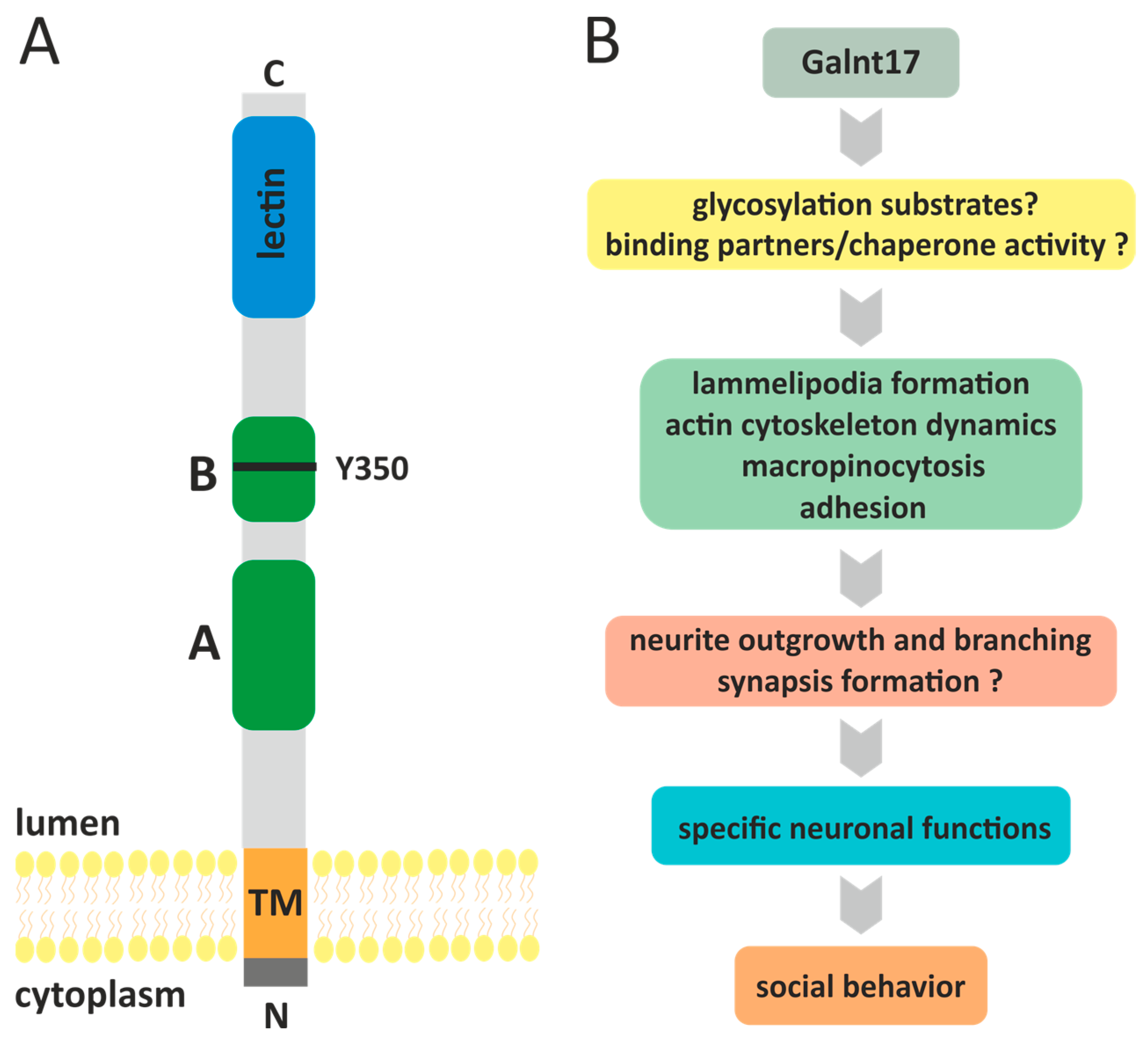
Human Galnt17 is a type II transmembrane protein of 598 amino acids. Galnt17 is composed of a short N-terminal cytoplasmic tail of only 7 aa long, transmembrane domain of 20 aa and extracellular domain that includes GT1 motif, Galnt motif and lectin domain (Figure 3) [83,86]. Upon transient expression in HEK293 cells, Galnt17 is glycosylated and, like most of the Galnt family proteins, is localized to the Golgi complex [83]. This is in line with the notion that mucin-type O-glycosylation is induced in the Golgi complex and continues as glycosylated protein is translocated from cis- to trans-Golgi compartment [84]. Despite the high sequence similarity with Galnt family members, initially it was discovered that, unlike Galnt1, recombinant Galnt17 (w/o cytoplasmic and transmembrane domains) does not glycosylate peptide substrates derived from MUC1a, MUC5AC and MUC7 proteins [83].
In further studies, low activity of Galnt17 towards peptide substrates from MUC7, MUC5AC-1, MUC5AC-2 proteins, but not towards MUC1 or MUC5AC-3, was shown [86], indicating that Galnt17 is catalytically active. It is noteworthy that Galnt17, similarly to several the so-called Y-subfamily Galnts, contains amino acid substitution of the conserved tryptophane residue for tyrosine in the GALNT motif (W->Y) [86]. More specifically, prototypical Galnt1 enzyme contains two invariant tryptophane residues in the GALNT motif within catalytic domain (W316 and W328). Substitution of W328 with any amino acid renders the enzyme completely inactive, while substitution of W316 with aromatic amino acid, such as tyrosine, significantly, but not completely, reduced the catalytic activity of the enzyme [87]. Tryptophane residue in the W328 position of Galnt1 is invariant in all members of the Galnt family, while Galnt enzymes of the Y-subfamily (Galnt8, Galnt9, Galnt17, Galnt18) carry W->Y substitution in the position similar to W316 in Galnt1 [86,87]. Therefore, this substitution (Y350 in Galnt17) is likely to explain the low catalytic activity of Galnt17. Nevertheless, analysis of O-glycosylation profile in cells with Galnt17 knockdown revealed several differentially glycosylated proteins, thus indicating the existence of protein targets for Galnt17-mediated O-glycosylation [86]. Currently these putative Galnt17 targets are unknown as it is not clear whether the changes in the glycosylation profile are direct or indirect consequences of Galnt17 activity. It would be interesting to determine if the reverse substitution (Y350W) would increase the catalytic activity of Galnt17.
At the present time it is not clear what the mechanisms of Galnt17 functioning in cells are and, in particular, what is the significance of its low catalytic activity. Probably, Galnt17 performs certain tasks that are not related to its catalytic activity. For example, it was demonstrated that another Y-subfamily protein Galnt18 can act as a chaperone for other Galnts thus regulating the homeostasis of endoplasmic reticulum [88,89]. Thus, except for several in vitro-tested peptides, Galnt17 substrates as well as protein-binding partners are unknown.
Most data regarding cellular functions of Galnt17 were obtained by Nakayama et al. [86]. siRNA-mediated knockdown of GALNT17 transcript in HEK293T cells cultured on fibronectin led to decrease in cell area, cell rounding, dissolution of lamellipodia, decrease in F-actin and paxillin (marker of focal contacts) staining [86]. These data allowed us to conclude that Galnt17 stimulates lamellipodia formation and promotes adhesive phenotype. Lamellipodia plays an important role in the regulation of cellular morphology, adhesion and motility [90]. Formation of lamellipodia and filopodia lies at the basis of neurite outgrowth, axon guidance and branching thus allowing for generation of novel synaptic contacts [91,92]. It is interesting to note that, similarly to Galnt17, Auts2 protein regulates formation of lamellipodia and filopodia, cell motility and neurite outgrowth [93]. Cellular functions of these two proteins seem to be tightly connected to the development of contact between neurons.
Accordingly, it might be speculated that cognitive and behavioral disturbances in model animals with Galnt17 KO and in patients with WBS are the consequences of impaired formation of neuronal contacts due to Galnt17 dysfunction. However, the exact role of Galnt17 in the regulation of actin cytoskeleton dynamics in neurons and the contribution of Galnt17 KO/knockdown to the development of the behavioral phenotypes are not known. It should be noted that so far, the effects of Galnt17 knockdown have not been evaluated in neuronal cell lines. It would be interesting to find out how Galnt17 knockdown affects actin cytoskeleton and neurite outgrowth in neuronal cell lines or in primary neurons. In support of the Galnt17 role in the regulation of neurite formation, Nakayama et al. have shown that Galnt17 might be involved in axonal outgrowth in zebrafish brain [94] and, in Galnt17 KO mice, abnormal development of axons and dendrites in Purkinje cells in cerebellum is observed [65].
Besides lamellipodia formation, Galnt17 is involved in the regulation of endocytosis. In one of the pioneer works, GALNT17 gene was identified as a candidate gene that regulates endocytosis, cell proliferation and N-glycan branching [95]. Indeed, it was later shown that Galnt17 negatively regulates micropinocytosis and upon Galnt17 knockdown cells accumulated enlarged macropinocytic vesicles, although Galnt17 is not likely to affect clathrin/caveolin-mediated endocytosis [86]. High concentration of N-acetylglucoseamine (GlcNAc) increases GALNT17 gene expression [86,95], that in turn can suppress micropinocytosis via Galnt17-mediated O-glycosylation of unknown substrates. The relationship between GlcNAc-induced GALNT17 expression and regulation of micropinocytosis warrants further studies.

5.2. Galnt17 Knockout Mouse

Most of the data on functions of Galnt17 at the level of organism were derived from the analysis of Galnt17 KO mice [65] that were generated by inserting the selective expression cassette into the first exon of Galnt17 gene thus disrupting its expression. Transcript-level analysis in the hippocampus and cerebellum of homozygous Galnt17 KO animals revealed a near-complete absence of the transcript. These data confirm the presence of only one transcript variant of this gene. Authors observed lethality of the homozygous KO mice at the C57BL/6J genetic background, but not at the hybrid C57BL/6J X C3H/He background. Reason for the observed lethality requires further investigation.
It turned out that many of the phenotype features in mice with reciprocal translocation with breakage site between Auts2 and Galnt17 genes [62] are recapitulated in Galnt17 KO mice [65]. Developmental delay, particularly in the early neonatal period (lower body weight, delayed eye opening) was one of the prominent phenotypic features of the homozygous Galnt17 knockout mice. Heterozygous knockout animals also exhibit developmental delay in the early neonatal period, which is compensated for with age. Homozygous knockout animals exhibit motor coordination problems, decreased social and exploratory activity while maintaining normal memory and learning ability, and abnormalities in the structure of the cerebellar vermis.
At the molecular level, RNA-seq identified many differentially expressed genes in the cerebellum of knockout animals; in particular, the largest number of downregulated genes belonged to the functional categories/terms that included neuronal differentiation, nervous system development, axon guidance, synaptic organization, cholinergic signaling and heparan sulfate synthesis that is required for axon guidance [65]. Downregulation of Mid1 and Folr1 gene might explain vermis development anomalies in the KO animals. Upregulated pathways included apoptotic signaling and integrin biosynthesis. Accordingly, TUNEL assay revealed increased apoptotic granular cells in vermis [65].
Reduced intensity of mucin-type O-glycosylated protein bands on lectin-probed blots of cerebellar protein extracts from Galnt17−/− animals clearly indicate that there are direct or indirect Galnt17 glycosylation targets in cells. Despite some efforts these proteins have not been identified so far.
Summarizing, GALNT17 gene is not deleted in WBS patients and its role in the development of WBS is not clear. On the other hand, observed behavioral and social phenotypes of the Galnt17 KO mice at least in part are similar to those seen in WBS and AUTS2 syndrome patients. These data imply that dysregulation of GALNT17 gene expression might contribute to the development of symptoms of these disorders. In support of this idea, it was shown that mouse translocation that affect Auts2 and Galnt17 genes leads to the significant downregulation of their transcripts. Therefore, one might propose that in WBS or WBDS patients, as a result of large chromosomal reengagements, the boundaries of chromatin loops and/or TADs are dramatically changed thus leading to abnormal downregulation of GALNT17/AUTS2 transcriptional activity. This hypothesis is supported by the evidence of AUTS2 downregulation in WBS patients [76], but unfortunately none of the previous studies have addressed the expression level of GALNT17 mRNA [74,76,77,80].

5.3. Galnt17, Human Diseases and Phenotypic Traits in Animals

Genetic studies allowed us to identify several SNPs that link GALNT17 with human diseases or phenotypic traits in animals. Large-scale GWAS identified novel SNP rs9638616:T in intron of GALNT17 gene in Asian populations that was associated with development of PD [64,96]. This SNP is associated with white matter tract and functional connectivity dysfunction in the supplementary motor area in cortex [97] and is located near miRNA genes in GALNT17 gene intron. Therefore, it is not clear whether the increased risk of PD development is associated with dysregulation of GALNT17 gene or miRNAs. Another intronic SNP rs17058752 in GALNT17 gene is associated with the development of age-related cataract in the Korean population [98]. Finally, in human, GALNT17 gene is differentially methylated in breast cancer and is included in seven gene signatures that allow us to distinguish between low- and high-risk breast cancers [99]. Significance of this finding is not clear since normally GALNT17 is not expressed in the mammary gland. Further studies are required to elucidate the role of GALNT17 methylation/expression in breast cancer.
In cows, SNP in GALNT17 gene was identified as associated with milk oligosaccharides synthesis [100] which has a clear connection to enzymatic function of Galnt17. Expression of GALNT17 gene is downregulated in the endometrium of pregnant cows, but significance of this observation is unknown [101]. In addition, Galnt17 protein level is increased in serum of rats 2 weeks after traumatic brain injury [102]. These data suggest that Galnt17 might have protective/regenerative role in the CNS. Based on the previous finding it is tempting to speculate that Galnt17 is increased in order to promote axon outgrowth, guidance and formation of synaptic contacts to follow the injury, but on the other hand the increase in Galnt17 might reflect the release of the protein from injured brain cells. Both hypotheses require further experimental confirmation.
Finally, circular RNA circ_WBSCR17 (circ_0080425, located in the fifth exon of GALNT17) is significantly upregulated in a mouse model of a diabetic nephropathy and functions as a pathogenic circular RNA acting as a molecular sponge for miR-185-5p, preventing the microRNA from binding to its target SOX6. As a result, SOX6 is overexpressed to promote inflammatory responses and fibrosis in kidney cells by inhibiting cell proliferation and promoting apoptosis [103]. Molecular mechanism of circ_WBSCR17 should be clarified in further studies.
To briefly summarize, Gant17 is an emerging player in the field of social behavior in animals and neurodevelopmental disorders in humans. Preliminary observations from GWAS and other genetic studies indicate that GALNT17 gene could be associated with CNS diseases in humans and with phenotypic traits in animals. These data further emphasize the importance of dissecting molecular mechanisms of both GALNT17 gene regulation and Galnt17 protein activity in cells. In mouse Galnt17 knockout leads to behavioral abnormalities and symptoms of autism spectrum. At the molecular level functions of Galnt17 are far from being understood and the relationship between ASD phenotype and the loss of Galnt17 are not entirely clear. Galnt17 regulates lamellipodia formation, cell adhesion and micropinocytosis in HEK293T cells but its glycosylation substrates and/or protein binding partners are not known. A plethora of genes are differentially expressed in cerebellum of Galnt17 KO mice and several proteins are differentially glycosylated but exact functions of Galnt17 in neuronal cells have not been investigated in detail so far. Recent advances in the dissection of genetic basis of dog’s domestication place Galnt17 central to social behaviors in dogs. Taken together the recent data suggest that GALNT17 is an attractive target for genome editing for the purpose of changing social behavior in animals. Besides that, it is of a great interest to dissect the molecular basis of Galnt17 functions in cells, and, in particular, in CNS.

6. Conclusions

Domestication is a multifaceted process, with its genetic foundations often being polygenic and not entirely clear. The primary indicator of successful domestication is the animal’s friendliness towards humans. The quest to identify the genetic basis of this behavioral trait in animals led to the discovery of the WBSCR locus, a deletion of which in humans causes WBS. However, deleting an entire locus in humans not only alters the behavioral phenotype but also causes a range of severe physical health issues. Therefore, finding individual genes that are crucial for domestication without affecting physical health is a highly promising and urgent task. One such candidate gene is the GALNT17. In particular, it has been found that certain polymorphisms in this gene differ between domestic dogs and wolves. The molecular pathways in which the GALNT17 gene product participates are not well-known, but this gene may be considered as a potential target for genome editing in domesticated animals to achieve the desired phenotype. A body of experimental data suggest that copy number changes and structural variants in the WBSCR affect the gene regulations directly as well as through alteration of 3D genome organization thus affecting both domestication of animals and development of neurobehavioral disorders in human (Figure 4). We emphasize the importance of further research aimed at unraveling the complexity of the relationships between genetic variants in the WBSCR and phenotypic traits in different animal species and in human. Advanced 3D genome analysis techniques and genome editing technologies are the methods of choice for solving these tasks.

Author Contributions

Conceptualization, M.V.S., T.T.G., G.Y.K. and Y.Y.S.; writing—original draft preparation, M.V.S., O.I.S., T.T.G., D.V.P., D.E.V. and Y.Y.S.; writing—review and editing, M.V.S., O.I.S., O.G.M., P.G.G., G.Y.K. and Y.Y.S.; visualization, M.V.S. and O.G.M.; funding acquisition, P.G.G. and G.Y.K. All authors have read and agreed to the published version of the manuscript.

Funding

This study was supported by the Russian Science Foundation, grant number 19-74-30026-P, https://rscf.ru/en/project/23-74-33005/ (accessed on 28 July 2025) (section “3D genome alterations in WBSCR region and social behavior”) and by the Ministry of Science and Higher Education of the Russian Federation (Government Assignment No. 075-00503-25-01).

Institutional Review Board Statement

Not applicable.

Informed Consent Statement

Not applicable.

Data Availability Statement

Data are contained within the article.

Acknowledgments

The authors would like to honor the memory of Valery I. Glazko, who made a significant contribution to this article but, unfortunately, passed away before its completion.

Conflicts of Interest

The authors declare no conflicts of interest.

Abbreviations

The following abbreviations are used in this manuscript:
ASDAutism Spectrum Disorder
CNSCentral nervous system
CNVCopy Number Variant
GalNAcN-acetylgalactosamine
GlcNAcN-acetylglucoseamine
GWASGenome-wide association studies
iPSCinduced pluripotent stem cell
KOknockout
LCRlow-copy repeat elements
NCCneural crest cell
PDParkinson disease
SINEShort-Interspersed Nucleotide Element
SNPsingle-nucleotide polymorphism
SVstructural variant
TADtopology associated domain
TEtransposon element
UDP-GalNAcuridine-5′-diphospho-N-acetylgalactoseamine
WBDSWilliams–Beuren region duplication syndrome
WBSWilliams–Beuren syndrome
WBSCRWilliams–Beuren syndrome control region
WSTFWilliams syndrome transcription factor

References

  1. Darwin, C. The Variation of Animals and Plants Under Domestication; John Murray: London, UK, 1868; Volume 2. [Google Scholar]
  2. Brown, T.A.; Jones, M.K.; Powell, W.; Allaby, R.G. The Complex Origins of Domesticated Crops in the Fertile Crescent. Trends Ecol. Evol. 2009, 24, 103–109. [Google Scholar] [CrossRef]
  3. Leach, H.M. Human Domestication Reconsidered. Curr. Anthropol. 2003, 44, 349–368. [Google Scholar] [CrossRef]
  4. Trut, L.N.; Plyusnina, I.Z.; Oskina, I.N. An Experiment on Fox Domestication and Debatable Issues of Evolution of the Dog. Russ. J. Genet. 2004, 40, 644–655. [Google Scholar] [CrossRef]
  5. Trut, L.; Oskina, I.; Kharlamova, A. Animal Evolution during Domestication: The Domesticated Fox as a Model. BioEssays News Rev. Mol. Cell. Dev. Biol. 2009, 31, 349–360. [Google Scholar] [CrossRef] [PubMed]
  6. Gilbert, S.F.; Opitz, J.M.; Raff, R.A. Resynthesizing Evolutionary and Developmental Biology. Dev. Biol. 1996, 173, 357–372. [Google Scholar] [CrossRef]
  7. Neural Crest Cells: Evolution, Development and Disease; Trainor, P., Ed.; Academic Press: Cambridge, MA, USA, 2013; ISBN 978-0-12-401730-6. [Google Scholar]
  8. Vega-Lopez, G.A.; Cerrizuela, S.; Aybar, M.J. Trunk Neural Crest Cells: Formation, Migration and Beyond. Int. J. Dev. Biol. 2017, 61, 5–15. [Google Scholar] [CrossRef]
  9. Wilkins, A.S.; Wrangham, R.W.; Fitch, W.T. The “Domestication Syndrome” in Mammals: A Unified Explanation Based on Neural Crest Cell Behavior and Genetics. Genetics 2014, 197, 795–808. [Google Scholar] [CrossRef] [PubMed]
  10. Rubio, A.O.; Summers, K. Neural Crest Cell Genes and the Domestication Syndrome: A Comparative Analysis of Selection. PLoS ONE 2022, 17, e0263830. [Google Scholar] [CrossRef] [PubMed]
  11. Gleeson, B.T.; Wilson, L.A.B. Shared Reproductive Disruption, Not Neural Crest or Tameness, Explains the Domestication Syndrome. Proc. R. Soc. B Biol. Sci. 2023, 290, 20222464. [Google Scholar] [CrossRef]
  12. Carneiro, M.; Piorno, V.; Rubin, C.-J.; Alves, J.M.; Ferrand, N.; Alves, P.C.; Andersson, L. Candidate Genes Underlying Heritable Differences in Reproductive Seasonality between Wild and Domestic Rabbits. Anim. Genet. 2015, 46, 418–425. [Google Scholar] [CrossRef]
  13. Bar-On, Y.M.; Phillips, R.; Milo, R. The Biomass Distribution on Earth. Proc. Natl. Acad. Sci. USA 2018, 115, 6506–6511. [Google Scholar] [CrossRef]
  14. Zeder, M.A. Core Questions in Domestication Research. Proc. Natl. Acad. Sci. USA 2015, 112, 3191–3198. [Google Scholar] [CrossRef]
  15. Wang, G.; Zhai, W.; Yang, H.; Fan, R.; Cao, X.; Zhong, L.; Wang, L.; Liu, F.; Wu, H.; Cheng, L.; et al. The Genomics of Selection in Dogs and the Parallel Evolution between Dogs and Humans. Nat. Commun. 2013, 4, 1860. [Google Scholar] [CrossRef]
  16. Belyaev, D.K. Destabilizing Selection as a Factor in Domestication. J. Hered. 1979, 70, 301–308. [Google Scholar] [CrossRef] [PubMed]
  17. vonHoldt, B.M.; Ji, S.S.; Aardema, M.L.; Stahler, D.R.; Udell, M.A.R.; Sinsheimer, J.S. Activity of Genes with Functions in Human Williams-Beuren Syndrome Is Impacted by Mobile Element Insertions in the Gray Wolf Genome. Genome Biol. Evol. 2018, 10, 1546–1553. [Google Scholar] [CrossRef]
  18. Kozel, B.A.; Barak, B.; Kim, C.A.; Mervis, C.B.; Osborne, L.R.; Porter, M.; Pober, B.R. Williams Syndrome. Nat. Rev. Dis. Primer 2021, 7, 42. [Google Scholar] [CrossRef] [PubMed]
  19. Hou, Y.; Qi, F.; Bai, X.; Ren, T.; Shen, X.; Chu, Q.; Zhang, X.; Lu, X. Genome-Wide Analysis Reveals Molecular Convergence Underlying Domestication in 7 Bird and Mammals. BMC Genom. 2020, 21, 204. [Google Scholar] [CrossRef] [PubMed]
  20. Sinha, S.; Jones, B.M.; Traniello, I.M.; Bukhari, S.A.; Halfon, M.S.; Hofmann, H.A.; Huang, S.; Katz, P.S.; Keagy, J.; Lynch, V.J.; et al. Behavior-Related Gene Regulatory Networks: A New Level of Organization in the Brain. Proc. Natl. Acad. Sci. USA 2020, 117, 23270–23279. [Google Scholar] [CrossRef]
  21. López-Tobón, A.; Trattaro, S.; Testa, G. The Sociability Spectrum: Evidence from Reciprocal Genetic Copy Number Variations. Mol. Autism 2020, 11, 50. [Google Scholar] [CrossRef]
  22. Genovese, A.; Butler, M.G. The Autism Spectrum: Behavioral, Psychiatric and Genetic Associations. Genes 2023, 14, 677. [Google Scholar] [CrossRef]
  23. Madlon-Kay, S.; Montague, M.J.; Brent, L.J.N.; Ellis, S.; Zhong, B.; Snyder-Mackler, N.; Horvath, J.E.; Skene, J.H.P.; Platt, M.L. Weak Effects of Common Genetic Variation in Oxytocin and Vasopressin Receptor Genes on Rhesus Macaque Social Behavior. Am. J. Primatol. 2018, 80, e22873. [Google Scholar] [CrossRef] [PubMed]
  24. Ike, K.G.O.; Lamers, S.J.C.; Kaim, S.; de Boer, S.F.; Buwalda, B.; Billeter, J.-C.; Kas, M.J.H. The Human Neuropsychiatric Risk Gene Drd2 Is Necessary for Social Functioning across Evolutionary Distant Species. Mol. Psychiatry 2024, 29, 518–528. [Google Scholar] [CrossRef]
  25. Gao, W.-J.; Mack, N.R. From Hyposociability to Hypersociability—The Effects of PSD-95 Deficiency on the Dysfunctional Development of Social Behavior. Front. Behav. Neurosci. 2021, 15, 618397. [Google Scholar] [CrossRef]
  26. Feyder, M.; Karlsson, R.-M.; Mathur, P.; Lyman, M.; Bock, R.; Momenan, R.; Munasinghe, J.; Scattoni, M.L.; Ihne, J.; Camp, M.; et al. Association of Mouse Dlg4 (PSD-95) Gene Deletion and Human DLG4 Gene Variation With Phenotypes Relevant to Autism Spectrum Disorders and Williams’ Syndrome. Am. J. Psychiatry 2010, 167, 1508–1517. [Google Scholar] [CrossRef]
  27. Hörnberg, H.; Pérez-Garci, E.; Schreiner, D.; Hatstatt-Burklé, L.; Magara, F.; Baudouin, S.; Matter, A.; Nacro, K.; Pecho-Vrieseling, E.; Scheiffele, P. Rescue of Oxytocin Response and Social Behaviour in a Mouse Model of Autism. Nature 2020, 584, 252–256. [Google Scholar] [CrossRef]
  28. Leblond, C.S.; Nava, C.; Polge, A.; Gauthier, J.; Huguet, G.; Lumbroso, S.; Giuliano, F.; Stordeur, C.; Depienne, C.; Mouzat, K.; et al. Meta-Analysis of SHANK Mutations in Autism Spectrum Disorders: A Gradient of Severity in Cognitive Impairments. PLoS Genet. 2014, 10, e1004580. [Google Scholar] [CrossRef]
  29. Miles, J.H. Autism Spectrum Disorders—A Genetics Review. Genet. Med. 2011, 13, 278–294. [Google Scholar] [CrossRef]
  30. Varghese, M.; Keshav, N.; Jacot-Descombes, S.; Warda, T.; Wicinski, B.; Dickstein, D.L.; Harony-Nicolas, H.; De Rubeis, S.; Drapeau, E.; Buxbaum, J.D.; et al. Autism Spectrum Disorder: Neuropathology and Animal Models. Acta Neuropathol. 2017, 134, 537–566. [Google Scholar] [CrossRef] [PubMed]
  31. Peoples, R.; Franke, Y.; Wang, Y.-K.; Pérez-Jurado, L.; Paperna, T.; Cisco, M.; Francke, U. A Physical Map, Including a BAC/PAC Clone Contig, of the Williams-Beuren Syndrome–Deletion Region at 7q11.23. Am. J. Hum. Genet. 2000, 66, 47–68. [Google Scholar] [CrossRef] [PubMed]
  32. Serrano-Juárez, C.A.; Prieto-Corona, B.; Rodríguez-Camacho, M.; Sandoval-Lira, L.; Villalva-Sánchez, Á.F.; Yáñez-Téllez, M.G.; López, M.F.R. Neuropsychological Genotype-Phenotype in Patients with Williams Syndrome with Atypical Deletions: A Systematic Review. Neuropsychol. Rev. 2023, 33, 891–911. [Google Scholar] [CrossRef]
  33. Miezah, D.; Porter, M.; Rossi, A.; Kazzi, C.; Batchelor, J.; Reeve, J. Cognitive Profile of Young Children with Williams Syndrome. J. Intellect. Disabil. Res. JIDR 2021, 65, 784–794. [Google Scholar] [CrossRef]
  34. Bayés, M.; Magano, L.F.; Rivera, N.; Flores, R.; Pérez Jurado, L.A. Mutational Mechanisms of Williams-Beuren Syndrome Deletions. Am. J. Hum. Genet. 2003, 73, 131–151. [Google Scholar] [CrossRef]
  35. Etokebe, G.E.; Axelsson, S.; Svaerd, N.H.; Storhaug, K.; Dembić, Z. Detection of Hemizygous Chromosomal Copy Number Variants in Williams-Beuren Syndrome (WBS) by Duplex Quantitative PCR Array: An Unusual Type of WBS Genetic Defect. Int. J. Biomed. Sci. IJBS 2008, 4, 161–170. [Google Scholar]
  36. Ferrero, G.B.; Howald, C.; Micale, L.; Biamino, E.; Augello, B.; Fusco, C.; Turturo, M.G.; Forzano, S.; Reymond, A.; Merla, G. An Atypical 7q11.23 Deletion in a Normal IQ Williams–Beuren Syndrome Patient. Eur. J. Hum. Genet. 2010, 18, 33–38. [Google Scholar] [CrossRef] [PubMed]
  37. Antonell, A.; Del Campo, M.; Magano, L.F.; Kaufmann, L.; de la Iglesia, J.M.; Gallastegui, F.; Flores, R.; Schweigmann, U.; Fauth, C.; Kotzot, D.; et al. Partial 7q11.23 Deletions Further Implicate GTF2I and GTF2IRD1 as the Main Genes Responsible for the Williams-Beuren Syndrome Neurocognitive Profile. J. Med. Genet. 2010, 47, 312–320. [Google Scholar] [CrossRef]
  38. Campbell, L.E.; Daly, E.; Toal, F.; Stevens, A.; Azuma, R.; Karmiloff-Smith, A.; Murphy, D.G.M.; Murphy, K.C. Brain Structural Differences Associated with the Behavioural Phenotype in Children with Williams Syndrome. Brain Res. 2009, 1258, 96–107. [Google Scholar] [CrossRef]
  39. Sanders, S.J.; Ercan-Sencicek, A.G.; Hus, V.; Luo, R.; Murtha, M.T.; Moreno-De-Luca, D.; Chu, S.H.; Moreau, M.P.; Gupta, A.R.; Thomson, S.A.; et al. Multiple Recurrent de Novo Copy Number Variations (CNVs), Including Duplications of the 7q11.23 Williams-Beuren Syndrome Region, Are Strongly Associated with Autism. Neuron 2011, 70, 863–885. [Google Scholar] [CrossRef]
  40. Van der Aa, N.; Rooms, L.; Vandeweyer, G.; van den Ende, J.; Reyniers, E.; Fichera, M.; Romano, C.; Delle Chiaie, B.; Mortier, G.; Menten, B.; et al. Fourteen New Cases Contribute to the Characterization of the 7q11.23 Microduplication Syndrome. Eur. J. Med. Genet. 2009, 52, 94–100. [Google Scholar] [CrossRef]
  41. López-Tobón, A.; Shyti, R.; Villa, C.E.; Cheroni, C.; Fuentes-Bravo, P.; Trattaro, S.; Caporale, N.; Troglio, F.; Tenderini, E.; Mihailovich, M.; et al. GTF2I Dosage Regulates Neuronal Differentiation and Social Behavior in 7q11.23 Neurodevelopmental Disorders. Sci. Adv. 2023, 9, eadh2726. [Google Scholar] [CrossRef] [PubMed]
  42. Beunders, G.; van de Kamp, J.M.; Veenhoven, R.H.; van Hagen, J.M.; Nieuwint, A.W.M.; Sistermans, E.A. A Triplication of the Williams-Beuren Syndrome Region in a Patient with Mental Retardation, a Severe Expressive Language Delay, Behavioural Problems and Dysmorphisms. J. Med. Genet. 2010, 47, 271–275. [Google Scholar] [CrossRef] [PubMed]
  43. Segura-Puimedon, M.; Sahún, I.; Velot, E.; Dubus, P.; Borralleras, C.; Rodrigues, A.J.; Valero, M.C.; Valverde, O.; Sousa, N.; Herault, Y.; et al. Heterozygous Deletion of the Williams–Beuren Syndrome Critical Interval in Mice Recapitulates Most Features of the Human Disorder. Hum. Mol. Genet. 2014, 23, 6481–6494. [Google Scholar] [CrossRef]
  44. Morris, C.A.; Mervis, C.B.; Hobart, H.H.; Gregg, R.G.; Bertrand, J.; Ensing, G.J.; Sommer, A.; Moore, C.A.; Hopkin, R.J.; Spallone, P.A.; et al. GTF2I Hemizygosity Implicated in Mental Retardation in Williams Syndrome: Genotype–Phenotype Analysis of Five Families with Deletions in the Williams Syndrome Region. Am. J. Med. Genet. A. 2003, 123A, 45–59. [Google Scholar] [CrossRef]
  45. van Hagen, J.M.; van der Geest, J.N.; van der Giessen, R.S.; Lagers-van Haselen, G.C.; Eussen, H.J.F.M.M.; Gille, J.J.P.; Govaerts, L.C.P.; Wouters, C.H.; de Coo, I.F.M.; Hoogenraad, C.C.; et al. Contribution of CYLN2 and GTF2IRD1 to Neurological and Cognitive Symptoms in Williams Syndrome. Neurobiol. Dis. 2007, 26, 112–124. [Google Scholar] [CrossRef]
  46. Makeyev, A.V.; Bayarsaihan, D. Molecular Basis of Williams-Beuren Syndrome: TFII-I Regulated Targets Involved in Craniofacial Development. Cleft Palate-Craniofacial J. Off. Publ. Am. Cleft Palate-Craniofacial Assoc. 2011, 48, 109–116. [Google Scholar] [CrossRef]
  47. Lopatina, O.L.; Komleva, Y.K.; Gorina, Y.V.; Olovyannikova, R.Y.; Trufanova, L.V.; Hashimoto, T.; Takahashi, T.; Kikuchi, M.; Minabe, Y.; Higashida, H.; et al. Oxytocin and Excitation/Inhibition Balance in Social Recognition. Neuropeptides 2018, 72, 1–11. [Google Scholar] [CrossRef]
  48. Sohal, V.S.; Rubenstein, J.L.R. Excitation-Inhibition Balance as a Framework for Investigating Mechanisms in Neuropsychiatric Disorders. Mol. Psychiatry 2019, 24, 1248–1257. [Google Scholar] [CrossRef] [PubMed]
  49. Enkhmandakh, B.; Bitchevaia, N.; Ruddle, F.; Bayarsaihan, D. The Early Embryonic Expression of TFII-I during Mouse Preimplantation Development. Gene Expr. Patterns 2004, 4, 25–28. [Google Scholar] [CrossRef]
  50. Roy, A.L. Biochemistry and Biology of the Inducible Multifunctional Transcription Factor TFII-I: 10years Later. Gene 2012, 492, 32–41. [Google Scholar] [CrossRef] [PubMed]
  51. Makeyev, A.V.; Bayarsaihan, D. New TFII-I Family Target Genes Involved in Embryonic Development. Biochem. Biophys. Res. Commun. 2009, 386, 554–558. [Google Scholar] [CrossRef] [PubMed]
  52. Chimge, N.-O.; Makeyev, A.V.; Ruddle, F.H.; Bayarsaihan, D. Identification of the TFII-I Family Target Genes in the Vertebrate Genome. Proc. Natl. Acad. Sci. USA 2008, 105, 9006–9010. [Google Scholar] [CrossRef]
  53. Nir Sade, A.; Levy, G.; Schokoroy Trangle, S.; Elad Sfadia, G.; Bar, E.; Ophir, O.; Fischer, I.; Rokach, M.; Atzmon, A.; Parnas, H.; et al. Neuronal Gtf2i Deletion Alters Mitochondrial and Autophagic Properties. Commun. Biol. 2023, 6, 1269. [Google Scholar] [CrossRef] [PubMed]
  54. Mervis, C.B.; Dida, J.; Lam, E.; Crawford-Zelli, N.A.; Young, E.J.; Henderson, D.R.; Onay, T.; Morris, C.A.; Woodruff-Borden, J.; Yeomans, J.; et al. Duplication of GTF2I Results in Separation Anxiety in Mice and Humans. Am. J. Hum. Genet. 2012, 90, 1064–1070. [Google Scholar] [CrossRef]
  55. Twite, M.D.; Stenquist, S.; Ing, R.J. Williams Syndrome. Pediatr. Anesth. 2019, 29, 483–490. [Google Scholar] [CrossRef]
  56. Li, D.Y.; Faury, G.; Taylor, D.G.; Davis, E.C.; Boyle, W.A.; Mecham, R.P.; Stenzel, P.; Boak, B.; Keating, M.T. Novel Arterial Pathology in Mice and Humans Hemizygous for Elastin. J. Clin. Invest. 1998, 102, 1783–1787. [Google Scholar] [CrossRef]
  57. Zanella, M.; Vitriolo, A.; Andirko, A.; Martins, P.T.; Sturm, S.; O’Rourke, T.; Laugsch, M.; Malerba, N.; Skaros, A.; Trattaro, S.; et al. Dosage Analysis of the 7q11.23 Williams Region Identifies BAZ1B as a Major Human Gene Patterning the Modern Human Face and Underlying Self-Domestication. Sci. Adv. 2019, 5, eaaw7908. [Google Scholar] [CrossRef]
  58. Barnett, C.; Yazgan, O.; Kuo, H.-C.; Malakar, S.; Thomas, T.; Fitzgerald, A.; Harbour, W.; Henry, J.J.; Krebs, J.E. Williams Syndrome Transcription Factor Is Critical for Neural Crest Cell Function in Xenopus Laevis. Mech. Dev. 2012, 129, 324–338. [Google Scholar] [CrossRef]
  59. Gregory, M.D.; Mervis, C.B.; Elliott, M.L.; Kippenhan, J.S.; Nash, T.; Czarapata, J.B.; Prabhakaran, R.; Roe, K.; Eisenberg, D.P.; Kohn, P.D.; et al. Williams Syndrome Hemideletion and LIMK1 Variation Both Affect Dorsal Stream Functional Connectivity. Brain 2019, 142, 3963–3974. [Google Scholar] [CrossRef]
  60. Todorovski, Z.; Asrar, S.; Liu, J.; Saw, N.M.N.; Joshi, K.; Cortez, M.A.; Snead, O.C.; Xie, W.; Jia, Z. LIMK1 Regulates Long-Term Memory and Synaptic Plasticity via the Transcriptional Factor CREB. Mol. Cell. Biol. 2015, 35, 1316–1328. [Google Scholar] [CrossRef]
  61. Biel, A.; Castanza, A.S.; Rutherford, R.; Fair, S.R.; Chifamba, L.; Wester, J.C.; Hester, M.E.; Hevner, R.F. AUTS2 Syndrome: Molecular Mechanisms and Model Systems. Front. Mol. Neurosci. 2022, 15, 858582. [Google Scholar] [CrossRef] [PubMed]
  62. Weisner, P.A.; Chen, C.-Y.; Sun, Y.; Yoo, J.; Kao, W.-C.; Zhang, H.; Baltz, E.T.; Troy, J.M.; Stubbs, L. A Mouse Mutation That Dysregulates Neighboring Galnt17 and Auts2 Genes Is Associated with Phenotypes Related to the Human AUTS2 Syndrome. G3 Genes Genomes Genet. 2019, 9, 3891–3906. [Google Scholar] [CrossRef] [PubMed]
  63. Hori, K.; Nagai, T.; Shan, W.; Sakamoto, A.; Abe, M.; Yamazaki, M.; Sakimura, K.; Yamada, K.; Hoshino, M. Heterozygous Disruption of Autism Susceptibility Candidate 2 Causes Impaired Emotional Control and Cognitive Memory. PLoS ONE 2015, 10, e0145979. [Google Scholar] [CrossRef]
  64. Foo, J.N.; Chew, E.G.Y.; Chung, S.J.; Peng, R.; Blauwendraat, C.; Nalls, M.A.; Mok, K.Y.; Satake, W.; Toda, T.; Chao, Y.; et al. Identification of Risk Loci for Parkinson Disease in Asians and Comparison of Risk Between Asians and Europeans: A Genome-Wide Association Study. JAMA Neurol. 2020, 77, 746–754. [Google Scholar] [CrossRef]
  65. Chen, C.-Y.; Seward, C.H.; Song, Y.; Inamdar, M.; Leddy, A.M.; Zhang, H.; Yoo, J.; Kao, W.-C.; Pawlowski, H.; Stubbs, L.J. Galnt17 Loss-of-Function Leads to Developmental Delay and Abnormal Coordination, Activity, and Social Interactions with Cerebellar Vermis Pathology. Dev. Biol. 2022, 490, 155–171. [Google Scholar] [CrossRef]
  66. Liu, M.; Chen, Y.; Sun, M.; Du, Y.; Bai, Y.; Lei, G.; Zhang, C.; Zhang, M.; Zhang, Y.; Xi, C.; et al. Auts2 Regulated Autism-like Behavior, Glucose Metabolism and Oxidative Stress in Mice. Exp. Neurol. 2023, 361, 114298. [Google Scholar] [CrossRef]
  67. Sanchez-Jimeno, C.; Blanco-Kelly, F.; López-Grondona, F.; Losada-Del Pozo, R.; Moreno, B.; Rodrigo-Moreno, M.; Martinez-Cayuelas, E.; Riveiro-Alvarez, R.; Fenollar-Cortés, M.; Ayuso, C.; et al. Attention Deficit Hyperactivity and Autism Spectrum Disorders as the Core Symptoms of AUTS2 Syndrome: Description of Five New Patients and Update of the Frequency of Manifestations and Genotype-Phenotype Correlation. Genes 2021, 12, 1360. [Google Scholar] [CrossRef]
  68. vonHoldt, B.M.; Pollinger, J.P.; Lohmueller, K.E.; Han, E.; Parker, H.G.; Quignon, P.; Degenhardt, J.D.; Boyko, A.R.; Earl, D.A.; Auton, A.; et al. Genome-Wide SNP and Haplotype Analyses Reveal a Rich History Underlying Dog Domestication. Nature 2010, 464, 898–902. [Google Scholar] [CrossRef]
  69. vonHoldt, B.M.; Shuldiner, E.; Koch, I.J.; Kartzinel, R.Y.; Hogan, A.; Brubaker, L.; Wanser, S.; Stahler, D.; Wynne, C.D.L.; Ostrander, E.A.; et al. Structural Variants in Genes Associated with Human Williams-Beuren Syndrome Underlie Stereotypical Hypersociability in Domestic Dogs. Sci. Adv. 2017, 3, e1700398. [Google Scholar] [CrossRef] [PubMed]
  70. Tandon, D.; Ressler, K.; Petticord, D.; Papa, A.; Jiranek, J.; Wilkinson, R.; Kartzinel, R.Y.; Ostrander, E.A.; Burney, N.; Borden, C.; et al. Homozygosity for Mobile Element Insertions Associated with WBSCR17 Could Predict Success in Assistance Dog Training Programs. Genes 2019, 10, 439. [Google Scholar] [CrossRef]
  71. Glazko, V.I.; Kosovsky, G.Y.; Blokhina, T.V.; Zhirkova, A.A.; Glazko, T.T. Socialization and Genetic Variability as a Driver of Domestication (by the Example of Dog Breeds). Selskokhozyaistvennaya Biol. Agric. Biol. 2021, 56, 292–303. [Google Scholar] [CrossRef]
  72. Gnanadesikan, G.E.; Tandon, D.; Bray, E.E.; Kennedy, B.S.; Tennenbaum, S.R.; MacLean, E.L.; vonHoldt, B.M. Transposons in the Williams–Beuren Syndrome Critical Region Are Associated with Social Behavior in Assistance Dogs. Behav. Genet. 2023, 54, 196–211. [Google Scholar] [CrossRef] [PubMed]
  73. Tiukacheva, E.A.; Ulianov, S.V.; Karpukhina, A.; Razin, S.V.; Vassetzky, Y. 3D Genome Alterations and Editing in Pathology. Mol. Ther. J. Am. Soc. Gene Ther. 2023, 31, 924–933. [Google Scholar] [CrossRef]
  74. Okhovat, M.; VanCampen, J.; Nevonen, K.A.; Harshman, L.; Li, W.; Layman, C.E.; Ward, S.; Herrera, J.; Wells, J.; Sheng, R.R.; et al. TAD Evolutionary and Functional Characterization Reveals Diversity in Mammalian TAD Boundary Properties and Function. Nat. Commun. 2023, 14, 8111. [Google Scholar] [CrossRef] [PubMed]
  75. Choudhary, M.N.K.; Quaid, K.; Xing, X.; Schmidt, H.; Wang, T. Widespread Contribution of Transposable Elements to the Rewiring of Mammalian 3D Genomes. Nat. Commun. 2023, 14, 634. [Google Scholar] [CrossRef]
  76. Merla, G.; Howald, C.; Henrichsen, C.N.; Lyle, R.; Wyss, C.; Zabot, M.-T.; Antonarakis, S.E.; Reymond, A. Submicroscopic Deletion in Patients with Williams-Beuren Syndrome Influences Expression Levels of the Nonhemizygous Flanking Genes. Am. J. Hum. Genet. 2006, 79, 332–341. [Google Scholar] [CrossRef] [PubMed]
  77. Gheldof, N.; Witwicki, R.M.; Migliavacca, E.; Leleu, M.; Didelot, G.; Harewood, L.; Rougemont, J.; Reymond, A. Structural Variation-Associated Expression Changes Are Paralleled by Chromatin Architecture Modifications. PLoS ONE 2013, 8, e79973. [Google Scholar] [CrossRef]
  78. Wang, Y.; Song, F.; Zhang, B.; Zhang, L.; Xu, J.; Kuang, D.; Li, D.; Choudhary, M.N.K.; Li, Y.; Hu, M.; et al. The 3D Genome Browser: A Web-Based Browser for Visualizing 3D Genome Organization and Long-Range Chromatin Interactions. Genome Biol. 2018, 19, 151. [Google Scholar] [CrossRef]
  79. Pletenev, I.A.; Bazarevich, M.; Zagirova, D.R.; Kononkova, A.D.; Cherkasov, A.V.; Efimova, O.I.; Tiukacheva, E.A.; Morozov, K.V.; Ulianov, K.A.; Komkov, D.; et al. Extensive Long-Range Polycomb Interactions and Weak Compartmentalization Are Hallmarks of Human Neuronal 3D Genome. Nucleic Acids Res. 2024, 52, 6234–6252. [Google Scholar] [CrossRef]
  80. Engmann, O.; Labonté, B.; Mitchell, A.; Bashtrykov, P.; Calipari, E.S.; Rosenbluh, C.; Loh, Y.-H.E.; Walker, D.M.; Burek, D.; Hamilton, P.J.; et al. Cocaine-Induced Chromatin Modifications Associate With Increased Expression and Three-Dimensional Looping of Auts2. Biol. Psychiatry 2017, 82, 794–805. [Google Scholar] [CrossRef]
  81. Tandon, D.; Kubinyi, E.; Sándor, S.; Faughnan, H.; Miklósi, Á.; vonHoldt, B.M. Canine Hyper-Sociability Structural Variants Associated with Altered Three-Dimensional Chromatin State. BMC Genom. 2024, 25, 767. [Google Scholar] [CrossRef] [PubMed]
  82. Merla, G.; Ucla, C.; Guipponi, M.; Reymond, A. Identification of Additional Transcripts in the Williams-Beuren Syndrome Critical Region. Hum. Genet. 2002, 110, 429–438. [Google Scholar] [CrossRef]
  83. Nakamura, N.; Toba, S.; Hirai, M.; Morishita, S.; Mikami, T.; Konishi, M.; Itoh, N.; Kurosaka, A. Cloning and Expression of a Brain-Specific Putative UDP-GalNAc: Polypeptide N-Acetylgalactosaminyltransferase Gene. Biol. Pharm. Bull. 2005, 28, 429–433. [Google Scholar] [CrossRef] [PubMed]
  84. Reily, C.; Stewart, T.J.; Renfrow, M.B.; Novak, J. Glycosylation in Health and Disease. Nat. Rev. Nephrol. 2019, 15, 346–366. [Google Scholar] [CrossRef]
  85. Hussain, M.R.M.; Hoessli, D.C.; Fang, M. N-Acetylgalactosaminyltransferases in Cancer. Oncotarget 2016, 7, 54067–54081. [Google Scholar] [CrossRef]
  86. Nakayama, Y.; Nakamura, N.; Oki, S.; Wakabayashi, M.; Ishihama, Y.; Miyake, A.; Itoh, N.; Kurosaka, A. A Putative Polypeptide N-Acetylgalactosaminyltransferase/Williams-Beuren Syndrome Chromosome Region 17 (WBSCR17) Regulates Lamellipodium Formation and Macropinocytosis. J. Biol. Chem. 2012, 287, 32222–32235. [Google Scholar] [CrossRef]
  87. Tenno, M.; Saeki, A.; Elhammer, Å.P.; Kurosaka, A. Function of Conserved Aromatic Residues in the Gal/GalNAc-glycosyltransferase Motif of UDP-GalNAc:Polypeptide N-acetylgalactosaminyltransferase 1. FEBS J. 2007, 274, 6037–6045. [Google Scholar] [CrossRef]
  88. Li, X.; Wang, J.; Li, W.; Xu, Y.; Shao, D.; Xie, Y.; Xie, W.; Kubota, T.; Narimatsu, H.; Zhang, Y. Characterization of ppGalNAc-T18, a Member of the Vertebrate-Specific Y Subfamily of UDP-N-Acetyl-α-D-Galactosamine:Polypeptide N-Acetylgalactosaminyltransferases. Glycobiology 2012, 22, 602–615. [Google Scholar] [CrossRef]
  89. Shan, A.; Lu, J.; Xu, Z.; Li, X.; Xu, Y.; Li, W.; Liu, F.; Yang, F.; Sato, T.; Narimatsu, H.; et al. Polypeptide N-Acetylgalactosaminyltransferase 18 Non-Catalytically Regulates the ER Homeostasis and O-Glycosylation. Biochim. Biophys. Acta Gen. Subj. 2019, 1863, 870–882. [Google Scholar] [CrossRef]
  90. Krause, M.; Gautreau, A. Steering Cell Migration: Lamellipodium Dynamics and the Regulation of Directional Persistence. Nat. Rev. Mol. Cell Biol. 2014, 15, 577–590. [Google Scholar] [CrossRef] [PubMed]
  91. Dent, E.W.; Gupton, S.L.; Gertler, F.B. The Growth Cone Cytoskeleton in Axon Outgrowth and Guidance. Cold Spring Harb. Perspect. Biol. 2011, 3, a001800. [Google Scholar] [CrossRef] [PubMed]
  92. Schneider, F.; Metz, I.; Rust, M.B. Regulation of Actin Filament Assembly and Disassembly in Growth Cone Motility and Axon Guidance. Brain Res. Bull. 2023, 192, 21–35. [Google Scholar] [CrossRef]
  93. Hori, K.; Nagai, T.; Shan, W.; Sakamoto, A.; Taya, S.; Hashimoto, R.; Hayashi, T.; Abe, M.; Yamazaki, M.; Nakao, K.; et al. Cytoskeletal Regulation by AUTS2 in Neuronal Migration and Neuritogenesis. Cell Rep. 2014, 9, 2166–2179. [Google Scholar] [CrossRef]
  94. Nakayama, Y.; Nakamura, N.; Kawai, T.; Kaneda, E.; Takahashi, Y.; Miyake, A.; Itoh, N.; Kurosaka, A. Identification and Expression Analysis of Zebrafish Polypeptide α-N-Acetylgalactosaminyltransferase Y-Subfamily Genes during Embryonic Development. Gene Expr. Patterns GEP 2014, 16, 1–7. [Google Scholar] [CrossRef]
  95. Lau, K.S.; Khan, S.; Dennis, J.W. Genome-Scale Identification of UDP-GlcNAc-Dependent Pathways. Proteomics 2008, 8, 3294–3302. [Google Scholar] [CrossRef]
  96. Grover, S.; Kumar-Sreelatha, A.A.; Bobbili, D.R.; May, P.; Domenighetti, C.; Sugier, P.-E.; Schulte, C.; COURAGE-PD Consortium; Elbaz, A.; Krüger, R.; et al. Replication of a Novel Parkinson’s Locus in a European Ancestry Population. Mov. Disord. Off. J. Mov. Disord. Soc. 2021, 36, 1689–1695. [Google Scholar] [CrossRef]
  97. Welton, T.; Teo, T.W.J.; Chan, L.L.; Tan, E.-K.; Tan, L.C.S. Parkinson’s Disease Risk Variant Rs9638616 Is Non-Specifically Associated with Altered Brain Structure and Function. J. Park. Dis. 2024, 14, 713–724. [Google Scholar] [CrossRef] [PubMed]
  98. Jee, D.; Kang, S.; Park, S. Association of Age-Related Cataract Risk with High Polygenetic Risk Scores Involved in Galactose-Related Metabolism and Dietary Interactions. Lifestyle Genom. 2022, 15, 55–66. [Google Scholar] [CrossRef] [PubMed]
  99. Zhang, C.; Zhao, H.; Li, J.; Liu, H.; Wang, F.; Wei, Y.; Su, J.; Zhang, D.; Liu, T.; Zhang, Y. The Identification of Specific Methylation Patterns across Different Cancers. PLoS ONE 2015, 10, e0120361. [Google Scholar] [CrossRef] [PubMed]
  100. Poulsen, N.A.; Robinson, R.C.; Barile, D.; Larsen, L.B.; Buitenhuis, B. A Genome-Wide Association Study Reveals Specific Transferases as Candidate Loci for Bovine Milk Oligosaccharides Synthesis. BMC Genom. 2019, 20, 404. [Google Scholar] [CrossRef]
  101. Forde, N.; Duffy, G.B.; McGettigan, P.A.; Browne, J.A.; Mehta, J.P.; Kelly, A.K.; Mansouri-Attia, N.; Sandra, O.; Loftus, B.J.; Crowe, M.A.; et al. Evidence for an Early Endometrial Response to Pregnancy in Cattle: Both Dependent upon and Independent of Interferon Tau. Physiol. Genom. 2012, 44, 799–810. [Google Scholar] [CrossRef]
  102. Thelin, E.P.; Just, D.; Frostell, A.; Häggmark-Månberg, A.; Risling, M.; Svensson, M.; Nilsson, P.; Bellander, B.-M. Protein Profiling in Serum after Traumatic Brain Injury in Rats Reveals Potential Injury Markers. Behav. Brain Res. 2018, 340, 71–80. [Google Scholar] [CrossRef]
  103. Li, G.; Qin, Y.; Qin, S.; Zhou, X.; Zhao, W.; Zhang, D. Circ_WBSCR17 Aggravates Inflammatory Responses and Fibrosis by Targeting miR-185-5p/SOX6 Regulatory Axis in High Glucose-Induced Human Kidney Tubular Cells. Life Sci. 2020, 259, 118269. [Google Scholar] [CrossRef] [PubMed]
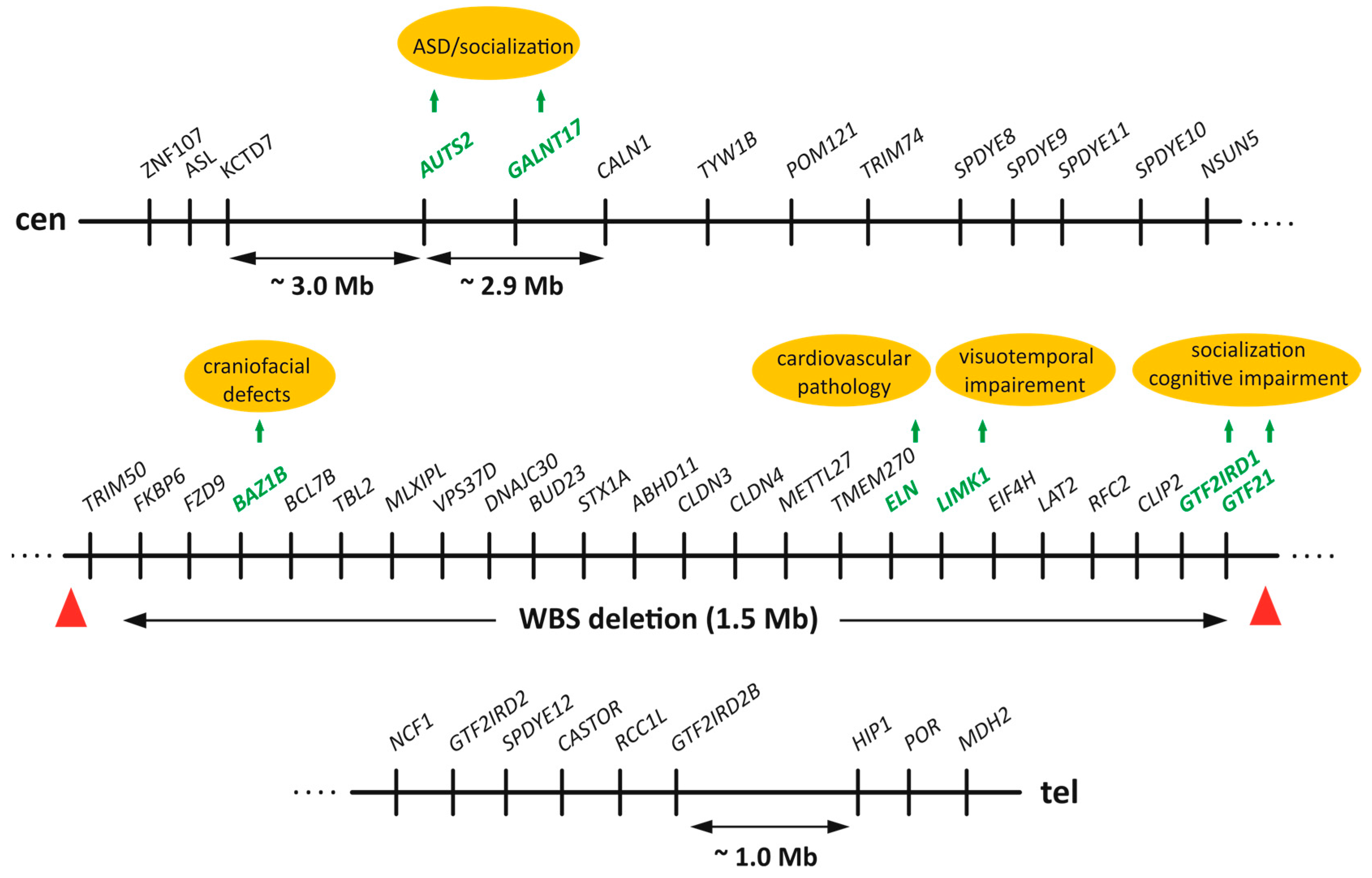
Figure 1. Scheme of the human Williams–Beuren Syndrome Critical Region (WBSCR) genomic locus. All protein-coding genes from AUTS2 to GTF2IRD2B in the ~5.26 Mb region are shown in a centromere (cen)-to-telomere (tel) orientation. Notably, more than half of the ~5.26 Mb genomic range (~2.9 Mb) is occupied only by three genes: AUTS2, GALNT17 and CALN1. The commonly deleted region in Williams–Beuren Syndrome (WBS) patients (1.5 Mb) is marked by red triangles and include genes from TRIM50 to GTF21. Several flanking genes on the centromeric (ZNF107, ASL, KCTD7) and telomeric (HIP1, POR, MDH2) flanks of the WBSCR locus are also shown. Arrows indicate the approximate distance between genes. Several genes with known genotype–phenotype associations are highlighted in green. Scheme is not drawn to scale.
Ijms 26 08549 g001
Figure 3. (A) Scheme of human Galnt17 protein. Galnt17 is a type II transmembrane protein that is anchored in the plasma membrane of Golgi apparatus by a 20 aa- long transmembrane domain (TM) with the C-terminal part of the protein facing the lumen and short N-terminal cytoplasmic tail (7 aa, dark gray). According to Uniprot (Q6IS24, Hs Galnt17), the catalytic domain is divided into subdomains A (151–262 aa) and B (319–381 aa), shown in green. GALNT motif (334–366 aa) is located within catalytic subdomain B. Ricin-like lectin domain (465–594 aa) is located at the C-terminus. Y350—a tyrosine residue within GALNT motif that is typical for Y-subfamily of GALNTs. (B) Diagram showing the putative role of Galnt17 in the regulation of social behavior in mammals. At the molecular level Galnt17 by glycosylating its substrates or binding to protein partners regulates cellular processes (e.g., actin cytoskeleton dynamics and adhesion) which are critical for proper neuron function in certain areas of brain thus specifically affecting social behavior of mammals.
Ijms 26 08549 g003
Figure 4. Scheme of the putative relationships between changes in copy number variants (CNVs), structural variants (SVs) and 3D genome organization in the Williams–Beuren Syndrome Critical Region (WBSCR) locus and domestication and neurobehavioral disorders.
Ijms 26 08549 g004
Disclaimer/Publisher’s Note: The statements, opinions and data contained in all publications are solely those of the individual author(s) and contributor(s) and not of MDPI and/or the editor(s). MDPI and/or the editor(s) disclaim responsibility for any injury to people or property resulting from any ideas, methods, instructions or products referred to in the content.