Article

Mitogenomic Alterations in Breast Cancer: Identification of Potential Biomarkers of Risk and Prognosis

by
Carlos Jhovani Pérez-Amado
1,
Amellalli Bazan-Cordoba
1,2,
Laura Gómez-Romero
3,4,
Julian Ramírez-Bello
5,
Verónica Bautista-Piña
6,
Alberto Tenorio-Torres
6,
Eva Ruvalcaba-Limón
6,
Felipe Villegas-Carlos
6,
Diana Karen Mendiola-Soto
1,7,
Alfredo Hidalgo-Miranda
8 and
Silvia Jiménez-Morales
1,*
1
Laboratorio de Innovación y Medicina de Precisión Núcleo “A”, Instituto Nacional de Medicina Genómica, Mexico City 14610, Mexico
2
Programa de Maestría y Doctorado, Posgrado en Ciencias Bioquímicas, Universidad Nacional Autónoma de México, Mexico City 04510, Mexico
3
Subdirección de Bioinformática, Instituto Nacional de Medicina Genómica, Mexico City 14610, Mexico
4
School of Medicine and Health Sciences, Tecnológico de Monterrey, Mexico City 14380, Mexico
5
Subdirección de Investigación Clínica, Instituto Nacional de Cardiología Ignacio Chávez, Mexico City 14080, Mexico
6
Fundación de Cáncer de Mama, FUCAM, Mexico City 04980, Mexico
7
Programa de Doctorado en Ciencias Biomédicas, Universidad Nacional Autónoma de México, Mexico City 04510, Mexico
8
Laboratorio de Genómica del Cáncer, Instituto Nacional de Medicina Genómica, Mexico City 14610, Mexico
*
Author to whom correspondence should be addressed.
Int. J. Mol. Sci. 2025, 26(17), 8456; https://doi.org/10.3390/ijms26178456
Submission received: 8 July 2025 / Revised: 13 August 2025 / Accepted: 27 August 2025 / Published: 30 August 2025
(This article belongs to the Special Issue Molecular Genetics of Breast Cancer—Recent Progress)

Abstract

Alterations in the mitochondrial genome (mtDNA) have been shown to be key in cancer development and could be useful as biomarkers for diagnosis, prognosis, and treatment. To identify mtDNA variants associated with breast cancer, we analyzed the whole mtDNA sequence from paired tissues (tumor–peripheral blood) of women with this malignancy and from peripheral blood samples of healthy women. The mtDNA mutational landscape, heteroplasmy levels of the variants, and mitochondrial ancestry were established. Comparative analysis between cases and controls revealed significant differences in the number and location of variants, as well as in the heteroplasmy levels. Cases showed higher mutation number in MT-ND5, tRNAs, and rRNAs genes; increased proportion of missense variants; and elevated mtDNA content, than controls. Notably, a high blood mtDNA mutational burden (OR = 3.83, CI: 1.89–7.95, p = 5.3 × 10⁻⁵) and five mtDNA variants showed association with the risk of breast cancer. Furthermore, a low tumor mutational burden (HR = 7.82, CI: 1.0–63.6, p = 0.05) and the haplogroup L (HR = 12.16, CI: 2.0–72.8, p = 0.0062) were associated with decreased overall and disease-free survival, respectively. Our study adds evidence of the potential usefulness of mtDNA variants as risk and prognosis biomarkers for breast cancer.

Graphical Abstract

1. Introduction

Breast cancer is the most common tumor in women around the world [1,2,3]. Nowadays, the unceasing campaigns to prevent and early detect this disease have contributed to improving the 5-year survival rates; however, in Mexico, as in many countries of the world, breast cancer is the leading cause of cancer-related deaths among women [3,4]. Breast cancer is a diverse group of diseases that is clinically and biologically heterogeneous and classified into four subtypes based on receptor protein expression and gene expression profiles. These subtypes are luminal A, luminal B, HER2-enriched, and triple-negative (also known as basal-like) [5,6]. Although the identification of the molecular subtypes of breast cancer is essential for an appropriate clinical and therapeutic approach, late diagnosis and differential response to treatment still represent the main limitations for increasing survival rates of these malignancies [3]. Furthermore, many of the biological mechanisms underlying the development and progression of breast cancer are still unknown [7].
Due to mitochondria’s involvement in numerous cellular processes, such as ATP synthesis via oxidative phosphorylation (OXPHOS), metabolic pathways, production of reactive oxygen species (ROS), apoptosis, and regulation of signaling pathways involved in cell growth and development, their study is highly relevant in numerous human diseases [8,9]. Mitochondria possess their own genome, whose alterations have been shown to promote bioenergetic changes needed for cellular transformation and oncogenesis [8,10,11,12]. The mitochondrial genome (mtDNA) is a circular 16.5 kb double-stranded DNA that contains a total of 37 genes (2 rRNA, 22 tRNA, and 13 protein subunits) encoding essential subunits of the OXPHOS system. mtDNA is maternally inherited, presents a high mutational rate, and can be found in a large number of copies per cell. Because of this last feature, a cell can be homoplasmic if all its mtDNA molecules have the same sequence or heteroplasmic if it contains a mixture of mtDNA molecules with different sequences [8,13,14].
Although the mutational landscape of mtDNA has been described in several types of tumors [8,15,16,17,18,19], the role of these mutations in the oncogenesis and cancer progression processes is unknown, and it seems to depend on the type of mutation, its functional consequences, and the proportion of mutated mtDNA within a cell (heteroplasmy level) [8,14,17]. However, studies have suggested that specific mtDNA variants, such as A10398G and haplogroups (a group of variants that are inherited together without any recombination and correlate with the geographic origin of populations) as D, are associated with breast cancer and have been proposed as risk factors for tumor development. In addition, the number of somatic variants in tumors has been linked with the overall survival of breast cancer patients [8,20,21,22]. Recent evidence even suggests that alterations in mtDNA copy number are involved in tumor biology and could be a potential tool to early detect women with breast cancer [23].
Thus, to identify potential biomarkers in breast cancer, we analyzed the whole mtDNA of paired samples (tumor–peripheral blood) from women with breast cancer and peripheral blood (PB) samples of women without cancer. Our genomic approach allowed us to determine the mutational landscape of mtDNA from peripheral samples, heteroplasmy levels of the variants, mtDNA copy number (mtCN), mitochondrial ancestry, and identify specific variants associated with breast cancer. Additionally, it allowed us to establish the correlation among mtDNA variants and clinical variables of the disease, such as clinical stage, tumor subtype, metastasis, and overall survival.

2. Results

2.1. Study Population

We retrieved the complete mtDNA sequences of 92 paired breast tumor-PB samples analyzed in our previous work [18]. As we described previously, the ages of this cohort ranged from 33 to 92 years old ( x ¯ = 53.83 ± 11.5). In 90 cases, tumors were classified with immunohistochemical (IHC) markers [3], being 63.3% (n = 57) luminal A, 24.4% (n = 22) luminal B, 5.6% (n = 5) HER2, and 6.7% (n = 6) triple negatives (Table 1). Additionally, we sequenced the mtDNA from 75 PB samples from healthy women with ages ranging from 37 to 66 years old ( x ¯ = 48.1 ± 4.3). Regarding age, no significant differences between patients and controls were observed (p = 0.06).

2.2. Landscape of Mitochondrial DNA Germline Variants in Breast Tumors

The landscape of the somatic mutations in breast tumors was previously published by our group [18]; thus, in the present work, we focus on the study of the germline mtDNA variants in breast tumors. A total of 869 variable positions and 42.7 ± 11.4 mtDNA germline variants on average per tumor (range: 24–90 variants) were identified. Based on the variant type, 95.6% (n = 831) were single-nucleotide variants (SNVs), 2.2% (n = 19) deletions, and 2.2% (n = 19) insertions (Supplementary Table S1). According to gene location, 59.95% (n = 521) were located in protein-coding genes and 40.05% (n = 348) in non-coding regions. The D-Loop region (n = 167), MT-ND5 (n = 89), MT-ND4 (n = 62), MT-CYB (n = 56), and MT-CO1 (n = 61) genes exhibited the highest number of variants. Missenses were the most frequent coding variants (48.6%, n = 253), followed by synonymous (46.0%, n = 240), stop gained (4.2%, n = 22), frameshift (0.8%, n = 4), inframe (0.2%, n = 1), and start lost (0.2%, n = 1) variants. Otherwise, 49.7% (n = 173) of the non-coding variants were located in rRNA and tRNA genes, 47.4% (n = 165) had regulatory functions, and 2.9% (n = 10) were intergenic (Supplementary Figure S1).
Heteroplasmy levels of the identified mtDNA germline variants were widely heterogeneous among tumors (from 0.8 to 95.0%), showing a mutant allele fraction (MAF) mean of 29.28%. Additionally, tumors had a mtDNA content mean of 38.96 ± 28.77 copies, while 30.36 ± 24.04 copies were detected in PB; although no significant differences among paired tumor–PB samples were observed (p = 0.086) (Supplementary Table S2 and Figure S2).

2.3. High Mutational Rate of Mitochondrial Genome from Peripheral Blood of Patients with Breast Cancer

Our analysis revealed that all patients with breast cancer carried mtDNA alterations in PB, and a mtDNA mutation rate of 52.3 muts/kb was estimated. mtDNA germline variants ranged from 27 to 109 ( x ¯ = 46.1 ± 16.8) per patient. A total of 866 different mtDNA germline variants distributed throughout the entire genome was found. Overall, of the total variant types, 95.9% (n = 831) corresponded to SNVs, 2.2% (n = 19) were deletions, and 1.9% (n = 16) were insertions (Table 2). The highest number of variants was observed in the D-Loop region (n = 164), followed by the coding genes MT-ND5 (n = 94), MT-ND4 (n = 66), MT-CYB (n = 58), and MT-CO1 (n = 54). Despite the fact that the highest mutational rate occurred in the D-Loop region (146.4 muts/kb), genes that transcribe tRNA, such as MT-TT (107.7 muts/kb), MT-TC (92.3 muts/kb), and MT-TR (62.5 muts/kb), and coding genes, such as MT-ATP6 (72.1 muts/kb) and MT-ATP8 (68.0 muts/kb), also presented high mutation rates.
Based on the mtDNA germiline variants effect prediction, 62% (n = 534) were located in coding regions, of which 52.13% (n = 281) were synonymous, 43.23% (n = 233) were missense, 3.53% (n = 19) were stop-gained, 0.93% (n = 5) were frameshift, and 0.19% (n = 1) were inframe. The variants in non-coding regions represented 38% (n = 332) of the total variants, and those with regulatory effect were the most predominant (49.40%, n = 164), followed by those affecting tRNA and rRNA transcripts (48.19%, n = 160), and intergenic variants (2.41%, n = 8) (Figure 1).

2.4. Low Mutational Rate of Mitochondrial Genome from Peripheral Blood of Healthy Women

The mtDNA mutational rate in blood samples of controls was 24.2 muts/kb, and the mtDNA germline variants were in the range from 21 to 49 ( x ¯ = 33.6 ± 6) variants per individual (Table 2). The identified variants were located in 401 positions distributed throughout the entire mtDNA. D-Loop region presented the greatest number of variants and the highest mutation rate (n = 119 variants, 106.3 muts/kb, respectively), which were similar to patients’ findings. In spite of the fact that coding genes such as MT-CO1 (n = 32), MT-CYB (n = 28), and MT-ATP6 (n = 26) presented a high number of variants, the MT-TT (76.9 muts/kb), MT-ATP8 (38.8 muts/kb), MT-ATP6 (38.2 muts/kb), and MT-TL2 (28.6 muts/kb) genes showed the highest mutation rate.
Regarding the mtDNA location of the identified variants in healthy women, 59% (n = 238) and 41% (n = 167) were located in coding and non-coding regions, respectively. Synonymous coding variants showed the highest proportion (70.59%, n = 168), followed by missense (28.15%, n = 67) and frameshift (0.42%, n = 1) variant types. Of the total number of non-coding variants, 71.26% (n = 119) were regulatory, 26.95% (n = 45) occurred in rRNA/tRNA transcripts, and 1.80% (n = 3) were in intergenic regions (Figure 1).

2.5. The Landscape of mtDNA Germline Variants Differs Between Patients with Breast Cancer and Healthy Women

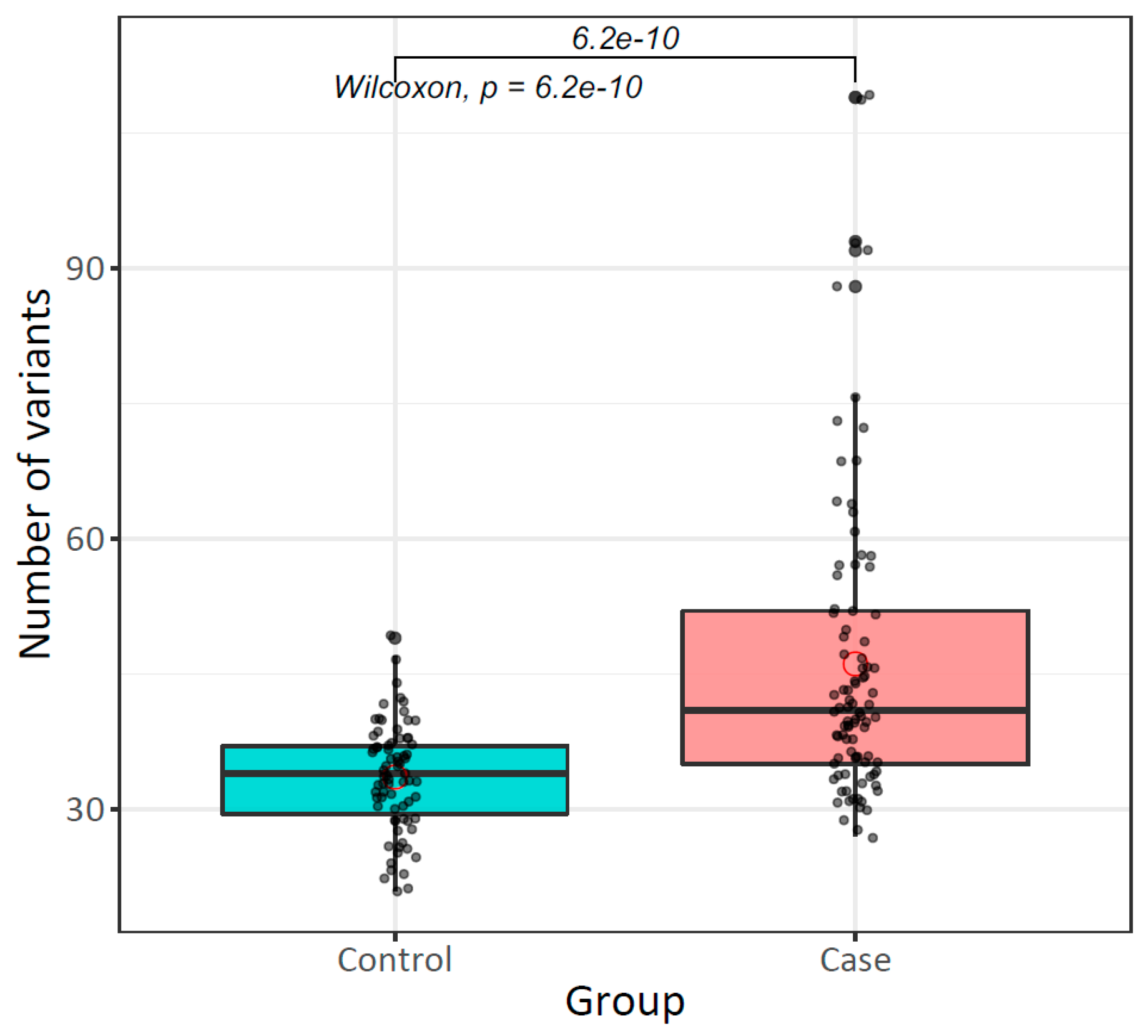
Our comparative analysis of mtDNA germline variants between patients with breast cancer and healthy women showed differences in the number and type of variants. The mtDNA mutation rate and the number of variants per individual (52.3 vs. 24.2 muts/kb, and x ¯ = 46.1 ± 16 vs. 33.5 ± 6, respectively) were higher in cases than controls (p = 6.2 × 10⁻¹⁰) (Table 2).
In terms of the types of variants (SNVs and indels), the differences observed between healthy women and patients were not statistically significant (p > 0.05). However, the proportion of missense variants and variants affecting rRNA/tRNAs was significantly higher in cases than controls (missense: 43.2% vs. 28.1%, p = 9.6 × 10⁻⁵; rRNA/tRNA: 48.19% vs. 26.9%, p = 0.0014). Otherwise, the proportion of synonymous and regulatory variants was lower in patients than healthy women (synonymous: 52.13% vs. 70.59, p = 0.0013; regulatory: 49.40% vs. 71.26%, p = 2.72 × 10⁻⁵). Stop-gained and in-frame variants were only detected in patients.
Although variants were distributed throughout the entire mtDNA, the genes MT-ND5 (p = 0.011), tRNA (p = 0.022), and rRNA (p = 0.045) were significantly more mutated in patients with breast cancer in comparison to healthy women. Otherwise, the D-Loop region was significantly less mutated in cases than controls (p = 2.7 × 10⁻⁵), and MT-TF, MT-TP, and MT-TS1 tRNA genes were exclusively mutated in controls (Table 3).

2.6. Higher Heteroplasmy Variation in Peripheral Blood of Patients with Breast Cancer than Healthy Women

The MAF estimation allowed us to determine the heteroplasmy levels of all mtDNA germline variants. In blood samples of patients, heteroplasmy levels ranged from 0.015 to 94.8% ( x ¯ = 10.8%). Of the total variants, 66.5% (n = 576) were heteroplasmic (MAF ≤ 95%) and 33.5% (n = 290) homoplasmic (MAF > 95%). The heteroplasmic variants showed different heteroplasmy levels: high heteroplasmy (MAF = 20–95%, n = 54), low heteroplasmy (MAF = 0.5–20%, n = 222), and very low heteroplasmy or rare variants (MAF < 0.5%, n = 370).
In relation to the mtDNA germline variants of the control group, we observed a lower range of heteroplasmy (from 22.2 to 95.0%, x ¯ = 63.3%) than in cases. A total of 78.8% (n = 316) of these variants were homoplasmic and 21.2% (n = 85) were heteroplasmic. Notably, we did not identify variants with low heteroplasmy degree or rare variants in this group. The distribution of heteroplasmic and homoplasmic mtDNA germline variants was statistically different (p < 2.2 × 10⁻¹⁶) between cases and controls (Table 4).
Additionally, we observed that 30.77% (n = 212) of mtDNA germline variants presented differences in the heteroplasmic levels among individuals; even so, some of the carriers were homoplasmic for these variants, which were differentially distributed between cases and controls (165 vs. 47 variants, respectively; p = 0.0015).
We identified 208 mtDNA germline variants shared between cases and controls, which showed a wide range of heteroplasmy levels among individuals. Overall, shared variants, 85.6% (n = 178), were detected in more than two subjects. Of these, we observed that 29.2% (n = 52) showed lower heteroplasmy levels in cases compared with controls; in 4.5% (n = 8), heteroplasmy levels were higher in cases than in controls, and 66.3% (n = 118) did not present changes in heteroplasmy levels in both groups.
Interestingly, 35 variants showed significant differences between cases and controls in their heteroplasmy levels. Most of the variants (n = 31) were heteroplasmic in cases, but homoplasmic in controls. For example, the heteroplasmy of A16183C was higher than 67.4% in controls, while, in most cases, it was lower than 70%. On the other hand, the AC493A and A15805G variants were homoplasmic (>95%) in cases, but heteroplasmic (30–70%) in controls (Figure 2).

2.7. Association Analysis Between Mitochondrial DNA Variants and Breast Cancer

2.7.1. The Mitochondrial Mutational Burden in Peripheral Blood Increases Risk for Breast Cancer

Based on the higher number of mtDNA germline variants per individual observed in cases than in controls (Figure 3), we investigated whether mitochondrial mutational burden was a risk factor for breast cancer. The median value of the number of mtDNA variants identified in controls (34 variants) was used as a cut-off point to categorize individuals with low (<34 variants) or high (≥34 variants) mutational burden. We observed that a high mitochondrial mutational burden in PB increases the risk of breast cancer (OR = 3.83, CI: 1.89–7.95, p = 5.3 × 10⁻⁵) (Table 5).

2.7.2. Mitochondrial DNA Germline Variants Are Associated with Breast Cancer Development Risk

Comparing cases and controls, we identified 208 shared mtDNA germline variants between both groups, and variants that were unique for patients (n = 658) or controls (n = 193). Regarding shared variants, only A1438G and A15326G (located in MT-12S and MT-CYB, respectively) were present in all analyzed subjects. Relating to exclusive variants of patients, 31.2% (n = 205) occurred in more than two individuals, and the most frequent showed proportions between 6.5 and 11.9%. In contrast, variants detected solely in the control group showed high heterogeneity among women, and only 9.7% of them were carried by more than one individual, with the C309CCCT, located in the D-Loop region, being the most frequent (5.3%) variant. Another interesting finding was that the average levels of heteroplasmy of the shared variants between the cases and controls were very similar to those observed in the unique variants detected in healthy women (89.36% and 93.35%, respectively), contrasting with the low levels of heteroplasmy observed for the unique variants in cases (44.14%).
Based on the knowledge that mtDNA variants have been associated with the risk of breast cancer, we performed a case-control association analysis including all shared mtDNA germline variants. We identified 25 (12%) shared variants, for which the frequencies between both groups were significantly different. The A16183C (OR = 10.7, CI: 2.5–97.3, p-adj = 2.3 × 10⁻⁴), C14766T (OR = 18.8, CI: 2.7–816.3, p-adj = 3.5 × 10⁻³), and C7028T (OR = 17.1, CI: 2.4–746.2, p-adj = 0.02) mtDNA germline variants increased the risk of developing breast cancer. In contrast, A263G (OR = 0.2, CI: 0.1–0.4, p-adj = 1.5 × 10⁻⁴) and A235G (OR = 0.32, CI: 0.2–0.6, p-adj = 0.02) were identified as protective variants to this neoplasia (Table 6).
The mtDNA variants associated with breast cancer were located mostly in the D-Loop (n = 3 variants) region and the MT-CO1 (n = 1 variant) and MT-CYB genes (n = 1 variant). According to the functional effect, the C14766T and C7028T coding variants were missense and synonymous, respectively. Regarding the A263G, A235G, and A16183C non-coding variants, only A263G and A235G showed a potential regulatory function because they were located in the H-strand replication origin region (OHR), binding sites for mitochondrial transcription factor (mtTF1), and conserved sequence blocks (CSBs).
Another relevant observation is that the heteroplasmy levels of the mtDNA variants associated with breast cancer differed between the studied groups. For instance, the A16183C variant, located in the D-Loop region, showed MAF average values of 50.6% (from 0.1 to 99.8%) and 83.1% (from 67.4 to 98.7%) in the cases and controls, respectively.

2.7.3. Higher Peripheral Blood Mitochondrial DNA Content in Patients with Breast Cancer than Healthy Women

The mtCN in peripheral blood samples from patients and controls showed statistically significant differences (26.2 ± 17.6 vs. 18.5 ± 11.5 copies, respectively; p = 0.021), but no association between mtDNA content and the risk for breast cancer developing (OR = 1.9, CI: 0.96–3.78, p = 0.066) was observed (Supplementary Tables S3 and S4, Figure S3).

2.8. Native American Mitochondrial Haplogroups Were Enriched in Patients with Breast Cancer and Healthy Women

In terms of mitochondrial ancestry, Native American haplogroups (A: 58.7%, C: 14.7%, B: 12%, and D: 8%) were the most frequent in control group, while European (U: 2.7%, R: 1.3%, and T: 1.3%) and African (L: 1.3%) haplogroups occurred with less frequency.
In patients, the most common haplogroups were also Native American origin (91.3%, n = 84). Nevertheless, in contrast to the control group, the African haplogroup was slightly more frequent than the European haplogroups (5.4% vs. 3.3%, respectively). The frequencies of the identified haplogroups were 44.6%, 22.8%, 12%, 12%, 5.4%, 2.2%, and 1.1% (A, B, C, D, L, H, and J, respectively).
By comparing cases and controls, we observed a higher frequency of B (22% vs. 12%), D (12% vs. 8%), and L (5.4% vs. 1.3%) haplogroups in patients than in controls. Moreover, T and R haplogroups were identified exclusively in controls, while H and J haplogroups were exclusive in patients. However, differences were not statistically significant (p > 0.05), and no association with the risk of breast cancer was observed (Supplementary Table S5).

2.9. Tumor Mitochondrial DNA Mutational Burden Is Associated with Overall Survival in Breast Cancer

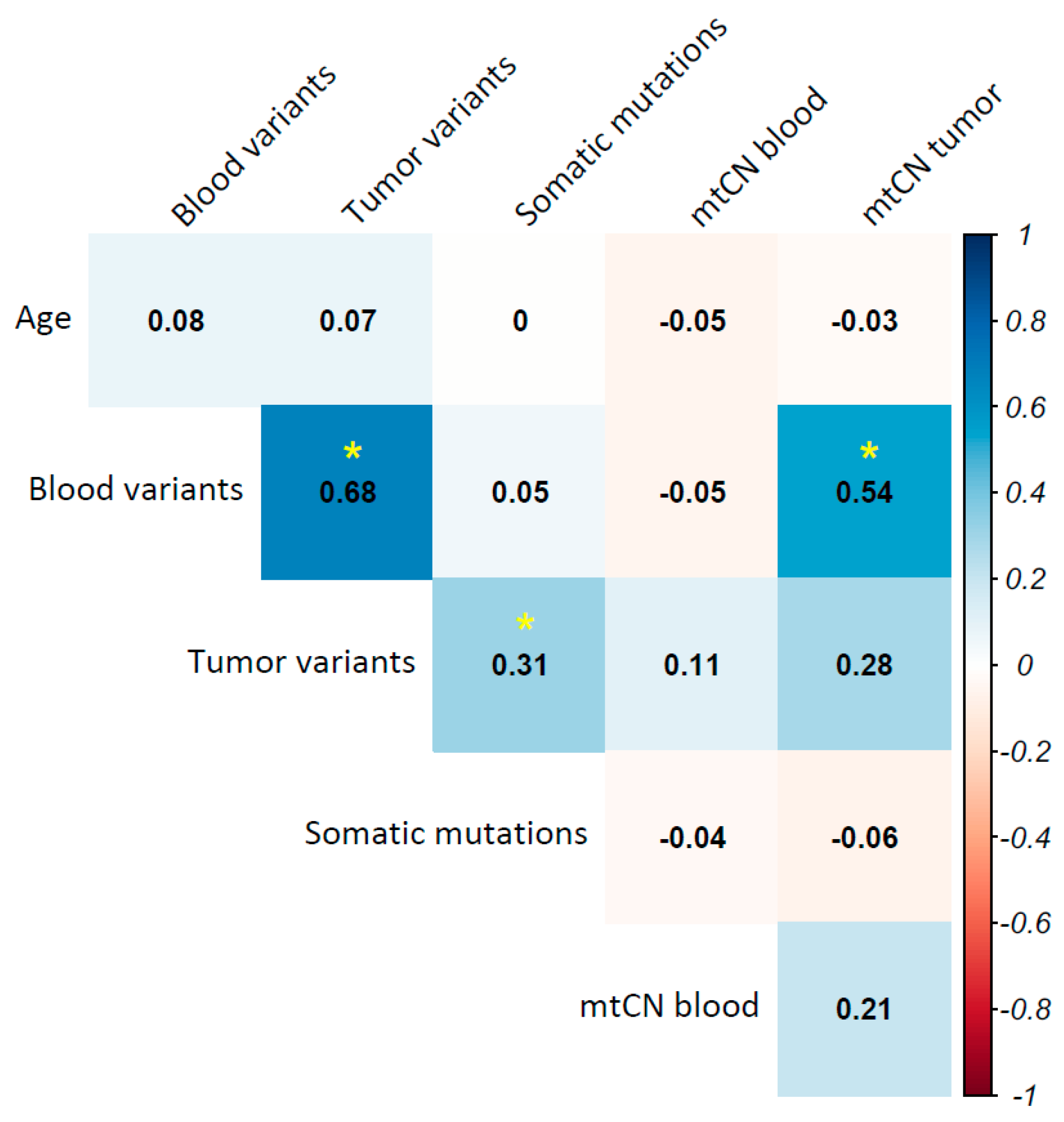
A correlation analysis between mtDNA alterations and clinical variables of breast cancer, such as molecular subtype, clinical stage, metastasis, and death, was performed. mtDNA alterations included blood and tumor mutational burden (total variants per individual), total of somatic mutations, and mtCN in blood and tumor. The age of patients was accounted for based on their reports showing an association between mitochondrial mutation rate and aging; however, no correlation was observed (p > 0.05). Blood mutational burden correlated with both tumor mutational burden (R = 0.68, p = 6.5 × 10⁻¹³) and tumor mtCN (R = 0.54, p = 0.022). Additionally, a correlation between tumor mutational burden and the total of somatic mutations was observed (R = 0.31, p = 0.0003) (Figure 4).
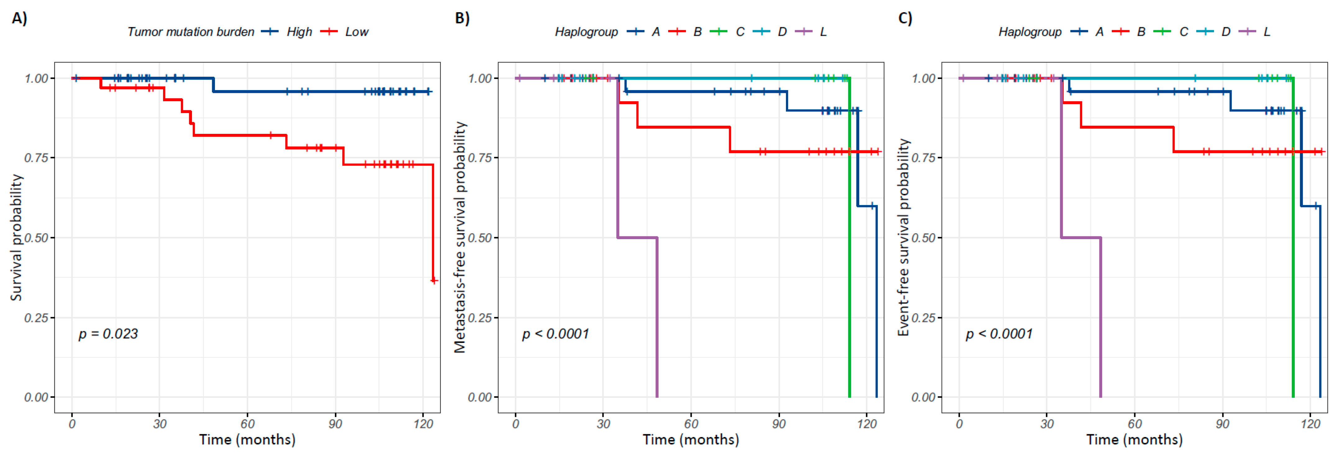
The association of mtDNA alterations with clinical variables of breast cancer was evaluated (Supplementary Table S6). A significant reduction in mutational burden in blood (p = 0.04) and tumor (p = 0.0025) was observed in patients who died. To determine if tumor mutational burden is associated with the prognosis of patients, an over-survival analysis was carried out. For this, patients with an average clinical follow-up of 64 months (from 1 to 124 months) were included, of which 11.84% (n = 9) died, 14.47% (n = 11) developed metastasis, and 17.11% (n = 13) presented one or both events. Patients were categorized with a somatic, tumoral, or blood mutational burden that was high or low using the median value of variants per individual as a cut-off point (Supplementary Table S7). Kaplan–Meier analysis showed that patients with less than 39 mtDNA variants in the tumor had a poor prognosis (p = 0.023) (Figure 5A). Mantel–Cox regression analysis estimated that a low tumor mutational burden increases the risk of death (HR = 7.82, CI: 1.0–63.6, p = 0.05).

2.10. Mitochondrial Haplogroups Are Associated with Prognosis in Breast Cancer

The association between mitochondrial haplogroups and clinical variables of breast cancer was also estimated. Our analysis did not show an association among mitochondrial haplogroups nor with breast cancer molecular subtype, tumor grade, or clinical stage (p > 0.05). However, an association between mitochondrial ancestry and the prognosis of the patients was observed. Survival analysis included the most frequent haplogroups identified (A, B, C, D, and L). Patients with breast cancer carrying the L haplogroup had a poor prognosis in comparison with patients bearing the D haplogroup (p = 6 × 10⁻⁶). Mantel–Cox regression estimated that the L haplogroup increases the risk for metastasis development 27.4-fold in patients with breast tumors (HR = 27.35, CI: 4.5–166.1, p = 0.0003). Finally, an association between mitochondrial ancestry and event-free survival was also observed, with patients carrying the L haplogroup being at a higher risk (12.16-fold) of death or developing metastasis (HR = 12.16, IC: 2.0–72.8, p = 0.0062) (Figure 5).

3. Discussion

Breast cancer is currently a serious worldwide public health problem, with incidence and mortality rates constantly increasing [4,7]. Although there is a vast amount of research around this neoplasia, the current breast cancer screening tests and early detection tools are still limited for detecting the disease or its precursor lesion at an early stage prior to the onset of symptoms. Thus, several studies have focused on the identification of biomarkers that can be used for early diagnosis of breast cancer, to predict its progression, or as a therapeutic target. Most of these investigations have been carried out through analyzing nuclear DNA; however, studies of the mitochondrial genome have shown enormous potential for this purpose. The mtDNA is essential for adequate maintenance of cellular bioenergetics, and it is well recognized that alterations in its sequence and content have been associated with the metabolic reprogramming of tumor cells, promoting their survival and progression [8,14]. However, the clinical implications of these variations are uncertain in most types of cancer [16,17,22,24,25].
The aim of this study was to determine the landscape of mitochondrial alterations in PB of patients with breast cancer and to identify biomarkers with potential clinical applications. For this, we analyzed the complete mtDNA of paired (blood–tumor) samples of breast cancer patients and blood samples of healthy women. Most of the studies around mitochondrial oncogenomics are focused on the identification of somatic mutations in tumors through their comparison with a reference tissue, as normal adjacent tissue or/and PB [11,19,26,27,28,29]. Since we have already reported the landscape of mutation signatures of breast tumors [18], we have focused on the germline variants.
The similar mutational rate of breast cancer tumors and paired PBs (52.4 vs. 52.3 muts/kb, respectively) observed in our analysis contrasts with previous studies, in which a differential mutational rate between tumor and normal tissue, and a higher frequency of mutations in MT-ATP6, MT-ND3, and MT-CYB genes have been reported [15,16,19,22,28,29,30,31]. Further, a wide range of heteroplasmy levels of the mtDNA germline variants across tumors and a higher tumoral mtDNA content than their matched PB samples was detected. These findings can be explained by the hypoxic and oxidative environment in which the tumors are immersed [9,14,18,24]. High concentrations of ROS damage mtDNA and promote dysfunction of essential OXPHOS system protein subunits, changing the cellular bioenergetic and activating signaling pathways implicated in the adaptation and progression of tumors [13,22].
Our case-control approach allowed us to identify noticeable differences between the two groups. We found a higher mtDNA mutation rate, wider ranges in heteroplasmy levels, and elevated mtDNA content in PB from patients than controls. As well, significant differences in the mutation type distribution and the location of the mtDNA variants between the two groups were observed. For instance, mtDNA variants in the MT-ND5, tRNA, and rRNA genes, plus non-synonymous type variants, were enriched in patients, but variants in the D-Loop and regulatory type were more frequent in the controls than in their counterparts. In contrast with these findings, a study performed in patients with breast cancer and healthy women from Sri Lanka did not report significant differences between the cases and controls, which could be due to the small sample size or ancestry background [32]. However, in accordance with our study, these authors also found a higher proportion of missense variants (58.3%) in patients with breast cancer than in controls [32]. No data are available to explain the biological meaning of our findings. Nonetheless, it has been reported in lung carcinomas that the MT-ND5 gene is also highly mutated. Functional experiments using base editing tools to generate mtDNA truncating mutations in the MT-ND5 gene showed decreased expression of complex 1 accessory proteins encoded in the nDNA [33,34]. Additionally, depending on the heteroplasmy levels, mutations can decrease proton-motive potential, modify the NAD+/NADH ratio, and alter concentrations of malate, lactate, fumarate, and arginosuccinate, promoting a metabolic imbalance that could impact the antitumor response [34]. Mutational enrichment in mitochondrial rRNA and tRNA genes of patients with breast cancer suggests a partial impairment in mitochondrial protein synthesis derived from a carcinogenic environment. Studies indicate that these genes retain and better tolerate deleterious variants in comparison to the mtDNA coding region, and even loss of function could be compensated through negative selection of the variants [35,36,37]. Mutations in mitochondrial tRNAs could alter the secondary structure and stability of transcripts, affecting tRNA–amino acid coupling and its insertion into the ribosome. Different types of cancer have shown an enrichment in mitochondrial variants in tRNAs, which are clustered specifically at the anticodon region [15]. Specifically, in patients with breast cancer, mutations in tRNAs such as G1606A (tRNAVal), A4300G (tRNAIle), T7505C (tRNASer1), A14693G (tRNAGlu), and G15927A (tRNAThr) have shown decreased mtDNA content and ATP production, suggesting that these regions are critical for oncogenesis [38].
Furthermore, heteroplasmy is a complex phenomenon that determines pathological phenotypes and their severity levels [16,24,25,36,39]. We observed that mtDNA from the PB of cases with breast cancer showed a wider range of heteroplasmy and a lower proportion of homoplasmic variants than in the controls. In addition to this, the detection of variants with heterogeneous heteroplasmy levels between individuals, such as T16092C, T489C, and A16183C, suggests the presence of mutational events regulated by selection mechanisms based on the functional effect of the mutation and its repercussion in tumoral bioenergetics [27,28]. mtDNA analyses in lung, colon, and liver carcinomas have found that heteroplasmy patterns are tissue- and region-specific, and arise from selection pressures caused by the biochemical and environmental context [24,36]. For example, coding and RNA transcription variants have been identified at low levels of heteroplasmy, and the MT-ND5 gene has proven to be the most susceptible to heteroplasmic changes [11,33,34,40,41,42]. Specific heteroplasmy patterns have been observed for each cancer type [16,24]. The effect and functional mechanisms of mtDNA variants and their heteroplasmy levels in cancer, including breast cancer, have not been sufficiently explored, mainly due to technical limitations in the selection of specific genotypes. Research on tumors has proposed that the deleterious effect of the non-synonymous mtDNA variants can be compensated with modifications in heteroplasmy levels, promoting mitochondrial dynamics that are reflected as changes in mtDNA copy number or even through the co-gain of other mutations [9,11,16,17,23,24,36,41]. However, the explanation for the behavior of the mtDNA variants in PB of patients with breast cancer must be further investigated.
What has been interesting is that case-control studies analyzing PB mtDNA alterations in breast cancer and other types of neoplasia have shown differences between patients and healthy subjects, even years before cancer diagnosis [29,43,44,45]. For instance, Hofmann et al., by studying the mtDNA copy number in renal cell carcinoma pre-diagnostic leukocytes, found that a high mtDNA content is associated with an increased risk for this neoplasia even six years prior to diagnosis [44].
Our analyses suggest that mtDNA mutational burden and the presence of specific variants in PB could have an impact on the identification of women at risk of developing breast cancer, as well as to know the prognosis of the patients. At present, there is no evidence regarding mtDNA mutational burden in PB and the increased risk of breast cancer; however, a higher proportion of mtDNA variants in cases with respect to controls has been previously reported [27,29,46]. A detailed comparison of mtDNA variation between women with breast cancer and controls allowed us to identify potential variants associated with the risk of developing breast tumors. Although studies with similar approaches have been performed, the association of these variants with the risk of this malignancy has not been reported [32,43].
Around 64% of the mtDNA germline variants with differential frequencies between patients with breast cancer and healthy women detected in the present work have been found in other types of cancer, such as oral, nasopharyngeal, ovarian, endometrial, prostate, lung, colorectal, melanoma, glioblastoma, thyroid, gastric, and pancreatic [47]. The association between mtDNA variants and breast cancer risk is debatable, and it has been suggested that these findings could be influenced by demography, as well as clinicopathological and genetic variables, such as tumor origin, molecular subtype, ethnic group, and the co-occurrence with other mtDNA variants [20,48,49,50,51,52,53].
Based on our data and other works, the detection of mtDNA alterations in PB seems to be a promising tool to evaluate the risk of breast cancer developing. A recent study in Swedish women with breast cancer identified concordance among mutations in the mtDNA of paired samples (tumor–PB) prior to diagnosis, suggesting that these genomic changes can be detected before tumor development [29]. Furthermore, our findings, indicating that tumor mtDNA mutational burden is associated with overall survival, suggest that the use of mtDNA variants as prognosis biomarkers could be expanded even to women who have already been diagnosed with breast cancer, allowing for the identification of those patients with poor prognosis. The association of mtDNA mutational burden and cancer prognosis has been previously reported in patients with breast and colorectal tumors [15,22], although these studies have only considered somatic mutations. Other authors have found that specific mutated regions (e.g., D-Loop) and mutational profiles are associated with poor overall survival, disease-free progression, and recurrence-free survival in lung and liver cancer [26,33]. The implementation of mitochondrial mutational burden as a potential tool to determine the prognosis of breast cancer would allow opportune therapeutic intervention in patients; however, analyses in large cohorts that validate these findings are required.
Regarding haplogroups, there is increasing evidence showing an association among some of them with cell biochemistry changes [54]. Studies suggest that haplogroups could modify the incidence, cell mass, invasion capacity, and tissue heterogeneity of tumors through the activation of signaling pathways that alter the antitumor immune response, thus promoting a favorable environment for cancer cells’ progression [15,55,56,57,58,59]. For instance, it has been reported that haplogroups J and T increase the risk of developing gliomas [54], while haplogroup C has been associated with the development of gastric cancer [60].
Our results showed a higher frequency of haplogroups B and D in patients with breast cancer in comparison with controls, but no statistical differences were observed. Haplogroup B has been reported to increase 2-fold the risk of developing cervical cancer in Mexican women [61], and haplogroup D has been reported as a risk factor for breast cancer in Asian and European populations [8].
Additionally, we observed that haplogroup L increases the risk of presenting a metastatic event and/or death. Paradoxically, functional studies have found that haplogroup L increases the expression of genes encoding respiratory complexes and correlates with a lower rate of ATP turnover and production of ROS that give rise to a more efficient OXPHOS system [62]. However, the preservation of a functional OXPHOS system could support optimal cellular bioenergetics for tumor growth and dissemination. Furthermore, we cannot discard that the additive effect of other variants acquired during carcinogenesis also contributes to the unfavorable prognosis of the patients. Due to the sample size, our findings have low statistical power; thus, additional studies increasing the number of haplogroup L carriers are needed to know whether haplogroup L is associated with the risk and progression of breast cancer and if it has clinical relevance. According to other authors, we suggest that mitochondrial haplotypes could be the effector and modifier of tumorigenesis and its progression, through mediating metabolism, redox state, cell death, and immune response [15,58,59].
Because of the intrinsic characteristics of mtDNA, mitochondrial oncogenomics is an extremely complex field of research, displaying huge technical and analytical challenges. For example, the incorporation of nuclear mitochondrial segments (NUMTs) could alter the determination of heteroplasmy levels [63,64,65]; thus, we enriched the mtDNA by a long PCR before sequencing it. Also, sequencing was performed at a depth coverage greater than 300x per base, which, in addition to the use of Strelka2 algorithms for variant calling, allowed us to detect mutations with a minimum proportion of 0.02%. In fact, due to its ability to detect somatic mutations in low proportions, Strelka2 has been proposed as a valuable tool for cancer mutational clonality studies [66]. However, heteroplasmy is a complex phenomenon where numerous biological and technical variables intervene [65,67]. The integration of single-cell sequencing strategies will allow more accurate analysis of heteroplasmy [63,64,68,69] and conclude the correlation between heteroplasmy, breast tumor development, and clinical variables.
For clarifying the impact of mitochondrial alterations in cancer, it is required to design experimental and multivariate studies that integrate omics and functional approaches [15,70,71]. High-throughput technologies that allow for the study of genomics, transcriptomics, proteomics, metabolomics, epigenomics, etc., in a nuclear context, have made great progress and could be applicable in the mitochondrial context [24,41,72,73,74].
Although experimental approaches have to be developed for deciphering the biological implications of haplogroups and specific mtDNA variants in the oncogenesis processes, it is highly valuable that a wide variety of studies have indicated that mtDNA variants could be used as biomarkers of risk, diagnosis, and prognosis in cancer, including breast cancer [29,40,41,42,75,76].
The evaluation of these mitogenomic biomarkers in a minimally invasive and easily accessible sample, such as PB, increases their potential clinical implementation. Why these mtDNA alterations can be detected in the peripheral blood of patients with breast cancer remains a cornerstone question. Several studies have suggested that an mtDNA alteration detected in peripheral blood may come from tumor cell-free mtDNA, tumor extracellular vesicles, mitochondria derived from circulating tumor cells, or due to alterations produced by a highly oxidative microenvironment, which could be characteristic of patients with breast cancer [75,77,78,79]. In lung carcinoma, mtDNA variants identified in tumors are also detected in circulating tumor DNA from the plasma in 90% of patients, even with concordant heteroplasmy levels [42]. Estimating the mitochondrial mutation rate in plasmatic extracellular vesicles from patients with pancreatic cancer showed an increased number of variants in MT-ND1, MT-CYB, and MT-ND5 genes in comparison with controls [75]. The presence of alterations in mtDNA that we observed in the PB of patients could also be explained by hormonal stimulation associated with breast tumors. Studies in endometrial carcinoma have shown that the presence of estrogens and their metabolites promotes the production of ROS, which increases mtDNA mutations and probably contributes to cancer origin and progression [80].
In summary, our findings suggest that certain mitochondrial DNA variants may be potential biomarkers of risk and prognosis for breast cancer; however, these data require additional validation. Longitudinal studies that include an independent cohort that comprises a larger number of cases and controls, which should be collected with a rigorous clinical and demographic characterization, will allow us to decipher the mitochondrial mutational dynamics and their contribution to breast tumorigenesis, as well as to determine their value as a risk predictor of this malignancy [64]. If the above is achieved, strategies for the opportune detection of breast cancer could be implemented, and those women already diagnosed could have more effective therapeutic options.

4. Materials and Methods

4.1. Study Population

mtDNA sequences of paired tumor–peripheral blood (PB) samples from 92 women diagnosed with breast cancer attended at the Instituto de Enfermedades de la Mama FUCAM (Mexico City, Mexico) were included. Patients with antineoplastic treatment prior to mastectomy were excluded. Breast tumors were classified using IHC markers as luminal A (LA), luminal B (LB), HER2-positive (HER2), and triple negative (TN). Additionally, 75 peripheral blood samples from women without breast cancer were involved. Control samples were obtained from blood donor women of the biobanks at the Instituto Nacional de Medicina Genómica (Mexico City, Mexico) and Instituto Nacional de Cardiología Ignacio Chávez (Mexico City, Mexico). All participating women signed the informed consent letter. This study was conducted in accordance with the projects CEI 2015/51 and 24/2016/I, approved in October 2015 and September 2016, respectively, by the Ethics Committee of Instituto Nacional de Medicina Genómica.

4.2. Genomic DNA Extraction

Peripheral blood samples were obtained by venipuncture and centrifuged at 1850× g to separate leukocytes. Tumor samples were taken during mastectomy and stored at −80 °C until further use. The cellularity of the breast tumors was evaluated, and samples with >80% tumor cells were included. Total DNA from PB and tumor tissue samples was extracted using the Maxi Kit (Qiagen, Hilden, Germany) and AllPrep DNA/RNA MiniKit (Qiagen, Hilden, Germany), respectively, according to the manufacturer’s specifications. Genomic DNA concentration and purity were assessed by spectrophotometry (NanoDrop One, Thermo Fisher Scientific, Waltham, MA, USA).

4.3. Direct Mitochondrial DNA Sequencing

The mtDNA was enriched by long PCR using two overlapping primer pairs to avoid contamination with NUMTS, as we reported previously [18]. Paired-end genomic libraries of 150 bp length were generated using the Nextera DNA Flex Library Prep Kit (Illumina, San Diego, CA, USA), and sequencing was performed on the MiSeq platform (Illumina, San Diego, CA, USA). A minimum depth coverage of 300x per base was used.

4.4. Mitochondrial DNA Content Quantification

The determination of the mtDNA copy number was realized by quantitative PCR in real time through a standard curve. Specific primers for the mitochondrial MT-ND1 gene and the nuclear HBB gene reference were used. The relative mtDNA copy number was calculated with the 2−∆CT method.

4.5. Bioinformatic Analysis

Reads with a depth coverage equal to or greater than 300x per base were analyzed. The quality control of the reads was evaluated with the FastQC algorithm (Braham Bioinformatics) [81]. Reads with a Phred score greater than 25 were considered for alignment with the mitochondrial genome reference (NC_012920.1) using the Burrows–Wheeler–Aligner (BWA) algorithm [82]. The reads were indexed, the duplicates were marked, and the bases were recalibrated with the Genome Analysis Tool Kit (GATK, Broad Institute, MA, USA) [83]. The mitochondrial ancestry was assigned by haplogroups determination using the sequence from blood samples with the mtDNA server Haplocheck algorithms [84,85]. Variant calling was performed with Strelka2 (Illumina Inc.) [86], and those variants flagged as PASS were filtered. To determine the heteroplasmy of the variants, the MAF was calculated based on the depth of sequencing. The variants were defined as heteroplasmic if MAF < 95%, or homoplasmic if MAF > 95% [25,68]. The variants identified in PB of the patients were compared with those detected in control women. To predict the functional effect of the variants, the Variant Effect Predictor (VEP) algorithms were used with default parameters [87]. All variants were searched in the MITOMAP [47] database to find out previously reported or associated with breast cancer or other types of neoplasia.

4.6. Statistical Analysis

The total number of variants distributed along mtDNA was counted, and their frequency was estimated considering all the individuals of each group. The number of variants per gene and the frequencies between cases and controls were compared using Chi-square and Fisher’s exact tests. Mutational frequencies, mutational burden, and mtDNA content were correlated with clinical variables (molecular subtype, clinical stage, tumor grade, and survival) of patients with breast cancer. The association of the variants and their risk for breast cancer developing was estimated using the Odds Ratio (OR) with 95% confidence intervals. Overall survival was assessed using Kaplan–Meier curves adjusted to a Cox Regression model. p-values < 0.05 were considered statistically significant and were adjusted (p-adj) by the Bonferroni method when required. All graphs and statistical tests were performed using the R programming language (R version 4.0.2) [88].

5. Conclusions

This research describes the mtDNA germline variants landscape of Mexican patients with breast cancer and healthy women. Our data show that mtDNA alterations in the PB of women with breast tumors differ significantly from healthy women. Although the biological mechanism of these alterations, as well as their relevance in the oncogenesis process, must be investigated in more detail, detection of specific variants and determination of mitochondrial haplogroups could be a tool with high potential for predicting the risk of breast cancer and establishing the prognosis. Longitudinal studies in larger and independent case-control cohorts are required to decipher the potential clinical application of our findings and whether they are meaningful for early diagnosis of breast cancer.

Supplementary Materials

The following supporting information can be downloaded at: https://www.mdpi.com/article/10.3390/ijms26178456/s1.

Author Contributions

Conceptualization and design: C.J.P.-A. and S.J.-M.; sample identification and clinical follow-up: C.J.P.-A., A.B.-C., V.B.-P., A.T.-T., E.R.-L., F.V.-C. and J.R.-B.; data analysis: C.J.P.-A., A.B.-C. and L.G.-R.; supervision: S.J.-M.; writing of the original draft: C.J.P.-A. and S.J.-M.; manuscript revision and approval of the submitted version: C.J.P.-A., A.B.-C., L.G.-R., J.R.-B., V.B.-P., A.T.-T., E.R.-L., F.V.-C., D.K.M.-S., A.H.-M. and S.J.-M. All authors have read and agreed to the published version of the manuscript.

Funding

This study was funded by the Instituto Nacional de Medicina Genómica (C2_19/2009 and 24/2016/I). Carlos Jhovani Pérez Amado (CVU 821714) was supported by a fellowship from the Consejo Nacional de Humanidades, Ciencia y Tecnología (CONAHCYT). The funders had no role in the study design, data collection and analysis, decision to publish, or preparation of the manuscript.

Institutional Review Board Statement

This study was conducted in accordance with the Declaration of Helsinki and approved by the Ethics Committee of Instituto Nacional de Medicina Genómica with project codes CEI 2015/51 and 24/2016/I, approved on 9 October 2009 and 5 September 2016, respectively.

Informed Consent Statement

Informed consent was obtained from all subjects involved in this study.

Data Availability Statement

The datasets generated and/or analyzed during the current study are not publicly available due to being subject to protection by the Instituto Nacional de Medicina Genómica, but are available from the corresponding author on reasonable request.

Acknowledgments

The authors thank the Unit of Sequencing (USeq) at INMEGEN for technical support. Data of the germline mtDNA variants from paired samples (tumor–peripheral blood) were obtained from our previous study, which was focused on the description of the mtDNA somatic mutation [18]. Finally, we are extremely grateful to all the patients who participated in this study.

Conflicts of Interest

The authors declare no conflicts of interest.

Abbreviations

The following abbreviations are used in this manuscript:
BWABurrows–Wheeler Aligner
CSBConserved sequence blocks
EREstrogen receptor
GATKGenome Analysis Tool Kit
HBBHemoglobin Subunit Beta
H2HER2 positive breast cancer subtype
HER2Human epidermal growth factor receptor 2
HRHazzard ratio
IHCImmunohistochemistry
LALuminal A breast cancer subtype
LBLuminal B breast cancer subtype
MAFMutant allele fraction
MT-ATP6Mitochondrial ATP Synthase Membrane Subunit 6
MT-ATP8Mitochondrial ATP Synthase Membrane Subunit 8
MT-CO (1-3)Mitochondrial Cytochrome C Oxidase Subunit (1-3)
mtCNMitochondrial DNA copy number
MT-CYBMitochondrial Cytochrome B
mtDNAMitochondrial DNA
MT-ND (1-6)Mitochondrial NADH Dehydrogenase Subunits (1-6)
MT-ND4LMitochondrial NADH Dehydrogenase Subunits 4L
mtTF1Mitochondrial transcription factor 1
NUMTSNuclear mitochondrial segments
OHRH-strand replication origin region
OROdds ratio
OXPHOSOxidative phosphorylation
PBPeripheral blood
PCRPolymerase Chain Reaction
PRProgesterone receptor
ROSReactive oxygen species
rRNARibosomal RNA
SNVSingle-nucleotide variant
TNTriple negative
tRNATransfer RNA
VEPVariant Effect Predictor

References

  1. Breastcancer org. Available online: https://www.breastcancer.org/ (accessed on 1 January 2019).
  2. GLOBOCAN. The Global Cancer Observatory: Globocan 2020 (México). Available online: https://gco.iarc.fr/today/home (accessed on 5 October 2024).
  3. Consenso Mexicano sobre el Diagnóstico y Tratamiento del Cáncer Mamario. Décima Revisión del Consenso Mexicano sobre el Diagnóstico y Tratamiento del Cáncer Mamario; Consenso Mexicano sobre el Diagnóstico y Tratamiento del Cáncer Mamario: Colima, México, 2023; p. 119. [Google Scholar]
  4. Bray, F.; Ferlay, J.; Soerjomataram, I.; Siegel, R.L.; Torre, L.A.; Jemal, A. Global cancer statistics 2018: GLOBOCAN estimates of incidence and mortality worldwide for 36 cancers in 185 countries. CA Cancer J. Clin. 2018, 68, 394–424. [Google Scholar] [CrossRef]
  5. Provenzano, E.; Ulaner, G.A.; Chin, S.F. Molecular Classification of Breast Cancer. PET Clin. 2018, 13, 325–338. [Google Scholar] [CrossRef]
  6. Turashvili, G.; Brogi, E. Tumor Heterogeneity in Breast Cancer. Front. Med. 2017, 4, 227. [Google Scholar] [CrossRef]
  7. Nielsen, T.O.; Parker, J.S.; Leung, S.; Voduc, D.; Ebbert, M.; Vickery, T.; Davies, S.R.; Snider, J.; Stijleman, I.J.; Reed, J.; et al. A comparison of PAM50 intrinsic subtyping with immunohistochemistry and clinical prognostic factors in tamoxifen-treated estrogen receptor-positive breast cancer. Clin. Cancer Res. 2010, 16, 5222–5232. [Google Scholar] [CrossRef]
  8. Jimenez-Morales, S.; Perez-Amado, C.J.; Langley, E.; Hidalgo-Miranda, A. Overview of mitochondrial germline variants and mutations in human disease: Focus on breast cancer (Review). Int. J. Oncol. 2018, 53, 923–936. [Google Scholar] [CrossRef] [PubMed]
  9. Wallace, D.C. Mitochondria and cancer. Nat. Rev. Cancer 2012, 12, 685–698. [Google Scholar] [CrossRef]
  10. Dominguez-de-la-Cruz, E.; Munoz, M.L.; Perez-Munoz, A.; Garcia-Hernandez, N.; Moctezuma-Meza, C.; Hinojosa-Cruz, J.C. Reduced mitochondrial DNA copy number is associated with the haplogroup, and some clinical features of breast cancer in Mexican patients. Gene 2020, 761, 145047. [Google Scholar] [CrossRef]
  11. Guo, W.; Liu, Y.; Ji, X.; Guo, S.; Xie, F.; Chen, Y.; Zhou, K.; Zhang, H.; Peng, F.; Wu, D.; et al. Mutational signature of mtDNA confers mechanistic insight into oxidative metabolism remodeling in colorectal cancer. Theranostics 2023, 13, 324–338. [Google Scholar] [CrossRef] [PubMed]
  12. Hou, X.S.; Wang, H.S.; Mugaka, B.P.; Yang, G.J.; Ding, Y. Mitochondria: Promising organelle targets for cancer diagnosis and treatment. Biomater. Sci. 2018, 6, 2786–2797. [Google Scholar] [CrossRef] [PubMed]
  13. Wallace, D.C. Bioenergetic origins of complexity and disease. Cold Spring Harb. Symp. Quant. Biol. 2011, 76, 1–16. [Google Scholar] [CrossRef]
  14. Warburg, O.; Wind, F.; Negelein, E. The Metabolism of Tumors in the Body. J. Gen. Physiol. 1927, 8, 519–530. [Google Scholar] [CrossRef]
  15. Gorelick, A.N.; Kim, M.; Chatila, W.K.; La, K.; Hakimi, A.A.; Berger, M.F.; Taylor, B.S.; Gammage, P.A.; Reznik, E. Respiratory complex and tissue lineage drive recurrent mutations in tumour mtDNA. Nat. Metab. 2021, 3, 558–570. [Google Scholar] [CrossRef]
  16. Grandhi, S.; Bosworth, C.; Maddox, W.; Sensiba, C.; Akhavanfard, S.; Ni, Y.; LaFramboise, T. Heteroplasmic shifts in tumor mitochondrial genomes reveal tissue-specific signals of relaxed and positive selection. Hum. Mol. Genet. 2017, 26, 2912–2922. [Google Scholar] [CrossRef]
  17. Ju, Y.S.; Alexandrov, L.B.; Gerstung, M.; Martincorena, I.; Nik-Zainal, S.; Ramakrishna, M.; Davies, H.R.; Papaemmanuil, E.; Gundem, G.; Shlien, A.; et al. Origins and functional consequences of somatic mitochondrial DNA mutations in human cancer. eLife 2014, 3, e02935. [Google Scholar] [CrossRef]
  18. Perez-Amado, C.J.; Tovar, H.; Gomez-Romero, L.; Beltran-Anaya, F.O.; Bautista-Pina, V.; Dominguez-Reyes, C.; Villegas-Carlos, F.; Tenorio-Torres, A.; Alfaro-Ruiz, L.A.; Hidalgo-Miranda, A.; et al. Mitochondrial DNA Mutation Analysis in Breast Cancer: Shifting From Germline Heteroplasmy Toward Homoplasmy in Tumors. Front. Oncol. 2020, 10, 572954. [Google Scholar] [CrossRef]
  19. Yuan, Y.; Ju, Y.S.; Kim, Y.; Li, J.; Wang, Y.; Yoon, C.J.; Yang, Y.; Martincorena, I.; Creighton, C.J.; Weinstein, J.N.; et al. Comprehensive molecular characterization of mitochondrial genomes in human cancers. Nat. Genet. 2020, 52, 342–352. [Google Scholar] [CrossRef] [PubMed]
  20. Canter, J.A.; Kallianpur, A.R.; Parl, F.F.; Millikan, R.C. Mitochondrial DNA G10398A polymorphism and invasive breast cancer in African-American women. Cancer Res. 2005, 65, 8028–8033. [Google Scholar] [CrossRef] [PubMed]
  21. Darvishi, K.; Sharma, S.; Bhat, A.K.; Rai, E.; Bamezai, R.N. Mitochondrial DNA G10398A polymorphism imparts maternal Haplogroup N a risk for breast and esophageal cancer. Cancer Lett. 2007, 249, 249–255. [Google Scholar] [CrossRef]
  22. McMahon, S.; LaFramboise, T. Mutational patterns in the breast cancer mitochondrial genome, with clinical correlates. Carcinogenesis 2014, 35, 1046–1054. [Google Scholar] [CrossRef] [PubMed]
  23. Reznik, E.; Miller, M.L.; Senbabaoglu, Y.; Riaz, N.; Sarungbam, J.; Tickoo, S.K.; Al-Ahmadie, H.A.; Lee, W.; Seshan, V.E.; Hakimi, A.A.; et al. Mitochondrial DNA copy number variation across human cancers. eLife 2016, 5, e10769. [Google Scholar] [CrossRef]
  24. Perez-Amado, C.J.; Bazan-Cordoba, A.; Hidalgo-Miranda, A.; Jimenez-Morales, S. Mitochondrial Heteroplasmy Shifting as a Potential Biomarker of Cancer Progression. Int. J. Mol. Sci. 2021, 22, 7369. [Google Scholar] [CrossRef] [PubMed]
  25. Stewart, J.B.; Chinnery, P.F. The dynamics of mitochondrial DNA heteroplasmy: Implications for human health and disease. Nat. Rev. Genet. 2015, 16, 530–542. [Google Scholar] [CrossRef]
  26. Yin, C.; Li, D.Y.; Guo, X.; Cao, H.Y.; Chen, Y.B.; Zhou, F.; Ge, N.J.; Liu, Y.; Guo, S.S.; Zhao, Z.; et al. NGS-based profiling reveals a critical contributing role of somatic D-loop mtDNA mutations in HBV-related hepatocarcinogenesis. Ann. Oncol. 2019, 30, 953–962. [Google Scholar] [CrossRef]
  27. Liu, J.; Wang, L.D.; Sun, Y.B.; Li, E.M.; Xu, L.Y.; Zhang, Y.P.; Yao, Y.G.; Kong, Q.P. Deciphering the signature of selective constraints on cancerous mitochondrial genome. Mol. Biol. Evol. 2012, 29, 1255–1261. [Google Scholar] [CrossRef]
  28. Larman, T.C.; DePalma, S.R.; Hadjipanayis, A.G.; Cancer Genome Atlas Research, N.; Protopopov, A.; Zhang, J.; Gabriel, S.B.; Chin, L.; Seidman, C.E.; Kucherlapati, R.; et al. Spectrum of somatic mitochondrial mutations in five cancers. Proc. Natl. Acad. Sci. USA 2012, 109, 14087–14091. [Google Scholar] [CrossRef]
  29. Li, Y.; Sundquist, K.; Vats, S.; Hong, M.G.; Wang, X.; Chen, Y.; Hedelius, A.; Saal, L.H.; Sundquist, J.; Memon, A.A. Mitochondrial heteroplasmic shifts reveal a positive selection of breast cancer. J. Transl. Med. 2023, 21, 696. [Google Scholar] [CrossRef] [PubMed]
  30. Tseng, L.M.; Yin, P.H.; Yang, C.W.; Tsai, Y.F.; Hsu, C.Y.; Chi, C.W.; Lee, H.C. Somatic mutations of the mitochondrial genome in human breast cancers. Genes Chromosomes Cancer 2011, 50, 800–811. [Google Scholar] [CrossRef]
  31. Weerts, M.J.A.; Sleijfer, S.; Martens, J.W.M. The role of mitochondrial DNA in breast tumors. Drug Discov. Today 2019, 24, 1202–1208. [Google Scholar] [CrossRef]
  32. Jayasekera, L.P.; Ranasinghe, R.; Senathilake, K.S.; Kotelawala, J.T.; de Silva, K.; Abeygunasekara, P.H.; Goonesinghe, R.; Tennekoon, K.H. Mitochondrial genome in sporadic breast cancer: A case control study and a proteomic analysis in a Sinhalese cohort from Sri Lanka. PLoS ONE 2023, 18, e0281620. [Google Scholar] [CrossRef]
  33. Kim, M.Y.; Kim, H.; Sung, J.A.; Koh, J.; Cho, S.; Chung, D.H.; Jeon, Y.K.; Lee, S.D. Whole Mitochondrial Genome Analysis in Non-Small Cell Lung Carcinoma Reveals Unique Tumor-Specific Somatic Mutations. Arch. Pathol. Lab. Med. 2023, 147, 1268–1277. [Google Scholar] [CrossRef] [PubMed]
  34. Mahmood, M.; Liu, E.M.; Shergold, A.L.; Tolla, E.; Tait-Mulder, J.; Huerta Uribe, A.; Shokry, E.; Young, A.L.; Lilla, S.; Kim, M.; et al. Tumour mitochondrial DNA mutations drive aerobic glycolysis to enhance checkpoint blockade. bioRxiv 2023. [Google Scholar] [CrossRef]
  35. Kauppila, J.H.K.; Baines, H.L.; Bratic, A.; Simard, M.L.; Freyer, C.; Mourier, A.; Stamp, C.; Filograna, R.; Larsson, N.G.; Greaves, L.C.; et al. A Phenotype-Driven Approach to Generate Mouse Models with Pathogenic mtDNA Mutations Causing Mitochondrial Disease. Cell Rep. 2016, 16, 2980–2990. [Google Scholar] [CrossRef]
  36. Pereira, C.V.; Gitschlag, B.L.; Patel, M.R. Cellular mechanisms of mtDNA heteroplasmy dynamics. Crit. Rev. Biochem. Mol. Biol. 2021, 56, 510–525. [Google Scholar] [CrossRef]
  37. Stewart, J.B.; Freyer, C.; Elson, J.L.; Wredenberg, A.; Cansu, Z.; Trifunovic, A.; Larsson, N.G. Strong purifying selection in transmission of mammalian mitochondrial DNA. PLoS Biol. 2008, 6, e10. [Google Scholar] [CrossRef] [PubMed]
  38. Ding, H.J.; Zhao, Y.P.; Jiang, Z.C.; Zhou, D.T.; Zhu, R. Analysis of Mitochondrial Transfer RNA Mutations in Breast Cancer. Balk. J. Med. Genet. 2023, 25, 15–22. [Google Scholar] [CrossRef]
  39. Stefano, G.B.; Kream, R.M. Mitochondrial DNA heteroplasmy in human health and disease. Biomed. Rep. 2016, 4, 259–262. [Google Scholar] [CrossRef] [PubMed]
  40. Cararo-Lopes, E.; Sawant, A.; Moore, D.; Ke, H.; Shi, F.; Laddha, S.; Chen, Y.; Sharma, A.; Naumann, J.; Guo, J.Y.; et al. Integrated metabolic and genetic analysis reveals distinct features of primary differentiated thyroid cancer and its metastatic potential in humans. medRxiv 2023. [Google Scholar] [CrossRef] [PubMed]
  41. Ji, X.; Guo, W.; Gu, X.; Guo, S.; Zhou, K.; Su, L.; Yuan, Q.; Liu, Y.; Guo, X.; Huang, Q.; et al. Mutational profiling of mtDNA control region reveals tumor-specific evolutionary selection involved in mitochondrial dysfunction. EBioMedicine 2022, 80, 104058. [Google Scholar] [CrossRef]
  42. Xu, Y.; Yang, Y.; Wang, Y.; Su, J.; Chan, T.; Zhou, J.; Gong, Y.; Wang, K.; Gu, Y.; Zhang, C.; et al. Molecular fingerprints of nuclear genome and mitochondrial genome for early diagnosis of lung adenocarcinoma. J. Transl. Med. 2023, 21, 250. [Google Scholar] [CrossRef]
  43. Tipirisetti, N.R.; Lakshmi, R.K.; Govatati, S.; Govatati, S.; Vuree, S.; Singh, L.; Raghunadha Rao, D.; Bhanoori, M.; Vishnupriya, S. Mitochondrial genome variations in advanced stage breast cancer: A case-control study. Mitochondrion 2013, 13, 372–378. [Google Scholar] [CrossRef]
  44. Hofmann, J.N.; Hosgood, H.D., 3rd; Liu, C.S.; Chow, W.H.; Shuch, B.; Cheng, W.L.; Lin, T.T.; Moore, L.E.; Lan, Q.; Rothman, N.; et al. A nested case-control study of leukocyte mitochondrial DNA copy number and renal cell carcinoma in the Prostate, Lung, Colorectal and Ovarian Cancer Screening Trial. Carcinogenesis 2014, 35, 1028–1031. [Google Scholar] [CrossRef]
  45. Yang, K.; Li, X.; Forman, M.R.; Monahan, P.O.; Graham, B.H.; Joshi, A.; Song, M.; Hang, D.; Ogino, S.; Giovannucci, E.L.; et al. Pre-diagnostic leukocyte mitochondrial DNA copy number and colorectal cancer risk. Carcinogenesis 2019, 40, 1462–1468. [Google Scholar] [CrossRef]
  46. Li, L.; Chen, L.; Li, J.; Zhang, W.; Liao, Y.; Chen, J.; Sun, Z. Correlational study on mitochondrial DNA mutations as potential risk factors in breast cancer. Oncotarget 2016, 7, 31270–31283. [Google Scholar] [CrossRef] [PubMed]
  47. Lott, M.T.; Leipzig, J.N.; Derbeneva, O.; Xie, H.M.; Chalkia, D.; Sarmady, M.; Procaccio, V.; Wallace, D.C. mtDNA Variation and Analysis Using Mitomap and Mitomaster. Curr. Protoc. Bioinform. 2013, 44, 1.23.1–1.23.26. [Google Scholar] [CrossRef]
  48. Bai, R.K.; Leal, S.M.; Covarrubias, D.; Liu, A.; Wong, L.J. Mitochondrial genetic background modifies breast cancer risk. Cancer Res. 2007, 67, 4687–4694. [Google Scholar] [CrossRef]
  49. Covarrubias, D.; Bai, R.K.; Wong, L.J.; Leal, S.M. Mitochondrial DNA variant interactions modify breast cancer risk. J. Hum. Genet. 2008, 53, 924–928. [Google Scholar] [CrossRef]
  50. Tengku Baharudin, N.; Jaafar, H.; Zainuddin, Z. Association of mitochondrial DNA 10398 polymorphism in invasive breast cancer in malay population of peninsular malaysia. Malays. J. Med. Sci. 2012, 19, 36–42. [Google Scholar] [PubMed]
  51. Czarnecka, A.M.; Krawczyk, T.; Zdrozny, M.; Lubinski, J.; Arnold, R.S.; Kukwa, W.; Scinska, A.; Golik, P.; Bartnik, E.; Petros, J.A. Mitochondrial NADH-dehydrogenase subunit 3 (ND3) polymorphism (A10398G) and sporadic breast cancer in Poland. Breast Cancer Res. Treat. 2010, 121, 511–518. [Google Scholar] [CrossRef] [PubMed]
  52. Jahani, M.M.; Azimi Meibody, A.; Karimi, T.; Banoei, M.M.; Houshmand, M. An A10398G mitochondrial DNA alteration is related to increased risk of breast cancer, and associates with Her2 positive receptor. Mitochondrial DNA. Part A DNA Mapp. Seq. Anal. 2020, 31, 11–16. [Google Scholar] [CrossRef]
  53. Setiawan, V.W.; Chu, L.H.; John, E.M.; Ding, Y.C.; Ingles, S.A.; Bernstein, L.; Press, M.F.; Ursin, G.; Haiman, C.A.; Neuhausen, S.L. Mitochondrial DNA G10398A variant is not associated with breast cancer in African-American women. Cancer Genet. Cytogenet. 2008, 181, 16–19. [Google Scholar] [CrossRef]
  54. Gonzalez, M.D.M.; Santos, C.; Alarcon, C.; Ramos, A.; Cos, M.; Catalano, G.; Acebes, J.J.; Aluja, M.P. Mitochondrial DNA haplogroups J and T increase the risk of glioma. Mitochondrion 2021, 58, 95–101. [Google Scholar] [CrossRef]
  55. Chattopadhyay, M.; Jenkins, E.C.; Lechuga-Vieco, A.V.; Nie, K.; Fiel, M.I.; Rialdi, A.; Guccione, E.; Enriquez, J.A.; Sia, D.; Lujambio, A.; et al. The portrait of liver cancer is shaped by mitochondrial genetics. Cell Rep. 2022, 38, 110254. [Google Scholar] [CrossRef]
  56. Brinker, A.E.; Vivian, C.J.; Beadnell, T.C.; Koestler, D.C.; Teoh, S.T.; Lunt, S.Y.; Welch, D.R. Mitochondrial Haplotype of the Host Stromal Microenvironment Alters Metastasis in a Non-cell Autonomous Manner. Cancer Res. 2020, 80, 1118–1129. [Google Scholar] [CrossRef] [PubMed]
  57. Brinker, A.E.; Vivian, C.J.; Koestler, D.C.; Tsue, T.T.; Jensen, R.A.; Welch, D.R. Mitochondrial Haplotype Alters Mammary Cancer Tumorigenicity and Metastasis in an Oncogenic Driver-Dependent Manner. Cancer Res. 2017, 77, 6941–6949. [Google Scholar] [CrossRef]
  58. Gaude, E.; Schmidt, C.; Gammage, P.A.; Dugourd, A.; Blacker, T.; Chew, S.P.; Saez-Rodriguez, J.; O’Neill, J.S.; Szabadkai, G.; Minczuk, M.; et al. NADH Shuttling Couples Cytosolic Reductive Carboxylation of Glutamine with Glycolysis in Cells with Mitochondrial Dysfunction. Mol. Cell 2018, 69, 581–593.e587. [Google Scholar] [CrossRef]
  59. Tasdogan, A.; McFadden, D.G.; Mishra, P. Mitochondrial DNA Haplotypes as Genetic Modifiers of Cancer. Trends Cancer 2020, 6, 1044–1058. [Google Scholar] [CrossRef] [PubMed]
  60. Cavalcante, G.C.; Marinho, A.N.R.; Anaissi, A.K.; Vinasco-Sandoval, T.; Ribeiro-Dos-Santos, A.; Vidal, A.F.; de Araujo, G.S.; Demachki, S.; Ribeiro-Dos-Santos, A. Whole mitochondrial genome sequencing highlights mitochondrial impact in gastric cancer. Sci. Rep. 2019, 9, 15716. [Google Scholar] [CrossRef] [PubMed]
  61. Guardado-Estrada, M.; Juarez-Torres, E.; Medina-Martinez, I.; Wegier, A.; Macias, A.; Gomez, G.; Cruz-Talonia, F.; Roman-Bassaure, E.; Pinero, D.; Kofman-Alfaro, S.; et al. A great diversity of Amerindian mitochondrial DNA ancestry is present in the Mexican mestizo population. J. Hum. Genet. 2009, 54, 695–705. [Google Scholar] [CrossRef]
  62. Kenney, M.C.; Chwa, M.; Atilano, S.R.; Falatoonzadeh, P.; Ramirez, C.; Malik, D.; Tarek, M.; Del Carpio, J.C.; Nesburn, A.B.; Boyer, D.S.; et al. Molecular and bioenergetic differences between cells with African versus European inherited mitochondrial DNA haplogroups: Implications for population susceptibility to diseases. Biochim. Biophys. Acta 2014, 1842, 208–219. [Google Scholar] [CrossRef]
  63. Bi, C.; Wang, L.; Fan, Y.; Yuan, B.; Ramos-Mandujano, G.; Zhang, Y.; Alsolami, S.; Zhou, X.; Wang, J.; Shao, Y.; et al. Single-cell individual full-length mtDNA sequencing by iMiGseq uncovers unexpected heteroplasmy shifts in mtDNA editing. Nucleic Acids Res. 2023, 51, e48. [Google Scholar] [CrossRef]
  64. Bi, C.; Wang, L.; Fan, Y.; Yuan, B.; Alsolami, S.; Zhang, Y.; Zhang, P.Y.; Huang, Y.; Yu, Y.; Izpisua Belmonte, J.C.; et al. Quantitative haplotype-resolved analysis of mitochondrial DNA heteroplasmy in Human single oocytes, blastoids, and pluripotent stem cells. Nucleic Acids Res. 2023, 51, 3793–3805. [Google Scholar] [CrossRef]
  65. Duan, M.; Tu, J.; Lu, Z. Recent Advances in Detecting Mitochondrial DNA Heteroplasmic Variations. Molecules 2018, 23, 323. [Google Scholar] [CrossRef]
  66. Roberts, N.D.; Kortschak, R.D.; Parker, W.T.; Schreiber, A.W.; Branford, S.; Scott, H.S.; Glonek, G.; Adelson, D.L. A comparative analysis of algorithms for somatic SNV detection in cancer. Bioinformatics 2013, 29, 2223–2230. [Google Scholar] [CrossRef] [PubMed]
  67. Gonzalez, M.D.M.; Ramos, A.; Aluja, M.P.; Santos, C. Sensitivity of mitochondrial DNA heteroplasmy detection using Next Generation Sequencing. Mitochondrion 2020, 50, 88–93. [Google Scholar] [CrossRef] [PubMed]
  68. Li, X.; Guo, X.; Li, D.; Du, X.; Yin, C.; Chen, C.; Fang, W.; Bian, Z.; Zhang, J.; Li, B.; et al. Multi-regional sequencing reveals intratumor heterogeneity and positive selection of somatic mtDNA mutations in hepatocellular carcinoma and colorectal cancer. Int. J. Cancer 2018, 143, 1143–1152. [Google Scholar] [CrossRef] [PubMed]
  69. Almeida, J.; Perez-Figueroa, A.; Alves, J.M.; Valecha, M.; Prado-Lopez, S.; Alvarino, P.; Cameselle-Teijeiro, J.M.; Chantada, D.; Fonseca, M.M.; Posada, D. Single-cell mtDNA heteroplasmy in colorectal cancer. Genomics 2022, 114, 110315. [Google Scholar] [CrossRef]
  70. Ludwig, L.S.; Lareau, C.A.; Ulirsch, J.C.; Christian, E.; Muus, C.; Li, L.H.; Pelka, K.; Ge, W.; Oren, Y.; Brack, A.; et al. Lineage Tracing in Humans Enabled by Mitochondrial Mutations and Single-Cell Genomics. Cell 2019, 176, 1325–1339.E22. [Google Scholar] [CrossRef]
  71. Mori, K.M.; McElroy, J.P.; Weng, D.Y.; Chung, S.; Fadda, P.; Reisinger, S.A.; Ying, K.L.; Brasky, T.M.; Wewers, M.D.; Freudenheim, J.L.; et al. Lung mitochondrial DNA copy number, inflammatory biomarkers, gene transcription and gene methylation in vapers and smokers. eBioMedicine 2022, 85, 104301. [Google Scholar] [CrossRef]
  72. Wei, W.; Tuna, S.; Keogh, M.J.; Smith, K.R.; Aitman, T.J.; Beales, P.L.; Bennett, D.L.; Gale, D.P.; Bitner-Glindzicz, M.A.K.; Black, G.C.; et al. Germline selection shapes human mitochondrial DNA diversity. Science 2019, 364, eaau6520. [Google Scholar] [CrossRef]
  73. Kopinski, P.K.; Janssen, K.A.; Schaefer, P.M.; Trefely, S.; Perry, C.E.; Potluri, P.; Tintos-Hernandez, J.A.; Singh, L.N.; Karch, K.R.; Campbell, S.L.; et al. Regulation of nuclear epigenome by mitochondrial DNA heteroplasmy. Proc. Natl. Acad. Sci. USA 2019, 116, 16028–16035. [Google Scholar] [CrossRef]
  74. McMillan, R.P.; Stewart, S.; Budnick, J.A.; Caswell, C.C.; Hulver, M.W.; Mukherjee, K.; Srivastava, S. Quantitative Variation in m.3243A > G Mutation Produce Discrete Changes in Energy Metabolism. Sci. Rep. 2019, 9, 5752. [Google Scholar] [CrossRef] [PubMed]
  75. Vikramdeo, K.S.; Anand, S.; Khan, M.A.; Khushman, M.; Heslin, M.J.; Singh, S.; Singh, A.P.; Dasgupta, S. Detection of mitochondrial DNA mutations in circulating mitochondria-originated extracellular vesicles for potential diagnostic applications in pancreatic adenocarcinoma. Sci. Rep. 2022, 12, 18455. [Google Scholar] [CrossRef]
  76. Xu, Y.; Zhou, J.; Yuan, Q.; Su, J.; Li, Q.; Lu, X.; Zhang, L.; Cai, Z.; Han, J. Quantitative detection of circulating MT-ND1 as a potential biomarker for colorectal cancer. Bosn. J. Basic Med. Sci. 2021, 21, 577–586. [Google Scholar] [CrossRef]
  77. Sansone, P.; Savini, C.; Kurelac, I.; Chang, Q.; Amato, L.B.; Strillacci, A.; Stepanova, A.; Iommarini, L.; Mastroleo, C.; Daly, L.; et al. Packaging and transfer of mitochondrial DNA via exosomes regulate escape from dormancy in hormonal therapy-resistant breast cancer. Proc. Natl. Acad. Sci. USA 2017, 114, E9066–E9075. [Google Scholar] [CrossRef]
  78. Zhou, X.; Liu, S.; Lu, Y.; Wan, M.; Cheng, J.; Liu, J. MitoEVs: A new player in multiple disease pathology and treatment. J. Extracell. Vesicles 2023, 12, e12320. [Google Scholar] [CrossRef] [PubMed]
  79. Malkin, E.Z.; De Michino, S.; Lambie, M.; Gill, R.; Zhao, Z.; Rostami, A.; Arruda, A.; Minden, M.D.; Bratman, S.V. Cell-free DNA topology depends on its subcellular and cellular origins in cancer. JCI Insight 2022, 7, e159590. [Google Scholar] [CrossRef] [PubMed]
  80. Young, M.J.; Sachidanandam, R.; Hales, D.B.; Brard, L.; Robinson, K.; Rahman, M.M.; Khadka, P.; Groesch, K.; Young, C.K.J. Identification of Somatic Mitochondrial DNA Mutations, Heteroplasmy, and Increased Levels of Catenanes in Tumor Specimens Obtained from Three Endometrial Cancer Patients. Life 2022, 12, 562. [Google Scholar] [CrossRef]
  81. Andrews, S. FastQC: A Quality Control Tool for High Throughput Sequence Data. Available online: https://www.bioinformatics.babraham.ac.uk/projects/fastqc/ (accessed on 1 January 2018).
  82. Li, H.; Durbin, R. Fast and accurate long-read alignment with Burrows-Wheeler transform. Bioinformatics 2010, 26, 589–595. [Google Scholar] [CrossRef]
  83. McKenna, A.; Hanna, M.; Banks, E.; Sivachenko, A.; Cibulskis, K.; Kernytsky, A.; Garimella, K.; Altshuler, D.; Gabriel, S.; Daly, M.; et al. The Genome Analysis Toolkit: A MapReduce framework for analyzing next-generation DNA sequencing data. Genome Res. 2010, 20, 1297–1303. [Google Scholar] [CrossRef]
  84. van Oven, M.; Kayser, M. Updated comprehensive phylogenetic tree of global human mitochondrial DNA variation. Hum. Mutat. 2009, 30, E386–E394. [Google Scholar] [CrossRef]
  85. Weissensteiner, H.; Pacher, D.; Kloss-Brandstatter, A.; Forer, L.; Specht, G.; Bandelt, H.J.; Kronenberg, F.; Salas, A.; Schonherr, S. HaploGrep 2: Mitochondrial haplogroup classification in the era of high-throughput sequencing. Nucleic Acids Res. 2016, 44, W58–W63. [Google Scholar] [CrossRef] [PubMed]
  86. Kim, S.; Scheffler, K.; Halpern, A.L.; Bekritsky, M.A.; Noh, E.; Kallberg, M.; Chen, X.; Kim, Y.; Beyter, D.; Krusche, P.; et al. Strelka2: Fast and accurate calling of germline and somatic variants. Nat. Methods 2018, 15, 591–594. [Google Scholar] [CrossRef] [PubMed]
  87. McLaren, W.; Gil, L.; Hunt, S.E.; Riat, H.S.; Ritchie, G.R.; Thormann, A.; Flicek, P.; Cunningham, F. The Ensembl Variant Effect Predictor. Genome Biol. 2016, 17, 122. [Google Scholar] [CrossRef] [PubMed]
  88. R Core Team. R: A Language and Environment for Statistical Computing. R Foundation for Statistical Computing. Available online: http://www.R-project.org/ (accessed on 1 August 2018).
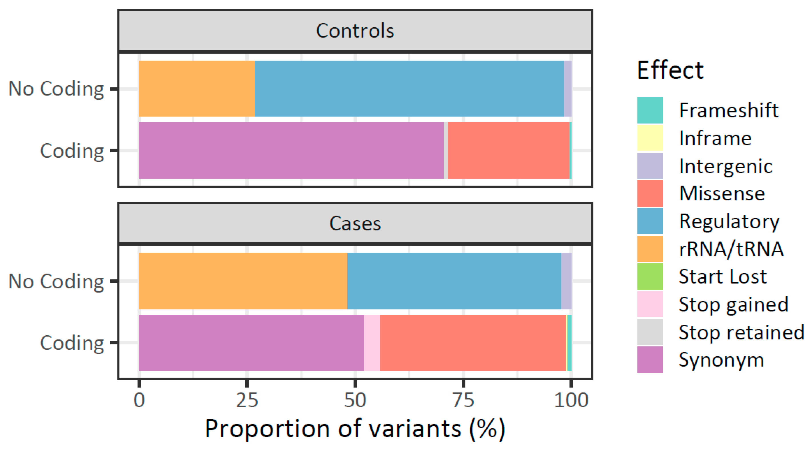
Figure 1. Functional effect of mtDNA variants in patients with breast cancer and healthy women. Distribution of mtDNA variants in peripheral blood of healthy women (Controls) and women with breast cancer (Cases) is represented. Variants are stratified by their location (coding and non-coding region) and in silico potential functional effect.
Ijms 26 08456 g001
Figure 2. Heteroplasmy level distributions of mtDNA germline variants in peripheral blood of patients with breast cancer and healthy women. Each box represents a variant. The heteroplasmy levels are reported on the y-axis, and the evaluated groups are shown on the x-axis (C = control, B = breast cancer). Points represent individuals and bars represent heteroplasmy levels (yellow: control, blue: case). Variants detected in at least 10 individuals are displayed.
Ijms 26 08456 g002
Figure 3. Mitochondrial DNA mutational burden in peripheral blood of patients with breast cancer and healthy women. The distribution of the total number of mtDNA germline variants identified in the peripheral blood of both groups is represented. Each black dot corresponds to the total number of variants per individual, while red circles represent the mean of total variants per group.
Ijms 26 08456 g003
Figure 4. Correlation among mitochondrial DNA alterations in breast cancer. The correlation of mitochondrial alterations analysis (variants in blood, variants in tumor, somatic mutations, mtCN in blood, and mtCN in tumor) in patients with breast cancer is shown. The right gradient color bar represents the degree of correlation (Spearman Correlation) among each alteration. Blue: positive correlation, red: negative correlation, yellow asterisks (*) indicate statistical significance (p < 0.05).
Ijms 26 08456 g004
Figure 5. Association of tumor mtDNA mutational burden and mitochondrial haplogroups with breast cancer prognosis. (A) Overall survival analysis stratified by mitochondrial mutational burden in tumor (low: <39 variants and high: ≥39 variants). (B) Metastasis-free survival analysis stratified by haplogroups. (C) Event-free survival analysis stratified by haplogroups. Kaplan–Meier curves: vertical lines (Ijms 26 08456 i001) indicate the event (death, metastasis, death and/or metastasis, as appropriate), while crosses (+) indicate patients with censored follow-up.
Ijms 26 08456 g005
Table 1. Clinical features of patients with breast cancer.
Clinical VariableFrequency
n (%)
Histological diagnosisInvasive Ductal Carcinoma76 (82.6)
Ductal Carcinoma In Situ1 (1.51)
Invasive Lobular Carcinoma11 (11.9)
Mixed Carcinoma3 (3.3)
NA1 (1.1)
Clinical StageI8 (8.7)
IIA43 (46.7)
IIB24 (26.1)
IIIA7 (7.6)
IIIB5 (5.4)
IIIC3 (3.3)
NA2 (2.2)
IHC classificationLuminal A57 (61.9)
Luminal B22 (24.0)
HER25 (5.4)
Triple Negative6 (6.5)
NA2 (2.2)
MetastasisPositive11 (11.9)
Negative65 (70.7)
NA16 (17.4)
DeathPositive9 (9.8)
Negative67 (72.8)
NA16 (17.4)
NA: Data not available; IHC: immunohistochemical.
Table 2. Number of mitochondrial DNA variants in peripheral blood of patients with breast cancer and controls.
Controls (N = 75)
n (%)
Cases (N = 92)
n (%)
p-Value
Total variants401866-
Mutational rate (mut/kb)24.252.3-
SNVs378 (94.3)831(95.9)0.23 a
Insertions11 (2.7)16 (1.9)0.41 a
Deletions12 (3.0)19 (2.2)0.51 a
Variants per individual ( x ¯ ± SE)33.6 ± 5.946.1 ± 16.86.225 × 10−10 *b
Range of variants per individual21–4927–109-
SNV: single nucleotide variant; x ¯ : mean; SE: standard deviation; a Chi-square test; b Wilcoxon test; * Statistically significant values.
Table 3. Number of variants per gene/region in peripheral blood mtDNA from patients with breast cancer and healthy women.
ControlsCases
Gen/RegionTotal Variants a
N = 402 (100%)
Total Variants a
N = 863 (100%)
p-Value c
D-Loop119 (29.6)164 (19.0)2.7 × 10⁻⁵ *
MT-ND525 (6.2)94 (10.9)0.011 *
tRNAs13 (3.2)57 (6.6)0.022 *
rRNAs32 (8.0)103 (11.9)0.045 *
MT-CO132 (8.0)54 (6.3)0.179
MT-ND66 (1.5)24 (2.8)0.234
MT-CO214 (3.5)22 (2.5)0.443
MT-ND426 (6.5)66 (7.6)0.542
MT-ND4L4 (1.0)13 (1.5)0.643
MT-ATP6 b26 (6.5)49 (5.7)0.651
MT-CO317 (4.2)32 (3.7)0.756
MT-ATP8 b8 (2.0)14 (1.6)0.803
MT-ND220 (5.0)46 (5.3)0.915
MT-CYB28 (7.0)58 (6.7)0.946
MT-ND123 (5.7)49 (5.7)1
MT-ND39 (2.2)18 (2.1)1
a Total number of mtDNA variants does not consider intergenic regions (eight and three variants in cases and controls, respectively); b variants on MT-ATP6 and MT-ATP8 genes may overlap (eight and four variants in cases and controls, respectively, were in both genes); c Chi-square test; * statistically significant values.
Table 4. Total heteroplasmic/homoplasmic mitochondrial DNA germline variants in patients with breast cancer and healthy women.
GroupHeteroplasmyHomoplasmy
Total Vars
n (%)
x ¯ MAF (Range)Total Vars
n (%)
x ¯ MAF (Range)
Controls85 (21.2)63.3% (22.2–95.0%)316 (78.8)99.8% (95–100%)
Cases576 (66.5)10.8% (0.02–94.8%)290 (33.5)99.7% (95.1–100%)
p-Value a<2.2 × 10⁻¹⁶ *<2.2 × 10⁻¹⁶ *
Total vars: total of mtDNA germline variants; x ¯ MAF: Mean of Mutant Allele Fraction; a Chi-square test; * statistically significant values.
Table 5. Peripheral blood mitochondrial DNA mutational burden association with risk of breast cancer.
Mutational BurdenControls (N = 75)Cases (N = 92)OR [CI]p-Value a
n (%)n (%)
High (>34 variants)35 (46.67)71 (77.17)3.83 [1.89–7.95]5.3 × 10⁻⁵ *
Low (≤34 variants)40 (53.33)21 (22.83)
OR: odds ratio; CI: confidence interval at 95%; a Chi-square test; * statistically significant values.
Table 6. Mitochondrial DNA germline variants associated with the risk of breast cancer.
VariantGene/RegionFunctional EffectControls (N = 75)
n (%)
Cases (N = 92)
n (%)
OR [CI]p-Value ap-adj b
A263GD-LoopRegulatory: OHR38 (50.7)16 (17.4)0.2 [0.1–0.4]5.8 × 10−61.5 × 10−4
A16183CD-LoopNC2 (2.6)21 (22.8)10.7 [2.5–97.3]9.3 × 10−52.3 × 10−4
C14766TMT-CYBMissense: T/I62 (82.6)91 (98.9)18.8 [2.7–816.3]1.4 × 10−43.5 × 10−3
C7028TMT-CO1Synonymous: A63 (84.0)91 (98.9)17.1 [2.4–746.2]6 × 10−40.02
A235GD-LoopRegulatory: OHR, mtTF1 BSX, CSB139 (52)24 (26.1)0.32 [0.2–0.6]7.3 × 10−40.02
G4820AMT-ND2Synonymous: E9 (12)28 (30.4)3.2 [1.3–8.3]4.8 × 10−30.12
G11719AMT-ND4Synonymous: G65 (86.6)90 (97.8)6.9 [1.4–66.5]6.5 × 10−30.16
T16519CD-LoopNC25 (33.3)50 (54.3)2.4 [1.2–4.7]7.9 × 10−30.16
T16189CD-LoopNC6 (8)21 (22.8)3.4 [1.2–10.8]0.010.27
C6473TMT-CO1Synonymous: I9 (12)26 (28.3)2.9 [1.2–7.5]0.010.32
CT16188CD-LoopNC1 (1.3)11 (11.9)9.9 [1.4–437.4]0.010.32
G11914AMT-ND4Synonymous: T9 (12.0)25 (27.2)2.7 [1.1–7.1]0.010.40
T4977CMT-ND2Synonymous: L9 (12.0)24 (26.1)2.6 [1.1–6.8]0.030.77
A10398GMT-ND3Missense: T/A13 (17.3)30 (32.6)2.3 [1.1–4.8]0.030.81
T16325CD-LoopNC13 (17.3)30 (32.6)2.3 [1.1–5.3]0.030.81
C10400TMT-ND3Synonymous: T11 (14.7)26 (28.3)2.3 [1.0–5.6]0.041
T9950CMT-CO3Synonymous: V11 (14.7)26 (28.3)2.3 [1.0–5.6]0.041
A8701GMT-ATP6Missense: T/A17 (22.7)35 (38.0)2.1 [1.0–4.4]0.041
C15535TMT-CYBSynonymous: N12 (16.0)27 (29.3)2.2 [1.0–5.1]0.041
T10873CMT-ND4Synonymous: P9 (12.0)23 (25.0)2.4 [1.0–6.4]0.041
A302ACD-LoopRegulatory: OHR, mtTF1 BSY, CSB23 (4.0)12 (13.0)3.6 [1.0–20.5]0.051
OR: Odds Ratio; CI: Confidence Intervals at 95%; a Fisher’s exact test; b p-adj: p-value adjusted by Bonferroni method; OHR: H-strand origin replication; mtTF1: Mitochondrial Transcription Factor 1 binding site; CSB: Conserved Sequence Blocks; NC: Non-coding; BSX: Binding sites X; BSY: Binding sites Y.
Disclaimer/Publisher’s Note: The statements, opinions and data contained in all publications are solely those of the individual author(s) and contributor(s) and not of MDPI and/or the editor(s). MDPI and/or the editor(s) disclaim responsibility for any injury to people or property resulting from any ideas, methods, instructions or products referred to in the content.