Review

Light and Shadows: Insights from Large-Scale Visual Screens for Arabidopsis Leaf Morphology Mutants

by
Lucía Juan-Vicente
,
Alejandro Ruiz-Bayón
and
José Luis Micol
*
Instituto de Bioingeniería, Universidad Miguel Hernández, Campus de Elche, 03202 Elche, Spain
*
Author to whom correspondence should be addressed.
These authors contributed equally to this work.
Int. J. Mol. Sci. 2025, 26(17), 8332; https://doi.org/10.3390/ijms26178332
Submission received: 1 August 2025 / Revised: 20 August 2025 / Accepted: 26 August 2025 / Published: 28 August 2025
(This article belongs to the Section Molecular Plant Sciences)

Abstract

Screens for specific phenotypes have long been a cornerstone of biology. Here, we present an updated synthesis of our large-scale visual screens for Arabidopsis (Arabidopsis thaliana) mutants that exhibit leaf morphology defects. In our 2009 review, we used phenotypes to group the leaf mutants that we had isolated and characterized since 1992; here, by contrast, we functionally classified the mutations that we studied over the last 16 years based on the biological programs they disrupt. Since 2009, we have identified and analyzed 38 genes required for proper leaf development; these genes are involved in translation, chloroplast function, cell wall construction, auxin homeostasis, microRNA biogenesis, and epigenetic regulation. Many of the identified mutants have pleiotropic phenotypes, consistent with the central roles of the affected pathways in development. In this review, we systematically link morphological traits to specific molecular dysfunctions, highlighting the enduring utility of forward genetic approaches. We found that the Arabidopsis leaf is a model organ of a model organism, and we have used this model-in-a-model system to dissect whole-plant traits such as cell proliferation and expansion, and to improve our understanding of the genetic control of plant form and size.

1. Introduction

Mutants, particularly from curated mutant collections, have long served as powerful tools for uncovering the genetic and molecular mechanisms underlying biological principles [1]. The first systematic mutant collections were established in the fruit fly Drosophila melanogaster by identifying mutations that altered wing shape or wing venation pattern [2]. D. melanogaster also offers a paradigmatic example of the usefulness of mutant collections: the mutants affecting embryonic development obtained by Nüsslein-Volhard and Wieschaus [3]. Other examples of collections of mutants in a model species include those affecting vulval differentiation in the nematode Caenorhabditis elegans [4] and those exhibiting diverse phenotypes in the mouse (Mus musculus) [5,6], which led to the identification of genes involved in fertility [7] and clonal B-cell lymphomagenesis [8].
In the plant kingdom, maize (Zea mays) and Arabidopsis (Arabidopsis thaliana) are prime examples of how mutant analysis has advanced our understanding of plant biology. Our research focuses on Arabidopsis, which was proposed as a model organism by Friedrich Laibach in 1943 [9] and has become a pillar of plant genetics research. Arabidopsis mutant collections generated through chemical, physical, and insertional mutagenesis have helped researchers elucidate diverse phenomena, including embryo development [10,11,12] and leaf morphogenesis [13]. These efforts have deepened our understanding of plant development and yielded an invaluable set of genetic resources for the plant science community [14].
The collections mentioned above primarily represent forward genetic approaches, in which mutagenesis was followed by screening to identify mutants with a phenotype of interest and characterization of the underlying genetic alterations. By contrast, reverse genetic approaches involve generating collections of mutants with altered expression of individual genes, allowing researchers to assess the phenotypes associated with known, and in most cases indexed, gene disruptions. Examples of reverse genetics resources include RNA interference (RNAi) lines targeting 71% of all genes in D. melanogaster [15], lines obtained using clustered regularly interspaced short palindromic repeats (CRISPR)/CRISPR-associated protein (Cas) for gene knockout or gene overexpression also in D. melanogaster [16], mutants obtained by transposon-based mutagenesis in C. elegans [17], and a comprehensive knockout collection in mice [18]. In Arabidopsis, two landmark collections of T-DNA insertion lines have enabled systematic functional studies across the genome: the SALK lines [19] and the Syngenta Arabidopsis Insertion Library (SAIL) lines [20].
Although mutants are invaluable for identifying genes involved in specific aspects of biology, characterization of mutants often reflects the research focus of the laboratories analyzing them. For example, David W. Meinke, who pioneered the study of embryo-defective (emb) mutants in Arabidopsis, noted that researchers who focused on gametophyte development frequently overlooked embryonic phenotypes. Conversely, Meinke and his collaborators generally excluded mutations that caused gametophytic lethality. He also highlighted a bias introduced by the prevalence of reverse genetic studies: the overrepresentation of emb mutants corresponding to well-studied protein families, reflecting research trends rather than biological importance [21,22].
Forward genetics helps circumvent researcher biases and enables the discovery of unexpected gene functions. For example, FLOWERING CONTROL LOCUS A (FCA) encodes an RNA-binding protein involved in post-transcriptional regulation; its effect on flowering time was discovered by a forward genetic approach [23]. The outcomes of forward genetic approaches, however, are not always aligned with the expectations of the researchers. For example, in our laboratory, screening for mutants with altered leaf morphology has led to the identification of genes affecting leaf morphogenesis as well as fundamental developmental processes throughout the Arabidopsis body and life cycle (Table S1). This observation mirrors the findings of Jürgens and colleagues, whose screens for embryonic patterning mutants also uncovered alleles of genes controlling essential cellular functions [11], rather than exclusively identifying specialized regulators of early development.
Over the years, we have conducted three large-scale screens for leaf morphological mutants in Arabidopsis: one using ethyl methanesulfonate (EMS) treatment [13], one using fast-neutron (FN) bombardment [24], and a third selecting publicly available lines from The Arabidopsis Information Service (AIS) collection [25]. To expand our collection, we also screened the SALK T-DNA insertion lines, identifying 706 additional leaf mutants, which we cataloged in our PhenoLeaf database [26].
In recent years, several reviews have been published on different facets of leaf development [27,28,29,30,31,32,33,34,35,36,37,38,39,40,41,42,43,44,45,46,47,48,49]. Some of these examine leaf organogenesis through the lens of mechanics [30,34,37,40] or evolution [39,45], and others discuss the contribution of ribosomal proteins [42] and hormones [41,48], or focus on genetics [47] or development of the leaf epidermis [36,49]. In the present review, we update and functionally reinterpret the collection of Arabidopsis leaf morphology mutants isolated in our laboratory, and characterized by our group and others since our previous synthesis in 2009 [50], incorporating the last 16 years of our research. In contrast to our previous phenotypic classification, we now organize our mutants based on the biological programs that their causal mutations disrupt, thereby aiming to provide a clearer view of gene function. We also highlight one exceptional mutant from the PhenoLeaf collection [26], notable for its unusual loss of leaf bilateral symmetry, and present a comprehensive table summarizing all characterized and pending mutant lines. In addition, we compiled the results of all double mutant combinations generated and published by our group since 1992, whose synergistic or additive morphological phenotypes either reveal or allow us to exclude putative genetic interactions. These results help define the functions of individual genes and the functional relationships within and between developmental pathways. In Section 2.1, Section 2.2, Section 2.3, Section 2.4, Section 2.5, Section 2.6 and Section 3, we examine the functional categories of our leaf morphology mutants, i.e., translation, chloroplast biogenesis and function, cell wall biosynthesis, auxin homeostasis, miRNA biogenesis and function, and epigenetic regulation. These categories, spanning several major processes of plant cells, give an indication of the extent of the insights provided by studying mutants exhibiting altered leaf morphology. All the mutations we have obtained and will discuss in this review are loss-of-function mutations, except where explicitly stated otherwise.

2. Leaf Morphology Mutants Studied Since 2009

2.1. Leaf Morphology Mutants with Defects in Translation

At least 18 mutants from our whole EMS collection (~9%) carry mutations in genes associated with translation; many of these mutations damage genes encoding ribosomal proteins (Tables S1 and S2). Although ribosomal proteins have been named based on whether they belong to the large subunit or the small subunit (RPLs and RPSs, respectively), an updated and consistent cross-kingdom nomenclature has been proposed [51,52]. However, here we use their originally published names for the sake of simplicity and provide their updated names in Table S1.
The first plant mutant affecting a gene encoding a ribosomal protein (RPS18A) was Arabidopsis pointed first leaves (pfl), which was isolated in the laboratory of Mieke Van Lijsebettens [53]. As its name suggests, the phenotype of pfl is most apparent on the first true leaves of seedlings, which are pointed instead of rounded [53]. Our angusta3 (ang3) mutant, which carries a mutation in RPL5B (uL18y), displays narrower leaves with a lower trichome density, smaller abaxial epidermal cells, and shorter hypocotyls and primary roots compared to the wild type (Figure 1B and Figure S1B; ref. [54]). Our apiculata2 (api2) mutant harbors a mutation in RPL36aB (eL42y), has pointed leaves and a main stem with reduced height (Figure 1C and Figure S1C; ref. [55]).
The mutants we initially named denticulata5 (den5), den12, and den30 carry mutations in RPL7B (uL30y), RPL10aB (uL1y), and RPL39C (eL39x), respectively, and were thus renamed rpl7b-1, rpl10ab-3, and rpl39c-1, respectively. Additionally, the den29 mutant carries a deletion of two adjacent genes, RPS15aB (uS8mz) and RPL28A (eL28z), and was renamed as the rps15ab-1 rpl28a-3 double mutant. All den mutants have small, reticulated, narrow and pointed leaves, with teeth visible on their first- and second-node leaves (Figure 1E–I and Figure S1E–I). The palisade mesophyll cells of these mutants are larger than those of the wild type and display increased ploidy levels. Several den mutants have additional distinct traits: the leaves of the den5 and den29 mutants show parallel veins on their proximal lamina, and den29 and den30 show a disorganized palisade mesophyll [56]. The den2 mutant has pointed, toothed leaves (Figure 1E and Figure S1E), and carries a mutation in SMALL ORGAN4 (SMO4), which encodes a ribosome biogenesis factor involved in 5.8S rRNA maturation; it was characterized in the laboratory of María Rosa Ponce [57]. We are currently studying the den3, den4, den6, den7, den9, den13, den14, and den15 mutants (Table S2), all of which exhibit a similar phenotype; most, but not all, of these mutants carry mutations in genes related to the translational apparatus.
Figure 1. Leaf phenotypes of mutants with defects in translation. Rosettes of the wild-type Landsberg erecta (Ler) (A), and the homozygous mutants ang3 (B), api2 (C), api7-1 (D), den2 (E), den5 (rpl7b-1) (F), den12 (rpl10ab-3) (G), den29 (rps15ab-1 rpl28a-3) (H), and den30 (rpl39c-1) (I). Photographs were taken 21 days after stratification (das). Scale bars, 2 mm. These mutants were described in [54] (B), ref. [55] (C), ref. [58] (D), ref. [57] (E), and [56] (FI). All figures in this review feature photographs taken concurrently from plants cultivated in parallel under the same controlled growth conditions.
Ijms 26 08332 g001
The apiculata7-1 (api7-1) mutant carries a mutation in ATP-BINDING CASSETTE E2 (ABCE2), which encodes a protein responsible for dissociating cytoplasmic ribosomes at the end of translation and thus contributes to ribosome recycling. The api7-1 plants exhibit a phenotype typical of ribosomal protein mutants, namely a small rosette and pointed, toothed, and pale leaves that contain reduced levels of photosynthetic pigments and have a simple venation pattern (Figure 1D and Figure S1D). Additionally, both the leaf epidermal cells and the overall size of api7-1 plants are reduced compared to the wild type [58].
All the mutants affected in genes encoding components of the translational apparatus that we isolated in our screens share a common phenotype characterized by pointed, narrow, and toothed leaves, along with alterations in cell size across all leaf layers. However, why mutations affecting translation cause this phenotype remains unclear. Reduced protein synthesis can explain the diminished or retarded growth of these mutants, as was suggested long ago for the Minute phenotype of Drosophila melanogaster, which includes prolonged development and short, thin bristles. Indeed, the connection between the Minute mutants and defects in protein translation was first proposed in the late 1970s and early 1980s, when researchers began discovering that many Minute genes encode ribosomal proteins [59,60].
The narrow-leaf phenotype of the den mutants may reflect the role of ribosomal proteins in regulating the translation of transcripts that harbor upstream open reading frames (uORFs). For example, in rice (Oryza sativa), loss of OsRPS3A compromises the reinitiation of translation in uORF-containing mRNAs such as those transcribed from AUXIN RESPONSE FACTOR 11 (OsARF11), OsARF16, and WUSCHEL-RELATED HOMEOBOX (OsWOX3A), ultimately leading to a narrow-leaf phenotype [61]. Indeed, despite their shared traits, the phenotypes of our den mutants differ, possibly due to the depletion of specific ribosomal proteins that may differentially affect the translation of certain transcripts, leading to distinct phenotypes [56].
In Arabidopsis, 249 genes encode the 81 distinct proteins that constitute the cytosolic ribosome, reflecting a remarkable heterogeneity compared to animals, whose genomes usually harbor fewer than 100 ribosomal protein genes. One plausible explanation for the large number of ribosome protein genes in plants is that highly proliferative tissues, such as leaf primordia, may require elevated expression of ribosomal protein genes. The correlation between differential expression of ribosomal protein paralogs and cell division rates suggests that the phenotypic differences that we observed may result from dosage effects rather than functional specialization [42,62,63].

2.2. Leaf Morphology Mutants with Defects in Chloroplast Biogenesis and Function
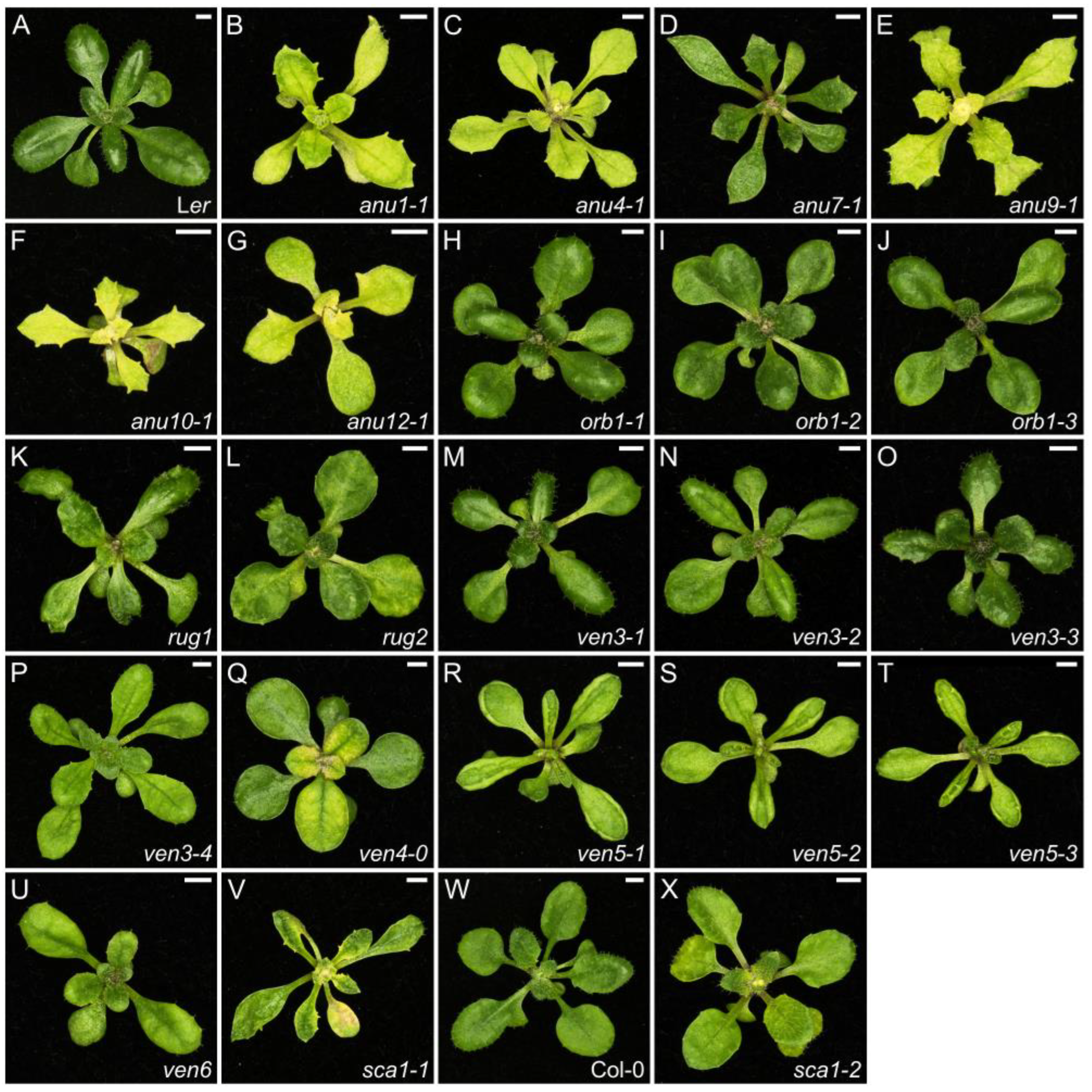
The chloroplast consists of thylakoids organized in grana and surrounded by stroma, all enclosed by two membrane layers. Many chloroplast proteins are encoded by nuclear genes and chloroplasts affect nuclear gene expression by retrograde signaling [64]. We identified 27 mutants (~13%) in our screens with mutations in genes related to chloroplast function (Tables S1 and S2). We used next-generation sequencing to identify the causal mutations of the phenotypes of four angulata (anu) mutants with pale-yellow leaf laminae and irregular leaf margins with prominent teeth (anu1-1, anu4-1, anu9-1, and anu12-1; Figure 2B,C,E,G and Figure S2B,C,E,G; ref. [65]). The anu1-1 and anu4-1 mutants were found to harbor mutations in the SECA2 and TRANSLOCON AT THE OUTER MEMBRANE OF CHLOROPLASTS33 (TOC33) genes, respectively. The secretory pathway SECA2 protein, together with the TOC33 protein, participate in the import of proteins from the cytosol into chloroplasts [66,67]. NON-INTRINSIC ABC PROTEIN14 (NAP14; [68]) encodes a protein that is truncated by the anu9-1 mutation and has a role in chloroplast homeostasis of transition metals such as iron and manganese, which are essential for photosynthesis [69]. The anu12-1 mutation damages the coding sequence of CLP PROTEASE PROTEOLYTIC SUBUNIT1 (CLPR1), whose product is involved in protein degradation in chloroplasts [70].
The two remaining anu mutants with chloroplast aberrations identified in our screens carry mutations that disrupt genes with functions that remain to be elucidated. The anu7-1 and anu10-1 plants exhibit leaves that are toothed, pale green, reduced in length, and display variation in the size of palisade mesophyll cells, as well as low levels of chlorophylls and carotenoids (Figure 2D,F and Figure S2D,F; ref. [71,72]). In addition, the anu10-1 mutant is compromised in leaf lateral expansion and accumulates H2O2 in its chloroplasts, which are small and abnormally shaped with fewer thylakoid membranes per granum [71,72]. ANU10 is present in the thylakoid membrane and is presumably required for thylakoid biogenesis and grana stacking [71]. ANU7 has a conserved DnaJ-like cysteine-rich domain, and other DnaJ proteins participate in various processes that take place in chloroplasts, like maintenance of photosystem II under chilling stress or chloroplast iron–sulfur cluster biogenesis [72,73,74]. Furthermore, we combined the anu7-1 mutation with a loss-of-function allele of GENOMES UNCOUPLED 1 (GUN1), which encodes a chloroplast-localized pentatricopeptide repeat (PPR) protein with a C-terminal small MutS-related domain. GUN1 plays a central role in chloroplast-to-nucleus retrograde signaling, but its specific function remains controversial, although it apparently involves the maturation of plastid transcripts [75,76,77]. The anu7-1 gun1 double mutant (Table S3) shows a synergistic phenotype with albino to completely green leaf sectors, thus revealing a genetic interaction [72].
The rugosa1 (rug1) plants are shorter and flower later than the wild type; they also produce irregularly shaped leaves that develop lesions (Figure 2K and Figure S2K). The rug1 mutation damages the gene that encodes HYDROXYMETHYLBILANE SYNTHASE (HEMC, also reported as PORPHOBILINOGEN DEAMINASE [PBGD]), which is involved in tetrapyrrole biosynthesis, and therefore, in chlorophyll biosynthesis [78]. The hemc-1 mutant exhibits altered RNA editing efficiency of four chloroplast transcripts [79]. Since chloroplast RNA editing and retrograde signaling appear to be somewhat related [80], RUG1 may also have a role in retrograde signaling, but this has not yet been experimentally confirmed. The rug2 mutant has pale and small hypocotyls, rosettes, stems and fruits, and short roots, as well as leaves with green and pale sectors, the latter containing sparsely packed mesophyll cells with chloroplasts abnormally shaped and reduced in number compared to wild-type cells (Figure 2L and Figure S2L; ref. [81]). RUG2 is also known as BELAYA SMERT (BSM) and encodes a homolog of metazoan mitochondrial transcription termination factor (mTERF4). Arabidopsis mTERF4 has a role in intron splicing in chloroplasts [82,83].
VENOSA3 (VEN3) and VEN6 encode the large and small subunits, respectively, of carbamoyl phosphate synthetase (CPS) [84]. This enzyme is located in chloroplasts and generates carbamoyl phosphate, which is required for arginine and pyrimidine biosynthesis [85,86]. The ven3 and ven6 mutants have reticulated leaves with green veins and a generally pale lamina; their juvenile leaves are hyponastic, and adult leaves are more toothed than those of the wild type (Figure 2M–P,U and Figure S2M–P,U). These reticulated leaves have fewer palisade mesophyll cells [84]. We discovered that VEN3 and VEN6 are overexpressed in the orbiculata1-3 (orb1-3) mutant. ORB1 encodes the chloroplast-localized GLUTAMATE SYNTHASE1 (GLU1), which is also referred to as FERREDOXIN-DEPENDENT GLUTAMINE OXOGLUTARATE AMINOTRANSFERASE1 (FD-GOGAT1) [87,88,89]. The leaf lamina of orb1 mutants is smaller and exhibits diminished growth caused by small palisade mesophyll cells, and pale green pigmentation due to low levels of chlorophyll a, chlorophyll b, and carotenoids (Figure 2H–J and Figure S2H–J). As CPS and GLU1 both use glutamine as their substrate, the overexpression of VEN3 and VEN6 in the orb1-3 mutant may be a compensatory mechanism to lower its high glutamine levels [90].
Another mutant with reticulated and pale leaves is ven4-0 (Figure 2Q and Figure S2Q), which carries a point mutation in VEN4, encoding the most likely Arabidopsis ortholog of human Sterile alpha motif and histidine-aspartate domain containing protein 1 (SAMHD1). Like human SAMHD1, Arabidopsis VEN4 is a deoxynucleotide triphosphohydrolase (dNTPase) involved in maintaining the dNTP pool [91] and in repairing DNA double-stranded breaks by homologous recombination [92]. The ven4-0 mutant also accumulates lower levels of chlorophyll and has smaller chloroplasts in palisade mesophyll cells, with more plastoglobuli and poorly organized thylakoids [91]. This lower chloroplast number may be a consequence of insufficient chloroplast genome replication due to lower dNTP levels, a trait also observed in the crinkled leaves 8-1 (cls8-1) mutant, which carries a mutation in CLS8 encoding a protein involved in de novo dNTP biosynthesis [93]. VEN5, also known as RETICULATA-RELATED3 (RER3), encodes a protein of unknown function localized in the chloroplast envelope. We renamed our ven5-1, ven5-2, and ven5-3 mutants, all of which carry point mutations in RER3, to rer3-1, rer3-2, and rer3-3, respectively. These mutants exhibit reticulated, hyponastic, and toothed leaves (Figure 2R–T and Figure S2R–T). Additionally, the leaf cells of these mutants show lower ploidy levels, their epidermal cells are smaller and have a disorganized mesophyll structure. Accumulation of H2O2 in the tissue surrounding their vasculature leads to localized cell death and causes leaf reticulation [94].
SCABRA1 (SCA1) encodes PLASTID RIBOSOMAL PROTEINS5 (RPS5). The sca1-1 and sca1-2 plants are smaller than the wild type, produce pale green leaves with lower contents of chlorophyll a and b and lower maximum efficiency of photosystem II than the wild type, together with an uneven leaf surface, prominent marginal teeth, and small palisade mesophyll cells (Figure 2V,X and Figure S2V,X). The sca1-1 mutation enhances the phenotype observed for asymmetric leaves2-1 (as2-1; ref. [95]), as shown by the leaf polarity defects of the sca1-1 as2-1 double mutant (Table S3), which produced radialized leaves and some trumpet-shaped leaves, uncovering a role for plastids in the establishment of adaxial–abaxial polarity [96]. As mentioned above, the den10 and den17 mutants are currently under study in our laboratory and carry mutations in genes that encode chloroplast components (Table S2).
Figure 2. Leaf phenotypes of mutants with altered chloroplast biogenesis and function. Rosettes of the wild-types Ler (A) and Columbia (Col-0) (W), and the anu1-1 (B), anu4-1 (C), anu7-1 (D), anu9-1 (E), anu10-1 (F), anu12-1 (G), orb1-1 (H), orb1-2 (I), orb1-3 (J), rug1 (K), rug2 (L), ven3-1 (M), ven3-2 (N), ven3-3 (O), ven3-4 (P), ven4-0 (Q), ven5-1 (rer3-1) (R), ven5-2 (rer3-2) (S), ven5-3 (rer3-3) (T), ven6 (U), sca1-1 (V), and sca1-2 (X) homozygous mutants. Photographs were taken 21 das. Scale bars, 2 mm. These mutants were described in [65] (B,C), ref. [72] (D), ref. [65] (E), ref. [71] (F), ref. [65] (G), ref. [90] (HJ), ref. [78] (K), ref. [81] (L), ref. [84] (MP), ref. [91] (Q), ref. [94] (RT), ref. [84] (U), ref. [96] (V), and [96] (X).
Ijms 26 08332 g002
Not unexpectedly, mutations in genes associated with the biosynthesis of photosynthetic pigments and chloroplast structure affect leaf morphology. The lower accumulation of photosynthetic pigments results in pale or reticulated leaves, whereas abnormalities in chloroplast structure and function impair cell expansion, altering the final leaf shape and size, leading, in this case, to toothed leaf margins and smaller rosettes.

2.3. Leaf Morphology Mutants Altered in Cell Wall Biosynthesis

Organ size and shape in plants are governed by cell proliferation and expansion, both of which depend on precise control of cell wall biogenesis. Cell division involves the formation of new cell walls at the division plane and cell expansion requires the insertion of newly synthesized polysaccharides to enable wall loosening and elongation [97]. Therefore, mutations that disrupt any aspect of cell wall construction may directly influence leaf shape. From our EMS mutagenesis, we have identified seven mutants (~3%) affected in genes related to cell wall construction (Tables S1 and S2).
Our serrata4-1 (sea4-1) and sea4-2 mutations are the only known viable mutant alleles of KEULE (KEU), which encodes a Sec1/Munc18 (SM) protein that interacts with the syntaxin KNOLLE (KN) in the plane of division [98]. This interaction facilitates the formation of trans-SNARE [soluble N-ethylmaleimide-sensitive factor (NSF)-attachment protein receptor] complexes between vesicles that then fuse to form the new cell wall between the two daughter cells during cytokinesis [99]. The sea4-1 and sea4-2 plants have small rosettes, with fewer leaves at bolting compared to wild type, as well as shorter stems and roots. Their leaves are serrated, small, and wavy, displaying a more complex venation pattern than the wild type, and undergoing premature senescence (Figure 3G,H and Figure S3G,H). Their leaf mesophyll cells are generally smaller, but epidermal cells have a simpler shape with fewer protuberances, and both mutants show increased levels of endoreduplication. The transcriptomic changes observed in the sea4 mutants suggest a drop in cell wall integrity, which may contribute to the observed phenotype [100].
The exigua1 (exi1), exi2, and exi5 mutants carry point mutations in the CELLULOSE SYNTHASE A8 (CESA8), CESA4, and CESA7 genes, respectively. These genes encode enzymes that synthesize cellulose microfibrils in the secondary cell wall during its formation [101]. These exi mutants exhibit small, dark-green leaves, short stems, roots and siliques, and small flowers (Figure 3B–E and Figure S3B–E). Additionally, their leaf cells are smaller and have a lower ploidy level compared to wild type, as also observed in other cesa mutants such as cesa6 [102]. In the exi1, exi2, and exi5 mutants, CESA dysfunction appears to cause the collapse of xylem vessels, which in turn reduces the turgor pressure necessary for proper cell expansion [103].
Figure 3. Leaf phenotypes of mutants altered in cell wall biosynthesis. Rosettes of the wild-type Ler (A), and the exi1-1 (B), exi1-2 (C), exi1-3 (D), exi2 (E), exi5 (F), sea4-1 (G), and sea4-2 (H) homozygous mutants. Photographs were taken 21 das. Scale bars, 2 mm. These mutants were described in [103] (BF), and [100] (G,H).
Ijms 26 08332 g003
Due to the importance of cell wall construction for the complete development of the plant body, mutations in genes involved in cell wall biosynthesis result in a pleiotropic morphological phenotype. Indeed, the sea4, exi1, exi2, and exi5 mutants exhibit alterations in rosette size, stem and root length, leaf shape, color, size, venation pattern and internal structure, inflorescences, and ploidy. Cytokinesis is required for leaf growth as shown in sea4 mutants. In addition, those mutants have lost cell wall integrity, which triggers an immune response followed by senescence. Defective cell wall construction also compromises water transport due to vessel collapse, leading to decreased turgor in the exi mutant cells; this negatively influences cell expansion, thus explaining their small size.

2.4. Leaf Morphology Mutants with Defects in Auxin Homeostasis
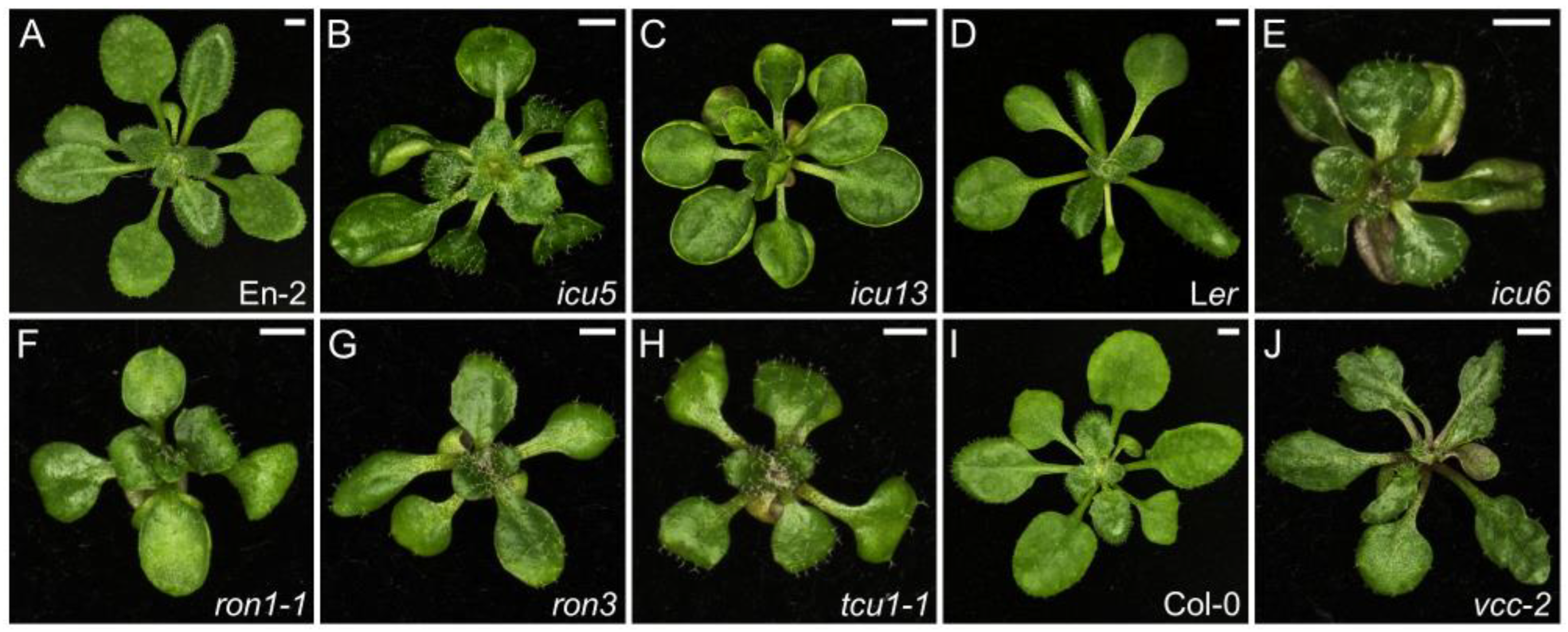
Many aspects of plant development rely on the phytohormone auxin, which promotes cellular differentiation and enhances or inhibits growth depending on the cellular context and its concentration. Auxin responses lead to changes in the transcriptome, but these responses can also occur rapidly and directly at the cell surface, although the underlying molecular mechanism is not completely understood (reviewed in [104,105]). Ten of the mutants (~5%) identified in our screens are defective in auxin homeostasis (Tables S1 and S2).
The incurvata13 (icu13) mutant carries a C → T transition that generates a new splicing donor site in AUXIN RESISTANT6 (AXR6), which encodes the CULLIN1 (CUL1) component of the Skip–Cullin–F-box TIR1 (SCFTIR1) ubiquitin ligase complex [106]. The icu13 plants have a pleiotropic phenotype including diminished auxin responsiveness and leaves that are hyponastic, with a simple venation pattern, small adaxial pavement cells, densely packed palisade, and spongy mesophyll cells, as well as increased numbers of rosette leaves at bolting, early flowering, and reduced plant stature and apical dominance (Figure 4C and Figure S4C; ref. [107]). We isolated the icu6 mutant in our EMS screen and found that it carries a semi-dominant gain-of-function allele of AXR3 (also named INDOLE-3-ACETIC ACID INDUCIBLE17 [IAA17]), which encodes a transcriptional regulator that represses auxin-inducible gene expression. The heterozygous icu6/AXR3 plants also have hyponastic leaves and smaller adaxial pavement cells but do not share any other phenotypes with icu13, as icu6/AXR3 plants have smaller rosettes, pronounced apical dominance, and enhanced responsiveness to auxin (Figure 4E and Figure S4E; ref. [108,109,110,111]). The icu5 mutant turned out to carry a hypermorphic allele of IAA3 (also named SHORT HYPOCOTYL2 [SHY2]), encoding another repressor of auxin responses; we renamed icu5 as shy2-10 accordingly [107,112,113]. Leaves of the shy2-10 mutant are smaller than those of the wild type and have more free-ending veins (Figure 4B and Figure S4B; ref. [107]).
The loss-of-function rotunda3 (ron3) mutant allele of ROTUNDA3 (RON3) is characterized by shorter rosette lamina, smaller leaf width and area, together with a late-flowering phenotype, shorter roots exhibiting hypergravitropic growth, and lower apical dominance (Figure 4G and Figure S4G; ref. [114]). A role for RON3 in splicing and microRNA (miRNA) biogenesis was previously reported through the analysis of its sickle-1 (sic-1) allele [115]. A Tandem Affinity Purification (TAP) assay revealed that RON3 interacts with six subunits of the cytosolic Protein Phosphatase 2A (PP2A) complex, which is involved in the polar localization of the PIN-FORMED (PIN) family of auxin efflux carriers [114,116,117,118]. In situ immunodetection of PIN1 and PIN2 in the root cells of the ron3 mutant revealed a role for RON3 in PIN trafficking, promoting both basal internalization and apical delivery or maintenance of PIN auxin carriers [114]. Another physical interactor of RON3 is DEBRANCHING ENZYME1 (DBR1), which enables debranching of the lariat intronic RNAs produced by intron excision during splicing [119]. RON3 also represses photomorphogenic growth through its interaction with ELONGATED HYPOCOTYL 5 (HY5) and PHYTOCHROME INTERACTING FACTOR 4 (PIF4), two antagonistic transcription factors that specifically regulate growth largely under light [120].
Other genes under study in our laboratory are not directly related to auxin signaling, but their loss of function leads to altered auxin responses. The ron1-1 mutant exhibits rounded leaves, with an open venation pattern, fewer lateral roots, late flowering, and loss of apical dominance (Figure 4F and Figure S4F). RON1 was previously reported as FIERY1 (FRY1; [121]), SAL1 [122], HIGH EXPRESSION OF OSMOTICALLY RESPONSIVE GENES2 (HOS2; ref. [123]), and ALTERED EXPRESSION OF APX2 (ALX8; ref. [124]). Although RON1 encodes an enzyme with inositol polyphosphate 1-phosphatase and 3′(2′),5′-bisphosphate nucleotidase activities, which are required for inositol triphosphate (InsP3) metabolism, the venation pattern phenotypes of the ron1-1 mutant suggest a role in auxin perception, as this phytohormone has a prevalent role in leaf vascular patterning [125,126]. Indeed, an interplay between inositol and auxin-mediated development has been reported, but has not been demonstrated yet [127,128].
Another example of a mutation that is not directly involved in auxin signaling but does cause an altered auxin response is transcurvata1-1 (tcu1-1), an allele of NUCLEOPORIN58 (NUP58). NUP58 is a component of the central barrier of the nuclear pore complex, along with NUP54 and NUP62, and thus, it is involved in nucleocytoplasmic trafficking of macromolecules [129,130]. The tcu1-1 mutant has a pleiotropic phenotype with longer hypocotyls and petioles, reticulated, smaller, and asymmetrically epinastic leaf lamina, and early flowering (Figure 4H and Figure S4H). Other nup mutants also show an early flowering phenotype, such as nup54, nup62, nup96, nup98a nup98b, nup136, and nup160 [131,132,133,134,135,136]. Double mutant combinations (Table S3) indicated a functional relationship between TCU1 and ICU5, AXR1 and AXR3, suggesting that TCU1 has a role in auxin signaling [130]. Indeed, AXR1 and AXR3 are partially retained in the cytoplasm of nup54 nup58 nup62 triple mutant protoplasts [137]. The early flowering phenotype of the tcu1-1 mutant is caused by the retention of the MADS-box transcription factor FLOWERING LOCUS C (FLC) in the cytoplasm [137].
Figure 4. Leaf phenotypes of mutants with defects in auxin homeostasis. Rosettes of the wild-types Enkheim-2 (En-2) (A), Ler (D), and Col-0 (I), and the icu5 (shy2-10) (B), icu13 (C), icu6 (E), ron1-1 (F), ron3 (G), tcu1-1 (H), and deal1-1 (vcc-2) (J) homozygous mutants. Photographs were taken 21 das. Scale bars, 2 mm. These mutants were described in [107] (B,C), ref. [111] (E), ref. [125] (F), ref. [114] (G), ref. [130] (H), and [138] (J).
Ijms 26 08332 g004
In our PhenoLeaf collection (https://genetics.umh.es/query-phenoleaf-db/ (accessed on 1 July 2025); ref. [26]), the desigual1-1 (deal1-1) mutant line stood out because of its unusual phenotype, as its leaves clearly deviated from the bilateral symmetry normally seen in the wild type, and without visible alterations in dorsoventral polarity [138]. We later identified DEAL1 as the previously reported gene VASCULATURE COMPLEXITY AND CONNECTIVITY (VCC; ref. [139]), which prompted us to rename the deal1 mutants as vcc. The leaf margins of vcc plants show aberrant lobes and sinuses that are bigger than those of the wild type, whose randomness ultimately causes loss of bilateral symmetry (Figure 4J and Figure S4J). Although alterations to the leaf medio-lateral axis are usually caused by changes in dorsoventral polarity [140], vcc mutants only show alterations in the medio-lateral axis without disturbing either the leaf dorsoventral axis or the proximo-distal axis. Notably, these differences in growth are related to cell proliferation, not cell expansion. The auxin maxima are unevenly located in the leaf primordia of vcc mutants, which do not show alterations in the venation pattern. CUP-SHAPED COTYLEDON2 (CUC2), a transcriptional regulator that influences auxin distribution, is expressed in well-defined and evenly spaced domains in leaf primordia of the Columbia-0 (Col-0) wild type. This pattern is lost in the vcc mutants, which show abnormally close or distant consecutive CUC2 domains, whose disposition was asymmetrical in primordia margins. VCC appears to be necessary for communication between growth-promoting and growth-repressing signals by mediating auxin and CUC2 signaling [138].
Our mutants with defects in auxin signaling are difficult to classify due to the diverse phenotypes arising from the complexity of auxin responses, which can alter the transcriptome in vastly different ways depending on the mutated gene. In fact, some mutants exhibit opposite phenotypes, such as lower or greater auxin responsiveness, early or late flowering, more complex or simplified venation patterns, and hyponastic or epinastic leaves with rounded or aberrant lobes and sinuses. This diversity can be explained because auxin response depends on both the correct auxin trafficking mediated by PIN proteins and apparently modulated by RON3, RON1, and VCC; and the proper SCF-mediated ubiquitination of IAA proteins followed by 26S proteasome degradation, which is presumably impaired in icu13, icu6, icu5, and tcu1 mutants. Given the high number of leaf morphology mutants that exhibit altered auxin responses (Table S1), the groups of Karin Ljung and Ondřej Novák developed a high-throughput method to screen for altered auxin metabolite profiles, which they tested using our collection of mutants, and which should be valuable for other large-scale mutant collections [141].

2.5. Leaf Morphology Mutants with Impaired miRNA Biogenesis and Function

An important mechanism of post-transcriptional regulation of gene expression is accomplished via endogenous small RNA molecules known as miRNAs, which impose translation inhibition, mRNA cleavage, and/or RNA-directed DNA methylation depending on the target transcript or gene (reviewed in [142]). At least eight mutants (~4%) from our screens are related to miRNA biogenesis (Tables S1 and S2).
In addition to leaf upward curvature, the incurvata mutants icu8, icu9, and icu15 share additional leaf phenotypes, including a poorly defined boundary between lamina and petiole and the presence of trichomes on the abaxial epidermis, fewer stomata per leaf, and a simpler venation pattern than in the wild type (Figure 5B–F and Figure S5B–F). These icu mutants are late flowering, have small and compact inflorescences with prematurely open flowers, incompletely fused carpels, and reduced fertility. The phenotypic similarities among these mutants suggested that they function in the same developmental program, and in fact all have been shown to participate in miRNA biogenesis and function. Indeed, icu8, icu9, and icu15 were shown in the laboratory of María Rosa Ponce to carry new alleles of the well-characterized genes HYPONASTIC LEAVES1 (HYL1), ARGONAUTE1 (AGO1), and HUA ENHANCER1 (HEN1), respectively [143]. HYL1 acts in several steps of miRNA biogenesis, from the transcriptional regulation of MIR genes to the inhibition of target mRNA translation, and has other roles not related to miRNA signaling such as the transcriptional regulation of genes related to plastid organization (reviewed in [144]). HEN1 methylates the miRNA/miRNA* duplex at the 3′ terminal nucleotides [145] and interacts with HYL1 [146]. AGO1 is the catalytic subunit of the RNA-induced silencing complex (RISC), the effector complex that mediates gene silencing [147].
The TCU2 gene, which encodes the auxiliary subunit of the NatB N-alpha-acetyltransferase complex [148], was serendipitously found to interact with AGO10. Our tcu2-1 mutant was isolated in the Landsberg erecta (Ler) background and showed a stronger phenotype than the tcu2-2 mutant in the Col-0 background, suggesting the presence of a genetic modifier in Ler. This genetic modifier was zwille-2 (zll-2) [149], a previously described allele of AGO10, and the tcu2-1 zll-2 double mutant showed a synergistic leaf phenotype with strong folding of the leaf (Table S3), which suggests a functional relationship between TCU2 and AGO10. Otherwise, the loss of TCU2 function alone suggest some degree of auxin deficiency, since the tcu2 mutants show leaf reticulation and downward folding at an oblique angle relative to the primary vein, early flowering, unfertilized or aborted ovules in siliques, and a greater proportion of siliques with more than two valves than in the Ler and Col-0 wild-type accessions (Figure 5G and Figure S5G).
Our mutants with perturbed silencing by miRNAs, like those in auxin signaling, have no easily predictable effects on the overall plant phenotype. Depending on the specific mutated component of the miRNA pathway, the resulting phenotype can vary substantially, even displaying opposite traits, as seen with the icu and tcu2 mutants. The icu mutants have hyponastic leaves and flower late, whereas the tcu2 mutants have epinastic leaves and early flowering. This variability in phenotypic outcomes presents challenges when identifying mutants with altered miRNA biogenesis based solely on plant morphology but evidences the need for spatial and temporal gene expression fine-tuning for proper morphogenesis.

2.6. Leaf Morphology Mutants with Altered Epigenetic Machinery

In plants, acclimation, vernalization, systemic acquired resistance, and imprinting are mitotically heritable and epigenetically regulated [150,151]. Other aspects of plant development and physiology that have been considered to be at least partially under epigenetic control include morphological plasticity, light signaling, stress responses, male and female gametogenesis, and flowering time. A recent review of the epigenetic machinery in plants highlights the revolutionary potential of advanced epigenetic tools to enhance crop productivity [152].
Polycomb group (PcG) proteins are key components of the epigenetic machinery, are highly conserved among eukaryotes, and epigenetically repress the expression of many genes, including those controlling growth, development, and environmental adaptation [153,154]. PcG proteins form part of two heteromultimeric Polycomb Repressive Complexes (PRCs) with different epigenetic functions: PRC1 is a ubiquitin ligase targeting histone H2A, whereas PRC2 is an H3K27 methyltransferase [155]. Seventeen mutants (~8%) of our mutant collection carry mutations in genes belonging to the epigenetic machinery (Tables S1 and S2).
The proteins encoded by seven genes identified from our mutant screens and/or studied prior to 2009 have epigenetic functions: CURLY LEAF [156] (CLF, which we initially called INCURVATA1 [ICU1] [157]), a PRC2 core component; the histone acetyltransferases ELONGATA1–4 (ELO1–ELO4) [158,159]; HISTONE MONOUBIQUITINATION1 (HUB1, which we initially named ANGUSTA4 [ANG4]), an E3 ligase that monoubiquitinates H2B [160]; and INCURVATA2 (ICU2), the catalytic subunit of DNA polymerase α, which interacts with chromatin remodeler complexes and whose loss of function affects the maintenance of repressive chromatin states at loci that are PRC2 targets [157,161,162].
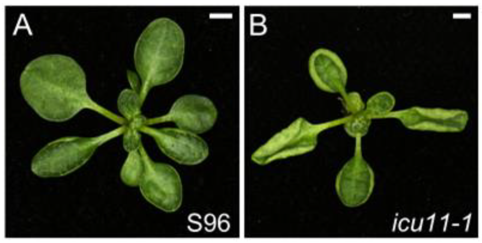
The icu11-1 mutant has a pleiotropic phenotype that includes hyponastic leaves, early flowering, smaller palisade mesophyll cells, and lower fertility than the wild type (Figure 6 and Figure S6). ICU11 encodes a 2-oxoglutarate and Fe(II)-dependent dioxygenase [163] that appears to be a PRC2 accessory protein that facilitates demethylation of the H3K36me3 active mark [163,164]. ICU11 and its closest paralog, CUPULIFORMIS2 (CP2), show unequal functional redundancy and are required for vegetative development in Arabidopsis, as icu11 cp2 double mutant plants skip the vegetative phase and develop floral organs immediately upon germination (Table S3; ref. [163,165]). CP2 may also act as a PRC2 accessory protein, and a transcriptome landscape of the icu11-5 cp2-1 double mutant resembles that of the loss-of-function PRC2 mutant embryonic flower 2-3 (emf2-3), which lacks EMF2, a core PRC2 component [166].
Comparing the phenotypes of the icu11-1 mutant and epigenetic signaling mutants previously studied in our laboratory [160,161] does not reveal a distinct characteristic phenotype for this class of mutants. As mentioned in previous sections, this may be due to the broad influence of these mutations on plant functions that affect the expression of many genes. Indeed, the phenotype of these mutants can be explained by the heterochronic and ectopic derepression of genes. For example, the clf, icu2, and icu11 mutants show an early flowering phenotype caused by the expression of floral homeotic genes such as AGAMOUS or SEPALLATA3 [156,161,163].

3. Concluding Remarks

From our mutant screens, we identified 208 mutants affecting the morphology of Arabidopsis leaves, which we found to fall into 144 complementation groups (Table S2). This set of mutants does not include the insertional mutants that we selected from the Arabidopsis SALK collection and described in the PhenoLeaf database, which comprises 706 mutant lines [26]. Of these 208 mutants, 83 were found to carry alleles of 41 previously characterized genes, and characterization of 30 additional mutants identified 22 genes not previously studied at the functional level (Table S2). In the present review, we have summarized the findings obtained from the analysis of 52 mutants (corresponding to 38 genes) since 2009, in some cases in collaboration with other groups or by other groups. We are currently studying 13 DEN genes, but we still have 74 mutant lines yet to be studied, which presumably correspond to 61 different genes (Table S2). Further analysis of these mutants will surely unveil previously missed functions related to Arabidopsis leaf development.
Some functional classes have conspicuous phenotypes. For example, all mutants related to protein translation mentioned above have either narrow or toothed leaves (Figure 1), whereas most mutants with defects in auxin signaling show defects in their leaf venation pattern (Figure 4). Loss of chloroplast function can lead to chlorotic or reticulated phenotypes, associated with a lower accumulation of chlorophyll a and/or b; this relationship is rather obvious, but it can also produce unexpected effects such as irregular leaf margins with prominent teeth, small laminae, or necrotic patches within leaves (Figure 2).
Connections between two apparently unrelated biological programs were revealed when the relationship between protein function and phenotype was not as straightforward. This was the case with api7, an allele of ABCE2, which encodes a protein involved in ribosome dissociation, but the simpler venation pattern of the api7 mutant suggested an alteration in auxin signaling, as was previously proposed for the ABCE2 ortholog of Cardamine hirsuta [58,167]. This is also the case for ven4-0, as VEN4 is primarily involved in maintaining the dNTP pool. Loss of VEN4 function disrupts chloroplast development, because it is highly dependent on the fine-tuning of dNTP levels [91,168]. The late-flowering phenotype of rug1 [78] presumably reflects a defect in chloroplast-to-nucleus retrograde signaling. The loss of function of RUG1, which is involved in RNA editing, or the loss of the central regulator of retrograde signaling GUN1 leads to a late-flowering phenotype [78,169]. Furthermore, RNA editing of chloroplast transcripts and retrograde signaling have a complex regulatory relationship [80]. Another example is the InsP3 metabolic enzyme RON1, which shows a phenotype reminiscent of mutants defective in auxin signaling, consistent with the interplay previously described for those pathways [125,127,128].
Other mutants encoding components of regulatory pathways that affect multiple developmental programs have pleiotropic phenotypes inherently unpredictable prior to empirical observation. For example, icu11-1 has an early flowering phenotype caused by the ectopic and heterochronic derepression of genes encoding MADS-box transcription factors that promote flowering, such as AGAMOUS (AG), SEPALLATA1 (SEP1), SEP2, or SEP3, but it also has higher expression levels of the flowering repressor FLC [163,164]. Therefore, given that ICU11 influences the expression of opposite components of the same pathway, it is not easy to predict what evokes the early flowering phenotype of icu11 mutants. This unpredictability is reflected in the disparity of phenotypic traits among the miRNA biogenesis and epigenetic mutants mentioned in this review.
It is of note that our forward genetic screens resulted in the first isolation and characterization of viable mutant alleles of the SECA2, NAP14, ANU7, VEN3, and VEN6 genes, and the first mutant alleles of ANU10 and RUG1. In addition, the rug2 mutation caused phenotypes different from that of its previously characterized allele bms, which is embryonic lethal.
There are still open questions regarding the mutants discussed in this review. We still do not know the molecular functions of ANU7, ANU10, or VEN5, nor do we understand the nature of the genetic interaction between TCU1 and AGO10. The main question that remains is the one that prompted these screens for leaf morphology mutants: how does a leaf form? Although these screens have been fruitful, and the analysis of the above mutants allowed us to make important contributions to the field, the genetics of leaf morphogenesis still needs many more studies to fully elucidate this complex developmental program.
Looking ahead, advances in automated image analysis and/or machine learning-based phenomics—such as those described in [170,171,172]—may transform how Arabidopsis leaf mutants are characterized, enabling cross-scale analyses from individual cells to whole rosettes. Although these methods still require rigorous benchmarking and community standards, their integration with classical genetic approaches has the potential to reshape future mutant screens and broaden our understanding of leaf development.

Supplementary Materials

The following supporting information can be downloaded at https://www.mdpi.com/article/10.3390/ijms26178332/s1, References [173,174,175,176,177,178,179,180] are cited in the Supplementary Materials.

Author Contributions

Conceptualization, J.L.M.; funding acquisition, J.L.M.; investigation, A.R.-B., L.J.-V. and J.L.M.; writing—original draft preparation, A.R.-B. and L.J.-V.; writing—review and editing, J.L.M.; supervision, J.L.M. All authors have read and agreed to the published version of the manuscript.

Funding

Research in the laboratory of J.L.M. has been funded in recent years by grants from the Ministerio de Ciencia, Innovación y Universidades of Spain (PGC2018-093445-B-I00, PID2021-127725NB-I00, EQC2018-005181-P and EQC2019-006592-P [MCI/AEI/FEDER, UE]), and the Generalitat Valenciana (PROMETEO/2019/117, CIPROM/2022/2, IDIFEDER/2020/019 and IDIFEDER/2021/033). L.J.-V. held a predoctoral fellowship from the Ministerio de Universidades of Spain (FPU16/03772).

Institutional Review Board Statement

Not applicable.

Informed Consent Statement

Not applicable.

Acknowledgments

The authors would like to express their gratitude to J. Castelló and M. Gomariz for their excellent technical assistance.

Conflicts of Interest

The authors declare no conflicts of interest. The funders had no role in the design of the study; in the collection, analyses, or interpretation of data; in the writing of the manuscript; or in the decision to publish the results.

References

  1. Parinov, S.; Sundaresan, V. Functional genomics in Arabidopsis: Large-scale insertional mutagenesis complements the genome sequencing project. Curr. Opin. Biotechnol. 2000, 11, 157–161. [Google Scholar] [CrossRef]
  2. Waddington, C.H. The genetic control of wing development in Drosophila. J. Genet. 1940, 41, 75–113. [Google Scholar] [CrossRef]
  3. Nüsslein-Volhard, C.; Wieschaus, E. Mutations affecting segment number and polarity in Drosophila. Nature 1980, 287, 795–801. [Google Scholar] [CrossRef] [PubMed]
  4. Ferguson, E.L.; Horvitz, H.R. Identification and characterization of 22 genes that affect the vulval cell lineages of the nematode Caenorhabditis elegans. Genetics 1985, 110, 17–72. [Google Scholar] [CrossRef] [PubMed]
  5. Nolan, P.M.; Peters, J.; Strivens, M.; Rogers, D.; Hagan, J.; Spurr, N.; Gray, I.C.; Vizor, L.; Brooker, D.; Whitehill, E.; et al. A systematic, genome-wide, phenotype-driven mutagenesis programme for gene function studies in the mouse. Nat. Genet. 2000, 25, 440–443. [Google Scholar] [CrossRef]
  6. de Angelis, M.H.; Flaswinkel, H.; Fuchs, H.; Rathkolb, B.; Soewarto, D.; Marschall, S.; Heffner, S.; Pargent, W.; Wuensch, K.; Jung, M.; et al. Genome-wide, large-scale production of mutant mice by ENU mutagenesis. Nat. Genet. 2000, 25, 444–447. [Google Scholar] [CrossRef]
  7. Furnes, B.; Schimenti, J. Fast forward to new genes in mammalian reproduction. J. Physiol. 2007, 578, 25–32. [Google Scholar] [CrossRef]
  8. Weber, J.; de la Rosa, J.; Grove, C.S.; Schick, M.; Rad, L.; Baranov, O.; Strong, A.; Pfaus, A.; Friedrich, M.J.; Engleitner, T.; et al. PiggyBac transposon tools for recessive screening identify B-cell lymphoma drivers in mice. Nat. Commun. 2019, 10, 1415. [Google Scholar] [CrossRef]
  9. Laibach, F.J.B.A. Arabidopsis thaliana (L.) Heynh. als Objekt für genetische und entwicklungsphysiologische Untersuchungen. Bot. Arch. 1943, 44, 439–455. [Google Scholar]
  10. Meinke, D.W.; Sussex, I.M. Embryo-lethal mutants of Arabidopsis thaliana. A model system for genetic analysis of plant embryo development. Dev. Biol. 1979, 72, 50–61. [Google Scholar] [CrossRef]
  11. Mayer, U.; Torres-Ruiz, R.A.; Berleth, T.; Miséra, S.; Jürgens, G. Mutations affecting body organization in the Arabidopsis embryo. Nature 1991, 353, 402–407. [Google Scholar] [CrossRef]
  12. McElver, J.; Tzafrir, I.; Aux, G.; Rogers, R.; Ashby, C.; Smith, K.; Thomas, C.; Schetter, A.; Zhou, Q.; Cushman, M.A.; et al. Insertional mutagenesis of genes required for seed development in Arabidopsis thaliana. Genetics 2001, 159, 1751–1763. [Google Scholar] [CrossRef]
  13. Berná, G.; Robles, P.; Micol, J.L. A mutational analysis of leaf morphogenesis in Arabidopsis thaliana. Genetics 1999, 152, 729–742. [Google Scholar] [CrossRef]
  14. Page, D.R.; Grossniklaus, U. The art and design of genetic screens: Arabidopsis thaliana. Nat. Rev. Genet. 2002, 3, 124–136. [Google Scholar] [CrossRef]
  15. Perkins, L.A.; Holderbaum, L.; Tao, R.; Hu, Y.; Sopko, R.; McCall, K.; Yang-Zhou, D.; Flockhart, I.; Binari, R.; Shim, H.S.; et al. The Transgenic RNAi Project at Harvard Medical School: Resources and validation. Genetics 2015, 201, 843–852. [Google Scholar] [CrossRef]
  16. Zirin, J.; Hu, Y.; Liu, L.; Yang-Zhou, D.; Colbeth, R.; Yan, D.; Ewen-Campen, B.; Tao, R.; Vogt, E.; VanNest, S.; et al. Large-scale transgenic Drosophila resource collections for loss- and gain-of-function studies. Genetics 2020, 214, 755–767. [Google Scholar] [CrossRef]
  17. Bazopoulou, D.; Tavernarakis, N. The NemaGENETAG initiative: Large scale transposon insertion gene-tagging in Caenorhabditis elegans. Genetica 2009, 137, 39–46. [Google Scholar] [CrossRef]
  18. The International Mouse Knockout Consortium. A mouse for all reasons. Cell 2007, 128, 9–13. [Google Scholar] [CrossRef]
  19. Alonso, J.M.; Stepanova, A.N.; Leisse, T.J.; Kim, C.J.; Chen, H.; Shinn, P.; Stevenson, D.K.; Zimmerman, J.; Barajas, P.; Cheuk, R.; et al. Genome-wide insertional mutagenesis of Arabidopsis thaliana. Science 2003, 301, 653–657. [Google Scholar] [CrossRef] [PubMed]
  20. Sessions, A.; Burke, E.; Presting, G.; Aux, G.; McElver, J.; Patton, D.; Dietrich, B.; Ho, P.; Bacwaden, J.; Ko, C.; et al. A high-throughput Arabidopsis reverse genetics system. Plant Cell 2002, 14, 2985–2994. [Google Scholar] [CrossRef] [PubMed]
  21. Meinke, D.W. Genome-wide identification of EMBRYO-DEFECTIVE (EMB) genes required for growth and development in Arabidopsis. New Phytol. 2020, 226, 306–325. [Google Scholar] [CrossRef]
  22. Ponce, M.R.; Micol, J.L. A cornucopia of mutants for understanding plant embryo development. New Phytol. 2020, 226, 289–291. [Google Scholar] [CrossRef] [PubMed]
  23. Macknight, R.; Bancroft, I.; Page, T.; Lister, C.; Schmidt, R.; Love, K.; Westphal, L.; Murphy, G.; Sherson, S.; Cobbett, C.; et al. FCA, a gene controlling flowering time in Arabidopsis, encodes a protein containing RNA-binding domains. Cell 1997, 89, 737–745. [Google Scholar] [CrossRef] [PubMed]
  24. Robles, P.; Micol, J.L. Genome-wide linkage analysis of Arabidopsis genes required for leaf development. Mol. Genet. Genomics 2001, 266, 12–19. [Google Scholar] [CrossRef]
  25. Serrano-Cartagena, J.; Robles, P.; Ponce, M.R.; Micol, J.L. Genetic analysis of leaf form mutants from the Arabidopsis Information Service collection. Mol. Genet. Genomics 1999, 261, 725–739. [Google Scholar] [CrossRef]
  26. Wilson-Sánchez, D.; Rubio-Díaz, S.; Muñoz-Viana, R.; Pérez-Pérez, J.M.; Jover-Gil, S.; Ponce, M.R.; Micol, J.L. Leaf phenomics: A systematic reverse genetic screen for Arabidopsis leaf mutants. Plant J. 2014, 79, 878–891. [Google Scholar] [CrossRef]
  27. Ali, S.; Khan, N.; Xie, L. Molecular and Hormonal Regulation of Leaf Morphogenesis in Arabidopsis. Int. J. Mol. Sci. 2020, 21, 5132. [Google Scholar] [CrossRef]
  28. Aneja, P.; Sanyal, R.; Ranjan, A. Leaf growth in third dimension: A perspective of leaf thickness from genetic regulation to ecophysiology. New Phytol. 2025, 245, 989–999. [Google Scholar] [CrossRef]
  29. Coen, E.; Prusinkiewicz, P. Developmental timing in plants. Nat. Commun. 2024, 15, 2674. [Google Scholar] [CrossRef]
  30. Coen, E.; Cosgrove, D.J. The mechanics of plant morphogenesis. Science 2023, 379, eade8055. [Google Scholar] [CrossRef] [PubMed]
  31. Dong, Q.; Hu, B.; Zhang, C. microRNAs and Their Roles in Plant Development. Front. Plant Sci. 2022, 13, 824240. [Google Scholar] [CrossRef]
  32. Fakih, Z.; Germain, H. Implication of ribosomal protein in abiotic and biotic stress. Planta 2025, 261, 85. [Google Scholar] [CrossRef] [PubMed]
  33. Gorelova, V.; Sprakel, J.; Weijers, D. Plant cell polarity as the nexus of tissue mechanics and morphogenesis. Nat. Plants 2021, 7, 1548–1559. [Google Scholar] [CrossRef]
  34. Guo, K.; Huang, C.; Miao, Y.; Cosgrove, D.J.; Hsia, K.J. Leaf morphogenesis: The multifaceted roles of mechanics. Mol. Plant 2022, 15, 1098–1119. [Google Scholar] [CrossRef] [PubMed]
  35. Lapointe, B.P.; Kaur, N.S.; Routier-Kierzkowska, A.L.; Burian, A. From stress to growth: Mechanical tissue interactions in developing organs. Curr. Opin. Plant Biol. 2025, 86, 102759. [Google Scholar] [CrossRef]
  36. Liu, S.; Jobert, F.; Rahneshan, Z.; Doyle, S.M.; Robert, S. Solving the Puzzle of Shape Regulation in Plant Epidermal Pavement Cells. Annu. Rev. Plant Biol. 2021, 72, 525–550. [Google Scholar] [CrossRef]
  37. Lv, Z.; Zhao, W.; Kong, S.; Li, L.; Lin, S. Overview of molecular mechanisms of plant leaf development: A systematic review. Front. Plant Sci. 2023, 14, 1293424. [Google Scholar] [CrossRef]
  38. Ma, J.; Li, H. The formation of shapes: Interplay of genes during leaf developmental processes. Forests 2022, 13, 1726. [Google Scholar] [CrossRef]
  39. Nakayama, H. Leaf form diversity and evolution: A never-ending story in plant biology. J. Plant Res. 2024, 137, 547–560. [Google Scholar] [CrossRef]
  40. Nakayama, H.; Koga, H.; Long, Y.; Hamant, O.; Ferjani, A. Looking beyond the gene network—Metabolic and mechanical cell drivers of leaf morphogenesis. J. Cell Sci. 2022, 135, jcs259611. [Google Scholar] [CrossRef]
  41. Navarro-Cartagena, S.; Micol, J.L. Is auxin enough? Cytokinins and margin patterning in simple leaves. Trends Plant Sci. 2023, 28, 54–73. [Google Scholar] [CrossRef] [PubMed]
  42. Norris, K.; Hopes, T.; Aspden, J.L. Ribosome heterogeneity and specialization in development. Wiley Interdiscip. Rev. RNA 2021, 12, e1644. [Google Scholar] [CrossRef]
  43. Poethig, R.S.; Fouracre, J. Temporal regulation of vegetative phase change in plants. Dev. Cell 2024, 59, 4–19. [Google Scholar] [CrossRef]
  44. Scarpella, E. Leaf Vein Patterning. Annu. Rev. Plant Biol. 2024, 75, 377–398. [Google Scholar] [CrossRef]
  45. Schneider, M.; Van Bel, M.; Inzé, D.; Baekelandt, A. Leaf growth—Complex regulation of a seemingly simple process. Plant J. 2024, 117, 1018–1051. [Google Scholar] [CrossRef]
  46. Tsukaya, H. The leaf meristem enigma: The relationship between the plate meristem and the marginal meristem. Plant Cell 2021, 33, 3194–3206. [Google Scholar] [CrossRef]
  47. Wang, H.; Kong, F.; Zhou, C. From genes to networks: The genetic control of leaf development. J. Integr. Plant Biol. 2021, 63, 1181–1196. [Google Scholar] [CrossRef]
  48. Zhao, B.; Liu, Q.; Wang, B.; Yuan, F. Roles of phytohormones and their signaling pathways in leaf development and stress responses. J. Agric. Food Chem. 2021, 69, 3566–3584. [Google Scholar] [CrossRef] [PubMed]
  49. Zuch, D.T.; Doyle, S.M.; Majda, M.; Smith, R.S.; Robert, S.; Torii, K.U. Cell biology of the leaf epidermis: Fate specification, morphogenesis, and coordination. Plant Cell 2022, 34, 209–227. [Google Scholar] [CrossRef] [PubMed]
  50. Pérez-Pérez, J.M.; Candela, H.; Robles, P.; Quesada, V.; Ponce, M.R.; Micol, J.L. Lessons from a search for leaf mutants in Arabidopsis thaliana. Int. J. Dev. Biol. 2009, 53, 1623–1634. [Google Scholar] [CrossRef]
  51. Ban, N.; Beckmann, R.; Cate, J.H.; Dinman, J.D.; Dragon, F.; Ellis, S.R.; Lafontaine, D.L.; Lindahl, L.; Liljas, A.; Lipton, J.M.; et al. A new system for naming ribosomal proteins. Curr. Opin. Struct. Biol. 2014, 24, 165–169. [Google Scholar] [CrossRef] [PubMed]
  52. Scarpin, M.R.; Busche, M.; Martinez, R.E.; Harper, L.C.; Reiser, L.; Szakonyi, D.; Merchante, C.; Lan, T.; Xiong, W.; Mo, B.; et al. An updated nomenclature for plant ribosomal protein genes. Plant Cell 2023, 35, 640–643. [Google Scholar] [CrossRef] [PubMed]
  53. Van Lijsebettens, M.; Vanderhaeghen, R.; De Block, M.; Bauw, G.; Villarroel, R.; Van Montagu, M. An S18 ribosomal protein gene copy at the Arabidopsis PFL locus affects plant development by its specific expression in meristems. EMBO J. 1994, 13, 3378–3388. [Google Scholar] [CrossRef] [PubMed]
  54. Van Minnebruggen, A.; Neyt, P.; De Groeve, S.; Coussens, G.; Ponce, M.R.; Micol, J.L.; Van Lijsebettens, M. The ang3 mutation identified the ribosomal protein gene RPL5B with a role in cell expansion during organ growth. Physiol. Plant 2010, 138, 91–101. [Google Scholar] [CrossRef]
  55. Casanova-Sáez, R.; Candela, H.; Micol, J.L. Combined haploinsufficiency and purifying selection drive retention of RPL36a paralogs in Arabidopsis. Sci. Rep. 2014, 4, 4122. [Google Scholar] [CrossRef]
  56. Horiguchi, G.; Mollá-Morales, A.; Pérez-Pérez, J.M.; Kojima, K.; Robles, P.; Ponce, M.R.; Micol, J.L.; Tsukaya, H. Differential contributions of ribosomal protein genes to Arabidopsis thaliana leaf development. Plant J. 2011, 65, 724–736. [Google Scholar] [CrossRef]
  57. Micol-Ponce, R.; Sarmiento-Mañús, R.; Fontcuberta-Cervera, S.; Cabezas-Fuster, A.; de Bures, A.; Sáez-Vásquez, J.; Ponce, M.R. SMALL ORGAN4 is a ribosome biogenesis factor involved in 5.8S ribosomal RNA maturation. Plant Physiol. 2020, 184, 2022–2039. [Google Scholar] [CrossRef]
  58. Navarro-Quiles, C.; Mateo-Bonmatí, E.; Candela, H.; Robles, P.; Martínez-Laborda, A.; Fernández, Y.; Šimura, J.; Ljung, K.; Rubio, V.; Ponce, M.R.; et al. The Arabidopsis ATP-Binding Cassette E protein ABCE2 is a conserved component of the translation machinery. Front. Plant Sci. 2022, 13, 1009895. [Google Scholar] [CrossRef]
  59. Bridges, C.B.; Morgan, T.H. The Third-Chromosome Group of Mutant Characters of Drosophila melanogaster; Carnegie Institution of Washington: Washington, DC, USA, 1923; No. 327. [Google Scholar] [CrossRef]
  60. Marygold, S.J.; Roote, J.; Reuter, G.; Lambertsson, A.; Ashburner, M.; Millburn, G.H.; Harrison, P.M.; Yu, Z.; Kenmochi, N.; Kaufman, T.C.; et al. The ribosomal protein genes and Minute loci of Drosophila melanogaster. Genome Biol. 2007, 8, R216. [Google Scholar] [CrossRef]
  61. Uzair, M.; Long, H.; Zafar, S.A.; Patil, S.B.; Chun, Y.; Li, L.; Fang, J.; Zhao, J.; Peng, L.; Yuan, S.; et al. Narrow Leaf21, encoding ribosomal protein RPS3A, controls leaf development in rice. Plant Physiol. 2021, 186, 497–518. [Google Scholar] [CrossRef]
  62. Williams, M.E.; Sussex, I.M. Developmental regulation of ribosomal protein L16 genes in Arabidopsis thaliana. Plant J. 1995, 8, 65–76. [Google Scholar] [CrossRef]
  63. Weijers, D.; Franke-van Dijk, M.; Vencken, R.J.; Quint, A.; Hooykaas, P.; Offringa, R. An Arabidopsis Minute-like phenotype caused by a semi-dominant mutation in a RIBOSOMAL PROTEIN S5 gene. Development 2001, 128, 4289–4299. [Google Scholar] [CrossRef] [PubMed]
  64. Jan, M.; Liu, Z.; Rochaix, J.D.; Sun, X. Retrograde and anterograde signaling in the crosstalk between chloroplast and nucleus. Front. Plant Sci. 2022, 13, 980237. [Google Scholar] [CrossRef]
  65. Mateo-Bonmatí, E.; Casanova-Sáez, R.; Candela, H.; Micol, J.L. Rapid identification of angulata leaf mutations using next-generation sequencing. Planta 2014, 240, 1113–1122. [Google Scholar] [CrossRef]
  66. Skalitzky, C.A.; Martin, J.R.; Harwood, J.H.; Beirne, J.J.; Adamczyk, B.J.; Heck, G.R.; Cline, K.; Fernandez, D.E. Plastids contain a second sec translocase system with essential functions. Plant Physiol. 2011, 155, 354–369. [Google Scholar] [CrossRef]
  67. Jarvis, P.; Chen, L.J.; Li, H.; Peto, C.A.; Fankhauser, C.; Chory, J. An Arabidopsis mutant defective in the plastid general protein import apparatus. Science 1998, 282, 100–103. [Google Scholar] [CrossRef]
  68. Shimoni-Shor, E.; Hassidim, M.; Yuval-Naeh, N.; Keren, N. Disruption of Nap14, a plastid-localized non-intrinsic ABC protein in Arabidopsis thaliana results in the over-accumulation of transition metals and in aberrant chloroplast structures. Plant Cell Environ. 2010, 33, 1029–1038. [Google Scholar] [CrossRef] [PubMed]
  69. Voith von Voithenberg, L.; Park, J.; Stübe, R.; Lux, C.; Lee, Y.; Philippar, K. A novel prokaryote-type ECF/ABC transporter module in chloroplast metal homeostasis. Front. Plant Sci. 2019, 10, 1264. [Google Scholar] [CrossRef]
  70. Koussevitzky, S.; Stanne, T.M.; Peto, C.A.; Giap, T.; Sjögren, L.L.; Zhao, Y.; Clarke, A.K.; Chory, J. An Arabidopsis thaliana virescent mutant reveals a role for ClpR1 in plastid development. Plant Mol. Biol. 2007, 63, 85–96. [Google Scholar] [CrossRef] [PubMed]
  71. Casanova-Sáez, R.; Mateo-Bonmatí, E.; Kangasjärvi, S.; Candela, H.; Micol, J.L. Arabidopsis ANGULATA10 is required for thylakoid biogenesis and mesophyll development. J. Exp. Bot. 2014, 65, 2391–2404. [Google Scholar] [CrossRef]
  72. Muñoz-Nortes, T.; Pérez-Pérez, J.M.; Ponce, M.R.; Candela, H.; Micol, J.L. The ANGULATA7 gene encodes a DnaJ-like zinc finger-domain protein involved in chloroplast function and leaf development in Arabidopsis. Plant J. 2017, 89, 870–884. [Google Scholar] [CrossRef]
  73. Kong, F.; Deng, Y.; Zhou, B.; Wang, G.; Wang, Y.; Meng, Q. A chloroplast-targeted DnaJ protein contributes to maintenance of photosystem II under chilling stress. J. Exp. Bot. 2014, 65, 143–158. [Google Scholar] [CrossRef]
  74. Zhang, J.; Bai, Z.; Ouyang, M.; Xu, X.; Xiong, H.; Wang, Q.; Grimm, B.; Rochaix, J.D.; Zhang, L. The DnaJ proteins DJA6 and DJA5 are essential for chloroplast iron–sulfur cluster biogenesis. EMBO J. 2021, 40, e106742. [Google Scholar] [CrossRef]
  75. Susek, R.E.; Ausubel, F.M.; Chory, J. Signal transduction mutants of Arabidopsis uncouple nuclear CAB and RBCS gene expression from chloroplast development. Cell 1993, 74, 787–799. [Google Scholar] [CrossRef]
  76. Wu, G.Z.; Bock, R. GUN control in retrograde signaling: How GENOMES UNCOUPLED proteins adjust nuclear gene expression to plastid biogenesis. Plant Cell 2021, 33, 457–474. [Google Scholar] [CrossRef] [PubMed]
  77. Tang, Q.; Xu, D.; Lenzen, B.; Brachmann, A.; Yapa, M.M.; Doroodian, P.; Schmitz-Linneweber, C.; Masuda, T.; Hua, Z.; Leister, D.; et al. GENOMES UNCOUPLED PROTEIN1 binds to plastid RNAs and promotes their maturation. Plant Commun. 2024, 5, 101069. [Google Scholar] [CrossRef]
  78. Quesada, V.; Sarmiento-Mañús, R.; González-Bayón, R.; Hricová, A.; Ponce, M.R.; Micol, J.L. PORPHOBILINOGEN DEAMINASE deficiency alters vegetative and reproductive development and causes lesions in Arabidopsis. PLoS ONE 2013, 8, e53378. [Google Scholar] [CrossRef] [PubMed]
  79. Huang, C.; Yu, Q.B.; Li, Z.R.; Ye, L.S.; Xu, L.; Yang, Z.N. Porphobilinogen deaminase HEMC interacts with the PPR-protein AtECB2 for chloroplast RNA editing. Plant J. 2017, 92, 546–556. [Google Scholar] [CrossRef]
  80. Zhao, X.; Huang, J.; Chory, J. Unraveling the linkage between retrograde signaling and RNA metabolism in plants. Trends Plant Sci. 2020, 25, 141–147. [Google Scholar] [CrossRef] [PubMed]
  81. Quesada, V.; Sarmiento-Mañús, R.; González-Bayón, R.; Hricová, A.; Pérez-Marcos, R.; Graciá-Martínez, E.; Medina-Ruiz, L.; Leyva-Díaz, E.; Ponce, M.R.; Micol, J.L. Arabidopsis RUGOSA2 encodes an mTERF family member required for mitochondrion, chloroplast and leaf development. Plant J. 2011, 68, 738–753. [Google Scholar] [CrossRef]
  82. Babiychuk, E.; Vandepoele, K.; Wissing, J.; Garcia-Diaz, M.; De Rycke, R.; Akbari, H.; Joubès, J.; Beeckman, T.; Jänsch, L.; Frentzen, M.; et al. Plastid gene expression and plant development require a plastidic protein of the mitochondrial transcription termination factor family. Proc. Natl. Acad. Sci. USA 2011, 108, 6674–6679. [Google Scholar] [CrossRef] [PubMed]
  83. Robles, P.; Quesada, V. Research progress in the molecular functions of plant mTERF proteins. Cells 2021, 10, 205. [Google Scholar] [CrossRef]
  84. Mollá-Morales, A.; Sarmiento-Mañús, R.; Robles, P.; Quesada, V.; Pérez-Pérez, J.M.; González-Bayón, R.; Hannah, M.A.; Willmitzer, L.; Ponce, M.R.; Micol, J.L. Analysis of ven3 and ven6 reticulate mutants reveals the importance of arginine biosynthesis in Arabidopsis leaf development. Plant J. 2011, 65, 335–345. [Google Scholar] [CrossRef]
  85. Shibata, H.; Ochiai, H.; Sawa, Y.; Miyoshi, S. Localization of carbamoylphosphate synthetase and aspartate carbamoyltransferase in chloroplasts. Plant Physiol. 1986, 80, 126–129. [Google Scholar] [CrossRef]
  86. Piérard, A.; Glansdorff, N.; Mergeay, M.; Wiame, J.M. Control of the biosynthesis of carbamoyl phosphate in Escherichia coli. J. Mol. Biol. 1965, 14, 23–36. [Google Scholar] [CrossRef]
  87. Coschigano, K.T.; Melo-Oliveira, R.; Lim, J.; Coruzzi, G.M. Arabidopsis gls mutants and distinct Fd-GOGAT genes: Implications for photorespiration and primary nitrogen assimilation. Plant Cell 1998, 10, 741–752. [Google Scholar] [CrossRef] [PubMed]
  88. Somerville, C.R. Isolation of photorespiration mutants in Arabidopsis thaliana. In Methods in Chloroplast Molecular Biology; Edelman, M., Hallick, R.B., Chua, N.-H., Eds.; Elsevier Biomedical Press: Amsterdam, The Netherlands; New York, NY, USA, 1982; pp. 129–138. [Google Scholar]
  89. Somerville, C.R.; Ogren, W.L. Inhibition of photosynthesis in Arabidopsis mutants lacking leaf glutamate synthase activity. Nature 1980, 286, 257–259. [Google Scholar] [CrossRef]
  90. Muñoz-Nortes, T.; Pérez-Pérez, J.M.; Sarmiento-Mañús, R.; Candela, H.; Micol, J.L. Deficient glutamate biosynthesis triggers a concerted upregulation of ribosomal protein genes in Arabidopsis. Sci. Rep. 2017, 7, 6164. [Google Scholar] [CrossRef] [PubMed]
  91. Sarmiento-Mañús, R.; Fontcuberta-Cervera, S.; González-Bayón, R.; Hannah, M.A.; Álvarez-Martínez, F.J.; Barrajón-Catalán, E.; Micol, V.; Quesada, V.; Ponce, M.R.; Micol, J.L. Analysis of the Arabidopsis venosa4-0 mutant supports the role of VENOSA4 in dNTP metabolism. Plant Sci. 2023, 335, 111819. [Google Scholar] [CrossRef]
  92. Sarmiento-Mañús, R.; Fontcuberta-Cervera, S.; Kawade, K.; Oikawa, A.; Tsukaya, H.; Quesada, V.; Micol, J.L.; Ponce, M.R. Functional conservation and divergence of Arabidopsis VENOSA4 and human SAMHD1 in DNA repair. Heliyon 2024, 11, e41019. [Google Scholar] [CrossRef]
  93. Garton, S.; Knight, H.; Warren, G.J.; Knight, M.R.; Thorlby, G.J. crinkled leaves 8—A mutation in the large subunit of ribonucleotide reductase—Leads to defects in leaf development and chloroplast division in Arabidopsis thaliana. Plant J. 2007, 50, 118–127. [Google Scholar] [CrossRef]
  94. Pérez-Pérez, J.M.; Esteve-Bruna, D.; González-Bayón, R.; Kangasjärvi, S.; Caldana, C.; Hannah, M.A.; Willmitzer, L.; Ponce, M.R.; Micol, J.L. Functional redundancy and divergence within the Arabidopsis RETICULATA-RELATED gene family. Plant Physiol. 2013, 162, 589–603. [Google Scholar] [CrossRef]
  95. Rédei, G.P.; Hirono, I. Linkage studies. Arab. Inf. Serv. 1964, 1, 9–10. [Google Scholar]
  96. Mateo-Bonmatí, E.; Casanova-Sáez, R.; Quesada, V.; Hricová, A.; Candela, H.; Micol, J.L. Plastid control of abaxial-adaxial patterning. Sci. Rep. 2015, 5, 15975. [Google Scholar] [CrossRef] [PubMed]
  97. Somerville, C.; Bauer, S.; Brininstool, G.; Facette, M.; Hamann, T.; Milne, J.; Osborne, E.; Paredez, A.; Persson, S.; Raab, T.; et al. Toward a systems approach to understanding plant cell walls. Science 2004, 306, 2206–2211. [Google Scholar] [CrossRef]
  98. Assaad, F.F.; Huet, Y.; Mayer, U.; Jürgens, G. The cytokinesis gene KEULE encodes a Sec1 protein that binds the syntaxin KNOLLE. J. Cell Biol. 2001, 152, 531–543. [Google Scholar] [CrossRef] [PubMed]
  99. Park, M.; Touihri, S.; Müller, I.; Mayer, U.; Jürgens, G. Sec1/Munc18 protein stabilizes fusion-competent syntaxin for membrane fusion in Arabidopsis cytokinesis. Dev. Cell 2012, 22, 989–1000. [Google Scholar] [CrossRef]
  100. Ruiz-Bayón, A.; Cara-Rodríguez, C.; Sarmiento-Mañús, R.; Muñoz-Viana, R.; Lozano, F.M.; Ponce, M.R.; Micol, J.L. Roles of the Arabidopsis KEULE gene in postembryonic development. Int. J. Mol. Sci. 2024, 25, 6667. [Google Scholar] [CrossRef]
  101. Du, J.; Vandavasi, V.G.; Molloy, K.R.; Yang, H.; Massenburg, L.N.; Singh, A.; Kwansa, A.L.; Yingling, Y.G.; O’Neill, H.; Chait, B.T.; et al. Evidence for plant-conserved region mediated trimeric CESAs in plant Cellulose Synthase Complexes. Biomacromolecules 2022, 23, 3663–3677. [Google Scholar] [CrossRef] [PubMed]
  102. Fagard, M.; Desnos, T.; Desprez, T.; Goubet, F.; Refregier, G.; Mouille, G.; McCann, M.; Rayon, C.; Vernhettes, S.; Höfte, H. PROCUSTE1 encodes a cellulose synthase required for normal cell elongation specifically in roots and dark-grown hypocotyls of Arabidopsis. Plant Cell 2000, 12, 2409–2424. [Google Scholar] [CrossRef]
  103. Rubio-Díaz, S.; Pérez-Pérez, J.M.; González-Bayón, R.; Muñoz-Viana, R.; Borrega, N.; Mouille, G.; Hernández-Romero, D.; Robles, P.; Höfte, H.; Ponce, M.R.; et al. Cell expansion-mediated organ growth is affected by mutations in three EXIGUA genes. PLoS ONE 2012, 7, e36500. [Google Scholar] [CrossRef]
  104. Gallei, M.; Luschnig, C.; Friml, J. Auxin signalling in growth: Schrödinger’s cat out of the bag. Curr. Opin. Plant Biol. 2020, 53, 43–49. [Google Scholar] [CrossRef]
  105. Dubey, S.M.; Serre, N.B.C.; Oulehlová, D.; Vittal, P.; Fendrych, M. No time for transcription—Rapid auxin responses in plants. Cold Spring Harb. Perspect. Biol. 2021, 13, a039891. [Google Scholar] [CrossRef]
  106. Gray, W.M.; del Pozo, J.C.; Walker, L.; Hobbie, L.; Risseeuw, E.; Banks, T.; Crosby, W.L.; Yang, M.; Ma, H.; Estelle, M. Identification of an SCF ubiquitin–ligase complex required for auxin response in Arabidopsis thaliana. Genes Dev. 1999, 13, 1678–1691. [Google Scholar] [CrossRef]
  107. Esteve-Bruna, D.; Pérez-Pérez, J.M.; Ponce, M.R.; Micol, J.L. incurvata13, a novel allele of AUXIN RESISTANT6, reveals a specific role for auxin and the SCF complex in Arabidopsis embryogenesis, vascular specification, and leaf flatness. Plant Physiol. 2013, 161, 1303–1320. [Google Scholar] [CrossRef] [PubMed]
  108. Leyser, H.M.; Pickett, F.B.; Dharmasiri, S.; Estelle, M. Mutations in the AXR3 gene of Arabidopsis result in altered auxin response including ectopic expression from the SAUR-AC1 promoter. Plant J. 1996, 10, 403–413. [Google Scholar] [CrossRef]
  109. Rouse, D.; Mackay, P.; Stirnberg, P.; Estelle, M.; Leyser, O. Changes in auxin response from mutations in an AUX/IAA gene. Science 1998, 279, 1371–1373. [Google Scholar] [CrossRef] [PubMed]
  110. Ouellet, F.; Overvoorde, P.J.; Theologis, A. IAA17/AXR3: Biochemical insight into an auxin mutant phenotype. Plant Cell 2001, 13, 829–841. [Google Scholar] [CrossRef] [PubMed]
  111. Pérez-Pérez, J.M.; Candela, H.; Robles, P.; López-Torrejón, G.; del Pozo, J.C.; Micol, J.L. A role for AUXIN RESISTANT3 in the coordination of leaf growth. Plant Cell Physiol. 2010, 51, 1661–1673. [Google Scholar] [CrossRef]
  112. Tian, Q.; Reed, J.W. Control of auxin-regulated root development by the Arabidopsis thaliana SHY2/IAA3 gene. Development 1999, 126, 711–721. [Google Scholar] [CrossRef]
  113. Tian, Q.; Uhlir, N.J.; Reed, J.W. Arabidopsis SHY2/IAA3 inhibits auxin-regulated gene expression. Plant Cell 2002, 14, 301–319. [Google Scholar] [CrossRef] [PubMed]
  114. Karampelias, M.; Neyt, P.; De Groeve, S.; Aesaert, S.; Coussens, G.; Rolčik, J.; Bruno, L.; De Winne, N.; Van Minnebruggen, A.; Van Montagu, M.; et al. ROTUNDA3 function in plant development by phosphatase 2A-mediated regulation of auxin transporter recycling. Proc. Natl. Acad. Sci. USA 2016, 113, 2768–2773. [Google Scholar] [CrossRef] [PubMed]
  115. Zhan, X.; Wang, B.; Li, H.; Liu, R.; Kalia, R.K.; Zhu, J.K.; Chinnusamy, V. Arabidopsis proline-rich protein important for development and abiotic stress tolerance is involved in microRNA biogenesis. Proc. Natl. Acad. Sci. USA 2012, 109, 18198–18203. [Google Scholar] [CrossRef]
  116. Petrášek, J.; Mravec, J.; Bouchard, R.; Blakeslee, J.J.; Abas, M.; Seifertová, D.; Wiśniewska, J.; Tadele, Z.; Kubeš, M.; Čovanová, M.; et al. PIN proteins perform a rate-limiting function in cellular auxin efflux. Science 2006, 312, 914–918. [Google Scholar] [CrossRef]
  117. Michniewicz, M.; Zago, M.K.; Abas, L.; Weijers, D.; Schweighofer, A.; Meskiene, I.; Heisler, M.G.; Ohno, C.; Zhang, J.; Huang, F.; et al. Antagonistic regulation of PIN phosphorylation by PP2A and PINOID directs auxin flux. Cell 2007, 130, 1044–1056. [Google Scholar] [CrossRef]
  118. Zhang, J.; Nodzyński, T.; Pěnčik, A.; Rolčik, J.; Friml, J. PIN phosphorylation is sufficient to mediate PIN polarity and direct auxin transport. Proc. Natl. Acad. Sci. USA 2010, 107, 918–922. [Google Scholar] [CrossRef] [PubMed]
  119. Wu, C.; Wang, X.; Zhen, W.; Nie, Y.; Li, Y.; Yuan, P.; Liu, Q.; Guo, S.; Shen, Z.; Zheng, B.; et al. SICKLE modulates lateral root development by promoting degradation of lariat intronic RNA. Plant Physiol. 2022, 190, 548–561. [Google Scholar] [CrossRef]
  120. Li, T.; Li, H.; Lian, H.; Song, P.; Wang, Y.; Duan, J.; Song, Z.; Cao, Y.; Xu, D.; Li, J.; et al. SICKLE represses photomorphogenic development of Arabidopsis seedlings via HY5- and PIF4-mediated signaling. J. Integr. Plant Biol. 2022, 64, 1706–1723. [Google Scholar] [CrossRef]
  121. Xiong, L.; Lee, B.; Ishitani, M.; Lee, H.; Zhang, C.; Zhu, J.K. FIERY1 encoding an inositol polyphosphate 1-phosphatase is a negative regulator of abscisic acid and stress signaling in Arabidopsis. Genes Dev. 2001, 15, 1971–1984. [Google Scholar] [CrossRef]
  122. Quintero, F.J.; Garciadeblás, B.; Rodríguez-Navarro, A. The SAL1 gene of Arabidopsis, encoding an enzyme with 3′(2′),5′-bisphosphate nucleotidase and inositol polyphosphate 1-phosphatase activities, increases salt tolerance in yeast. Plant Cell 1996, 8, 529–537. [Google Scholar] [CrossRef]
  123. Xiong, L.; Lee, H.; Huang, R.; Zhu, J.K. A single amino acid substitution in the Arabidopsis FIERY1/HOS2 protein confers cold signaling specificity and lithium tolerance. Plant J. 2004, 40, 536–545. [Google Scholar] [CrossRef]
  124. Wilson, P.B.; Estavillo, G.M.; Field, K.J.; Pornsiriwong, W.; Carroll, A.J.; Howell, K.A.; Woo, N.S.; Lake, J.A.; Smith, S.M.; Harvey Millar, A.; et al. The nucleotidase/phosphatase SAL1 is a negative regulator of drought tolerance in Arabidopsis. Plant J. 2009, 58, 299–317. [Google Scholar] [CrossRef]
  125. Robles, P.; Fleury, D.; Candela, H.; Cnops, G.; Alonso-Peral, M.M.; Anami, S.; Falcone, A.; Caldana, C.; Willmitzer, L.; Ponce, M.R.; et al. The RON1/FRY1/SAL1 gene is required for leaf morphogenesis and venation patterning in Arabidopsis. Plant Physiol. 2010, 152, 1357–1372. [Google Scholar] [CrossRef]
  126. Biedrón, M.; Banasiak, A. Auxin-mediated regulation of vascular patterning in Arabidopsis thaliana leaves. Plant Cell Rep. 2018, 37, 1215–1229. [Google Scholar] [CrossRef]
  127. Lin, W.H.; Wang, Y.; Mueller-Roeber, B.; Brearley, C.A.; Xu, Z.H.; Xue, H.W. At5PTase13 modulates cotyledon vein development through regulating auxin homeostasis. Plant Physiol. 2005, 139, 1677–1691. [Google Scholar] [CrossRef]
  128. Zhang, J.; Vanneste, S.; Brewer, P.B.; Michniewicz, M.; Grones, P.; Kleine-Vehn, J.; Löfke, C.; Teichmann, T.; Bielach, A.; Cannoot, B.; et al. Inositol trisphosphate-induced Ca2+ signaling modulates auxin transport and PIN polarity. Dev. Cell 2011, 20, 855–866. [Google Scholar] [CrossRef]
  129. Guan, T.; Müller, S.; Klier, G.; Panté, N.; Blevitt, J.M.; Haner, M.; Paschal, B.; Aebi, U.; Gerace, L. Structural analysis of the p62 complex, an assembly of O-linked glycoproteins that localizes near the central gated channel of the nuclear pore complex. Mol. Biol. Cell 1995, 6, 1591–1603. [Google Scholar] [CrossRef] [PubMed]
  130. Ferrández-Ayela, A.; Alonso-Peral, M.M.; Sánchez-García, A.B.; Micol-Ponce, R.; Pérez-Pérez, J.M.; Micol, J.L.; Ponce, M.R. Arabidopsis TRANSCURVATA1 encodes NUP58, a component of the nucleopore central channel. PLoS ONE 2013, 8, e67661. [Google Scholar] [CrossRef]
  131. Parry, G.; Ward, S.; Cernac, A.; Dharmasiri, S.; Estelle, M. The Arabidopsis SUPPRESSOR OF AUXIN RESISTANCE proteins are nucleoporins with an important role in hormone signaling and development. Plant Cell 2006, 18, 1590–1603. [Google Scholar] [CrossRef] [PubMed]
  132. Dong, C.H.; Hu, X.; Tang, W.; Zheng, X.; Kim, Y.S.; Lee, B.H.; Zhu, J.K. A putative Arabidopsis nucleoporin, AtNUP160, is critical for RNA export and required for plant tolerance to cold stress. Mol. Cell. Biol. 2006, 26, 9533–9543. [Google Scholar] [CrossRef] [PubMed]
  133. Tamura, K.; Fukao, Y.; Iwamoto, M.; Haraguchi, T.; Hara-Nishimura, I. Identification and characterization of nuclear pore complex components in Arabidopsis thaliana. Plant Cell 2010, 22, 4084–4097. [Google Scholar] [CrossRef]
  134. Parry, G. Components of the Arabidopsis nuclear pore complex play multiple diverse roles in control of plant growth. J. Exp. Bot. 2014, 65, 6057–6067. [Google Scholar] [CrossRef] [PubMed]
  135. Jiang, S.; Xiao, L.; Huang, P.; Cheng, Z.; Chen, F.; Miao, Y.; Fu, Y.F.; Chen, Q.; Zhang, X.M. Nucleoporin Nup98 participates in flowering regulation in a CONSTANS-independent mode. Plant Cell Rep. 2019, 38, 1263–1271. [Google Scholar] [CrossRef]
  136. Zhao, Q.; Meier, I. Identification and characterization of the Arabidopsis FG-repeat nucleoporin Nup62. Plant Signal. Behav. 2011, 6, 330–334. [Google Scholar] [CrossRef]
  137. Chen, G.; Xu, D.; Liu, Q.; Yue, Z.; Dai, B.; Pan, S.; Chen, Y.; Feng, X.; Hu, H. Regulation of FLC nuclear import by coordinated action of the NUP62-subcomplex and importin β SAD2. J. Integr. Plant Biol. 2023, 65, 2086–2106. [Google Scholar] [CrossRef]
  138. Wilson-Sánchez, D.; Martínez-López, S.; Navarro-Cartagena, S.; Jover-Gil, S.; Micol, J.L. Members of the DEAL subfamily of the DUF1218 gene family are required for bilateral symmetry but not for dorsoventrality in Arabidopsis leaves. New Phytol. 2018, 217, 1307–1321. [Google Scholar] [CrossRef] [PubMed]
  139. Roschzttardtz, H.; Paez-Valencia, J.; Dittakavi, T.; Jali, S.; Reyes, F.C.; Baisa, G.; Anne, P.; Gissot, L.; Palauqui, J.C.; Masson, P.H.; et al. The VASCULATURE COMPLEXITY AND CONNECTIVITY gene encodes a plant-specific protein required for embryo provasculature development. Plant Physiol. 2014, 166, 889–902. [Google Scholar] [CrossRef]
  140. Moubayidin, L.; Østergaard, L. Symmetry matters. New Phytol. 2015, 207, 985–990. [Google Scholar] [CrossRef]
  141. Pěnčík, A.; Casanova-Sáez, R.; Pilařová, V.; Žukauskaitė, A.; Pinto, R.; Micol, J.L.; Ljung, K.; Novák, O. Ultra-rapid auxin metabolite profiling for high-throughput mutant screening in Arabidopsis. J. Exp. Bot. 2018, 69, 2569–2579. [Google Scholar] [CrossRef] [PubMed]
  142. Pegler, J.L.; Grof, C.P.L.; Eamens, A.L. The plant microRNA Pathway: The production and action stages. Methods. Mol. Biol. 2019, 1932, 15–39. [Google Scholar] [CrossRef]
  143. Jover-Gil, S.; Candela, H.; Robles, P.; Aguilera, V.; Barrero, J.M.; Micol, J.L.; Ponce, M.R. The microRNA pathway genes AGO1, HEN1 and HYL1 participate in leaf proximal–distal, venation and stomatal patterning in Arabidopsis. Plant Cell Physiol. 2012, 53, 1322–1333. [Google Scholar] [CrossRef]
  144. Gonzalo, L.; Giudicatti, A.J.; Manavella, P.A. HYL1’s multiverse: A journey through miRNA biogenesis and beyond canonical and non-canonical functions of HYL1. Curr. Opin. Plant Biol. 2024, 80, 102546. [Google Scholar] [CrossRef]
  145. Li, J.; Yang, Z.; Yu, B.; Liu, J.; Chen, X. Methylation protects miRNAs and siRNAs from a 3′-end uridylation activity in Arabidopsis. Curr. Biol. 2005, 15, 1501–1507. [Google Scholar] [CrossRef]
  146. Baranauskė, S.; Mickutė, M.; Plotnikova, A.; Finke, A.; Venclovas, C.; Klimašauskas, S.; Vilkaitis, G. Functional mapping of the plant small RNA methyltransferase: HEN1 physically interacts with HYL1 and DICER-LIKE 1 proteins. Nucleic Acids Res. 2015, 43, 2802–2812. [Google Scholar] [CrossRef]
  147. Baumberger, N.; Baulcombe, D.C. Arabidopsis ARGONAUTE1 is an RNA Slicer that selectively recruits microRNAs and short interfering RNAs. Proc. Natl. Acad. Sci. USA 2005, 102, 11928–11933. [Google Scholar] [CrossRef]
  148. Ferrández-Ayela, A.; Micol-Ponce, R.; Sánchez-García, A.B.; Alonso-Peral, M.M.; Micol, J.L.; Ponce, M.R. Mutation of an Arabidopsis NatB N-alpha-terminal acetylation complex component causes pleiotropic developmental defects. PLoS ONE 2013, 8, e80697. [Google Scholar] [CrossRef] [PubMed]
  149. Moussian, B.; Schoof, H.; Haecker, A.; Jürgens, G.; Laux, T. Role of the ZWILLE gene in the regulation of central shoot meristem cell fate during Arabidopsis embryogenesis. EMBO J. 1998, 17, 1799–1809. [Google Scholar] [CrossRef]
  150. Pikaard, C.S.; Mittelsten Scheid, O. Epigenetic regulation in plants. Cold Spring Harb. Perspect. Biol. 2014, 6, a019315. [Google Scholar] [CrossRef] [PubMed]
  151. Crisp, P.A.; Ganguly, D.; Eichten, S.R.; Borevitz, J.O.; Pogson, B.J. Reconsidering plant memory: Intersections between stress recovery, RNA turnover, and epigenetics. Sci. Adv. 2016, 2, e1501340. [Google Scholar] [CrossRef]
  152. Zhang, H.; Zhu, J.K. Epigenetic gene regulation in plants and its potential applications in crop improvement. Nat. Rev. Mol. Cell Biol. 2025, 26, 51–67. [Google Scholar] [CrossRef] [PubMed]
  153. Xiao, J.; Wagner, D. Polycomb repression in the regulation of growth and development in Arabidopsis. Curr. Opin. Plant Biol. 2015, 23, 15–24. [Google Scholar] [CrossRef]
  154. Jiao, H.; Xie, Y.; Li, Z. Current understanding of plant Polycomb group proteins and the repressive histone H3 Lysine 27 trimethylation. Biochem. Soc. Trans. 2020, 48, 1697–1706. [Google Scholar] [CrossRef] [PubMed]
  155. Bratzel, F.; Lopez-Torrejón, G.; Koch, M.; Del Pozo, J.C.; Calonje, M. Keeping cell identity in Arabidopsis requires PRC1 RING-finger homologs that catalyze H2A monoubiquitination. Curr. Biol. 2010, 20, 1853–1859. [Google Scholar] [CrossRef] [PubMed]
  156. Goodrich, J.; Puangsomlee, P.; Martin, M.; Long, D.; Meyerowitz, E.M.; Coupland, G. A Polycomb-group gene regulates homeotic gene expression in Arabidopsis. Nature 1997, 386, 44–51. [Google Scholar] [CrossRef]
  157. Serrano-Cartagena, J.; Candela, H.; Robles, P.; Ponce, M.R.; Pérez-Pérez, J.M.; Piqueras, P.; Micol, J.L. Genetic analysis of incurvata mutants reveals three independent genetic operations at work in Arabidopsis leaf morphogenesis. Genetics 2000, 156, 1363–1377. [Google Scholar] [CrossRef] [PubMed]
  158. Nelissen, H.; Fleury, D.; Bruno, L.; Robles, P.; De Veylder, L.; Traas, J.; Micol, J.L.; Van Montagu, M.; Inzé, D.; Van Lijsebettens, M. The elongata mutants identify a functional Elongator complex in plants with a role in cell proliferation during organ growth. Proc. Natl. Acad. Sci. USA 2005, 102, 7754–7759. [Google Scholar] [CrossRef]
  159. Woloszynska, M.; Le Gall, S.; Van Lijsebettens, M. Plant Elongator-mediated transcriptional control in a chromatin and epigenetic context. Biochim. Biophys. Acta 2016, 1859, 1025–1033. [Google Scholar] [CrossRef]
  160. Fleury, D.; Himanen, K.; Cnops, G.; Nelissen, H.; Boccardi, T.M.; Maere, S.; Beemster, G.T.; Neyt, P.; Anami, S.; Robles, P.; et al. The Arabidopsis thaliana homolog of yeast BRE1 has a function in cell cycle regulation during early leaf and root growth. Plant Cell 2007, 19, 417–432. [Google Scholar] [CrossRef]
  161. Barrero, J.M.; González-Bayón, R.; del Pozo, J.C.; Ponce, M.R.; Micol, J.L. INCURVATA2 encodes the catalytic subunit of DNA Polymerase α and interacts with genes involved in chromatin-mediated cellular memory in Arabidopsis thaliana. Plant Cell 2007, 19, 2822–2838. [Google Scholar] [CrossRef]
  162. Hyun, Y.; Yun, H.; Park, K.; Ohr, H.; Lee, O.; Kim, D.H.; Sung, S.; Choi, Y. The catalytic subunit of Arabidopsis DNA polymerase α ensures stable maintenance of histone modification. Development 2013, 140, 156–166. [Google Scholar] [CrossRef]
  163. Mateo-Bonmatí, E.; Esteve-Bruna, D.; Juan-Vicente, L.; Nadi, R.; Candela, H.; Lozano, F.M.; Ponce, M.R.; Pérez-Pérez, J.M.; Micol, J.L. INCURVATA11 and CUPULIFORMIS2 are redundant genes that encode epigenetic machinery components in Arabidopsis. Plant Cell 2018, 30, 1596–1616. [Google Scholar] [CrossRef] [PubMed]
  164. Bloomer, R.H.; Hutchison, C.E.; Bäurle, I.; Walker, J.; Fang, X.; Perera, P.; Velanis, C.N.; Gümüs, S.; Spanos, C.; Rappsilber, J.; et al. The Arabidopsis epigenetic regulator ICU11 as an accessory protein of Polycomb Repressive Complex 2. Proc. Natl. Acad. Sci. USA 2020, 117, 16660–16666. [Google Scholar] [CrossRef]
  165. Nadi, R.; Juan-Vicente, L.; Mateo-Bonmatí, E.; Micol, J.L. The unequal functional redundancy of Arabidopsis INCURVATA11 and CUPULIFORMIS2 is not dependent on genetic background. Front. Plant Sci. 2023, 14, 1239093. [Google Scholar] [CrossRef]
  166. Nadi, R.; Juan-Vicente, L.; Lup, S.D.; Fernández, Y.; Rubio, V.; Micol, J.L. Overlapping roles of Arabidopsis INCURVATA11 and CUPULIFORMIS2 as Polycomb Repressive Complex 2. bioRxiv 2024. [Google Scholar] [CrossRef]
  167. Kougioumoutzi, E.; Cartolano, M.; Canales, C.; Dupré, M.; Bramsiepe, J.; Vlad, D.; Rast, M.; Dello Ioio, R.; Tattersall, A.; Schnittger, A.; et al. SIMPLE LEAF3 encodes a ribosome-associated protein required for leaflet development in Cardamine hirsuta. Plant J. 2013, 73, 533–545. [Google Scholar] [CrossRef] [PubMed]
  168. Xu, D.; Leister, D.; Kleine, T. VENOSA4, a human dNTPase SAMHD1 homolog, contributes to chloroplast development and abiotic stress tolerance. Plant Physiol. 2020, 182, 721–729. [Google Scholar] [CrossRef] [PubMed]
  169. Wu, G.Z.; Chalvin, C.; Hoelscher, M.; Meyer, E.H.; Wu, X.N.; Bock, R. Control of retrograde signaling by rapid turnover of GENOMES UNCOUPLED1. Plant Physiol. 2018, 176, 2472–2495. [Google Scholar] [CrossRef]
  170. Swartz, L.G.; Liu, S.; Dahlquist, D.; Kramer, S.T.; Walter, E.S.; McInturf, S.A.; Bucksch, A.; Mendoza-Cózatl, D.G. OPEN leaf: An open-source cloud-based phenotyping system for tracking dynamic changes at leaf-specific resolution in Arabidopsis. Plant J. 2023, 116, 1600–1616. [Google Scholar] [CrossRef]
  171. Wan, Z.; Kong, W.; Tang, Y.; Ma, F.; Ji, Y.; Peng, Y.; Zhu, Z. A computer vision-based approach for high-throughput automated analysis of Arabidopsis seedling phenotypes. Plant Physiol. 2025, 198, kiaf275. [Google Scholar] [CrossRef]
  172. Liu, H.; Zhu, H.; Liu, F.; Deng, L.; Wu, G.; Han, Z.; Zhao, L. From organelle morphology to whole-plant phenotyping: A phenotypic detection method based on deep learning. Plants 2024, 13, 1177. [Google Scholar] [CrossRef]
  173. Wilson-Sánchez, D.; Lup, S.D.; Sarmiento-Mañús, R.; Ponce, M.R.; Micol, J.L. Next-generation forward genetic screens: Using simulated data to improve the design of mapping-by-sequencing experiments in Arabidopsis. Nucleic Acids Res. 2019, 47, e140. [Google Scholar] [CrossRef]
  174. Reinholz, E. X-Ray mutations in Arabidopsis thaliana (L.) Heynh. and their significance for plant breeding and the theory of evolution. FIAT Report 1947, 1006, 1–70. [Google Scholar]
  175. Azpiroz, R.; Wu, Y.; LoCascio, J.C.; Feldmann, K.A. An Arabidopsis brassinosteroid-dependent mutant is blocked in cell elongation. Plant Cell 1998, 10, 219–230. [Google Scholar] [CrossRef]
  176. Ochando, I.; Jover-Gil, S.; Ripoll, J.J.; Candela, H.; Vera, A.; Ponce, M.R.; Martínez-Laborda, A.; Micol, J.L. Mutations in the microRNA complementarity site of the INCURVATA4 gene perturb meristem function and adaxialize lateral organs in arabidopsis. Plant Physiol. 2006, 141, 607–619. [Google Scholar] [CrossRef] [PubMed]
  177. Hricová, A.; Quesada, V.; Micol, J.L. The SCABRA3 nuclear gene encodes the plastid RpoTp RNA polymerase, which is required for chloroplast biogenesis and mesophyll cell proliferation in Arabidopsis. Plant Physiol. 2006, 141, 942–956. [Google Scholar] [CrossRef] [PubMed]
  178. Pérez-Pérez, J.M.; Ponce, M.R.; Micol, J.L. The UCU1 Arabidopsis gene encodes a SHAGGY/GSK3-like kinase required for cell expansion along the proximodistal axis. Dev. Biol. 2002, 242, 161–173. [Google Scholar] [CrossRef] [PubMed]
  179. Pérez-Pérez, J.M.; Ponce, M.R.; Micol, J.L. The ULTRACURVATA2 gene of Arabidopsis encodes an FK506-binding protein involved in auxin and brassinosteroid signaling. Plant Physiol. 2004, 134, 101–117. [Google Scholar] [CrossRef]
  180. González-Bayón, R.; Kinsman, E.A.; Quesada, V.; Vera, A.; Robles, P.; Ponce, M.R.; Pyke, K.A.; Micol, J.L. Mutations in the RETICULATA gene dramatically alter internal architecture but have little effect on overall organ shape in Arabidopsis leaves. J. Exp. Bot. 2006, 57, 3019–3031. [Google Scholar] [CrossRef]
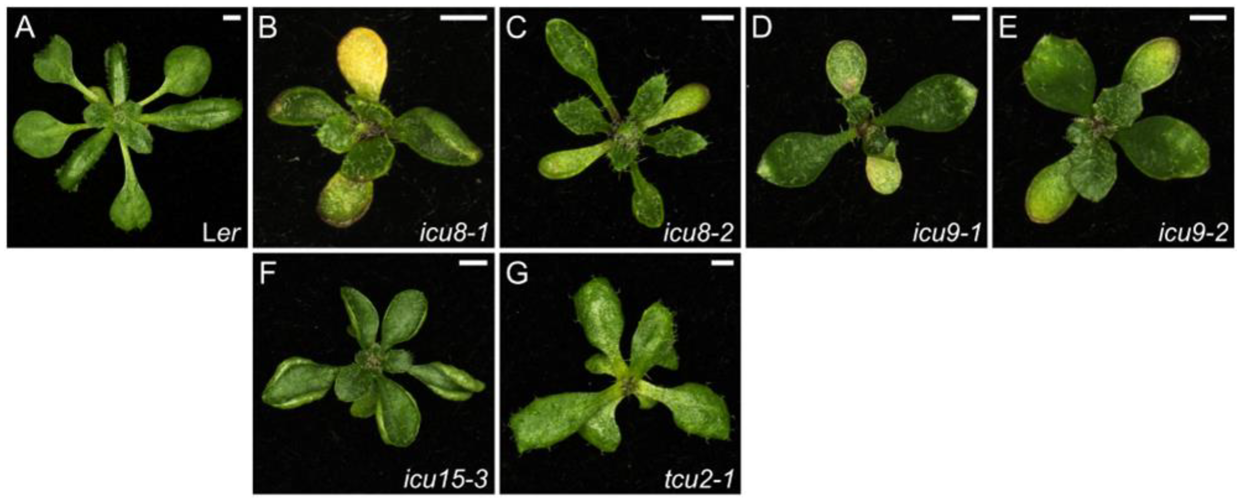
Figure 5. Leaf phenotypes of mutants altered in miRNA biogenesis and function. Rosettes of the wild-type Ler (A), and the icu8-1 (hyl1-11) (B), icu8-2 (hyl1-12) (C), icu9-1 (ago1-51) (D), icu9-2 (ago1-52) (E), icu15-3 (hen1-13) (F), and tcu2-1 (G) homozygous mutants. Photographs were taken 21 das. Scale bars, 2 mm. These mutants were described in [143] (BF) and [148] (G).
Ijms 26 08332 g005
Figure 6. Leaf phenotype of icu11-1, a mutant altered in the epigenetic machinery. Rosettes of the wild-type S96 (A) and the icu11-1 homozygous mutant (B). Photographs were taken 21 das. Scale bars, 2 mm. This mutant was described in [163] (B).
Ijms 26 08332 g006
Disclaimer/Publisher’s Note: The statements, opinions and data contained in all publications are solely those of the individual author(s) and contributor(s) and not of MDPI and/or the editor(s). MDPI and/or the editor(s) disclaim responsibility for any injury to people or property resulting from any ideas, methods, instructions or products referred to in the content.