Article

The DIR Gene Family in Watermelon: Evolution, Stress Expression Profiles, and Functional Exploration of ClDIR8

1
Key Laboratory of Horticultural Crop Germplasm Innovation and Utilization (Co-Construction by Ministry and Province), Institute of Horticulture, Anhui Academy of Agricultural Sciences, Hefei 230001, China
2
College of Agriculture, Anhui Science and Technology University, Fengyang 233100, China
3
Anhui Provincial Key Laboratory for Germplasm Resources Creation and High-Efficiency Cultivation of Horticultural Crops, Hefei 230001, China
4
Institute of Vegetables, Anhui Academy of Agricultural Sciences, Hefei 230001, China
*
Authors to whom correspondence should be addressed.
These authors contributed equally to this work.
Int. J. Mol. Sci. 2025, 26(16), 7730; https://doi.org/10.3390/ijms26167730
Submission received: 9 July 2025 / Revised: 22 July 2025 / Accepted: 8 August 2025 / Published: 10 August 2025
(This article belongs to the Special Issue Plant Stress Biology)

Abstract

Dirigent proteins (DIR) are involved in lignan biosynthesis, stress responses, and disease resistance in plants. However, systematic characterization of the DIR gene family in watermelon (Citrullus lanatus) remains limited. Here, we identified 22 ClDIR genes in watermelon using bioinformatics methods, designated ClDIR1 to ClDIR22, which were unevenly distributed across eight chromosomes and classified into three subfamilies (DIR-a, DIR-b/d, DIR-e) based on phylogenetic analysis, with DIR-b/d being the largest. Synteny analysis revealed that tandem duplication primarily drove ClDIR family expansion, and collinear relationships with Arabidopsis, rice, and cucurbit species indicated evolutionary conservation. Cis-acting element analysis showed abundant stress- and hormone-responsive elements in ClDIR promoters, suggesting roles in stress regulation. Tissue-specific expression analysis demonstrated distinct patterns, with most genes highly expressed in roots. Expression profiling under 16 abiotic and biotic stresses showed 18 ClDIR genes responded to stress, with ClDIR8 differentially expressed across all conditions. qRT-PCR validation of six key genes (ClDIR5, ClDIR8, ClDIR9, ClDIR12, ClDIR16, ClDIR22) confirmed their expression patterns under high-temperature, drought, salt, and low-temperature stresses, showing a high degree of consistency with transcriptome data. Subcellular localization indicated ClDIR8 is peroxisome-localized. Yeast two-hybrid (Y2H) and bimolecular fluorescence complementation (BiFC) assays validated two ClDIR8-interacting proteins, Cla97C02G049920 (encoding peroxidase) and Cla97C08G152180 (encoding catalase). These findings provide insights into ClDIR genes in watermelon, highlighting ClDIR8 as a key stress-responsive candidate for further functional studies and breeding.

1. Introduction

Plants continuously face dual challenges of abiotic and biotic stresses during growth and development. These environmental stressors disrupt cellular redox homeostasis, dismantle hormonal signaling networks, and inhibit the activity of photosynthetic electron transport chains [1,2,3], consequently leading to reduced photosynthetic carbon assimilation rates, imbalanced source–sink transport of assimilates, and blocked secondary metabolite biosynthesis. This ultimately results in crop yield loss and quality degradation. Due to their sessile growth habit, plants cannot evade environmental pressures through physical displacement and must rely on physical and chemical strategies to counteract biotic and abiotic stresses [4]. For instance, plants enhance cell wall rigidity by synthesizing lignin to resist environmental pressures [5]. Notably, lignin biosynthesis is closely associated with DIR (dirigent) proteins, which are encoded by the DIR gene family and specifically regulate the stereoselectivity of lignin monomer coupling, a key step in secondary cell wall thickening [6]. Additionally, these proteins dynamically regulate lignin deposition in cell walls in response to environmental signals [7,8]. Thus, DIR-mediated lignin modulation represents a conserved strategy for plants to integrate environmental cues with cell wall remodeling during stress adaptation.
The core function of DIR proteins lies in regulating the biosynthesis of plant secondary metabolites. These proteins participate in the formation of lignin and lignans by mediating radical coupling reactions of monolignols (e.g., coniferyl alcohol). As key components of the secondary cell walls in vascular plants, lignin and lignans collaboratively construct the rigid framework of cell walls, which not only maintains organ morphology and imparts mechanical strength and compression resistance but also participates in biotic and abiotic stress defense [9]. Recent functional studies have demonstrated that DIR genes play multidimensional roles in plant stress resistance mechanisms: for instance, PIDIR1 and AtsDIR23 regulate lignin accumulation in Phryma leptostachya and Acorus tatarinowii, respectively [10,11]. FvDIR13 enhances disease resistance in strawberry by regulating JA and SA response genes [12]. Overexpression of GmDIR27 promotes soybean pod dehiscence [13]. ScDIR7 in sugarcane responds to drought stress [14]. ZmDIR11 enhances drought tolerance in maize by regulating abscisic acid and lignan metabolism [15]. VvDIR4 in grapevine participates in regulating hormone and lignin biosynthesis pathways for anthracnose resistance [16]. These findings reveal the functional diversity of the DIR family in plant adaptive evolution.
In-depth analysis of the biological functions of the DIR gene family in plant growth and development can not only provide new insights into the molecular regulatory mechanisms of crops responding to abiotic and biotic stresses but also lay an important theoretical foundation for genetic improvement of crop stress resistance. Watermelon (Citrullus lanatus) is a representative cucurbit crop of significant economic value. During its growth and development, plants are often challenged by various abiotic and biotic stresses, including high temperature, low temperature, salt, drought, Fusarium wilt, powdery mildew, etc. Numerous studies have validated the pivotal role of DIR family genes in plant stress responses. Currently, the functions of DIR genes in responding to abiotic stresses (e.g., drought, salinity) and biotic stresses (e.g., pathogen infection) have been widely studied in various plants such as cassava [17], pigeonpea [18], rice [19], mung bean [20], and Panax notoginseng [21]. However, although the genome-wide identification of DIR gene family has been reported in watermelon and their expression patterns under powdery mildew stress were analyzed by quantitative real-time PCR [22], their functions in abiotic stresses and other biotic stresses have not yet been analyzed, and thus their potential roles in these stress responses remain unclear. Therefore, functional characterization of DIR genes in watermelon under diverse stress conditions is crucial for deciphering their specific regulatory roles in stress adaptation mechanisms.
In this study, 22 members of the DIR gene family in watermelon (designated as ClDIR genes) were systematically identified through genome-wide screening. Physicochemical properties, chromosomal locations, gene structures, conserved motifs, phylogenetic relationships, cis-acting elements, and synteny analysis of ClDIR genes were conducted using bioinformatics approaches. Furthermore, based on 21 transcriptome sequencing datasets, tissue-specific expression and stress-responsive expression patterns of ClDIR genes were analyzed to preliminarily explore their biological functions in watermelon. Additionally, subcellular localization of the ClDIR8 protein and screening of its interacting proteins were performed. These findings will provide valuable insights for further functional characterization of ClDIR genes and offer a theoretical basis for stress-resistant watermelon breeding.

2. Results

2.1. Genome-Wide Identification of DIR Gene Family in Watermelon

According to the published genomic data of watermelon (97103_v2.5), a total of 22 DIR family members were identified and designated as ClDIR1ClDIR22. These genes were unevenly distributed across chromosomes 1, 2, 3, 5, 6, 7, 9, and 10. Chromosome 2 contained the highest number of ClDIR genes (8), whereas chromosomes 1 and 3 harbored the fewest (1 ClDIR gene each). Coding sequence (CDS) lengths of ClDIR genes ranged from 525 bp (ClDIR3) to 1200 bp (ClDIR10). The deduced ClDIR proteins varied from 174 amino acids (ClDIR3) to 399 amino acids (ClDIR10), with molecular weights spanning 18.44 kDa (ClDIR3) to 43.45 kDa (ClDIR10). Theoretical isoelectric points (pI) ranged from 4.36 (ClDIR6) to 10.04 (ClDIR9). Instability indices of ClDIR proteins varied from 9.42 (ClDIR3) to 54.36 (ClDIR22); proteins with instability indices < 40 were classified as stable, whereas those with instability indices > 40 were considered unstable. Accordingly, ClDIR6, ClDIR10, ClDIR13, ClDIR14, ClDIR17, and ClDIR22 encoded unstable proteins, while the remaining ClDIR genes encoded stable ClDIR proteins. Aliphatic indices of ClDIR proteins ranged from 68.86 (ClDIR7) to 101.18 (ClDIR9), and grand average of hydropathicity (GRAVY) values spanned −0.337 (ClDIR10) to 0.332 (ClDIR2). Positive GRAVY values indicate hydrophobic tendencies in ClDIR proteins, whereas negative values suggest hydrophilic properties. Thus, ClDIR4, ClDIR6, ClDIR7, ClDIR10, ClDIR13, and ClDIR20 encoded hydrophilic ClDIR proteins, while the remaining ClDIR genes encoded hydrophobic ClDIR proteins. Notably, most ClDIR proteins had GRAVY values near zero, indicating amphipathic characteristics (Table 1).

2.2. Phylogenetic Analysis of ClDIR Genes

To decipher the evolutionary trajectories of ClDIR genes, a phylogenetic analysis was performed using 167 DIR proteins, including 25 AtDIR proteins from Arabidopsis, 49 OsDIR proteins from rice, 24 CaDIR proteins from pepper, 23 CsDIR proteins from cucumber, 24 SmDIR proteins from eggplant, and 22 ClDIR proteins from watermelon. The resultant phylogenetic tree resolved five distinct subgroups: DIR-a, DIR-b/d, DIR-c, DIR-g, and DIR-e. The majority of DIR genes clustered into three subgroups: DIR-a, DIR-b/d, and DIR-e. Notably, the DIR-b/d subgroup contained the highest number of genes (76), suggesting substantial expansion during DIR gene evolution compared to other subgroups. ClDIR genes from watermelon were primarily distributed across DIR-a, DIR-b/d, and DIR-e subgroups. Intriguingly, the DIR-c and DIR-g subgroups harbored only OsDIR genes from rice, with no representation from other species (Figure 1). These differential distributions imply functional divergence among subgroups. Furthermore, sequence homology analysis revealed strong conservation between ClDIR genes and their cucumber orthologs, underscoring the evolutionary preservation of DIR genes across Cucurbitaceae species.

2.3. Gene Structure and Conserved Motif Analyses of ClDIR Genes

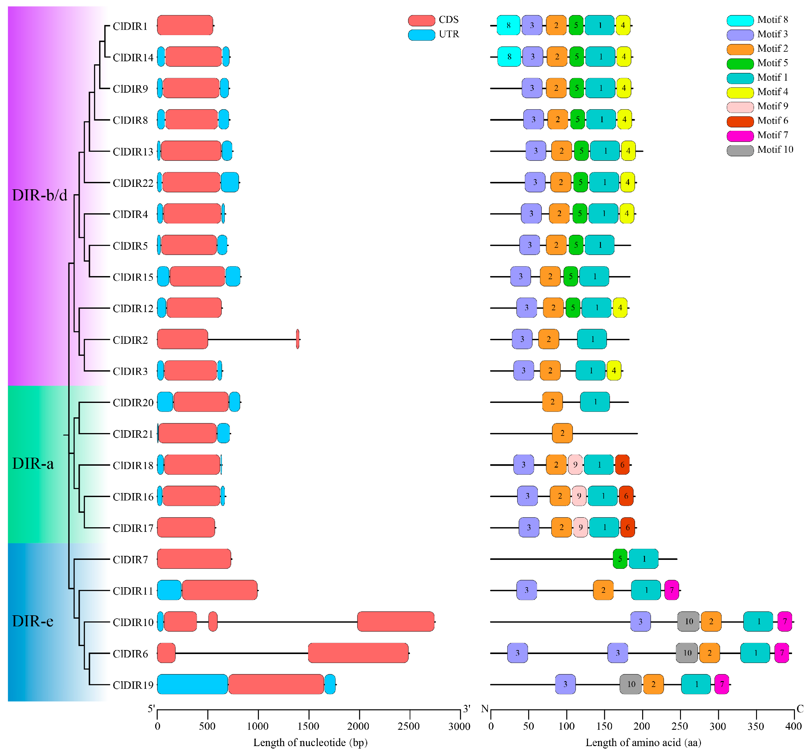
Structural analysis of the 22 ClDIR genes revealed three canonical subfamilies (DIR-a, DIR-b/d, and DIR-e) based on phylogenetic clustering, consistent with comparative analyses of watermelon, Arabidopsis, rice, eggplant, pepper, and cucumber. Specifically, the DIR-a subfamily comprised 5 ClDIR members, DIR-b/d emerged as the largest clade with 12 ClDIR genes, and DIR-e contained 5 ClDIR genes. Exon–intron architecture analysis showed that most ClDIR genes exhibited a typical single-exon structure without introns, though notable variations existed: ClDIR2 and ClDIR6 displayed a two-exon structure, while ClDIR10 harbored a three-exon configuration (Figure 2), potentially reflecting annotation specificity or evolutionary structural remodeling.
Conserved motif analysis of the 22 ClDIR proteins identified 10 motifs with lengths ranging from 21 to 41 amino acids, sequentially designated motif1 to motif10 (Table S1). Subfamily-specific motif distributions were evident: DIR-a members predominantly contained motif1, motif2, motif3, motif6, and motif9; DIR-b/d members typically harbored motif1, motif2, motif3, motif4, and motif5; and DIR-e members were characterized by motif1, motif2, motif3, motif7, and motif10. Notably, motif1 and motif2 were conserved in all ClDIR proteins except ClDIR21 (lacking motif1) and ClDIR7 (lacking motif2) (Figure 2). Pfam annotation revealed that both motif1 and motif2 correspond to the dirigent protein domain, indicating their critical functional significance in the DIR gene family.

2.4. Synteny Analysis of ClDIR Genes

Collinearity analysis of the ClDIR gene family identified four pairs of tandem duplicated genes (ClDIR4/ClDIR5, ClDIR6/ClDIR7, ClDIR8/ClDIR9, ClDIR16/ClDIR17), accounting for 36.36% of family members, and one pair of segmental duplicated genes (ClDIR2/ClDIR3) (Figure 3A), comprising 9.09% of the family. These results indicated that tandem duplication was the primary driver of rapid expansion and evolution in the watermelon DIR gene family. Ka/Ks (non-synonymous substitution rates/synonymous substitution rates) ratio analysis of duplicated gene pairs revealed that three tandem duplicated pairs (ClDIR4/ClDIR5, ClDIR8/ClDIR9, ClDIR16/ClDIR17) and one segmental pair (ClDIR2/ClDIR3) had Ka/Ks < 1, suggesting purifying selection and conserved functions. By contrast, the tandem pair ClDIR6/ClDIR7 exhibited Ka/Ks > 1, indicating positive selection (Table 2).
To trace evolutionary trajectories, interspecific collinearity analysis was conducted between ClDIR genes in watermelon and DIR genes from the dicotyledonous plant Arabidopsis thaliana and the monocotyledonous plant rice (Oryza sativa). Genomic comparisons revealed 3 DIR orthologs between watermelon and rice and 12 between watermelon and Arabidopsis (Figure 3B), indicating a closer divergence time between watermelon and Arabidopsis relative to rice. Collinearity analyses with other cucurbit species showed 15 collinear gene pairs between watermelon and cucumber and 17 between watermelon and melon (Figure 3C). Notably, ClDIR6 and ClDIR11 exhibited collinearity with all analyzed species. Additionally, ClDIR8 and ClDIR19 also showed collinearity with Arabidopsis, cucumber, and melon (Table S2). These findings suggest that ClDIR6, ClDIR8, ClDIR11, and ClDIR19 may represent ancestral genes conserved across watermelon, cucumber, and melon, retaining similar structures and functions through shared chromosomal rearrangements. Variations in collinear pair numbers among species likely stem from evolutionary gene rearrangement, deletion, or insertion events.

2.5. Analysis of Cis-Acting Elements in ClDIR Promoters

Cis-acting element analysis of ClDIR gene promoters identified 39 distinct types of regulatory motifs, systematically categorized into three functional classes: abiotic and biotic stress-responsive cis-acting elements, phytohormone response-related cis-acting elements, and plant growth and development-related cis-acting elements (Figure 4).
Among the abiotic and biotic stress-responsive cis-acting elements, 10 key motifs (ARE, DRE core, LTR, MBS, MYB, MYC, STRE, TC-rich repeats, W box, and WUN-motif) were identified, mediating responses to oxidative, drought, low-temperature, osmotic, high-temperature stresses, and wound signaling. The MYB transcription factor binding site exhibited the highest prevalence, detected in 21 ClDIR promoters, followed by the ARE element (anaerobic response under waterlogging), present in 20 ClDIR promoters.
In the phytohormone response-related cis-acting elements, 10 characteristic motifs (ABRE, as-1, CGTCA-motif, ERE, P-box, TCA-element, TGACG-motif, TGA-element, AuxRR-core, and GARE-motif) regulated signaling pathways for abscisic acid (ABA), salicylic acid (SA), methyl jasmonate (MeJA), ethylene, gibberellin (GA), and auxin. The ethylene-responsive element (ERE) was most widely distributed, occurring in 86% of ClDIR promoters.
In the plant growth and development-related cis-acting elements, 19 motifs (A-box, AE-box, Box 4, CAT-box, circadian, GA-motif, GATA-motif, G-box, GCN4-motif, GT1-motif, I-box, MRE, RY-element, ACE, chs-CMA1a, L-box, Sp1, TCCC-motif, TCT-motif) governed light signal transduction, cell cycle regulation, seed germination, stem elongation, and circadian rhythms. All ClDIR promoters contained light-responsive elements, with Box 4 (95%) the most prevalent, followed by G-box (64%) and TCT-motif (59%).
Collectively, 646 cis-acting elements were identified across ClDIR promoters. ClDIR8, ClDIR16, and ClDIR17 harbored the richest element diversity (42, 44, and 43 elements, respectively), whereas ClDIR13 had only 18 elements, highlighting substantial variation in cis-element composition. This variability suggested diverse transcriptional regulation and functional specialization among ClDIR genes.

2.6. Tissue-Specific Expression Analysis of ClDIR Genes

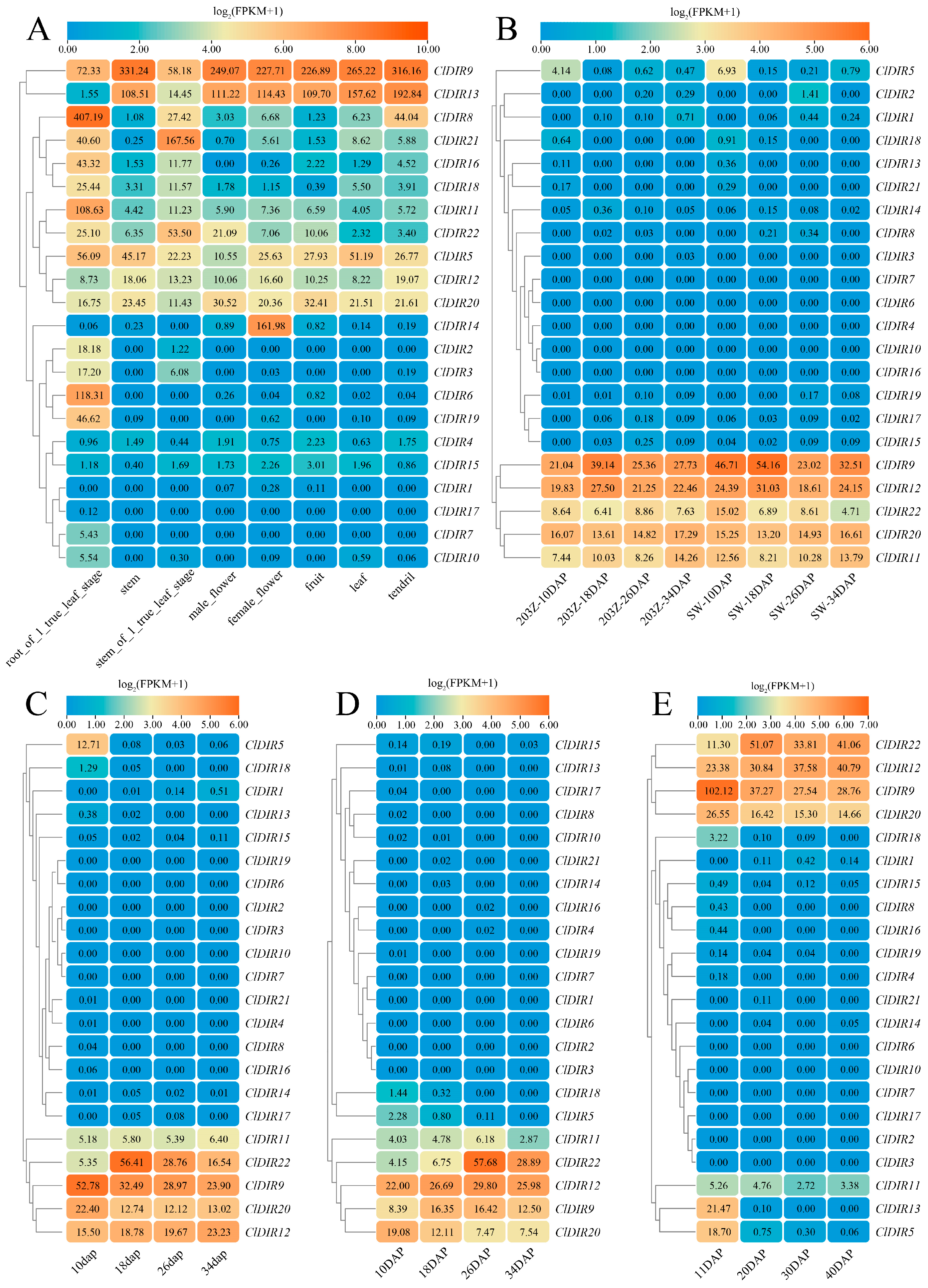
Tissue-specific expression analysis revealed that the ClDIR9 gene exhibited high expression in watermelon roots, stems, flowers, fruits, leaves, and tendrils. A subset of ClDIR genes (ClDIR4, ClDIR15, ClDIR1, ClDIR17, ClDIR7, and ClDIR10) displayed no expression or extremely low expression levels across all detected tissues. The ClDIR13 gene was expressed in all tissues but showed reduced expression in true leaf-stage roots. ClDIR8 and ClDIR11 were universally expressed with specific high expression in true leaf-stage roots. The ClDIR21 gene was expressed in all tissues, peaking in true leaf-stage stems. Additionally, ClDIR16 was expressed in all tissues except male flowers, with the highest expression in true leaf-stage roots. ClDIR18 showed universal expression, peaking in true leaf-stage roots and being the lowest in fruits. ClDIR22 was expressed in all tissues, highest in true leaf-stage stems and lowest in leaves. ClDIR5 showed universal expression, highest in true leaf-stage roots and lowest in male flowers. ClDIR12 was expressed in all tissues, highest in tendrils and lowest in leaves. ClDIR20 showed universal expression, highest in fruits and lowest in true leaf-stage stems. Notably, ClDIR14 exhibited specific expression in female flowers, while ClDIR2, ClDIR3, ClDIR6, and ClDIR19 showed tissue-specific expression in true leaf-stage roots (Figure 5A).
During fruit development, ClDIR9, ClDIR11, ClDIR12, ClDIR20, and ClDIR22 maintained high expression at 10, 18, 26, and 34 days after pollination (DAP). In contrast, 15 genes (ClDIR1, ClDIR2, ClDIR3, ClDIR4, ClDIR6, ClDIR7, ClDIR8, ClDIR10, ClDIR14, ClDIR15, ClDIR16, ClDIR17, ClDIR18, ClDIR19, and ClDIR21) were either non-expressed or showed minimal expression during these stages. The ClDIR5 gene displayed specific expression at 10 DAP (Figure 5B–E). Collectively, these results demonstrated that ClDIR genes exhibited distinct expression patterns across tissues and developmental stages, highlighting their tissue-specific and spatiotemporal expression characteristics.

2.7. Expression Analysis of ClDIR Genes Under Abiotic Stresses

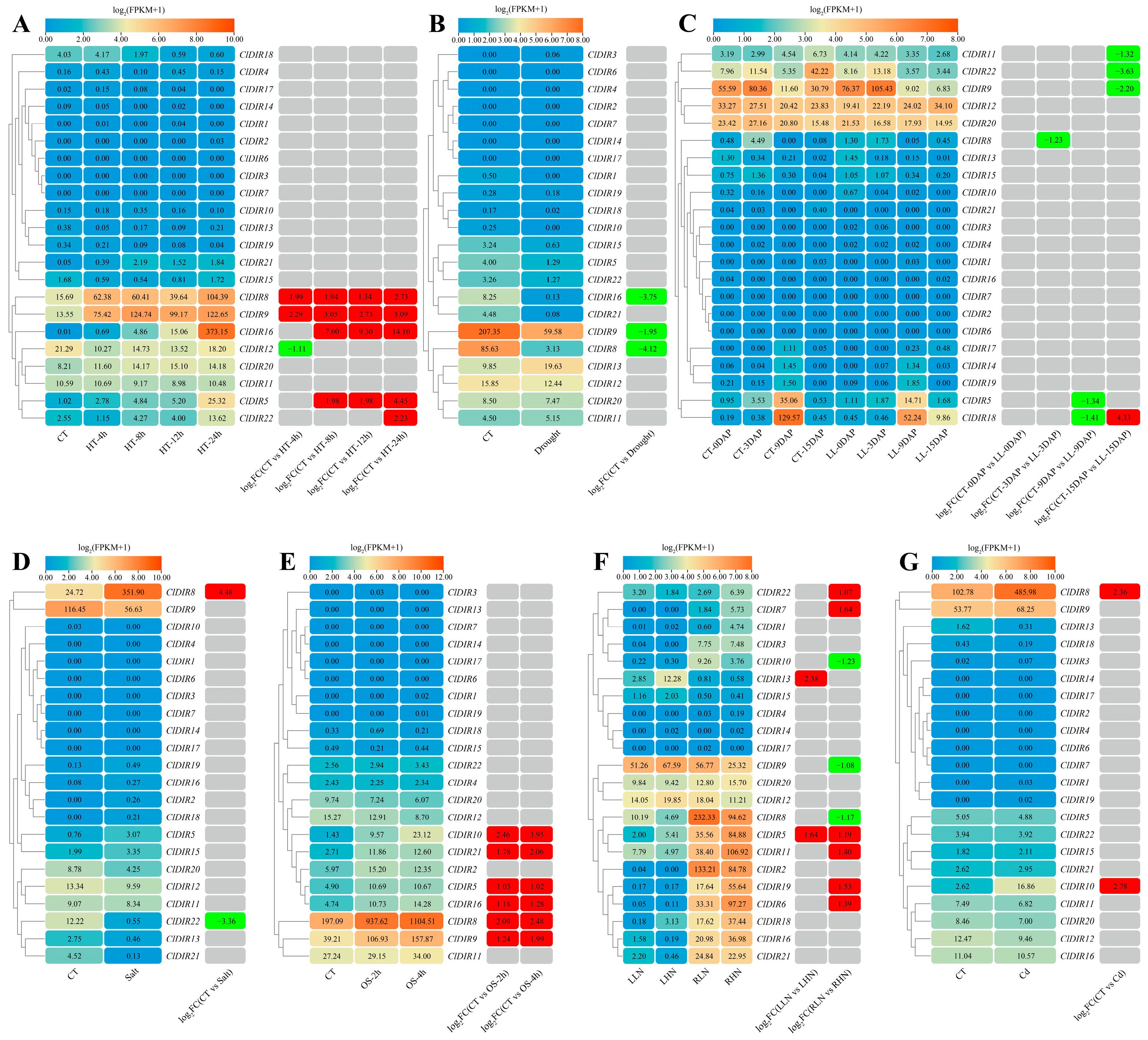
Expression pattern analysis based on transcriptome sequencing data showed that under high-temperature stress, only the ClDIR12 gene was significantly downregulated, while ClDIR8, ClDIR9, ClDIR16, ClDIR5, and ClDIR22 were significantly upregulated. ClDIR8 and ClDIR9 were upregulated at 4, 8, 12, and 24 h post-stress. ClDIR16 and ClDIR5 were significantly upregulated at 8, 12, and 24 h. ClDIR22 was only upregulated at 24 h, while the remaining 16 ClDIR genes showed no significant expression changes (Figure 6A). Under drought stress, ClDIR16, ClDIR9, and ClDIR8 were significantly downregulated, and the other 19 ClDIR genes showed no changes (Figure 6B). Under low-light treatment, ClDIR18 was significantly downregulated at 9 DAP and upregulated at 15 DAP. ClDIR11, ClDIR22, ClDIR9, ClDIR8, and ClDIR5 were significantly downregulated. ClDIR11, ClDIR22, and ClDIR9 were downregulated only at 15 DAP, and ClDIR8 and ClDIR5 were downregulated only at 9 DAP. The remaining 16 ClDIR genes were unaffected (Figure 6C). Under salt stress, ClDIR8 was significantly upregulated, ClDIR22 was downregulated, and the other 20 ClDIR genes showed no expression differences (Figure 6D). During osmotic stress, ClDIR10, ClDIR21, ClDIR5, ClDIR16, ClDIR8, and ClDIR9 were significantly upregulated at 2 and 4 h post-stress, while the remaining 16 ClDIR genes showed no response (Figure 6E). Under high-nitrogen stress, in roots, ClDIR22, ClDIR7, ClDIR11, ClDIR19, and ClDIR6 were significantly upregulated, whereas ClDIR10, ClDIR9, and ClDIR8 were downregulated. ClDIR13 was upregulated in leaves, and ClDIR5 was upregulated in both leaves and roots. The other 12 ClDIR genes were unchanged (Figure 6F). Under cadmium stress, ClDIR8 and ClDIR10 were significantly upregulated, and the remaining 20 ClDIR genes showed no response (Figure 6G).

2.8. Expression Analysis of ClDIR Genes Under Biotic Stresses

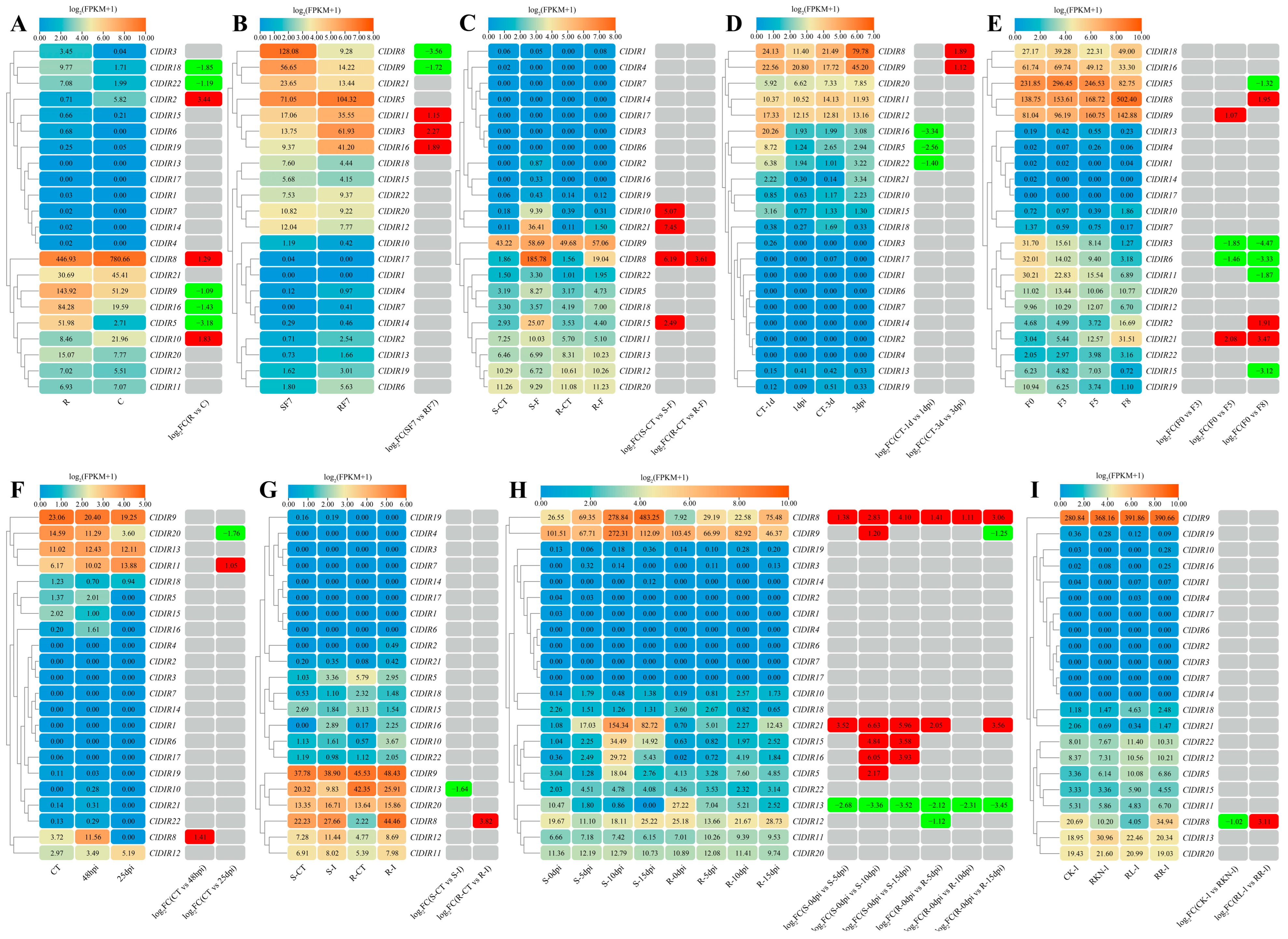
Transcriptome sequencing data of five sets of watermelons under Fusarium wilt stress were analyzed to explore the expression patterns of ClDIR genes. The results showed that compared with watermelon–oilseed rape rotation (R), the expressions of ClDIR2, ClDIR8, and ClDIR10 were significantly upregulated, while the expressions of ClDIR5, ClDIR9, ClDIR16, ClDIR18, and ClDIR22 were significantly downregulated under continuous watermelon monocropping (C) (Figure 7A). When compared with the susceptible materials 7 days after Fusarium wilt inoculation (SF7), the expression levels of ClDIR11, ClDIR3, and ClDIR16 were significantly upregulated, and the expression levels of ClDIR8 and ClDIR9 were significantly downregulated in the resistant materials 7 days after Fusarium wilt inoculation (RF7) (Figure 7B). After inoculation with Fusarium wilt, the susceptible materials (S-F) and resistant materials (R-F) were compared with their respective controls (S-CT and R-CT). The expression levels of ClDIR10, ClDIR21, and ClDIR15 were only significantly upregulated in the susceptible materials, while the expression level of ClDIR8 was significantly upregulated in both resistant and susceptible materials (Figure 7C). Time-course analysis showed that ClDIR5, ClDIR16, and ClDIR22 were downregulated at 1 day post inoculation (1 dpi), and ClDIR8 and ClDIR9 were repressed at 3 dpi relative to controls (CT-1d, CT-3d) (Figure 7D). At 8 days post inoculation with Fusarium wilt (F8), ClDIR5, ClDIR11, and ClDIR15 were specifically downregulated compared with the control (F0), while ClDIR8 and ClDIR2 were induced. ClDIR9 was upregulated only at 5 days post inoculation (F5). ClDIR3 and ClDIR6 were repressed at both F5 and F8, whereas ClDIR21 was consistently upregulated at both time points (Figure 7E).
Transcriptome sequencing data of watermelons under stresses of cucumber green mottle mosaic virus, powdery mildew, squash vein yellowing virus, and root-knot nematodes were further utilized to analyze the expression patterns of ClDIR genes under biotic stresses. Under cucumber green mottle mosaic virus stress, ClDIR8 was significantly upregulated at 48 dpi, while ClDIR11 was induced at 25 dpi and ClDIR20 was repressed at the same time point (Figure 7F). For powdery mildew stress, inoculation of susceptible and resistant materials revealed that ClDIR13 was downregulated in susceptible lines, whereas ClDIR8 was upregulated in resistant genotypes relative to controls (Figure 7G). Under squash vein yellowing virus stress, ClDIR8 and ClDIR21 were upregulated in both resistant and susceptible materials, while ClDIR13 was downregulated in both resistant and susceptible lines. ClDIR15, ClDIR16, and ClDIR5 were uniquely upregulated in susceptible lines, and ClDIR12 was specifically downregulated in resistant lines. Notably, ClDIR9 was induced in susceptible materials but repressed in resistant materials (Figure 7H). For root-knot nematode stress, Meloidogyne incognita infection decreased ClDIR8 expression in watermelon leaves under white light compared with water-treated controls, but induced ClDIR8 under red-light treatment relative to red-light-treated controls (Figure 7I).

2.9. Expression Patterns of ClDIR Genes Under Abiotic and Biotic Stresses

To analyze the expression patterns of ClDIR genes under abiotic and biotic stresses, differentially expressed genes were marked and visualized as a heatmap (Figure 8). Results showed that 18 of the 22 ClDIR genes exhibited stress responses, with only 4 genes (ClDIR1, ClDIR4, ClDIR14, ClDIR17) showing no response. Notably, ClDIR8 displayed significant differential expression across all 16 abiotic and biotic stress conditions. ClDIR5 responded significantly to four abiotic and four biotic stresses. ClDIR9 was differentially expressed in five abiotic and five biotic stresses. Some DIR genes responded exclusively to abiotic stresses, such as ClDIR6, ClDIR7, and ClDIR19, which were upregulated only under nitrogen treatment. In contrast, genes like ClDIR2, ClDIR3, and ClDIR15 showed differential expression only under biotic stresses, while ClDIR20 was downregulated exclusively under cucumber green mottle mosaic virus stress. The remaining 11 ClDIR genes responded to both stress types. Significantly, ClDIR8 was expressed in all 16 stress conditions, indicating its critical research value in regulating biotic and abiotic stress responses.

2.10. Validation of the Expression Profiles of ClDIR Genes Under High-Temperature, Drought, Salt and Low-Temperature Stresses

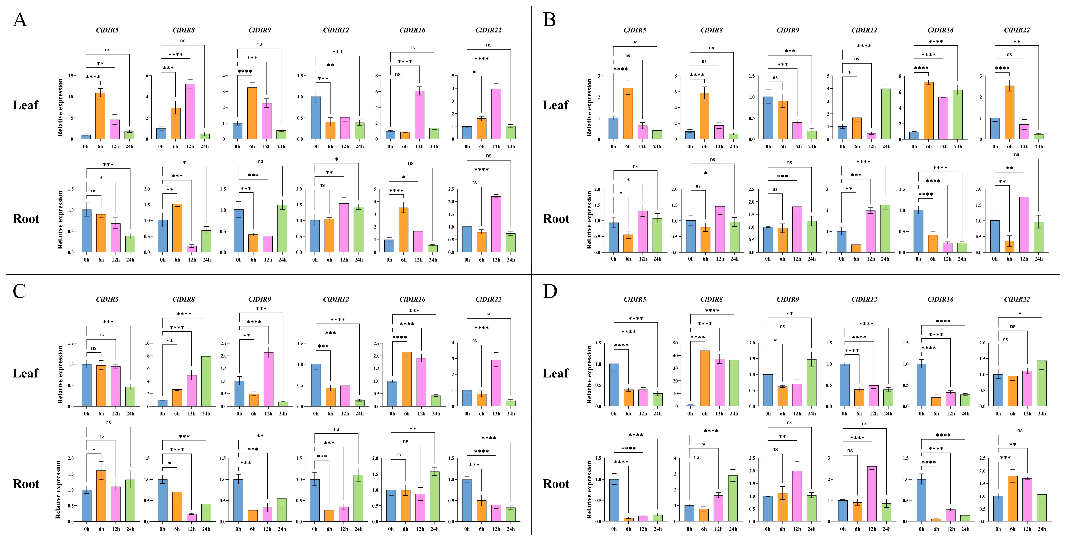
To elucidate the functional roles of ClDIR genes in responding to abiotic stresses, six candidate ClDIR genes (ClDIR5, ClDIR8, ClDIR9, ClDIR12, ClDIR16, ClDIR22) with significant differential expression under abiotic stresses were selected based on transcriptome analysis. qRT-PCR was then employed to analyze their temporal dynamic expression patterns (0, 6, 12, and 24 h) in roots and leaves in response to four abiotic stresses: high-temperature (45 °C), drought (20% PEG6000), salt (500 mmol·L−1 NaCl), and low-temperature (4 °C).
Under high-temperature stress, the expressions of ClDIR genes exhibited tissue-specific divergence. In leaves, ClDIR5, ClDIR8, ClDIR9, and ClDIR22 displayed transient upregulation at 6–12 h, reverting to the 0 h levels by 24 h; ClDIR12 showed persistent downregulation from 6 to 24 h; and ClDIR16 was only upregulated at 12 h. In roots, ClDIR5 was downregulated at 12–24 h; ClDIR8 presented early upregulation at 6 h followed by downregulation; ClDIR9 decreased at 6–12 h and then recovered; ClDIR12 remained static at 6 h but was upregulated at 12–24 h; ClDIR16 showed upregulation at 6 h and downregulation at 12 h; and ClDIR22 was only upregulated at 12 h (Figure 9A).
Under drought stress, tissue-specific expression patterns emerged. In leaves, ClDIR5, ClDIR8, ClDIR12, and ClDIR22 showed stage-specific upregulation (at 6 h or 24 h); ClDIR9 was downregulated at 12–24 h; and ClDIR16 was consistently upregulated from 6 to 24 h. In roots, ClDIR5 was downregulated at 6 h and upregulated at 12 h; ClDIR8 and ClDIR9 were only upregulated at 12 h; ClDIR12 was downregulated at 6 h and upregulated at 12–24 h; ClDIR16 was consistently downregulated from 6 to 24 h; and ClDIR22 was downregulated at 6 h, upregulated at 12 h, and recovered to the 0 h level at 24 h (Figure 9B).
Under salt stress, distinct tissue-specific responses were observed. In leaves, ClDIR5 was downregulated at 24 h; ClDIR8 was consistently upregulated from 6 to 24 h; and ClDIR9, ClDIR12, ClDIR16, and ClDIR22 exhibited bidirectional expression changes (e.g., ClDIR9 was downregulated at 6 h, upregulated at 12 h, and downregulated again at 24 h). In roots, ClDIR5 was upregulated at 6 h; ClDIR8, ClDIR9, and ClDIR22 were consistently downregulated; ClDIR12 was downregulated at 6–12 h and recovered to the 0 h level at 24 h; and ClDIR16 was upregulated only at 24 h (Figure 9C).
Under low-temperature stress, tissue-specific divergence of ClDIR expression occurred. In leaves, ClDIR5, ClDIR12, and ClDIR16 were consistently downregulated from 6 to 24 h; ClDIR8 was consistently upregulated; ClDIR9 was downregulated at 6 h, showed recovery at 12 h, and was upregulated at 24 h; and ClDIR22 was upregulated only at 24 h. In roots, ClDIR5 and ClDIR16 were consistently downregulated; ClDIR9 and ClDIR12 were upregulated only at 12 h; ClDIR8 was upregulated at 12–24 h; and ClDIR22 was upregulated at 6–12 h and recovered to the 0 h level at 24 h (Figure 9D).

2.11. Subcellular Localization of ClDIR8 Protein

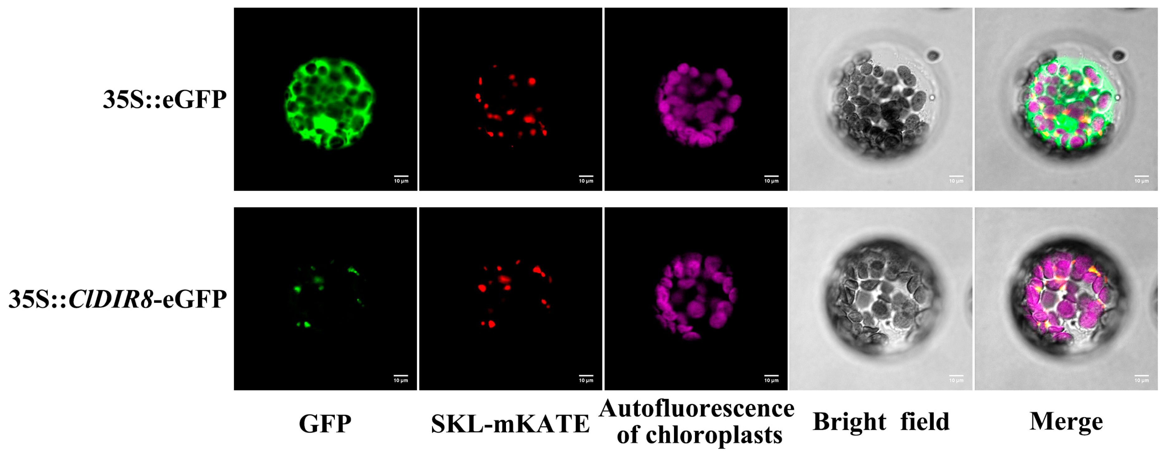
To characterize the subcellular localization of ClDIR8 protein, the 35S::ClDIR8-eGFP fusion expression vector was constructed. The PEG-mediated transient transformation method was employed to introduce the 35S::ClDIR8-eGFP recombinant plasmid, the control plasmid 35S::eGFP, and the peroxisomal marker plasmid 35S::SKL-mKate into Arabidopsis protoplasts. Fluorescence imaging analysis revealed that the 35S::ClDIR8-eGFP fusion protein was specifically localized to peroxisomes (Figure 10). This result indicated that ClDIR8 protein was a peroxisome-localized protein, providing important clues for elucidating its biological function.

2.12. Screening of Proteins Interacting with ClDIR8 Protein

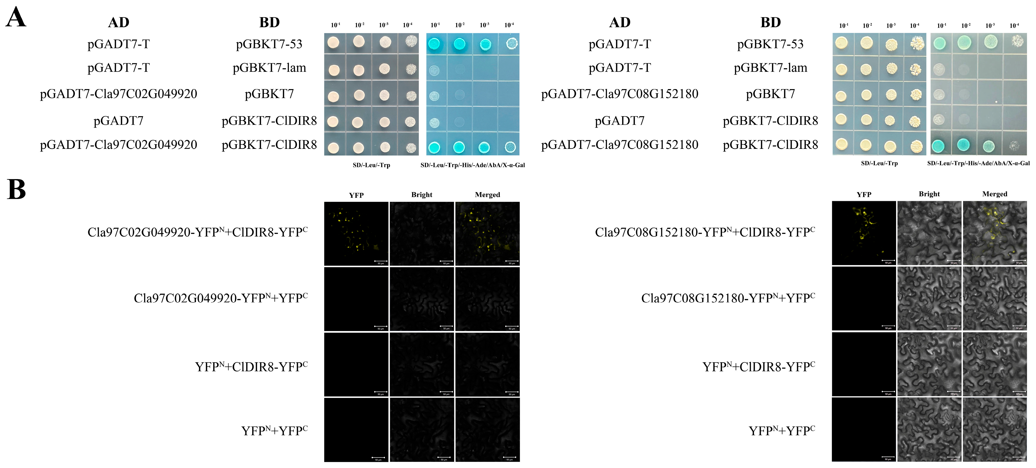
To dissect the molecular interaction network of the ClDIR8 gene, a yeast two-hybrid (Y2H) library was constructed for initial screening of interacting proteins, yielding 81 positive clones (Figure S1). Following Sanger sequencing and BLAST homology analysis, 50 candidate interacting proteins were identified (Table S3). For verification, ClDIR8 and candidate genes were cloned into pGBKT7 and pGADT7 vectors, respectively. Co-transformed yeast strains were assayed on SD-Leu-Trp and SD-Leu-Trp-His-Ade + X-α-Gal plates. Combinations of pGBKT7-ClDIR8 + pGADT7-Cla97C02G049920 and pGBKT7-ClDIR8 + pGADT7-Cla97C08G152180 grew and turned blue on SD-Leu-Trp-His-Ade + X-α-Gal plates (Figure 11A), indicating transcriptional activation interactions between ClDIR8 protein and these two candidates.
Subsequently, YFP fusion vectors of ClDIR8 and candidates were constructed in pCAMBIA1300-35S-N and pCAMBIA1300-35S-C, respectively, and co-transformed into Nicotiana benthamiana leaves via Agrobacterium tumefaciens GV3101-mediated infiltration. Laser confocal microscopy revealed YFP (yellow fluorescent protein) fluorescence complementation signals for both ClDIR8/Cla97C02G049920 and ClDIR8/Cla97C08G152180 combinations, whereas negative controls (empty vectors and non-fused YFPN/YFPC) showed no obvious fluorescence (Figure 11B), verifying interactions at the subcellular level. In conclusion, via Y2H and bimolecular fluorescence complementation (BiFC) cross-verification, Cla97C02G049920 (encoding peroxidase) and Cla97C08G152180 (encoding catalase) were confirmed as genuine ClDIR8-interacting proteins.

3. Discussion

3.1. Identification of DIR Gene Family in Watermelon and Its Evolutionary Context

The dirigent protein was initially discovered to regulate the regional and stereoselective coupling reaction of lignin biosynthesis [23]. Recent studies have shown that DIR proteins can control the formation of specific chemical bonds during the polymerization of monolignols into lignin polymers. These DIR proteins not only promote plant lignification but also enable plants to respond to biotic and abiotic stresses, playing a central role in biotic resistance and abiotic adaptation [24]. Currently, the DIR gene family has been identified in a variety of plant species. In monocotyledons, it includes 49 DIR members in Oryza sativa [19], 38 in Panicum italicum [25], and 47 in Phyllostachys edulis [26]. In dicotyledons, there are 25 in Arabidopsis thaliana [27], 24 in Capsicum annuum [28], 24 in Solanum melongena [29], 25 in Cajanus cajan [18], and 23 in Cucumis sativus [30]. In this study, 22 ClDIR family genes were successfully identified from the watermelon genome using bioinformatics approaches, and the number of identified ClDIR genes is consistent with previous research findings. Compared with the above-identified species, the number of ClDIR genes was significantly lower than that in monocots but similar to that in dicot crops. This difference suggests that the DIR gene family may have undergone lineage-specific adaptive expansion during the evolution of monocots.

3.2. Mechanisms of DIR Gene Family Expansion in Watermelon

The expansion of gene families mainly relies on three molecular mechanisms: tandem duplication, segmental duplication, and transposition [31]. For example, in Medicago truncatula, 45 MtDIR genes have been identified, and 82.22% (37) of MtDIR genes arose from tandem duplication events [32]. In Phyllostachys edulis, 47 PeDIR genes have been identified, with 67.6% (32) of PeDIR genes resulting from tandem duplications and 10 pairs of segmental duplications identified [26]. These results indicate that numerous duplication events within species drive the expansion of DIR family members. In this study, four pairs of tandemly duplicated genes (ClDIR4/ClDIR5, ClDIR6/ClDIR7, ClDIR8/ClDIR9, ClDIR16/ClDIR17) were found in the watermelon DIR gene family, comprising 36.36% of the gene family, and one pair of segmentally duplicated genes (ClDIR2/ClDIR3), accounting for 9.09%. Therefore, tandem duplication likely served as the primary driving force for the expansion of ClDIR genes.

3.3. Phylogenetic Classification and Subfamily-Specific Features of ClDIR Genes

A phylogenetic tree constructed from 167 DIR proteins across Arabidopsis thaliana, Oryza sativa, Citrullus lanatus (watermelon), Cucumis sativus, Solanum melongena, and Capsicum annuum classified DIR proteins into five subfamilies: DIR-a, DIR-b/d, DIR-c, DIR-e, and DIR-g. Notably, watermelon ClDIR genes were exclusively distributed in DIR-a, DIR-b/d, and DIR-e subfamilies, with no members detected in DIR-c or DIR-g. The DIR-a subfamily has a well-established functional research history: its members were first linked to lignin biosynthesis in 1997 [33], and in 2012, biochemical analysis of recombinant AtDIR5 and AtDIR6 proteins showed they synergize with laccase to drive oxidative coupling of coniferyl alcohol into (−)-pinoresinol in vitro [34]. Five watermelon genes (ClDIR16, ClDIR17, ClDIR18, ClDIR20, ClDIR21) clustered with AtDIR5/AtDIR6 in DIR-a, suggesting their potential role in lignin synthesis. DIR-b/d subfamily proteins mediate both abiotic and biotic stress responses [35,36], and previous studies as well as this clustering analysis confirm DIR-b/d as the largest DIR subfamily, implying broad functional diversity. Conversely, DIR-c is monocot-specific, featuring low sequence conservation and a ~140-amino-acid C-terminal extension similar to Jacalin-like domains [37]. This domain confers defense functions and abiotic stress (salt/drought) responsiveness [38], and may contribute to insect resistance [39] and disease resistance [40,41]. Consistent with prior reports [22,37], DIR-c members were detected only in O. sativa among the studied species, absent in all dicots including watermelon.

3.4. Gene Structure and Conserved Motifs of ClDIR Genes

The typical structure of DIR genes usually contains only one exon [37]. Analysis of the gene structure of watermelon ClDIR genes showed that 86.4% (19) of the ClDIR genes retained the canonical single-exon architecture, while only 3 genes (ClDIR2, ClDIR6, ClDIR10) contained introns. Notably, all members of the DIR-a subfamily maintained the single-exon structure, indicating higher structural conservation of DIR-a compared to the other subfamilies described earlier. It has been proposed that introns may delay regulatory responses, whereas intron-lacking genes accelerate transcription rates, enabling plants to promptly respond to stresses [42]. Additionally, reduced intron numbers may enhance gene function, improving plant tolerance to various stresses [43]. Conserved motifs (e.g., motif1, motif2, motif3) were identified in the majority of ClDIR genes, reflecting their structural conservation. However, subfamily-specific motifs were also observed: motif9 was exclusive to the DIR-a subfamily, and motif10 was unique to the DIR-e subfamily, highlighting functional diversification across DIR subfamilies.

3.5. Cis-Acting Elements in ClDIR Promoters

Bioinformatics analysis of cis-acting elements in the 2000 bp promoter region upstream of ClDIR genes identified multiple cis-acting elements associated with hormone responses (e.g., ABRE, as-1, CGTCA-motif, ERE), abiotic and biotic stresses (e.g., STRE, MBS), and growth and development (e.g., G-box, AE-box). This indicates that the ClDIR genes play a significant role in the growth, development, and stress responses of watermelon. Among them, 59.1% (13) of ClDIR genes contain cis-acting elements responsive to methyl jasmonate (MeJA) regulation (i.e., CGTCA-motif or TGACG-motif), and a high proportion of 68.18% (15) of ClDIR genes possess ABRE elements responsive to abscisic acid (ABA) regulation. Relevant studies have shown that hormones such as MeJA and ABA can enhance the activities of plant cell protective enzymes (such as POD, CAT, and SOD), scavenge free radicals, and increase the content of osmotic adjustment substances, thereby reducing stress damage to plants [44]. Therefore, it is speculated that ClDIR genes may have functions related to plant hormones. They may participate in stress regulation processes by responding to hormones such as MeJA, ABA, and gibberellin (GA), thereby enhancing plants’ tolerance to adverse stresses. However, the specific mechanisms of action still require in-depth investigation. Previous studies have found that in Pyrus bretschneideri, the expression of PbDIR4 increases significantly upon induction by SA, ABA, and MeJA, indicating that PbDIR4 plays a crucial role in responses to biotic and abiotic stresses [24]. In addition, MYB transcription factor binding sites are present in the promoter regions of 95.5% (21) of ClDIR genes. Given that MYB transcription factors have been proven to play key regulatory roles in responses to abiotic stresses such as drought, salinity, and low temperature [45], it is speculated that MYB transcription factors may interact with cis-acting elements (such as MBS, MRE, W-box) in the promoters of ClDIR genes to coordinate the expression of ClDIR genes under hormone signaling and abiotic stress conditions, thereby enhancing plants’ environmental adaptability and regulating their growth and development processes.

3.6. Tissue-Specific and Fruit Development Expression Patterns of ClDIR Genes

The DIR gene family displayed distinct expression patterns across various tissues and developmental stages in watermelon. In this study, all 22 ClDIR genes were expressed in roots, stems, flowers, fruits, leaves, and tendrils, but with significant variations in expression levels. Notably, the majority of ClDIR genes showed higher expression in root tissues, a pattern consistent with tissue-specific expression of SmDIR genes in eggplant, where most SmDIR genes exhibited high root expression [29]. Previous studies have shown that DIR genes promote organ lignification, suggesting that the ClDIR genes are crucial for watermelon root development and may coordinate the regulation of other organ growth. Through comprehensive analysis of four transcriptome datasets, we investigated the gene expression of ClDIR genes during watermelon fruit development. Results showed that ClDIR9, ClDIR12, ClDIR22, ClDIR20, and ClDIR11 maintained high expression throughout fruit development. These findings suggest that these five ClDIR genes may regulate fruit morphogenesis or exert critical functions in fruit quality formation.

3.7. Expression Profiles of ClDIR Genes Under Abiotic Stresses

To systematically investigate the functions of ClDIR genes in watermelon in response to abiotic stresses, we first analyzed the expression profiles of ClDIR genes using 7 sets of publicly available abiotic stress transcriptome datasets, including high temperature, drought, low light, salt, osmotic stress, nitrogen treatment, and cadmium treatment. Transcriptome analysis revealed that 63.64% of ClDIR genes exhibited significant differential expression under abiotic stresses, suggesting that this gene family is widely involved in the adaptive regulation of watermelon to abiotic adversities. Based on the differential expression results from transcriptome data, 6 key ClDIR genes (ClDIR5, ClDIR8, ClDIR9, ClDIR12, ClDIR16, and ClDIR22) with differential expression in abiotic stress responses were selected from 22 ClDIR genes, and their expression patterns under high-temperature, drought, and salt stresses were validated using qRT-PCR. The results showed that the expression trends of these six key genes were generally consistent with the transcriptome data. Among them, ClDIR8, ClDIR9, ClDIR12, and ClDIR22 displayed highly consistent expression changes with the transcriptome data, with ClDIR8 showing the highest consistency across all stress conditions. However, the expression characteristics of ClDIR5 under high-temperature stress and ClDIR16 under drought stress showed certain discrepancies with the transcriptome results, which may be attributed to differences in experimental conditions (e.g., stress intensity, duration) and plant materials. Nevertheless, the high consistency between qRT-PCR validation results and transcriptome data confirmed the reliability of the preliminary analysis. In addition, qRT-PCR analysis further revealed the response characteristics of these six genes to low-temperature stress. Compared with the 0 h control, the expression levels of all genes showed significant differences at most time points in both roots and leaves. Notably, ClDIR8 exhibited extremely significant induced expression at all time points in leaves under low-temperature stress, with its expression level being approximately 30–40 fold higher than that of the control, suggesting that ClDIR8 may play a central role in watermelon adaptation to low temperature. Combined transcriptome analysis and qRT-PCR validation indicated that members of the ClDIR genes play important regulatory roles in watermelon responses to multiple abiotic stresses. These findings are consistent with previous studies: HpDIR16 and HpDIR17 in Herpetospermum pedunculosum were significantly upregulated under salt stress, and silencing these two genes aggravated oxidative damage caused by salt stress, confirming their positive role in enhancing plant salt tolerance [46]. PeDIR20 and PeDIR43 in Phyllostachys edulis were significantly upregulated under drought and high-temperature stresses [26]. In maize, ZmDIR11 was significantly upregulated in leaves under drought stress, and silencing ZmDIR11 significantly reduced drought tolerance in maize seedlings, indicating its positive regulatory role in drought response [15]. Similarly, a study on sugarcane found that ScDIR5, ScDIR7, ScDIR11, and ScDIR40 could enhance drought resistance in transgenic tobacco [14]. In this study, most ClDIR genes in watermelon were found to be induced by multiple abiotic stresses, further highlighting the critical regulatory roles of ClDIR genes in watermelon responses to abiotic stresses. Particularly, ClDIR8, which showed a robust response to abiotic stresses, may serve as a key candidate gene for watermelon adaptation to abiotic stresses, warranting in-depth investigation.

3.8. Role of ClDIR Genes in Biotic Stress Responses, with a Focus on ClDIR8

Previous studies have demonstrated that DIR family members play key regulatory roles in lignin biosynthesis and are involved in plant defense responses against microorganisms and insects [47,48,49]. Lignans, as widely distributed secondary metabolites in plants, are products of DIR-mediated reactions and exhibit significant antifungal activity. They enhance plant resistance to biotic stresses (e.g., pathogen infection) by inhibiting pathogen growth and spread [50]. For example, overexpression of GhDIR1 in cotton (Gossypium hirsutum), GmDIR22 in soybean (Glycine max), and TaDIR13 in tobacco (Nicotiana tabacum) increases lignin accumulation and enhances resistance to Verticillium dahliae, Phytophthora, and Pseudomonas syringae, respectively [51,52,53]. Similarly, overexpression of Fragaria vesca DIR13 in transgenic Arabidopsis thaliana lines increases lignin levels and activates genes in the phenylpropane and jasmonic acid signaling pathways, thereby enhancing resistance to Colletotrichum higginsianum [54]. Conversely, silencing pepper CaDIR7 reduces root activity and increases plant susceptibility to Phytophthora capsica [28]. This study also analyzed the expression patterns of watermelon ClDIR genes under biotic stresses using nine sets of published biotic stress-related transcriptome datasets, including those for Fusarium wilt, cucumber green mottle mosaic virus, powdery mildew, squash vein yellowing virus, and root-knot nematodes. It was revealed that each of the 22 ClDIR genes exhibited differential expression under at least one biotic stress. Notably, ClDIR8 showed significant differential expression across all nine biotic stress transcriptome datasets. These results indicate that ClDIR genes are widely involved in the transcriptional response of watermelon to diverse biotic stresses (e.g., fungal, viral, and nematode infections), and the unique expression pattern of ClDIR8 suggests it may act as a key hub gene in watermelon’s response to biotic stresses. Future studies should validate the function of ClDIR8 in watermelon or model crops using overexpression, gene editing, and other techniques to elucidate its molecular mechanism in regulating biotic stress responses, particularly whether it enhances resistance by promoting the synthesis of lignans and other disease-resistant metabolites.

3.9. Subcellular Localization of ClDIR8 and Identification of Interacting Proteins

Co-localization analysis with the peroxisomal marker protein SKL-mKate (red fluorescence) confirmed that ClDIR8 is localized to peroxisomes (Figure 10). Y2H assays showed that ClDIR8 can interact with the proteins Cla97C02G049920 and Cla97C08G152180. Further BiFC assays verified that Cla97C02G049920 and Cla97C08G152180 are genuine interacting proteins of ClDIR8. In the BiFC assays, punctate-distributed YFP fluorescence signals (yellow) were detected for both the ClDIR8-Cla97C02G049920 and ClDIR8-Cla97C08G152180 combinations. This fluorescence distribution pattern is consistent with the typical granular structural characteristics of peroxisomes and completely matches the peroxisomal localization result of ClDIR8 itself. Therefore, the BiFC assays not only confirmed that Cla97C02G049920 and Cla97C08G152180 are interacting proteins of ClDIR8, but also directly demonstrated, through the exhibited peroxisomal morphological characteristics, that ClDIR8 interacts with these two proteins inside peroxisomes.

3.10. Functional Implications of ClDIR8 Interactions with Peroxidase and Catalase, and Future Perspectives

This study is the first to experimentally confirm that ClDIR8 interacts with Cla97C02G049920 (encoding peroxidase) and Cla97C08G152180 (encoding catalase). Previous studies have reported that Arabidopsis thaliana AtDIR5/AtDIR6 can synergize with laccase to guide the directional coupling of coniferyl alcohol radicals, forming (−)-pinoresinol, an intermediate in the lignin pathway [34]. Similarly, in sesame, the initial stage of lignan synthesis was found to rely on laccase (LAC) and/or peroxidase (POD) to catalyze the oxidative coupling of two coniferyl alcohol molecules, with the stereoselective guidance of DIR proteins generating (+)-pinoresinol, a key precursor of lignans [55]. The above-mentioned studies indicate that the production of both (−)-pinoresinol and (+)-pinoresinol depends on the coordinated participation of DIR proteins and oxidases (LAC and/or POD). This study clarifies the interaction between ClDIR8 and the peroxidase-encoding gene Cla97C02G049920, and it is speculated that the two may guide the coupling of coniferyl alcohol radicals to generate pinoresinol with a specific configuration through a similar mechanism, thereby participating in the synthesis of lignans or lignin.
Notably, this study also revealed the interaction between ClDIR8 and the catalase-encoding gene Cla97C08G152180. Peroxidases can decompose various peroxides (ROOH) or aerobic organic compounds to generate H2O2 [56], while catalases can use H2O2 as a substrate to scavenge it and prevent cell damage [57]. Catalases play an important protective role when reactive oxygen species (ROS) are involved in biological reactions. As an essential substrate for peroxidases, the concentration of H2O2 may be mutually regulated by these two enzymes: when the activity of catalase is too high, it will lead to insufficient H2O2, inhibiting the peroxidase-mediated oxidation of lignin or lignan monomers; conversely, when the activity of catalase is low, the accumulation of H2O2 can enhance the catalytic efficiency of peroxidase. Given that the synthesis of both lignin and lignan requires oxidases (LAC and/or POD) to catalyze coniferyl alcohol to form a common precursor, the interaction between ClDIR8 and Cla97C08G152180 (catalase) may regulate the concentration of H2O2 and the scavenging of ROS at the reaction site, thereby affecting the synthesis efficiency of lignin or lignan.
Regarding the discovery that ClDIR8 interacts with peroxidase (POD) and catalase (CAT) inside peroxisomes, there are still many scientific questions awaiting in-depth investigation, for example, exploring whether ClDIR8, Cla97C02G049920, and Cla97C08G152180 might form a functional complex and deciphering the resistance regulatory mechanism of ClDIR8.

4. Materials and Methods

4.1. Identification of ClDIR Genes and Analysis of Physicochemical Properties of Their Encoded Proteins

To identify the members of the DIR gene family in watermelon, the genomic file of watermelon (97103_v2.5) was downloaded from the Cucurbit Genomics Database website (http://cucurbitgenomics.org/v2/, accessed on 15 March 2025) [58]. Then, the Hidden Markov Model (HMM) file (PF03018) corresponding to the DIR conserved domain was downloaded from the Pfam database (http://pfam.xfam.org/, accessed on 15 March 2025) [59]. Subsequently, the HMMER3.0 software was used to search for genes containing the DIR domain sequence in the watermelon protein database as candidate ClDIR genes [60]. Furthermore, by using the bam files from transcriptome sequencing of different watermelon tissues, the gene structure refinement of candidate ClDIR genes was performed using IGV-GSAman v0.6.83 software. Following refinement, the CDS and protein sequences of the candidate ClDIR genes were extracted. Online tools such as NCBI-CDD (https://www.ncbi.nlm.nih.gov/Structure/cdd/wrpsb.cgi, accessed on 15 March 2025) [61], Pfam (https://www.ebi.ac.uk/interpro/search/sequence/, accessed on 15 March 2025) [62], and SMART (https://smart.embl.de/, accessed on 15 March 2025) [63] were used to verify the candidate ClDIR genes. Genes containing the entire dirigent domain were selected as the final confirmed members of the ClDIR gene family. The Protein Parameter Calc (ProrParam-based) plugin in TBtools (v2.322) software [64] was utilized to analyze the physicochemical properties of DIR family proteins in watermelon, including coding sequence (CDS) length, amino acid number, molecular weight, isoelectric point (pI), instability index, aliphatic index, and the grand average of hydropathicity (GRAVY).

4.2. Phylogenetic Tree, Gene Structure, and Conserved Motif Analysis

The phylogenetic tree was constructed using MEGA-X (v10.2.6) software for 25 DIR proteins from Arabidopsis thaliana [27], 49 DIR proteins from rice (Oryza sativa) [19], 24 DIR proteins from pepper (Capsicum annuum) [28], 24 DIR proteins from eggplant (Solanum melongena) [29], 23 DIR proteins from cucumber (Cucumis sativus) [65], and 22 ClDIR proteins. The neighbor-joining (NJ) method was adopted with 1000 bootstrap replicates, using the Poisson model. The phylogenetic tree was visualized using the online software Evolview 2.0 (https://evolgenius.info/evolview-v2/, accessed on 16 March 2025) [66]. The exon, intron, and UTR sequences of each ClDIR gene were retrieved from the GFF3 file of the watermelon (97103_v2.5) genome. Conserved motifs of ClDIR proteins were identified using the MEME website (http://meme-suite.org/, accessed on 16 March 2025) [67], with parameters set as follows: 10 conserved motifs; optimal width of 6–100 amino acids. The Gene Structure View (Advanced) plugin in TBtools software was used for visual analysis of ClDIR gene exon/intron structures and ClDIR protein conserved motifs.

4.3. Synteny, Gene Duplication, and Selective Pressure Analysis of ClDIR Genes

To explore the collinearity of DIR family genes between watermelon and two model crops (Arabidopsis thaliana and rice), as well as between watermelon and two Cucurbitaceae species (cucumber and melon), the One Step MCScanX plugin in TBtools software was used. Collinearity analyses were conducted based on GFF3 and genome files, focusing on DIR family members in pairwise comparisons: watermelon vs. A. thaliana, watermelon vs. rice, watermelon vs. cucumber, and watermelon vs. melon. The Multiple Synteny Plot plugin in TBtools software was then employed to visualize interspecific collinear gene pairs.
Within the watermelon DIR gene family, tandemly and segmentally duplicated genes were first identified using MCScanX (v1.0.0) software [68]. Tandem duplication events were further verified via the One Step MCScanX function in TBtools. Interspecific collinearity relationships were visualized using the Advanced Circos plugin in TBtools software.
Selective pressure analysis was performed using KaKs_calculator (v2.0) [69] to calculate non-synonymous substitution rates (Ka), synonymous substitution rates (Ks), and Ka/Ks ratios for duplicated ClDIR gene pairs.

4.4. Cis-Acting Element Analysis of ClDIR Gene Promoters

The PlantCARE database (http://bioinformatics.psb.ugent.be/webtools/plantcare/html/, accessed on 17 March 2025) [70] was used for bioinformatics prediction of cis-acting elements in the 2000 bp promoter region upstream of ClDIR genes. Raw data obtained were systematically organized, classified, and statistically analyzed using Microsoft Excel 2019. The heatmap plugin in TBtools software was then employed to visualize the cis-acting elements of ClDIR genes. Furthermore, Origin 2021 software was used to generate a stacked bar chart depicting the number of cis-acting elements in each ClDIR gene for intuitive analysis.

4.5. Reanalysis of Watermelon Transcriptome Sequencing Data

Watermelon transcriptome data were retrieved and downloaded from the NCBI SRA database (https://www.ncbi.nlm.nih.gov/sra, accessed on 4 August 2024) and National Genomics Data Center of China (https://ngdc.cncb.ac.cn/, accessed on 5 August 2024). The downloaded SRA-format data were converted to Fastq format using fasterq-dump (v2.11.0) (https://github.com/ncbi/sra-tools/wiki/HowTo:-fasterq-dump, accessed on 10 August 2024), and data quality was assessed via a quality report generated by FastQC (v0.11.9) [71]. Low-quality sequences were filtered using Trimmomatic (v0.39) to obtain high-quality reads [72]. Filtered Fastq files were aligned to the watermelon (97103_v2.5) genome using STAR (v2.7.11b), generating SAM-format files [73]. These were converted to BAM format and sorted using SAMtools (v1.18) [74]. Transcript expression levels were calculated with StringTie (v2.2.1) [75], and differentially expressed genes (DEGs) were identified using DESeq2 (v1.40.2) by inputting gene count matrices, thus completing the DEG analysis [76].

4.6. Analysis of ClDIR Gene Expression Patterns in Different Tissues and Under Abiotic and Biotic Stresses

Transcriptome sequencing data for different watermelon tissues and stress conditions (abiotic/biotic) were retrieved from the NCBI SRA database (https://www.ncbi.nlm.nih.gov/sra, accessed on 4 August 2024) and National Genomics Data Center of China (https://ngdc.cncb.ac.cn/, accessed on 5 August 2024) (Table 3). Using watermelon genomic information (97103_v2.5), the transcriptome data were reanalyzed following the pipeline described above. TBtools software was then used to generate expression heatmaps of ClDIR genes in different tissues and during abiotic and biotic stress responses. Additionally, differentially expressed ClDIR genes were screened based on DEG results from each treatment group.

4.7. Plant Materials and Stress Treatments

An inbred watermelon line W-22-13 was used to analyze the expression patterns of ClDIR genes in response to abiotic stresses. Seeds of W-22-13 were sterilized with 55 °C water for 10 min, soaked for 8 h, and germinated in an incubator at 28 °C. Post-germination, seeds were planted in a growing medium and cultured in a growth chamber under controlled conditions: 27 °C/16 h (day), 25 °C/8 h (night), 25,000 lux light intensity, and 60% humidity. At the two-leaf stage, seedlings with uniform growth were selected for abiotic stress treatments. The experiment included four groups—high-temperature treatment (45 °C), low-temperature treatment (4 °C), salt stress (root application of 500 mmol·L−1 NaCl), and drought stress (root application of 20% PEG6000)—with 24 seedlings per group. For temperature stresses, seedlings were transferred to growth chambers set at target temperatures. For salt/drought treatments, 10 mL of each solution was evenly applied to the root system to ensure full root exposure to stressors. Root and leaf samples were collected at 0, 6, 12, and 24 h post-treatment. At each time point, samples from two seedlings were pooled to form one biological replicate, with three replicates per time point. All samples were immediately frozen in liquid nitrogen and stored at −80 °C for subsequent analysis.

4.8. Total RNA Extraction and qRT-PCR Analysis

Total RNA was extracted from samples using the FastPure Universal Plant Total RNA Isolation Kit (Vazyme Biotech Co., Ltd., Nanjing, China). RNA concentration was measured with a NanDrop 2000c Spectrophotometer (Thermo Scientific, Waltham, MA, USA), and its integrity was verified by 1% agarose gel electrophoresis. cDNA was synthesized according to the manufacturer’s protocol using the HiScript® III RT SuperMix for qPCR (+gDNA wiper) (Vazyme Biotech Co., Ltd., Nanjing, China). Quantitative real-time polymerase chain reaction (qRT-PCR) was performed on a ViiA7 Real-Time PCR System (Applied Biosystems, Waltham, MA, USA) with ChamQ Universal SYBR qPCR Master Mix (Vazyme Biotech Co., Ltd., Nanjing, China) to determine the relative expression levels of ClDIR genes under four abiotic stress conditions. The β-Actin gene was used as an internal reference gene for normalization in qRT-PCR analysis. The relative expression levels were calculated following the 2−ΔΔCT method [91]. Primer sequences are listed in Table S4.

4.9. Subcellular Localization Analysis of ClDIR8 Protein

The coding sequence of ClDIR8 (excluding the stop codon) was cloned into the plant expression vector pAN580-eGFP, harboring the enhanced green fluorescent protein (eGFP) reporter gene under the control of the constitutive 35S Cauliflower Mosaic Virus (CaMV) promoter. The resultant constructs, 35S::ClDIR8-eGFP (experimental), 35S::eGFP (empty vector control), and 35S::SKL-mKate (peroxisomal marker) [92], were introduced into Arabidopsis thaliana mesophyll protoplasts using a polyethylene glycol (PEG)-mediated transient expression system [93]. Fluorescence signals were observed under a Nikon C2-ER laser confocal microscope. Primer sequences are listed in Table S4.

4.10. Y2H Library Screening

The full-length coding sequence of the ClDIR8 gene was cloned into the bait vector pGBKT7 and transformed into the yeast strain Y2HGold. Prior to library screening, self-activation and toxicity of the ClDIR8 bait protein were validated on SD/-Trp/-Leu/-His/-Ade and SD/-Trp selective media. A cDNA library derived from watermelon root and leaf tissues was constructed in the prey vector pGADT7 using Gateway® recombination technology. The pGBKT7-ClDIR8 bait vector and the prey cDNA library were co-transformed into Y2HGold cells. Transformants were screened on SD/-Trp/-Leu/-His/-Ade plates supplemented with 20 μg/mL X-α-Gal at 30 °C for 3–5 days. Blue-colored positive clones were subjected to colony PCR, Sanger sequencing, and BLAST analysis to identify interacting proteins.

4.11. Y2H Validation and BiFC Assays

For Y2H validation, candidate prey plasmids were co-transformed with the bait plasmid into Y2HGold cells, and interactions were confirmed on SD/-Trp/-Leu/-His/-Ade plates and by β-galactosidase filter lift assays. For BiFC assays, the bait and prey genes (ClDIR8 and interacting partners) were cloned into pCAMBIA1300-35S-N and pCAMBIA1300-35S-C vectors, respectively, to fuse them with the N-terminal (YFPN) and C-terminal (YFPC) fragments of YFP. Recombinant constructs were co-transformed into Nicotiana benthamiana leaves via Agrobacterium tumefaciens GV3101-mediated infiltration. Following 48 h incubation at 25 °C, YFP fluorescence was visualized using a Zeiss LSM880 confocal laser scanning microscope with excitation at 514 nm and emission at 527–535 nm. Negative controls included empty vector transformations and co-expression of non-fused YFPN and YFPC fragments to exclude self-complementation artifacts. Primer sequences are listed in Table S4.

5. Conclusions

In this study, we identified 22 ClDIR genes in watermelon. By integrating analyses of physicochemical properties, chromosomal localization, gene structure, phylogeny, collinearity, and expression patterns, we gained insights into the evolution and expression characteristics of ClDIR genes under diverse environments. Our results revealed that ClDIR8 was involved in responses to all 16 stress conditions in transcriptome analysis, and its responsiveness was validated by qRT-PCR under high-temperature, low-temperature, salt, and drought stresses. Additionally, the ClDIR8 protein was localized to peroxisomes, and its interacting proteins (Cla97C02G049920 and Cla97C08G152180) were identified using yeast two-hybrid (Y2H) and bimolecular fluorescence complementation (BiFC) assays. Overall, these findings lay a foundation for further functional studies of ClDIR genes and provide potential candidate genes for breeding stress-resistant watermelon.

Supplementary Materials

The following supporting information can be downloaded at: https://www.mdpi.com/article/10.3390/ijms26167730/s1.

Author Contributions

Conceptualization, C.Y. and L.J.; methodology, K.Z.; software, Z.W.; validation, K.Z., Z.W. and J.G.; formal analysis, H.T.; investigation, R.C.; resources, Y.S.; data curation, Q.D.; writing—original draft preparation, K.Z. and Z.W.; writing—review and editing, C.Y. and L.J.; visualization, J.G.; supervision, C.Y. and L.J.; project administration, C.Y. and L.J.; funding acquisition, K.Z. and C.Y. All authors have read and agreed to the published version of the manuscript.

Funding

This research was funded by Open Research Fund Program of Key Laboratory of Horticultural Crop Germplasm Innovation and Utilization (Co-construction by Ministry and Province) (AHYY2023008), the Anhui Province Vegetable Industry Technology System (2021-711), and the College Students’ Innovative Entrepreneurial Training Plan Program (202410879072).

Institutional Review Board Statement

Not applicable.

Informed Consent Statement

Not applicable.

Data Availability Statement

All associated data are presented in the article.

Conflicts of Interest

The authors declare no conflicts of interest.

Abbreviations

The following abbreviations are used in this manuscript:
DIRDirigent proteins
BiFCBimolecular fluorescence complementation
Y2HYeast two-hybrid
GRAVYGrand average of hydropathicity
pITheoretical isoelectric points
KaNon-synonymous substitution rates
KsSynonymous substitution rates
ABAAbscisic acid
SASalicylic acid
MeJAMethyl jasmonate
GAGibberellin
DAPDays after pollination
dpiDay post inoculation
YFPYellow fluorescent protein
CDSCoding sequence
NJNeighbor-joining method
DEGsDifferentially expressed genes
qRT-PCRQuantitative real-time polymerase chain reaction
eGFPEnhanced green fluorescent protein

References

  1. Mittler, R. Abiotic stress, the field environment and stress combination. Trends Plant Sci. 2006, 11, 15–19. [Google Scholar] [CrossRef] [PubMed]
  2. Zhang, Z.; Zhang, S.; Zhang, Y.; Wang, X.; Li, D.; Li, Q.; Yue, M.; Li, Q.; Zhang, Y.-e.; Xu, Y. Arabidopsis floral initiator SKB1 confers high salt tolerance by regulating transcription and pre-mRNA splicing through altering histone H4R3 and small nuclear ribonucleoprotein LSM4 methylation. Plant Cell 2011, 23, 396–411. [Google Scholar] [CrossRef]
  3. Zhou, J.; Ma, J.; Yang, C.; Zhu, X.; Li, J.; Zheng, X.; Li, X.; Chen, S.; Feng, L.; Wang, P. A non-canonical role of ATG8 in Golgi recovery from heat stress in plants. Nat. Plants 2023, 9, 749–765. [Google Scholar] [CrossRef]
  4. Imran, Q.M.; Falak, N.; Hussain, A.; Mun, B.-G.; Yun, B.-W. Abiotic stress in plants; stress perception to molecular response and role of biotechnological tools in stress resistance. Agronomy 2021, 11, 1579. [Google Scholar] [CrossRef]
  5. Yadav, S.; Chattopadhyay, D. Lignin: The building block of defense responses to stress in plants. J. Plant Growth Regul. 2023, 42, 6652–6666. [Google Scholar] [CrossRef]
  6. Dalisay, D.S.; Kim, K.W.; Lee, C.; Yang, H.; Rübel, O.; Bowen, B.P.; Davin, L.B.; Lewis, N.G. Dirigent protein-mediated lignan and cyanogenic glucoside formation in flax seed: Integrated omics and MALDI mass spectrometry imaging. J. Nat. Prod. 2015, 78, 1231–1242. [Google Scholar] [CrossRef] [PubMed]
  7. Bardin, M.; Rousselot-Pailley, P.; Tron, T.; Robert, V. Investigation of dirigent like domains from bacterial genomes. BMC Bioinform. 2022, 23, 313. [Google Scholar] [CrossRef]
  8. Gao, Y.-Q.; Huang, J.-Q.; Reyt, G.; Song, T.; Love, A.; Tiemessen, D.; Xue, P.-Y.; Wu, W.-K.; George, M.W.; Chen, X.-Y. A dirigent protein complex directs lignin polymerization and assembly of the root diffusion barrier. Science 2023, 382, 464–471. [Google Scholar] [CrossRef]
  9. Li, Q.; Fu, C.; Liang, C.; Ni, X.; Zhao, X.; Chen, M.; Ou, L. Crop lodging and the roles of lignin, cellulose, and hemicellulose in lodging resistance. Agronomy 2022, 12, 1795. [Google Scholar] [CrossRef]
  10. Guo, Z.; Xu, W.; Wei, D.; Zheng, S.; Liu, L.; Cai, Y. Functional analysis of a dirigent protein AtsDIR23 in Acorus tatarinowii. J. Plant Physiol. 2023, 290, 154098. [Google Scholar] [CrossRef]
  11. Pei, Y.; Cao, W.; Yu, W.; Peng, C.; Xu, W.; Zuo, Y.; Wu, W.; Hu, Z. Identification and functional characterization of the dirigent gene family in Phryma leptostachya and the contribution of PlDIR1 in lignan biosynthesis. BMC Plant Biol. 2023, 23, 291. [Google Scholar] [CrossRef]
  12. Shi, Y.; Shen, Y.; Ahmad, B.; Yao, L.; He, T.; Fan, J.; Liu, Y.; Chen, Q.; Wen, Z. Genome-wide identification and expression analysis of dirigent gene family in strawberry (Fragaria vesca) and functional characterization of FvDIR13. Sci. Hortic. 2022, 297, 110913. [Google Scholar] [CrossRef]
  13. Ma, X.; Xu, W.; Liu, T.; Chen, R.; Zhu, H.; Zhang, H.; Cai, C.; Li, S. Functional characterization of soybean (Glycine max) DIRIGENT genes reveals an important role of GmDIR27 in the regulation of pod dehiscence. Genomics 2021, 113, 979–990. [Google Scholar] [CrossRef] [PubMed]
  14. Li, X.; Liu, Z.; Zhao, H.; Deng, X.; Su, Y.; Li, R.; Chen, B. Overexpression of sugarcane ScDIR genes enhances drought tolerance in Nicotiana benthamiana. Int. J. Mol. Sci. 2022, 23, 5340. [Google Scholar] [CrossRef]
  15. Zhao, Z.; Guan, Y.; Qin, T.; Zheng, H.; Wang, H.; Xu, W.; Gu, W.; Yu, D.; Wei, J.; Hu, Y. A Dirigent Gene, ZmDIR11, Positively Regulates Drought Tolerance in Maize. Agronomy 2025, 15, 604. [Google Scholar] [CrossRef]
  16. Zhang, Q.; Luo, N.; Dai, X.; Lin, J.; Ahmad, B.; Chen, Q.; Lei, Y.; Wen, Z. Ectopic and transient expression of VvDIR4 gene in Arabidopsis and grapes enhances resistance to anthracnose via affecting hormone signaling pathways and lignin production. BMC Genom. 2024, 25, 895. [Google Scholar] [CrossRef] [PubMed]
  17. Dong, S.-M.; Liang, X.; Li, Z.-B.; Jie, S.; Yan, H.-B.; Li, S.-X.; Liao, W.-B.; Ming, P. A novel long non-coding RNA, DIR, increases drought tolerance in cassava by modifying stress-related gene expression. J. Integr. Agric. 2022, 21, 2588–2602. [Google Scholar] [CrossRef]
  18. Dokka, N.; Tyagi, S.; Ramkumar, M.; Rathinam, M.; Senthil, K.; Sreevathsa, R. Genome-wide identification and characterization of DIRIGENT gene family (CcDIR) in pigeonpea (Cajanus cajan L.) provide insights on their spatial expression pattern and relevance to stress response. Gene 2024, 914, 148417. [Google Scholar] [CrossRef]
  19. Liao, Y.; Liu, S.; Jiang, Y.; Hu, C.; Zhang, X.; Cao, X.; Xu, Z.; Gao, X.; Li, L.; Zhu, J.; et al. Genome-wide analysis and environmental response profiling of dirigent family genes in rice (Oryza sativa). Genes Genom. 2017, 39, 47–62. [Google Scholar] [CrossRef]
  20. Xu, W.; Liu, T.; Zhang, H.; Zhu, H. Mungbean DIRIGENT gene subfamilies and their expression profiles under salt and drought stresses. Front. Genet. 2021, 12, 658148. [Google Scholar] [CrossRef]
  21. Deng, J.; Guan, R.; Liang, T.; Su, L.; Ge, F.; Cui, X.; Liu, D. Dirigent gene family is involved in the molecular interaction between Panax notoginseng and root rot pathogen Fusarium solani. Ind. Crops Prod. 2022, 178, 114544. [Google Scholar] [CrossRef]
  22. Yadav, V.; Wang, Z.; Yang, X.; Wei, C.; Changqing, X.; Zhang, X. Comparative analysis, characterization and evolutionary study of dirigent gene family in cucurbitaceae and expression of novel dirigent peptide against powdery mildew stress. Genes 2021, 12, 326. [Google Scholar] [CrossRef]
  23. Paniagua, C.; Bilkova, A.; Jackson, P.; Dabravolski, S.; Riber, W.; Didi, V.; Houser, J.; Gigli-Bisceglia, N.; Wimmerova, M.; Budínská, E. Dirigent proteins in plants: Modulating cell wall metabolism during abiotic and biotic stress exposure. J. Exp. Bot. 2017, 68, 3287–3301. [Google Scholar] [CrossRef]
  24. Cheng, X.; Su, X.; Muhammad, A.; Li, M.; Zhang, J.; Sun, Y.; Li, G.; Jin, Q.; Cai, Y.; Lin, Y. Molecular characterization, evolution, and expression profiling of the dirigent (DIR) family genes in Chinese white pear (Pyrus bretschneideri). Front. Genet. 2018, 9, 136. [Google Scholar] [CrossRef]
  25. Gong, L.; Li, B.; Zhu, T.; Xue, B. Genome-wide identification and expression profiling analysis of DIR gene family in Setaria italica. Front. Plant Sci. 2023, 14, 1243806. [Google Scholar] [CrossRef]
  26. Xuan, X.; Su, S.; Chen, J.; Tan, J.; Yu, Z.; Jiao, Y.; Cai, S.; Zhang, Z.; Ramakrishnan, M. Evolutionary and functional analysis of the DIR gene family in Moso bamboo: Insights into rapid shoot growth and stress responses. Front. Plant Sci. 2025, 16, 1535733. [Google Scholar] [CrossRef]
  27. Ralph, S.; Park, J.-Y.; Bohlmann, J.; Mansfield, S.D. Dirigent proteins in conifer defense: Gene discovery, phylogeny, and differential wound-and insect-induced expression of a family of DIR and DIR-like genes in spruce (Picea spp.). Plant Mol. Biol. 2006, 60, 21–40. [Google Scholar] [CrossRef]
  28. Khan, A.; Li, R.-J.; Sun, J.-T.; Ma, F.; Zhang, H.-X.; Jin, J.-H.; Ali, M.; Haq, S.u.; Wang, J.-E.; Gong, Z.-H. Genome-wide analysis of dirigent gene family in pepper (Capsicum annuum L.) and characterization of CaDIR7 in biotic and abiotic stresses. Sci. Rep. 2018, 8, 5500. [Google Scholar] [CrossRef]
  29. Zhang, K.; Xing, W.; Sheng, S.; Yang, D.; Zhen, F.; Jiang, H.; Yan, C.; Jia, L. Genome-wide identification and expression analysis of eggplant DIR gene family in response to biotic and abiotic stresses. Horticulturae 2022, 8, 732. [Google Scholar] [CrossRef]
  30. Liu, C.; Qin, Z.; Zhou, X.; Xin, M.; Wang, C.; Liu, D.; Li, S. Expression and functional analysis of the Propamocarb-related gene CsDIR16 in cucumbers. BMC Plant Biol. 2018, 18, 16. [Google Scholar] [CrossRef] [PubMed]
  31. Cannon, S.B.; Mitra, A.; Baumgarten, A.; Young, N.D.; May, G. The roles of segmental and tandem gene duplication in the evolution of large gene families in Arabidopsis thaliana. BMC Plant Biol. 2004, 4, 10. [Google Scholar] [CrossRef] [PubMed]
  32. Song, M.; Peng, X. Genome-wide identification and characterization of DIR genes in Medicago truncatula. Biochem. Genet. 2019, 57, 487–506. [Google Scholar] [CrossRef]
  33. Davin, L.B.; Wang, H.-B.; Crowell, A.L.; Bedgar, D.L.; Martin, D.M.; Sarkanen, S.; Lewis, N.G. Stereoselective bimolecular phenoxy radical coupling by an auxiliary (dirigent) protein without an active center. Science 1997, 275, 362–367. [Google Scholar] [CrossRef]
  34. Kim, K.-W.; Moinuddin, S.G.; Atwell, K.M.; Costa, M.A.; Davin, L.B.; Lewis, N.G. Opposite stereoselectivities of dirigent proteins in Arabidopsis and Schizandra species. J. Biol. Chem. 2012, 287, 33957–33972. [Google Scholar] [CrossRef]
  35. Wu, R.; Wang, L.; Wang, Z.; Shang, H.; Liu, X.; Zhu, Y.; Qi, D.; Deng, X. Cloning and expression analysis of a dirigent protein gene from the resurrection plant Boea hygrometrica. Prog. Nat. Sci. 2009, 19, 347–352. [Google Scholar] [CrossRef]
  36. Uchida, K.; Akashi, T.; Aoki, T. The missing link in leguminous pterocarpan biosynthesis is a dirigent domain-containing protein with isoflavanol dehydratase activity. Plant Cell Physiol. 2017, 58, 398–408. [Google Scholar] [CrossRef]
  37. Corbin, C.; Drouet, S.; Markulin, L.; Auguin, D.; Lainé, É.; Davin, L.B.; Cort, J.R.; Lewis, N.G.; Hano, C. A genome-wide analysis of the flax (Linum usitatissimum L.) dirigent protein family: From gene identification and evolution to differential regulation. Plant Mol. Biol. 2018, 97, 73–101. [Google Scholar] [CrossRef] [PubMed]
  38. Jiang, H.; Peng, J.; Li, Q.; Geng, S.; Zhang, H.; Shu, Y.; Wang, R.; Zhang, B.; Li, C.; Xiang, X. Genome-wide identification and analysis of monocot-specific chimeric jacalins (MCJ) genes in Maize (Zea mays L.). BMC Plant Biol. 2024, 24, 636. [Google Scholar] [CrossRef] [PubMed]
  39. Blanchard, D.J.; Cicek, M.; Chen, J.; Esen, A. Identification of β-glucosidase aggregating factor (BGAF) and mapping of BGAF binding regions on maize β-glucosidase. J. Biol. Chem. 2001, 276, 11895–11901. [Google Scholar] [CrossRef]
  40. Kittur, F.S.; Yu, H.Y.; Bevan, D.R.; Esen, A. Deletion of the N-terminal dirigent domain in maize β-glucosidase aggregating factor and its homolog sorghum lectin dramatically alters the sugar-specificities of their lectin domains. Plant Physiol. Bioch. 2010, 48, 731–734. [Google Scholar] [CrossRef]
  41. Ma, Q.-H. Monocot chimeric jacalins: A novel subfamily of plant lectins. Crit. Rev. Biotechnol. 2014, 34, 300–306. [Google Scholar] [CrossRef] [PubMed]
  42. Zhou, Y.; Zeng, L.; Chen, R.; Wang, Y.; Song, J. Genome-wide identification and characterization of stress-associated protein (SAP) gene family encoding A20/AN1 zinc-finger proteins in Medicago truncatula. Arch. Biol. Sci. 2018, 70, 087–098. [Google Scholar] [CrossRef]
  43. Chen, Y.; Ma, T.; Zhang, T.; Ma, L. Trends in the evolution of intronless genes in Poaceae. Front. Plant Sci. 2023, 14, 1065631. [Google Scholar] [CrossRef] [PubMed]
  44. Wang, F.; Wan, C.; Wu, W.; Zhang, Y.; Pan, Y.; Chen, X.; Li, C.; Pi, J.; Wang, Z.; Ye, Y. Methyl jasmonate (MeJA) enhances salt tolerance of okra (Abelmoschus esculentus L.) plants by regulating ABA signaling, osmotic adjustment substances, photosynthesis and ROS metabolism. Sci. Hortic. 2023, 319, 112145. [Google Scholar] [CrossRef]
  45. Qu, X.; Zou, J.; Wang, J.; Yang, K.; Wang, X.; Le, J. A rice R2R3-type MYB transcription factor OsFLP positively regulates drought stress response via OsNAC. Int. J. Mol. Sci. 2022, 23, 5873. [Google Scholar] [CrossRef]
  46. Chen, D.; Sun, M.; Yang, Y.; Tan, B.; Ren, D.; Tao, Y.; Li, R.; Zhao, Q. Biochemistry; Comprehensive genome-wide analysis and functional characterization of the DIR gene family in Herpetospermum pedunculosum: Insights from HpDIR16 and HpDIR17. Plant Physiol. Bioch. 2025, 226, 110074. [Google Scholar] [CrossRef]
  47. Burlat, V.; Kwon, M.; Davin, L.B.; Lewis, N.G. Dirigent proteins and dirigent sites in lignifying tissues. Phytochemistry 2001, 57, 883–897. [Google Scholar] [CrossRef]
  48. Gallego-Giraldo, L.; Jikumaru, Y.; Kamiya, Y.; Tang, Y.; Dixon, R.A. Selective lignin downregulation leads to constitutive defense response expression in alfalfa (Medicago sativa L.). New Phytol. 2011, 190, 627–639. [Google Scholar] [CrossRef]
  49. Li, X.; Liu, Z.; Wu, H.; Yu, Z.; Meng, J.; Zhao, H.; Deng, X.; Su, Y.; Chen, B.; Li, R. Four sugarcane ScDIR genes contribute to lignin biosynthesis and disease resistance to Sporisorium scitamineum. Phytopathol. Res. 2024, 6, 17. [Google Scholar] [CrossRef]
  50. Harmatha, J.; Dinan, L. Biological activities of lignans and stilbenoids associated with plant-insect chemical interactions. Phytochem. Rev. 2003, 2, 321–330. [Google Scholar] [CrossRef]
  51. Shi, H.; Liu, Z.; Zhu, L.; Zhang, C.; Chen, Y.; Zhou, Y.; Li, F.; Li, X. Overexpression of cotton (Gossypium hirsutum) dirigent1 gene enhances lignification that blocks the spread of Verticillium dahliae. Acta Bioch. Bioph. Sin. 2012, 44, 555–564. [Google Scholar] [CrossRef] [PubMed]
  52. Li, N.; Zhao, M.; Liu, T.; Dong, L.; Cheng, Q.; Wu, J.; Wang, L.; Chen, X.; Zhang, C.; Lu, W. A novel soybean dirigent gene GmDIR22 contributes to promotion of lignan biosynthesis and enhances resistance to Phytophthora sojae. Front. Plant Sci. 2017, 8, 1185. [Google Scholar] [CrossRef] [PubMed]
  53. Ma, Q.-H.; Liu, Y.-C. TaDIR13, a dirigent protein from wheat, promotes lignan biosynthesis and enhances pathogen resistance. Plant Mol. Biol. Rep. 2015, 33, 143–152. [Google Scholar] [CrossRef]
  54. Zhang, L.; Sun, M.; Zhang, H. Comprehensive genome wide analysis of the dirigent (DIR) gene family in Arabidopsis thaliana and Brassica species: Evolutionary dynamics and abiotic stress response mechanisms. Genet. Resour. Crop Evol. 2025, 1–21. [Google Scholar] [CrossRef]
  55. Zoclanclounon, Y.A.B.; Rostás, M.; Chung, N.-J.; Mo, Y.; Karlovsky, P.; Dossa, K. Characterization of peroxidase and laccase gene families and in silico identification of potential genes involved in upstream steps of lignan formation in sesame. Life 2022, 12, 1200. [Google Scholar] [CrossRef]
  56. De Oliveira, F.K.; Santos, L.O.; Buffon, J.G. Mechanism of action, sources, and application of peroxidases. Food Res. Int. 2021, 143, 110266. [Google Scholar] [CrossRef]
  57. Alfonso-Prieto, M.; Biarnés, X.; Vidossich, P.; Rovira, C. The molecular mechanism of the catalase reaction. J. Am. Chem. Soc. 2009, 131, 11751–11761. [Google Scholar] [CrossRef]
  58. Zheng, Y.; Wu, S.; Bai, Y.; Sun, H.; Jiao, C.; Guo, S.; Zhao, K.; Blanca, J.; Zhang, Z.; Huang, S. Cucurbit Genomics Database (CuGenDB): A central portal for comparative and functional genomics of cucurbit crops. Nucleic Acids Res. 2019, 47, D1128–D1136. [Google Scholar] [CrossRef]
  59. Bateman, A.; Haft, D.H. HMM-based databases in InterPro. Brief. Bioinform. 2002, 3, 236–245. [Google Scholar] [CrossRef]
  60. Lowry, J.A.; Atchley, W.R. Molecular evolution of the GATA family of transcription factors: Conservation within the DNA-binding domain. J. Mol. Evol. 2000, 50, 103–115. [Google Scholar] [CrossRef] [PubMed]
  61. Marchler-Bauer, A.; Bo, Y.; Han, L.; He, J.; Lanczycki, C.J.; Lu, S.; Chitsaz, F.; Derbyshire, M.K.; Geer, R.C.; Gonzales, N.R. CDD/SPARCLE: Functional classification of proteins via subfamily domain architectures. Nucleic Acids Res. 2017, 45, D200–D203. [Google Scholar] [CrossRef] [PubMed]
  62. Studholme, D.J.; Rawlings, N.D.; Barrett, A.J.; Bateman, A. A comparison of Pfam and MEROPS: Two databases, one comprehensive, and one specialised. BMC Bioinform. 2003, 4, 17. [Google Scholar] [CrossRef]
  63. Letunic, I.; Khedkar, S.; Bork, P. SMART: Recent updates, new developments and status in 2020. Nucleic Acids Res. 2021, 49, D458–D460. [Google Scholar] [CrossRef] [PubMed]
  64. Chen, C.; Chen, H.; Zhang, Y.; Thomas, H.R.; Frank, M.H.; He, Y.; Xia, R. TBtools: An integrative toolkit developed for interactive analyses of big biological data. Mol Plant. 2020, 13, 1194–1202. [Google Scholar] [CrossRef]
  65. Zhang, K.; He, S.; Jia, L.; Hu, Y.; Yang, D.; Lu, X.; Zhang, Q.; Yan, C. Genome-wide identification and expression analysis of DIR gene family in Cucumber. Sci. Agric. Sin. 2023, 56, 711–728. [Google Scholar]
  66. He, Z.; Zhang, H.; Gao, S.; Lercher, M.J.; Chen, W.-H.; Hu, S. Evolview v2: An online visualization and management tool for customized and annotated phylogenetic trees. Nucleic Acids Res. 2016, 44, W236–W241. [Google Scholar] [CrossRef]
  67. Bailey, T.L.; Johnson, J.; Grant, C.E.; Noble, W.S. The MEME suite. Nucleic Acids Res. 2015, 43, W39–W49. [Google Scholar] [CrossRef]
  68. Wang, Y.; Tang, H.; DeBarry, J.D.; Tan, X.; Li, J.; Wang, X.; Lee, T.-h.; Jin, H.; Marler, B.; Guo, H. MCScanX: A toolkit for detection and evolutionary analysis of gene synteny and collinearity. Nucleic Acids Res. 2012, 40, e49. [Google Scholar] [CrossRef]
  69. Zhang, Z.; Li, J.; Zhao, X.-Q.; Wang, J.; Wong, G.K.-S.; Yu, J. KaKs_Calculator: Calculating Ka and Ks through model selection and model averaging. Genom Proteom Bioinform. 2006, 4, 259–263. [Google Scholar] [CrossRef] [PubMed]
  70. Lescot, M.; Déhais, P.; Thijs, G.; Marchal, K.; Moreau, Y.; Van de Peer, Y.; Rouzé, P.; Rombauts, S. PlantCARE, a database of plant cis-acting regulatory elements and a portal to tools for in silico analysis of promoter sequences. Nucleic Acids Res. 2002, 30, 325–327. [Google Scholar] [CrossRef]
  71. Brown, J.; Pirrung, M.; McCue, L.A. FQC Dashboard: Integrates FastQC results into a web-based, interactive, and extensible FASTQ quality control tool. Bioinformatics 2017, 33, 3137–3139. [Google Scholar] [CrossRef] [PubMed]
  72. Bolger, A.M.; Lohse, M.; Usadel, B. Trimmomatic: A flexible trimmer for Illumina sequence data. Bioinformatics 2014, 30, 2114–2120. [Google Scholar] [CrossRef] [PubMed]
  73. Dobin, A.; Davis, C.A.; Schlesinger, F.; Drenkow, J.; Zaleski, C.; Jha, S.; Batut, P.; Chaisson, M.; Gingeras, T.R. STAR: Ultrafast universal RNA-seq aligner. Bioinformatics 2013, 29, 15–21. [Google Scholar] [CrossRef]
  74. Li, H.; Handsaker, B.; Wysoker, A.; Fennell, T.; Ruan, J.; Homer, N.; Marth, G.; Abecasis, G.; Durbin, R. The sequence alignment/map format and SAMtools. Bioinformatics 2009, 25, 2078–2079. [Google Scholar] [CrossRef]
  75. Pertea, M.; Pertea, G.M.; Antonescu, C.M.; Chang, T.-C.; Mendell, J.T.; Salzberg, S.L. StringTie enables improved reconstruction of a transcriptome from RNA-seq reads. Nat. Biotechnol. 2015, 33, 290–295. [Google Scholar] [CrossRef]
  76. Varet, H.; Brillet-Guéguen, L.; Coppée, J.-Y.; Dillies, M.-A. SARTools: A DESeq2-and EdgeR-based R pipeline for comprehensive differential analysis of RNA-Seq data. PLoS ONE 2016, 11, e0157022. [Google Scholar] [CrossRef]
  77. Umer, M.J.; Bin Safdar, L.; Gebremeskel, H.; Zhao, S.; Yuan, P.; Zhu, H.; Kaseb, M.; Anees, M.; Lu, X.; He, N. Identification of key gene networks controlling organic acid and sugar metabolism during watermelon fruit development by integrating metabolic phenotypes and gene expression profiles. Hortic. Res. 2020, 7, 193. [Google Scholar] [CrossRef]
  78. Gong, C.; Diao, W.; Zhu, H.; Umer, M.J.; Zhao, S.; He, N.; Lu, X.; Yuan, P.; Anees, M.; Yang, D. Metabolome and transcriptome integration reveals insights into flavor formation of ‘Crimson’watermelon flesh during fruit development. Front. Plant Sci. 2021, 12, 629361. [Google Scholar] [CrossRef]
  79. Wu, S.; Wang, X.; Reddy, U.; Sun, H.; Bao, K.; Gao, L.; Mao, L.; Patel, T.; Ortiz, C.; Abburi, V.L. Genome of ‘Charleston Gray’, the principal American watermelon cultivar, and genetic characterization of 1,365 accessions in the US National Plant Germplasm System watermelon collection. Plant Biotechnol. J. 2019, 17, 2246–2258. [Google Scholar] [CrossRef] [PubMed]
  80. Song, Q.; Joshi, M.; DiPiazza, J.; Joshi, V. Functional relevance of citrulline in the vegetative tissues of watermelon during abiotic stresses. Front. Plant Sci. 2020, 11, 512. [Google Scholar] [CrossRef]
  81. Gao, W.; She, F.; Sun, Y.; Han, B.; Wang, X.; Xu, G. Transcriptome analysis reveals the genes related to water-melon fruit expansion under low-light stress. Plants 2023, 12, 935. [Google Scholar] [CrossRef]
  82. Song, Q.; Joshi, M.; Joshi, V. Transcriptomic analysis of short-term salt stress response in watermelon seedlings. Int. J. Mol. Sci. 2020, 21, 6036. [Google Scholar] [CrossRef] [PubMed]
  83. Zhu, F.; Li, M.; Yan, M.; Qiao, F.; Jiang, X. Integrated transcriptome analysis and single-base resolution methylomes of watermelon (Citrullus lanatus) reveal epigenome modifications in response to osmotic stress. Front. Plant Sci. 2021, 12, 769712. [Google Scholar] [CrossRef]
  84. Nawaz, M.A.; Chen, C.; Shireen, F.; Zheng, Z.; Sohail, H.; Afzal, M.; Ali, M.A.; Bie, Z.; Huang, Y. Genome-wide expression profiling of leaves and roots of watermelon in response to low nitrogen. BMC Genom. 2018, 19, 456. [Google Scholar] [CrossRef]
  85. Zhu, F.; Wang, Z.; Su, W.; Tong, J.; Fang, Y.; Luo, Z.; Yuan, F.; Xiang, J.; Chen, X.; Wang, R. Study on the role of salicylic acid in watermelon-resistant fusarium wilt under different growth conditions. Plants 2022, 11, 293. [Google Scholar] [CrossRef]
  86. Zhu, F.; Wang, Z.; Fang, Y.; Tong, J.; Xiang, J.; Yang, K.; Wang, R. Study on the role of phytohormones in resistance to watermelon fusarium wilt. Plants 2022, 11, 156. [Google Scholar] [CrossRef] [PubMed]
  87. Chen, Z.; Wang, Z.; Xu, W. Bacillus velezensis WB induces systemic resistance in watermelon against Fusarium wilt. Pest Manag. Sci. 2024, 80, 1423–1434. [Google Scholar] [CrossRef]
  88. Zhang, Y.; Xiao, J.; Yang, K.; Wang, Y.; Tian, Y.; Liang, Z. Transcriptomic and metabonomic insights into the biocontrol mechanism of Trichoderma asperellum M45a against watermelon Fusarium wilt. PLoS ONE 2022, 17, e0272702. [Google Scholar] [CrossRef] [PubMed]
  89. Yadav, V.; Wang, Z.; Guo, Y.; Zhang, X. Comparative transcriptome profiling reveals the role of phytohormones and phenylpropanoid pathway in early-stage resistance against powdery mildew in watermelon (Citrullus lanatus L.). Front. Plant Sci. 2022, 13, 1016822. [Google Scholar] [CrossRef]
  90. Lv, G.; Zheng, X.; Duan, Y.; Wen, Y.; Zeng, B.; Ai, M.; He, B. The GRAS gene family in watermelons: Identification, characterization and expression analysis of different tissues and root-knot nematode infestations. Peer J. 2021, 9, e11526. [Google Scholar] [CrossRef] [PubMed]
  91. Livak, K.J.; Schmittgen, T.D. Analysis of relative gene expression data using real-time quantitative PCR and the 2−ΔΔCT method. Methods 2001, 25, 402–408. [Google Scholar] [CrossRef] [PubMed]
  92. Rodríguez-Serrano, M.; Romero-Puertas, M.C.; Sparkes, I.; Hawes, C.; del Río, L.A.; Sandalio, L.M. Peroxisome dynamics in Arabidopsis plants under oxidative stress induced by cadmium. Free Radic Biol. Med. 2009, 47, 1632–1639. [Google Scholar] [CrossRef] [PubMed]
  93. Yoo, S.-D.; Cho, Y.-H.; Sheen, J. Arabidopsis mesophyll protoplasts: A versatile cell system for transient gene expression analysis. Nat. Protoc. 2007, 2, 1565–1572. [Google Scholar] [CrossRef] [PubMed]
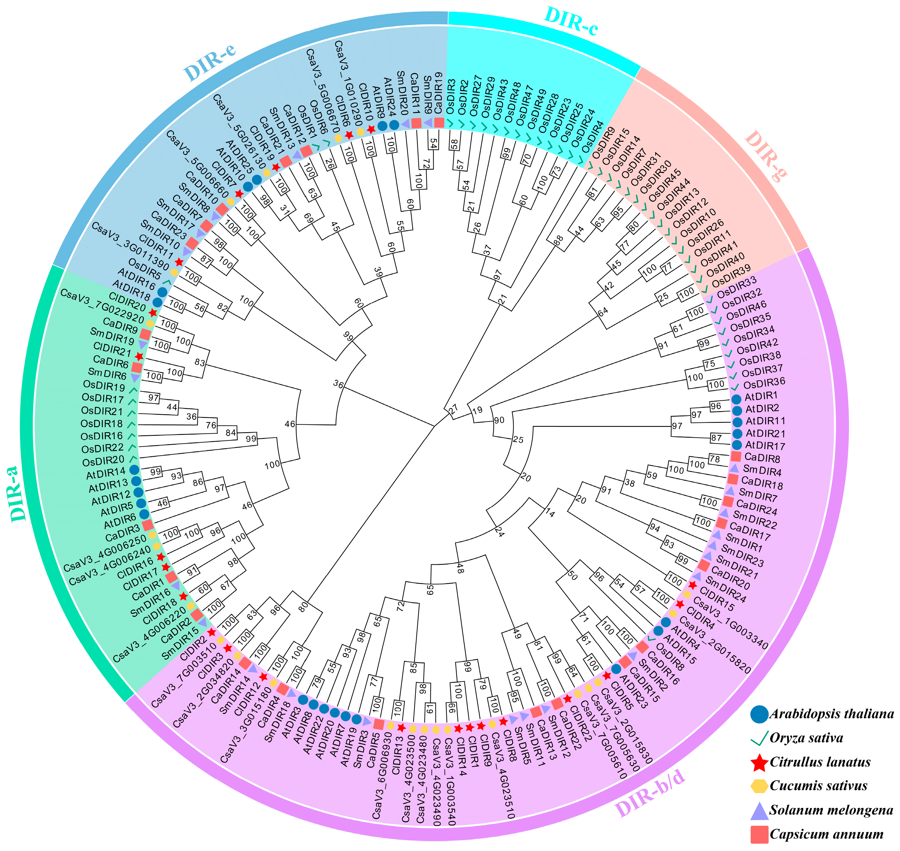
Figure 1. Phylogenetic analysis of DIR proteins from Arabidopsis, rice, watermelon, cucumber, eggplant, and pepper.
Ijms 26 07730 g001
Figure 2. Exon–intron structures of ClDIR genes and conserved motifs of ClDIR proteins.
Ijms 26 07730 g002
Figure 3. Syntenic relationships of DIR family genes among Citrullus lanatus and four representative species. (A) Intraspecific collinearity analysis of ClDIR genes in C. lanatus. (B) Interspecific syntenic relationships of DIR genes between C. lanatus and two model species (Arabidopsis thaliana, Oryza sativa). (C) Interspecific syntenic relationships of DIR genes between C. lanatus and two Cucurbitaceae species (Cucumis sativus, Cucumis melo).
Ijms 26 07730 g003
Figure 4. Cis-elements analysis of the promoters of ClDIR genes. Different shades of red indicate the number of cis-acting elements, with deeper colors corresponding to higher counts.
Ijms 26 07730 g004
Figure 5. Expression heatmap of ClDIR genes in diverse tissues (A) and fruit developmental stages (BE) of watermelon. 203Z: sweet watermelon; SW: sour watermelon; DAP or dap: days after pollination. The data in the boxes indicate the original FPKM values.
Ijms 26 07730 g005
Figure 6. The expression heatmaps of ClDIR genes under abiotic stresses in watermelon. (A) The expression patterns of ClDIR genes under high-temperature stress. CT: Control treatment; HT-4h, HT-8h, HT-12h, and HT-24h: high-temperature treatment for 4, 8, 12, and 24 h. (B) The expression patterns of ClDIR genes under drought stress. (C) The expression patterns of ClDIR genes under low-light stress. CT: Control treatment; LL: low light; 0DAP, 3DAP, 9DAP, and 15DAP: 0, 3, 9, and 15 days after pollination. (D) The expression patterns of ClDIR genes under salt stress. (E) The expression patterns of ClDIR genes under osmotic stress. CT: Control treatment; OS-2h and OS-4h: osmotic stress for 2 and 4 h. (F) The expression patterns of ClDIR genes under nitrogen treatment. LLN: leaf low nitrogen; LHN: leaf high nitrogen; RLN: root low nitrogen; RHN: root high nitrogen. (G) The expression patterns of ClDIR genes under cadmium stress. CT: Control treatment; Cd: cadmium stress. Original FPKM values were shown in heatmap boxes; differentially expressed genes were highlighted in red (upregulation) and green (downregulation) with log2(fold-change) values.
Ijms 26 07730 g006
Figure 7. The expression heatmaps of ClDIR genes under biotic stresses in watermelon. (AE) The expression patterns of ClDIR genes under Fusarium wilt stress. R: oilseed rape rotation cropping; C: continuous watermelon monocropping; SF7: susceptible cultivar at 7 days post inoculation with Fusarium oxysporum f. sp. niveum; RF7: resistant cultivar at 7 days post inoculation with Fusarium oxysporum f. sp. Niveum; S-CT: susceptible cultivar under non-inoculated control conditions; S-F: susceptible cultivar inoculated with Fusarium oxysporum f. sp. niveum; R-CT: resistant cultivar under non-inoculated control conditions; S-F: resistant cultivar inoculated with Fusarium oxysporum f. sp. niveum; CT-1d: non-inoculated control plants at 1 day; 1 dpi: plants at 1 day post inoculation with Fusarium oxysporum f. sp. niveum; CT-3d: non-inoculated control plants at 3 days; 3 dpi: plants at 3 days post inoculation with Fusarium oxysporum f. sp. niveum; F0, F3, F5, and F8: 0, 3, 5, and 8 days after inoculation with Fusarium oxysporum f. sp. niveum. (F) The expression patterns of ClDIR genes under cucumber green mottle mosaic virus stress. CT: control treatment; 48 hpi and 25 dpi: 48 h and 25 days post inoculation. (G) The expression patterns of ClDIR genes under powdery mildew stress. S-CT: non-inoculated control of susceptible cultivar; S-I: susceptible cultivar inoculated with powdery mildew; R-CT: non-inoculated control of resistant cultivar; R-I: resistant cultivar inoculated with powdery mildew. (H) The expression patterns of ClDIR genes under squash vein yellowing virus stress. S: susceptible plants; R: resistant plants; 0 dpi, 5 dpi, 10 dpi, and 15 dpi were 0, 5, 10, and 15 days post inoculation, respectively. (I) The expression patterns of ClDIR genes under root-knot nematode stress. CK-l: leaf samples under white light with water treatment; RKN-l: leaf samples under white light inoculated with Meloidogyne incognita; RL-l: leaf samples under red light with water treatment; RR-l: leaf samples under red light inoculated with Meloidogyne incognita. Original FPKM values are shown in heatmap boxes; differentially expressed genes are highlighted in red (upregulation) and green (downregulation) with log2(fold-change) values.
Ijms 26 07730 g007
Figure 8. The heatmap for expression patterns of ClDIR genes under abiotic and biotic stresses. Gray indicates no differential expression; red indicates upregulation; green indicates downregulation; and blue indicates both up- and downregulation.
Ijms 26 07730 g008
Figure 9. Expression profiles of ClDIR genes in response to diverse abiotic stresses. (A) Expression profiles of ClDIR genes under high-temperature stress (45 °C). (B) Expression profiles of ClDIR genes under drought stress (20% PEG6000). (C) Expression profiles of ClDIR genes under salt stress (500 mmol·L−1 NaCl). (D) Expression profiles of ClDIR genes under low-temperature stress (4 °C). Data are presented as means ± SE (n = 3) from three independent biological replicates. Statistical significance is denoted as ns (p > 0.05, not significant), * (p < 0.05), ** (p < 0.01), *** (p < 0.001), and **** (p < 0.0001).
Ijms 26 07730 g009
Figure 10. Subcellular localization analysis of ClDIR8 protein.
Ijms 26 07730 g010
Figure 11. Validation of ClDIR8 protein interactions via Y2H and BiFC assays. (A) Y2H confirmation of physical interactions between ClDIR8 and candidate proteins. (B) BiFC visualization of ClDIR8 interactions in Nicotiana benthamiana leaves.
Ijms 26 07730 g011
Table 1. The physiochemical characteristics of DIR family genes in watermelon.
Gene NameGene IDCDS Size (bp)Number of Amino Acids (aa)Molecular Weight (kDa)Theoretical pIInstability IndexAliphatic IndexGrand Average of Hydropathicity
ClDIR1Cla97C01G00679056118620.368.8929.3290.700.118
ClDIR2Cla97C02G02840054918219.886.7124.0096.980.332
ClDIR3Cla97C02G03538052517418.449.649.4299.770.272
ClDIR4Cla97C02G03729057619121.009.1030.2786.75−0.127
ClDIR5Cla97C02G03730055518419.809.1930.5992.660.147
ClDIR6Cla97C02G039150119139641.464.3643.1879.34−0.140
ClDIR7Cla97C02G03916073824527.509.6333.3468.86−0.329
ClDIR8Cla97C02G05002057018920.919.3928.2394.870.145
ClDIR9Cla97C02G05003056418720.4510.0429.08101.180.167
ClDIR10Cla97C03G055500120039943.456.1452.0077.74−0.337
ClDIR11Cla97C05G08435075325025.625.0036.2688.600.224
ClDIR12Cla97C05G08817054918220.065.4138.8293.630.116
ClDIR13Cla97C06G11031060320022.536.5142.1489.80−0.007
ClDIR14Cla97C06G12458056418720.628.4744.1386.520.016
ClDIR15Cla97C06G12495055218320.047.8936.8992.190.236
ClDIR16Cla97C07G13489057319021.158.3822.7583.740.127
ClDIR17Cla97C07G13490057919221.167.7444.4186.350.169
ClDIR18Cla97C07G13492055818520.598.9532.2984.430.039
ClDIR19Cla97C09G18168095131632.504.9534.9180.220.002
ClDIR20Cla97C09G18427054618120.249.6938.1785.75−0.223
ClDIR21Cla97C10G18475058219321.157.3427.1094.870.172
ClDIR22Cla97C10G20065057919220.899.6954.3693.440.223
Table 2. Ka/Ks analysis for the duplicated ClDIR paralogous gene pairs in watermelon.
Duplicated Gene PairsKaKsKa/KsDuplicationSelection Pattern
ClDIR4/ClDIR50.481.330.36TandemPurifying selection
ClDIR8/ClDIR90.291.090.26TandemPurifying selection
ClDIR16/ClDIR170.190.550.35TandemPurifying selection
ClDIR2/ClDIR30.432.060.21SegmentalPurifying selection
ClDIR6/ClDIR71.060.831.28TandemPositive selection
Table 3. Transcriptome datasets of watermelon for analyzing ClDIR gene expression patterns in diverse tissues and under stresses.
ProjectNo.ExperimentAccession NumberSampled TissueReference
Tissue-specific expression1Different tissuesPRJNA1031825Root, stem, leaf, male flower, female flower, fruit, tendril-
2Fruit developmentPRJNA407607Fruit flesh[77]
3Fruit developmentPRJNA718123Fruit flesh-
4Fruit developmentPRJNA703434Fruit flesh[78]
5Fruit developmentPRJNA520808Fruit flesh[79]
Abiotic stresses6High temperaturePRJNA504354Ovule-
7DroughtPRJNA604984Leaf[80]
8Low lightPRJNA602124Fruit flesh[81]
9SaltPRJNA609260Leaf[82]
10Osmotic stressPRJNA770012Leaf[83]
11Nitrogen treatmentPRJNA422970Leaf, root[84]
12Cadmium stressPRJNA1079538Leaf-
Biotic stresses13Fusarium wiltPRJNA641525Root[85]
14Fusarium wiltPRJNA794199Root[86]
15Fusarium wiltPRJNA973274Root-
16Fusarium wiltPRJNA929333Leaf[87]
17Fusarium wiltPRJNA783543Root[88]
18Cucumber green mottle mosaic virusPRJNA534308Leaf-
19Powdery mildewPRJNA881394Leaf[89]
20Squash vein yellowing virusPRJNA1086032Leaf-
21Root-knot nematodesPRJCA001078Leaf[90]
Disclaimer/Publisher’s Note: The statements, opinions and data contained in all publications are solely those of the individual author(s) and contributor(s) and not of MDPI and/or the editor(s). MDPI and/or the editor(s) disclaim responsibility for any injury to people or property resulting from any ideas, methods, instructions or products referred to in the content.