Article

Axially Disubstituted Silicon(IV) Phthalocyanine as a Potent Sensitizer for Antimicrobial and Anticancer Photo and Sonodynamic Therapy

by
Marcin Wysocki
1,2,
Daniel Ziental
1,
Zekeriya Biyiklioglu
3,*,
Malgorzata Jozkowiak
2,4,5,
Jolanta Dlugaszewska
6,
Hanna Piotrowska-Kempisty
4,
Emre Güzel
7 and
Lukasz Sobotta
1,*
1
Chair and Department of Inorganic and Analytical Chemistry, Poznan University of Medical Sciences, Rokietnicka 3, 60-806 Poznan, Poland
2
Doctoral School, Poznan University of Medical Sciences, Bukowska 70, 60-812 Poznan, Poland
3
Department of Chemistry, Faculty of Science, Karadeniz Technical University, Trabzon 61080, Türkiye
4
Chair and Department of Toxicology, Poznan University of Medical Sciences, Rokietnicka 3, 60-806 Poznan, Poland
5
Division of Anatomy, Department of Human Morphology and Embryology, Faculty of Medicine, Wroclaw Medical University, Chalubinskiego 6a, 50-368 Wroclaw, Poland
6
Chair and Department of Genetics and Pharmaceutical Microbiology, Poznan University of Medical Sciences, Rokietnicka 3, 60-806 Poznan, Poland
7
Department of Engineering Fundamental Sciences, Sakarya University of Applied Sciences, Sakarya 54050, Türkiye
*
Authors to whom correspondence should be addressed.
Int. J. Mol. Sci. 2025, 26(15), 7447; https://doi.org/10.3390/ijms26157447 (registering DOI)
Submission received: 14 July 2025 / Revised: 28 July 2025 / Accepted: 29 July 2025 / Published: 1 August 2025
(This article belongs to the Section Bioactives and Nutraceuticals)

Abstract

The unique properties of phthalocyanines (Pcs), such as strong absorption, high photostability, effective singlet oxygen generation, low toxicity and biocompatibility, versatile chemical modifications, broad spectrum of antimicrobial activity, and synergistic effects with other treatment modalities, make them a preferred superior sensitizer in the field of antimicrobial photodynamic therapy. The photodynamic and sonodynamic activity of 3-(3-(diethylamino)phenoxy)propanoxy substituted silicon(IV) Pc were evaluated against bacteria and cancer cells. Stability and singlet oxygen generation upon light irradiation and ultrasound (1 MHz, 3 W) were assessed with 1,3-diphenylisobenzofuran. The phthalocyanine revealed high photostability in DMF and DMSO, although the singlet oxygen yields under light irradiation were low. On the other hand, the phthalocyanine revealed excellent sonostability and caused a high rate of DPBF degradation upon excitation by ultrasounds at 1 MHz. The silicon phthalocyanine presented significant bacterial reduction growth, up to 5 log against MRSA and S. epidermidis upon light excitation, whereas the sonodynamic effect was negligible. The phthalocyanine revealed high activity in both photodynamic and sonodynamic manner toward hypopharyngeal tumor (FaDu, 95% and 42% reduction, respectively) and squamous cell carcinoma (SCC-25, 96% and 62% reduction, respectively). The sensitizer showed ca. 30% aldehyde dehydrogenase inhibition in various concentrations and up to 85% platelet-activating factor acetylhydrolase for 0.25 μM, while protease-activated protein C was stimulated up to 66% for 0.75 μM.

1. Introduction

Phthalocyanines (Pcs) are synthetic macrocyclic compounds whose structures are related to the naturally occurring porphyrins. They are known as potent photosensitizers (PSs) with strong absorption between 600 and 800 nm, in the red-light part of the spectrum [1,2,3,4,5]. As with other porphyrinoids, Pcs are prone to various modifications, the most common of which is the formation of complexes with other elements, usually metals. The incorporation of metals and metalloids often enhances the properties of macrocyclic compounds. The principle of PDT is focused on ROS formation in the target site [3,4,6,7,8,9,10].
The Pcs are widely recognized sensitizers in terms of photodynamic therapy (PDT) and sonodynamic therapy (SDT), two similar techniques that benefit from the interaction of the sensitizer with light or ultrasounds, respectively. Such interactions can initiate further chemical reactions between the excited sensitizer and surrounding molecules, such as molecular oxygen or organic moieties [11]. Despite their similarities, both techniques have different mechanisms. For PDT, the key interaction is photon absorption by the sensitizer following its excitation. Next, the excited sensitizer undergoes deactivation by forming reactive oxygen species (ROS), including singlet oxygen. The drawback of this approach is associated with limited tissue penetration by light (photons), strongly dependent on the wavelength. Phthalocyanines are good candidates for PS in PDT due to strong light absorption within the so-called “phototherapeutic window” range (600–800 nm) while light provides enough energy to the molecule [8,12]. Interestingly, the processes occurring during SDT work in a slightly different manner. The energy is passed to the sensitizer (sonosensitizer, SS) via acoustic cavitation, the phenomenon of creating oscillating microbubbles in response to ultrasound. Irregular oscillation of those bubbles results in their collapse, locally providing extreme conditions that promote the formation of radicals from affected molecules, such as solvent, sensitizer, or other moieties. The radicals may then react with other molecules and generate other radicals, but may also excite the SS. Additionally, the mechanical effects of the ultrasound provide other beneficial effects, such as an increase in cell membrane permeability and mechanical rupture of cellular membranes [13,14,15]. Both processes are characterized by specific chemical reactions that result in the generation of particular ROS, which are potent oxidizers that are utilized to directly oxidize the cellular membranes of targeted cells, either bacterial or cancerous ones [3,13,14,16,17,18,19,20,21].
Interestingly, the unusual coordination of silicon(IV) allows for bearing two additional substituents placed axially to the Pc ring that may effectively improve the compounds’ properties, including solubility and hamper aggregation tendency, due to increased steric hindrance of the macrocycle. The possibility to form the aggregates is one of the key drawbacks in terms of (PDT), due to self-quenching ROS [3,4,7,8]. Axial substituents, characterized by large, branched ligands, reduce the risk of formation of dimers or larger clusters typical of phthalocyanines. Therefore, various silicon(IV) Pcs have been described so far. However, the research on their use in photodynamic and sonodynamic therapy remains very limited, despite versatile synthesis and potentially high therapeutic efficacy. Thanks to analyses conducted by, among others, Chen et al. [22] the introduction of axial substituents in phthalocyanines has been recognized as a promising strategy for modifying their photochemical properties. Axial substitution enhances nonlinear optical absorption by introducing a dipole moment perpendicular to the macrocycle and improving excited-state absorption through stabilization of the excited state. It also allows fine-tuning of spectral characteristics and excited-state lifetimes, contributing to greater control over the photophysical behavior of the molecule [23]. In Si(IV) phthalocyanines, axial ligands can modulate the electron density of the macrocycle through inductive or mesomeric effects without directly perturbing the conjugated system. Electron-donating groups such as diethylamino may induce a slight bathochromic shift in absorption, potentially extending it toward the near-infrared region, which is favorable for applications requiring deeper light penetration, such as photodynamic therapy [24,25]. In biological studies, diethylamino groups are one of the most important compounds thanks to their various biological activities such as anticancer, photodynamic therapy [26,27,28]. For this purpose, (3-(diethylamino)phenoxy) propanoxy groups were preferred as substituents because they contain propanoxy chains for enhanced organic solubility and quaternizable nitrogen atoms. Motivated by these facts, it has been focused on non-aggregated silicon(IV) phthalocyanine as a strategy to explain Pcs in conjunction with the 3-(3-(diethylamino)phenoxy)propanoxy group to study antimicrobial and anticancer photo-sonodynamic therapy. There is no study in the literature in which diethylamino groups substituted with silicon phthalocyanine are used as sensitizers for either photodynamic or sonodynamic activity. This is the first study of axially substituted silicon phthalocyanine containing 3-(3-(diethylamino)phenoxy)propanoxy groups as potential sensitizers for photodynamic and sonodynamic purposes. Thus, we present the photo- and sonochemical properties and activity toward bacterial and cancer cells in vitro of silicon(IV) Pc bearing axial 3-(3-(diethylamino)phenoxy)propanoxy substituents (SiPc, Figure 1). The synthesis and electrochemical properties of the compound have been reported before [29].

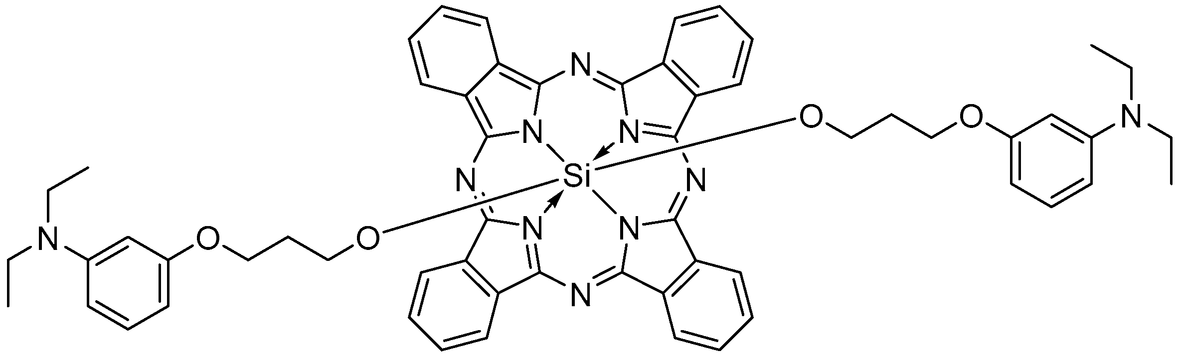
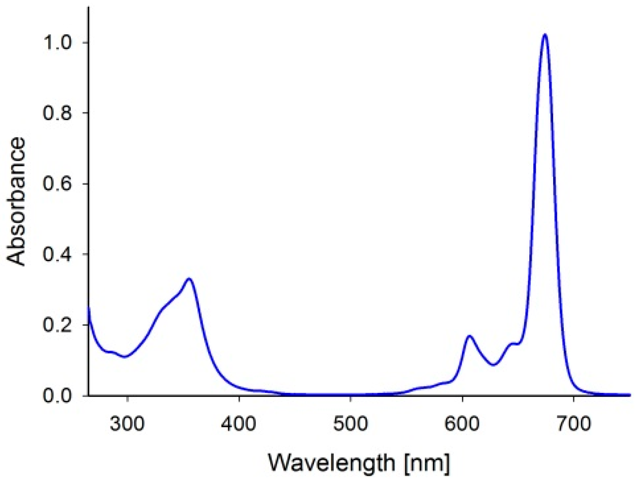
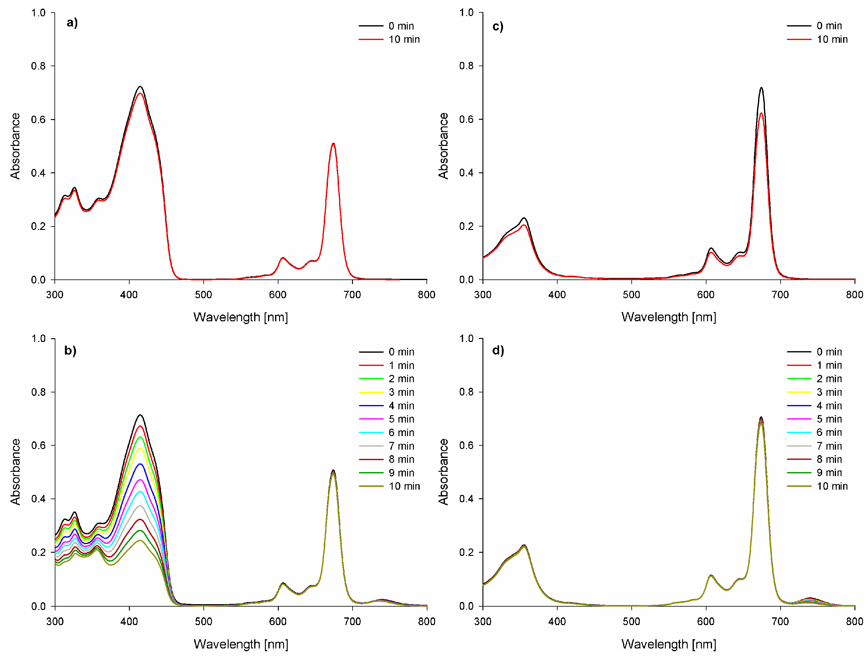
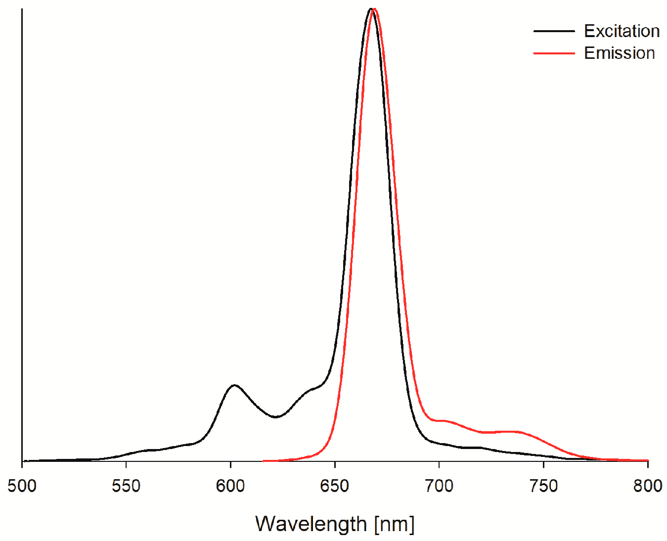
2. Results and Discussion

Evaluated silicon(IV) phthalocyanine bearing axial 3-(3-(diethylamino)phenoxy)propanoxy substituents (SiPc) revealed two absorption bands in the 300–400 nm range—the Soret band and in the 600–700 nm range—the Q band (Figure 2).
These absorption bands are typical for phthalocyanines and the most important from the PDT point of view is the Q band located at the “therapeutic window” region [1]. Following light activation, singlet oxygen quantum yield values were close to zero—see Table 1.
Interestingly, the rate of DPBF decomposition in the presence of phthalocyanine differed significantly depending on the used trigger—light or ultrasounds (Figure 3). It is well-known that silicon(IV) phthalocyanines can produce singlet oxygen both with very high and low ΦΔ values [34,35,36,37]. SiPc is a very poor singlet oxygen generator under light exposure, which can be associated with axial substituents bearing nitrogen atoms in the aliphatic chain. When confronted with the relatively high biological activity, these results suggest that other molecular factors may play a key role in the photodynamic process. Some previously described silicon phthalocyanines exhibit relatively long excited-state lifetimes, which may compensate for lower ROS generation efficiency [38,39]. Additionally, the positive charge of the molecule may enhance its interactions with the cell membrane or wall, thereby potentiating the phototoxic effect [40]. In comparison with other silicon phthalocyanines exhibiting higher singlet oxygen generation potential, a key distinguishing factor appears to be the presence or absence of strong electron-donating groups, long-chain alkyl or bulky substituents, or halogen atoms such as bromine, chlorine, or fluorine [41].
A similar phenomenon has been reported by van de Winckel et al. [33] for compounds IIII (Figure 4). Values of ΦΔ dependent on substituent were in the range from ca. 0.03 for II to ca. 0.38 for III [34,35]. Surprisingly, the nitrogen atom which is included in the aromatic ring does not cause a dramatic drop in singlet oxygen formation. It was reported high ΦΔ values for phthalocyanines IV (0.62) and V (0.86) [36,42]. Additionally, axial substituents bearing aliphatic chains in their structure without nitrogen atoms present high ΦΔ values, i.e., VI (0.48) [35]. On the other hand, the presence of quaternized nitrogen atoms, results in quenching 1O2 formation as well, i.e., VII (0.11), VIII (0.11), and IX (0.08) (Figure 4) [37].
The photostability of SiPc was evaluated as well in a liquid environment (DMF and DMSO). SiPc under light exposure decomposed according to the photobleaching mechanism. It was observed that the absorption bands were vanishing, without forming new ones (Figure 3c). The quantum yield values (18.9 × 10⁻⁶ and 34.1 × 10⁻⁶ in DMF and DMSO, respectively) of photodegradation of the presented photosensitizer classify it as a stable compound [43]. The ability of SiPc to emit light (Figure 5) has also been evaluated. Its fluorescence quantum yields were determined at the level of 0.07 (Table 1, Figure 5), which are much lower in comparison to the values of the Φf for the standard.
In the sonochemical experiments, it was noticed that SiPc upon sonication efficiently decomposes DPBF. The half-life time of DPBF dropped over 5-fold in the presence of sensitizer (Table 2). The decomposition DPBF rate for SiPc under sonication is over two times faster in comparison to lately reported porphyrazines [12]. This potentially enables to achievement of high sonodynamic activity in biological evaluation. Notably, the presented compounds demonstrated the highest sonostability (T0.5 = 7737.3 min, Figure 3d) reported to date among silicon phthalocyanines studied in sonochemistry [12]. Low frequencies resulted in a decomposition sensitizer. On the other hand, high frequencies >> 1 MHz (i.e., 2 MHz) resulted in no ROS formation.
The antibacterial evaluation indicated that SiPc under light irradiation is extremely active. For activation light at the dose of 100 and 150 J/cm2 was used. Surprisingly, PS activation at 100 J/cm2 did not induce any activity against MRSA, whereas against Staphylococcus epidermidis high—ca. 5 log reduction in bacterial growth was noticed (Table 3). When the light dose is raised to 150 J/cm2 the bacterial growth reduction achieves the rate of ca. 5 log for both species. The obtained here results in comparison to those reported by van de Winckel et al. where silicon(IV) phthalocyanines at the concentration of 20 µM (here only 10 µM) were activated with comparable light dose have given maximally ca. 4 log reduction in S. aureus growth [34]. In the sonodynamic evaluation, no activity against bacteria was noted.
Similarly, in the SiPc activity evaluation against cancer cells, much higher anticancer activity in the photodynamic than sonodynamic pathway was observed (Table 4). For the experiments, the squamous cell carcinoma (SCC-25) and hypopharyngeal tumor cell line (FaDu) cell lines were chosen. These tumors are one of the most common problems of head and neck oncology. Interestingly, an increase in light dose from 5 to 10 J/cm2 did not result in a rise in the activity for all tested cells. On the other hand, when comparing the viability of cancer cells treated with PDT-SiPc (viability reduction about >94%) to the data obtained before, for similar silicon(IV) phthalocyanine (10 µM), viability reduction was about ca. 60% [44]. Unfortunately, the highest activity of the tested sensitizer was shown against MRC-5 fibroblasts. This indicated that in the next stage of evaluation, application, and excitation with light of the sensitizer have to be performed only within the diseased area. In contrast, to what was reported before for sonodynamically inactive porphyrazine/phthalocyanine hybrids against cancer cells, studied here, SiPc shows moderate activity with cell viability reduction 41.5–82.5% (Table 4).
The impact of SiPc on the activity of enzymes was also evaluated. Highly inhibited was aldehyde dehydrogenase at about 85% in the sensitizer concentration of 0.75 µM (Figure 6). Such a rate of inhibition may indicate additional independent of the studied compound’s light mechanism of anticancer activity. Otherwise, tested phthalocyanine reduced the activity of PAF acetylhydrolase at ca. 40% independently of the concentration within the studied range. Interestingly, our sensitizer increased the activity of protease-activated protein C, which is responsible for anticoagulant and anti-inflammatory activity [45]. Additional stimulation of protease-activated protein C may limit inflammation associated with the PDT procedure. Nevertheless, promising results of the impact of SiPc on enzymatic activity have to be further evaluated.
The obtained results represent an interesting alternative to currently available treatment methods, particularly for surface infections. The development of optimal sonosensitizers will enable further combination with carriers, such as hydrogel patches propagating ultrasound, as innovative solutions for treating diabetic foot ulcers and hard-to-heal, superinfected wounds. This technology appears particularly promising for multidrug-resistant and mixed infections, where selecting the optimal antibiotic therapy can be especially challenging. Moreover, in cancer treatment, this method could serve as a valuable adjuvant therapy with a relatively low risk of severe adverse effects and side effects.

3. Materials and Methods

3.1. Materials

Dimethyl sulfoxide (DMSO, 99.9% for spectroscopy) and N,N-dimethylformamide (DMF, ≥99.8% for spectroscopy ACS) were purchased from Fisher (Waltham, MA, USA). Zinc(II) phthalocyanine (ZnPc) and 1,3-diphenylisobenzofuran (DPBF) were purchased from Aldrich (St. Louis, MO, USA). Brain-heart infusion (BHI) broth was purchased from Oxoid (Waltham, MA, USA). The SCC-25 and FaDu human squamous cell carcinoma cell lines and the MRC-5 human diploid lung fibroblasts were purchased from the American Type Culture Collection (ATCC) (Manassas, VA, USA). Phosphate-buffered saline, Dulbecco’s Modified Eagle’s Medium/F-12 (DMEM/F-12), Fetal Bovine Serum (FBS), L-glutamine, Penicillin, Streptomycin, Eagle’s Minimum Essential Medium (EMEM), Trypsin-EDTA solution, Dimethyl sulfoxide (DMSO, for cell culture), and MTT were purchased from Merck (Rahway, NJ, USA). Purified acetylcholinesterase (C3389), acetylcholinesterase inhibitor screening kit (MAK324), purified aldehyde dehydrogenase (A6338), aldehyde dehydrogenase inhibitor screening kit (MAK327), and activated protein C (APC) inhibitor screening kit (MAK346) were purchased from Merck. A platelet-activating factor acetylhydrolase (PAF-AH) inhibitor screening kit was purchased from BioVision (Exton, PA, USA).

3.2. Photochemistry Measurements

3.2.1. Singlet Oxygen Quantum Yields Determination Under Light Exposure

The measurements of singlet oxygen quantum yields were conducted in DMSO and DMF at ambient temperature, according to the comparative method described previously [12,46,47,48]. The method involves using DPBF as a chemical quencher of singlet oxygen and ZnPc as a reference compound. Firstly, irradiation of the respective sensitizer-quencher mixtures with light of fixed power (0.5 mW/cm2 measured with RD 0.2/2 TD probe radiometer, Optel, Opole, Poland) was performed, and the proper wavelength of the Q band region (660 nm) was determined. Then, spectral changes allowed calculation of the quencher (DPBF), and decomposition kinetics were recorded. Such data obtained for mixtures of DPBF with ZnPc (reference) and mixtures of DBPF with the studied compound (SiPc) were then compared according to Equation (1):
Φ Δ = Φ Δ s t d R I A b s S t d R S t d I A b s ,
where Φ Δ s t d is the quantum yield of singlet oxygen known for ZnPc (0.56 in DMF and 0.67 in DMSO), R and RStd stand for rates of DPBF decomposition, and IAbs with I a b s S t d stand for rates of light absorption of both investigated SiPc and the standard, respectively. The light emitted by 150 W xenon lamp (Optel, Opole, Poland) was adjusted to the proper wavelength with the monochromator (M250/1200/U with 2 nm/mm (Dk = 4 nm) dispersionOptel, Opole, Poland). The absorption spectra and their changes (within 10 min of the experiment provided energy of 300 mJ/cm2) were gathered using an OceanOptics Flame spectrophotometer coupled with DT-MINI-2-GS light source. The obtained data were then used for calculations of singlet oxygen quantum yields using the presented equation [12,31,46,47,48].

3.2.2. Photodecomposition Quantum Yields Determination

The measurements of photostability were performed in DMF and DMSO. The samples were prepared under aerobic conditions and kept at ambient temperature and the investigation was conducted according to the method described before [12,46,48]. As a light source served the 150 W xenon lamp (Optel, Opole, Poland), and the visible light (>450 nm; 20 mW/cm2) was filtered with HCC-16 cutting glass filter. Absorption spectra changes during the irradiation (10 min provided total energy of 12 J/cm2) were recorded with OceanOptics Flame spectrophotometer equipped with DT-MINI-2-GS light source, and the photodegradation quantum yield was then calculated according to the equation presented before [12].

3.2.3. Fluorescence Measurements

The fluorescence spectra were recorded in DMF and DMSO at 21 °C using Jasco FP-6200 spectrofluorometer with the slit width set to match 5 nm bandwidth. Before measurements, the absorbance values of the samples in the excitation maximum were set to 0.1 and placed in quartz cuvettes (l = 10 mm), and then the excitation and emission spectra were recorded. For calculations of the fluorescence quantum yields, ZnPc was employed as a reference compound. The fluorescence quantum yields were then calculated using Equation (2):
Φ f = Φ f s t d F A E x s t d F s t d A E x ,
where Φ f s t d —fluorescence quantum yield for ZnPc (0.30 in DMF and 0.20 in DMSO), F and FStd—fluorescence intensity described as the spectrum integral for the sample and the standard, AEx and A E x s t d —absorbance of the samples at the excitation wavelength (for SiPc 667 nm in DMF, 669 nm in DMSO) [30,33].

3.3. Sonochemistry Measurements

3.3.1. DPBF Decomposition Under Sonication

The SiPc-mediated decomposition of DPBF was measured in the dark and at ambient temperature. The mixtures of the phthalocyanine with DPBF were aerated for 10 min, then moved into quartz cuvettes (l = 10 mm), and sealed with plugs. Before the measurements, each cuvette was shaken and the spectrum before sonication was recorded using a Shimadzu U-1900 spectrophotometer (Shimadzu, Tokyo, Japan). The cuvettes were then thoroughly shaken again and placed in the ultrasonic apparatus (constructed by the Institute of Fundamental Technological Research, Polish Academy of Sciences, Warsaw, Poland). The ultrasounds of 1 MHz frequency 3 W power and 400% duty cycle were transmitted via an ultrasonic head for ten 1 min intervals. The absorption spectra were recorded after each interval and the samples were subsequently shaken before further sonication. For the calculations, mean values of the data obtained from five independent measurements were taken.

3.3.2. Sonostability of Sensitizers

The sonostability measurements of SiPc were conducted according to the same steps described in Section 3.3.1.

3.4. Antibacterial Activity

3.4.1. Cell Culture

The methicillin-resistant Staphylococcus aureus (MRSA, clinical strain) and Staphylococcus epidermidis (clinical strain) strains were cultured aerobically in brain heart infusion (BHI) broth at 36 ± 1 °C for 24 h. The microbes were then centrifuged at 3000 rpm for 15 min, harvested, and resuspended in 10 mM phosphate-buffered saline (pH = 7.0). The cells were later diluted to approximately 107 colony-forming units per ml (CFU/mL). The bacterial suspension was placed into wells of a 96-well plate, and then aliquots of a proper amount from the stock solution prepared in DMSO to obtain the final concentration of 10 µM were added. Control cells were cultured under the same conditions with 0.1% DMSO. The reduction in bacterial growth was calculated using the colony counting method following the guidelines of CLSI. Experiments were performed in triplicate.

3.4.2. Photodynamic Pathway

The plate was placed in the dark for 30 min to incubate properly, then subjected to irradiation with 660 nm light using a custom LED lamp (20 mW/cm2 Led-Byt, Bytom, Poland), achieving the dose of 100 (80 min) and 150 J/cm2 (120 min). The dose and irradiation time were evaluated with RD 0.2/2 radiometer (Optel, Opole, Poland). Control samples without the sensitizer presence were performed as well. After the given irradiation interval, the bacterial suspensions were diluted, placed on tryptic soy agar plates and incubated for 24 h at 36 ± 1 °C.

3.4.3. Sonodynamic Pathway

The incubation was performed via placing the plate in dark place for 30 min. After incubation, the plate was positioned above the ultrasonic head (the Institute of Fundamental Technological Research, Polish Academy of Sciences, Warsaw, Poland) and subjected to sonication (1 MHz, 3 W, 40% duty cycle), reaching a final dose of 1024 J/cm2. Control samples, not including the sensitizer and ultrasound, were also incorporated. The bacterial cultures after the sonication were moved to tryptic soy agar plates and incubated at 36 ± 1 °C for 24 h.

3.5. Anticancer Activity

3.5.1. Cell Culture and Viability Assay

The chosen cell lines: human squamous cell carcinoma SCC-25 and FaDu, as well as the reference human diploid lung fibroblasts MRC-5 were cultivated in the optimal conditions (37 °C, 95% humidity, 5% CO2 content) using an incubator. FaDu and MRC-5 cell lines were cultivated in phenol red-free Eagle’s Minimum Essential Medium (EMEM) supplemented with 10% fetal bovine serum (FBS), 2.5 mM L-glutamine, penicillin (100 U/mL), and streptomycin (0.1 mg/mL), while the SCC-25 cell line was cultured in phenol red-free Dulbecco’s Modified Eagle Medium F-12 (DMEM/F-12) medium complemented with 10% FBS, 2.5 mM L-glutamine, penicillin (100 U/mL), and streptomycin (0.1 mg/mL). The cell cultures were harvested using trypsin-EDTA solution and seeded in 6-well cell culture plates at 106 cells per well. The cells were left overnight to attach and then the stock solution of the silicon phthalocyanine in DMSO was added to maintain the final concentration of 10 μM. Control cells were cultured in the same conditions with 0.1% DMSO instead. Cell viability was assessed with an MTT test the next day after treatment. The culture medium was replaced with mixtures of MTT and EMEM or DMEM/F-12 (both 1:8), depending on the investigated cell lines. The resulting formazan crystals were then dissolved by adding 1 mL of DMSO, and the absorbance was measured using an Elx-800 plate reader (BioTek, Bad Friedrichshall, Germany) at 570 nm (reference wavelength 650 nm). Experiments were performed in triplicate.

3.5.2. Photodynamic Pathway

The cells were incubated for 5 h and then exposed to 660 nm LED light (Led-Byt, Bytom, Poland) to achieve the final light doses of 5 and 10 J/cm2. The cell viability was then measured using an MTT assay.

3.5.3. Sonodynamic Pathway

After 5 h of incubation, the cells were exposed to ultrasound using a transducer with a resonance frequency of 3 MHz (continuous mode) until the energy dose reached 96 J/cm2. Then, the cell viability was assessed with an MTT assay.

3.6. The Impact of Sensitizer Activity on Endogenous Enzymes

The assays were performed according to the procedures provided by suppliers. The evaluated macrocycle was added in the concentration range of 0.1–1.0 µM. Absorbance was recorded with Tecan Infinite 200 Pro MNano (Tecan Life Sciences, Zürich, Switzerland), equipped with an absorbance–monochromator module.

4. Conclusions

In a nutshell, silicon(IV) phthalocyanine bearing axial 3-(3-(diethylamino)phenoxy)propanoxy substituents (SiPc) was evaluated, and when excited, this sensitizer decomposed DPBF, and the rate of DPBF bleaching differed significantly depending on the used trigger—light or ultrasounds. The studied SiPc under light exposure decomposed according to the photobleaching mechanism with a low quantum yield. Upon sonication, the efficiently studied sensitizer decomposes DPBF. This potentially enables to achievement of high sonodynamic activity in biological tests. The antibacterial evaluation indicated that SiPc studied here under light irradiation is extremely active. In the sonodynamic triggering of ROS formation by SiPc, no activity against bacteria was noted. Similarly, in the SiPc activity evaluation against cancer cells, much higher anticancer activity in the photodynamic than sonodynamic pathway was observed. The impact of SiPc on the activity of enzymes was also evaluated. Highly inhibited was aldehyde dehydrogenase, about 85%; such a rate of inhibition could indicate additional independence of the light mechanism of the anticancer activity of the studied compound. Interestingly, as presented here, the sensitizer increased the activity of protease-activated protein C. Eventually, SiPc shows promise as a potential sonophotodynamic agent in antimicrobial PDT applications.

Supplementary Materials

The following supporting information can be downloaded at: https://www.mdpi.com/article/10.3390/ijms26157447/s1.

Author Contributions

Conceptualization, D.Z., Z.B., M.J., E.G. and L.S.; data curation, M.W., D.Z., M.J., J.D., H.P.-K. and E.G.; formal analysis, M.W., D.Z., M.J., H.P.-K., E.G. and L.S.; funding acquisition, M.W. and L.S.; investigation, M.W., D.Z., Z.B., M.J. and J.D.; methodology, M.W., D.Z., Z.B., M.J., J.D. and L.S.; project administration, L.S.; resources, Z.B., J.D., H.P.-K. and L.S.; software, M.W. and L.S.; supervision, D.Z., H.P.-K., E.G. and L.S.; validation, M.W., D.Z., M.J., J.D. and H.P.-K.; writing—original draft, M.W. and L.S.; writing—review and editing, D.Z., Z.B., E.G. and L.S. All authors have read and agreed to the published version of the manuscript.

Funding

This work was supported by the Poznan University of Medical Sciences under grant no. SDUM-DGB 16/04/23 and no. SDUM-MGB 15/05/24—small and large research grants from statutory funding for young researchers-doctoral students for 2023 and 2024. The research was supported by the FENG.02.02-IP.05-0257/23 grant from the Foundation for Polish Science, co-financed by the European Union funds under the European Funds for Smart Economy 2021–2027 (FENG). and the Scientific Research Projects Coordination Unit of Sakarya University of Applied Sciences (Grant no. 244-2024). Marcin Wysocki is a participant in the STER Internationalisation of Doctoral Schools Programme from NAWA Polish National Agency for Academic Exchange No. PPI/STE/2020/1/00014/DEC/02.

Institutional Review Board Statement

Not applicable.

Informed Consent Statement

Not applicable.

Data Availability Statement

All data are included in the manuscript or the Supplementary Materials. Additional details will be provided upon request.

Conflicts of Interest

The authors declare no conflicts of interest.

Abbreviations

The following abbreviations are used in this manuscript:
PSPhotosensitizer
PDTPhotodynamic therapy
ROSReactive oxygen species
SSSonosensitizer
SDTSonodynamic therapy
PcPhthalocyanine
DPBF1,3-diphenylisobenzofuran
DMSODimethyl sulfoxide
DMFN,N-dimethylformamide
ZnPcUnsubstituted zinc(II) phthalocyanine
CFUColony-forming units
SiPcEvaluated here silicon(IV) phthalocyanine
MRSAMethicillin-resistant Staphylococcus aureus

References

  1. Pang, E.; Zhao, S.; Wang, B.; Niu, G.; Song, X.; Lan, M. Strategies to Construct Efficient Singlet Oxygen-Generating Photosensitizers. Coord. Chem. Rev. 2022, 472, 214780. [Google Scholar] [CrossRef]
  2. Ömeroğlu, İ.; Göksel, M.; Kussovski, V.; Mantareva, V.; Durmuş, M. Novel Water-Soluble Silicon(IV) Phthalocyanine for Photodynamic Therapy and Antimicrobial Inactivations. Macroheterocycles 2019, 12, 255–263. [Google Scholar] [CrossRef]
  3. Yenilmez, H.Y.; Farajzadeh, N.; Tollu, G.; Kuşçulu, N.G.; Bahar, D.; Özdemir, S.; Bayır, Z.A. Silicon Phthalocyanines as Anti-infectious, Antioxidant, and Anticancer Agents. ChemistrySelect 2023, 8, e202300856. [Google Scholar] [CrossRef]
  4. Mitra, K.; Hartman, M.C.T. Silicon Phthalocyanines: Synthesis and Resurgent Applications. Org. Biomol. Chem. 2021, 19, 1168–1190. [Google Scholar] [CrossRef] [PubMed]
  5. Kessel, D. Photodynamic Therapy: Critical PDT Theory. Photochem. Photobiol. 2023, 99, 199–203. [Google Scholar] [CrossRef]
  6. Güngördü Solğun, D.; Ağırtaş, M.S. Synthesis of Novel an Axially Substituted Silicon Phthalocyanine and Determination of Its Selectivity for Fe3+. J. Mol. Struct. 2023, 1286, 135577. [Google Scholar] [CrossRef]
  7. Balcik-Ercin, P.; Ekineker, G.; Salik, N.; Aydoğdu, B.; Yagci, T.; Göksel, M. Arginine Mediated Photodynamic Therapy with Silicon(IV) Phthalocyanine Photosensitizers. Photodiagnosis Photodyn. Ther. 2023, 43, 103667. [Google Scholar] [CrossRef]
  8. Granados-Tavera, K.; Zambrano-Angulo, M.; Montenegro-Pohlhammer, N.; Yaşa Atmaca, G.; Sobotta, L.; Güzel, E.; Cárdenas-Jirón, G.; Erdoğmuş, A.; Gürek, A.G. Synergistic Effect of Ultrasound and Light to Efficient Singlet Oxygen Formation for Photodynamic Purposes. Dye. Pigment. 2023, 210, 110986. [Google Scholar] [CrossRef]
  9. Pucelik, B.; Dąbrowski, J.M. Photodynamic Inactivation (PDI) as a Promising Alternative to Current Pharmaceuticals for the Treatment of Resistant Microorganisms. In Advances in Inorganic Chemistry; Elsevier: Amsterdam, The Netherlands, 2022; Volume 79, pp. 65–103. ISBN 978-0-323-99972-4. [Google Scholar]
  10. Dabrowski, J.M.; Arnaut, L.G. Photodynamic Therapy (PDT) of Cancer: From Local to Systemic Treatment. Photochem. Photobiol. Sci. 2015, 14, 1765–1780. [Google Scholar] [CrossRef]
  11. Chen, J.; Zhou, Q.; Cao, W. Multifunctional Porphyrin-Based Sonosensitizers for Sonodynamic Therapy. Adv. Funct. Mater. 2024, 34, 2405844. [Google Scholar] [CrossRef]
  12. Wysocki, M.; Ziental, D.; Jozkowiak, M.; Dlugaszewska, J.; Piotrowska-Kempisty, H.; Güzel, E.; Sobotta, L. Porphyrazine/Phthalocyanine Hybrid Complexes—Antibacterial and Anticancer Photodynamic and Sonodynamic Activity. Synth. Met. 2023, 299, 117474. [Google Scholar] [CrossRef]
  13. Nowak, K.M.; Schwartz, M.R.; Breza, V.R.; Price, R.J. Sonodynamic Therapy: Rapid Progress and New Opportunities for Non-Invasive Tumor Cell Killing with Sound. Cancer Lett. 2022, 532, 215592. [Google Scholar] [CrossRef]
  14. Son, S.; Kim, J.H.; Wang, X.; Zhang, C.; Yoon, S.A.; Shin, J.; Sharma, A.; Lee, M.H.; Cheng, L.; Wu, J.; et al. Multifunctional Sonosensitizers in Sonodynamic Cancer Therapy. Chem. Soc. Rev. 2020, 49, 3244–3261. [Google Scholar] [CrossRef]
  15. Moyano, D.B.; Paraiso, D.A.; González-Lezcano, R.A. Possible Effects on Health of Ultrasound Exposure, Risk Factors in the Work Environment and Occupational Safety Review. Healthcare 2022, 10, 423. [Google Scholar] [CrossRef]
  16. Spesia, M.B.; Durantini, E.N. Evolution of Phthalocyanine Structures as Photodynamic Agents for Bacteria Inactivation. Chem. Rec. 2022, 22, e202100292. [Google Scholar] [CrossRef]
  17. Yang, B.; Chen, Y.; Shi, J. Reactive Oxygen Species (ROS)-Based Nanomedicine. Chem. Rev. 2019, 119, 4881–4985. [Google Scholar] [CrossRef]
  18. Ogilby, P.R. Singlet Oxygen: There Is Indeed Something New under the Sun. Chem. Soc. Rev. 2010, 39, 3181. [Google Scholar] [CrossRef]
  19. Borah, B.M.; Cacaccio, J.; Durrani, F.A.; Bshara, W.; Turowski, S.G.; Spernyak, J.A.; Pandey, R.K. Sonodynamic Therapy in Combination with Photodynamic Therapy Shows Enhanced Long-Term Cure of Brain Tumor. Sci. Rep. 2020, 10, 21791. [Google Scholar] [CrossRef] [PubMed]
  20. An, J.; Hu, Y.-G.; Cheng, K.; Li, C.; Hou, X.-L.; Wang, G.-L.; Zhang, X.-S.; Liu, B.; Zhao, Y.-D.; Zhang, M.-Z. ROS-Augmented and Tumor-Microenvironment Responsive Biodegradable Nanoplatform for Enhancing Chemo-Sonodynamic Therapy. Biomaterials 2020, 234, 119761. [Google Scholar] [CrossRef] [PubMed]
  21. Xu, M.; Zhou, L.; Zheng, L.; Zhou, Q.; Liu, K.; Mao, Y.; Song, S. Sonodynamic Therapy-Derived Multimodal Synergistic Cancer Therapy. Cancer Lett. 2021, 497, 229–242. [Google Scholar] [CrossRef]
  22. Chen, Y.; Gao, L.; Feng, M.; Gu, L.; He, N.; Wang, J.; Araki, Y.; Blau, W.; Ito, O. Photophysical and Optical Limiting Properties of Axially Modified Phthalocyanines. Mini-Rev. Org. Chem. 2009, 6, 55–65. [Google Scholar] [CrossRef]
  23. Pişkin, M. Phthalocyanine Photosensitizers with Bathochromic Shift, of Suitable Brightness, Capable of Producing Singlet Oxygen with Effective Efficiency. J. Photochem. Photobiol. A Chem. 2023, 435, 114325. [Google Scholar] [CrossRef]
  24. Claessens, C.G.; Hahn, U.; Torres, T. Phthalocyanines: From Outstanding Electronic Properties to Emerging Applications. Chem. Rec. 2008, 8, 75–97. [Google Scholar] [CrossRef]
  25. Huang, Y.; Liang, J.; Fan, Z.; Yin, C. Benzopyrylium Salt Conjugate with 4-Diethylamino Group Enhances D-π-A-D′ to Boost Type-I/II ROS Generation for Photodynamic Therapy. Sens. Actuators B Chem. 2025, 427, 137209. [Google Scholar] [CrossRef]
  26. Wang, A.; Ma, Y.; Wang, Y.; Lu, S.; Lin, Y.; Zhou, J.; Zhou, L.; Wei, S. Effects of Protonation Degree on Photodynamic Activity of Zinc Phthalocyanine Substituted with 1,2-Diethylamino. Inorg. Chem. Commun. 2014, 48, 107–110. [Google Scholar] [CrossRef]
  27. Queirós, C.; Leite, A.; Moura, N.M.M.; Cerqueira, A.F.R.; Serra, V.V.; Neves, M.G.P.M.S.; Tomé, A.C.; Silva, A.M.G. Exploring the Reactivity of Formylporphyrins with 3-(Diethylamino)Phenol. Synthesis, Spectroscopic Properties and Singlet Oxygen Generation of a New Porphyrin–Rosamine Conjugate. Dye. Pigment. 2023, 217, 111431. [Google Scholar] [CrossRef]
  28. Biyiklioglu, Z.; Alp, H. Synthesis, Characterization, Electropolymerization and Aggregation Properties of Axially Diethyl-Dimethylaminophenoxypropanoxy Substituted Silicon Phthalocyanines and Their Water Soluble Derivatives. Dye. Pigment. 2016, 132, 213–222. [Google Scholar] [CrossRef]
  29. Seybold, P.G.; Gouterman, M. Porphyrins. J. Mol. Spectrosc. 1969, 31, 1–13. [Google Scholar] [CrossRef]
  30. Sobotta, L.; Lijewski, S.; Dlugaszewska, J.; Nowicka, J.; Mielcarek, J.; Goslinski, T. Photodynamic Inactivation of Enterococcus Faecalis by Conjugates of Zinc(II) Phthalocyanines with Thymol and Carvacrol Loaded into Lipid Vesicles. Inorganica Chim. Acta 2019, 489, 180–190. [Google Scholar] [CrossRef]
  31. Ogunsipe, A.; Maree, D.; Nyokong, T. Solvent Effects on the Photochemical and Fluorescence Properties of Zinc Phthalocyanine Derivatives. J. Mol. Struct. 2003, 650, 131–140. [Google Scholar] [CrossRef]
  32. Kuznetsova, N.A.; Fedorova, T.M.; Solovyova, L.I.; Shevchenko, E.N.; Bordaev, E.B.; Bulgakov, R.A.; Kaliya, O.L.; Lukyanets, E.A. Zinc Carboxyphthalocyanines: Synthesis, Properties, Photocatalysis. Macroheterocycles 2018, 11, 21–28. [Google Scholar] [CrossRef]
  33. van de Winckel, E.; Schneider, R.J.; de la Escosura, A.; Torres, T. Multifunctional Logic in a Photosensitizer with Triple-Mode Fluorescent and Photodynamic Activity. Chem. A Eur. J. 2015, 21, 18551–18556. [Google Scholar] [CrossRef]
  34. Taşkın, G.C.; Durmuş, M.; Yüksel, F.; Mantareva, V.; Kussovski, V.; Angelov, I.; Atilla, D. Axially Paraben Substituted Silicon(IV) Phthalocyanines towards Dental Pathogen Streptococcus Mutans: Synthesis, Photophysical, Photochemical and in Vitro Properties. J. Photochem. Photobiol. A Chem. 2015, 306, 31–40. [Google Scholar] [CrossRef]
  35. Atmaca, G.Y.; Celep, K.; Erdoğmuş, A. Application of Comparative Sonochemical, Photochemical, and Sono-Photochemical Methods to Obtain New Silicon Phthalocyanine with High Singlet Oxygen Efficiency. J. Organomet. Chem. 2024, 1004, 122952. [Google Scholar] [CrossRef]
  36. Gamelas, S.R.D.; Vieira, C.; Bartolomeu, M.; Faustino, M.A.F.; Tomé, J.P.C.; Tomé, A.C.; Almeida, A.; Lourenço, L.M.O. Photodynamic Inactivation of Pathogenic Gram-Negative and Gram-Positive Bacteria Mediated by Si(IV) Phthalocyanines Bearing Axial Ammonium Units. J. Photochem. Photobiol. B Biol. 2022, 233, 112502. [Google Scholar] [CrossRef]
  37. Fujitsuka, M.; Ito, O.; Konami, H. Photoexcited State Properties of Silicon Phthalocyanine Monomer, Dimer, and Trimer. Bull. Chem. Soc. Jpn. 2001, 74, 1823–1829. [Google Scholar] [CrossRef]
  38. Doane, T.L.; Chuang, C.-H.; Chomas, A.; Burda, C. Photophysics of Silicon Phthalocyanines in Aqueous Media. ChemPhysChem 2013, 14, 321–330. [Google Scholar] [CrossRef]
  39. Domínguez, A.B.; Ziental, D.; Dlugaszewska, J.; Sobotta, L.; Torres, T.; Rodríguez-Morgade, M.S. Multicationic Ruthenium Phthalocyanines as Photosensitizers for Photodynamic Inactivation of Multiresistant Microbes. Eur. J. Med. Chem. 2025, 285, 117214. [Google Scholar] [CrossRef]
  40. Gümrükçü Köse, G. A Novel Diaxially Silicon Phthalocyanine Sensitizer for the Generation of High Efficiency Singlet Oxygen in Photochemical and Sono-Photochemical Studies. J. Organomet. Chem. 2023, 998, 122814. [Google Scholar] [CrossRef]
  41. Güzel, E.; Günsel, A.; Bilgiçli, A.T.; Atmaca, G.Y.; Erdoğmuş, A.; Yarasir, M.N. Synthesis and Photophysicochemical Properties of Novel Thiadiazole-Substituted Zinc (II), Gallium (III) and Silicon (IV) Phthalocyanines for Photodynamic Therapy. Inorganica Chim. Acta 2017, 467, 169–176. [Google Scholar] [CrossRef]
  42. Nyokong, T.; Ahsen, V. (Eds.) Photosensitizers in Medicine, Environment, and Security; Springer: Dordrecht, The Netherlands, 2012; ISBN 978-90-481-3870-8. [Google Scholar]
  43. Barut, B.; Biyiklioglu, Z.; Yalçın, C.Ö.; Abudayyak, M. Non-Aggregated Axially Disubstituted Silicon Phthalocyanines: Synthesis, DNA Cleavage and in Vitro Cytotoxic/Phototoxic Anticancer Activities against SH-SY5Y Cell Line. Dye. Pigment. 2020, 172, 107794. [Google Scholar] [CrossRef]
  44. Griffin, J.H.; Zlokovic, B.V.; Mosnier, L.O. Activated Protein C, Protease Activated Receptor 1, and Neuroprotection. Blood 2018, 132, 159–169. [Google Scholar] [CrossRef]
  45. Chauke, V.; Ogunsipe, A.; Durmuş, M.; Nyokong, T. Novel Gallium(III) Phthalocyanine Derivatives—Synthesis, Photophysics and Photochemistry. Polyhedron 2007, 26, 2663–2671. [Google Scholar] [CrossRef]
  46. Ogunsipe, A.; Durmuş, M.; Atilla, D.; Gürek, A.G.; Ahsen, V.; Nyokong, T. Synthesis, Photophysical and Photochemical Studies on Long Chain Zinc Phthalocyanine Derivatives. Synth. Met. 2008, 158, 839–847. [Google Scholar] [CrossRef]
  47. Skupin-Mrugalska, P.; Koczorowski, T.; Szczolko, W.; Dlugaszewska, J.; Teubert, A.; Piotrowska-Kempisty, H.; Goslinski, T.; Sobotta, L. Cationic Porphyrazines with Morpholinoethyl Substituents—Syntheses, Optical Properties, and Photocytotoxicities. Dye. Pigment. 2022, 197, 109937. [Google Scholar] [CrossRef]
  48. Seotsanyana-Mokhosi, I.; Kuznetsova, N.; Nyokong, T. Photochemical Studies of Tetra-2,3-Pyridinoporphyrazines. J. Photochem. Photobiol. A Chem. 2001, 140, 215–222. [Google Scholar] [CrossRef]
Figure 1. Molecular structure of the studied SiPc.
Ijms 26 07447 g001
Figure 2. Spectrum of the studied SiPc dissolved in DMF (6.08 × 10⁻⁶ M).
Ijms 26 07447 g002
Figure 3. DPBF decomposition in the mixture with SiPc in DMF activated by: (a) light (0.5 mW/cm2; 660 nm; cSiPc = 2.58 × 10⁻⁶ M, cDPBF = 3.42 × 10⁻⁵ M); (b) ultrasounds (3 W, 1 MHz, 40% duty cycle); (cSiPc = 2.57 × 10⁻⁶ M, cDPBF = 3.46 × 10⁻⁵ M); SiPc decomposition in DMF exposed to: (c) light (>450 nm); (d) ultrasounds (3 W, 1 MHz, 40% duty cycle).
Ijms 26 07447 g003
Figure 4. The molecular structures of silicon(IV) derivatives IIX.
Ijms 26 07447 g004
Figure 5. The fluorescence spectra for SiPc in DMF (excitation wavelength λex = 667 nm).
Ijms 26 07447 g005
Figure 6. Enzymatic inhibition activity of SiPc (p < 0.05).
Ijms 26 07447 g006
Table 1. Quantum yields of fluorescence, photodecomposition and singlet oxygen formation (SiPc—studied sensitizer; ZnPc—unsubstituted zinc(II) phthalocyanine; Φf—quantum yield of fluorescence; ΦP—quantum yield of photodecomposition; ΦΔ—quantum yield of singlet oxygen formation).
CompoundSolventΦf106 ΦPΦΔ
SiPcDMF0.0718.90.01
DMSO0.0734.10.09
ZnPcDMF0.30 [30]10.2 [31]0.56 [32]
DMSO0.20 [33]3.5 [31]0.67 [32]
Table 2. Kinetic parameters of: sonosensitizer decomposition upon sonication; DPBF decomposition by sensitizer excited with ultrasounds [1 MHz] (brackets denote the parameters of sensitizer decomposition under ROS formation in the experiment with DPBF).
DPBFSiPcDPBF in the Presence of SiPc
k (min−1)0.02450.00008960.134 [0.00081]
t0.5 (min)28.37737.35.2 [858.2]
Ln (A)−0.01650.009−0.086 [0.011]
Table 3. Antibacterial activity in photodynamic and sonodynamic pathway (n.a.—no activity, * for p < 0.05).
Log Reduction in Bacterial Growth
photodynamic activitypositive control
Light dose [J/cm2]MRSAS. epidermidisLight at 660 nmSiPc in the dark
100n.a.4.96 ± 0.53 *n.a.
1505.25 ± 0.36 *4.96 ± 0.47 *
sonodynamic activitypositive control
Ultrasound dose [J/cm2]MRSAS. epidermidisUltrasoundSiPc in the dark without ultrasound
512n.a.n.a.n.a.
1024
Table 4. Anticancer activity via photodynamic and sonodynamic pathway. It is shown the percentage of viable cells. (sensitizer concentration—10 µM is a table).
Cell LineControlPositive ControlPhotodynamic TherapySonodynamic
Therapy
SiPcLight at 660 nm [5 J/cm2]Light at 660 nm [10 J/cm2]USSiPc Activated with 660 nm LightSiPc Activated with US [3 W, 1 MHz, 40% Duty Cycle]
5 [J/cm2]10 [J/cm2]
viability [%]
MRC-5100.00 ± 6.8763.20 ± 3.92 *67.41 ± 0.87 *67.55 ± 2.51 *93.51 ± 7.101.27 ± 0.05 *1.2 ± 0.05 *18.5 ± 1.88 *
SCC-25100.00 ± 2.9878.89 ± 0.79 *85.89 ± 1.48 *92.95 ± 1.3994.08 ± 4.883.56 ± 0.07 *5.79 ± 0.95 *37.45 ± 14.28 *
FaDu100.00 ± 3.0494.75 ± 4.2965.86 ± 5.28 *57.54 ± 2.91 *77.64 ± 13.36 *5.99 ± 0.44 *4.72 ± 0.05 *58.53 ± 14.03 *
* for p < 0.05.
Disclaimer/Publisher’s Note: The statements, opinions and data contained in all publications are solely those of the individual author(s) and contributor(s) and not of MDPI and/or the editor(s). MDPI and/or the editor(s) disclaim responsibility for any injury to people or property resulting from any ideas, methods, instructions or products referred to in the content.

