Article

Structural Features of Nucleoproteins from the Recently Discovered Orthonairovirus songlingense and Norwavirus beijiense

1
State Research Center of Virology and Biotechnology “Vector”, 630559 Kol’tsovo, Russia
2
Physics Department, Novosibirsk State University, 630090 Novosibirsk, Russia
3
Scientific Educational Center Institute of Chemical Technology, Novosibirsk State University, 630090 Novosibirsk, Russia
4
SRF “SKIF”, 630559 Kol’tsovo, Russia
5
Research Center for Molecular Mechanisms of Aging and Age-Related Diseases, Moscow Institute of Physics and Technology, 141701 Dolgoprudny, Russia
6
National Facility for Protein Science Shanghai, Shanghai Advanced Research Institute, No. 99, Haike Road, Shanghai 201210, China
*
Author to whom correspondence should be addressed.
Int. J. Mol. Sci. 2025, 26(15), 7445; https://doi.org/10.3390/ijms26157445 (registering DOI)
Submission received: 30 June 2025 / Revised: 26 July 2025 / Accepted: 30 July 2025 / Published: 1 August 2025
(This article belongs to the Collection State-of-the-Art Macromolecules in Russia)

Abstract

The recent discovery of Orthonairovirus songlingense (SGLV) and Norwavirus beijiense (BJNV) in China has raised significant concern due to their potential to cause severe human disease. However, little is known about the structural features and function of their nucleoproteins, which play a key role in the viral life cycle. By combining small-angle X-ray scattering (SAXS) data and AlphaFold 3 simulations, we reconstructed the BJNV and SGLV nucleoprotein structures for the first time. The SGLV and BJNV nucleoproteins have structures that are broadly similar to those of Orthonairovirus haemorrhagiae (CCHFV) nucleoproteins despite low sequence similarity. Based on structural analysis, several residues located in the positively charged region of BJNV and SGLV nucleoproteins have been indicated to be important for viral RNA binding. A positively charged RNA-binding crevice runs along the interior of the SGLV and BJNV ribonucleoprotein complex (RNP), shielding the viral RNA. Despite the high structural similarity between SGLV and BJNV nucleoprotein monomers, their RNPs adopt distinct conformations. These findings provide important insights into the molecular mechanisms of viral genome packaging and replication in these emerging pathogens. Also, our work demonstrates that experimental SAXS data can validate and improve predicted AlphaFold 3 structures to reflect their solution structure and also provides the first low-resolution structures of the BJNV and SGLV nucleoproteins for the future development of POC tests, vaccines, and antiviral drugs.

1. Introduction

Emerging pathogenic tick-borne viruses capable of infecting humans and animals have attracted much attention due to the increasing incidence of tick-borne viral infections and their significant impact on human health [1]. Ticks, acting as carriers, play a crucial role in the transmission of these viruses to humans and animals. Ixodid ticks are among the most versatile carriers due to their ability to transmit several types of pathogens to humans and animals at once, including bacteria, protozoa, and viruses [2]. In recent years, NGS has been used to identify emerging and re-emerging putative human infections caused by tick-borne viruses belonging to the Nairoviridae family that are phylogenetically distinct from previously reported human pathogenic viruses. These include Orthonairovirus tomdiense, Sulina virus (Orthonairovirus sulinaense), Tacheng tick virus 1 (Orthonairovirus tachengense), Songling virus (Orthonairovirus songlingense, SGLV), Yezo virus (Orthonairovirus yezoense, YEZV), Beiji nairovirus (Norwavirus beijiense, BJNV), and Iwanai valley virus [3,4,5,6,7,8,9].
SGLV was first discovered in 42 patients from China in regions bordering Russia between 2017 and 2018 with symptoms including headache, fever, depression, fatigue, and dizziness [6]. BJNV was first identified in 67 patients from Inner Mongolia, China, in 2017–2018 with similar symptoms. In one case, the disease caused by BJNV resulted in the death of the patient [8]. In addition, SGLV and BJNV genetic material was found in ticks: Ixodes persulcatus (SGLV, BJNV), Ixodes crenulatus (SGLV, BJNV), Demacentor silvarum (BJNV), Demacentor nuttalli (SGLV, BJNV), Haemaphysalis conicinna (SGLV, BJNV), Haemaphysalis longicornis (SGLV, BJNV), Rhipicephalus sanguineus (BJNV), and Rhipicephalus microplus (BJNV) [10,11,12,13].
SGLV belongs to the Orthonairovirus genus of the Nairoviridae family. The SGLV genome is represented by tri-segmented, single-stranded RNA of negative polarity (ssRNA(−)). The S segment is approximately 1.5 kb long and encodes the 487 aa viral nucleoprotein (N). The M segment is approximately 4 kb long and encodes the 1355 aa viral glycoprotein precursor (GPC). The L segment is approximately 12 kb long and encodes the 3951 aa large protein with its RNA-dependent RNA polymerase (RdRp), which is essential for viral replication. BJNV belongs to the Norwavirus genus of the Nairoviridae family. Unlike SGLV, the BJNV genome is represented by a bi-segmented ssRNA(−) of approximately 18.6 kb. The S segment is approximately 3.7 kb long and encodes the 552 aa N; the L segment is approximately 14.9 kb long and encodes the 4811 aa large protein with its RdRp. The Nairoviridae family includes several important tick-borne pathogens that cause serious disease in humans and animals. The most prominent member of this genus is the Crimean–Congo hemorrhagic fever virus (Orthonairovirus haemorrhagiae, CCHFV), which causes severe hemorrhagic fever in humans with reported mortality rates of 5% to 30% [14].
Both viruses encode N proteins in their genome structure, which play an important role in encapsulating the viral genome to form a ribonucleoprotein complex (RNP). RNP is necessary for protecting the viral genome from degradation, folding of viral RNA, and replication and transcription of the viral genome. However, it has been shown that the role of the N protein of ssRNA(−) viruses is much broader and includes participation in the host’s innate immune responses to viral infection [15].
This work is devoted to the first decoding of the SGLV N and BJNV N tertiary structures and the prediction of their probable functions using modern methods of synchrotron small-angle X-ray scattering and structural bioinformatics. This information has not been described in the literature so far and is fundamentally important for the further development of preventive measures against viral threats associated with recently discovered SGLV and BJNV, which have already caused serious infectious diseases in humans.

2. Results

In an effort to elucidate the structural features of full-length SGLV N and BJNV N, we investigated the native-state conformations of SGLV N and BJNV N through an integrated approach combining SAXS across multiple protein concentrations and tertiary structure modeling with AlphaFold 3.

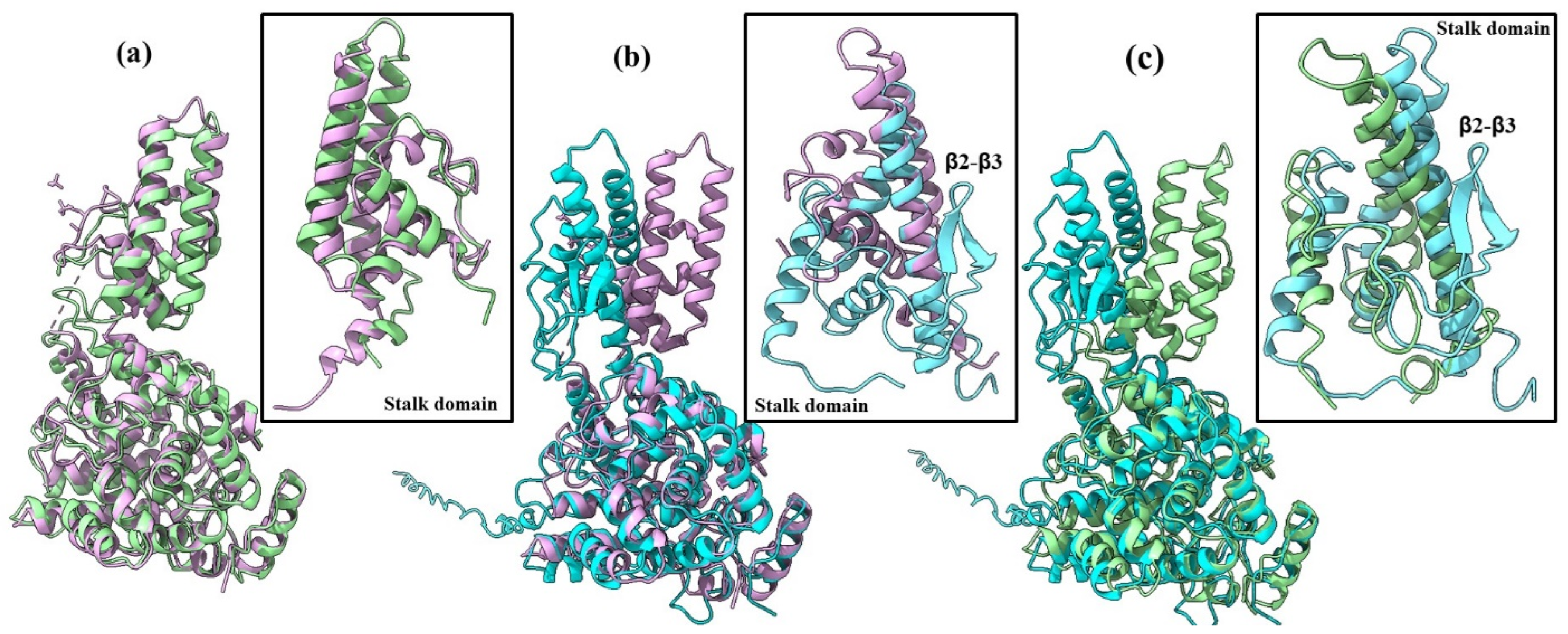
2.1. AlphaFold 3 Tertiary Structure Models of SGLV N and BJNV N

Tertiary structure models of SGLV N with pLDDT = 81 and BJNV N with pLDDT = 73 were generated using AlphaFold 3 (Figure S1). SGLV N and BJNV N were found to have the highest topological similarity to CCHFV N with TM-score 0.87 (PDB ID: 4AQF) and TM-score 0.80 (PDB ID: 6Z0O), respectively, despite extremely low amino acid sequence identity (Table 1). While when comparing the tertiary structures of SGLV N, BJNV N, and the N crystal structures of other closely related viruses, significantly lower similarity coefficients were observed. Also, the tertiary structure model of the SGLV N differs from those of the BJNV, with TM-score 0.78 and RMSD 4.34 Å (Table 1).
The SGLV N and BJNV N have a two-domain racket-shaped structure typical for orthonairoviruses nucleoproteins, consisting of a head domain and a stalk domain. Both SGLV N and BJNV N domains mainly consist of α-helices (Figure 1). In addition, the BJNV N has an extended unstructured N-terminal tail of 35 amino acids, which is not observed in the SGLV N.
The structure of SGLV N and BJNV N head domains are more conserved than the stalk domains. The SGLV N and the BJNV N structures show high flexibility of the stalk domain with TM-score 0.59 and RMSD 3.50 Å (Table 1).
While the SGLV N and BJNV N head domains align very closely, the SGLV N and BJNV N stalk domains adopt radically different positions. Significant conformational differences are exhibited in the orientation between the head and the stalk domains. The SGLV N stalk domain contains four alpha helices (α9–α12) with several tight turns, while the BJNV N stalk domain has four α-helices (α9–α12) and one beta sheet with two β-strands (β2 and β3) and a tight turn. It is noteworthy that the tertiary structure of the CCHFV does not contain a beta sheet (Figure 1). A two β-strand motif was observed in the stalk domain of Kupe virus N (PDP ID: 4XZC) (Figure S2).
However, SGLV N and BJNV N AlphaFold 3 models may have some regions that are incorrectly predicted due to the lack of structural information from homologous viral proteins in the PDB database and the limited ability of the AlphaFold 3 to predict viral proteins. In order to test the suitability of the SGLV N and BJNV N AlphaFold 3 models for structural analysis, we produced recombinant SGLV N and BJNV N and measured them using the SAXS method under native conditions (20 mM Tris-HCl and 150 mM NaCl, pH 7.5; see Section 4. Materials and Methods).

2.2. Recombinant SGLV N and BJNV N

We successfully obtained highly purified (>95% by SDS-PAGE) full-length recombinant SGLV N (487 aa, pI 7.66) and BJNV N (556 aa, pI 7.73) in a soluble form, without affinity tags and fusion proteins, using a prokaryotic cell system. SGLV N eluted at a peak of 14.9 mL on a Superdex®200 Increase 10/300 GL column, corresponding to the SGLV N monomeric form with a molecular weight of 54.5 kDa, and BJNV N eluted at a peak of 16.1 mL, corresponding to the BJNV N monomeric form with a molecular weight of 61.4 kDa (Figure 2a,c and Figure S31). The A260/A280 ratio of approximately 0.5 indicates that the SGLV N and BJNV N were successfully stripped of endogenous nucleic acids from the expression host. SEC and DLS analysis showed that SGLV N and BJNV N mainly exist in a monomeric form with DNase/RNase treatment similar to CCHFV N [17]. It is noteworthy that the SGLV N size was 6.02 nm ± 1.8, while the BJNV N size was 8.47 nm ± 2.05, which is significantly larger than that of the SGLV N (Figure 2b). This pronounced difference in hydrodynamic size—with BJNV N appearing significantly larger than SGLV N by DLS—is surprising, given that the molecular weight of BJNV N (61.4 kDa) is only slightly higher than that of SGLV N (54.5 kDa). A likely explanation is the presence of an extended, intrinsically disordered region at the C-terminus of BJNV N (residues 516–551), which can substantially increase the apparent hydrodynamic radius. This hypothesis is supported by hydrodynamic modeling based on AlphaFold 3 structures using the HullRad server, which predicts diameters of 6.30 nm and 7.62 nm for SGLV N and BJNV N, respectively—closely matching the experimental DLS data (Table S1 and Figure 2b).

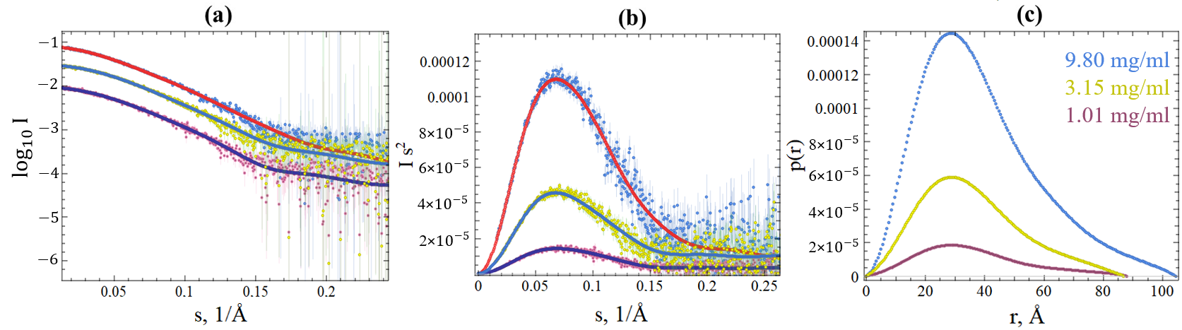
2.3. SAXS Data of SGLV N and BJNV N

Overall structural parameters of SGLV N and BJNV N, such as the molecular weight (MW), the radius of gyration of the molecule (Rg), the maximum linear dimension (Dmax), and the distance distribution function (P(r)), were calculated using the PRIMUS of the ATSAS v4.0 package. The SGLV N and BJNV N ab initio models were reconstructed in 3D at low resolution using DAMMIF by ensuring P1 symmetry. Then, averaging of the SGLV N and BJNV N ab initio models (15 for each protein) was performed in DAMAVER. The SGLV N and BJNV N structures modeled in the AlphaFold 3 were fitted to the SGLV N and BJNV N ab initio models from SAXS data using the ChimeraX v1.15rc (Figures S3–S8).
SGLV N and BJNV N show an increase in the molecule’s structures in solution (slightly larger Rg and Dmax) compared to the structures predicted by the AlphaFold 3 and DLS data. This is especially pronounced for the stalk domain, reflecting the SGLV N and BJNV N conformational dynamics in solution. The shape of the SAXS curve does not change depending on the SGLV N and BJNV N concentration in the solution (Figure 3a,d). Rg increases slightly by ~0.2 nm for SGLV N and does not change for BJNV N with increasing concentration (Table 2). P(r) exhibits one maximum. The position of the P(r) maximum does not change depending on the SGLV N and BJNV concentration (Figure 3c,f). The absence of significant upward or downward slope in the intensity profiles and the linearity of the Guinier plots for the three concentrations of SGLV N and BJNV N indicate the absence of protein aggregation or significant interparticle effect in the concentration range studied. A slight increase in Rg, Dmax, and V(Porod) for SGLV N at the highest concentration (9.8 mg/mL) suggests minor concentration-dependent oligomerization. However, this effect is negligible at 1.01 and 3.15 mg/mL, where scattering patterns are consistent. Thus, data from the lower concentrations were used for further analysis. The Kratky plots provide additional insight: for SGLV N, the peak position closely aligns with the theoretical value expected for a compact globular particle, supporting its well-folded, compact conformation (Figure 3b and Figures S3–S5). The BJNV N Kratky profile shows a small shift from this reference point, which is consistent with the presence of an intrinsically disordered C-terminal segment (Figure 3e and Figures S6–S8). The difference between the expected and experimentally obtained SGLV N and BJNV N molecular weights may arise due to error of theoretical molecular weight calculations (Table 2).
To test the plausibility of the SAXS models, AlphaFold 3 structural models of the BJNV N and SGLV N and crystal structures of N of viruses having the highest topological similarity were generated and fitted into the corresponding SAXS-derived shells. We compared the SAXS data for SGLV N at 3.15 mg/mL and BJNV N at 3.46 mg/mL with both the crystallographic data of viral N and the in silico predictions from AlphaFold 3. The experimental SAXS curves and theoretical curves were compared by analyzing the associated chi-square (χ2) test for the BJNV N and SGLV N (Figure S9). The χ2 variance should be equal to 1 for the ideal case. Our results show a χ2 = 1.1–1.3 for BJNV N and a χ2 = 1.2–1.3 for SGLV N (Table 3). Overall, SAXS measurements indicate fewer compact structures. The differences observed may be due to the fact that the structure in solution may differ slightly from the structure in the crystal and, in particular, reflect multiple conformational spaces in one averaged model. However, given the low resolution of the SAXS method, we conclude that the structures of the SGLV N and BJNV N are well preserved in solution and the AlphaFold 3 structural models of the BJNV N and SGLV N are adequate.
The solved tertiary structures obtained from ab initio modeling based on the SAXS scattering curves are shown in Figure 4. SGLV N and BJNV N tertiary structures have similar elongated shapes and are similar in size. The AlphaFold 3 structure fits well into the ab initio tertiary structure models of SGLV N and BJNV N, confirming that there are no major conformational changes between the structures in solution and confirming the suitability of the AlphaFold 3 tertiary structure models of SGLV N and BJNV N for further structural and functional analysis.

2.4. Interaction of SGLV N and BJNV N with Their ssRNA(−)

Three positively charged regions are found in the SGLV N and BJNV N structures (Figure 5a,c). Two regions, representing a deep positively charged crevice (BJNV N crevice: Lys49, Arg53, Arg185, Lys363, Lys364, Lys365, His392, His401, Arg402, His472, His475; SGLV N crevice: Arg54, Arg175, Arg383, Lys342, Lys344, His452, His455) and a platform (BJNV N platform 1: Arg62, Lys88, Lys136, His489; SGLV N platform 1: Arg87, Arg88, Lys95, Lys147), are located in the head domains, and a third positively charged region (BJNV N platform 2: Lys191, Lys193, Lys258; SGLV N platform 2: Lys187, His192, Lys216, Arg219) is located in the stalk domains. Structural alignment revealed that the SGLV N and BJNV N RNA-binding regions correspond to those within CCHFV N (Figures S10 and S11). The essential residues identified in these regions are well conserved in N proteins of other members of the Nairoviridae family and thus suggest a role in RNA binding [18].
Modeling of the structure of SGLV N and BJNV N monomers in complex with fragments of their ssRNA(−) of the S segment (SGLV RNA: 3′-UUUGAAUAUGGGGUGUGCA-5′ and BJNV RNA: 3′-GGGUUAGAUAUCUUCGAGA-5′) showed that SGLV RNA and BJNV RNA interact with N proteins in the crevice and platform 2, which supports the hypothesis that these positively charged regions are RNA-binding (Figure 5b,d). SGLV RNA and BJNV RNA literally penetrate SGLV N and BJNV N in the crevice, forming extensive hydrogen bonds, and extend out through platform 2, leaving their ends available for interaction with the next SGLV N and BJNV N monomers. In the crevice of SGLV N and BJNV N there are two extended RNA-binding regions. Both RNA-binding regions of the crevice in SGLV N and BJNV N have a contact distance with the RNA of less than 3.2 Å (Figures S10 and S11).
Interestingly, when modeling BJNV N and SGLV N in complex with another ssRNA(−) fragment of the BJNV S segment, for example, 3′-CUCACGGAUUACUGGUUGA-5′, we observed movement of the BJNV N and SGLV N stalk domain toward the formation of a more compact BJNV N tertiary structure (Figures S12 and S13). Notably, platform 1 of both SGLV N and BJNV N remains uninvolved in RNA-binding in these simulations. It is possible that this region becomes functional upon oligomerization of viral N proteins or upon binding of large RNA, or that double-stranded RNA is required for this region to function.
We attempted to reproduce the oligomeric state of SGLV N and BJNV N in complex with their ssRNA(−) using AlphaFold 3. SGLV RNPs and BJNV RNPs exhibit distinct oligomeric structures (Figure 6 and Figures S14–S23). The trimers, tetramers, pentamers, and hexamers of SGLV RNPs and BJNV RNPs adopt ring-shaped configurations. The oligomeric structures of SGLV RNPs form a large central cavity (~50–60 Å in diameter), whereas BJNV N RNPs assemble into more compact structures with multiple smaller cavities within the ring. The heptamers of SGLV RNPs and BJNV RNPs adopt a helical conformation, consisting of repeating six-monomer units, with the seventh monomer coiled into a helical turn (Figure 6a,b). The SGLV N-SGLV N interface comprises domain contacts between tight turns within α9–α10 and α11–α12 of the stalk domain and the region within α15–α16 of the head domain (Figures S24 and S25). The BJNV N-BJNV N interface comprises domain contacts between α9-TT-β2 of the stalk domain and α2 and α14–α15 of the head domain (Figure 6c,d, Figures S26 and S27). A positively charged RNA-binding crevice runs along the interior of SGLV RNP and BJNV RNP, shielding the viral ssRNA(−).

3. Discussion

In this study, we present the first structural investigation of BJNV N and SGLV N through an integrative approach combining SAXS experiments with AlphaFold 3 computational modeling.
The BJNV N and SGLV N monomers have a linear molecular dimension of approximately 9–10 nm (~Rg = 2.7   ±   0.1–3.1   ±   0.1) and a molecular weight of 47.6–62.0 kDa that varies slightly depending on the concentration. The BJNV N and SGLV N have a racket-shaped tertiary structure with distinct head and stalk domains that are broadly similar to those of CCHFV N, despite sequence similarities of 22% and 35.6%, respectively. The similarity of the BJNV N and SGLV N tertiary structures with the CCHFV N tertiary structure may indicate the similarity of viral functions and mechanisms of implementation of viral genetic information. CCHFV is a tick-borne orthonairovirus capable of causing hemorrhagic fever in humans, with mortality rates reaching up to 40%. The CCHFV N is widely distributed in the virion, participates in genome encapsidation, RNP formation, viral replication, and transcription, interacts with various cellular proteins and factors, plays an important role in viral pathogenesis, and stimulates innate, humoral, and cellular immune responses [19]. The observed preservation of tertiary structure across divergent ssRNA(−) viruses suggests conservation of fundamental functional mechanisms in viral genome processing.
According to crystal structures, the head domains of the orthonairoviruses N present the most conserved architecture and consist predominantly of α-helices. Interestingly, the head domain residues of BJNV N and SGLV N form a parallel double-β-stranded sheet, similar to the low-homology structures of Erve virus N (PDB ID: 4XZ8) and Kupe virus N (PDB ID: 4XZC). In contrast, the highly homologous head domains of CCHFV N (PDB ID: 4AQF) and Hazara virus N (PDB ID: 4XZE) lack this feature (Figure S28). Despite this structural difference, the head domains of SGLV N and BJNV N closely resemble those of CCHFV N, whereas their stalk domains adopt radically different positions. On the one hand, this may be due to the limitations of the chosen SAXS and AlphaFold 3 methods. On the other hand, this rearrangement may have important consequences that result in different activities in critical SGLV N and BJNV N functions, such as protein oligomerization or viral RNA binding.
Interestingly, bacterially expressed and purified SGLV N and BJNV N mainly exist in a monomeric form, similar to CCHFV N, according to data of DLS, SEC, and SAXS. This is quite different from the situation during the other reported ssRNA(−) viruses, which remained strongly and stably bound to host cell nucleic acids and oligomerized [20,21].
Notably, for SGLV N and BJNV N and the N proteins of homologous viruses, one of the regions of coupling between the head and stalk domains is not constructed for crystal structures due to the lack of interpretable electron density, which also indicates its structural flexibility. This coupling region corresponds to the α6 helix in the Lassa virus N, which adopts an open state upon binding to RNA during the so-called “gating mechanism” [22]. The other remaining region of coupling between these two domains in nucleoproteins is the α12, but this single helix is not sufficient to establish a rigid connection between the head and stalk domains (Figure S29). Thus, the coupling region between these two domains is very flexible in N proteins, which allows for interchangeable conformational changes for some key N functions such as RNP oligomerization or binding to viral RNA.
Next, we investigated the interaction of SGLV N and BJNV N with their ssRNA(−) and the oligomerization of SGLV and BJNV RNPs. It has been previously reported that CCHFV N has different binding modes for viral RNA and undergoes a conformational change in the presence of RNA [23,24]. A similar phenomenon is observed for BJNV N, but not for SGLV N. SGLV N and BJNV N binds RNA differently, possibly due to divergent evolutionary adaptations. This observation highlights a critical structural and functional feature shared by many ssRNA(−) viruses, particularly in the Bunyavirales order. Also, both SGLV N and BJNV N form trimers, tetramers, pentamers, and hexamers in ring-shaped arrangements and helical heptamers, typical of many nucleoproteins of ssRNA(−) viruses. However, SGLV RNPs have a larger central cavity, suggesting a more open architecture and the ability to accommodate bulkier RNA segments or allow dynamic rearrangements during viral replication.
Determining the structure of viral proteins is an important step in the development of antiviral drugs. Bioinformatic analysis has repeatedly identified candidate antiviral drugs, thereby expanding our arsenal of means to combat viral infections [25,26,27]. Being highly conserved proteins, the analogues of which are absent in human cells, N proteins are an interesting target for the search for new antiviral drugs. Thus, several substances have been shown to block the work of the nucleoprotein of the influenza A virus [28,29]. A recent study showed that the FDA-approved drug lurazidone suppresses severe fever caused by the thrombocytopenia syndrome virus [30]. The authors showed that lurazidone has an antiviral effect by directly binding to the viral nucleoprotein, thereby disrupting genome replication. These results provide a valuable theoretical basis for the rational development of new antiviral drugs targeting viral nucleoproteins.
The combination of SAXS with high-resolution structural techniques and computer analysis represents a powerful strategy for converting structural knowledge into effective low-molecular-weight inhibitors. This comprehensive approach is particularly promising for combating viruses carried by ticks and mosquitoes, where there is an urgent need for new therapeutic agents. Thus, our work demonstrates that experimental SAXS data can validate and improve predicted AlphaFold 3 structures of recently discovered viruses to reflect their solution structure and can be used in concert to directly analyze functions and conformational changes in nucleoproteins of recently discovered viruses that have extremely low genomic sequence identity compared to annotated viral genomes. Understanding the structure and function of viral nucleoproteins offers advantages in finding new therapeutic targets and also enhances our understanding of viral evolution. The results of this study are fundamentally important for the further development of preventive measures against viral threats associated with recently discovered SGLV and BJNV, which have already caused serious infectious diseases in humans. Further studies of nucleoproteins of poorly understood and novel ssRNA(−) viruses will undoubtedly reveal even more fascinating and diverse aspects.

4. Materials and Methods

4.1. Synthetic Assembly of DNA Copies of Genes Encoding Viral Nucleoproteins

Synthetic, codon-optimized DNA copies of the genes encoding the SGLV N (1449 bases, GenBank Acc. No. NC_079002, codon-optimized) and BJNV N (1653 bases, GenBank Acc. No. MW315111, codon-optimized) were obtained by sequential annealing of partially complementary oligonucleotides followed by PCR amplification (Tables S2 and S3). DNA fragments were amplified using the Q5 High-Fidelity DNA Polymerase kit (NEB, Hitchin, UK). Amplicon analysis was performed at each stage using electrophoresis in 2% agarose gel. Amplicons were purified using the Cleanup Standard kit (Eurogen, Moscow, Russia). The assembled DNA copies of the SGLV N and BJNV N genes were directly cloned into the pJET1.2/blunt plasmid (Thermo Fisher Scientific, Waltham, MA, USA) to create a genetic construct that ensures long-term storage of the synthesized DNA copies. Then, using the “heat shock” method, the pJET1.2/SGLV-N and pJET1.2/BJNV-N genetic constructs, containing DNA copies of the SGLV N and BJNV N genes, were transformed into chemically competent cells of Escherichia coli strain NEB Stable (NEB, UK) and plated on Petri dishes with LB agar medium (AppliChem, Darmstadt, Germany) with ampicillin at a concentration of 100 μg/mL as a selective marker, at a temperature of 30 °C for 16–18 h. Untransformed Escherichia coli cells of the NEB Stable strain served as a negative control. Recombinant plasmid DNA was isolated using the Plasmid Miniprep Color kit (Eurogen, Russia). The correctness of the nucleotide sequence of the pJET1.2/SGLV-N and pJET1.2/BJNV-N genetic constructs was confirmed by sequencing according to the Sanger method using the BigDye Terminator v3.1 kit (Thermo Fisher Scientific, USA) on an ABI 3500/3500xl (Applied Biosystems, Waltham, MA, USA). The obtained sequence data were processed using SnapGene v.3.2.1 (GSL Biotech LLC, San Diego, CA, USA).

4.2. Production of Recombinant Viral Nucleoproteins in Escherichia coli

To obtain genetic constructs that ensure the synthesis of SGLV N and BJNV N, DNA amplification of copies of the SGLV N and BJNV N genes from pJET1.2/SGLV-N and pJET1.2/BJNV-N was performed, followed by treatment of the PCR products with the restriction endonuclease Mal I (SibEnzyme, Novosibirsk, Russia). SGLV N and BJNV N were cloned into the pET28b-14xHis (Invitrogen, Waltham, MA, USA) vector with EcoR I and Xho I restriction sites using the cloning primers (Table S4). The pET28b-14xHis construct was designed in frame with an N-terminal 14xHis tag followed by a small ubiquitin-like modifier fusion protein and a picornain 3C A28 cleavage site (Figure 7). The resulting genetic constructs were transformed into chemically competent Escherichia coli cells of the NEB Stable strain. After 16 h of growth on LB agar medium (100 μg/mL kanamycin) at 30 °C, single colonies were selected and grown for 12 h in liquid LB nutrient medium with a selective antibiotic (100 μg/mL kanamycin) at 30 °C and 220 rpm. The recombinant plasmids encoding SGLV N and BJNV N were transformed into Escherichia coli strain KRX and overexpressed as an N-terminal 14xHis tag, a small ubiquitin-like modifier fusion protein, and a picornain 3C A28 proteolysis site. Expression of chimeric recombinant SGLV N and BJNV N transgenes was induced by adding IPTG to a concentration of 1 mM and L-rhamnose to a concentration of 0.1%. Induction was carried out for 20–24 h at 18 °C with constant stirring at 180 rpm in a thermostat with a built-in shaker NB-205QF (BioLabs, Shanghai, China).

4.3. Purification of Recombinant Viral Nucleoproteins

Cells were collected by centrifugation at 4500× g for 15–30 min at 4 °C and resuspended in lysis buffer (20 mM Tris, 500 mM NaCl, 20 mM imidazole, pH 7.5) with the addition of DNase (Biolabmix, Novosibirsk, Russia) and MgCl2 (New England BioLab, Hitchin, UK) in a ratio of 1 μL per 10 mL of cell suspension and a cocktail of protease inhibitors (Trans Gene Biotech, Beijing, China) in a ratio of 20 μL per 1 mL of cell suspension. Resuspended cells were sonicated with 20 kHz and 320W on ice using a b.braun LABSONIC 2000 homogenizer (B.Braun, Melsungen, Germany) and purified by centrifugation. The cleared cell lysate was loaded onto a HisPur Ni-NTA Resin (Thermo Fisher Scientific, USA), pre-equilibrated with the lysis buffer. The bound chimeric nucleoproteins were eluted by stepwise increasing the imidazole concentration to 75, 100, 150, 300, and 500 mM by mixing the flow of buffer A (20 mM Tris, 500 mM NaCl, 20 mM imidazole, pH 7.5) with buffer B (20 mM Tris, 500 mM NaCl, 500 mM imidazole, pH 7.5). The optical density was monitored using the HBBiolab software v.1.0 (Hanbon Sci. & Tech., Huaian, China), guided by the readings of the flow photometer included in the Bio-Lab 100 Chromatography System (Hanbon Sci. & Tech., China). Proteolytic cleavage of the N-terminal tract from chimeric nucleoproteins was performed by treatment with picornaine 3C A28 (SRC VB “Vector” Rospotrebnadzor, Koltsovo, Russia) in a molar ratio of 1:8. Proteolysis was carried out at 4 °C for 12–16 h. The proteolysis products were reapplied to a pre-equilibrated column with HisPur Ni-NTA Resin sorbent (Figure S30). Eluted nucleoproteins were further dialyzed using dialysis membrane with MWCO 10 kDa against 300 volumes of buffer C (20 mM Tris and 150 mM NaCl, pH 7.5), concentrated to 10–12 mg/mL, and purified using Superdex®200 Increase 10/300 GL (GE Healthcare, Stockholm, Sweden) size exclusion chromatography (SEC). Sodium dodecyl-sulfate polyacrylamide gel electrophoresis (SDS-PAGE) revealed over 95% purity of the final purified recombinant nucleoproteins. The nucleoproteins were quantified and stored in 100 μL aliquots at −80 °C.

4.4. Dynamic Light Scattering (DLS) Measurement

DLS data were collected using a BeNano 180 Zeta Pro (Bettersize, Dandong, China) with a light source wavelength of 671 nm, a fixed scattering angle of 173°, and a temperature of 25 °C. The HullRad server was used for calculating hydrodynamic properties of AlphaFold 3-predicted structures and the theoretical molecular weight of proteins, http://52.14.70.9/Run_hullrad.html (accessed on 20 May 2025) [31].

4.5. Small-Angle X-Ray Scattering (SAXS)

The SAXS data of nucleoproteins were collected at beamline BL19U2 of the National Facility for Protein Science Shanghai (NFPS) at the Shanghai Synchrotron Radiation Facility (SSRF, Shanghai, China) [32]. The X-ray beam size on the stage was 0.33 mm (H) × 0.05 mm (V). A two-dimensional Pilatus3 2 M detector (DECTRIS Ltd., Baden, Switzerland) was placed at a sample-to-detector distance of 2.7 m. The scattering vector magnitude range (q = (4π/λ)sinθ, where 2θ is the scattering angle and λ = 0.1033 nm is the wavelength) was 0.07–4.5 nm−1. SAXS profiles were obtained for different nucleoprotein concentrations. BJNV N samples were prepared at 1.12, 3.46, and 10.65 mg/mL, and SGLV N samples were prepared at 0.99, 3.15, and 9.8 mg/mL concentrations in the buffer (20 mM Tris-HCl and 150 mM NaCl, pH 7.5). Twenty frames with an integration time of 1.0 s were collected for each nucleoprotein’s concentration. The data were normalized to the intensity of the transmitted beam and radially averaged using the BioXTAS RAW program [33]. Scattering profiles of the buffer alone were measured under identical experimental conditions and subtracted from the corresponding sample profiles to account for background contributions and correct for solvent scattering effects. All subsequent data processing and analysis were performed using the ATSAS software package, version 4.0.1 (https://www.embl-hamburg.de/biosaxs/, accessed on 20 May 2025). Initial steps involved standard procedures, including averaging of the scattering curves [34]. Preliminary assessments of data quality and linearity in the low-angle region were carried out using the PRIMUS [35] module.
Key structural parameters such as the radius of gyration (Rg) and forward scattering intensity I(0) were extracted through Guinier analysis:
I e x p s = I 0 exp s 2 R g 2 3 ,
was applied within the regime, satisfying the approximation sRg < 1.3, ensuring the linearity criterion was met [36]. These parameters provide essential insight into the overall size and compactness of the macromolecular species in solution. A Bayesian statistical framework was employed to infer the molecular mass of the nucleoproteins directly from the SAXS data, providing both median estimates and credibility intervals with higher reliability. To further dissect the underlying shape and size distribution, p(r) pair-distance distribution functions were computed via an Indirect Fourier Transformation (IFT) approach implemented in GNOM [37] according to Equation (2):
p r = 1 2 π 2 0 s r I s sin s r d s .
This real-space transformation allowed for accurate determination of both Rg and the maximum intraparticle dimension (Dmax). The Rg values presented in Table 2 reflect those obtained through p(r) fitting rather than Guinier extrapolation, offering higher resilience to noise. For ab initio low-resolution structural modeling, we employed DAMMIF [38], which reconstructs particle envelopes using a bead-model approach and optimizes conformation via simulated annealing (SA). The modeling strives to minimize the discrepancy between experimental and calculated scattering intensities using a χ2 -like target function:
χ 2 = 1 N 1 j I e x p s j c I c a l c s j σ s j 2 ,
where N is the number of measured data points, I e x p and I c a l c denote the experimental and computed scattering intensities at momentum transfer s, σ represents the uncertainty at each point, and c is a scale factor optimized during the fit.
To ensure structural consistency and reproducibility, multiple independent ab initio reconstructions were generated and compared. Resulting models were spatially superimposed using SUPCOMB [39], and the most representative solutions were identified using the DAMAVER [40] suite via an averaging and filtering protocol. To validate and extend the analysis, theoretical scattering profiles were also calculated using CRYSOL [41], applied to both available crystal structures of related proteins and predicted structural models obtained from AlphaFold 3. This step enabled us to assess the agreement of the experimental data with both atomic-resolution and computationally derived models, providing deeper insight into potential conformational states in solution.

4.6. Tertiary Structure Modeling of Nucleoproteins and Their Analysis

Nucleotide sequences determined in this study are available in the GenBank database. Predictive modeling of nucleoprotein tertiary structures was carried out using AlphaFold 3 (Google DeepMind, London, UK), https://alphafoldserver.com/welcome (accessed on 10 May 2025) [42]. A search for closely related proteins whose spatial structures had already been solved experimentally was performed using NCBI BLAST (version 2.17.0) (National Center for Biotechnology Information Basic Local Alignment Search Tool, https://blast.ncbi.nlm.nih.gov) against the PDB database (https://www.rcsb.org/) using the blastp (protein–protein BLAST) algorithm (accessed on 10 May 2025). Nucleoproteins were screened against known viral domains using the FoldSeek server (Seoul National University, Seoul, South Korea), https://search.foldseek.com/ (accessed on 10 May 2025) [43]. Structural inferences were based on per-residue confidence scores provided via the AlphaFold 3 predicted Local Distance Difference Test (pLDDT) metric, which quantitatively estimates deviations in Cα–Cα interatomic distances between the reference and predicted structural models, with values ranging from 0 to 100. The resulting models were selected based on high-confidence structural prediction outputs, with particular emphasis on pLDDT scoring, and aligned against their respective structural homologs using the TM-align algorithm, implemented via the RCSB Pairwise Structure Alignment Tool (Research Collaboratory for Structural Bioinformatics Protein Data Bank, RCSB PDB, Piscataway, NJ, USA), https://www.rcsb.org/alignment/; accessed 10 May 2025). Structural alignments were evaluated according to two key metrics: root mean square deviation (RMSD) to estimate atomic displacement between aligned backbone atoms and TM-score, a length-independent metric that reflects topological similarity on a scale between 0 and 1, with values above 0.5 generally indicating correct fold prediction and values near 1.0 indicating high structural congruency.
Tertiary structure models of viral proteins were visualized using UCSF ChimeraX v1.15rc (University of California, San Francisco, CA, USA) [16]. Comparative secondary structure alignments of viral proteins were generated with ESPript v3.0 (Institute for the Biology and Chemistry of Proteins, Lyon, France), https://espript.ibcp.fr/ (accessed on 10 May 2025) [44], allowing for the graphical representation of conserved secondary structure motifs across multiple aligned sequences.

Supplementary Materials

The following supporting information can be downloaded at: https://www.mdpi.com/article/10.3390/ijms26157445/s1.

Author Contributions

Conceptualization, A.V.G.; methodology, A.O.Y., D.I.I., V.Y.T., I.A.O., A.E.T., I.R.I., and A.V.G.; software, A.O.Y.; validation, A.O.Y., Y.L.R., I.R.I., and A.V.G.; formal analysis, A.V.G.; investigation, A.O.Y., D.I.I., V.Y.T., I.A.O., A.E.T., S.E.O., E.O.U., N.S.R., Y.L.R., I.R.I., and A.V.G.; resources, S.G.A., Y.L.R., N.L., I.R.I., and A.V.G.; data curation, A.V.G.; writing—original draft preparation, A.O.Y. and A.V.G.; writing—review and editing, A.O.Y., D.I.I., V.Y.T., I.A.O., A.E.T., S.E.O., E.O.U., N.S.R., S.G.A., Y.L.R., N.L., A.P.A., I.R.I., and A.V.G.; visualization, A.O.Y. and A.V.G.; supervision, I.R.I. and A.V.G.; project administration, A.P.A. and A.V.G.; funding acquisition, A.P.A. All authors have read and agreed to the published version of the manuscript.

Funding

This study was supported by the Ministry of Science and Higher Education of the Russian Federation (agreement No. 075-15-2025-452) as part of the implementation of certain activities of the Federal Scientific and Technical Program for the Development of Synchrotron and Neutron Research and Research Infrastructure. Yury L. Ryzhykau acknowledges the Ministry of Science and Higher Education of the Russian Federation (agreement 075-03-2025-662, project FSMG-2025-0003) for the support of his contribution to the design of the small-angle scattering experiment using a concentration series and to the analysis of concentration dependence.

Institutional Review Board Statement

Not applicable.

Informed Consent Statement

Not applicable.

Data Availability Statement

The original contributions presented in this study are included in the article/Supplementary Material; further inquiries can be directed to the corresponding author.

Acknowledgments

We thank Valentin Borschevsky (Moscow Institute of Physics and Technology, Dolgoprudny, Russia) and the staff of the Shanghai Synchrotron Radiation Facility beamline BL19U2 for help with SAXS data collection.

Conflicts of Interest

The authors declare no conflicts of interest.

Abbreviations

The following abbreviations are used in this manuscript:
SGLVOrthonairovirus songlingense, Songling virus
YEZVOrthonairovirus yezoense, Yezo virus
BJNVNorwavirus beijiense, Beiji nairovirus
SAXSSmall-angle X-ray scattering
CCHFVOrthonairovirus haemorrhagiae, Crimean–Congo hemorrhagic fever virus
ssRNA(−)Single-stranded RNA of negative polarity
NNucleoprotein
GPCGlycoprotein precursor
RdRpRNA-dependent RNA polymerase
SECSize exclusion chromatography
DLSDynamic light scattering

References

  1. Zhou, H.; Xu, L.; Shi, W. The Human-Infection Potential of Emerging Tick-Borne Viruses Is a Global Public Health Concern. Nat. Rev. Microbiol. 2023, 21, 215–217. [Google Scholar] [CrossRef]
  2. Moraga-Fernández, A.; Muñoz-Hernández, C.; Sánchez-Sánchez, M.; Fernández de Mera, I.G.; de la Fuente, J. Exploring the Diversity of Tick-Borne Pathogens: The Case of Bacteria (Anaplasma, Rickettsia, Coxiella and Borrelia) Protozoa (Babesia and Theileria) and Viruses (Orthonairovirus, Tick-Borne Encephalitis Virus and Louping Ill Virus) in the European Continent. Vet. Microbiol. 2023, 286, 109892. [Google Scholar] [CrossRef]
  3. Liu, X.; Zhang, X.; Wang, Z.; Dong, Z.; Xie, S.; Jiang, M.; Song, R.; Ma, J.; Chen, S.; Chen, K.; et al. A Tentative Tamdy Orthonairovirus Related to Febrile Illness in Northwestern China. Clin. Infect. Dis. 2020, 70, 2155–2160. [Google Scholar] [CrossRef]
  4. Tomazatos, A.; von Possel, R.; Pekarek, N.; Holm, T.; Rieger, T.; Baum, H.; Bialonski, A.; Maranda, I.; Erdelyi-Molnár, I.; Spînu, M.; et al. Discovery and Genetic Characterization of a Novel Orthonairovirus in Ixodes Ricinus Ticks from Danube Delta. Infect. Genet. Evol. 2021, 88, 104704. [Google Scholar] [CrossRef]
  5. Kodama, F.; Yamaguchi, H.; Park, E.; Tatemoto, K.; Sashika, M.; Nakao, R.; Terauchi, Y.; Mizuma, K.; Orba, Y.; Kariwa, H.; et al. A Novel Nairovirus Associated with Acute Febrile Illness in Hokkaido, Japan. Nat. Commun. 2021, 12, 5539. [Google Scholar] [CrossRef]
  6. Ma, J.; Lv, X.-L.; Zhang, X.; Han, S.-Z.; Wang, Z.-D.; Li, L.; Sun, H.-T.; Ma, L.-X.; Cheng, Z.-L.; Shao, J.-W.; et al. Identification of a New Orthonairovirus Associated with Human Febrile Illness in China. Nat. Med. 2021, 27, 434–439. [Google Scholar] [CrossRef]
  7. Zhang, M.-Z.; Bian, C.; Ye, R.-Z.; Cui, X.-M.; Yao, N.-N.; Yang, J.-H.; Chu, Y.-L.; Su, X.-L.; Wu, Y.-F.; Ye, J.-L.; et al. A Series of Patients Infected with the Emerging Tick-Borne Yezo Virus in China: An Active Surveillance and Genomic Analysis. Lancet Infect. Dis. 2025, 25, 390–398. [Google Scholar] [CrossRef]
  8. Wang, Y.-C.; Wei, Z.; Lv, X.; Han, S.; Wang, Z.; Fan, C.; Zhang, X.; Shao, J.; Zhao, Y.-H.; Sui, L.; et al. A New Nairo-like Virus Associated with Human Febrile Illness in China. Emerg. Microbes Infect. 2021, 10, 1200–1208. [Google Scholar] [CrossRef]
  9. Matsumura, R.; Kobayashi, D.; Yamauchi, T.; Park, E.; Nishino, A.; Maeda, K.; Kasai, S.; Itoyama, K.; Isawa, H. Isolation and Characterization of Iwanai Valley Virus, a New Tick-Borne Nairovirus from Ixodes Ovatus Ticks in Hokkaido, Japan. Arch. Virol. 2024, 170, 23. [Google Scholar] [CrossRef]
  10. Liu, Z.; Li, L.; Xu, W.; Yuan, Y.; Liang, X.; Zhang, L.; Wei, Z.; Sui, L.; Zhao, Y.; Cui, Y.; et al. Extensive Diversity of RNA Viruses in Ticks Revealed by Metagenomics in Northeastern China. PLoS Negl. Trop. Dis. 2022, 16, e0011017. [Google Scholar] [CrossRef]
  11. Liu, Z.; Wei, Z.; Liu, Y.; Wang, Z. The Extensive Diversity of Nairoviruses Reveals the Complexity and High Risk of Tick-Borne Diseases in Northeastern China. bioRxiv 2025. [Google Scholar] [CrossRef]
  12. Li, D.; Li, J.; Wang, R.; Zhang, W.; Nie, K.; Yin, Q.; Fu, S.; Cui, Q.; Xu, S.; Li, F.; et al. Detection and Genetic Analysis of Songling Virus in Haemaphysalis Concinna near the China-North Korea Border. Zoonoses 2024, 4. [Google Scholar] [CrossRef]
  13. Wang, G.; Tian, X.; Peng, R.; Huang, Y.; Li, Y.; Li, Z.; Hu, X.; Luo, Z.; Zhang, Y.; Cui, X.; et al. Genomic and Phylogenetic Profiling of RNA of Tick-Borne Arboviruses in Hainan Island, China. Microbes Infect. 2024, 26, 105218. [Google Scholar] [CrossRef]
  14. Bente, D.A.; Forrester, N.L.; Watts, D.M.; McAuley, A.J.; Whitehouse, C.A.; Bray, M. Crimean-Congo Hemorrhagic Fever: History, Epidemiology, Pathogenesis, Clinical Syndrome and Genetic Diversity. Antiviral Res. 2013, 100, 159–189. [Google Scholar] [CrossRef]
  15. Sabsay, K.R.; Te Velthuis, A.J.W. Negative and Ambisense RNA Virus Ribonucleocapsids: More than Protective Armor. Microbiol. Mol. Biol. Rev. MMBR 2023, 87, e0008223. [Google Scholar] [CrossRef]
  16. Pettersen, E.F.; Goddard, T.D.; Huang, C.C.; Meng, E.C.; Couch, G.S.; Croll, T.I.; Morris, J.H.; Ferrin, T.E. UCSF ChimeraX: Structure Visualization for Researchers, Educators, and Developers. Protein Sci. 2021, 30, 70–82. [Google Scholar] [CrossRef]
  17. Guo, Y.; Wang, W.; Ji, W.; Deng, M.; Sun, Y.; Zhou, H.; Yang, C.; Deng, F.; Wang, H.; Hu, Z.; et al. Crimean–Congo Hemorrhagic Fever Virus Nucleoprotein Reveals Endonuclease Activity in Bunyaviruses. Proc. Natl. Acad. Sci. USA 2012, 109, 5046–5051. [Google Scholar] [CrossRef]
  18. Carter, S.D.; Surtees, R.; Walter, C.T.; Ariza, A.; Bergeron, É.; Nichol, S.T.; Hiscox, J.A.; Edwards, T.A.; Barr, J.N. Structure, Function, and Evolution of the Crimean-Congo Hemorrhagic Fever Virus Nucleocapsid Protein. J. Virol. 2012, 86, 10914–10923. [Google Scholar] [CrossRef]
  19. Pirincal, A.; Doymaz, M.Z. The Role of Nucleocapsid Protein (NP) in the Immunology of Crimean-Congo Hemorrhagic Fever Virus (CCHFV). Viruses 2024, 16, 1547. [Google Scholar] [CrossRef]
  20. Dong, H.; Li, P.; Elliott, R.M.; Dong, C. Structure of Schmallenberg Orthobunyavirus Nucleoprotein Suggests a Novel Mechanism of Genome Encapsidation. J. Virol. 2013, 87, 5593–5601. [Google Scholar] [CrossRef]
  21. Zheng, W.; Olson, J.; Vakharia, V.; Tao, Y.J. The Crystal Structure and RNA-Binding of an Orthomyxovirus Nucleoprotein. PLoS Pathog. 2013, 9, e1003624. [Google Scholar] [CrossRef]
  22. Wang, W.; Liu, X.; Wang, X.; Dong, H.; Ma, C.; Wang, J.; Liu, B.; Mao, Y.; Wang, Y.; Li, T.; et al. Structural and Functional Diversity of Nairovirus-Encoded Nucleoproteins. J. Virol. 2015, 89, 11740–11749. [Google Scholar] [CrossRef]
  23. Ohta, K.; Saka, N.; Nishio, M. Identification of Critical Residues for RNA Binding of Nairovirus Nucleoprotein. J. Virol. 2024, 98, e0144624. [Google Scholar] [CrossRef]
  24. Jeeva, S.; Pador, S.; Voss, B.; Ganaie, S.S.; Mir, M.A. Crimean-Congo Hemorrhagic Fever Virus Nucleocapsid Protein Has Dual RNA Binding Modes. PLoS ONE 2017, 12, e0184935. [Google Scholar] [CrossRef]
  25. Moquin, S.A.; Simon, O.; Karuna, R.; Lakshminarayana, S.B.; Yokokawa, F.; Wang, F.; Saravanan, C.; Zhang, J.; Day, C.W.; Chan, K.; et al. NITD-688, a Pan-Serotype Inhibitor of the Dengue Virus NS4B Protein, Shows Favorable Pharmacokinetics and Efficacy in Preclinical Animal Models. Sci. Transl. Med. 2021, 13, eabb2181. [Google Scholar] [CrossRef]
  26. Gejji, V.; Svoboda, P.; Stefanik, M.; Wang, H.; Salat, J.; Eyer, L.; Ruzek, D.; Fernando, S. An RNA-Dependent RNA Polymerase Inhibitor for Tick-Borne Encephalitis Virus. Virology 2020, 546, 13–19. [Google Scholar] [CrossRef]
  27. Zhang, S.; Garzan, A.; Haese, N.; Bostwick, R.; Martinez-Gzegozewska, Y.; Rasmussen, L.; Streblow, D.N.; Haise, M.T.; Pathak, A.K.; Augelli-Szafran, C.E.; et al. Pyrimidone Inhibitors Targeting Chikungunya Virus NsP3 Macrodomain by Fragment-Based Drug Design. PLoS ONE 2021, 16, e0245013. [Google Scholar] [CrossRef]
  28. Hu, Y.; Sneyd, H.; Dekant, R.; Wang, J. Influenza A Virus Nucleoprotein: A Highly Conserved Multi-Functional Viral Protein as a Hot Antiviral Drug Target. Curr. Top. Med. Chem. 2017, 17, 2271–2285. [Google Scholar] [CrossRef]
  29. Yang, F.; Pang, B.; Lai, K.K.; Cheung, N.N.; Dai, J.; Zhang, W.; Zhang, J.; Chan, K.-H.; Chen, H.; Sze, K.-H.; et al. Discovery of a Novel Specific Inhibitor Targeting Influenza A Virus Nucleoprotein with Pleiotropic Inhibitory Effects on Various Steps of the Viral Life Cycle. J. Virol. 2021, 95. [Google Scholar] [CrossRef]
  30. Cheng, T.; Xiao, Q.; Cui, J.; Dong, S.; Wu, Y.; Li, W.; Yang, X.; Ma, L.; Li, Z.; Sun, P.; et al. Identification of Lurasidone as a Potent Inhibitor of Severe Fever with Thrombocytopenia Syndrome Virus by Targeting the Viral Nucleoprotein. Front. Microbiol. 2025, 16, 1578844. [Google Scholar] [CrossRef]
  31. Fleming, P.J.; Fleming, K.G. HullRad: Fast Calculations of Folded and Disordered Protein and Nucleic Acid Hydrodynamic Properties. Biophys. J. 2018, 114, 856–869. [Google Scholar] [CrossRef]
  32. Li, Y.-W.; Liu, G.-F.; Wu, H.-J.; Zhou, P.; Hong, C.-X.; Li, N.; Bian, F.-G. BL19U2: Small-Angle X-Ray Scattering Beamline for Biological Macromolecules in Solution at SSRF. Nucl. Sci. Tech. 2020, 31, 117. [Google Scholar] [CrossRef]
  33. Hopkins, J.B. BioXTAS RAW 2: New Developments for a Free Open-Source Program for Small-Angle Scattering Data Reduction and Analysis. J. Appl. Crystallogr. 2024, 57, 194–208. [Google Scholar] [CrossRef]
  34. Petoukhov, M.V.; Franke, D.; Shkumatov, A.V.; Tria, G.; Kikhney, A.G.; Gajda, M.; Gorba, C.; Mertens, H.D.T.; Konarev, P.V.; Svergun, D.I. New Developments in the ATSAS Program Package for Small-Angle Scattering Data Analysis. J. Appl. Crystallogr. 2012, 45, 342–350. [Google Scholar] [CrossRef]
  35. Konarev, P.V.; Volkov, V.V.; Sokolova, A.V.; Koch, M.H.J.; Svergun, D.I. PRIMUS: A Windows PC-Based System for Small-Angle Scattering Data Analysis. J. Appl. Crystallogr. 2003, 36, 1277–1282. [Google Scholar] [CrossRef]
  36. Feigin, L.A.; Svergun, D.I.; Taylor, G.W. Structure Analysis by Small-Angle X-Ray and Neutron Scattering; Springer: New York, NY, USA, 2013; ISBN 9781475766264. [Google Scholar]
  37. Svergun, D.I. Determination of the Regularization Parameter in Indirect-Transform Methods Using Perceptual Criteria. J. Appl. Crystallogr. 1992, 25, 495–503. [Google Scholar] [CrossRef]
  38. Svergun, D.I. Restoring Low Resolution Structure of Biological Macromolecules from Solution Scattering Using Simulated Annealing. Biophys. J. 1999, 76, 2879–2886. [Google Scholar] [CrossRef]
  39. Kozin, M.B.; Svergun, D.I. Automated Matching of High- and Low-Resolution Structural Models. J. Appl. Crystallogr. 2001, 34, 33–41. [Google Scholar] [CrossRef]
  40. Volkov, V.V.; Svergun, D.I. Uniqueness of Ab Initio Shape Determination in Small-Angle Scattering. J. Appl. Crystallogr. 2003, 36, 860–864. [Google Scholar] [CrossRef]
  41. Svergun, D.; Barberato, C.; Koch, M.H.J. CRYSOL—A Program to Evaluate X-Ray Solution Scattering of Biological Macromolecules from Atomic Coordinates. J. Appl. Crystallogr. 1995, 28, 768–773. [Google Scholar] [CrossRef]
  42. Abramson, J.; Adler, J.; Dunger, J.; Evans, R.; Green, T.; Pritzel, A.; Ronneberger, O.; Willmore, L.; Ballard, A.J.; Bambrick, J.; et al. Accurate Structure Prediction of Biomolecular Interactions with AlphaFold 3. Nature 2024, 630, 493–500. [Google Scholar] [CrossRef] [PubMed]
  43. van Kempen, M.; Kim, S.S.; Tumescheit, C.; Mirdita, M.; Lee, J.; Gilchrist, C.L.M.; Söding, J.; Steinegger, M. Fast and Accurate Protein Structure Search with Foldseek. Nat. Biotechnol. 2024, 42, 243–246. [Google Scholar] [CrossRef] [PubMed]
  44. Robert, X.; Gouet, P. Deciphering Key Features in Protein Structures with the New ENDscript Server. Nucleic Acids Res. 2014, 42, W320–W324. [Google Scholar] [CrossRef]
Figure 1. Imposition models of SGLV N and BJNV N tertiary structures: (a) SGLV N tertiary structure (green) with CCHFV (PDP ID: 4AQF) (purple); (b) BJNV N tertiary structure (blue) with CCHFV (PDP ID: 4AQF) (purple); (c) SGLV N tertiary structure (green) with BJNV N tertiary structure (blue). The figure was generated using UCSF ChimeraX v1.15rc [16].
Ijms 26 07445 g001
Figure 2. Size exclusion chromatography (a), dynamic light scattering (b), and SDS-PAGE in 12% gel (c) analysis of the recombinant SGLV N and BJNV N. SGLV N is highlighted in blue in (a,b) and is located on the third lane in (c). BJNV N is highlighted in red in (a,b) and is located on the second lane in (c).
Ijms 26 07445 g002
Figure 3. SAXS analysis of full-length SGLV N (ac) at concentrations of 9.8 mg/mL (blue), 3.15 mg/mL (yellow), and 1.01 mg/mL (purple) and full-length BJNV N (df) at concentrations of 10.65 mg/mL (pink), 3.46 mg/mL (light blue), and 1.12 mg/mL (green). (a) SGLV N scattering experimental data of SGLV N; (b) Kratky plot of SGLV N; (c) distance distribution function p(r) of SGLV N; (d) BJNV N scattering experimental data; (e) Kratky plot of BJNV N; (f) distance distribution function p(r) of BJNV N. Regularized SAXS curves obtained in GNOM are shown in different colors together with corresponding experimental data.
Ijms 26 07445 g003aIjms 26 07445 g003b
Figure 4. Ab initio tertiary structure models of SGLV N (a) and BJNV N (b). The AlphaFold model is displayed as ribbon diagrams. The random dummy residues of the average DAMAVER models are shown as a surface.
Ijms 26 07445 g004
Figure 5. Spatial structures of SGLV N and BJNV N monomers with their RNA fragments of the S genome segment. Electrostatic surface potential of SGLV N (a) and BJNV N (c). Spatial structures of the SGLV N (purple) with RNA (green) (b) and BJNV N (blue) with RNA (orange) (d) in ribbon presentation. The positive surface potential is colored blue, and the negative surface potential is colored red. Key amino acids involved in interaction with RNA are highlighted.
Ijms 26 07445 g005
Figure 6. AlphaFold 3 structural models of viral RNP complexes: (a,d) SGLV RNP (top and side views); (b,c) BJNV RNP (top and side views). In SGLV RNPs, N monomers are shown in blue, and the RNA is colored green. In BJNV RNPs, N monomers are shown in green, and the RNA is colored cyan. Amino acids associated with the interaction of the BJNV N head domain with ssRNA(−) in the crevice are shown in the orange box and colored in pale pink. Amino acids associated with the interaction of the BJNV N stalk domain with ssRNA(−) in platform 2 are shown in the orange box and colored in pale yellow. Amino acids associated with the BJNV N-BJNV N interaction are shown in the blue box. Amino acids associated with the interaction of the SGLV N head domain with ssRNA(−) in the crevice are shown in the green box and colored in green. Amino acids associated with the interaction of the SGLV N stalk domain with ssRNA(−) in platform 2 are shown in the green box and colored in yellow. Amino acids associated with the SGLV N-SGLV N interaction are shown in the purple box.
Ijms 26 07445 g006aIjms 26 07445 g006b
Figure 7. Linear map of BJNV N (a) and SGLV N (b) expression cassettes.
Ijms 26 07445 g007
Table 1. Comparison of amino acid sequences and tertiary structures of SGLV N and BJNV N with N proteins of viruses having the highest topological similarity.
PDB IDName of VirusTM-ScoreRMSD, ÅAligned
Residues, a.a.
Amino Acid Sequence Identity (Blastp) 1, %Amino Acid Sequence Identity (RSCB) 2, %
SGLV N (full-length)
-BJNV0.784.3436525.023.0
4AQF_ACCHFV0.872.5343335.634.0
5A97_CHazara virus0.833.5248932.832.0
4AQF_CCCHFV0.823.2741335.434.0
4XZE_AHazara virus0.813.3838132.832.0
4XZC_AKupe virus0.783.6336733.928.1
3U3I_ACCHFV0.752.3036536.038.0
BJNV N (full-length)
-SGLV0.784.3436525.023.0
6Z0O_BCCHFV0.803.1341722.022.0
4AQF_BCCHFV0.773.4839024.022.0
4XZC_BKupe virus0.763.4038923.322.0
5A97_AHazara virus0.743.9836927.628.0
4XZA_AErve virus0.642.5333722.025.0
SGLV N head domain
-BJNV0.902.1534827.027.0
4AQF_ACCHFV0.941.4035139.040.0
5A97_CHazara virus0.931.7235236.037.0
4AQF_CCCHFV0.941.4634538.040.0
4XZE_AHazara virus0.951.2835535.038.0
4XZC_AKupe virus0.951.3735537.039.0
BJNV N head domain
6Z0O_BCCHFV0.902.2634224.026.0
4AQF_BCCHFV0.882.4033625.027.0
4XZC_BKupe virus0.892.3033924.026.0
5A97_AHazara virus0.842.3134027.029.0
4XZA_AErve virus0.892.5733723.024.0
SGLV N stalk domain
-BJNV0.553.008518.018.0
4AQF_ACCHFV0.692.839417.015.0
5A97_CHazara virus0.692.689721.018.0
4AQF_CCCHFV0.732.809917.016.0
4XZE_AHazara virus0.702.859522.017.0
4XZC_AKupe virus0.702.729221.015.0
BJNV N stalk domain
6Z0O_BCCHFV0.563.28216.014.0
4AQF_BCCHFV0.573.28115.013.0
4XZC_BKupe virus0.582.98113.011.0
5A97_AHazara virus0.603.08315.013.0
1 Amino acid sequence identity was calculated in the protein–protein BLAST (blastp). 2 Amino acid sequence identity was calculated relative to the aligned residues in the tertiary structure.
Table 2. Overall structural parameters of SGLV N and BJNV N obtained from SAXS data.
SampleConcentration, mg/mL R g , nm * D m a x , nm *V(Porod), A 3 *MW (exp), kDa *MW (Theory), kDa *
SGLV N9.8 3.1   ±   0.110.3 103 × 10 3 53.1
[93% CI: 47.2, 57.5]
54.5
3.15 2.8   ±   0.18.7 86 × 10 3 52.0
[94% CI: 49.2, 55.0]
54.5
1.01 2.8   ±   0.18.8 81 × 10 3 47.6
[95% CI: 46.2, 51.5]
54.5
BJNV N10.7 3.2   ±   0.19.7 110 × 10 3 58.1
[91% CI: 53.8, 61.6]
61.4
3.46 3.0   ±   0.19.8 110 × 10 3 53.1
[91% CI: 50.3, 56.2]
61.4
1.12 3.2   ±   0.19.9 114 × 10 3 53.2
[91% CI: 53.0, 60.2]
61.4
* V(Porod) is the particle volume, MW (exp) is the experimental molecular weight, and MW (theory) is the theoretical molecular weight calculated according to the SGLV N and BJNV N amino acid sequences.
Table 3. Comparison of SGLV N and BJNV N SAXS data with crystal structures of N of viruses having the highest topological similarity and AlphaFold 3 structural models of SGLV N and BJNV N.
Model R g , nm * D m a x (Envelope), nmV (Envelope), A 3 MW, kDaχ2
CCHFV N,
PDB ID: 4AQF
2.69.1 81 × 10 3 52.4BJNV N: 1.2
SGLV N: 1.2
Hazara virus N,
PDB ID: 5A97
2.58.8 82 × 10 3 53.1BJNV N: 1.3
SGLV N: 1.3
BJNV N,
AlphaFold 3 model
3.09.6 92 × 10 3 61.41.1
SGLV N,
AlphaFold 3 model
2.59.1 84 × 10 3 54.51.3
* Rg calculated from the slope of net intensity curve generated by CRYSOL.
Disclaimer/Publisher’s Note: The statements, opinions and data contained in all publications are solely those of the individual author(s) and contributor(s) and not of MDPI and/or the editor(s). MDPI and/or the editor(s) disclaim responsibility for any injury to people or property resulting from any ideas, methods, instructions or products referred to in the content.

