Article

Microbial Lipopolysaccharide Regulates Host Development Through Insulin/IGF-1 Signaling

Shanghai Key Laboratory of Metabolic Remodeling and Health, Institute of Metabolism and Integrative Biology, Fudan University, Shanghai 200438, China
*
Author to whom correspondence should be addressed.
Int. J. Mol. Sci. 2025, 26(15), 7399; https://doi.org/10.3390/ijms26157399 (registering DOI)
Submission received: 3 June 2025 / Revised: 22 July 2025 / Accepted: 24 July 2025 / Published: 31 July 2025
(This article belongs to the Special Issue C. elegans as a Disease Model: Molecular Perspectives: 2nd Edition)

Abstract

Lipopolysaccharide (LPS), the defining outer membrane component of Gram-negative bacteria, is a potent immunostimulant recognized by Toll-like receptor 4 (TLR4). While extensively studied for its roles in immune activation and barrier disruption, the potential function of LPS as a developmental cue remains largely unexplored. By leveraging Caenorhabditis elegans and its genetic and gnotobiotic advantages, we screened a panel of Escherichia coli LPS biosynthesis mutants. This screen revealed that the loss of outer core glycosylation in the ∆rfaG mutant causes significant developmental delay independent of bacterial metabolism. Animals exhibited developmental delay that was rescued by exogenous LPS or amino acid supplementation, implicating that LPS triggers nutrient-sensing signaling. Mechanistically, this developmental arrest was mediated by the host FOXO transcription factor DAF-16, which is the key effector of insulin/IGF-1 signaling (IIS). Our findings uncover an unprecedented role for microbial LPS as a critical regulator of host development, mediated through conserved host IIS pathways, fundamentally expanding our understanding of host–microbe crosstalk.

1. Introduction

The microbiome plays crucial roles in regulating host physiology, with microbes implicated in host diseases ranging from metabolic syndrome to neurodegeneration. Among abundant microbial molecules and metabolites, lipopolysaccharide (LPS), a hallmark component of Gram-negative bacterial outer membranes, is secreted via outer membrane vesicles (OMVs) and potently activates mammalian innate immunity [1,2,3]. LPS comprises three domains: the hydrophobic lipid A, inner core oligosaccharide, and outer core O-antigen. Lipid A, the conserved endotoxic moiety, anchors the LPS molecule to the bacterial outer membrane and drives innate immune activation via Toll-like receptor 4 (TLR4) in mammals. The O-antigen determines serotype specificity and resistance to host defenses, while the inner core oligosaccharide containing 3-deoxy-D-manno-oct-2-ulosonic acid (KDO) is essential for the synthesis of bioactive lipid A and influences host–pathogen interactions [4,5,6,7,8]. Structural modifications to the outer core (e.g., acetylation) further tune LPS immunogenicity and environmental adaptability [9]. This tripartite architecture differentiates Gram-negative bacteria from Gram-positive species. In contrast to Gram-negative bacteria, Gram-positive bacteria lack LPS but possess teichoic acid-embedded peptidoglycan [10,11]. LPS acts as a pathogen-associated molecular pattern (PAMP), triggering oxidative stress, intestinal barrier disruption, and immune signaling in metazoan hosts [12,13,14,15,16,17,18,19]. Paradoxically, while LPS is best characterized as an inflammatory toxin, its potential role as a developmental cue remains unexplored.
The nematode Caenorhabditis elegans offers unparalleled advantages for dissecting host–microbe interactions: genetic tractability, transparent anatomy, and gnotobiotic control enable precise analysis of microbes [20,21,22]. A simplified yet conserved intestinal architecture featuring microvilli and antimicrobial peptide production mirrors human gut physiology [23,24,25,26]. Additionally, the clear genetic background and defined developmental cell lineage enable the tracking of gene functions at different developmental phases, while gnotobiotic culturing allows systematic investigation of the role of bacterial strains and mutants [22,27,28,29]. These features provide a robust foundation for understanding the genetic mechanisms underlying host–microbe interactions [30].
C. elegans encounters diverse pathogens whose invasion mechanisms illuminate universal virulence principles, and it also serves as a powerful model organism for dissecting host–pathogen interactions due to its genetic tractability, transparent anatomy, short lifecycle, and conserved innate immune pathways. Despite lacking adaptive immunity, C. elegans mounts robust defenses against diverse pathogens (bacteria, fungi, and viruses) through evolutionarily conserved mechanisms. Prior studies of LPS have focused predominantly on its cytotoxic effects [16,17,19], overlooking potential functions in host development and metabolism. Here, we bridge this gap through a genetic screening of E. coli LPS biosynthesis mutants. By employing a high throughput platform, we identified the E. coli mutant ∆rfaG, which is defective in outer core glycosylation synthesis, as a key regulator of C. elegans development. This developmental delay is independent of bacterial metabolism but is rescued by exogenous LPS or amino acids, implicating that LPS modulates host nutrient-sensing signaling. Mechanistically, we establish that this developmental delay is mediated by the host FOXO transcription factor DAF-16 in insulin signaling (IIS). Our work uncovers LPS as an essential cue that regulates host development through conserved nutrient-sensing pathways, revealing an unexpected crosstalk between microbial surface architecture and host developmental programming.

2. Results

2.1. Establishment of a High-Throughput Screening Platform for C. elegans Development

To systematically investigate the impact of bacteria on host development and metabolism, we developed a high-throughput assay using animal body size as a quantitative proxy for developmental progression (Figure 1A). We employed this platform to screen bacterial strains for their ability to modulate host development rate and body size (Table 1). Animals fed different bacterial strains displayed distinct developmental outcomes and varied body size, underscoring the complexity of host–microbe interactions (Figure 1B). Notably, animals fed Gram-negative bacteria (dark-shaded column) exhibited significantly larger body sizes compared to those fed Gram-positive species (red column) (Figure 1C), suggesting potentially enhanced nutritional composition or specific signaling molecules from Gram-negative microbes.

2.2. The LPS Biosynthesis Mutant ΔrfaG Impairs C. elegans Development

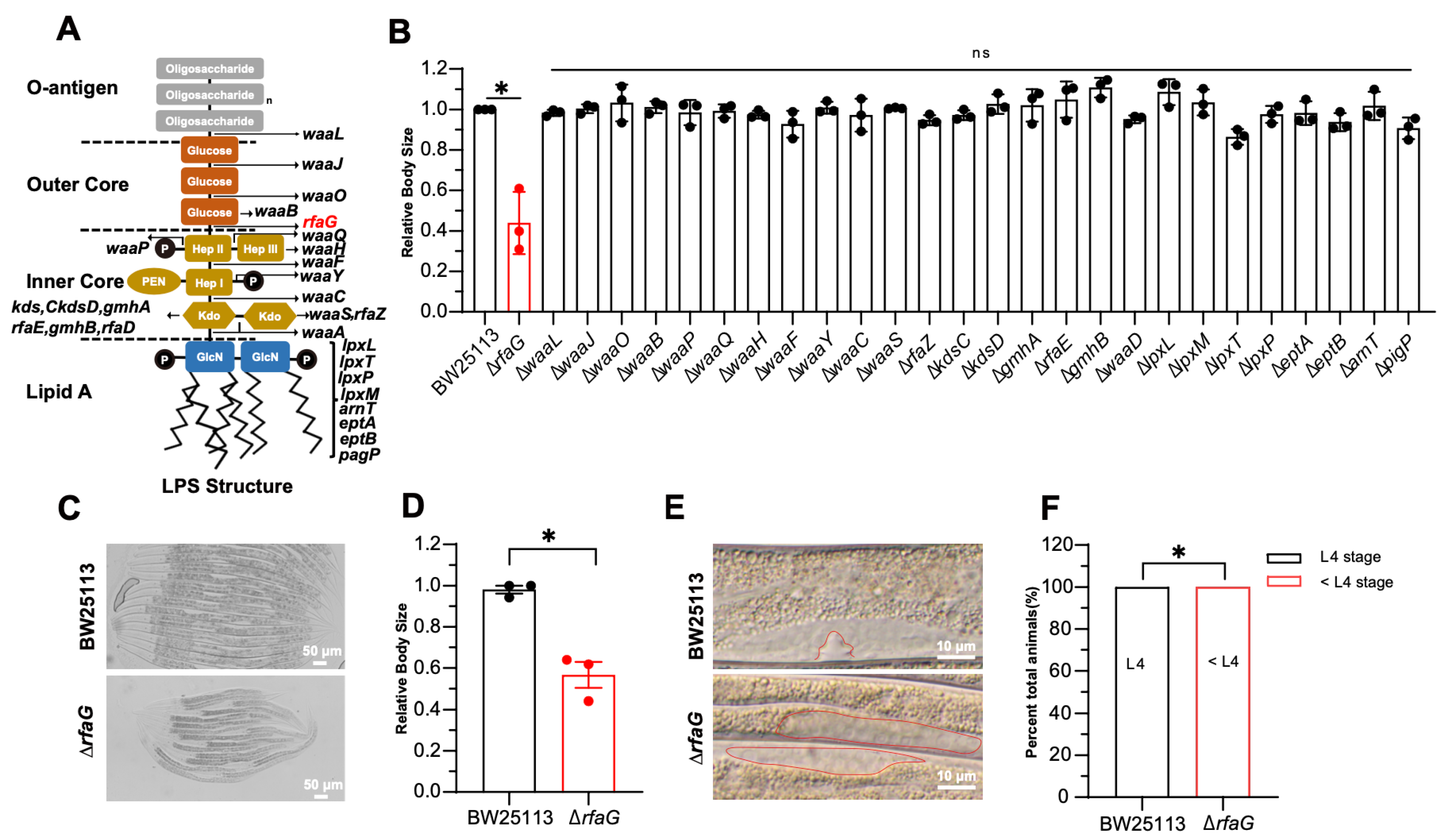
Given the structural divergence in bacterial cell walls, specifically the presence of lipopolysaccharide (LPS) in Gram-negative outer membranes, we hypothesized that LPS could act as a key modulator of C. elegans development. To test this hypothesis, we systematically screened a panel of E. coli LPS biosynthesis mutants. To exclude the pathogenic effects, we selected the E. coli Keio single-gene deletion library, which is non-pathogenic to C. elegans. The Escherichia coli LPS comprises three domains: lipid A, core polysaccharide, and O-antigen [31] (Figure 2A). We found that only the ΔrfaG mutant induced significantly smaller body size in C. elegans (Figure 2B–D). The rfaG gene encodes a glycosyltransferase critical for core polysaccharide biosynthesis [12,32,33,34]. To determine whether the reduced body size reflected developmental delay, we examined the developmental stage of C. elegans. Animals fed E. coli BW25113 reached the L4 stage, marked by the characteristic Christmas tree-like vulval morphology after 48 h, whereas animals fed the ΔrfaG mutant exhibited significant delay and failed to progress to the L4 stage (Figure 2E,F). Together, these results identify the ΔrfaG mutation as a specific inducer of C. elegans developmental delay, and this phenotype is independent of broader LPS biosynthesis defects or general bacterial fitness impairment.

2.3. LPS Structure Mediates ΔrfaG-Induced Developmental Delay

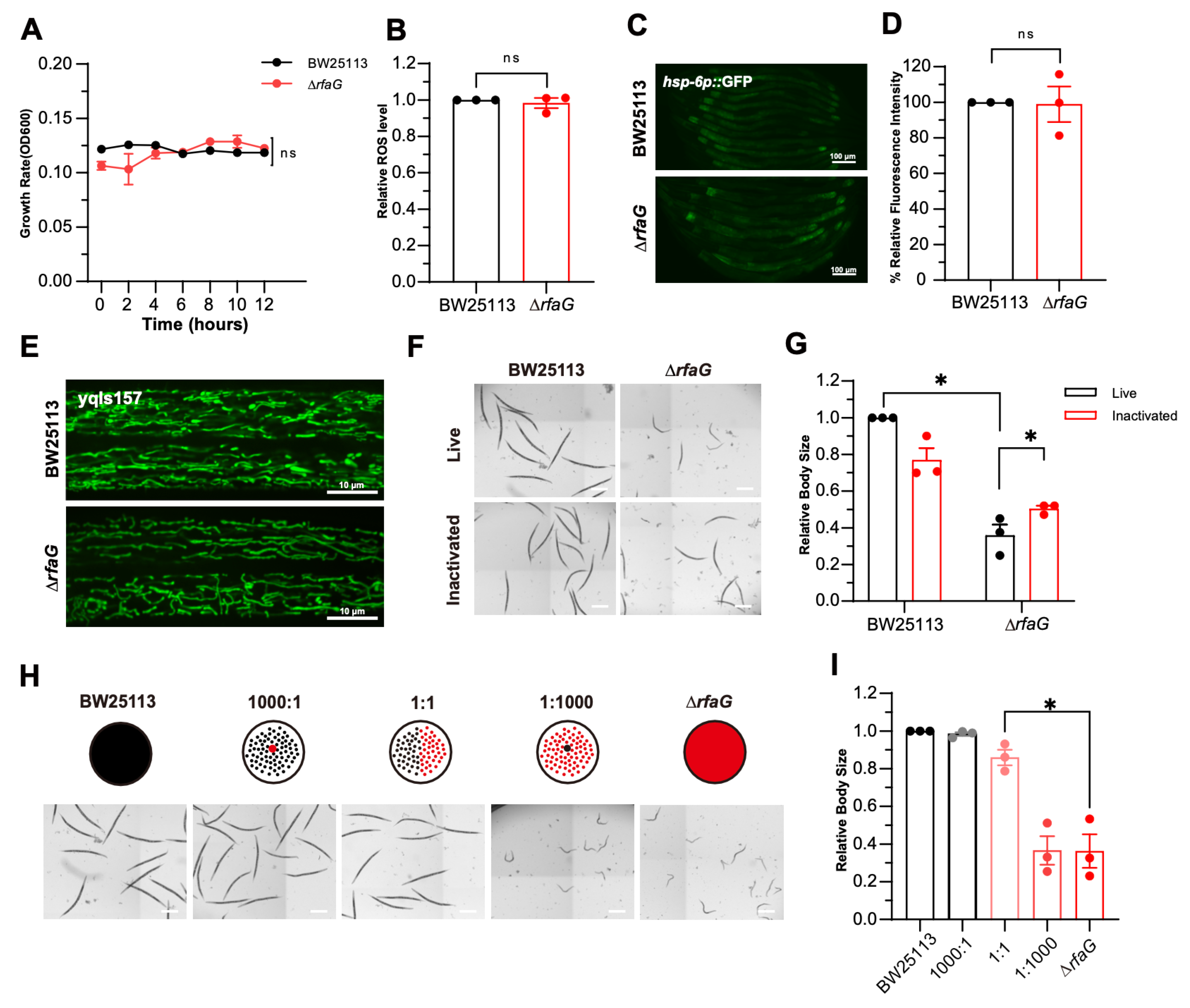
To dissect the mechanism underlying ∆rfaG-induced host developmental delay, we first assessed whether bacterial growth contributed to the host developmental delay. Both wild-type BW25113 and ΔrfaG strains exhibited no significant growth due to the limited nutrients in the medium, indicating that the host developmental delay was not attributable to a bacterial growth defect (Figure 3A). Deletion of LPS synthesis genes has been reported to increase bacterial reactive oxygen species (ROS) levels and oxidative stress [35]. ROS are reported to play roles in various biological processes like DNA replication and protein homeostasis [36,37,38,39]. To determine whether ROS levels induced C. elegans developmental delay, we measured bacterial ROS levels using the 2′,7′-dichlorodihydrofluorescein diacetate (H2DCFDA). There were no significant differences in ROS levels in the bacteria BW25113 and ΔrfaG (Figure 3B). The mitochondrial unfolded protein response (UPRmt) has been reported to be closely associated with a variety of oxidative stresses [40,41]. However, ΔrfaG did not activate UPRmt in C. elegans (Figure 3C,D). Mitochondrial morphology (visualized using yqIs157) also retained normal tubular structures (Figure 3E), further excluding oxidative stress as a driver of host developmental delay. The structure of the mitochondria in C. elegans fed ΔrfaG maintained normal tubular morphology (Figure 3E). These findings suggest that the ΔrfaG-induced developmental delay in C. elegans is not associated with oxidative stress.
To elucidate whether the effects of ΔrfaG are dependent on bacteria metabolism, we treated the bacteria with paraformaldehyde (PFA) to inactivate their metabolic activity [42]. Remarkably, the PFA-inactivated ΔrfaG exhibited the C. elegans developmental defect (Figure 3F,G). This result suggests that the LPS structure in ΔrfaG, rather than live bacteria metabolites, mediates delayed development in C. elegans.
Next, we determined the threshold at which wild-type BW25113 could rescue the growth of C. elegans fed on ΔrfaG. We titrated wild-type BW25113 into ΔrfaG with varying concentrations. Mixing small amounts of wild-type BW25113 with ∆rfaG did not alleviate C. elegans developmental delay; ≥50% wild-type bacteria restored normal development of C. elegans (Figure 3H,I). This demonstrates that intact LPS architecture is critical for C. elegans growth, with no compensatory capacity below this threshold. Collectively, our findings suggest that structural changes in LPS, rather than bacterial metabolites, drive ∆rfaG-induced host developmental delay, providing crucial insights into the role of LPS in host–microbe interactions.

2.4. The ΔrfaG Mutant Induced Developmental Defects via DAF-16

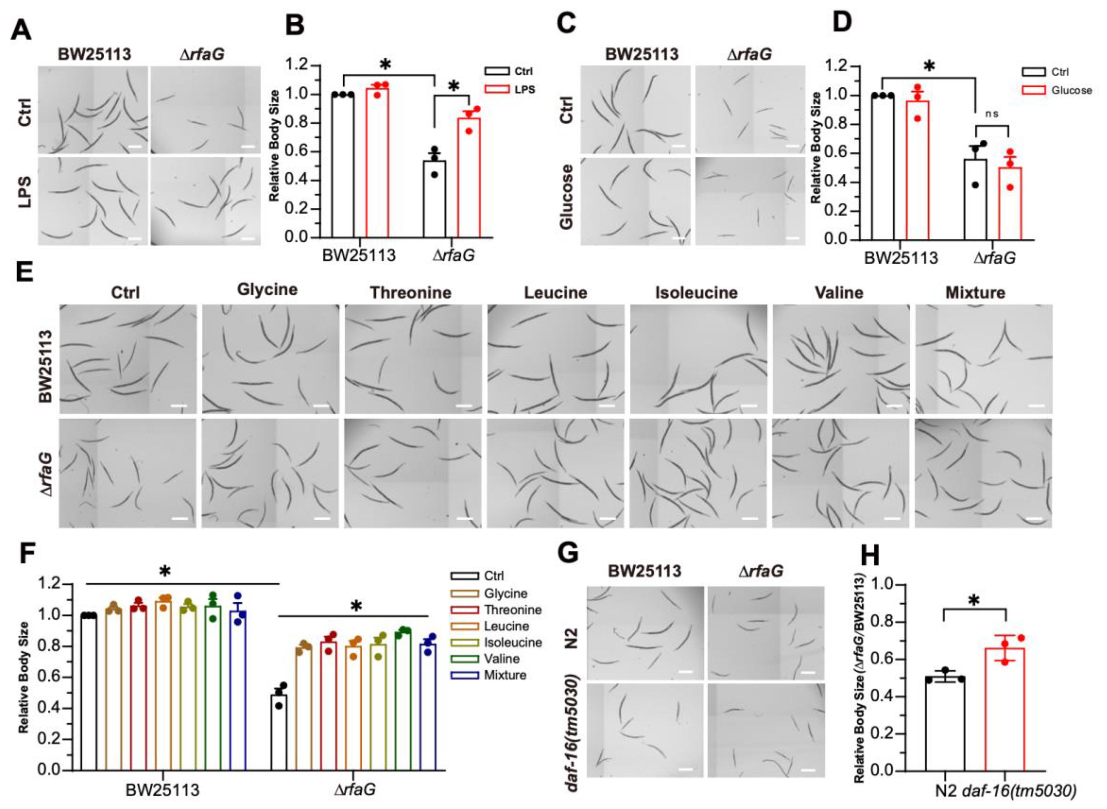
To determine whether LPS directly contributes to ∆rfaG-induced developmental defects in C. elegans, we supplemented ΔrfaG cultures with purified LPS, which fully rescued the developmental delay in C. elegans induced by ΔrfaG (Figure 4A,B). This result provides direct evidence for the essential role of bacterial LPS in promoting the growth of C. elegans. LPS influences the intestinal nutrient absorption in C. elegans [20], so we tested whether the ΔrfaG-induced developmental delay was caused by nutrient deficiency. We supplemented C. elegans with several nutrients. Supplementation of glucose did not alleviate the developmental delay of C. elegans induced by ΔrfaG (Figure 4C,D). We further tested a set of amino acids (glycine, threonine, leucine, isoleucine, and valine) for their ability to restore host growth (Figure 4E,F), suggesting amino acid scarcity underlies ΔrfaG-induced host developmental delay.
The insulin/IGF-1 signaling (IIS) integrates nutrient status via FOXO transcription factor DAF-16 and plays a crucial role in regulating developmental arrest and gene expression [43,44]. The FOXO transcription factor DAF-16, a downstream effector of the IIS pathway, is responsible for integrating stress signals and translocating from the cytoplasm to the nucleus [45]. To determine if DAF-16 mediates developmental arrest in C. elegans induced by ΔrfaG, we examined the development of daf-16(tm5030) animals fed on ΔrfaG; daf-16(tm5030) mutants fed on ΔrfaG bypassed developmental arrest (Figure 4G,H), indicating that DAF-16 mediates developmental arrest induced by ΔrfaG. Together, these data indicate a cascade wherein LPS limits host amino acid availability, exogenous LPS or amino acids alleviate the developmental delay, and the insulin-like signaling (IIS) pathway mediates the developmental delay.
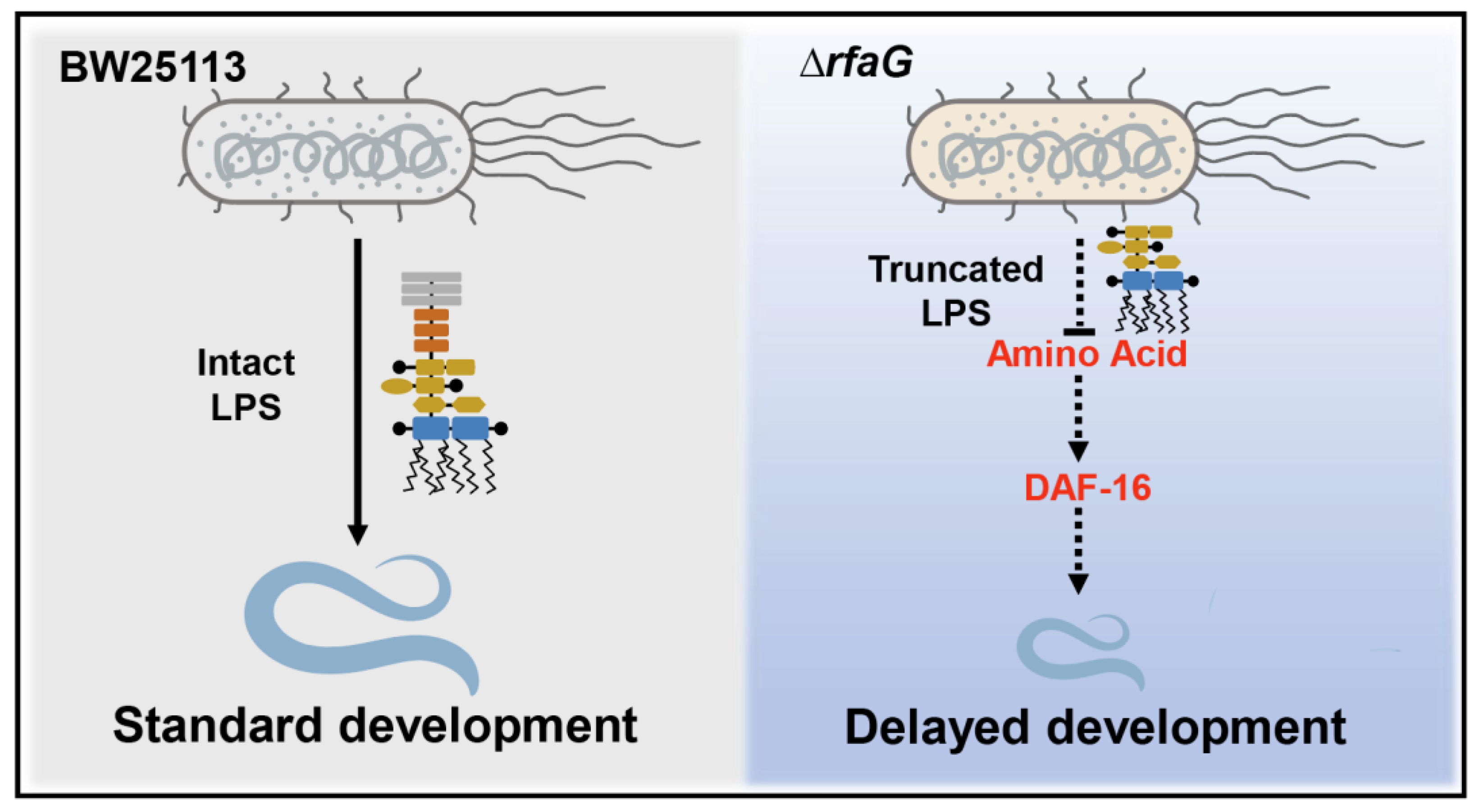
In summary, our findings demonstrate that E. coli LPS is essential for C. elegans development. The ΔrfaG mutant, with a truncated LPS structure, induces developmental defects by limiting host amino acid availability. These developmental defects are mediated by DAF-16, the FOXO transcription factor in the insulin/IGF-1 signaling pathway, and rescued by exogenous LPS or amino acids, highlighting LPS structure as a critical determinant of host–microbe metabolic crosstalk (Figure 5).

3. Discussion

Previous studies have emphasized LPS as a pro-inflammatory endotoxin that induces cellular damage and immune responses [14,19,46]. However, our work uncovered an unrecognized role of bacterial LPS in C. elegans development. We demonstrated that ΔrfaG with a structural defect in LPS profoundly disrupts C. elegans development. This finding challenges the traditional paradigm that bacterial metabolic outputs (e.g., nutrient provisioning) are the drivers of C. elegans developmental progression. Instead, our data positions LPS architecture as a critical structural signal in host–microbe crosstalk. This novel finding expands our understanding of the molecular mechanisms underlying host–microbe interactions in C. elegans.
A pivotal innovation of this study lies in disentangling LPS’s structural role from bacterial metabolic activity. The rescue of developmental delay by paraformaldehyde (PFA)-fixed ΔrfaG (Figure 3G) unequivocally demonstrates that LPS integrity—not live bacteria or secreted metabolites—is essential for normal development. Future studies could explore whether homologs of these receptors mediate LPS recognition in C. elegans.
LPS, a structural component of the bacterial cell wall, serves as a crucial signal molecule [7]. The rescue of ΔrfaG-induced defects by exogenous amino acids (Figure 4E,F) points to a novel mechanistic link between LPS structure and host nutrient utilization. We hypothesize that intact LPS facilitates amino acid uptake or signaling, possibly by maintaining intestinal barrier integrity or modulating nutrient-sensing pathways. This mirrors findings in mammals, where LPS dysbiosis impairs amino acid absorption and triggers metabolic stress [46]. Intriguingly, the insulin/IGF-1 signaling (IIS) pathway—a central regulator of nutrient sensing and longevity in C. elegans—emerged as a key player, as daf-16 mutants bypassed developmental arrest (Figure 4G,H). This parallels studies where IIS pathway components, including DAF-16, mediate lifespan extension under dietary restriction or protease inhibition [44,45], suggesting a conserved nexus between microbial signals, nutrient availability, and developmental plasticity. Given that amino acids can rescue the developmental defects induced by ΔrfaG, LPS may affect the uptake or utilization of amino acids in C. elegans. Further research is needed to explore this potential relationship between LPS and amino acids.
The suppression of ΔrfaG-induced defects in daf-16 mutants highlights DAF-16’s dual role as a stress-responsive transcription factor and a developmental checkpoint. While DAF-16 is best known for its role in longevity under stress, our work expands its function to include developmental modulation in response to microbial cues. This aligns with recent discoveries that DAF-16 integrates environmental signals, such as oxidative stress and pathogen exposure, to regulate organismal fitness. Future investigations should delineate whether LPS directly modulates DAF-16 nuclear translocation or collaborates with upstream IIS components like AGE-1/ PI3K.
Notably, our findings contrast with studies where LPS triggers neuroinflammation and behavioral deficits in mammals, underscoring the context-dependent nature of LPS signaling. In C. elegans, the absence of canonical TLR4 homologs may explain why LPS acts as a developmental cue rather than an inflammatory trigger. This divergence highlights the evolutionary plasticity of LPS–host interactions and positions C. elegans as a unique model to study LPS’s non-canonical roles.

4. Materials and Methods

4.1. C. elegans Strains and Maintenance

All nematode strains in this study were maintained on nematode growth medium (NGM) using standard protocols [47]. NGM was prepared as previously described [47] (1.7% agar, 0.3% NaCl, 0.25% peptone, 1 mM CaCl2, 1 mM MgSO4, 5 μg/mL cholesterol, and 25 mM KH2PO4 buffer pH 6.0). Gravid adults were treated with 5% hypochlorite solution and 0.5 M NaOH for 5 min to release embryos, which were washed 5 times with M9 buffer (22 mM KH2PO4, 42 mM Na2HPO4, 85 mM NaCl, and 1 mM MgSO4), followed by incubation in liquid NGM at 20 °C for 48 h to obtain synchronized L1 larvae. Synchronized L1 larvae were obtained via hypochlorite treatment of gravid adults, followed by incubating in liquid NGM at 20 °C for 48 h. The following strains were used in the study: wild-type N2, SJ4100 [zcls13 (Phsp-6::GFP)], yqIs157 [(Py37a1b.5::mito-GFP), and XV92[daf-16(tm5030)].

4.2. Bacterial Strains and Maintenance

E. coli BW25113 (WT) was cultured in Lysogeny Broth (LB) medium at 37 °C. E. coli single-gene deletion mutants from the Keio collection were grown in LB medium supplemented with 50 μg/mL kanamycin at 37 °C.

4.3. Reagents

Stock solutions were prepared as follows: LPS (5 mg/mL), glucose (350 mM), glycine (1.7 M), threonine (280 mM), leucine (64 mM), isoleucine (190 mM), and valine (170 mM); stored at −20 °C. Paraformaldehyde (PFA, 80096618) was purchased from Sinopharm (Beijing, China) and stored as a 4% solution at 4 °C. Liquid NGM was prepared as standard NGM without agar.

4.4. E. coli LPS Synthesis Mutants Screen in Liquid Culture System

C. elegans embryos were harvested from gravid worms fed E. coli BW25113. Adults were treated with alkaline hypochlorite solution and incubated for 20 h in M9 buffer at 20 °C. Synchronized L1 larvae (≈25 per well) in 10 μL M9 buffer were aliquoted into 96-well plates. Overnight cultures of E. coli LPS biosynthesis mutants were centrifuged (3000× g, 30 min), washed, and resuspended in liquid NGM to OD600 = 8.0. A total of 10 μL of bacterial suspension was added to each well containing larvae, followed by 50 μL liquid NGM. Plates were sealed and incubated statically at 20 °C for 48 h. Worms were then immobilized with M9 buffer containing 1 mM levamisole, imaged using an Invitrogen EVOS FL microscope (Thermo Fisher, Waltham, MA, USA), and body size was quantified using ImageJ software, version1.54K [48].

4.5. Assessment of Bacterial Growth Rate

A total of 10 μL of bacterial culture and 60 μL of liquid NGM were added to 96-well plates and incubated at 220 rpm at 20 °C. A total of 10 μL of bacterial culture was diluted to 100 μL to measure OD600. OD600 was measured every 2 h for 12 h using a microplate reader (TECAN, Mannedorf, Switzerland). A total of 10 μL bacterial culture was diluted with 90 μL liquid NGM to measure OD600.

4.6. Assessment of Bacterial ROS Levels

Logarithmic-phase bacterial cultures were centrifuged (3000× g, 10 min), washed, and resuspended in PBS (pH 7.4) to OD600 ≈ 0.5. Bacteria were incubated with 10 μM H2DCFDA (2′,7′-dichlorodihydrofluorescein diacetate; Invitrogen, Carlsbad, CA, USA, Cat. No. D399) in the dark at 37 °C for 1 h. Fluorescence intensity (excitation 492 nm, emission 535 nm) was measured in a black-walled 96-well plate using a microplate reader.

4.7. Paraformaldehyde Treatment of Bacteria

Overnight bacterial cultures were treated with 0.5% (v/v) PFA (final concentration) and incubated with shaking (220 rpm) at 37 °C for 1 h. Fixed bacteria were centrifuged (3000× g, 30 min) and washed five times with sterile LB medium. PFA-killed pellets were resuspended in liquid NGM for assays.

4.8. Bacterial Mixing Assay

Overnight cultures of E. coli BW25113 (WT) and ΔrfaG were centrifuged (3000× g, 10 min), washed, and resuspended in liquid NGM to OD600 = 8.0. Bacteria were mixed at WT: ΔrfaG ratios of 1000:1, 1:1, and 1:1000. Synchronized L1 larvae, bacterial mixtures, and liquid NGM were combined in 96-well plates. Plates were sealed, incubated at 20 °C with shaking (220 rpm) for 48 h, and imaged.

4.9. Statistical Analysis

Statistical analyses were performed using GraphPad Prism 8.0.2 (GraphPad Software) and Fiji (ImageJ). Data are presented as mean ± SEM. Significance was determined by unpaired two-tailed Student’s t-test (two groups) or one-way ANOVA with Tukey’s post hoc test (≥3 groups). Significance levels: * p < 0.05, ** p < 0.01, and ns: not significant. A minimum of three independent biological replicates were performed.

Author Contributions

L.T.: Methodology, validation, analysis, investigation, and writing—original draft preparation. J.Z.: Writing—review and editing, supervision, project administration, and funding acquisition. All authors have read and agreed to the published version of the manuscript.

Funding

This work was supported by the National Key R&D Program of China (No. 2022YFA0806400) and the National Natural Science Foundation of China (No. 32271215).

Institutional Review Board Statement

Not applicable.

Informed Consent Statement

Not applicable.

Data Availability Statement

The original contributions presented in the study are included in the article, further inquiries can be directed to the corresponding authors.

Conflicts of Interest

The authors declare no conflict of interest.

References

  1. Beveridge Terry, J. Structures of Gram-Negative Cell Walls and Their Derived Membrane Vesicles. J. Bacteriol. 1999, 181, 4725–4733. [Google Scholar] [CrossRef] [PubMed]
  2. Caroff, M.; Karibian, D. Structure of bacterial lipopolysaccharides. Carbohydr. Res. 2003, 338, 2431–2447. [Google Scholar] [CrossRef] [PubMed]
  3. Vatanen, T.; Kostic, A.D.; d’Hennezel, E.; Siljander, H.; Franzosa, E.A.; Yassour, M.; Kolde, R.; Vlamakis, H.; Arthur, T.D.; Hämäläinen, A.-M.; et al. Variation in Microbiome LPS Immunogenicity Contributes to Autoimmunity in Humans. Cell 2016, 165, 842–853. [Google Scholar] [CrossRef] [PubMed]
  4. Singh, S.; Wilksch, J.J.; Dunstan, R.A.; Mularski, A.; Wang, N.; Hocking, D.; Jebeli, L.; Cao, H.; Clements, A.; Jenney, A.W.J.; et al. LPS O Antigen Plays a Key Role in Klebsiella pneumoniae Capsule Retention. Microbiol. Spectr. 2022, 10, e0151721. [Google Scholar] [CrossRef]
  5. McGarry, N.; Roe, D.; Smith, S.G.J. Synergy between Group 2 capsules and lipopolysaccharide underpins serum resistance in extra-intestinal pathogenic Escherichia coli. Microbiology 2024, 170. [Google Scholar] [CrossRef]
  6. Kumar, A.; Mallik, D.; Pal, S.; Mallick, S.; Sarkar, S.; Chanda, A.; Ghosh, A.S. Escherichia coli O8-antigen enhances biofilm formation under agitated conditions. FEMS Microbiol. Lett. 2015, 362, fnv112. [Google Scholar] [CrossRef]
  7. Park, B.S.; Song, D.H.; Kim, H.M.; Choi, B.S.; Lee, H.; Lee, J.O. The structural basis of lipopolysaccharide recognition by the TLR4-MD-2 complex. Nature 2009, 458, 1191–1195. [Google Scholar] [CrossRef]
  8. Alexander, C.; Zähringer, U. Chemical structure of lipid A—The primary immunomodulatory center of bacterial lipopolysaccharides. Trends Glycosci. Glycotechnol. 2002, 14, 69–86. [Google Scholar] [CrossRef]
  9. Ruhal, R.; Kataria, R. Biofilm patterns in gram-positive and gram-negative bacteria. Microbiol. Res. 2021, 251, 126829. [Google Scholar] [CrossRef] [PubMed]
  10. Mai-Prochnow, A.; Clauson, M.; Hong, J.; Murphy, A.B. Gram positive and Gram negative bacteria differ in their sensitivity to cold plasma. Sci. Rep. 2016, 6, 38610. [Google Scholar] [CrossRef]
  11. Varghese, M.; Balachandran, M. Antibacterial efficiency of carbon dots against Gram-positive and Gram-negative bacteria: A review. J. Environ. Chem. Eng. 2021, 9, 106821. [Google Scholar] [CrossRef]
  12. Yethon Jeremy, A.; Vinogradov, E.; Perry Malcolm, B.; Whitfield, C. Mutation of the Lipopolysaccharide Core Glycosyltransferase Encoded by waaG Destabilizes the Outer Membrane of Escherichia coli by Interfering with Core Phosphorylation. J. Bacteriol. 2000, 182, 5620–5623. [Google Scholar] [CrossRef]
  13. Barua, S.; Yamashino, T.; Hasegawa, T.; Yokoyama, K.; Torii, K.; Ohta, M. Involvement of surface polysaccharides in the organic acid resistance of Shiga Toxin-producing Escherichia coli O157:H7. Mol. Microbiol. 2002, 43, 629–640. [Google Scholar] [CrossRef]
  14. Sivamaruthi, B.S.; Prasanth, M.I.; Balamurugan, K. Alterations in Caenorhabditis elegans and Cronobacter sakazakii lipopolysaccharide during interaction. Arch. Microbiol. 2015, 197, 327–337. [Google Scholar] [CrossRef] [PubMed]
  15. Kamaladevi, A.; Balamurugan, K. Lipopolysaccharide of Klebsiella pneumoniae attenuates immunity of Caenorhabditis elegans and evades by altering its supramolecular structure. RSC Adv. 2016, 6, 30070–30080. [Google Scholar] [CrossRef]
  16. Ma, J.; Xu, X.; Wang, R.; Yan, H.; Yao, H.; Zhang, H.; Jiang, S.; Xu, A. Lipopolysaccharide exposure induces oxidative damage in Caenorhabditis elegans: Protective effects of carnosine. BMC Pharmacol. Toxicol. 2020, 21, 85. [Google Scholar] [CrossRef]
  17. Zhang, X.; Li, W.; Feng, K.; Xiao, J.; Du, J.; Cao, Y.; Chen, Y. Immunomodulatory effect of pentagalloyl glucose in LPS-stimulated RAW264.7 macrophages and PAO1-induced Caenorhabditis elegans. Exp. Gerontol. 2021, 150, 111388. [Google Scholar] [CrossRef] [PubMed]
  18. Huang, Y.; Wang, Y.; Xu, J.; Feng, J.; He, X. Propacin, a coumarinolignoid isolated from durian, inhibits the lipopolysaccharide-induced inflammatory response in macrophages through the MAPK and NF-κB pathways. Food Funct. 2020, 11, 596–605. [Google Scholar] [CrossRef]
  19. Aballay, A.; Drenkard, E.; Hilbun, L.R.; Ausubel, F.M. Caenorhabditis elegans innate immune response triggered by Salmonella enterica requires intact LPS and is mediated by a MAPK signaling pathway. Curr. Biol. 2003, 13, 47–52. [Google Scholar] [CrossRef] [PubMed]
  20. Zhang, X.-P.; Ma, X.; Liu, J.-L.; Liu, A.-L. Exploring the potential use of Caenorhabditis elegans as an animal model for evaluating chemical-induced intestinal dysfunction. Toxicol. Appl. Pharmacol. 2024, 493, 117140. [Google Scholar] [CrossRef]
  21. Kumar, A.; Baruah, A.; Tomioka, M.; Iino, Y.; Kalita, M.C.; Khan, M. Caenorhabditis elegans: A model to understand host-microbe interactions. Cell. Mol. Life Sci. 2019, 77, 1229–1249. [Google Scholar] [CrossRef] [PubMed]
  22. Yen, C.A.; Curran, S.P. Gene-diet interactions and aging in C. elegans. Exp. Gerontol. 2016, 86, 106–112. [Google Scholar] [CrossRef]
  23. Park, Y.-J.; Yeon, J.; Cho, J.; Kim, D.-Y.; Bai, X.; Oh, Y.; Kim, J.; Nam, H.; Hwang, H.; Heo, W.; et al. PIEZO acts in an intestinal valve to regulate swallowing in C. elegans. Nat. Commun. 2024, 15, 10072. [Google Scholar] [CrossRef]
  24. Berg, M.; Stenuit, B.; Ho, J.; Wang, A.; Parke, C.; Knight, M.; Alvarez-Cohen, L.; Shapira, M. Assembly of the Caenorhabditis elegans gut microbiota from diverse soil microbial environments. ISME J. 2016, 10, 1998–2009. [Google Scholar] [CrossRef]
  25. Berg, M.; Zhou, X.Y.; Shapira, M. Host-specific functional significance of Caenorhabditis gut commensals. Front. Microbiol. 2016, 7. [Google Scholar] [CrossRef]
  26. Asan, A.; Raiders, S.A.; Priess, J.R. Morphogenesis of the C. elegans Intestine Involves Axon Guidance Genes. PLoS Genet. 2016, 12, e1005950. [Google Scholar] [CrossRef] [PubMed]
  27. Stroustrup, N.; Ulmschneider, B.E.; Nash, Z.M.; López-Moyado, I.F.; Apfeld, J.; Fontana, W. The Caenorhabditis elegans Lifespan Machine. Nat. Methods 2013, 10, 665–670. [Google Scholar] [CrossRef]
  28. Guzman, D.M.; Chakka, K.; Shi, T.; Marron, A.; Fiorito, A.E.; Rahman, N.S.; Ro, S.; Sucich, D.G.; Pierce, J.T. Transgenerational effects of alcohol on behavioral sensitivity to alcohol in Caenorhabditis elegans. PLoS ONE 2022, 17, e0271849. [Google Scholar] [CrossRef] [PubMed]
  29. Noma, K.; Jin, Y. Optogenetic mutagenesis in Caenorhabditis elegans. Nat. Commun. 2015, 6, 8868. [Google Scholar] [CrossRef]
  30. Davies, A.G.; McIntire, S.L.; Using, C. elegans to screen for targets of ethanol and behavior-altering drugs. Biol. Proced. Online 2004, 6, 113–119. [Google Scholar] [CrossRef]
  31. Raetz, C.R.; Whitfield, C. Lipopolysaccharide endotoxins. Annu. Rev. Biochem. 2002, 71, 635–700. [Google Scholar] [CrossRef]
  32. Parker, C.T.; Pradel, E.; Schnaitman, C.A. Identification and sequences of the lipopolysaccharide core biosynthetic genes rfaQ, rfaP, and rfaG of Escherichia coli K-12. J. Bacteriol. 1992, 174, 930–934. [Google Scholar] [CrossRef] [PubMed]
  33. Kadam, S.K.; Rehemtulla, A.; Sanderson, K.E. Cloning of rfaG, B, I, and J genes for glycosyltransferase enzymes for synthesis of the lipopolysaccharide core of Salmonella typhimurium. J. Bacteriol. 1985, 161, 277–284. [Google Scholar] [CrossRef] [PubMed]
  34. Parker, C.T.; Kloser, A.W.; Schnaitman, C.A.; Stein, M.A.; Gottesman, S.; Gibson, B.W. Role of the rfaG and rfaP genes in determining the lipopolysaccharide core structure and cell surface properties of Escherichia coli K-12. J. Bacteriol. 1992, 174, 2525–2538. [Google Scholar] [CrossRef] [PubMed]
  35. Seregina, T.A.; Petrushanko, I.Y.; Shakulov, R.S.; Zaripov, P.I.; Makarov, A.A.; Mitkevich, V.A.; Mironov, A.S. The Inactivation of LPS Biosynthesis Genes in E. coli Cells Leads to Oxidative Stress. Cells 2022, 11, 2667. [Google Scholar] [CrossRef]
  36. Taylor, P.L.; Blakely, K.M.; de Leon, G.P.; Walker, J.R.; McArthur, F.; Evdokimova, E.; Zhang, K.; Valvano, M.A.; Wright, G.D.; Junop, M.S. Structure and Function of Sedoheptulose-7-phosphate Isomerase, a Critical Enzyme for Lipopolysaccharide Biosynthesis and a Target for Antibiotic Adjuvants. J. Biol. Chem. 2008, 283, 2835–2845. [Google Scholar] [CrossRef] [PubMed]
  37. Srinivas, U.S.; Tan, B.W.Q.; Vellayappan, B.A.; Jeyasekharan, A.D. ROS and the DNA damage response in cancer. Redox Biol. 2019, 25, 101084. [Google Scholar] [CrossRef]
  38. Zhou, J.; Xin-Yu, L.; Yu-Jia, L.; Ji, F.; Yong, W.; Han-Ming, S.; Lu, G.-D. Full-coverage regulations of autophagy by ROS: From induction to maturation. Autophagy 2022, 18, 1240–1255. [Google Scholar] [CrossRef]
  39. Cui, Q.; Wang, J.-Q.; Assaraf, Y.G.; Ren, L.; Gupta, P.; Wei, L.; Ashby, C.R.; Yang, D.-H.; Chen, Z.-S. Modulating ROS to overcome multidrug resistance in cancer. Drug Resist. Updates 2018, 41, 1–25. [Google Scholar] [CrossRef]
  40. Shpilka, T.; Haynes, C.M. The mitochondrial UPR: Mechanisms, physiological functions and implications in ageing. Nat. Rev. Mol. Cell Biol. 2018, 19, 109–120. [Google Scholar] [CrossRef]
  41. Liu, Y.; Li, Q.; Tian, G.; Zhou, X.; Chen, P.; Chen, B.; Shan, Z.; Qi, B. Neuronal PRDX-2-Mediated ROS Signaling Regulates Food Digestion via peripheral UPRmt Activation. Nat. Commun. 2024, 15, 10582. [Google Scholar] [CrossRef]
  42. Beydoun, S.; Choi, H.S.; Dela-Cruz, G.; Kruempel, J.; Huang, S.; Bazopoulou, D.; Miller, H.A.; Schaller, M.L.; Evans, C.R.; Leiser, S.F. An alternative food source for metabolism and longevity studies in Caenorhabditis elegans. Commun. Biol. 2021, 4, 258. [Google Scholar] [CrossRef]
  43. Feng, M.; Gao, B.; Ruiz, D.; Garcia, L.R.; Sun, Q. Bacterial vitamin B6 is required for post-embryonic development in C. elegans. Commun. Biol. 2024, 7, 367. [Google Scholar] [CrossRef]
  44. Fernandes de Abreu, D.A.; Caballero, A.; Fardel, P.; Stroustrup, N.; Chen, Z.; Lee, K.; Keyes, W.D.; Nash, Z.M.; López-Moyado, I.F.; Vaggi, F.; et al. An Insulin-to-Insulin Regulatory Network Orchestrates Phenotypic Specificity in Development and Physiology. PLoS Genet. 2014, 10, e1004225. [Google Scholar] [CrossRef] [PubMed]
  45. Zečić, A.; Braeckman, B.P. DAF-16/FoxO in Caenorhabditis elegans and Its Role in Metabolic Remodeling. Cells 2020, 9, 109. [Google Scholar] [CrossRef] [PubMed]
  46. Stein, S.C.; Faber, E.; Bats, S.H.; Murillo, T.; Speidel, Y.; Coombs, N.; Josenhans, C. Helicobacter pylori modulates host cell responses by CagT4SS-dependent translocation of an intermediate metabolite of LPS inner core heptose biosynthesis. PLoS Pathog. 2017, 13, e1006514. [Google Scholar] [CrossRef] [PubMed]
  47. Brenner, S. The genetics of Caenorhabditis elegans. Genetics 1974, 77, 71–94. [Google Scholar] [CrossRef]
  48. Schindelin, J.; Arganda-Carreras, I.; Frise, E.; Kaynig, V.; Longair, M.; Pietzsch, T.; Preibisch, S.; Rueden, C.; Saalfeld, S.; Schmid, B.; et al. Fiji: An open-source platform for biological-image analysis. Nat. Methods 2012, 9, 676–682. [Google Scholar] [CrossRef]
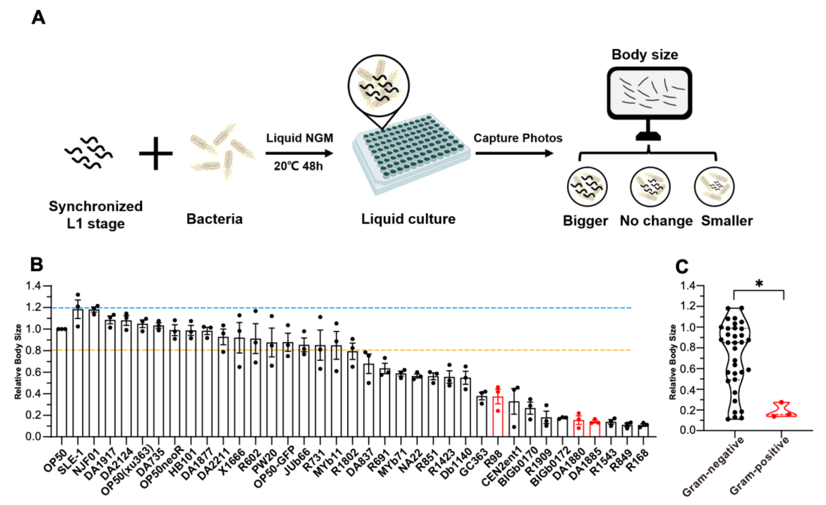
Figure 1. High-throughput screen identifying Gram-negative bacteria as potent promoters of C. elegans development. (A) Workflow for liquid culture developmental screening. Synchronized L1 larvae were co-cultured with bacterial suspensions. Bright-field images were captured using the Invitrogen EVOS FL imaging system, and body size was analyzed using ImageJ software, version1.54K. (B) Bar graph comparing C. elegans developmental rate across bacterial isolates. Data were normalized to the mean body size of nematodes fed wild-type Escherichia coli OP50 (control; dashed line). Body size outside this range exhibits significant differences. (C) Body size distribution of worms fed Gram-negative (dark) versus Gram-positive bacteria (red). * p < 0.05 (unpaired two-tailed t-test). Data represent mean ± SEM.
Ijms 26 07399 g001
Figure 2. The ΔrfaG mutant impairs C. elegans development. (A) Schematic of E. coli LPS structure. (B) Body size of C. elegans fed LPS biosynthesis mutants (ns, not significant; * p < 0.05 vs. BW25113 control). (C,D) Representative images and quantification of worms fed ΔrfaG (scale bar: 50 μm). (E,F) Developmental stage of C. elegans grown on ∆rfaG for 48 hour. Vulval morphology (Christmas tree-like structure, top) and germline (bottom) were visualized by red line(scale bar: 10 μm). Data are mean ± SEM (Data are from 3 independent biological replicates, more than 25 worms per group, and 3 independent experiments) (* p < 0.05 (two-tailed t-test)).
Ijms 26 07399 g002
Figure 3. Microbial LPS drives host developmental delay. (A) Bacterial growth rates of BW25113 and ΔrfaG. (B) Reactive oxygen species (ROS) levels in BW25113 vs. ∆rfaG, measured via H2DCFDA fluorescence. (C,D) Representative images and quantitative analysis of hsp-6p::GFP expression in C. elegans fed with BW25113 vs. ∆rfaG(Scale bar: 100 μm). (E) Representative images of mitochondria in C. elegans fed with BW25113 vs. ∆rfaG (Scale bar: 10 μm). (F,G) Metabolically inactivated (PFA-fixed) ΔrfaG rescues host developmental delay (scale bar: 500 μm). (H,I) Supplementation wild-type BW25113 restores the host developmental delay (scale bar: 500 μm). Data are mean ± SEM (Data are from 3 independent biological replicates, more than 20 worms per group, and 3 independent experiments) (ns, not significant; * p < 0.05 (two-tailed t-test)).
Ijms 26 07399 g003
Figure 4. Exogenous LPS and amino acids rescue ΔrfaG-induced developmental defects mediated by DAF-16. (A,B) LPS rescues ΔrfaG-induced developmental delay(scale bar: 500 μm). (C,D) Glucose supplementation fails to rescue the developmental defect. (E,F) Amino acid supplementation restores animal growth (scale bar: 500 μm,* p < 0.05 (one-way-ANOVA). (G,H) daf-16 loss-of-function mutants bypass ΔrfaG-induced developmental defect(scale bar: 500 μm). Data are mean ± SEM (Data are from 3 independent biological replicates, more than 20 worms per group, and 3 independent experiments) (ns, not significant; * p < 0.05 (two-tailed t-test)).
Ijms 26 07399 g004
Figure 5. Schematic model for ∆rfaG-induced developmental delay in C. elegans via FOXO/DAF-16.
Ijms 26 07399 g005
Table 1. Bacterial strains of the screening.
StrainGramGenus
OP50negativeEscherichia
SLE-1negativeEscherichia
NJF01negativeEscherichia
DA1917negativeEscherichia
DA2124negativeEscherichia
OP50(xu363)negativeEscherichia
DA735negativeEscherichia
OP50-NeoRnegativeEscherichia
HB101negativeEscherichia
DA1877negativeComamonas
DA2211negativeEscherichia
X1666negativeEscherichia
R602negativeAcidovorax
PW20negativeEscherichia
OP50-GFPnegativeEscherichia
JUb66negativeLelliottia
R731negativeSphingomonas
MYb11negativePseudomonas
R1802negativeAcidovorax
DA837negativeEscherichia
R691negativeChryseobacterium
MYb71negativeOchrobactrum
NA22negativeEscherichia
R851negativeSphingomonas
R1423negativeChryseobacterium
Db1140negativeSerratia
GC363negativeEscherichia
R98positiveAgrococcus
CEN2ent1negativeEnterobacter
BIGb0170negativeSphingobacterium
R1909negativeChryseobacterium
BIGb0172negativeComamonas
DA1880positiveBacillus
DA1885positiveBacillus
R1543negativePedobacter
R849negativeAzospirillum
R168negativeAzospirillum
Disclaimer/Publisher’s Note: The statements, opinions and data contained in all publications are solely those of the individual author(s) and contributor(s) and not of MDPI and/or the editor(s). MDPI and/or the editor(s) disclaim responsibility for any injury to people or property resulting from any ideas, methods, instructions or products referred to in the content.