Article

Molecular Determinants of TMC Protein Biogenesis and Trafficking

1
State Key Laboratory of Brain Function and Disorders, MOE Frontiers Center for Brain Science, Institutes of Brain Science, ENT Institute and Otorhinolaryngology Department of Eye & ENT Hospital, Fudan University, Shanghai 200032, China
2
NHC Key Laboratory of Hearing Medicine, Fudan University, Shanghai 200032, China
3
Shanghai Key Laboratory of Gene Editing and Cell Therapy for Rare Diseases, Fudan University, Shanghai 200031, China
4
Institutes of Biomedical Science, Fudan University, Shanghai 200032, China
5
Shanghai Key Laboratory of Medical Epigenetics, International Co-Laboratory of Medical Epigenetics and Metabolism (Ministry of Science and Technology), Department of Systems Biology for Medicine, Fudan University, Shanghai 200032, China
*
Author to whom correspondence should be addressed.
Int. J. Mol. Sci. 2025, 26(13), 6356; https://doi.org/10.3390/ijms26136356
Submission received: 7 May 2025 / Revised: 17 June 2025 / Accepted: 28 June 2025 / Published: 1 July 2025
(This article belongs to the Special Issue C. elegans as a Disease Model: Molecular Perspectives: 2nd Edition)

Abstract

Transmembrane channel-like (TMC) proteins are essential for hearing and balance; however, the molecular mechanisms that regulate their proper folding and membrane targeting remain poorly understood. Here, we establish Caenorhabditis elegans as a genetically tractable model to dissect TMC-1 trafficking by combining CRISPR knock-in strains, super-resolution microscopy, and genome-wide forward genetic screening. We show that TMC-1 robustly localizes to the plasma membrane in both neurons and muscle cells and identify a conserved valine (V803) in transmembrane domain 9 (TM9) as critical for its biogenesis and trafficking. Structural analyses guided by AlphaMissense and AlphaFold uncover two evolutionarily conserved functional hotspots, one in the extracellular loop adjacent to TM9 and the other in the TMC signature motif, which are interconnected by an evolutionarily conserved disulfide bond. Disrupting this bond in worm TMC-1 abolishes its cell-surface localization and destabilizes the mechanotransduction channel complex. Together, these findings provide a structural framework for interpreting deafness-causing mutations in human TMC1 and highlight disulfide-bond-linked hotspots as key molecular determinants of TMC protein biogenesis and trafficking.

1. Introduction

The senses of hearing and balance rely on hair cells in the inner ear, which detect mechanical stimuli. These specialized sensory cells convert mechanical forces, such as sound waves and head movements, into electrical signals through mechanotransduction, a process critical for auditory perception and vestibular function. The mechanotransduction machinery of hair cells comprises a macromolecular ion channel complex, including pore-forming α subunits and accessory β subunits. After decades of investigation, transmembrane channel-like protein 1 (TMC1) has been established as the pore-forming component of the transduction channel [1,2,3,4,5,6,7,8].
The TMC1 gene was first linked to hereditary hearing loss in 2002, with mutations identified in both human patients and deaf mouse models [9,10]. To date, over 90 mutations in TMC1 have been associated with various forms of hearing loss, including DFNA36 (autosomal dominant non-syndromic sensorineural deafness 36), DFNB7/11 (autosomal recessive non-syndromic sensorineural deafness 7/11), and early-onset presbycusis [11]. Consequently, TMC1 has emerged as a promising target for gene therapy and gene editing strategies aimed at treating hereditary hearing loss [12,13,14,15,16]. Nevertheless, despite its clinical significance, only a limited subset of TMC1 mutations has been functionally characterized in mouse hair cells [5,6,17,18,19,20,21,22]. Studying TMC1 is complicated by difficulties in expressing and localizing the protein to the cell surface in heterologous systems. Furthermore, experiments in mouse hair cells are time-consuming, low-throughput, and technically demanding, limiting large-scale functional studies and drug screening efforts.
To overcome these challenges, recent studies have tested various approaches, including point mutations in the transmembrane domains of TMC1/2, knockdown of endogenous genes such as UROD (a heme biosynthesis enzyme) in HEK293T cells, engineering of TMC1 with an N-terminal Fyn lipidation tag (a myristoylation/palmitoylation motif), and co-expression of TMC1 with LGR6, a GPCR chaperone naturally expressed in hair cells [23,24,25]. While these strategies have partially enabled TMC1 and TMC2 to be localized to the plasma membrane in heterologous cells and allowed for the detection of mechanosensitive currents, our own preliminary experiments failed to observe substantial membrane localization of TMC proteins, highlighting the need for further refinement and mechanistic insights into TMC1 trafficking.
The TMC gene family is evolutionarily conserved, with eight members in mammals and two in the nematode C. elegans. Our previous studies demonstrated that both endogenous C. elegans TMC-1 and exogenously expressed human TMC proteins localize to the cell surface and act as mechanosensors in worm neurons and muscle cells [26]. These findings suggest that the trafficking mechanisms directing TMC proteins to the plasma membrane in C. elegans closely resemble those operating in mammalian inner ear hair cells. Thus, TMC-1-expressing C. elegans provides a genetically tractable in vivo model to study TMC protein trafficking and to investigate the pathogenic mechanisms of deafness-associated mutations.
In mammalian inner ear hair cells, TMC1/2 proteins localize to the tips of stereocilia, as shown by advanced confocal and electron microscopy techniques [27,28,29]. However, their low abundance in stereocilia complicates efforts to determine whether these proteins are embedded in the plasma membrane or confined to nearby intracellular compartments. Although TMC1/2 and their orthologs are widely believed to function as surface-localized mechanotransduction channels, definitive evidence of their presence in the plasma membrane of any cell type or organism remains elusive. To address this gap, we used genome editing and super-resolution microscopy to examine the subcellular localization of endogenous TMC-1 in C. elegans, revealing robust enrichment at the plasma membrane of both neurons and muscle cells. A genome-wide forward genetic screen identified a conserved valine residue (V803) in transmembrane domain 9 (TM9) as essential for TMC-1 surface delivery. Guided by AlphaMissense and AlphaFold structural predictions, we further identified two functional hotspots connected by a conserved disulfide bond as critical determinants of TMC-1 maturation. Disruption of this disulfide linkage abolished plasma membrane localization and destabilized the TMC-1 mechanotransduction complex. Together, these results provide direct evidence supporting TMC-1 as a surface-localized channel and reveal key structural features that govern its proper folding and membrane targeting.

2. Results

2.1. MmTMC1 and MmTMC2 Do Not Localize to the Plasma Membrane of HEK 293T Cells

As the pore-forming subunits of the hair cell mechanotransduction channel, TMC1 and TMC2 proteins must localize to the cell surface. However, the mechanisms governing their cell surface trafficking remain poorly understood. Consistent with previous reports, when GFP-tagged MmTMC1 and MmTMC2 were expressed in HEK293T cells, they were predominantly retained in the cytoplasm, colocalizing with endoplasmic reticulum (ER) markers but not with plasma membrane markers (Figure S1A,D).
Recent studies have suggested that specific point mutations in the third transmembrane domain (TM3) of MmTMC1/2, such as H353A in MmTMC1 and H406A in MmTMC2, might promote plasma membrane localization when assessed by non-permeabilized staining methods [23]. To investigate this, we generated GFP-tagged versions of these mutants and compared their localization patterns to the wild-type proteins. However, no appreciable increase in surface localization was observed for the mutants compared to the wild type (Figure S1B,E). In contrast, fusing an 11-residue Fyn lipidation tag to the N-terminus of MmTMC1 and MmTMC2 partially promoted their plasma membrane localization in a subset of cells (Figure S1C,F). This tag facilitates myristoylation and palmitoylation, aiding in membrane anchoring [25]. Nonetheless, the effect was variable, and a significant proportion of the proteins remained intracellular. These findings indicate that, despite the mechanically activated currents reported for these mutants, their trafficking to the plasma membrane in HEK293T cells remains inefficient. This underscores the need for further investigation into the molecular determinants of TMC1/2 trafficking.

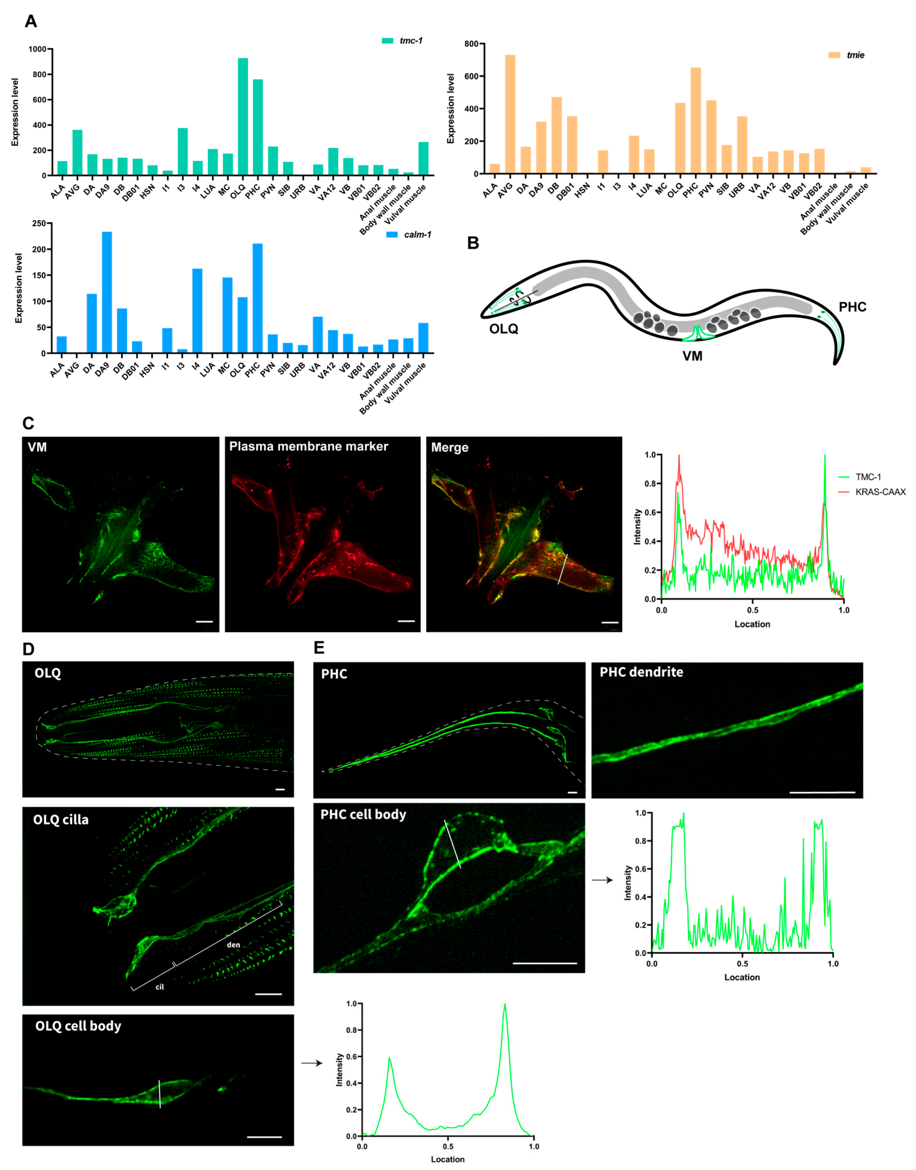
2.2. Intense Plasma Membrane Localization of TMC-1 in C. elegans Neurons and Muscle Cells

To establish a tractable in vivo system for observing the plasma membrane localization of TMC proteins and facilitating genetic manipulation to assess how specific mutations affect their cell surface trafficking, we selected C. elegans as our model organism. This nematode offers a unique combination of genetic simplicity and optical transparency, enabling precise genome editing and live imaging of protein dynamics. The C. elegans TMC mechanotransduction channel complex consists of TMC-1/2, TMIE, and CALM-1, as revealed by cryo-electron microscopy (cryo-EM) of the native TMC-1/2 complex [8,30]. To identify cells expressing all three genes, we analyzed publicly available single-cell transcriptomic data from C. elegans [31]. Our analysis showed that tmc-1, calm-1, and tmie (Y39A1C.1) are expressed in OLQ and PHC sensory neurons, as well as in body wall muscle (BWM) and vulva muscle (VM) cells (Figure 1A).
Using super-resolution microscopy, we visualized C. elegans TMC-1 (ceTMC-1) in CRISPR knock-in worms, where GFP was tagged to the endogenous tmc-1 locus. We found that TMC-1 was prominently localized in the plasma membrane of both muscle cells and neurons (Figure 1B–E). Remarkably, the membrane localization of TMC-1 appears to be more pronounced than that observed for a standard plasma membrane marker, underscoring the robust and specific targeting of TMC-1 to the cell membrane (Figure 1C). Furthermore, TMC-1 was detected in the plasma membrane of specialized cellular structures, including the cilia of OLQ neurons and the dendrites of PHC neurons (Figure 1D,E). These results provide clear evidence of the cell surface localization of TMC-1 in both C. elegans neurons and muscle cells.

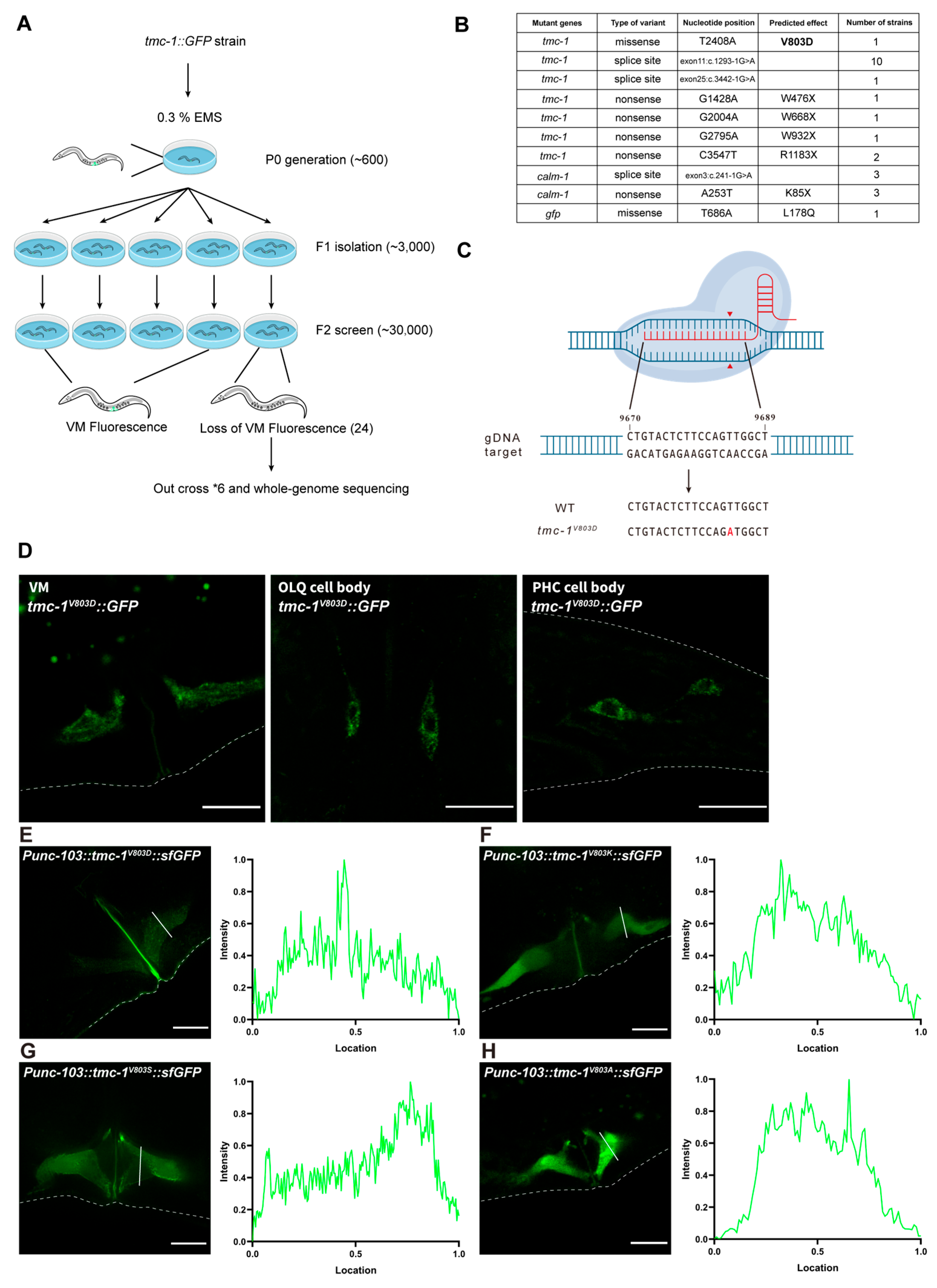
2.3. Forward Genetic Screen in C. elegans Identifies Critical Residues for the Plasma Membrane Localization of TMC-1

To elucidate the molecular mechanisms underlying TMC-1 plasma membrane localization, we performed a forward genetic screen using a tmc-1::GFP knock-in strain, focusing on GFP fluorescence in VM cells. Approximately 500 animals were mutagenized, and ~30,000 F2 progeny were screened, resulting in the identification of 24 mutants with reduced or absent GFP fluorescence (Figure 2A). Whole-genome sequencing revealed that most of these mutants harbored splice-site or nonsense mutations in either tmc-1 or calm-1, leading to loss of TMC-1::GFP signal (Figure 2B). Identifying calm-1 mutations therefore corroborates our earlier finding that CALM-1 is required for both TMC-1 expression and protein stability, and mirrors recent work showing that mammalian CIB2 governs the delivery of TMC1 and TMC2 to stereocilia in cochlear hair cells [8,26].
Among the identified mutations, the missense mutation TMC-1V803D, which substitutes a valine with aspartate in the ninth transmembrane domain (TM9) of TMC-1, was particularly intriguing. We introduced this mutation into the parental tmc-1::GFP strain using CRISPR/Cas9 genome editing and confirmed the phenotype (Figure 2C). The TMC-1V803D protein exhibited reduced expression and was retained in the cytoplasm across all examined cell types, including VM, OLQ, and PHC neurons, suggesting that this mutation disrupts TMC-1 biogenesis or trafficking (Figure 2D).
To determine whether the V803 residue is itself critical or whether the valine-to-aspartate substitution was simply too disruptive, we substituted V803 with several other amino acids and expressed the corresponding tmc-1::GFP variants in VM2 cells. While wild-type TMC-1::GFP localized robustly to the plasma membrane, all V803 mutants showed diminished expression and poor cell surface localization (Figure 2E–H). The V803S substitution caused a modest decrease in membrane localization, while V803A abolished it entirely. Substituting V803 with charged residues (aspartate or lysine) resulted in a complete loss of membrane localization and markedly reduced protein levels.

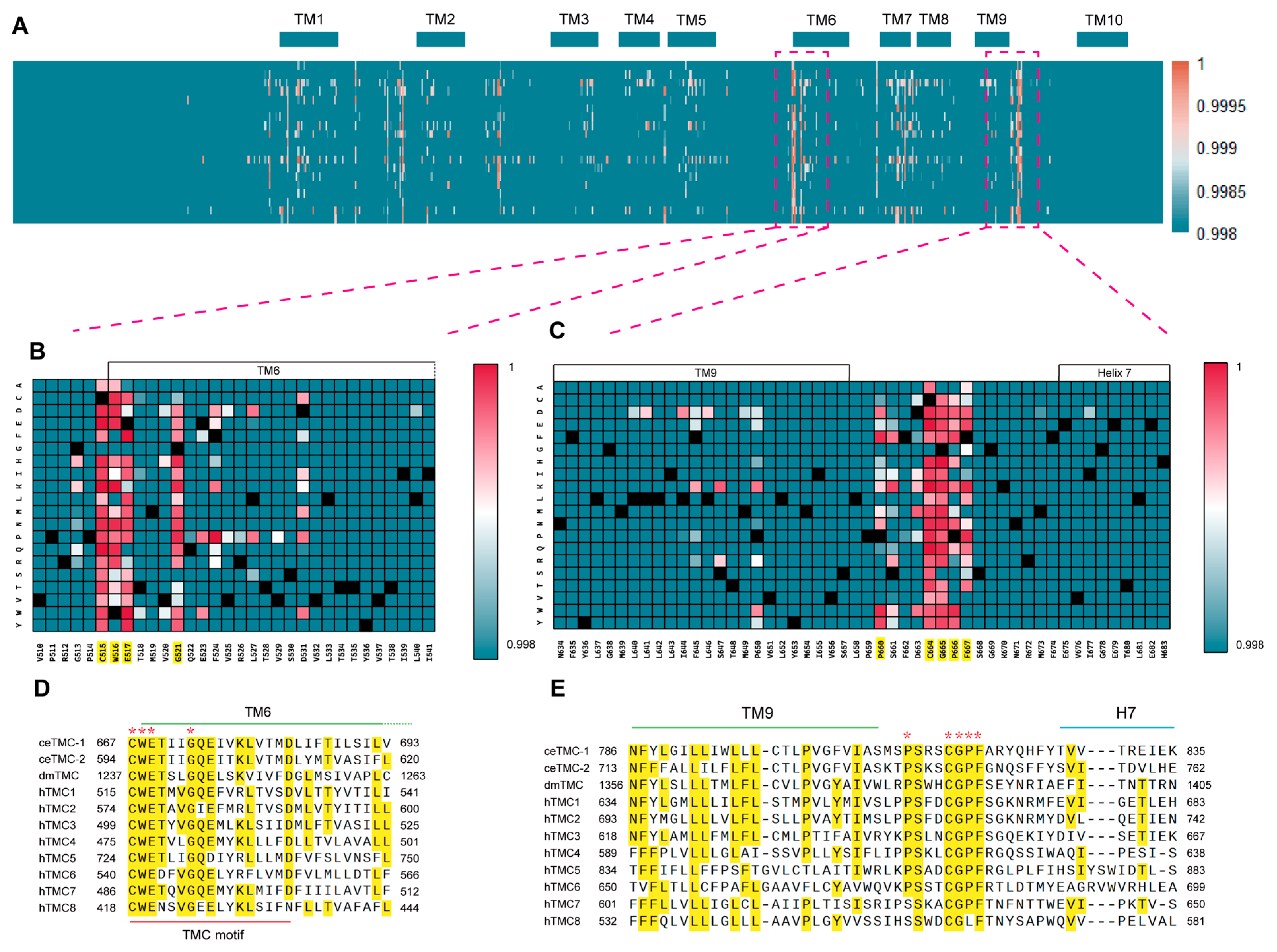
2.4. Human TMC1 TM9–TM10 Extracellular-Loop Mutation Hotspot and Its Conserved Equivalents in C. elegans TMC-1

To better understand why mutations at V803 impair TMC-1 trafficking, we examined the structural context of this residue. V803 is located within TM9, near the extracellular face of the membrane, adjacent to a loop region that connects TM9 and TM10. This loop includes a disordered segment flanked by two α-helices (Helix 7 and Helix 8; Figure 3A). Analysis of human deafness variant databases revealed multiple missense mutations in this extracellular loop of human TMC1. Notably, five of the mutated residues are evolutionarily conserved in C. elegans TMC-1 (Figure 3A,B), underscoring the potential functional relevance of this region.
To assess how these deafness-associated mutations affect ceTMC-1 trafficking, we introduced the corresponding substitutions into ceTMC-1 and evaluated their localization when overexpressed in VM cells. Two of these mutations, P812L (Figure 3D) and C816W (Figure 3E), caused cytoplasmic retention and reduced protein levels. Mutations V828D and V829T partially impaired plasma membrane localization, whereas E834Q retained robust membrane localization (Figure 3F–H). Furthermore, co-expression of TMC-1P812L or TMC-1C816W with ceTMIE led to mislocalization of ceTMIE, compared to co-expression with wild-type TMC-1 (Figure S2A–C). These results indicate that these mutations disrupt the proper trafficking of the TMC-1 mechanotransduction complex and likely contribute to hearing loss by impairing human TMC1 localization at the cell surface.

2.5. AlphaMissense Reveals Two Structurally Connected Functional Hotspots in Human TMC1

Our unbiased whole-genome EMS mutagenesis screen in C. elegans identified a critical residue, V803, in worm TMC-1. This position aligns to V651 in human TMC1 and is strictly conserved across the TMC family. A query of hereditary deafness mutation databases revealed multiple pathogenic variants clustered around V651, suggesting that this region is particularly sensitive to alteration. To determine whether other regions of TMC1 show similar susceptibility, we applied AlphaMissense, a deep learning model built upon AlphaFold2, which predicts the pathogenic impact of every possible missense substitution in the human proteome [32]. By generating pathogenicity scores for all single amino acid variants in human TMC1, we were able to map mutational hotspots and identify domains critical for its structural integrity and function. This analysis revealed two clusters of highly pathogenic residues in human TMC1, each forming a missense mutation hotspot (Figure 4A). Within these regions, AlphaMissense assigned consistently high pathogenicity scores to specific residues—such as C515, W516, E517, and G521 (Figure 4B), as well as C664 and G665 (Figure 4C)—when mutated to most other amino acids. Intriguingly, one of these hotspots, located at the beginning of TM6 (near the extracellular loop between TM5 and TM6), is the TMC signature sequence motif (short for TMC motif), a highly conserved sequence “CWETXVGQEly(K/R)LtvXD” that defines the TMC protein family (Figure 4D). The second hotspot is located between TM9 and Helix 7 and contains residue V651, which aligns with V803 in C. elegans TMC-1 and is conserved across the TMC family (Figure 4E).
Strikingly, analysis of the resolved cryo-EM structure of C. elegans TMC-1 and TMC-2 revealed that these two pathogenic regions are structurally connected by a disulfide bond (C667–C816 in TMC-1; C594–C743 in TMC-2) (Figure S3A,B). Using AlphaFold2 predictions [33], we extended this observation to other TMC homologs, including Drosophila melanogaster TMC and human TMC1–8. These predicted structures also featured disulfide bonds between equivalent cysteine residues (Figure S3C–K). Notably, in the predicted structure of human TMC1 (Figure S3D), C515 forms a disulfide bond with C664, both of which are identified as most likely pathogenic residues in the AlphaMissense database (Figure 4B,C). These findings underscore the evolutionary conservation and likely functional importance of this disulfide bond in TMC proteins.

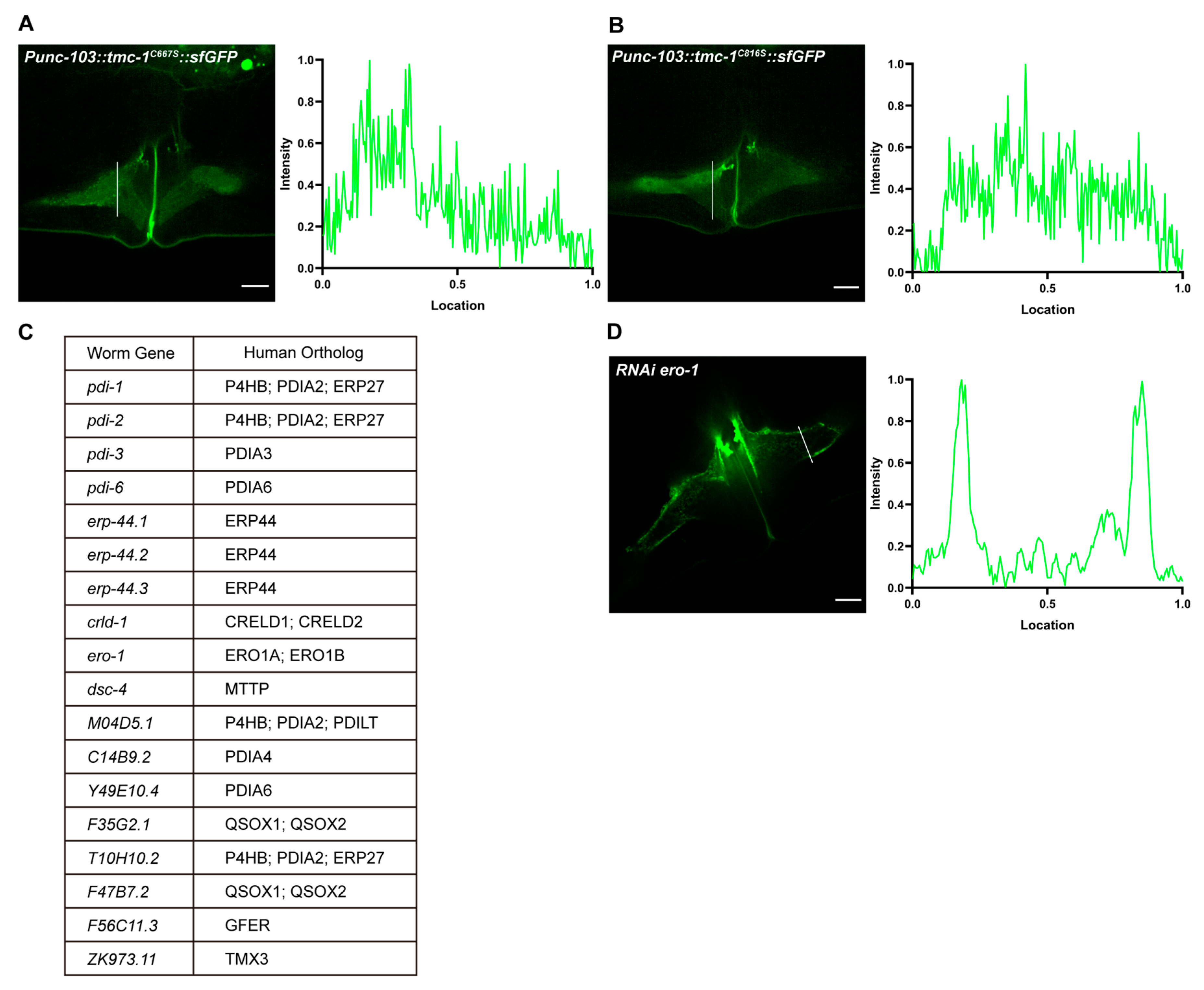
2.6. Disruption of the Disulfide Bond Impairs TMC-1 Trafficking

To investigate the functional significance of the disulfide bond, we introduced serine substitutions at cysteine residues C667 and C816 in C. elegans TMC-1, disrupting the bond without introducing charge. Overexpression of these mutants (TMC-1C667S and TMC-1C816S) in VM cells led to reduced protein levels and prominent cytoplasmic retention (Figure 5A,B), recapitulating the trafficking defects observed in GFP-tagged MmTMC proteins in HEK 293T cells (Figure S1A). Furthermore, co-expression of red fluorescently labeled ceTMIE with TMC-1C667S or TMC-1C816S resulted in mislocalization of TMIE, indicating that disruption of the disulfide bond compromises proper assembly or localization of the TMC-1 mechanotransduction channel complex (Figure S4A,B).
Interestingly, addition of an N-terminal Fyn tag, which directs proteins to the plasma membrane via lipid modification, was sufficient to restore membrane localization of the disulfide-bond-disrupted MmTMC1 mutants (Fyn-MmTMC1C516S and Fyn-MmTMC1C664S) in HEK 293T cells, as shown by their colocalization with a plasma membrane marker (Figure S5A,B). These findings suggest that the Fyn tag can override intrinsic trafficking defects caused by disulfide bond disruption, potentially through enforced membrane anchoring. However, this also highlights a caveat of the Fyn-tagged system: it may mask the impact of mutations that would otherwise impair proper protein trafficking, thereby limiting its utility in dissecting endogenous trafficking mechanisms of TMC proteins.

2.7. TMC-1 Disulfide Bond Formation May Involve Unknown Chaperones

Since disulfide bond formation is catalyzed by protein disulfide isomerases (PDIs) or sulfhydryl oxidases, we hypothesized that a PDI-like chaperone might facilitate disulfide bond formation and promote TMC-1 folding and trafficking. To test this, we performed feeding RNAi in C. elegans targeting 18 genes implicated in disulfide bond formation, including four PDI family members (pdi-1, pdi-2, pdi-3, and pdi-6), ero-1 (which reoxidizes PDI-1), and other genes encoding sulfhydryl oxidases or PDI-like proteins (Figure 5C). Surprisingly, none of these knockdowns recapitulated the trafficking defects observed in TMC-1C667S or TMC-1C816S mutant strains (Figure 5D). This lack of phenotype could be attributed to insufficient knockdown efficiency of the RNAi, or it may indicate that disulfide bond formation in TMC-1 occurs independently of these known enzymes or depends on molecular chaperones that have yet to be identified.

3. Discussion

This study establishes C. elegans as a robust model for investigating TMC protein trafficking and the molecular mechanisms underlying deafness. Through an integrative approach combining structural, functional, and genetic analyses, we provide critical insights into the determinants of TMC protein folding and trafficking. Our findings reveal that two functional hotspots, interconnected by a conserved disulfide bond, are essential for the structural integrity and plasma membrane localization of TMC proteins—key prerequisites for their role in mechanotransduction.

3.1. The TMC Motif and Disulfide Bond: Key Determinants of TMC Protein Folding and Trafficking

The TMC signature sequence motif, a highly conserved feature among TMC family members, emerges as a key structural element in our study. This motif, together with a disulfide bond linking two distinct functional hotspots, plays a pivotal role in protein folding and trafficking. Structural analyses, integrating AlphaMissense and AlphaFold predictions with cryo-EM data, have revealed that the TMC motif and the extracellular region between TM9 and Helix 7 are connected via a disulfide bond (e.g., C515-C664 in human TMC1) that is conserved across TMC orthologs. This covalent link appears to stabilize the tertiary structure of TMC proteins, facilitating correct folding and ensuring efficient trafficking to the plasma membrane. Disruption of either the conserved motif or the disulfide linkage—through missense mutations—results in misfolding, cytoplasmic retention, and, ultimately, loss of function. These results underscore the indispensable role of the disulfide bond as a structural linchpin for TMC protein stability and trafficking.

3.2. Disulfide-Bond-Connected Functional Hotspots and TMC-Associated Diseases

Mutations that affect the stability of disulfide bonds can have profound effects on protein function. In particular, changes at cysteine residues that are essential for disulfide bond formation, such as C667 and C816 in C. elegans TMC-1 (which correspond to C515 and C664 in human TMC1), can significantly reduce protein expression and its ability to localize to the plasma membrane. According to AlphaMissense predictions, this disulfide bond bridges two regions that are functional for pathogenic mutations. Many missense mutations near this disulfide bond in TMC1, including p.P514L, p.C515R, p.C515F, p.P660L, p.C664W, and p.S668R, have been identified and are associated with hearing loss [34,35,36,37,38]. Our research correlates these structural changes with known pathogenic mutations related to TMC1, highlighting the critical role of the C515-C664 disulfide bond in TMC1 function. Therefore, mutations that weaken the disulfide bond or its neighboring structural elements can impact the trafficking of the protein, potentially disrupting the mechanotransduction machinery in hair cells. This discovery establishes a molecular link between structural defects and the clinical presentation of deafness, enhancing our understanding of the underlying causes of hearing loss.
Building on these findings, we have also investigated the ClinVar database for missense mutations in human TMC1-8. Among these, we identified a TMC6 mutant (C540R) and a TMC8 mutant (C418W), both of which are likely to disrupt potential disulfide bonds. These mutations are associated with epidermodysplasia verruciformis, a dermatosis linked to TMC6/8 [39], and are currently classified as “Uncertain significance” due to the absence of functional validation. Our findings may provide valuable pathogenic evidence for these specific mutations and others that disrupt disulfide bonds in TMC family members, enhancing our comprehension of the molecular mechanisms underlying TMC-associated diseases.

3.3. ER Redox Regulation of TMC Protein Folding

The ER lumen maintains an oxidizing environment that is crucial for the formation of de novo disulfide bonds in nascent proteins. This process is facilitated by protein disulfide isomerases (PDIs), which are re-oxidized by ER oxidases such as Ero1α/β, peroxiredoxin IV, and vitamin K epoxide reductase (VKOR) [40,41,42,43]. These enzymatic pathways sustain a high GSSG:GSH ratio, which promotes the formation of native disulfide bonds and allows PDIs to resolve mispaired cysteines, ensuring that proteins acquire the correct tertiary structures [43].
Many multi-pass transmembrane proteins rely on disulfide bonds between conserved cysteines within luminal or extracellular loops to stabilize their folding and function. Disulfide bond formation often occurs co-translationally as luminal loops emerge into the ER, significantly reducing the entropy of the folding intermediates [44]. For instance, class A GPCRs require a conserved disulfide between TM3 and the second extracellular loop to exit the ER [45,46]. Disruption of this bond in rhodopsin leads to ER retention and retinitis pigmentosa [47]. Similarly, the inositol 1,4,5-trisphosphate receptor (IP3R) contains four luminal cysteine residues that form two disulfide bonds. One disulfide bond is essential for the formation of functional IP3R tetramers, while the other regulates channel activity. Oxidation of the regulatory disulfide bond by ERp46 activates IP3R, whereas its reduction by ERdj5 leads to channel inactivation [48,49].
ER quality control closely couples disulfide bond formation to folding and degradation pathways [43,44]. Chaperones such as BiP bind nascent loops to prevent premature or improper disulfide formation, while lectin chaperones (calnexin/calreticulin) coordinate glycan and disulfide maturation through ERp57 [50,51,52,53,54]. Reductive isomerases like ERdj5 and TMX4 further correct mispaired bonds, safeguarding proper folding and channeling irreparably misfolded proteins toward ER-associated degradation (ERAD) [55,56]. These processes are dynamically regulated by luminal calcium and the unfolded protein response to maintain ER redox homeostasis under stress.
By analogy to these systems, TMC proteins, which feature multiple luminal loops, likely depend on similar oxidative folding machinery. Our data show that disrupting a conserved disulfide bond (C667–C816) in C. elegans TMC-1 abolishes surface trafficking, highlighting the structural importance of this bond. However, targeting canonical PDIs and ER oxidases via RNAi did not fully replicate the trafficking defects, suggesting that additional, possibly TMC-specific, redox mechanisms or chaperones may be involved. Future studies should investigate ER redox dynamics during TMC biogenesis and identify key factors supporting proper folding, which may ultimately inform therapeutic strategies for trafficking-defective TMC mutants underlying hereditary deafness.

4. Materials and Methods

4.1. Experimental Worm Strains

The C. elegans strains used in this work are listed as Table 1.

4.2. Plasmids Construction

Plasmids for expression in C. elegans and cell lines were constructed using either the Multisite Gateway Three-Fragment cloning system (12537-023, Invitrogen, Waltham, MA, USA) or pEASY®-Basic Seamless Cloning and Assembly Kit (TransGen Biotech, Beijing, China). All genes amplified by PCR were sequenced to confirm accuracy.

4.3. Immunofluorescence and Super-Resolution Imaging

HEK293T cells were purchased from Cell Bank/Stem Cell Bank, Chinese Academy of Sciences (Shanghai, China). HEK 293T cells were cultured at 37 °C in a humidified incubator with 5% CO2 in Dulbecco’s Modified Eagle Medium (DMEM) supplemented with 10% fetal bovine serum (FBS) and penicillin–streptomycin. For imaging experiments, cells were seeded in 35 mm glass-bottom dishes (NEST) and transfected with plasmid DNA using polyethylenimine (PEI; Beyotime, Shanghai, China), following the manufacturer’s instructions. Live-cell staining was performed using Hoechst (Beyotime) for nuclear labeling and CellMask™ Plasma Membrane Stain (C10045, Invitrogen, USA) for membrane visualization, according to the manufacturers’ protocols. Forty-eight hours post-transfection, images were acquired using a super-resolution microscope (Olympus SpinFV-COMB, Tokyo, Japan) equipped with 60× or 100× oil-immersion objectives. Laser excitation settings were as follows: 405 nm for Hoechst, 488 nm for GFP, 594 nm for mCherry, and 647 nm for far-red signals. Identical imaging settings, including channel configuration, laser intensity, and exposure time, were maintained across experimental and control groups.
For worm imaging, live C. elegans were anesthetized with 25 mM muscimol and mounted between 3% agarose pads and microscope coverslips prior to imaging.

4.4. EMS Mutagenesis and Genetic Mapping

Ethyl methanesulfonate (EMS) mutagenesis was performed following established protocols using the tmc-1::GFP (TYQ28) C. elegans strain [57,58]. Synchronized L4-stage hermaphrodites were washed off NGM plates into 15 mL tubes with M9 buffer. After centrifugation at 700× g for 1 min, the supernatant was carefully removed, and the worms were washed four times with M9 buffer to eliminate residual bacteria. The worms were then resuspended in 2 mL of M9 buffer.
In a fume hood, a 0.1 M EMS stock solution was freshly prepared by adding 20 μL of EMS (Sigma-Aldrich, Waltham, MA, USA) to 2 mL of M9 buffer in a 15 mL tube, mixing gently until fully dissolved. An equal volume (2 mL) of the worm suspension was combined with the EMS solution to achieve a final concentration of 50 mM EMS. The mixture was sealed with Parafilm and incubated horizontally at 20 °C for 4 h with gentle rotation. Post mutagenesis, worms were washed five times with M9 buffer to remove residual EMS.
Mutants exhibiting altered fluorescence in VM cells were identified through screening of clonal F2 progeny, resulting in the isolation of 24 mutants. These mutants were backcrossed six times with the parental tmc-1::GFP (TYQ28) strain before whole-genome sequencing, which was performed by Novogene Biotechnologies, Inc. (Tianjin, China).

4.5. Generation of CRISPR/Cas9-Mediated V803D Mutant Strains

To generate the V803D point mutation, CRISPR/Cas9 genome editing was employed [59]. Lyophilized single-guide RNA (sgRNA) was resuspended in nuclease-free TE buffer to a concentration of 50 μM, and single-stranded oligodeoxynucleotide (ssODN) donors were resuspended in nuclease-free water to 100 μM. The injection mixture was prepared by sequentially combining the following components: 0.1 μL of 3M KCl (final concentration: 300 mM), 2.5 μL of tracrRNA (100 ng/μL), 1.4 μL of crRNA (56 ng/μL), 4.5 μL of asymmetric-hybrid double-stranded DNA (dsDNA) donors (200 ng/μL), 1 μL of pRF4 plasmid (50 ng/μL), and 0.5 μL of Cas9 protein (0.25 μg/μL). All components were centrifuged at maximum speed for 3 min prior to assembly. To form ribonucleoprotein (RNP) complexes, tracrRNA, crRNA, and Cas9 were pre-incubated at 37 °C for 10 min before adding DNA components. Premature addition of dsDNA donors was avoided, as it can reduce homology-directed repair (HDR) efficiency by over 60%. After RNP formation, 1.1 μL of ssODN donors (1 μg/μL) or 4 μg total of dsDNA donor cocktail were added. The final volume was adjusted to 10 μL with nuclease-free water. For optimal needle function, the mixture was centrifuged at 14,000 rpm for 2 min, and 8 μL of the clarified supernatant was transferred to a fresh tube. Injection needles were loaded within 15 min of preparation to maintain complex stability.

4.6. RNA Interference (RNAi) Feeding

RNAi feeding experiments were conducted using the C. elegans RNAi feeding library developed by Julie Ahringer’s group [60]. NGM agar plates supplemented with 100 μg/mL carbenicillin and 1 mM IPTG were prepared and stored at 4 °C for up to three weeks. Bacterial cultures were grown overnight at 37 °C in LB medium containing 50 μg/mL ampicillin. Approximately 400 μL of each culture was seeded onto the prepared NGM agar plates. All bacterial clones were verified by sequencing to ensure correct alignment of RNAi plasmids with their corresponding gene sequences.
After two days of bacterial growth, day 1 adult worms were bleached onto the RNAi plates. The phenotypes of the F1 progeny were assessed to determine the effects of gene knockdown. A control RNAi plate known to produce a distinct phenotype was included in each batch of experiments to confirm effective IPTG induction.

4.7. Protein Structure Accession Numbers

Protein structure coordinates were obtained from the Protein Data Bank (PDB) and the AlphaFold Database under the following accession numbers:
Resolved structures: C. elegans TMC-1: 7USW; C. elegans TMC-2: 8TKP.
Predicted structures: Drosophila melanogaster TMC: A0A0U1QT59; Human TMC1: Q8TDI8; Human TMC2: Q8TDI7; Human TMC3: Q7Z5M5; Human TMC4: Q7Z404; Human TMC5: Q6UXY8; Human TMC6: Q7Z403; Human TMC8: Q7Z402; Human TMC1: Q8IU68.

Supplementary Materials

The following supporting information can be downloaded at: https://www.mdpi.com/article/10.3390/ijms26136356/s1.

Author Contributions

Y.-Q.T., Y.S. and Q.Q. designed and supervised the study. D.S., J.T., and X.F. performed the experiments and analyzed the data. Y.-Q.T. and D.S. wrote the manuscript. All authors have read and agreed to the published version of the manuscript.

Funding

This work was supported by grants from the STI2030-Major Projects (2021ZD0203303 to Y.-Q.T.), the National Natural Science Foundation of China (32170694 to Y.-Q.T.), the National Key R&D Program of China (2024YFC2511100/2024YFC2511104 to Y.-Q.T.), the Program for Professor of Special Appointment (Eastern Scholar) at Shanghai Institutions of Higher Learning (Y.-Q.T.), Shanghai Municipal Science and Technology Major Project (2018SHZDZX01), ZJ Lab, and Shanghai Center for Brain Science and Brain-Inspired Technology (Y.-Q.T.).

Institutional Review Board Statement

Not applicable.

Informed Consent Statement

Not applicable.

Data Availability Statement

The data presented in this study are available upon request from the corresponding author.

Conflicts of Interest

The authors declare no conflicts of interest.

References

  1. Hudspeth, A.J.; Corey, D.P. Sensitivity, polarity, and conductance change in the response of vertebrate hair cells to controlled mechanical stimuli. Proc. Natl. Acad. Sci. USA 1977, 74, 2407–2411. [Google Scholar] [PubMed]
  2. Corey, D.P.; Hudspeth, A.J. Response latency of vertebrate hair cells. Biophys. J. 1979, 26, 499–506. [Google Scholar] [PubMed]
  3. Corey, D.P.; Hudspeth, A.J. Ionic basis of the receptor potential in a vertebrate hair cell. Nature 1979, 281, 675–677. [Google Scholar] [PubMed]
  4. Kawashima, Y.; Geleoc, G.S.G.; Kurima, K.; Labay, V.; Lelli, A.; Asai, Y.; Makishima, T.; Wu, D.K.; Della Santina, C.C.; Holt, J.R.; et al. Mechanotransduction in mouse inner ear hair cells requires transmembrane channel-like genes. J. Clin. Investig. 2011, 121, 4796–4809. [Google Scholar]
  5. Pan, B.; Geleoc, G.S.; Asai, Y.; Horwitz, G.C.; Kurima, K.; Ishikawa, K.; Kawashima, Y.; Griffith, A.J.; Holt, J.R. TMC1 and TMC2 are components of the mechanotransduction channel in hair cells of the mammalian inner ear. Neuron 2013, 79, 504–515. [Google Scholar]
  6. Pan, B.; Akyuz, N.; Liu, X.P.; Asai, Y.; Nist-Lund, C.; Kurima, K.; Derfler, B.H.; Gyorgy, B.; Limapichat, W.; Walujkar, S.; et al. TMC1 Forms the Pore of Mechanosensory Transduction Channels in Vertebrate Inner Ear Hair Cells. Neuron 2018, 99, 736–753 e6. [Google Scholar]
  7. Jia, Y.; Zhao, Y.; Kusakizako, T.; Wang, Y.; Pan, C.; Zhang, Y.; Nureki, O.; Hattori, M.; Yan, Z. TMC1 and TMC2 Proteins Are Pore-Forming Subunits of Mechanosensitive Ion Channels. Neuron 2020, 105, 310–321 e3. [Google Scholar]
  8. Jeong, H.; Clark, S.; Goehring, A.; Dehghani-Ghahnaviyeh, S.; Rasouli, A.; Tajkhorshid, E.; Gouaux, E. Structures of the TMC-1 complex illuminate mechanosensory transduction. Nature 2022, 610, 796–803. [Google Scholar]
  9. Kurima, K.; Peters, L.M.; Yang, Y.; Riazuddin, S.; Ahmed, Z.M.; Naz, S.; Arnaud, D.; Drury, S.; Mo, J.; Makishima, T.; et al. Dominant and recessive deafness caused by mutations of a novel gene, TMC1, required for cochlear hair-cell function. Nat. Genet. 2002, 30, 277–284. [Google Scholar]
  10. Vreugde, S.; Erven, A.; Kros, C.J.; Marcotti, W.; Fuchs, H.; Kurima, K.; Wilcox, E.R.; Friedman, T.B.; Griffith, A.J.; Balling, R.; et al. Beethoven, a mouse model for dominant, progressive hearing loss DFNA36. Nat. Genet. 2002, 30, 257–258. [Google Scholar]
  11. Boucher, S.; Tai, F.W.J.; Delmaghani, S.; Lelli, A.; Singh-Estivalet, A.; Dupont, T.; Niasme-Grare, M.; Michel, V.; Wolff, N.; Bahloul, A.; et al. Ultrarare heterozygous pathogenic variants of genes causing dominant forms of early-onset deafness underlie severe presbycusis. Proc. Natl. Acad. Sci. USA 2020, 117, 31278–31289. [Google Scholar] [PubMed]
  12. Gao, X.; Tao, Y.; Lamas, V.; Huang, M.; Yeh, W.H.; Pan, B.; Hu, Y.J.; Hu, J.H.; Thompson, D.B.; Shu, Y.; et al. Treatment of autosomal dominant hearing loss by in vivo delivery of genome editing agents. Nature 2018, 553, 217–221. [Google Scholar] [PubMed]
  13. Nist-Lund, C.A.; Pan, B.; Patterson, A.; Asai, Y.; Chen, T.; Zhou, W.; Zhu, H.; Romero, S.; Resnik, J.; Polley, D.B.; et al. Improved TMC1 gene therapy restores hearing and balance in mice with genetic inner ear disorders. Nat. Commun. 2019, 10, 236. [Google Scholar] [PubMed]
  14. Yeh, W.H.; Shubina-Oleinik, O.; Levy, J.M.; Pan, B.; Newby, G.A.; Wornow, M.; Burt, R.; Chen, J.C.; Holt, J.R.; Liu, D.R. In vivo base editing restores sensory transduction and transiently improves auditory function in a mouse model of recessive deafness. Sci. Transl. Med. 2020, 12, eaay9101. [Google Scholar]
  15. Wu, J.; Solanes, P.; Nist-Lund, C.; Spataro, S.; Shubina-Oleinik, O.; Marcovich, I.; Goldberg, H.; Schneider, B.L.; Holt, J.R. Single and Dual Vector Gene Therapy with AAV9-PHP.B Rescues Hearing in Tmc1 Mutant Mice. Mol. Ther. 2021, 29, 973–988. [Google Scholar]
  16. Zheng, Z.; Li, G.; Cui, C.; Wang, F.; Wang, X.; Xu, Z.; Guo, H.; Chen, Y.; Tang, H.; Wang, D.; et al. Preventing autosomal-dominant hearing loss in Bth mice with CRISPR/CasRx-based RNA editing. Signal Transduct. Target. Ther. 2022, 7, 79. [Google Scholar]
  17. Yu, X.; Zhao, Q.; Li, X.; Chen, Y.; Tian, Y.; Liu, S.; Xiong, W.; Huang, P. Deafness mutation D572N of TMC1 destabilizes TMC1 expression by disrupting LHFPL5 binding. Proc. Natl. Acad. Sci. USA 2020, 117, 29894–29903. [Google Scholar]
  18. Beurg, M.; Schimmenti, L.A.; Koleilat, A.; Amr, S.S.; Oza, A.; Barlow, A.J.; Ballesteros, A.; Fettiplace, R. New Tmc1 Deafness Mutations Impact Mechanotransduction in Auditory Hair Cells. J. Neurosci. 2021, 41, 4378–4391. [Google Scholar]
  19. Akyuz, N.; Karavitaki, K.D.; Pan, B.; Tamvakologos, P.I.; Brock, K.P.; Li, Y.; Marks, D.S.; Corey, D.P. Mechanical gating of the auditory transduction channel TMC1 involves the fourth and sixth transmembrane helices. Sci. Adv. 2022, 8, eabo1126. [Google Scholar]
  20. Ballesteros, A.; Swartz, K.J. Regulation of membrane homeostasis by TMC1 mechanoelectrical transduction channels is essential for hearing. Sci. Adv. 2022, 8, eabm5550. [Google Scholar]
  21. Fettiplace, R.; Furness, D.N.; Beurg, M. The conductance and organization of the TMC1-containing mechanotransducer channel complex in auditory hair cells. Proc. Natl. Acad. Sci. USA 2022, 119, e2210849119. [Google Scholar]
  22. Beurg, M.; Schwalbach, E.T.; Fettiplace, R. LHFPL5 is a key element in force transmission from the tip link to the hair cell mechanotransducer channel. Proc. Natl. Acad. Sci. USA 2024, 121, e2318270121. [Google Scholar]
  23. Fu, S.; Pan, X.; Lu, M.; Dong, J.; Yan, Z. Human TMC1 and TMC2 are mechanically gated ion channels. Neuron 2025, 113, 411–425 e4. [Google Scholar] [PubMed]
  24. Yang, Z.; Zhou, S.H.; Zhang, Q.Y.; Song, Z.C.; Liu, W.W.; Sun, Y.; Wang, M.W.; Fu, X.L.; Zhu, K.K.; Guan, Y.; et al. A force-sensitive adhesion GPCR is required for equilibrioception. Cell Res. 2025, 35, 243–264. [Google Scholar]
  25. Chen, Y.; Li, Y.; Liu, Y.; Sun, J.; Feng, W.; Chen, Y.; Tian, Y.; Lei, T.; Huang, P. Ectopic mouse TMC1 and TMC2 alone form mechanosensitive channels that are potently modulated by TMIE. Proc. Natl. Acad. Sci. USA 2025, 122, e2403141122. [Google Scholar]
  26. Tang, Y.Q.; Lee, S.A.; Rahman, M.; Vanapalli, S.A.; Lu, H.; Schafer, W.R. Ankyrin Is An Intracellular Tether for TMC Mechanotransduction Channels. Neuron 2020, 107, 112–125 e10. [Google Scholar]
  27. Kurima, K.; Ebrahim, S.; Pan, B.; Sedlacek, M.; Sengupta, P.; Millis, B.A.; Cui, R.; Nakanishi, H.; Fujikawa, T.; Kawashima, Y.; et al. TMC1 and TMC2 Localize at the Site of Mechanotransduction in Mammalian Inner Ear Hair Cell Stereocilia. Cell Rep. 2015, 12, 1606–1617. [Google Scholar]
  28. Mahendrasingam, S.; Furness, D.N. Ultrastructural localization of the likely mechanoelectrical transduction channel protein, transmembrane-like channel 1 (TMC1) during development of cochlear hair cells. Sci. Rep. 2019, 9, 1274. [Google Scholar]
  29. Wang, P.; Miller, K.K.; He, E.; Dhawan, S.S.; Cunningham, C.L.; Grillet, N. LOXHD1 is indispensable for maintaining TMC1 auditory mechanosensitive channels at the site of force transmission. Nat. Commun. 2024, 15, 7865. [Google Scholar]
  30. Clark, S.; Jeong, H.; Posert, R.; Goehring, A.; Gouaux, E. The structure of the Caenorhabditis elegans TMC-2 complex suggests roles of lipid-mediated subunit contacts in mechanosensory transduction. Proc. Natl. Acad. Sci. USA 2024, 121, e2314096121. [Google Scholar]
  31. Taylor, S.R.; Santpere, G.; Weinreb, A.; Barrett, A.; Reilly, M.B.; Xu, C.; Varol, E.; Oikonomou, P.; Glenwinkel, L.; McWhirter, R.; et al. Molecular topography of an entire nervous system. Cell 2021, 184, 4329–4347.e23. [Google Scholar] [PubMed]
  32. Cheng, J.; Novati, G.; Pan, J.; Bycroft, C.; Zemgulyte, A.; Applebaum, T.; Pritzel, A.; Wong, L.H.; Zielinski, M.; Sargeant, T.; et al. Accurate proteome-wide missense variant effect prediction with AlphaMissense. Science 2023, 381, eadg7492. [Google Scholar]
  33. Jumper, J.; Evans, R.; Pritzel, A.; Green, T.; Figurnov, M.; Ronneberger, O.; Tunyasuvunakool, K.; Bates, R.; Zidek, A.; Potapenko, A.; et al. Highly accurate protein structure prediction with AlphaFold. Nature 2021, 596, 583–589. [Google Scholar] [PubMed]
  34. Santos, R.L.; Wajid, M.; Khan, M.N.; McArthur, N.; Pham, T.L.; Bhatti, A.; Lee, K.; Irshad, S.; Mir, A.; Yan, K.; et al. Novel sequence variants in the TMC1 gene in Pakistani families with autosomal recessive hearing impairment. Hum. Mutat. 2005, 26, 396. [Google Scholar]
  35. Kitajiri, S.I.; McNamara, R.; Makishima, T.; Husnain, T.; Zafar, A.U.; Kittles, R.A.; Ahmed, Z.M.; Friedman, T.B.; Riazuddin, S.; Griffith, A.J. Identities, frequencies and origins of TMC1 mutations causing DFNB7/B11 deafness in Pakistan. Clin. Genet. 2007, 72, 546–550. [Google Scholar]
  36. Sadeghian, L.; Tabatabaiefar, M.A.; Fattahi, N.; Pourreza, M.R.; Tahmasebi, P.; Alavi, Z.; Hashemzadeh Chaleshtori, M. Next-generation sequencing reveals a novel pathological mutation in the TMC1 gene causing autosomal recessive non-syndromic hearing loss in an Iranian kindred. Int. J. Pediatr. Otorhinolaryngol. 2019, 124, 99–105. [Google Scholar]
  37. Zardadi, S.; Razmara, E.; Asgaritarghi, G.; Jafarinia, E.; Bitarafan, F.; Rayat, S.; Almadani, N.; Morovvati, S.; Garshasbi, M. Novel homozygous variants in the TMC1 and CDH23 genes cause autosomal recessive nonsyndromic hearing loss. Mol. Genet. Genom. Med. 2020, 8, e1550. [Google Scholar]
  38. Tollefson, M.R.; Gogal, R.A.; Weaver, A.M.; Schaefer, A.M.; Marini, R.J.; Azaiez, H.; Kolbe, D.L.; Wang, D.; Weaver, A.E.; Casavant, T.L.; et al. Assessing variants of uncertain significance implicated in hearing loss using a comprehensive deafness proteome. Hum. Genet. 2023, 142, 819–834. [Google Scholar]
  39. Ramoz, N.; Rueda, L.A.; Bouadjar, B.; Montoya, L.S.; Orth, G.; Favre, M. Mutations in two adjacent novel genes are associated with epidermodysplasia verruciformis. Nat. Genet. 2002, 32, 579–581. [Google Scholar]
  40. Tu, B.P.; Weissman, J.S. Oxidative protein folding in eukaryotes: Mechanisms and consequences. J. Cell Biol. 2004, 164, 341–346. [Google Scholar]
  41. Rutkevich, L.A.; Williams, D.B. Vitamin K epoxide reductase contributes to protein disulfide formation and redox homeostasis within the endoplasmic reticulum. Mol. Biol. Cell 2012, 23, 2017–2027. [Google Scholar] [PubMed]
  42. Ushioda, R.; Nagata, K. Redox-Mediated Regulatory Mechanisms of Endoplasmic Reticulum Homeostasis. Cold Spring Harb. Perspect. Biol. 2019, 11, a033910. [Google Scholar]
  43. Wang, L.; Wang, C.C. Oxidative protein folding fidelity and redoxtasis in the endoplasmic reticulum. Trends Biochem. Sci. 2023, 48, 40–52. [Google Scholar] [PubMed]
  44. Feige, M.J.; Hendershot, L.M. Disulfide bonds in ER protein folding and homeostasis. Curr. Opin. Cell Biol. 2011, 23, 167–175. [Google Scholar]
  45. Karnik, S.S.; Gogonea, C.; Patil, S.; Saad, Y.; Takezako, T. Activation of G-protein-coupled receptors: A common molecular mechanism. Trends Endocrinol. Metab. 2003, 14, 431–437. [Google Scholar]
  46. Tao, Y.X.; Conn, P.M. Chaperoning G protein-coupled receptors: From cell biology to therapeutics. Endocr. Rev. 2014, 35, 602–647. [Google Scholar]
  47. Hwa, J.; Reeves, P.J.; Klein-Seetharaman, J.; Davidson, F.; Khorana, H.G. Structure and function in rhodopsin: Further elucidation of the role of the intradiscal cysteines, Cys-110, -185, and -187, in rhodopsin folding and function. Proc. Natl. Acad. Sci. USA 1999, 96, 1932–1935. [Google Scholar]
  48. Joseph, S.K.; Young, M.P.; Alzayady, K.; Yule, D.I.; Ali, M.; Booth, D.M.; Hajnoczky, G. Redox regulation of type-I inositol trisphosphate receptors in intact mammalian cells. J. Biol. Chem. 2018, 293, 17464–17476. [Google Scholar]
  49. Fujii, S.; Ushioda, R.; Nagata, K. Redox states in the endoplasmic reticulum directly regulate the activity of calcium channel, inositol 1,4,5-trisphosphate receptors. Proc. Natl. Acad. Sci. USA 2023, 120, e2216857120. [Google Scholar]
  50. Lee, Y.K.; Brewer, J.W.; Hellman, R.; Hendershot, L.M. BiP and immunoglobulin light chain cooperate to control the folding of heavy chain and ensure the fidelity of immunoglobulin assembly. Mol. Biol. Cell 1999, 10, 2209–2219. [Google Scholar]
  51. Meunier, L.; Usherwood, Y.K.; Chung, K.T.; Hendershot, L.M. A subset of chaperones and folding enzymes form multiprotein complexes in endoplasmic reticulum to bind nascent proteins. Mol. Biol. Cell 2002, 13, 4456–4469. [Google Scholar] [PubMed]
  52. Feige, M.J.; Groscurth, S.; Marcinowski, M.; Shimizu, Y.; Kessler, H.; Hendershot, L.M.; Buchner, J. An unfolded CH1 domain controls the assembly and secretion of IgG antibodies. Mol. Cell 2009, 34, 569–579. [Google Scholar] [PubMed]
  53. Oliver, J.D.; Roderick, H.L.; Llewellyn, D.H.; High, S. ERp57 functions as a subunit of specific complexes formed with the ER lectins calreticulin and calnexin. Mol. Biol. Cell 1999, 10, 2573–2582. [Google Scholar] [PubMed]
  54. Mezghrani, A.; Fassio, A.; Benham, A.; Simmen, T.; Braakman, I.; Sitia, R. Manipulation of oxidative protein folding and PDI redox state in mammalian cells. EMBO J. 2001, 20, 6288–6296. [Google Scholar]
  55. Sugiura, Y.; Araki, K.; Iemura, S.; Natsume, T.; Hoseki, J.; Nagata, K. Novel thioredoxin-related transmembrane protein TMX4 has reductase activity. J. Biol. Chem. 2010, 285, 7135–7142. [Google Scholar]
  56. Ushioda, R.; Hoseki, J.; Araki, K.; Jansen, G.; Thomas, D.Y.; Nagata, K. ERdj5 is required as a disulfide reductase for degradation of misfolded proteins in the ER. Science 2008, 321, 569–572. [Google Scholar]
  57. Brenner, S. The genetics of Caenorhabditis elegans. Genetics 1974, 77, 71–94. [Google Scholar]
  58. Zuryn, S.; Le Gras, S.; Jamet, K.; Jarriault, S. A strategy for direct mapping and identification of mutations by whole-genome sequencing. Genetics 2010, 186, 427–430. [Google Scholar]
  59. Dokshin, G.A.; Ghanta, K.S.; Piscopo, K.M.; Mello, C.C. Robust Genome Editing with Short Single-Stranded and Long, Partial-ly Single-Stranded DNA Donors in Caenorhabditis elegans. Genetics 2018, 210, 781–787. [Google Scholar]
  60. Kamath, R.S.; Ahringer, J. Genome-wide RNAi screening in Caenorhabditis elegans. Methods 2003, 30, 313–321. [Google Scholar]
Figure 1. Intense plasma membrane localization of TMC-1 in C. elegans neurons and muscle cells. (A) Expression profiles of tmc-1, calm-1, and tmie (Y39A1C.1) in neuronal and non-neuronal cells of C. elegans. Data were obtained from the CeNGEN scRNA-seq dataset. (B) Diagram of anatomy location of OLQ, PHC, and VM in C. elegans. (C) Representative super-resolution fluorescence images of endogenous TMC-1 in C. elegans VM cells co-expressing the red fluorescently labeled plasma membrane marker, mCherry::KRAS-CAAX motif. (D) Representative super-resolution fluorescence images of endogenous TMC-1 in cell bodies and cilia of OLQ neurons. (E) Representative super-resolution fluorescence images of endogenous TMC-1 in cell bodies and dendrites of PHC neurons. Fluorescence intensity histograms for the corresponding images are shown on the right panels. Scale bars, 5 μm.
Ijms 26 06356 g001
Figure 2. Forward genetic screen in C. elegans identifies critical residues for the plasma membrane localization of TMC-1. (A) Schematic diagram of the EMS mutagenesis screening for tmc-1::GFP strain. (B) EMS mutation mapping results showing mutant genes, types of variants, nucleotide positions, predicted effects, and the number of each mutant for 24 mutant strains losing VM fluorescence in (A). (C) Schematic diagram of CRISPR/Cas9-mediated V803D mutagenesis in the tmc-1::GFP strain. (D) Representative fluorescence images of endogenous ceTMC-1V803D in VM cells, OLQ neurons, and PHC neurons. (EH) Representative fluorescence images for VM cells overexpressing ceTMC-1V803D (E), ceTMC-1V803K (F), ceTMC-1V803S (G), and ceTMC-1V803A (H) mutants. Fluorescence intensity histograms for the corresponding images are shown on the right panels. Scale bars, 10 μm.
Ijms 26 06356 g002
Figure 3. Deafness mutation hotspot in the extracellular loop between TM9 and TM10 of TMC-1. (A) Schematic representation of the ten-transmembrane helix topology of C. elegans TMC-1 (ceTMC-1) and the structure of TM9 and the adjacent extracellular loop. Residues that are conserved between ceTMC-1 and human TMC1 (hTMC1) are indicated in green. (B) Sequence alignment of residues 803-834 of ceTMC-1 with ceTMC-2 and hTMC1. Conserved residues identified by MUSCLE alignment are highlighted in yellow. Selected conserved residues are indicated by asterisks and listed below. (CH) Representative fluorescence images for VM cells overexpressing WT ceTMC-1 (C), ceTMC-1P812L (D), ceTMC-1C816W (E), ceTMC-1V828D (F), ceTMC-1V829T (G), and ceTMC-1E834Q (H) mutants. Fluorescence intensity histograms for the corresponding images are shown on the right panels. Scale bars, 10 μm.
Ijms 26 06356 g003
Figure 4. AlphaMissense predictions of two functional hotspots in human TMC1. (A) Heatmap of AlphaMissense scores for hTMC1. This heatmap displays the AlphaMissense pathogenicity scores for all possible amino acid substitutions at each position in the full-length hTMC1 protein sequence. The Y-axis represents the 19 alternative amino acid substitutions (excluding the wild-type), while the X-axis represents the full-length protein sequence. The color scale indicates the AlphaMissense-predicted pathogenicity scores, with darker colors representing more pathogenic substitutions (1: most pathogenic). (B,C) Enlarged heatmaps of the two hotspot regions with the highest predicted pathogenicity in hTMC1. Panel B focuses on residues 510–541, while panel C focuses on residues 634–683. The color scale matches that of panel A, with black bars denoting the wild-type residues. (D,E) Sequence alignment of conserved functional hotspots across species, including C. elegans TMC-1/2, Drosophila TMC, and human TMC1-8. Yellow highlights in the alignment indicate conserved residues, as determined by MUSCLE alignment. Asterisks in D,E indicate the hotspot residues, which are highlighted in yellow in B,C.
Ijms 26 06356 g004
Figure 5. Mutations disrupting the disulfide bond impair TMC-1 trafficking. (A,B) Representative fluorescence images for VM cells overexpressing ceTMC-1C667S (A) and ceTMC-1C816S (B) mutants. Fluorescence intensity histograms for the corresponding images are shown on the right panels. (C) A list of all 18 genes screened in RNAi experiments. (D) Representative fluorescence images of VM cells in the tmc-1::GFP strain following RNAi feeding. The example shown is the knockdown of ero-1. Scale bars, 5 μm.
Ijms 26 06356 g005
Table 1. C. elegans strains used in this work.
Experimental Worm StrainsSourceIdentifier
tmc-1(ok1859)CGCTYQ15
yqt1[tmc-1::GFP;Prps-0::HygR::unc-54 3’UTR]This studyTYQ28
yqt5[tmc-1::GFP;Prps-0::HygR::unc-54 3’UTR]; tmc-1V803DThis studyTYQ159
yqt6[tmc-1::GFP;Prps-0::HygR::unc-54 3’UTR]; tmc-1(splicing exon11:c.1293-1G>A)This studyTYQ160
yqt10[tmc-1::GFP;Prps-0::HygR::unc-54 3’UTR]; tmc-1R1183*This studyTYQ164
yqt12[tmc-1::GFP;Prps-0::HygR::unc-54 3’UTR]; tmc-1W668*This studyTYQ166
yqt13[tmc-1::GFP;Prps-0::HygR::unc-54 3’UTR]; calm-1(splicing exon3:c.241-1G>A)This studyTYQ167
yqt16[tmc-1::GFP;Prps-0::HygR::unc-54 3’UTR]; calm-1K85*This studyTYQ170
yqt21[tmc-1::GFP;Prps-0::HygR::unc-54 3’UTR]; GFPA110VThis studyTYQ175
yqt24[tmc-1::GFP;Prps-0::HygR::unc-54 3’UTR]; tmc-1W476*This studyTYQ178
yqt25[tmc-1::GFP;Prps-0::HygR::unc-54 3’UTR]; tmc-1(splicing exon25:c.3442-1G>A)This studyTYQ179
yqt29[tmc-1::GFP;Prps-0::HygR::unc-54 3’UTR]; tmc-1W932*This studyTYQ183
yqtEx138[Punc-103e::cetmc-1V803S::sfGFP(50ng/μ); Pmyo-2::mCherry(5ng/μl); pcDNA3.1(to total 150ng/μl)]This studyTYQ277
yqtEx139[Punc-103e::cetmc-1V803K::sfGFP(50ng/μl); Pmyo-2::mCherry(5ng/μl); pcDNA3.1(to total 150ng/μl)]This studyTYQ278
yqtEx148[Punc-103e::cetmc-1P812L::sfGFP(50ng/μl); Pmyo-2::mCherry(5ng/μl); pcDNA3.1(to total 150ng/μl)]This studyTYQ289
yqtEx157[Punc-103e::cetmc-1C816W::sfGFP(50ng/μl); Pmyo-2::mCherry(5ng/μl); pcDNA3.1(to total 150ng/μl)]This studyTYQ365
yqtEx158[Punc-103e::cetmc-1E834Q::sfGFP(50ng/μl); Pmyo-2::mCherry(5ng/μl); pcDNA3.1(to total 150ng/μl)]This studyTYQ366
yqtEx166[Punc-103e::cetmc-1V803D::sfGFP(50ng/μl); Pmyo-2::mCherry(5ng/μl); pcDNA3.1(to total 150ng/μl)]This studyTYQ374
yqtEx175[Punc-103e::cetmc-1V829T::sfGFP(50ng/μl); Pmyo-2::mCherry(5ng/μl); pcDNA3.1(to total 150ng/μl)]This studyTYQ384
yqtEx177[Punc-103e::cetmc-1P812L::sfGFP(50ng/μl);Ptmc-2-2kb::ctmie::mCherry(50ng/μl); Pmyo-2::mCherry(5ng/μl); pcDNA3.1(to total 150ng/μl)]This studyTYQ386
yqtEx178[Punc-103e::cetmc-1C816W::sfGFP(50ng/μl);Ptmc-2-2kb::ctmie::mCherry(50ng/μl); Pmyo-2::mCherry(5ng/μl); pcDNA3.1(to total 150ng/μl)]This studyTYQ387
yqtEx179[Punc-103e::cetmc-1C816S::sfGFP(50ng/μl); Pmyo-2::mCherry(5ng/μl); pcDNA3.1(to total 150ng/μl)]This studyTYQ408
yqtEx180[Punc-103e::cetmc-1C667R::sfGFP(50ng/μl); Pmyo-2::mCherry(5ng/μl); pcDNA3.1(to total 150ng/μl)]This studyTYQ409
yqtEx182[Ptmc-2-2kb::ctmie::mCherry(50ng/μl); Pmyo-2::GFP(2.5ng/μl); pcDNA3.1(to total 150ng/μl)); tmc-1(ok1859)This studyTYQ411
yqtEx183[Punc-103e::cetmc-1C816S::sfGFP(50ng/μl);Ptmc-2-2kb::ctmie::mCherry(50ng/μl);Pmyo-2::GFP(2.5ng/μl); pcDNA3.1(to total 150ng/μl)]; tmc-1(ok1859)This studyTYQ412
yqtEx184[Punc-103e::cetmc-1V828D::sfGFP(50ng/μl);Ptmc-2-2kb::ctmie::mCherry(50ng/μl); Pmyo-2::mCherry(5ng/μl); pcDNA3.1(to total 150ng/μl)]This studyTYQ413
yqtEx187[Punc-103e::mCherry::KRAS-CAAX(40ng/μl); Pmyo-2::GFP(2.5ng/μl); pcDNA3.1(to total 150ng/μl); yqt1[tmc-1::GFP;Prps-0::HygR::unc-54 3’UTR]]This studyTYQ416
Disclaimer/Publisher’s Note: The statements, opinions and data contained in all publications are solely those of the individual author(s) and contributor(s) and not of MDPI and/or the editor(s). MDPI and/or the editor(s) disclaim responsibility for any injury to people or property resulting from any ideas, methods, instructions or products referred to in the content.

