Article

Role of Dystrophic Calcification in Reparative Dentinogenesis After Rat Molar Pulpotomy

1
Division of Cariology, Operative Dentistry and Endodontics, Department of Oral Health Science, Niigata University Graduate School of Medical and Dental Sciences, Niigata 951-8126, Japan
2
Division of Oral Science for Health Promotion, Department of Oral Health and Welfare, Niigata University Graduate School of Medical and Dental Sciences, Niigata 951-8126, Japan
*
Author to whom correspondence should be addressed.
Int. J. Mol. Sci. 2025, 26(15), 7130; https://doi.org/10.3390/ijms26157130
Submission received: 8 June 2025 / Revised: 19 July 2025 / Accepted: 22 July 2025 / Published: 24 July 2025

Abstract

Vital pulp therapy with calcium hydroxide or mineral trioxide aggregate (MTA) rapidly induces dystrophic calcification and promotes the accumulation of two members of small integrin-binding ligand N-linked glycoproteins: osteopontin (OPN) and dentin matrix protein-1 (DMP1). However, the precise relationship between these initial events and their roles in reparative dentinogenesis remain unclear. This study aimed to clarify the relationship between dystrophic calcification, OPN and DMP1 accumulation, and reparative dentin formation. Pulpotomy was performed on rat molars using MTA or zirconium oxide (ZrO2). ZrO2 was used as a control to assess pulp healing in the absence of dystrophic calcification. Pulpal responses were evaluated from 3 h to 7 days postoperatively via elemental mapping, micro-Raman spectroscopy, and histological staining. In the MTA-treated group, a calcium-rich dystrophic calcification zone containing calcite and hydroxyapatite was observed at 3 h after treatment; OPN and DMP1 accumulated under the dystrophic calcification zone by day 3; reparative dentin formed below the region of OPN and DMP1 accumulation by day 7. In contrast, these reactions did not occur in the ZrO2-treated group. These results suggest that dystrophic calcification serves as a key trigger for OPN and DMP1 accumulation and plays a pivotal role in reparative dentinogenesis.

1. Introduction

The dentin–pulp complex has an inherent capacity for repair and regeneration. When the pulp is exposed due to dental trauma or caries, reparative dentin (RD) can form through the activity of newly differentiated odontoblast-like cells, provided that infection is controlled [1]. This capacity underlies the success of vital pulp therapies (VPTs).
VPT is increasingly recognized as a favorable alternative to conventional root canal treatment for the management of teeth with pulpal disease [2]. Although spontaneous pain and prolonged thermal sensitivity have traditionally been considered as hallmarks of irreversible pulpitis requiring root canal treatment [3], emerging evidence suggests that partial or full pulpotomy may be effective even in teeth presenting with these symptoms [4]. Compared with root canal treatment, VPT offers a less-invasive approach that reduces procedural complexity, preserves pulp vitality and function, and helps maintain the structural integrity of the tooth [5]. These benefits may lead to better long-term outcomes and enhanced preservation of natural teeth.
To further improve the clinical efficacy and predictability of VPT, it is essential to elucidate the mechanisms by which pulp capping agents promote tissue repair. Calcium hydroxide (CH) has traditionally been regarded as the gold standard for pulp capping owing to its strong antibacterial effects and capacity to stimulate dentin bridge formation [6]. However, CH has several drawbacks, including high solubility, poor long-term sealing ability, and the propensity to stimulate the formation of RD with tunnel defects, which may compromise the quality of the pulp seal and increase the risk of reinfection [7]. In recent years, mineral trioxide aggregate (MTA) has been widely used as a pulp capping material owing to its ability to effectively overcome the limitations of CH. MTA primarily comprises tricalcium and dicalcium silicates, with bismuth oxide for radiopacity, and small amounts of gypsum and other mineral phases [8]. When combined with water, it sets by forming calcium silicate hydrate, which ensures mechanical stability, and CH is produced as a byproduct, contributing to its bioactivity [8]. Compared with CH, MTA exhibits lower solubility, enhanced sealing ability, and greater mechanical strength [9]. Moreover, it induces the formation of a more homogeneous and structurally sound hard tissue barrier with fewer tunnel defects than CH [10]. Owing to its favorable physicochemical and biological properties, MTA is a suitable reference material for investigating the cellular and molecular mechanisms underlying pulp wound healing following VPT.
Previous studies have identified key early events in pulp wound healing. After CH or MTA is applied to the exposed pulp, noncollagenous proteins, such as osteopontin (OPN) and dentin matrix protein-1 (DMP1), accumulate at the exposed site prior to the appearance of odontoblast-like cells [11,12]. OPN and DMP1 are small integrin-binding ligand N-linked glycoproteins (SIBLING) and share common characteristics [13]. These proteins exhibit a high affinity for hydroxyapatite and regulate mineralization [14,15]. Furthermore, their arginine–glycine–aspartic acid motif binds to cell surface integrins, promoting cell adhesion and signaling [16,17]. Notably, strong expression of these proteins has been observed in the outermost layer of RD [11,12], indicating their involvement in odontoblast-like cell differentiation and early mineralization of RD.
Dystrophic calcification also occurs on the pulp wound surface during the initial healing phase after pulp capping with CH or MTA [18,19]. This process involves the deposition of calcium crystals on cellular debris and swollen collagen fibrils [18,20,21]. Although the exact role of dystrophic calcification in pulp tissue repair remains unclear, several studies suggest that it may contribute to the formation of RD. For example, Yoshiba et al. [18] and Tziafas et al. [22] demonstrated that fibronectin, a noncollagenous protein that promotes odontoblast-like cell differentiation [23], is adsorbed onto dystrophic calcification deposits in the dental pulp. Furthermore, Higashi and Okamoto [20] showed that RD forms in direct contact with the dystrophic calcification zone, indicating that dystrophic calcification serves as a scaffold or signaling source for the initiation of hard tissue regeneration.
The accumulation of OPN and DMP1 is presumed to be associated with dystrophic calcification, as they appear in the same locations and at similar time points during early pulp wound healing [11,12,18,19]. However, the nature of this association has yet to be fully elucidated. Moreover, the influence of these early events on RD formation is poorly understood, thus warranting further investigation.
Therefore, this study aimed to clarify the relationship between dystrophic calcification, OPN and DMP1 accumulation, and RD formation. To this end, pulpotomy was performed on rat molars using MTA or a bioinert material, zirconium oxide (ZrO2). ZrO2 served as a negative control to assess the pulp healing process in the absence of dystrophic calcification. The pulpal responses were subsequently evaluated through elemental mapping, micro-Raman spectroscopy, and histological staining.
This study hypothesized that dystrophic calcification is associated with the accumulation of OPN and DMP1 and contributes to the formation of RD.

2. Results

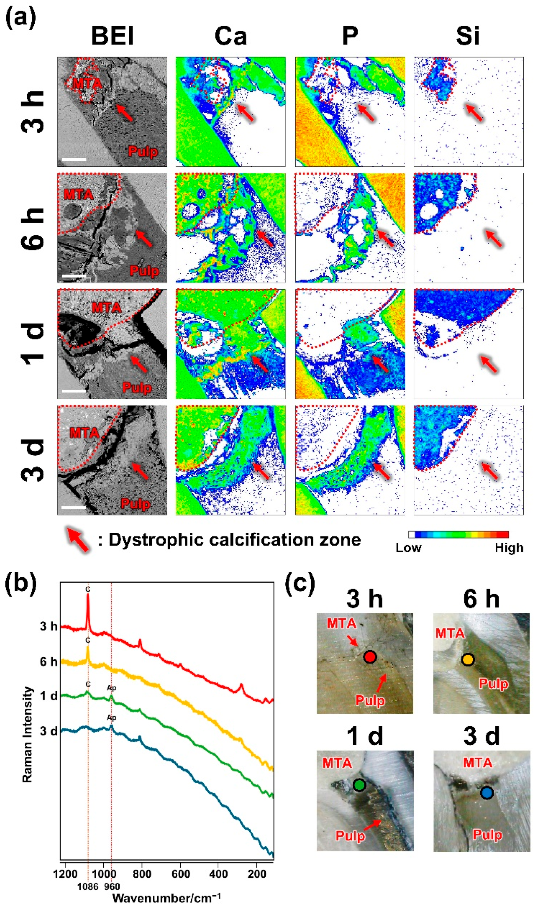
2.1. Dystrophic Calcification Beneath MTA

Using elemental mapping and Raman spectroscopy, we evaluated early mineral deposition beneath the MTA. A calcium-rich dystrophic calcification zone was observed beneath the silicon-rich MTA at 3 h, 6 h, 1 day, and 3 days (Figure 1, Table 1). The Raman spectra of the pulp under MTA exhibited peaks at 1086 and 960 cm−1, indicating the presence of calcite and hydroxyapatite [24], respectively (Figure 1).

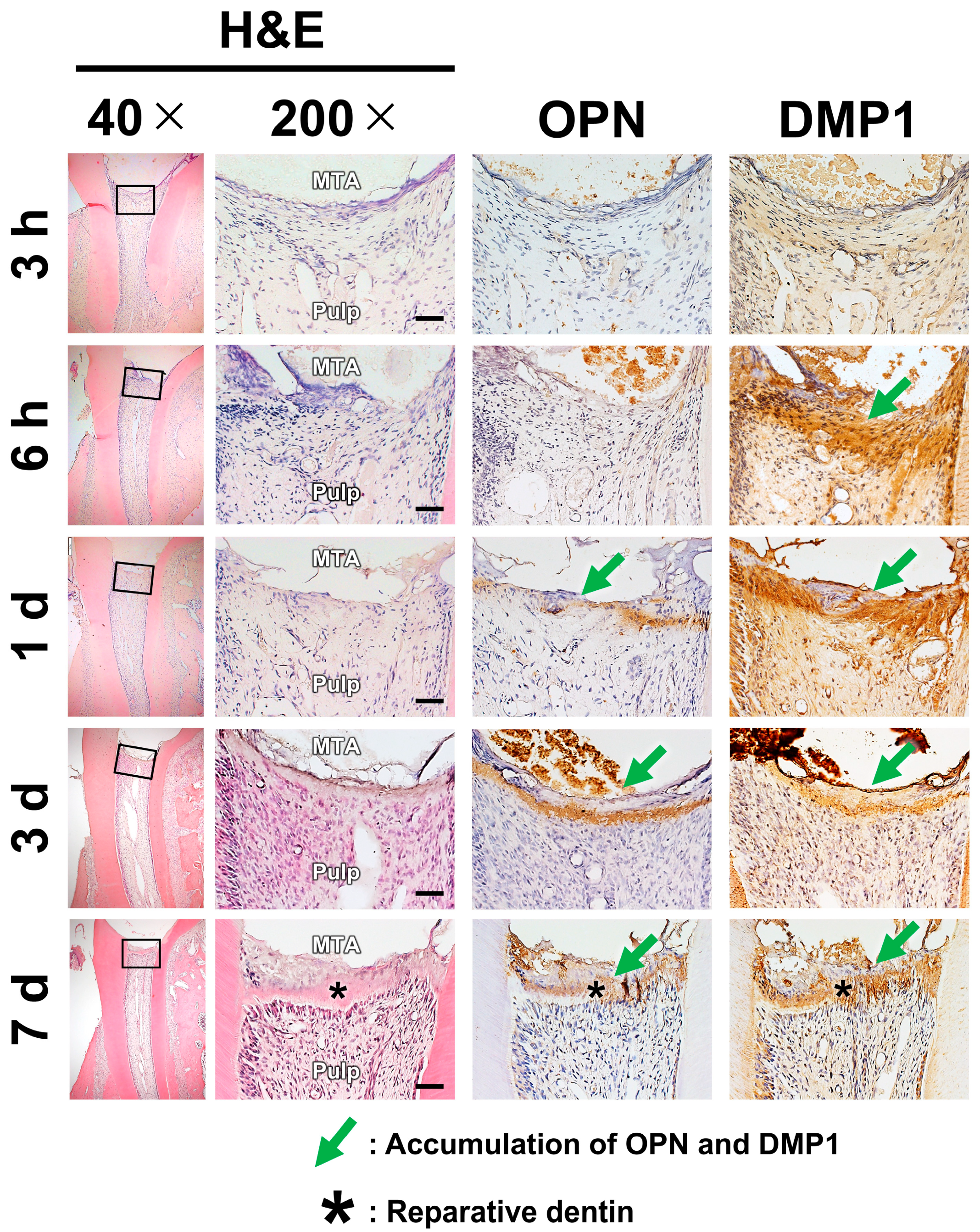
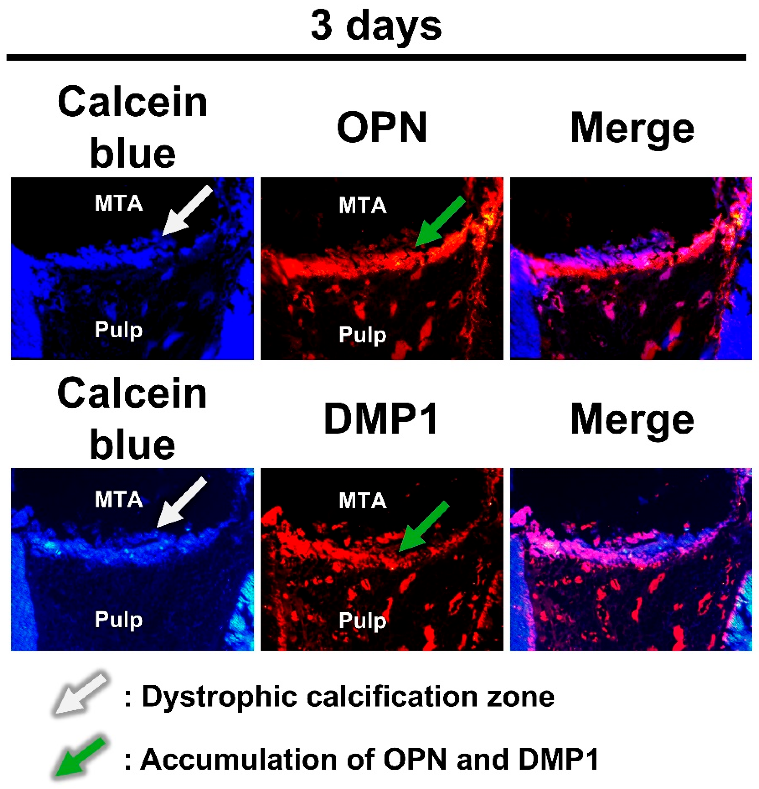
2.2. Pulpal Responses After MTA Pulpotomy

Three complementary staining techniques were employed to evaluate pulpal healing responses. Hematoxylin and eosin (H&E) staining was used to qualitatively evaluate RD formation and to confirm the absence of excessive inflammatory response, particularly in the ZrO2 group. Immunohistochemistry enabled the time-resolved detection of OPN and DMP1 expressions. Double fluorescence staining with calcein blue and immunofluorescent antibodies was employed to determine the spatial association between mineralized deposits and protein localization.
H&E staining showed the presence of RD on day 7. The accumulation of OPN or DMP1 was not detected via immunostaining at 3 h. DMP1 was detected at 6 h, whereas OPN was detected after 1 day (Figure 2, Table 1). Double staining with calcein blue and immunofluorescent antibodies revealed OPN and DMP1 expressions directly beneath the calcein blue–positive dystrophic calcification zone (Figure 3).

2.3. Dystrophic Calcification and Pulpal Responses After ZrO2 Pulpotomy

To assess pulpal responses in the absence of dystrophic calcification, the same analytical methods, i.e., elemental mapping, histological staining, and immunohistochemistry, were used in the ZrO2 group.
Elemental mapping revealed no calcium-rich zones under ZrO2. H&E staining revealed minimal to no inflammation in the pulp beneath ZrO2, and no RD formation was observed. No OPN or DMP1 accumulation was detected with immunostaining (Figure 4, Table 2).

3. Discussion

The results of this study elucidated the spatiotemporal distribution of dystrophic calcification, OPN and DMP1 accumulation, and RD formation after pulpotomy with MTA and ZrO2.
Dystrophic calcification is defined as the deposition of calcium crystals in degenerated or necrotic tissues, occurring despite normal serum calcium and phosphate levels [25]. The application of CH or MTA to exposed pulp rapidly induces calcium crystal precipitation in the pulp [18,19]. Based on ultrastructural analysis, calcium crystals formed under CH or MTA are associated with degenerated cells and swollen collagen, exhibiting characteristic features of dystrophic calcification [18,20,21]. CH and MTA are highly alkaline, causing tissue degeneration upon contact [1], and calcium release from CH and MTA promotes crystal nucleation and growth [26]. These factors likely contribute to dystrophic calcification in dental pulp.
Additionally, MTA forms a calcium silicate hydrate (CSH) gel during hydration. CSH gel is negatively charged, attracting calcium and phosphate ions and acting as nucleation sites for hydroxyapatite crystals [27,28]. Therefore, CSH gel formation by MTA may further promote dystrophic calcification by providing additional sites for mineral deposition in degenerated pulp tissue.
OPN and DMP1 are SIBLING family acidic glycoproteins. DMP1 is localized in the peritubular dentin and dental pulp, whereas OPN is found in predentin, mantle dentin, dentin–cementum interface, and tertiary dentin [29]. Previous studies have shown that their expression is upregulated in response to injury in the dentin–pulp complex, and that they colocalize in the superficial layer of RD following pulp capping with CH [12] or GaAlAs laser irradiation [30], indicating their involvement in reparative dentinogenesis.
Previous studies have demonstrated that dystrophic calcification occurs within 1 day in human [18] and canine teeth [20] after pulp capping with CH. In this study, elemental mapping detected a calcium-rich dystrophic calcification zone beneath MTA as early as 3 h after operation (Figure 1, Table 2). Moreover, micro-Raman spectroscopy confirmed the presence of calcite and hydroxyapatite in the zone (Figure 1), consistent with previous findings regarding calcite-like birefringent structures [31] and calcium–phosphorus crystals [19,20] in the pulp beneath CH or MTA.
The accumulation of OPN and DMP1 occurred later than dystrophic calcification (Table 2). Consistent with previous studies [11,12], DMP1 began accumulating after 6 h, whereas OPN accumulation became clear after 1 day. Furthermore, histological analysis of undecalcified sections revealed that areas of OPN and DMP1 accumulation were located just beneath the dystrophic calcification zone (Figure 3). Based on this spatiotemporal relationship, it is possible that dystrophic calcification induces the accumulation of OPN and DMP1.
The mechanism by which OPN and DMP1 accumulate adjacent to the dystrophic calcification zone remains unclear. One possible explanation is that OPN and DMP1 in tissue fluids are adsorbed onto hydroxyapatite within the dystrophic calcification zone. This hypothesis is supported by the high affinity of OPN [14] and DMP1 [15] for hydroxyapatite. Alternatively, OPN and DMP1 accumulation in the area adjacent to the dystrophic calcification zone may result from nanohydroxyapatite-induced upregulation of OPN and DMP1 expression in nearby cells. Supporting this possibility, previous studies have demonstrated that crystals in the dystrophic calcification zone are nanosized [18,19] and that nanohydroxyapatite enhances OPN and DMP1 expression in dental pulp stem cells [32,33]. These potential mechanisms warrant further investigation.
For this study, ZrO2 was selected owing to its well-established biological inertness. Previous studies have shown that ZrO2 exhibits negligible cytotoxicity and inflammatory responses when implanted in soft and hard tissues [34,35] and poses minimal risk of inducing hypersensitivity reactions or foreign body responses [36]. Moreover, contrary to MTA, ZrO2 does not release bioactive ions, such as calcium (Ca2+) or hydroxide (OH), and does not induce hydroxyapatite formation under physiological conditions [37]. The inert nature of ZrO2 limits its interaction with surrounding tissues, making it suitable as a negative control in studies assessing material-induced tissue responses.
Although the MTA group underwent time-course analysis from 3 h to 7 days to track the sequential progression of dystrophic calcification, OPN and DMP1 accumulation, and RD formation, the ZrO2 group was examined only at the final time point (day 7). This is because the purpose of the ZrO2 group was not to monitor temporal changes but to determine whether these events occur in the absence of any bioactive effects from the capping material.
As expected, even after 7 days, the pulp tissue beneath ZrO2 exhibited no signs of dystrophic calcification. Furthermore, no accumulation of OPN or DMP1 was detected during the same period (Figure 4, Table 2). These results indicate that OPN and DMP1 accumulation in the MTA-treated specimens is not a spontaneous response to pulp exposure. Instead, it appears to be specifically induced by MTA, likely due to its ability to promote dystrophic calcification.
Compared with the MTA group, the ZrO2 group exhibited delayed RD formation. Specifically, all samples in the MTA group exhibited RD by day 7 (Table 1), whereas none of the day 7 samples in the ZrO2 group showed this change (Table 2). Importantly, this difference was not attributable to differences in inflammatory status, as both groups exhibited minimal or no inflammation at day 7. These findings are consistent with those of previous studies showing no RD in pulp capped with bioinert materials such as Teflon [38] or 4-META/MMA-TBB resin [39].
The differences in tissue responses following pulpotomy with MTA and ZrO2 may, at least in part, be attributed to the distinct capacities of these materials to induce dystrophic calcification. Dystrophic calcification deposits adsorb fibronectin [18,22], which promotes odontoblast-like cell differentiation [23]. Additionally, nanohydroxyapatite, which is likely present in the dystrophic calcification zone, induces odontoblastic differentiation of dental pulp stem cells [32]. Moreover, RD is continuously formed beneath the dystrophic calcification zone [20]. Therefore, the dystrophic calcification zone may serve as a substrate for odontoblast-like cell differentiation and promote the formation of RD.
OPN and DMP1 accumulated at the pulp exposure site, and this was followed by the formation of RD in the MTA group (Table 1). However, these changes were not observed in the ZrO2 group (Table 2), suggesting that the accumulation of OPN and DMP1 may be a prerequisite for the formation of RD. Recombinant DMP1–impregnated collagen matrix has been shown to induce odontoblast-like cell differentiation in rat molars [40]. Similarly, OPN has been reported to stimulate the odontoblastic differentiation of human dental pulp cells in vitro [41]. Therefore, the presence of OPN and DMP1 at the pulp exposure site may facilitate the differentiation of odontoblast-like cells and promote the formation of RD. However, further research is needed to elucidate their specific roles at pulp exposure sites.
This study has several limitations. First, the observation period was limited to postoperative day 7. Although this time frame was selected to capture the early events associated with dystrophic calcification and the initial OPN and DMP1 expressions, it precludes the evaluation of long-term tissue responses such as RD maturation. Second, although this study mainly focused on OPN and DMP1 accumulation, other extracellular matrix (ECM) remodeling events, such as fibrillin-1 degradation [42] and increased collagen production [43,44], have also been reported to precede reparative dentinogenesis. However, whether these ECM changes are regulated by dystrophic calcification remains unclear. Third, this study exclusively used ProRoot MTA as the pulp capping material; however, many other tricalcium silicate–based materials are currently available for clinical use, including bismuth-free formulations to reduce cytotoxicity and discoloration [45] as well as materials with accelerated setting reactions or enhanced handling characteristics [46]. However, it is undetermined whether these alternative materials induce dystrophic calcification and comparable pulpal responses similar to MTA. Fourth, the sample size for each analysis was relatively small (n = 2–6), which may limit the generalizability of the findings. Collectively, more studies with longer follow-up, broader ECM analysis, larger sample sizes, and different materials are warranted to better understand the mechanisms of pulp healing and confirm the present findings.
In conclusion, dystrophic calcification occurred at an early stage after MTA pulpotomy, followed by OPN and DMP1 accumulation and the formation of RD. These processes did not occur in ZrO2-treated specimens. Our results suggest that dystrophic calcification triggers OPN and DMP1 accumulation and RD formation.

4. Materials and Methods

4.1. Materials

Two materials were utilized for pulpotomy: ProRoot MTA (Dentsply Sirona, York, PA, USA; Lot No. 333803) and ZrO2 (Wako, Osaka, Japan; Lot No. PAG2868). ProRoot MTA was prepared according to the manufacturer’s protocols. The powder was mixed with sterile distilled water at a powder-to-liquid ratio of 3:1 (w/w) on a silicone mixing pad using a plastic spatula until a putty-like consistency was achieved. Similarly, ZrO2 powder was mixed with sterile distilled water at a powder-to-liquid ratio of 5:1 (w/w) to produce a cohesive putty.

4.2. Pulpotomy Procedures

All animal experiments were approved by the Niigata University Animal Experimentation Committee (approval no. SA00903) and conducted according to relevant guidelines and regulations. The experiments included 58 male Wistar rats (Clea Japan, Tokyo, Japan) aged 8 weeks old. After the induction of anesthesia via intraperitoneal injection of medetomidine hydrochloride, midazolam, and butorphanol (Wako), pulpotomy was performed on the left maxillary first molar under a 20× magnification using a surgical microscope (S9D; Leica, Wetzlar, Germany). Specifically, the pulp chamber was accessed using a tungsten carbide bur (E0123 size 008; Dentsply), and the coronal pulp tissue was removed with the same bur. The exposed pulp stump was irrigated with 2.5% sodium hypochlorite (Neo Dental Chemical Products, Tokyo, Japan) and 3% hydrogen peroxide (Yoshida Pharmaceutical, Tokyo, Japan), followed by rinsing with saline (1 mL each), in accordance with previous studies [11,12]. Following hemostasis, MTA or ZrO2 was placed on the pulp stump, and the cavity was sealed with a flowable composite resin (Beautifil Flow; Shofu, Kyoto, Japan) using a bonding system (Clearfil Universal Bond Quick; Kuraray, Tokyo, Japan). The opposing lower first molars were extracted at the time of surgery to prevent fracture of the upper first molars. After surgery, the animals’ general health status was monitored daily by veterinary technicians. No signs of distress or abnormal behavior was observed throughout the observation period. At designated time points, 3 h, 6 h, 1 day, 3 days, and 7 days after pulpotomy, the animals were euthanized via carbon dioxide inhalation, and the maxillae containing the treated upper first molars were harvested for analysis.

4.3. Elemental Mapping and Micro-Raman Spectrometry

Specimens from the MTA (3 h, 6 h, 1 day, and 3 days; n = 4 per time) and ZrO2 (7 days; n = 4) groups were analyzed. The sample size was based on previous studies using similar models for qualitative analyses [47,48]. Specimens were fixed in 2.5% glutaraldehyde (Wako) buffered with 60 mmol/L HEPES (Dojindo Laboratories, Kumamoto, Japan), dehydrated in ethanol and acetone, embedded in methyl methacrylate (MMA) resin (Osteoresin; Wako), and ground using a rotary grinder (Vector Power Head; BUEHLER, Lake Bluff, IL, USA). The exposed surfaces containing the capping material and the pulp stump were gold-coated and analyzed for elemental distribution using an electron probe microanalyzer (EPMA1601; Shimadzu, Kyoto, Japan). The EPMA accelerating voltage was 15 kV, step size was 3 μm, and sampling time was 0.1 s per point. After EPMA analysis, the specimens were subjected to micro-Raman spectroscopy. The gold coating was removed by polishing with diamond pads (Struers, Champigny-sur-Marne, France). Subsequently, Raman spectra were obtained from the pulp stump beneath the MTA, targeting areas with dystrophic calcification. Measurements were performed using a micro-Raman spectrometer (NRS-3100; JASCO, Tokyo, Japan) equipped with a 100× objective lens. A 532 nm laser served as the excitation source, with an output power of 7.4 mW. To minimize thermal noise and enhance signal quality, the CCD detector was cooled to −50.0 °C.

4.4. Histological Staining of the Decalcified Sections

Specimens from the MTA (3 and 6 h and 1, 3, and 7 days; n = 6 per time point) and ZrO2 (7 days; n = 6) groups were analyzed. The sample size was based on previous studies using similar models for qualitative analyses [49,50]. After fixation with 4% paraformaldehyde (Wako), the specimens were demineralized in EDTA solution (OsteoSoft; Merck, Darmstadt, Germany), dehydrated in ethanol, cleared with xylene, embedded in paraffin, and sectioned (5 µm) using a microtome (HistoCore Multicut R; Leica, Wetzlar, Germany). Subsequently, the sections were stained with H&E (Wako) to evaluate the timing of RD formation and pulpal inflammation, particularly in the ZrO2 group, confirming the material’s biological inertness. Immunohistochemistry was performed using the primary antibodies listed in Table 3, following a previous protocol [43]. The staining results were qualitatively assessed by a trained observer in a blinded manner, with no knowledge of the group assignment or time point during the analysis.

4.5. Histological Staining of the Nondecalcified Sections

MTA specimens (3 days; n = 2) were fixed in phosphate-free 10% neutral formalin (Mutoh Chemical, Tokyo, Japan), dehydrated in ethanol and acetone, and then embedded in MMA resin (Technovit 9100; Kulzer, Wehrheim, Germany) via acetone evaporation [51]. Next, the embedded samples were cut into 3 µm sections using a hard tissue microtome (HistoCore Multicut R) and double-stained with calcein blue, a fluorescent dye that labels calcium, and an immunofluorescent antibody for OPN or DMP1. Sections were deresinized in methyl ethyl ketone and acetone, stained with calcein blue (10 mg/mL, 10 min; Wako), temporarily mounted, and imaged using a fluorescence microscope (Eclipse E800; Nikon, Tokyo, Japan). After imaging, the sections were subjected to decalcification with EDTA (OsteoSoft) for 20 min, antigen retrieval with hyaluronidase (Wako) at 37 °C for 20 min, and blocking with goat serum (Jackson Immuno Research Laboratories, West Grove, PA, USA) for 5 min, followed by overnight incubation with a primary antibody for OPN or DMP1 and then 1 h incubation with a secondary antibody (Alexa Fluor 546-conjugated anti-rabbit IgG; Invitrogen, Carlsbad, CA, USA). Subsequently, the sections were mounted again and reimaged. The resulting images were aligned and merged using Fiji (ImageJ 1.53t; National Institutes of Health, Bethesda, MD, USA) [52].

Author Contributions

Conceptualization: N.E.; Methodology: N.E.; Data curation: N.E., K.Y., R.S.I.B., N.Y., S.T. (Shoji Takenaka), N.O., S.T. (Shintaro Takahara), T.I., R.B., S.K. and P.T.; Formal analysis: N.E.; Writing—original draft: N.E.; Writing—review and editing: N.E., K.Y., R.S.I.B., N.Y., N.O. and Y.N. All authors have read and agreed to the published version of the manuscript.

Funding

This research was funded by Grants-in-Aid for Scientific Research from the Japan Society for the Promotion of Science (grant numbers 21K16966 and 23K09164 to N.E.).

Institutional Review Board Statement

All experiments were reviewed and approved by the Committee on the Guidelines for Animal Experimentation of Niigata University (approval number SA00903) and adhered to all international, national, and institutional guidelines for the care and use of animals.

Informed Consent Statement

For this type of study, formal consent is not required.

Data Availability Statement

Relevant data from this study are available upon reasonable request to the corresponding author.

Acknowledgments

This work was supported by CCRF, Niigata University. We thank Ayako Ikarashi for her technical support.

Conflicts of Interest

The authors declare no conflicts of interest.

Abbreviations

The following abbreviations are used in this manuscript:
BEIBackscattered electron image
CHCalcium hydroxide
CSHCalcium silicate hydrate
DMP1Dentin matrix protein-1
ECMExtracellular matrix
EPMAElectron probe microanalyzer
H&EHematoxylin and eosin
MMAMethyl methacrylate
MTAMineral trioxide aggregate
OPNOsteopontin
RDReparative dentin
SIBLINGSmall integrin-binding ligand N-linked glycoproteins
ZrO2Zirconium Oxide

References

  1. Takashi, O.; Kunihiko, Y. Reparative dentinogenesis induced by mineral trioxide aggregate: A review from the biological and physicochemical points of view. Int. J. Dent. 2009, 2009, 464280. [Google Scholar] [CrossRef] [PubMed]
  2. Nebu, P.; Bharat, S. Minimally invasive endodontics: A new era for pulpotomy in mature permanent teeth. Br. Dent. J. 2022, 233, 1035–1041. [Google Scholar] [CrossRef] [PubMed]
  3. Glickman, G.N. AAE Consensus Conference Recommended Diagnostic Terminology. J. Endod. 2009, 35, 1634. [Google Scholar] [CrossRef] [PubMed]
  4. Jassal, A.; Nawal, R.R.; Yadav, S.; Talwar, S.; Yadav, S.; Duncan, H.F. Outcome of partial and full pulpotomy in cariously exposed mature molars with symptoms indicative of irreversible pulpitis: A randomized controlled trial. Int. Endod. J. 2023, 56, 331–344. [Google Scholar] [CrossRef] [PubMed]
  5. Thibault, N.C.; Phillip, L.T. Vital pulp therapies in permanent teeth: What, when, where, who, why and how? Br. Dent. J. 2025, 238, 458–468. [Google Scholar] [CrossRef] [PubMed]
  6. Sangwan, P.; Sangwan, A.; Duhan, J.; Rohilla, A. Tertiary dentinogenesis with calcium hydroxide: A review of proposed mechanisms. Int. Endod. J. 2013, 46, 3–19. [Google Scholar] [CrossRef] [PubMed]
  7. Cox, C.F.; Sübay, R.K.; Ostro, E.; Suzuki, S.; Suzuki, S.H. Tunnel defects in dentin bridges: Their formation following direct pulp capping. Oper. Dent. 1996, 21, 4–11. [Google Scholar] [PubMed]
  8. Primus, C.M.; Tay, F.R.; Niu, L.-N. Bioactive tri/dicalcium silicate cements for treatment of pulpal and periapical tissues. Acta Biomater. 2019, 96, 35–54. [Google Scholar] [CrossRef] [PubMed]
  9. Parirokh, M.; Torabinejad, M. Mineral Trioxide Aggregate: A Comprehensive Literature Review-Part I: Chemical, Physical, and Antibacterial Properties. J. Endod. 2010, 36, 16–27. [Google Scholar] [CrossRef] [PubMed]
  10. Nair, P.N.R.; Duncan, H.F.; Ford, T.P.; Luder, H.U. Histological, ultrastructural and quantitative investigations on the response of healthy human pulps to experimental capping with mineral trioxide aggregate: A randomized controlled trial. Int. Endod. J. 2008, 41, 128–150. [Google Scholar] [CrossRef] [PubMed]
  11. Kuratate, M.; Yoshiba, K.; Shigetani, Y.; Yoshiba, N.; Ohshima, H.; Okiji, T. Immunohistochemical Analysis of Nestin, Osteopontin, and Proliferating Cells in the Reparative Process of Exposed Dental Pulp Capped with Mineral Trioxide Aggregate. J. Endod. 2008, 34, 970–974. [Google Scholar] [CrossRef] [PubMed]
  12. Shigetani, Y.; Yoshiba, K.; Kuratate, M.; Takei, E.; Yoshiba, N.; Yamanaka, Y.; Ohshima, H.; Okiji, T. Temporospatial localization of dentine matrix protein 1 following direct pulp capping with calcium hydroxide in rat molars. Int. Endod. J. 2015, 48, 573–581. [Google Scholar] [CrossRef] [PubMed]
  13. Staines, K.A.; MacRae, V.E.; Farquharson, C. The importance of the SIBLING family of proteins on skeletal mineralisation and bone remodelling. J. Endocrinol. 2012, 214, 241–255. [Google Scholar] [CrossRef] [PubMed]
  14. Wada, T.; McKee, M.D.; Steitz, S.; Giachelli, C.M. Calcification of vascular smooth muscle cell cultures: Inhibition by osteopontin. Circ. Res. 1999, 84, 166–178. [Google Scholar] [CrossRef] [PubMed]
  15. Gajjeraman, S.; Narayanan, K.; Hao, J.; Qin, C.; George, A. Matrix macromolecules in hard tissues control the nucleation and hierarchical assembly of hydroxyapatite. J. Biol. Chem. 2007, 282, 1193–1204. [Google Scholar] [CrossRef] [PubMed]
  16. Kulkarni, G.V.; Chen, B.; Malone, J.P.; Narayanan, A.; George, A. Promotion of selective cell attachment by the RGD sequence in dentine matrix protein 1. Arch. Oral. Biol. 2000, 45, 475–484. [Google Scholar] [CrossRef] [PubMed]
  17. Liaw, L.; Skinner, M.P.; Raines, E.W.; Ross, R.; Cheresh, D.A.; Schwartz, S.M.; Giachelli, C.M. The adhesive and migratory effects of osteopontin are mediated via distinct cell surface integrins. Role of alpha v beta 3 in smooth muscle cell migration to osteopontin in vitro. J. Clin. Investig. 1995, 95, 713–724. [Google Scholar] [CrossRef] [PubMed]
  18. Yoshiba, K.; Yoshiba, N.; Nakamura, H.; Iwaku, M.; Ozawa, H. Immunolocalization of fibronectin during reparative dentinogenesis in human teeth after pulp capping with calcium hydroxide. J. Dent. Res. 1996, 75, 1590–1597. [Google Scholar] [CrossRef] [PubMed]
  19. Tziafas, D.; Pantelidou, O.; Alvanou, A.; Belibasakis, G.; Papadimitriou, S. The dentinogenic effect of mineral trioxide aggregate (MTA) in short-term capping experiments. Int. Endod. J. 2002, 35, 245–254. [Google Scholar] [CrossRef] [PubMed]
  20. Higashi, T.; Okamoto, H. Characteristics and effects of calcified degenerative zones on the formation of hard tissue barriers in amputated canine dental pulp. J. Endod. 1996, 22, 168–172. [Google Scholar] [CrossRef] [PubMed]
  21. Schröder, U. Effects of calcium hydroxide-containing pulp-capping agents on pulp cell migration, proliferation, and differentiation. J. Dent. Res. 1985, 64, 541–548. [Google Scholar] [CrossRef] [PubMed]
  22. Tziafas, D.; Panagiotakopoulos, N.; Komnenou, A. Immunolocalization of fibronectin during the early response of dog dental pulp to demineralized dentine or calcium hydroxide-containing cement. Arch. Oral. Biol. 1995, 40, 23–31. [Google Scholar] [CrossRef] [PubMed]
  23. Tziafas, D.; Alvanou, A.; Kaidoglou, K. Dentinogenic activity of allogenic plasma fibronectin on dog dental pulp. J. Dent. Res. 1992, 71, 1189–1195. [Google Scholar] [CrossRef] [PubMed]
  24. Fausto, Z.; Carlo, P.; Paola, T.; Andrea, S.; Michele, D.F. Gandolfi Maria Giovanna. Chemical-Physical Properties and Bioactivity of New Premixed Calcium Silicate-Bioceramic Root Canal Sealers. Int. J. Mol. Sci. 2022, 23, 13914. [Google Scholar] [CrossRef]
  25. Stewart, V.L.; Herling, P.; Dalinka, M.K. Calcification in Soft Tissues. JAMA 1983, 250, 78–81. [Google Scholar] [CrossRef] [PubMed]
  26. Lu, X.; Leng, Y. Theoretical analysis of calcium phosphate precipitation in simulated body fluid. Biomaterials 2005, 26, 1097–1108. [Google Scholar] [CrossRef] [PubMed]
  27. Gandolfi, M.G.; Taddei, P.; Tinti, A.; Prati, C. Apatite-forming ability (bioactivity) of ProRoot MTA. Int. Endod. J. 2010, 43, 917–929. [Google Scholar] [CrossRef] [PubMed]
  28. Edanami, N.; Takenaka, S.; Saifullah, I.B.R.; Yoshiba, K.; Takahara, S.; Yoshiba, N.; Ohkura, N.; Noiri, Y. In Vivo Assessment of the Apatite-Forming Ability of New-Generation Hydraulic Calcium Silicate Cements Using a Rat Subcutaneous Implantation Model. J. Funct. Biomater. 2023, 14, 213. [Google Scholar] [CrossRef] [PubMed]
  29. Alberto, F.C.; Nancy, A.; Monica, P.G. The Roles of SIBLING Proteins in Dental, Periodontal and Craniofacial Development. Front. Dent. Med. 2022, 3, 898802. [Google Scholar] [CrossRef]
  30. Shigetani, Y.; Ohkura, N.; Yoshiba, K.; Ohshima, H.; Hosoya, A.; Yoshiba, N.; Okiji, T. GaAlAs laser-induced pulp mineralization involves dentin matrix protein 1 and osteopontin expression. Oral. Dis. 2016, 22, 399–405. [Google Scholar] [CrossRef] [PubMed]
  31. Roberto, H.; Carlos, E.P.; Waldericio, M.; Mauro, J.N.; de Souza, V. Histochemical analysis of the dogs’ dental pulp after pulp capping with calcium, barium, and strontium hydroxides. J. Endod. 1982, 8, 444–447. [Google Scholar] [CrossRef] [PubMed]
  32. Yoshida, S.; Sugii, H.; Itoyama, T.; Kadowaki, M.; Hasegawa, D.; Tomokiyo, A.; Hamano, S.; Ipposhi, K.; Yamashita, K.; Maeda, H. Development of a novel direct dental pulp-capping material using 4-META/MMA-TBB resin with nano hydroxyapatite. Mater. Sci. Eng. C Mater. Biol. Appl. 2021, 130, 112426. [Google Scholar] [CrossRef] [PubMed]
  33. Khaled, H.A.; Souzy, F.S.; Nour, E.G.; Riham, M.A. Nano Hydroxyapatite & Mineral Trioxide Aggregate Efficiently Promote Odontogenic Differentiation of Dental Pulp Stem Cells. Open Access Maced. J. Med. Sci. 2018, 6, 1727–1731. [Google Scholar] [CrossRef]
  34. Ralph, V.B.; Gert, J.M.; Willem, V.J.; John, J.; Cornelis, D.P.; Marco, S.C. Soft tissue response to zirconia and titanium implant abutments: An in vivo within-subject comparison. J. Clin. Periodontol. 2012, 39, 995–1001. [Google Scholar] [CrossRef]
  35. Warashina, H.; Sakano, S.; Kitamura, S.; Yamauchi, K.-I.; Yamaguchi, J.; Ishiguro, N.; Hasegawa, Y. Biological reaction to alumina, zirconia, titanium and polyethylene particles implanted onto murine calvaria. Biomaterials 2003, 24, 3655–3661. [Google Scholar] [CrossRef] [PubMed]
  36. Megumi, W.; Lipei, L.; Tetsuo, I. Are Allergy-Induced Implant Failures Actually Hypersensitivity Reactions to Titanium? A Literature Review. Dent. J. 2023, 11, 263. [Google Scholar] [CrossRef] [PubMed]
  37. Xuanyong, L.; Anping, H.; Chuanxian, D.; Paul, K.C. Bioactivity and cytocompatibility of zirconia (ZrO2) films fabricated by cathodic arc deposition. Biomaterials 2006, 27, 3904–3911. [Google Scholar] [CrossRef] [PubMed]
  38. Cvek, M.; Granath, L.; Cleaton-Jones, P.; Austin, J. Hard tissue barrier formation in pulpotomized monkey teeth capped with cyanoacrylate or calcium hydroxide for 10 and 60 minutes. J. Dent. Res. 1987, 66, 1166–1174. [Google Scholar] [CrossRef] [PubMed]
  39. Nakamura, M.; Inoue, T.; Shimono, M. Immunohistochemical study of dental pulp applied with 4-META/MMA-TBB adhesive resin after pulpotomy. J. Biomed. Mater. Res. 2000, 51, 241–248. [Google Scholar] [CrossRef]
  40. Almushayt, A.; Narayanan, K.; Zaki, A.E.; Anne, G. Dentin matrix protein 1 induces cytodifferentiation of dental pulp stem cells into odontoblasts. Gene. Ther. 2006, 13, 611–620. [Google Scholar] [CrossRef] [PubMed]
  41. Jia, T.; Youjing, Q.; Zehan, L. Osteopontin Facilitated Dental Pulp Cell Adhesion and Differentiation: A Laboratory Investigation. ACS Appl. Bio Mater. 2025, 8, 1320–1329. [Google Scholar] [CrossRef]
  42. Yoshiba, N.; Yoshiba, K.; Ohkura, N.; Hosoya, A.; Shigetani, Y.; Yamanaka, Y.; Izumi, N.; Nakamura, H.; Okiji, T. Expressional alterations of fibrillin-1 during wound healing of human dental pulp. J. Endod. 2012, 38, 177–184. [Google Scholar] [CrossRef] [PubMed]
  43. Edanami, N.; Yoshiba, N.; Ohkura, N.; Takeuchi, R.; Tohma, A.; Noiri, Y.; Yoshiba, K. Characterization of Dental Pulp Myofibroblasts in Rat Molars after Pulpotomy. J. Endod. 2017, 43, 1116–1121. [Google Scholar] [CrossRef] [PubMed]
  44. Yoshiba, N.; Edanami, N.; Tohma, A.; Takeuchi, R.; Ohkura, N.; Hosoya, A.; Noiri, Y.; Nakamura, H.; Yoshiba, K. Detection of bone marrow-derived fibrocytes in human dental pulp repair. Int. Endod. J. 2018, 51, 1187–1195. [Google Scholar] [CrossRef] [PubMed]
  45. Marciano, M.A.; Pelepenko, L.E.; Francati, T.M.; Antunes, T.B.M.; Janini, A.C.P.; Rohwedder, J.J.R.; Shelton, R.M.; Camilleri, J. Bismuth release from endodontic materials: In vivo analysis using Wistar rats. Sci. Rep. 2023, 13, 9738. [Google Scholar] [CrossRef] [PubMed]
  46. Parirokh, M.; Torabinejad, M.; Dummer, P.M.H. Mineral trioxide aggregate and other bioactive endodontic cements: An updated overview—Part I: Vital pulp therapy. Int. Endod. J. 2018, 51, 177–205. [Google Scholar] [CrossRef] [PubMed]
  47. Zeid, S.T.A.; Alamoudi, R.A.; Neel, E.A.A.; Saleh, A.A.M. Morphological and spectroscopic study of an apatite layer induced by fast-set versus regular-set endosequence root repair materials. Materials 2019, 12, 3678. [Google Scholar] [CrossRef]
  48. Zamparini, F.; Siboni, F.; Prati, C.; Taddei, P.; Gandolfi, M.G. Properties of calcium silicate-monobasic calcium phosphate materials for endodontics containing tantalum pentoxide and zirconium oxide. Clin. Oral Investig. 2019, 23, 445–457. [Google Scholar] [CrossRef] [PubMed]
  49. Saito, K.; Nakatomi, M.; Ida-Yonemochi, H.; Ohshima, H. Osteopontin Is Essential for Type i Collagen Secretion in Reparative Dentin. J. Dent. Res. 2016, 95, 1034–1041. [Google Scholar] [CrossRef] [PubMed]
  50. Seok, Y.I.; Seol, L.D.; Tae, P.J.; Joong, K.H.; Hyun, S.H.; Cheol, P.J. Tertiary dentin formation after direct pulp capping with odontogenic ameloblast-associated protein in rat teeth. J. Endod. 2010, 36, 1956–1962. [Google Scholar] [CrossRef] [PubMed]
  51. Steiniger, B.S.; Bubel, S.; Böckler, W.; Lampp, K.; Seiler, A.; Jablonski, B.; Guthe, M.; Stachniss, V. Immunostaining of pulpal nerve fibre bundle/arteriole associations in ground serial sections of whole human teeth embedded in technovit® 9100. Cells Tissues Organs 2013, 198, 57–65. [Google Scholar] [CrossRef] [PubMed]
  52. Piccinini, F.; Tazzari, M.; Tumedei, M.M.; Stellato, M.; Remondini, D.; Giampieri, E.; Martinelli, G.; Castellani, G.; Carbonaro, A. Data Science for Health Image Alignment: A User-Friendly Open-Source ImageJ/Fiji Plugin for Aligning Multimodality/Immunohistochemistry/Immunofluorescence 2D Microscopy Images. Sensors 2024, 24, 451. [Google Scholar] [CrossRef] [PubMed]
Figure 1. Dystrophic calcification after pulpotomy with mineral trioxide aggregate (MTA). (a) Backscattered electron images (BEIs) and elemental mapping images showing the distributions of calcium (Ca), phosphorus (P), and silicon (Si) at the pulp exposure site (453 × 453 μm). The MTA is outlined with red dotted lines. A Ca-rich dystrophic calcification zone was present during all observation periods. Scale: 100 μm. (b) Raman spectra of the pulp stump beneath the MTA. Raman peaks corresponding to hydroxyapatite (Ap) at 960 cm−1 and calcite (C) at 1086 cm−1 were detected. (c) Bright-field microscopic images of the specimens used for Raman spectroscopy. The colored dots in the bright-field images indicate the locations of the Raman measurements.
Ijms 26 07130 g001
Figure 2. Osteopontin (OPN) and dentin matrix protein-1 (DMP1) expressions as well as reparative dentin formation following pulpotomy with mineral trioxide aggregate (MTA). Consecutive sections stained with hematoxylin and eosin (H&E) and antibodies against OPN and DMP1 are presented. DMP1 accumulation was detected at 6 h, whereas OPN accumulation was detected after 1 day. RD formation was detected beneath the OPN and DMP1 accumulation on day 7. Images were obtained at an original magnification of 40× (low magnification) and 200× (high magnification). The area indicated by a square in the low-magnification image is shown at a higher magnification. H&E: hematoxylin and eosin staining. Scale: 50 μm.
Ijms 26 07130 g002
Figure 3. Spatial association between the dystrophic calcification zone and the accumulation of osteopontin (OPN) and dentin matrix protein-1 (DMP1). OPN and DMP1 immunoreactivities were directly localized beneath the calebin blue–positive dystrophic calcification zone. MTA: mineral trioxide aggregate.
Ijms 26 07130 g003
Figure 4. Dystrophic calcification and tissue responses in pulp treated with zirconium oxide (ZrO2). (a) A backscattered electron image (BEI) and elemental mapping images showing calcium (Ca), phosphorus (P), and zirconium (Zr) distributions in the pulp stump (453 × 453 μm). The ZrO2 is outlined with red dotted lines. No Ca-rich regions were detected beneath the ZrO2. Scale: 100 μm. (b) Hematoxylin and eosin staining (H&E) and immunohistochemical staining for osteopontin (OPN) and dentin matrix protein-1 (DMP1) were performed on consecutive sections. Only minimal inflammation was observed at the pulp amputation site, and neither OPN nor DMP1 accumulation was detected beneath ZrO2. Images were captured at original magnifications of 40× (low) and 200× (high). The area indicated by a square in the low-magnification image is shown at a higher magnification. Scale: 50 μm.
Ijms 26 07130 g004
Table 1. Pulpal responses after pulpotomy with mineral trioxide aggregate (Positive/Total).
3 h6 h1 Day3 Day7 Day
DC4/44/44/44/4-
OPN0/60/62/66/66/6
DMP10/62/66/66/66/6
RD0/60/60/60/66/6
The presence or absence of dystrophic calcification (DC) was determined based on elemental mapping results. Osteopontin (OPN) and dentin matrix protein-1 (DMP1) accumulation were evaluated using immunohistochemical staining, whereas reparative dentin (RD) was assessed through hematoxylin and eosin staining.
Table 2. Pulpal responses after pulpotomy with zirconium oxide (positive/total).
7 Day
DC0/4
OPN0/6
DMP10/6
RD0/6
The presence or absence of dystrophic calcification (DC) was determined based on elemental mapping results. Osteopontin (OPN) and dentin matrix protein-1 (DMP1) accumulation were evaluated using immunohistochemical staining, whereas reparative dentin (RD) was assessed through hematoxylin and eosin staining.
Table 3. Primary antibodies used in this study.
Antibody (Catalog Number)ConcentrationManufacturer
Rabbit polyclonal anti-osteopontin antibody (18628)1:300Immuno-Biological Laboratories, Gunma, Japan
Rabbit polyclonal anti-dentin matrix protein-1 antibody (M176)1:300Takara Bio, Shiga, Japan
Disclaimer/Publisher’s Note: The statements, opinions and data contained in all publications are solely those of the individual author(s) and contributor(s) and not of MDPI and/or the editor(s). MDPI and/or the editor(s) disclaim responsibility for any injury to people or property resulting from any ideas, methods, instructions or products referred to in the content.

