Article

Vitamin D3 Modulates Inflammatory and Antimicrobial Responses in Oral Epithelial Cells Exposed to Periodontitis-Associated Bacteria

by
Fadime Karaca
1,
Susanne Bloch
1,2,
Fabian L. Kendlbacher
2,
Christian Behm
1,
Christina Schäffer
2,* and
Oleh Andrukhov
1,*
1
Competence Center for Periodontal Research, University Clinic of Dentistry, Medical University of Vienna, 1090 Vienna, Austria
2
NanoGlycobiology Research Group, Institute of Biochemistry, Department of Natural Sciences and Sustainable Resources, University of Natural Resources and Life Sciences, 1190 Vienna, Austria
*
Authors to whom correspondence should be addressed.
Int. J. Mol. Sci. 2025, 26(14), 7001; https://doi.org/10.3390/ijms26147001
Submission received: 11 June 2025 / Revised: 11 July 2025 / Accepted: 18 July 2025 / Published: 21 July 2025

Abstract

The oral epithelium is essential for maintaining oral health and plays a key role in the onset and progression of periodontitis. It serves as both a mechanical and immunological barrier and possesses antimicrobial activity. Vitamin D3, a hormone with known immunomodulatory functions, may influence oral epithelial responses. This study investigated the effects of two vitamin D3 metabolites on key immunological and antimicrobial functions of oral epithelial cells, both under basal conditions and during bacterial challenge. Ca9-22 oral epithelial cells were treated with 1,25(OH)2D3 or 25(OH)D3 in the presence or absence of Tannerella forsythia, Fusobacterium nucleatum, or Porphyromonas gingivalis. Inflammatory responses were assessed by measuring gene and protein expression of IL-1β and IL-8. Antimicrobial activity was evaluated via expression of LL-37, hBD-2, and hBD-3, as well as direct bacterial killing assays. Expression of epithelial integrity markers E-cadherin and ICAM-1 was also analyzed. Vitamin D3 metabolites reduced IL-8 expression and significantly increased LL-37 expression and production in Ca9-22 cells. Both forms enhanced antimicrobial activity against all tested pathogens and modulated epithelial integrity markers. Vitamin D3 positively regulates antimicrobial and barrier functions in oral epithelial cells, suggesting a potential role in supporting oral health and preventing periodontitis progression.

1. Introduction

The oral cavity is a complex and dynamic environment where the maintenance of a balanced host–microbiota interaction is essential for oral health [1]. In healthy individuals, the oral microbiome forms a symbiotic relationship with the host’s immune system, sustaining a stable and mutually beneficial ecosystem [2,3,4,5]. Disruption of this homeostasis, often due to microbial dysbiosis, can lead to periodontitis—a prevalent inflammatory disease affecting the tooth-supporting tissues. Classically associated with members of the “red complex”, such as Porphyromonas gingivalis, Tannerella forsythia, and Treponema denticola [6], periodontitis is now understood as a polymicrobial infection involving a broader shift in microbial composition [7]. Notably, an increased abundance of genera such as Fusobacterium, Filifactor, Prevotella, Tannerella, and Porphyromonas, accompanied by a concurrent reduction in commensal genera such as Streptococcus, Actinomyces, and Haemophilus in subgingival biofilms, has been associated with the onset and progression of periodontal disease [8,9]. This microbial shift suggests a potential role for these genera in the pathogenesis of the disease.
While the pathogenesis of periodontitis is multifactorial and still incompletely understood [10], vitamin D3 deficiency has emerged as a potential risk factor [11]. Vitamin D3 is a fat-soluble secosteroid hormone synthesized in the skin upon UVB exposure or obtained via diet [12]. It undergoes two-step hydroxylation—first to 25-hydroxyvitamin D3 [25(OH)D3] in the liver, and then to its active form 1,25-dihydroxyvitamin D3 [1,25(OH)2D3] in the kidneys [13]. The biological activity of 1,25(OH)2D3 is mediated by the vitamin D receptor (VDR), which is expressed in various oral cell types [14]. VDR is an intracellular nuclear receptor functioning as a ligand-activated transcription factor which directly regulates gene expression (e.g., antimicrobial peptides), but may also exist in a membrane-associated form that mediates the non-genomic effects of vitamin D3 (e.g., calcium influx) [15]. The detailed mechanisms of VDR signalling specifically in oral epithelial cells remain incompletely understood.
Vitamin D3 status is commonly assessed by measuring serum 25(OH)D3 concentrations, with optimal levels typically ranging between 75 and 125 nmol/L (30–50 ng/mL). Serum levels of 25(OH)D3 above 50 nmol/L (20 ng/mL) are generally considered, while levels below 30 nmol/L (12 ng/mL) indicate severe vitamin D3 deficiency [16,17]. Salivary concentrations of 25(OH)D3 have been reported to be up to 5 times lower than those in serum [18,19]; however, no established reference ranges for salivary levels currently exist. Meta-analyses have shown that serum levels of 25(OH)D3 are significantly lower in patients with periodontitis compared to healthy individuals [20]. The mechanisms through which vitamin D3 contributes to oral health and modulates the progression of periodontitis are currently under active investigation. In particular, vitamin D3 is a key regulator of both bone homeostasis and immune function and has recently been implicated in host antimicrobial defense mechanisms [19,20,21].
The oral epithelium plays a critical role in preserving oral health by acting as both a physical and immunological barrier against microbial invasion [22]; it is in constant and dynamic interaction with the resident microbiota under both healthy and pathological conditions. Numerous studies have investigated how specific periodontitis-associated bacteria interact with epithelial cells, providing key insights into their role in disease progression. Among these, P. gingivalis is particularly well studied. This keystone pathogen is capable of adhering to, invading, and replicating within oral epithelial cells, allowing it to evade immune surveillance and establish persistent infections [23]. In addition, P. gingivalis can compromise epithelial barrier integrity, facilitating its translocation into deeper connective tissues [24,25]. Its effects on the host immune response are multifaceted, such as upregulating the expression of pro-inflammatory cytokines like IL-1β and IL-8 in oral epithelial cells [26], while simultaneously degrading these mediators through its proteolytic enzymes, thereby subverting host defenses. T. forsythia also exhibits strong epithelial tropism, with the ability to adhere to and invade epithelial cells [27,28]. It has also been shown to activate inflammatory signaling pathways, leading to the upregulation of various pro-inflammatory mediators in multiple oral epithelial cell lines [29]. Similarly, Fusobacterium can adhere to and invade oral epithelial cells, trigger robust inflammatory responses, and modulate the production of antimicrobial peptides such as defensins and cathelicidins [30,31].
While previous studies have addressed some of these bacterial interactions and the effects of vitamin D3 in isolation, little is known about how vitamin D3 modulates the epithelial response to bacterial infection. Multiple studies have examined the effects of vitamin D3 on oral and gingival epithelial cells, where vitamin D3 metabolites modulate critical components of the innate immune response, barrier and antimicrobial functions [32,33,34,35,36,37]. Loss of VDR signaling enhances proliferation and impairs differentiation of oral keratinocytes [32], while 1,25(OH)2D3 has been shown to promote epithelial cell proliferation and wound closure in vitro [38]. Moreover, 1,25(OH)2D3 consistently reduces the expression of pro-inflammatory cytokines, including IL-1β and IL-8, in various oral epithelial cells [33,34,35,36,37]. It also upregulates the expression of CD14, and the triggering receptor expressed on myeloid cells-1 (TREM-1), potentially enhancing epithelial innate immunity [39]. Importantly, gingival epithelial cells express the enzyme 1α-hydroxylase (CYP27B1), which catalyzes the local conversion of 25-hydroxyvitamin D3 [25(OH)D3] to its biologically active form, 1,25-dihydroxyvitamin D3 [1,25(OH)2D3] [33]. Both vitamin D3 metabolites are potent inducers of the antimicrobial peptide LL-37 in oral epithelial cells [33,39,40,41], and 1,25(OH)2D3 additionally increases the expression of human β-defensin 3 (hBD-3) [36]. These effects are functionally relevant, as vitamin D3-treated epithelial cells exhibit enhanced antimicrobial activity, particularly against P. gingivalis and Aggregatibacter actinomycetemcomitans [36,39].
In this study, we investigated how 1,25(OH)2D3 and 25(OH)D3 influence the immune and antimicrobial responses of Ca9-22 oral squamous carcinoma cells in the absence and presence of key periodontitis-associated bacteria: T. forsythia, F. nucleatum, and P. gingivalis. The use of Ca9-22 cells allows overcoming the difficulties in culturing the primary oral epithelial cells [42,43]. We analyzed the expression of inflammatory mediators (IL-1β, IL-8), antimicrobial peptides cathelicidin (LL-37), human β-defensins (hBD-2 and hBD-3), barrier-related molecules (E-cadherin, ICAM-1), as well as the overall antimicrobial activity of vitamin D3-treated cells. This study provides new insights into the multifaceted role of vitamin D3 in shaping epithelial defense mechanisms and highlights its potential as a modulator of host–microbiome interactions in the context of periodontal disease.

2. Results

2.1. Production of Pro-Inflammatory Mediators

The effect of 1,25(OH)2D3 and 25(OH)D3 on IL-1β and IL-8 expression and production in Ca9-22 cells, both in the presence and absence of different bacterial infections, is presented in Figure 1.
1,25(OH)2D3 significantly upregulated IL-1β gene expression in uninfected cells and in those infected with T. forsythia (Figure 1A)., whereas no significant changes were observed in cells infected with P. gingivalis or F. nucleatum. In contrast, 25(OH)D3 had no notable effect on IL-1β gene expression in any condition. IL-1β protein levels in the culture supernatants were significantly decreased in F. nucleatum-infected cells and significantly increased in P. gingivalis-infected cells following treatment with 1,25(OH)2D3, compared to untreated and 25(OH)D3-treated controls (Figure 1B).
Regarding IL-8, both vitamin D3 metabolites generally suppressed its gene expression (Figure 1C). This reduction was statistically significant in all groups, except for 25(OH)D3-treated cells infected with T. forsythia or P. gingivalis. At the protein level, a significant decrease in IL-8 secretion was observed only in F. nucleatum-infected cells treated with 25(OH)D3 (Figure 1D). In all other conditions, neither 1,25(OH)2D3 nor 25(OH)D3 significantly altered IL-8 protein production by Ca9-22 cells.

2.2. Production of Antimicrobial Peptides and Antimicrobial Activity

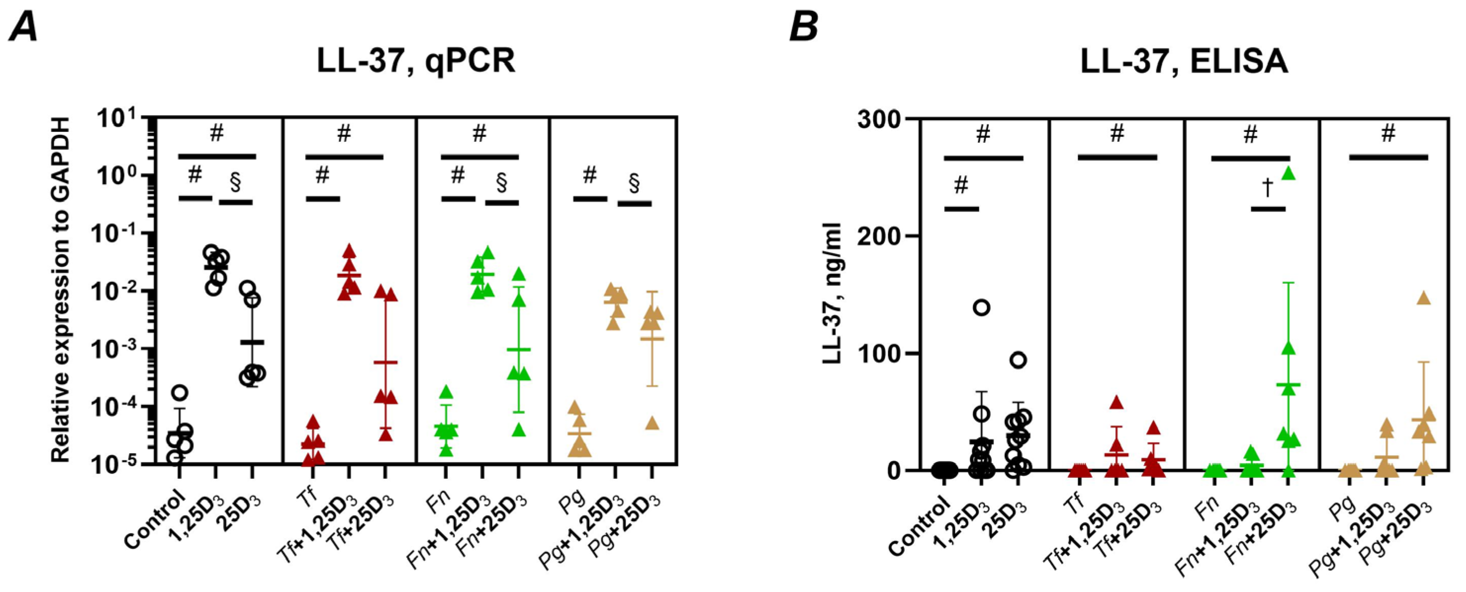
The effects of vitamin D3 metabolites on the expression and secretion of cathelicidin antimicrobial peptide (CAMP/LL-37) are presented in Figure 2. Both 1,25(OH)2D3 and 25(OH)D3 significantly upregulated CAMP gene expression in Ca9-22 cells across all tested conditions, including both uninfected and bacterially infected cells (Figure 2A). Notably, 1,25(OH)2D3 induced higher CAMP mRNA levels than 25(OH)D3. At the protein level, LL-37 levels in the conditioned media were also significantly elevated by both metabolites (Figure 2B), although 25(OH)D3 led to higher LL-37 peptide concentrations (mean values 14–73 ng/mL) than 1,25(OH)2D3 (mean values 4–24 ng/mL).
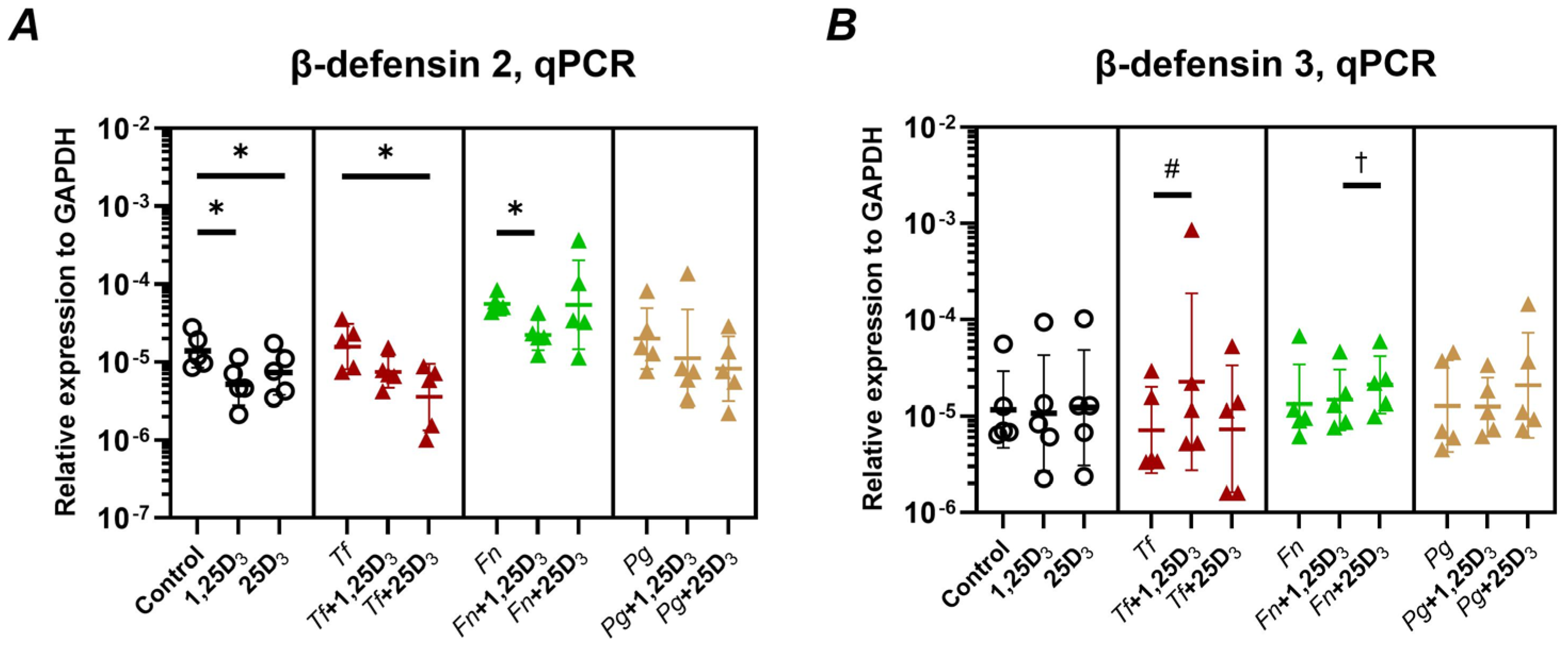
Figure 3 illustrates the effects of vitamin D3 metabolites on the expression of human β-defensins in Ca9-22 cells, with and without bacterial infection. Both 1,25(OH)2D3 and 25(OH)D3 significantly downregulated hBD-2 gene expression in uninfected cells (Figure 3A). In infected cells, 25(OH)D3 significantly suppressed hBD-2 expression following T. forsythia infection, while 1,25(OH)2D3 significantly decreased hBD-2 expression in F. nucleatum-infected cells. Regarding hBD-3, a significant increase in gene expression was observed only in T. forsythia-infected cells treated with 1,25(OH)2D3 (Figure 3B). Moreover, hBD-3 expression was higher in cells treated with 25(OH)D3 than those treated with 1,25(OH)2D3 when infected with F. nucleatum.
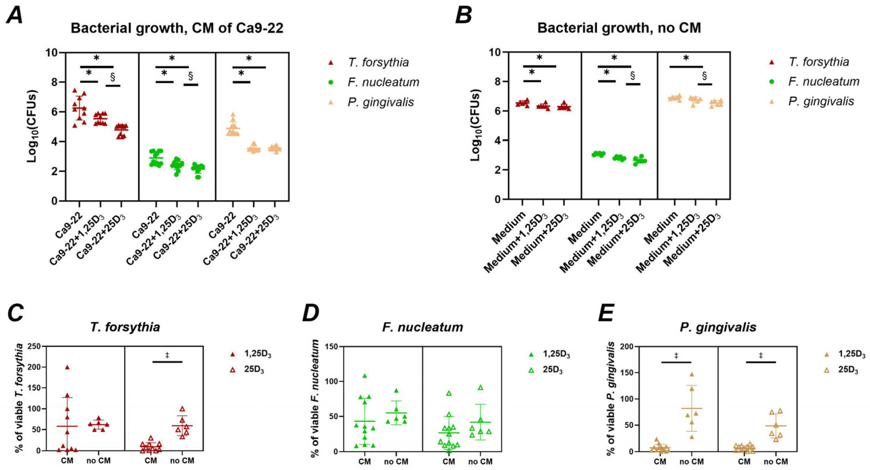
The antimicrobial activity of Ca9-22 cells treated with vitamin D3 metabolites was assessed as described in Section 4.7, and the results are presented in Figure 4. Conditioned media from cells treated with either 1,25(OH)2D3 or 25(OH)D3 significantly inhibited the growth of T. forsythia, F. nucleatum, and P. gingivalis (Figure 4A). A direct antimicrobial effect was also observed when bacteria were exposed to vitamin D3 metabolites alone, in the absence of conditioned media (Figure 4B). The percentages of viable bacterial cells following treatment with either conditioned media or vitamin D3 metabolites alone are summarized in Figure 4C–E. Notably, conditioned medium from 25(OH) D3-treated Ca9-22 cells exhibited a strong antibacterial effect against T. forsythia and P. gingivalis. In contrast, conditioned medium from 1,25(OH)2D3-treated cells showed pronounced inhibition only against P. gingivalis. In other cases, a slight reduction in bacterial viability was observed following treatment with conditioned media, although these changes did not reach statistical significance.

2.3. Gene and Protein Expression of Adhesion Molecules

Figure 5 shows the effects of 1,25(OH)2D3 and 25(OH)D3 on E-cadherin expression at both gene and protein levels in Ca9-22 cells. Treatment with 1,25(OH)2D3 significantly upregulated E-cadherin gene expression in both uninfected and bacterially infected cells (Figure 5A). In contrast, 25(OH)D3 had no significant impact on E-cadherin mRNA levels. At the protein level, flow cytometry analysis revealed minimal changes in the percentage of E-cadherin+ cells. A small but statistically significant decrease was observed with 25(OH)D3 in uninfected cells (from 94.2% to 93.3%) (Figure 5B). Interestingly, the mean fluorescence intensity (MFI) of E-cadherin+ cells was significantly reduced by 1,25(OH)2D3 in the absence of infection. Moreover, in both uninfected cells and cells infected with T. forsythia or P. gingivalis, the MFI was significantly lower after treatment with 1,25(OH)2D3 compared to 25(OH)D3.
Figure 6 illustrates the impact of vitamin D3 metabolites on ICAM-1 gene and protein expression in Ca9-22 cells. ICAM-1 gene expression was significantly decreased only by 1,25(OH)2D3 in P. gingivalis-infected cells (Figure 6A); no other significant changes were observed. The percentage of ICAM-1+ cells was significantly reduced by 1,25(OH)2D3 in uninfected cells (Figure 6B). In T. forsythia-infected cells, a trend toward reduction was seen, but no consistent or significant effects were observed for other conditions. Mean fluorescence intensity of ICAM-1+ cells was significantly decreased by both vitamin D3 metabolites in uninfected cells (Figure 6C). Specifically, 25(OH)D3 reduced MFI. in T. forsythia-infected cells, and 1,25(OH)2D3 decreased MFI in F. nucleatum-infected cells; however, 1,25(OH)2D3 significantly increased ICAM-1 MFI in T. forsythia-infected cells. When comparing metabolites, 1,25(OH)2D3-treatment resulted in significantly higher ICAM-1 MFI than 25(OH)D3 in both uninfected and T. forsythia-infected cells, whereas the opposite was observed in F. nucleatum-infected cells. It is worth noting that, despite statistical significance, the changes in ICAM-1+ cell percentages and MFI induced by either metabolite were relatively modest—generally not exceeding an 8% difference compared to untreated cells.

3. Discussion

The oral epithelium plays a central role in innate immune defense, acting as both a mechanical and immunological barrier against invading pathogens [22]. Vitamin D3, a hormone essential for immune regulation, has been implicated in maintaining oral health [11,44]. This study provides a comprehensive analysis of how two key metabolites of vitamin D3—1,25(OH)2D3 (the active form) and 25(OH)D3 (the circulating precursor)—modulate the response of oral epithelial cells to periodontitis-associated bacteria. Notably, our results reveal previously unrecognized roles for 25(OH)D3 in enhancing antimicrobial responses and gene regulation, independent of its conversion to the active form.
Using Ca9-22 cells, which mimic several features of oral epithelium despite inherent limitations [43,45], we investigated the regulation of inflammatory mediators [46], antimicrobial peptides, barrier-related proteins, and molecules involved in neutrophil migration. We found that 1,25(OH)2D3 upregulated IL-1β gene expression in the absence of infection and in the presence of T. forsythia (Figure 1A), but not at the protein level. This likely reflects the inherently low capacity of Ca9-22 cells to produce IL-1β protein, as levels were near the ELISA-detection threshold. A small but statistically significant increase in IL-1β protein was observed following treatment with 1,25(OH)2D3 in F. nucleatum- and P. gingivalis-infected cells (Figure 1B), although the biological relevance is limited due to low baseline levels. These findings contrast with previous work showing inhibitory effects of 1,25(OH)2D3 on lipopolysaccharide-induced IL-1β expression in human oral keratinocytes [35], possibly due to differences in experimental models. Interestingly, upregulation of IL-1β has been linked to improved barrier integrity in the oral epithelium [47], warranting further investigation into potential barrier-enhancing effects mediated by vitamin D3.
Both vitamin D3 metabolites suppressed IL-8 gene expression, regardless of bacterial infection, though not all effects reached statistical significance (Figure 1C). At the protein level, only 25(OH)D3 significantly reduced IL-8 in F. nucleatum-infected cells (Figure 1D). Notably, IL-8 production was minimal in response to P. gingivalis, likely due to the proteolytic degradation of chemokines by this pathogen [48,49]. These findings are consistent with studies showing vitamin D3-mediated suppression of IL-8 in gingival fibroblasts and periodontal ligament cells [50,51,52], and suggest an anti-inflammatory role through reduced neutrophil recruitment.
Our results also show that both vitamin D3 metabolites robustly induced LL-37, a key antimicrobial peptide [53] with anti-inflammatory properties believed to play a role in maintaining oral health [54,55]. This induction was observed at both the gene and protein levels (Figure 2A,B), regardless of bacterial presence. This confirms and extends previous studies showing vitamin D3-mediated upregulation of LL-37 in oral epithelial cells [33,39,40,41].
Additionally, we examined the expression of β-defensins [56]. The levels of hBD-2 in the gingival sulcus are increased in periodontitis [57]. hBD-3 was shown to inhibit the inflammatory response in vitro and the development of periodontitis in mice in vivo [58]. Both 1,25(OH)2D3 and 25(OH)D3 reduced hBD-2 expression in uninfected cells, with minimal effects during bacterial infection. Conversely, 25(OH)D3 enhanced hBD-3 gene expression, significantly so in T. forsythia-infected cells. Expression levels induced by 25(OH)D3 were also significantly higher than those induced by 1,25(OH)2D3. This finding aligns with a previous study showing upregulation of hBD-3 by 1,25(OH)2D3 [34] and suggests a more pronounced role for 25(OH)D3 in modulating defensin responses.
Crucially, both metabolites enhanced the antimicrobial activity of Ca9-22 cells against oral pathogens (Figure 4A), with 25(OH)D3 demonstrating greater potency, likely reflecting stronger induction of CAMP and hBD-3. This is the first study to demonstrate such an effect for 25(OH)D3, expanding current knowledge beyond its function as a precursor. Direct antimicrobial effects of both metabolites were also observed (Figure 4B), although substantially weaker than cell-mediated effects. Previous studies have demonstrated direct effects of 1,25(OH)2D3 at substantially higher concentrations [59] compared to the much lower levels used in our study (~3-00 µg/mL vs. ~4 ng/mL). Our findings suggest that even low-dose effects, particularly of 25(OH)D3, may be biologically relevant. Interestingly, the antimicrobial efficacy varied among the tested species. Notably, for F. nucleatum, a reduced number of viable bacteria was observed even in the control group lacking both host cells and vitamin D (Figure 4B), indicating that the experimental conditions alone may have negatively affected bacterial viability.
We also evaluated E-cadherin, a key molecule in epithelial barrier maintenance, often degraded during bacterial infection [60,61]. While 1,25(OH)2D3 increased E-cadherin gene expression (Figure 5A), this did not translate into increased surface expression. Instead, a slight decrease in surface protein levels was observed (Figure 5B,C), suggesting a disconnect between transcriptional and post-translational regulation. This contrasts with findings in other epithelial cells [62] and highlights the need for further studies in oral epithelium, particularly given the association between E-cadherin dysregulation and periodontitis [63] and that a potential regulation of E-cadherin by vitamin D3 might impact periodontal disease pathogenesis. When interpreting E-cadherin data, it should be noted that E-cadherin expression is diminished in oral squamous carcinoma compared to the normal epithelium [56,64].
Regarding ICAM-1, which mediates trans-epithelial leukocyte migration [65], we observed minor and context-specific changes. 1,25(OH)2D3 reduced ICAM-1 gene expression in P. gingivalis-infected cells (Figure 6A), while changes in surface protein expression were modest and did not exceed 8% compared to untreated cells (Figure 6B,C). Previous studies have demonstrated downregulation of ICAM-1 by vitamin D3 in lung epithelial cells [66]; however, the relevance of oral epithelium remains unclear and may depend on the inflammatory microenvironment.
Our findings underscore the complex and context-dependent role of vitamin D3 in oral epithelial immunity. Importantly, we demonstrate that 25(OH)D3 has independent, immunomodulatory activity at physiologically relevant concentrations (10 nmol/L), comparable to salivary levels [19]. This is in contrast to serum levels of 1,25(OH)2D3, which are much lower than the concentrations used in vitro [67]. While serum 25(OH)D3 levels are a known predictor of periodontitis risk [11], our data suggest that local actions of both vitamin D3 metabolites in the epithelium may contribute to this association.
Most oral cells can convert 25(OH)D3 to 1,25(OH)2D3 [44,68], but the physiological relevance of this conversion, particularly in inflamed tissue, remains to be defined. It is also possible that inflammatory conditions suppress vitamin D3 signaling [69,70], adding another layer of complexity to its function in periodontal disease. In our study, we observed that 25(OH)D3 exerts biological effects in Ca9-22 cells, suggesting that these cells are also capable of converting it into 1,25(OH)2D3. This still seems possible despite the expression of CYP27B1, an enzyme responsible for the conversion of 25(OH)D3 into 1,25(OH)2D3 is diminished in oral squamous carcinoma [71]. The physiological relevance of local vitamin D3 activation, as well as the exact cellular mechanisms of its action, needs to be clarified by further studies.
This study has several limitations. It was conducted in vitro using a squamous carcinoma cell line, which does not fully mimic the behavior of normal oral epithelium. Additionally, the infection model involved a single bacterial species, whereas in vivo exposure typically involves polymicrobial biofilms and complex host-microbe interactions. Moreover, the experimental design did not account for oral commensal bacteria, which play a crucial role in maintaining oral health and the resilience of the subgingival ecological niche to external stimuli. Therefore, investigating the influence of commensals on the effects mediated by vitamin D3 represents an essential step towards developing a model that more accurately mimics the in vivo oral ecosystem.

4. Materials and Methods

4.1. Cell Cultures

The human oral squamous cell carcinoma (OSCC) cell line Ca9-22 was employed as an in vitro model for oral epithelial cells [43]. Cells were maintained in Minimum Essential Medium Eagle (MEM; Sigma, St. Louis, MO, USA) supplemented with 10% fetal bovine serum (FBS; Gibco, Carlsbad, CA, USA), 100 U/mL penicillin, 100 μg/mL streptomycin (P/S; Gibco, Carlsbad, CA, USA) Cultures were incubated at 37 °C in a humidified atmosphere with 5% CO2. All experiments were conducted using cells between passages 3 and 7.

4.2. Cultivation of Bacteria

Tannerella forsythia ATCC 43,037 (American Type Culture Collection, Manassas, VA, USA) was cultured anaerobically at 37°C for 5 days on Fastidious Anaerobe agar (FA; Neogen, Lansing, MI, USA) supplemented with 5% horse blood (Sigma, St.-Louis, MO, USA) and 20 μg/mL N-acetylmuramic acid (NAMA; Sigma-Aldrich, St. Louis, MO, USA) [29,72]. Fusobacterium nucleatum subsp. nucleatum KP-F8 (OMZ598) and Porphyromonas gingivalis ATCC 33,277 were grown anaerobically at 37 °C for 5 days on Columbia Blood Agar (CBA; Oxoid, Altrincham, UK) supplemented with 5% horse blood [73].

4.3. Infection Procedure

Cells were seeded as a monolayer at a density of 1.7 × 10⁵ cells/well in 500 µL of MEM supplemented with 10% FBS and antibiotics in 24-well plates. After 24 h, the medium was replaced with MEM lacking FBS and antibiotics, and cells were treated with either 10 nmol/L of 1,25(OH)2D3 or 10 nmol/L 25(OH)D3 (both from Cayman Chemical, Ann Arbor, MI, USA). FBS was omitted to avoid potential interference from its components with host-bacteria interactions [74]. Subsequently, cells were infected with either T. forsythia, F. nucleatum, or P. gingivalis at a multiplicity of infection (MOI) of 50. For this purpose, bacterial cells were harvested from agar plates, washed with MEM, and the optical density at 600 nm (OD600) was measured using a Biochrom Ultrospec 10 cell density meter (Harvard Bioscience, Holliston, MA, USA). For each bacterial suspension, the correlation between OD600 and colony-forming units, as determined previously [48] was used as a basis for calculating the MOI. The concentrations of vitamin D3 metabolites and MOI were based on our previously established protocols [49,68,75]. Each treatment and infection combination was performed in duplicate. Cells were exposed to both vitamin D3 metabolites and bacteria for 24 h, and gene and protein expression of selected markers was analyzed.

4.4. Reverse Transcription-Quantitative Polymerase Chain Reaction (Rt-Qpcr)

Cells were lysed, and mRNA was reverse transcribed to cDNA using a TaqMan Gene Expression Cells-to-Ct kit (Invitrogen, Waltham, MA, USA) following the manufacturer’s protocol, as previously described [49]. cDNA synthesis was carried out on a Biometra TOne PCR thermal cycler (Analytik Jena, Jena, Germany) under the following conditions: 37 °C for 1 h, 95 °C for 5 min, followed by cooling to 4 °C. qPCR was performed to detect specific transcripts using TaqMan Gene Expression Assays (Applied Biosystems, Waltham, MA, USA). The following inventoried assay IDs were used: DEFB103 (β-defensin 103): Hs00218678_m1; IL-1β (interleukin-1β): Hs01555410_m1; IL-8 (interleukin-8): Hs00174103; DEFB4B (β-defensin 4B): Hs00823638_m1; CDH1 (E-cadherin): Hs01023894_m1; ICAM1 (intercellular adhesion molecule 1): Hs00164932_m1; CAMP (cathelicidin antimicrobial peptide, LL-37): Hs00189038_m1; GAPDH (housekeeping gene): Hs99999905_m1. qPCR was conducted using a QuantStudio 3 Real-Time PCR System (Applied Biosystems, Waltham, MA, USA) with the following thermal cycling conditions: 95 °C for 10 min, followed by 50 cycles at 95 °C for 15 s (denaturation) and 60 °C for 1 min (annealing and extension). Cycle threshold values were determined for each gene. The gene expression was quantified in relation to that of GAPDH by the 2−ΔCt method, where ΔCt = (Cttarget − CtGAPDH).

4.5. Protein and Peptide Production Measurement by Elisa

Supernatants were collected at the end of the treatment period and stored at −80 °C until analysis. The concentrations of IL-1β, IL-8, and CAMP were quantified using commercially available ELISA kits, according to the manufacturers’ protocols.: IL-1β (Cat. Nr. 88-7261-88, Invitrogen, Waltham, MA, USA); IL-8 (Cat. Nr. 88-8086-88, Invitrogen, Waltham, MA, USA); CAMP (Cat. Nr. SEC419Hu, Cloud-Clone Corp., Houston, TX, USA). Absorbance was measured using a microplate reader (BioTek Instruments, Winooski, VT, USA), and data were analyzed with Gen5 software (Version 2.09, BioTek Instruments, Winooski, VT, USA).

4.6. Analysis of Icam1 and E-Cadherin by Flow Cytometry

At the end of the experiment, Ca9-22 cells from two wells were pooled and transferred into FACS tubes. The culture medium was removed by centrifugation, and the cells were resuspended in 100 µL of FACS buffer (PBS supplemented with 3% BSA and 0.09% NaN3). Cells were stained with either PE-conjugated anti-ICAM-1 antibody (Cat. Nr. 12-0549-92, eBioscience, Santa Clara, CA, USA) or PE-conjugated anti-E-cadherin antibody (Cat. Nr. A15784, Life Technologies, Carlsbad, CA, USA), following the manufacturer’s instructions. Immediately after staining, samples were analyzed using an Attune NxT Flow Cytometer (Thermo Fisher Scientific, Waltham, MA, USA). Data were processed using Attune NxT Flow Cytometer Software v3.1, with results reported as mean fluorescence intensity (MFI) and percentage of positive cells relative to the stained untreated control.

4.7. Antibacterial Activity of Ca9-22 Cells

Ca9-22 cells were treated with either 10 nmol/L of 1,25(OH)2D3 or 10 nmol/L 25(OH) D3 in the absence of bacterial infection for 24 h, as described above. Following treatment, conditioned media were collected and subsequently applied to bacterial cultures. A total of 500 µL of conditioned media–either untreated or treated with 10 nmol/L of 1,25(OH)2D3 or 10 nmol/L of 25(OH)D3–was mixed with 500 µL of T. forsythia, F. nucleatum, or P. gingivalis suspended in MEM, at a multiplicity of infection (MOI) of 50. The mixtures were incubated under anaerobic conditions for 24 h, after which 50 µL from each was plated on agar plates for bacterial enumeration. Plating was performed using sterilized solid glass beads (4 mm diameter; (Sigma, St. Louis, MO, USA) to evenly distribute the bacterial suspension. Colony-forming units (CFU) were counted from seven technical replicates. As a control, cell culture medium containing 10 nmol/L of 1,25(OH)2D3 or 10 nmol/L of 25(OH)D3, not pre-conditioned with Ca9-22 cells, was used.

4.8. Statistical Analysis

Data are presented as mean values ± standard error of the mean (SEM) from at least five biological replicates. Statistical differences between experimental groups were assessed using the Wilcoxon signed-rank test. Differences in the percentage of viable bacteria between treatments with and without conditioned media were analyzed by the Mann–Whitney U test. Analyses were performed using IBM SPSS 27.0 software (IBM, Chicago, IL, USA). A p-value of <0.05 was considered statistically significant. Data visualization was carried out using dot plots generated in GraphPad Prism version 8.0.2. (La Jolla, CA, USA).

5. Conclusions

In conclusion, our study is the first to comprehensively show that 25(OH)D3, at physiologically relevant concentrations, exerts direct immunomodulatory and antimicrobial effects on oral epithelial cells, complementing and sometimes exceeding the effects of 1,25(OH)2D3. These findings expand our understanding of vitamin D3 biology in the oral cavity and underscore its potential as a modulator of epithelial defense. Further in vivo and clinical studies are needed to determine the translational relevance of these findings.

Author Contributions

Conceptualization, C.S. and O.A.; methodology, F.K., S.B., F.L.K. and C.B.; formal analysis, F.K., S.B., C.B. and O.A.; investigation, F.K., S.B., F.L.K. and C.B.; resources, C.S. and O.A.; data curation, F.K.; writing—original draft preparation, F.K. and O.A.; writing—review and editing, S.B., F.L.K., C.B. and C.S.; visualization, F.K. and O.A.; supervision, C.S. and O.A.; project administration, C.S. and O.A.; funding acquisition, C.S. and O.A. All authors have read and agreed to the published version of the manuscript.

Funding

This research was funded by the Austrian Science Fund FWF, grants number P34642 and PAT4147223.

Institutional Review Board Statement

Not applicable.

Informed Consent Statement

Not applicable.

Data Availability Statement

The original contributions presented in this study are included in the article. Further inquiries can be directed to the corresponding author(s).

Conflicts of Interest

The authors declare no conflicts of interest. The funders had no role in the design of the study; in the collection, analyses, or interpretation of data; in the writing of the manuscript; or in the decision to publish the results.

Abbreviations

The following abbreviations are used in this manuscript:
1,25(OH)2D3 1,25-dihydroxyvitamin D3 (active form of vitamin D3)
25(OH)D3 25-hydroxyvitamin D3 (circulating precursor of vitamin D3)
ATCCAmerican Type Culture Collection
BSABovine serum albumin
CAMP, LL-37Cathelicidin Antimicrobial Peptide
CBAColumbia blood agar
cDNAComplementary DNA
CFUColony-Forming Units
CtCycle threshold
E-cadherin Epithelial cadherin (a cell adhesion molecule)
ELISAEnzyme-Linked Immunosorbent Assay
FAA Fastidious Anaerobe Agar
FACSFluorescence-Activated Cell Sorting
FBSFetal Bovine Serum
GAPDHGlyceraldehyde 3-phosphate dehydrogenase (housekeeping gene)
hBD-2Human beta-defensin 2
hBD-3 Human beta-defensin 3
ICAM-1 Intercellular Adhesion Molecule 1
IL-1β Interleukin 1 beta
IL-8 Interleukin 8
MEMMinimum Essential Medium
MFIMean Fluorescence Intensity
MOI Multiplicity of Infection
NAMAN-acetylmuramic acid
OD600 Optical Density at 600 nm
OSCCOral Squamous Cell Carcinoma
P/S Penicillin/Streptomycin
PBSPhosphate-Buffered Saline
PE Phycoerythrin (a fluorescent dye)
SDStandard Deviation
SEMStandard Error of the Mean
VDRVitamin D Receptor

References

  1. Deo, P.N.; Deshmukh, R. Oral microbiome: Unveiling the fundamentals. J. Oral Maxillofac. Pathol. 2019, 23, 122–128. [Google Scholar] [CrossRef]
  2. Hajishengallis, G.; Lamont, R.J. Beyond the red complex and into more complexity: The polymicrobial synergy and dysbiosis (PSD) model of periodontal disease etiology. Mol. Oral Microbiol. 2012, 27, 409–419. [Google Scholar] [CrossRef] [PubMed]
  3. Radaic, A.; Kapila, Y.L. The oralome and its dysbiosis: New insights into oral microbiome-host interactions. Comput. Struct. Biotechnol. J. 2021, 19, 1335–1360. [Google Scholar] [CrossRef] [PubMed]
  4. Wade, W.G. The oral microbiome in health and disease. Pharmacol. Res. 2013, 69, 137–143. [Google Scholar] [CrossRef] [PubMed]
  5. Kilian, M.; Chapple, I.L.; Hannig, M.; Marsh, P.D.; Meuric, V.; Pedersen, A.M.; Tonetti, M.S.; Wade, W.G.; Zaura, E. The oral microbiome—An update for oral healthcare professionals. Br. Dent. J. 2016, 221, 657–666. [Google Scholar] [CrossRef]
  6. Socransky, S.; Haffajee, A.; Cugini, M.; Smith, C.; Kent, R. Microbial complexes in subgingival plaque. J. Clin. Periodontol. 1998, 25, 134–144. [Google Scholar] [CrossRef]
  7. Belibasakis, G.N.; Belstrøm, D.; Eick, S.; Gursoy, U.K.; Johansson, A.; Könönen, E. Periodontal microbiology and microbial etiology of periodontal diseases: Historical concepts and contemporary perspectives. Periodontol. 2000 2023, 1–17. [Google Scholar] [CrossRef]
  8. Costalonga, M.; Herzberg, M.C. The oral microbiome and the immunobiology of periodontal disease and caries. Immunol. Lett. 2014, 162, 22–38. [Google Scholar] [CrossRef]
  9. Wen, C.-C.; Lin, J.C.Y.; Salamanca, E.; Teng, N.-C.; Lin, Y.-J.; Wu, Y.-F.; Chang, H.-M.; Chang, W.-J. Full-length 16S rRNA sequencing reveals microbial characteristics in supragingival plaque of periodontitis patients. J. Dent. Sci. 2025, in press. [CrossRef]
  10. Loos, B.G.; Van Dyke, T.E. The role of inflammation and genetics in periodontal disease. Periodontol. 2000 2020, 83, 26–39. [Google Scholar] [CrossRef]
  11. Lu, E.M. The role of vitamin D in periodontal health and disease. J. Periodontal. Res. 2023, 58, 213–224. [Google Scholar] [CrossRef]
  12. Wacker, M.; Holick, M.F. Sunlight and Vitamin D: A global perspective for health. Derm. Endocrinol. 2013, 5, 51–108. [Google Scholar] [CrossRef]
  13. Lips, P. Vitamin D physiology. Prog. Biophys. Mol. Biol. 2006, 92, 4–8. [Google Scholar] [CrossRef] [PubMed]
  14. Pike, J.W. Intracellular receptors mediate the biologic action of 1,25-dihydroxyvitamin D3. Nutr. Rev. 1985, 43, 161–168. [Google Scholar] [CrossRef] [PubMed]
  15. Zmijewski, M.A.; Carlberg, C. Vitamin D receptor(s): In the nucleus but also at membranes? Exp. Dermatol. 2020, 29, 876–884. [Google Scholar] [CrossRef] [PubMed]
  16. Amrein, K.; Scherkl, M.; Hoffmann, M.; Neuwersch-Sommeregger, S.; Kostenberger, M.; Tmava Berisha, A.; Martucci, G.; Pilz, S.; Malle, O. Vitamin D deficiency 2.0: An update on the current status worldwide. Eur. J. Clin. Nutr. 2020, 74, 1498–1513. [Google Scholar] [CrossRef]
  17. Holick, M.F. Vitamin D status: Measurement, interpretation, and clinical application. Ann. Epidemiol. 2009, 19, 73–78. [Google Scholar] [CrossRef]
  18. Bahramian, A.; Falsafi, P.; Abbasi, T.; Ghanizadeh, M.; Abedini, M.; Kavoosi, F.; Kouhsoltani, M.; Noorbakhsh, F.; Dabbaghi Tabriz, F.; Rajaeih, S.; et al. Comparing serum and salivary levels of vitamin D in patients with recurrent aphthous stomatitis and healthy individuals. J. Dent. 2018, 19, 295–300. [Google Scholar]
  19. Davidopoulou, S.; Makedou, K.; Kourti, A.; Gkeka, I.; Karakostas, P.; Pikilidou, M.; Tolidis, K.; Kalfas, S. Vitamin D and LL-37 in serum and saliva: Insights into oral immunity. Curr. Issues Mol. Biol. 2025, 47, 102. [Google Scholar] [CrossRef]
  20. Machado, V.; Lobo, S.; Proenca, L.; Mendes, J.J.; Botelho, J. Vitamin D and periodontitis: A systematic review and meta-analysis. Nutrients 2020, 12, 2177. [Google Scholar] [CrossRef]
  21. Holick, M.F. Sunlight and vitamin D for bone health and prevention of autoimmune diseases, cancers, and cardiovascular disease. Am. J. Clin. Nutr. 2004, 80, 1678S–1688S. [Google Scholar] [CrossRef]
  22. Groeger, S.; Meyle, J. Oral mucosal epithelial cells. Front. Immunol. 2019, 10, 208. [Google Scholar] [CrossRef] [PubMed]
  23. Lamont, R.J.; Chan, A.; Belton, C.M.; Izutsu, K.T.; Vasel, D.; Weinberg, A. Porphyromonas gingivalis invasion of gingival epithelial cells. Infect. Immun. 1995, 63, 3878–3885. [Google Scholar] [CrossRef] [PubMed]
  24. Bugueno, I.M.; Batool, F.; Keller, L.; Kuchler-Bopp, S.; Benkirane-Jessel, N.; Huck, O. Porphyromonas gingivalis bypasses epithelial barrier and modulates fibroblastic inflammatory response in an in vitro 3D spheroid model. Sci. Rep. 2018, 8, 14914. [Google Scholar] [CrossRef] [PubMed]
  25. Chen, W.; Alshaikh, A.; Kim, S.; Kim, J.; Chun, C.; Mehrazarin, S.; Lee, J.; Lux, R.; Kim, R.H.; Shin, K.H.; et al. Porphyromonas gingivalis impairs oral epithelial barrier through targeting GRHL2. J. Dent. Res. 2019, 98, 1150–1158. [Google Scholar] [CrossRef]
  26. Yee, M.; Kim, S.; Sethi, P.; Duzgunes, N.; Konopka, K. Porphyromonas gingivalis stimulates IL-6 and IL-8 secretion in GMSM-K, HSC-3 and H413 oral epithelial cells. Anaerobe 2014, 28, 62–67. [Google Scholar] [CrossRef]
  27. Honma, K.; Mishima, E.; Sharma, A. Role of Tannerella forsythia NanH sialidase in epithelial cell attachment. Infect. Immun. 2011, 79, 393–401. [Google Scholar] [CrossRef]
  28. Mishima, E.; Sharma, A. Tannerella forsythia invasion in oral epithelial cells requires phosphoinositide 3-kinase activation and clathrin-mediated endocytosis. Microbiology 2011, 157, 2382–2391. [Google Scholar] [CrossRef]
  29. Schäffer, C.; Andrukhov, O. The intriguing strategies of Tannerella forsythia’s host interaction. Front. Oral Health 2024, 5, 1434217. [Google Scholar] [CrossRef]
  30. Groeger, S.; Zhou, Y.; Ruf, S.; Meyle, J. Pathogenic mechanisms of Fusobacterium nucleatum on oral epithelial cells. Front. Oral Health 2022, 3, 831607. [Google Scholar] [CrossRef]
  31. Gursoy, U.K.; Kononen, E.; Uitto, V.J. Stimulation of epithelial cell matrix metalloproteinase (MMP-2, -9, -13) and interleukin-8 secretion by fusobacteria. Oral Microbiol. Immunol. 2008, 23, 432–434. [Google Scholar] [CrossRef]
  32. Yuan, F.N.; Valiyaparambil, J.; Woods, M.C.; Tran, H.; Pant, R.; Adams, J.S.; Mallya, S.M. Vitamin D signaling regulates oral keratinocyte proliferation in vitro and in vivo. Int. J. Oncol. 2014, 44, 1625–1633. [Google Scholar] [CrossRef]
  33. Menzel, L.P.; Ruddick, W.; Chowdhury, M.H.; Brice, D.C.; Clance, R.; Porcelli, E.; Ryan, L.K.; Lee, J.; Yilmaz, O.; Kirkwood, K.L.; et al. Activation of vitamin D in the gingival epithelium and its role in gingival inflammation and alveolar bone loss. J. Periodontal Res. 2019, 54, 444–452. [Google Scholar] [CrossRef]
  34. Ge, X.; Wang, Y.; Xie, H.; Li, R.; Zhang, F.; Zhao, B.; Du, J. 1,25(OH)2 D3 blocks IFNb production through regulating STING in epithelial layer of oral lichen planus. J. Cell Mol. Med. 2022, 26, 3751–3759. [Google Scholar] [CrossRef]
  35. Ge, X.; Wang, L.; Li, M.; Xu, N.; Yu, F.; Yang, F.; Li, R.; Zhang, F.; Zhao, B.; Du, J. Vitamin D/VDR signaling inhibits LPS-induced IFNg and IL-1b in oral epithelia by regulating hypoxia-inducible factor-1a signaling pathway. Cell Commun. Signal. 2019, 17, 18. [Google Scholar] [CrossRef] [PubMed]
  36. De Filippis, A.; Fiorentino, M.; Guida, L.; Annunziata, M.; Nastri, L.; Rizzo, A. Vitamin D reduces the inflammatory response by Porphyromonas gingivalis infection by modulating human beta-defensin-3 in human gingival epithelium and periodontal ligament cells. Int. Immunopharmacol. 2017, 47, 106–117. [Google Scholar] [CrossRef] [PubMed]
  37. Kirkwood, K.L.; Van Dyke, T.E.; Kirkwood, C.L.; Zhang, L.; Panezai, J.; Duran-Pinedo, A.E.; Figgins, E.L.; Ryan, L.K.; Frias-Lopez, J.J.; Diamond, G. Topical vitamin D prevents bone loss and inflammation in a mouse model. J. Dent. Res. 2024, 103, 908–915. [Google Scholar] [CrossRef] [PubMed]
  38. Nazzal, A.; Tipton, D.A.; Karydis, A.; Slominski, A.; Stein, S.H. Vitamin D stimulates epithelial cell proliferation and facilitates wound closure via a cathelicidin independent pathway in vitro. Periodon. Prosthodon. 2016, 2, 8. [Google Scholar] [CrossRef]
  39. McMahon, L.; Schwartz, K.; Yilmaz, O.; Brown, E.; Ryan, L.K.; Diamond, G. Vitamin D-mediated induction of innate immunity in gingival epithelial cells. Infect. Immun. 2011, 79, 2250–2256. [Google Scholar] [CrossRef]
  40. Figgins, E.L.; Arora, P.; Gao, D.; Porcelli, E.; Ahmed, R.; Daep, C.A.; Keele, G.; Ryan, L.K.; Diamond, G. Enhancement of innate immunity in gingival epithelial cells by vitamin D and HDAC inhibitors. Front. Oral Health 2024, 5, 1378566. [Google Scholar] [CrossRef]
  41. Tada, H.; Shimizu, T.; Nagaoka, I.; Takada, H. Vitamin D3 analog maxacalcitol (OCT) induces hCAP-18/LL-37 production in human oral epithelial cells. Biomed. Res. 2016, 37, 199–205. [Google Scholar] [CrossRef] [PubMed]
  42. Bierbaumer, L.; Schwarze, U.Y.; Gruber, R.; Neuhaus, W. Cell culture models of oral mucosal barriers: A review with a focus on applications, culture conditions and barrier properties. Tissue Barriers 2018, 6, 1479568. [Google Scholar] [CrossRef]
  43. Rivera, C.; Venegas, B. Histological and molecular aspects of oral squamous cell carcinoma (Review). Oncol. Lett. 2014, 8, 7–11. [Google Scholar] [CrossRef] [PubMed]
  44. Andrukhov, O.; Blufstein, A.; Behm, C.; Moritz, A.; Rausch-Fan, X. Vitamin D3 and Dental Mesenchymal Stromal Cells. Appl. Sci. 2020, 10, 4527. [Google Scholar] [CrossRef]
  45. Tian, Z.; Zhao, Z.; Rausch, M.A.; Behm, C.; Tur, D.; Shokoohi-Tabrizi, H.A.; Andrukhov, O.; Rausch-Fan, X. A comparative study of the epithelial regeneration capacities of two biomaterials in vitro. BMC Oral Health 2025, 25, 640. [Google Scholar] [CrossRef]
  46. Eskan, M.A.; Benakanakere, M.R.; Rose, B.G.; Zhang, P.; Zhao, J.; Stathopoulou, P.; Fujioka, D.; Kinane, D.F. Interleukin-1b modulates proinflammatory cytokine production in human epithelial cells. Infect. Immun. 2008, 76, 2080–2089. [Google Scholar] [CrossRef]
  47. Stolte, K.N.; Pelz, C.; Yapto, C.V.; Raguse, J.D.; Dommisch, H.; Danker, K. IL-1b strengthens the physical barrier in gingival epithelial cells. Tissue Barriers 2020, 8, 1804249. [Google Scholar] [CrossRef]
  48. Braun, M.L.; Tomek, M.B.; Grunwald-Gruber, C.; Nguyen, P.Q.; Bloch, S.; Potempa, J.S.; Andrukhov, O.; Schäffer, C. Shut-down of type IX protein secretion alters the host immune response to Tannerella forsythia and Porphyromonas gingivalis. Front. Cell. Infect. Microbiol. 2022, 12, 835509. [Google Scholar] [CrossRef]
  49. Kendlbacher, F.L.; Bloch, S.; Hager-Mair, F.F.; Schäffer, C.; Andrukhov, O. Red-complex bacteria exhibit distinctly different interactions with human periodontal ligament stromal cells compared to Fusobacterium nucleatum. Arch. Oral Biol. 2024, 164, 106004. [Google Scholar] [CrossRef]
  50. Andrukhov, O.; Andrukhova, O.; Hulan, U.; Tang, Y.; Bantleon, H.P.; Rausch-Fan, X. Both 25-hydroxyvitamin-D3 and 1,25-dihydroxyvitamin-D3 reduces inflammatory response in human periodontal ligament cells. PLoS ONE 2014, 9, e90301. [Google Scholar] [CrossRef]
  51. Liu, K.; Meng, H.; Hou, J. Characterization of the autocrine/paracrine function of vitamin D in human gingival fibroblasts and periodontal ligament cells. PLoS ONE 2012, 7, e39878. [Google Scholar] [CrossRef]
  52. Nebel, D.; Svensson, D.; Arosenius, K.; Larsson, E.; Jonsson, D.; Nilsson, B.O. 1alpha,25-dihydroxyvitamin D3 promotes osteogenic activity and downregulates proinflammatory cytokine expression in human periodontal ligament cells. J. Periodontal Res. 2015, 50, 666–673. [Google Scholar] [CrossRef]
  53. Kosciuczuk, E.M.; Lisowski, P.; Jarczak, J.; Strzalkowska, N.; Jozwik, A.; Horbanczuk, J.; Krzyzewski, J.; Zwierzchowski, L.; Bagnicka, E. Cathelicidins: Family of antimicrobial peptides. A review. Mol. Biol. Rep. 2012, 39, 10957–10970. [Google Scholar] [CrossRef] [PubMed]
  54. Jonsson, D.; Nilsson, B.O. The antimicrobial peptide LL-37 is anti-inflammatory and proapoptotic in human periodontal ligament cells. J. Periodontal Res. 2012, 47, 330–335. [Google Scholar] [CrossRef] [PubMed]
  55. He, Y.; Zhou, Y.; Liu, N.; Zhang, W.; Chen, X.; Qiu, G.; Shen, Y. Cathelicidin LL-37 in periodontitis: Current research advances and future prospects—A review. Int. Immunopharmacol. 2025, 150, 114277. [Google Scholar] [CrossRef] [PubMed]
  56. Weinberg, A.; Jin, G.; Sieg, S.; McCormick, T.S. The yin and yang of human beta-defensins in health and disease. Front. Immunol. 2012, 3, 294. [Google Scholar] [CrossRef]
  57. Sidharthan, S.; Dharmarajan, G.; Kulloli, A. Gingival crevicular fluid levels of Interleukin-22 (IL-22) and human beta defensin-2 (hBD-2) in periodontal health and disease: A correlative study. J. Oral Biol. Craniofac Res. 2020, 10, 498–503. [Google Scholar] [CrossRef]
  58. Cui, D.; Lyu, J.; Li, H.; Lei, L.; Bian, T.; Li, L.; Yan, F. Human b-defensin 3 inhibits periodontitis development by suppressing inflammatory responses in macrophages. Mol. Immunol. 2017, 91, 65–74. [Google Scholar] [CrossRef]
  59. Grenier, D.; Morin, M.P.; Fournier-Larente, J.; Chen, H. Vitamin D inhibits the growth of and virulence factor gene expression by Porphyromonas gingivalis and blocks activation of the nuclear factor kappa B transcription factor in monocytes. J. Periodontal Res. 2016, 51, 359–365. [Google Scholar] [CrossRef]
  60. Devaux, C.A.; Mezouar, S.; Mege, J.L. The E-cadherin cleavage associated to pathogenic bacteria infections can favor bacterial invasion and transmigration, dysregulation of the immune response and cancer induction in humans. Front. Microbiol. 2019, 10, 2598. [Google Scholar] [CrossRef]
  61. Bloch, S.; Hager-Mair, F.F.; Bacher, J.; Tomek, M.B.; Janesch, B.; Andrukhov, O.; Schäffer, C. Investigating the role of a Tannerella forsythia HtrA protease in host protein degradation and inflammatory response. Front. Oral Health 2024, 5, 1425937. [Google Scholar] [CrossRef]
  62. Janubova, M.; Zitnanova, I. The effects of vitamin D on different types of cells. Steroids 2024, 202, 109350. [Google Scholar] [CrossRef]
  63. Lialios, P.; Alimperti, S. Role of E-cadherin in epithelial barrier dysfunction: Implications for bacterial infection, inflammation, and disease pathogenesis. Front. Cell. Infect. Microbiol. 2025, 15, 1506636. [Google Scholar] [CrossRef]
  64. Lopez-Verdin, S.; Martinez-Fierro, M.L.; Garza-Veloz, I.; Zamora-Perez, A.; Grajeda-Cruz, J.; Gonzalez-Gonzalez, R.; Molina-Frechero, N.; Arocena-Sutz, M.; Bologna-Molina, R. E-Cadherin gene expression in oral cancer: Clinical and prospective data. Med. Oral Patol. Oral Cir. Buc. 2019, 24, e444–e451. [Google Scholar] [CrossRef] [PubMed]
  65. Diamond, M.S.; Staunton, D.E.; de Fougerolles, A.R.; Stacker, S.A.; Garcia-Aguilar, J.; Hibbs, M.L.; Springer, T.A. ICAM-1 (CD54): A counter-receptor for Mac-1 (CD11b/CD18). J. Cell Biol. 1990, 111, 3129–3139. [Google Scholar] [CrossRef] [PubMed]
  66. Chen, Y.C.; Sung, H.C.; Chuang, T.Y.; Lai, T.C.; Lee, T.L.; Lee, C.W.; Lee, I.T.; Chen, Y.L. Vitamin D3 decreases TNF-a-induced inflammation in lung epithelial cells through a reduction in mitochondrial fission and mitophagy. Cell Biol. Toxicol. 2022, 38, 427–450. [Google Scholar] [CrossRef] [PubMed]
  67. Pagana, K.D.; Pagana, T.J.; Pagana, T.N. Mosby’s Diagnostic and laboratory Test Reference—E-Book, 14th ed.; Elsevier: Amsterdam, The Netherlands, 2018. [Google Scholar]
  68. Behm, C.; Blufstein, A.; Gahn, J.; Moritz, A.; Rausch-Fan, X.; Andrukhov, O. 25-hydroxyvitamin D(3) generates immunomodulatory plasticity in human periodontal ligament-derived mesenchymal stromal cells that is inflammatory context-dependent. Front. Immunol. 2023, 14, 1100041. [Google Scholar] [CrossRef]
  69. Blufstein, A.; Behm, C.; Kubin, B.; Gahn, J.; Moritz, A.; Rausch-Fan, X.; Andrukhov, O. Transcriptional activity of vitamin D receptor in human periodontal ligament cells is diminished under inflammatory conditions. J. Periodontol. 2020, 92, 137–148. [Google Scholar] [CrossRef]
  70. Blufstein, A.; Behm, C.; Kubin, B.; Gahn, J.; Rausch-Fan, X.; Moritz, A.; Andrukhov, O. Effect of vitamin D3 on the osteogenic differentiation of human periodontal ligament stromal cells under inflammatory conditions. J. Periodontal Res. 2021, 56, 579–588. [Google Scholar] [CrossRef]
  71. Guo, J.; Zhou, S.; Huang, P.; Xu, S.; Zhang, G.; He, H.; Zeng, Y.; Xu, C.X.; Kim, H.; Tan, Y. NNK-mediated upregulation of DEPDC1 stimulates the progression of oral squamous cell carcinoma by inhibiting CYP27B1 expression. Am. J. Cancer Res. 2020, 10, 1745–1760. [Google Scholar]
  72. Bloch, S.; Thurnheer, T.; Murakami, Y.; Belibasakis, G.N.; Schäffer, C. Behavior of two Tannerella forsythia strains and their cell surface mutants in multispecies oral biofilms. Mol. Oral Microbiol. 2017, 32, 404–418. [Google Scholar] [CrossRef]
  73. Lima, B.P.; Shi, W.; Lux, R. Identification and characterization of a novel Fusobacterium nucleatum adhesin involved in physical interaction and biofilm formation with Streptococcus gordonii. Microbiologyopen 2017, 6, e00444. [Google Scholar] [CrossRef]
  74. Santos, L.M.; Cardoso, P.E.S.; Diniz, E.A.; Rahhal, J.G.; Sipert, C.R. Different concentrations of fetal bovine serum affect cytokine modulation in lipopolysaccharide-activated apical papilla cells in vitro. J. Appl. Oral Sci. 2023, 31, e20230020. [Google Scholar] [CrossRef]
  75. Behm, C.; Miłek, O.; Schwarz, K.; Rausch-Fan, X.; Moritz, A.; Andrukhov, O. 1,25-dihydroxyvitamin-D(3) distinctly impacts the paracrine and cell-to-cell contact interactions between hPDL-MSCs and CD4(+) T lymphocytes. Front. Immunol. 2024, 15, 1448597. [Google Scholar] [CrossRef]
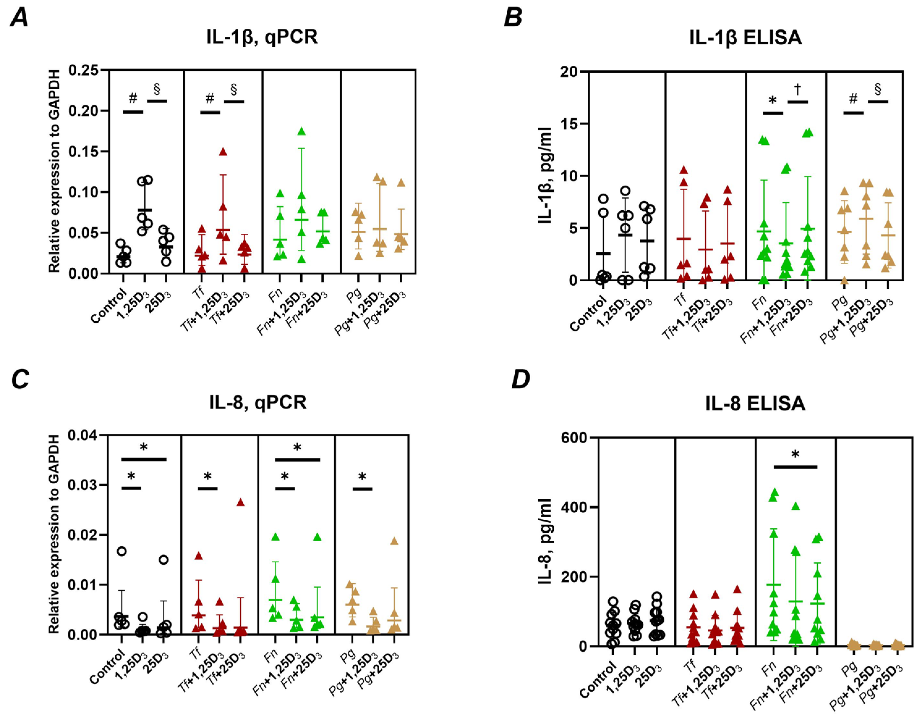
Figure 1. Effect of different vitamin D3 metabolites on the gene expression and production of inflammatory mediators in Ca9-22 cells. Ca9-22 cells were treated with 10 nmol/L of either 1,25(OH)2D3 or 25(OH)D3 for 24 h. Treatments were performed in both uninfected cells (circle) and cells infected with T. forsythia (red triangles), F. nucleatum (green triangles), or P. gingivalis (yellow triangles) at an MOI of 50. Following treatment, the gene expression and protein levels of IL-1β (A,B) and IL-8 (C,D) were assessed using qPCR and ELISA, respectively. Panels (A,C) show the gene expression relative to that of GAPDH calculated by the 2−ΔCt method. Panels (B,D) represent the concentrations of the corresponding proteins in the conditioned media after treatment and infection, respectively. Each data point represents an individual experiment; lines and error bars indicate the mean and SD, respectively. Symbols indicate statistically significant differences (p < 0.05). *—significantly reduced gene expression or protein production following vitamin D3 metabolite treatment. #—significantly increased gene expression following vitamin D3 metabolite treatment. §—significantly increased gene expression after the treatment with 1,25(OH)2D3 compared to 25(OH)D3. —significantly decreased gene expression after the treatment with 1,25(OH)2D3 compared to 25(OH)D3.
Ijms 26 07001 g001
Figure 2. Effect of vitamin D3 metabolites on the gene expression and production of cathelicidin antimicrobial peptide LL-37 in Ca9-22 cells. Ca9-22 cells were treated with 10 nmol/L of either 1,25(OH)2D3 or 25(OH)D3 for 24 h. Treatments were applied to both uninfected cells (circle) and cells infected with T. forsythia (red triangles), F. nucleatum (green triangles), or P. gingivalis (yellow triangles) at an MOI of 50. Following treatment, the gene expression and protein production of cathelicidin antimicrobial peptide (CAMP, LL-37) were measured by qPCR and ELISA, respectively. Panel (A) shows the gene expression relative to that of GAPDH calculated by the 2−ΔCt method. Panel (B) displays the concentration of LL-37 in the conditioned media after treatment. Each data point represents an individual experiment; lines and error bars indicate the mean and SD, respectively. Symbols indicate statistically significant differences (p < 0.05). #—significantly increased gene expression after vitamin D3 metabolite treatment §—significantly increased gene expression after treatment with 1,25(OH)2D3 compared to 25(OH)D3. —significantly lower peptide production after treatment with 1,25(OH)2D3 compared to 25(OH)D3.
Ijms 26 07001 g002
Figure 3. Effect of vitamin D3 metabolites on the gene expression and production of β-defensins in Ca9-22 cells. Ca9-22 cells were treated with 10 nmol/L of either 1,25(OH)2D3 or 25(OH)D3 for 24 h. Treatments were applied to both uninfected cells (circle) and cells infected with T. forsythia (red triangles), F. nucleatum (green triangles), or P. gingivalis (yellow triangles) at an MOI of 50. Following treatment, the gene expression of β-defensin 2 (A) and β-defensin-3 (B) was determined by qPCR. The gene expression relative to that of GAPDH was calculated by the 2−ΔCt method. Each data point represents an individual experiment; lines and error bars indicate the mean and SD, respectively. Symbols indicate statistically significant differences (p < 0.05). *—significantly lower gene expression following vitamin D3 metabolite treatment. #—significantly higher gene expression following vitamin D3 metabolite treatment. —significantly lower gene expression following the treatment with 1,25(OH)2D3 compared to 25(OH)D3.
Ijms 26 07001 g003
Figure 4. Antimicrobial activity of vitamin D3 metabolites mediated by Ca9-22 cells. Panel (A) Ca9-22 cells were treated with 10 nmol/L of either 1,25(OH)2D3 or 25(OH)D3 for 24 h. Conditioned media (CM) were then collected and used to treat T. forsythia (red triangles), F. nucleatum (green triangles) or P. gingivalis (yellow triangles). Panel (B) Media containing 10 nmol/L of either 1,25(OH)2D3 or 25(OH)D3, but without CA9-22 cells (no CM), was used to treat T. forsythia, F. nucleatum or P. gingivalis. Following treatment, bacteria were plated on selective agar plates and incubated anaerobically for 24 h, followed by counting colony-forming units (CFUs). Panels (CE) Percentage of viable cells relative to the corresponding groups not treated with 1,25(OH)2D3 or 25(OH)D3. Each data point represents an individual experiment; lines and error bars indicate the mean and SD, respectively. Symbols denote statistically significant differences (p < 0.05). *—significantly lower CFU counts following treatment with vitamin D3 metabolites. §—significantly higher CFUs counts after treatment with 1,25(OH)2D3 or 1,25(OH)2D3-conditioned media compared to 25(OH)D3. —significantly different % of live bacteria between treatment with and without CM.
Ijms 26 07001 g004
Figure 5. Effect of vitamin D3 metabolites on E-cadherin gene and surface protein expression in Ca9-22 cells. Ca9-22 cells were treated with 10nmol/L of either 1,25(OH)2D3 or 25(OH)D3 for 24 h. Treatments were performed on uninfected cells (circle) and cells infected with T. forsythia (red triangles), F. nucleatum (green triangles), or P. gingivalis (yellow triangles) at an MOI of 50. Following treatment, the expression of the E-cadherin gene and surface protein levels were assessed by qPCR and flow cytometry, respectively. Panel (A): Gene expression relative to that of GAPDH calculated by the 2−ΔCt method. Panel (B):—Percentage of Ca9-22 cells positive for E-cadherin (E-cadherin+ cells). Panel (C): Mean fluorescence intensities (MFI) of E-cadherin on positive CA9-22 cells. Each data point represents an individual experiment; lines and error bars indicate the mean and SD, respectively. Statistical significance (p < 0.05) is indicated as follows: *—significantly decreased percentage of E-cadherin+ cells or MFI after treatment with vitamin D3 metabolites. #—significantly increased gene expression following vitamin D3 metabolite treatment. §—significantly higher gene expression after treatment with 1,25(OH)2D3 compared to 25(OH)D3. —significantly decreased MFI of E-cadherin+ cells after treatment with 1,25(OH)2D3 compared to 25(OH)D3.
Ijms 26 07001 g005
Figure 6. Effect of vitamin D3 metabolites on ICAM-1 gene and surface protein expression in Ca9-22 cells. Ca9-22 cells were treated with 10 nmol/L of either 1,25(OH)2D3 or 25(OH)D3 for 24 h. Treatments were performed on uninfected cells (circle) and cells infected with T. forsythia (red triangles), F. nucleatum (green triangles), or P. gingivalis (yellow triangles) at an MOI of 50. Following treatment, the production of ICAM-1 gene and surface protein was assessed by qPCR and flow cytometry, respectively. Panel (A): Gene expression relative to that of GAPDH calculated by the 2−ΔCt method. Panel (B): Percentage of Ca9-22 cells positive for ICAM-1 (ICAM-1+ cells). Panel (C): Mean fluorescence intensities (MFI) of E-cadherin on positive CA9-22 cells. Each data point represents an individual experiment; lines and error bars indicate the mean and SD, respectively. Statistical significance (p < 0.05) is indicated as follows: *—significantly decreased ICAM-1 gene expression, percentage of ICAM-1+ cells or MFI of ICAM-1+ cells after vitamin D3 metabolite treatment. #—significantly increased MFI of ICAM-1+ cells following vitamin D3 metabolite treatment. §—significantly higher percentage of ICAM-1+ cells or MFI of ICAM-1+ cells after treatment with 1,25(OH)2D3 compared to 25(OH)D3. —significantly lower percentage of ICAM-1+ cells or MFI of ICAM-1+ cells after treatment with 1,25(OH)2D3 compared to 25(OH)D3.
Ijms 26 07001 g006
Disclaimer/Publisher’s Note: The statements, opinions and data contained in all publications are solely those of the individual author(s) and contributor(s) and not of MDPI and/or the editor(s). MDPI and/or the editor(s) disclaim responsibility for any injury to people or property resulting from any ideas, methods, instructions or products referred to in the content.