Article

Characterization and Expression of TGF-β Proteins and Receptor in Sea Cucumber (Holothuria scabra): Insights into Potential Applications via Molecular Docking Predictions

by
Siriporn Nonkhwao
1,
Jarupa Charoenrit
1,
Chanachon Ratanamungklanon
1,
Lanlalin Sojikul
1,
Supawadee Duangprom
1,
Sineenart Songkoomkrong
1,
Jirawat Saetan
2,
Nipawan Nuemket
1,
Prateep Amonruttanapun
1,
Prasert Sobhon
3 and
Napamanee Kornthong
1,*
1
Chulabhorn International College of Medicine, Rangsit Campus, Thammasat University, Pathumthani 12120, Bangkok, Thailand
2
Division of Health and Applied Sciences, Faculty of Science, Prince of Songkla University, Hat Yai 90110, Songkhla, Thailand
3
Department of Anatomy, Faculty of Science, Mahidol University, Bangkok 10110, Bangkok, Thailand
*
Author to whom correspondence should be addressed.
Int. J. Mol. Sci. 2025, 26(14), 6998; https://doi.org/10.3390/ijms26146998
Submission received: 14 June 2025 / Revised: 16 July 2025 / Accepted: 19 July 2025 / Published: 21 July 2025
(This article belongs to the Section Molecular Biology)

Abstract

Holothuria scabra has long been acknowledged in traditional medicine for its therapeutic properties. The transforming growth factor-beta (TGF-β) superfamily is crucial in regulating cellular processes, including differentiation, proliferation, and immune responses. This study marks the first exploration of the gene expression localization, sequence conservation, and functional roles of H. scabra TGF-β proteins, specifically activin (HolscActivin), inhibin (HolscInhibin), and the TGF-β receptor (HolscTGFBR), across various organs. In situ hybridization indicated that HolscActivin and HolscInhibin are expressed in the intestine, respiratory tree, ovary, testis, and inner body wall. This suggests their roles in nutrient absorption, gas exchange, reproduction, and extracellular matrix remodeling. Notably, HolscTGFBR demonstrated a similar tissue-specific expression pattern, except for its absence in the respiratory tree. Bioinformatics analysis reveals that HolscTGFBR shares significant sequence similarity with HomsaTGFBR, especially in regions essential for signal transduction and inhibition. Molecular docking results indicate that HolscActivin may promote receptor activation, while HolscInhibin functions as a natural antagonist, reflecting the signaling mechanisms of human TGF-β proteins. Interestingly, cross-species ternary complex docking with human TGF-β receptors further supports these findings, showing that HolscActivin moderately engages the receptors, whereas HolscInhibin exhibits strong binding, suggestive of competitive inhibition. These results indicate that H. scabra TGF-β proteins retain the structural and functional features of vertebrate TGF-β ligands, supporting their potential applications as natural modulators in therapeutic and functional food development.

1. Introduction

In recent years, research into marine organisms has revealed a wealth of bioactive compounds with significant implications for human health. The sea cucumber (Holothuria scabra), often referred to as “the Ginseng of the sea” in Japan, China, Korea, and Hong Kong, is consumed for its reputed medical properties [1]. Bioactive compounds extracted from H. scabra have been utilized in traditional medicine and have shown potential applications in wound healing, immune modulation, anti-cancer treatments, protection against degenerative processes, and neuroprotection [2,3,4]. Notably, a previous study identified a conserved family of growth factor and receptor genes in H. scabra, which are critical in directing tissue repair and regeneration [5,6]. Although activin and inhibin from H. scabra have been reported and characterized, details their expression and proposed function remain limited [6].
The transforming growth factor-β (TGF-β) superfamily, which includes activin and inhibin, plays fundamental roles in cellular signaling, embryonic development, immune regulation, reproduction, extracellular matrix remodeling, and tissue repair in both vertebrates and many invertebrates [7,8]. Activin and inhibin are dimeric glycoproteins within this superfamily of cytokines [9]. Activin typically comprises two β subunits (βA or βB), while inhibin consists of an α subunit linked to a β subunit (βA, βB, or βC) [10,11]. These structural differences result in distinct biological functions. Activin mainly regulates cell proliferation, differentiation, and apoptosis, whereas inhibin functions as a negative regulator of follicle-stimulating hormone (FSH) secretion, playing a crucial role in reproductive feedback regulation [12,13]. Beyond their reproductive roles, activin and inhibin are involved in diverse physiological processes. Activin A, for example, has been shown to suppress epithelial growth in developing gastrointestinal organs in rats and induce apoptosis in rapidly proliferating cells of the forestomach and glandular stomach [14]. In the nervous system, activin is critical for neurogenesis, synaptic plasticity, and neuronal survival [15]. Moreover, activin suppresses the production of nitric oxide (NO), tumor necrosis factor-alpha (TNF-α), and interleukin-6 (IL-6), thereby reducing neuroinflammation and promoting wound healing [16,17]. These findings underscore the broad physiological roles of activin and inhibin, highlighting their potential therapeutic applications in regenerative medicine, metabolic disorders, and neuroprotection [9,17].
TGF-β receptor type I (TGFBR1), also known as activin receptor-like kinase 5 (ALK5), is a serine/threonine kinase vital for mediating TGF-β signaling. Upon ligand binding, TGF-β ligands engage the type II receptor (TGFBR2), which recruits and phosphorylates TGFBR1. This activation initiates downstream signaling cascades that regulate gene expression [18]. Under normal physiological conditions, TGFBR1 signaling is crucial for maintaining cellular homeostasis by modulating cell proliferation, apoptosis, immune responses, and extracellular matrix production. It is also essential for preserving tissue integrity, contributing to processes such as wound healing, tissue repair, and immune tolerance [19]. In cancer cells, TGFBR1 exhibits a dual role depending on the tumor stage. During early tumorigenesis, it functions as a tumor suppressor by inhibiting cell growth and promoting apoptosis. However, in advanced stages, the overexpression or constitutive activation of TGFBR1 can facilitate cancer progression by promoting epithelial mesenchymal transition (EMT), invasion, metastasis, and immune evasion [20,21,22]. This duality has been documented in various malignancies, including breast, pancreatic, colorectal, and liver cancers. Given its role in cancer, TGFBR1 is a therapeutic target, particularly in tumors where TGF-β signaling promotes metastasis and immunosuppression [23].
In this study, we investigated the expression patterns of activin, inhibin, and their receptor (TGFBR1) transcripts in various organs of H. scabra, including the intestine, respiratory tree, ovary, testis, and the inner body wall, using In situ hybridization. Structural analyses provided additional evidence for the conservation of TGF-β receptors between H. scabra and Homo sapiens, particularly in regions critical for ligand and receptor interactions. To explore the potential functions of these proteins in humans, we conducted molecular docking analysis to predict their binding affinity with human TGF-β receptors. This approach helped identify possible cross-species functional similarities. Our findings may contribute to a deeper understanding of TGF-β signaling in marine invertebrates and highlight the biomedical significance of bioactive compounds derived from H. scabra.

2. Results

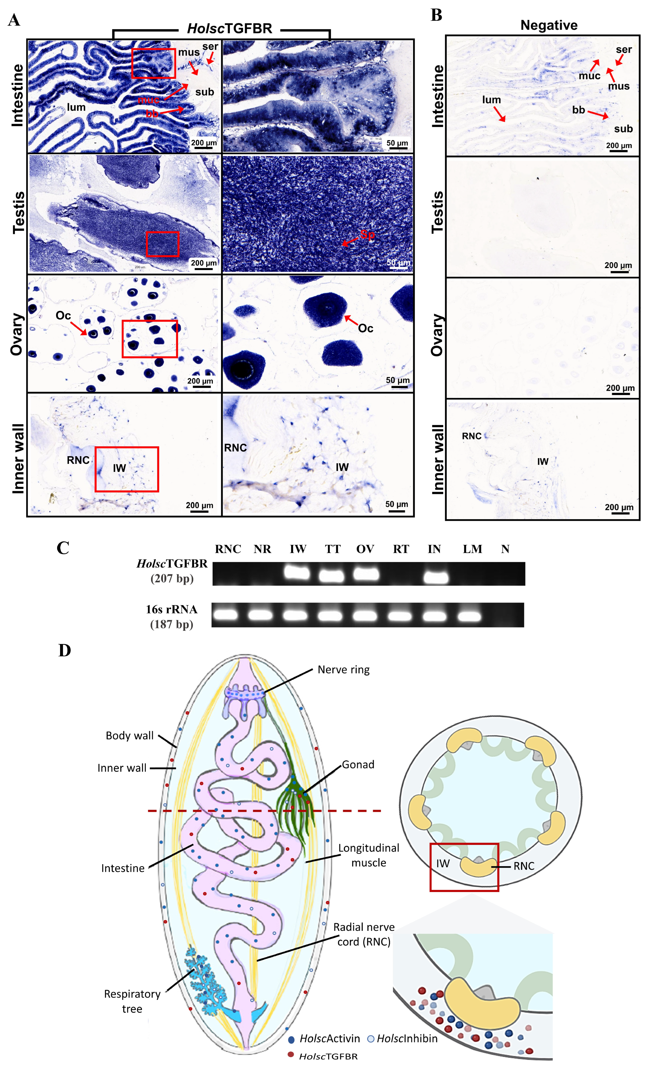
2.1. In Situ Hybridization of H. scabra Activin and Inhibin

In situ hybridization technique was performed to investigate the localization of activin and inhibin mRNA in the intestine, respiratory tree, testis, ovary, and inner body wall of H. scabra [24]. The signals were visualized as distinct blue staining, indicating the sites of mRNA expression. In the intestine, blue staining was prominently observed in epithelial lining of the intestine and respiratory tree. These mRNAs were also detected in the coelomic epithelium and connective tissue of intestine. In the reproductive organs, activin and inhibin localization were found in the spermatocytes of testis and oocytes of ovary. Finally, in the inner wall, blue staining was observed within specific cells and connective tissues (Figure 1A,B).

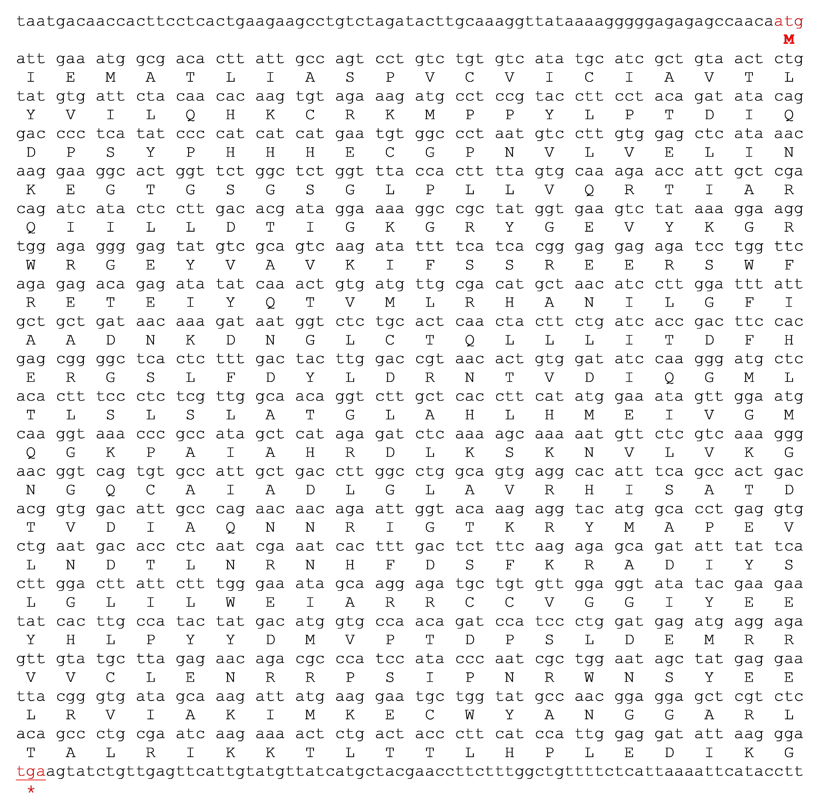
2.2. Characterization and Expression of H. scabra TGF-β Receptor (HolscTGFBR)

The full-length nucleotide sequence of HolscTGFBR, comprising 1850 bp, was successfully amplified, validated, and made available in NCBI with accession number PV173756 (Supplemental Data S1). It encodes a protein of 381 amino acids with an estimated molecular weight of approximately 43.2 kDa (Figure 2). Furthermore, for the specific localization of the TGF receptors in the target organs, we found that the HolscTGFBR mRNA was exclusively expressed in the intestinal epithelium, spermatocytes of the testis, oocytes of the ovary, and cells in the inner wall of the H. scabra (Figure 3A). The expression of HolscTGFBR was analyzed in the selected tissues using RT–PCR, which produced an amplicon of approximately 207 bp (Figure 3C). The results demonstrated that the HolscTGFBR transcript was widely expressed in the intestine, ovary, testis, and inner body wall, suggesting its involvement in diverse physiological processes within these organs. In contrast, the expression of HolscTGFBR was not detected in the respiratory tree, radial nerve cord, and nerve ring (Figure 3C). The specificity of the RT-PCR assay was validated by the absence of amplification in the non-template (negative) control. Additionally, the expression of 16S rRNA was used as a housekeeping gene to ensure the reliability and accuracy of the observed gene expression profiles.

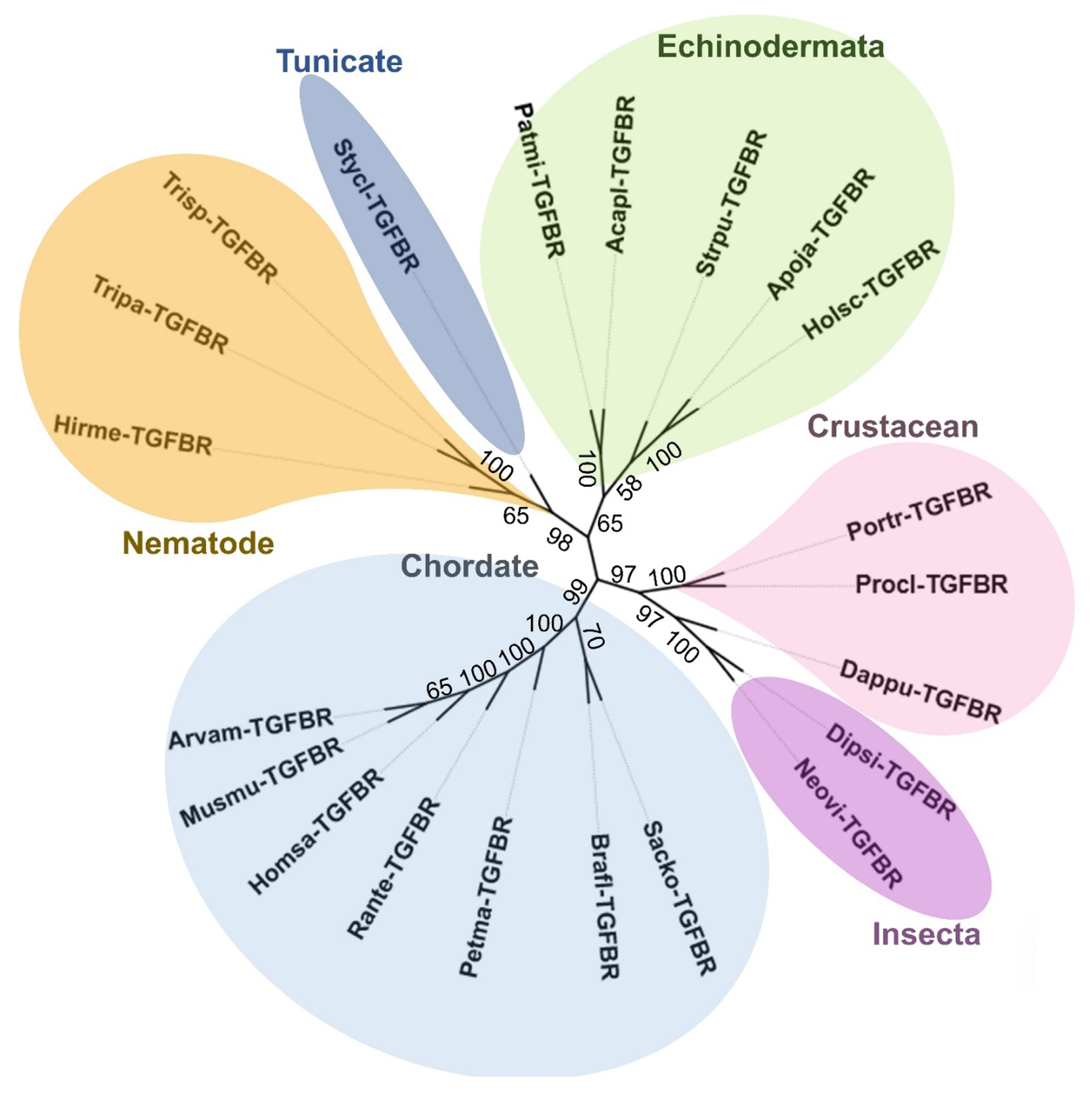
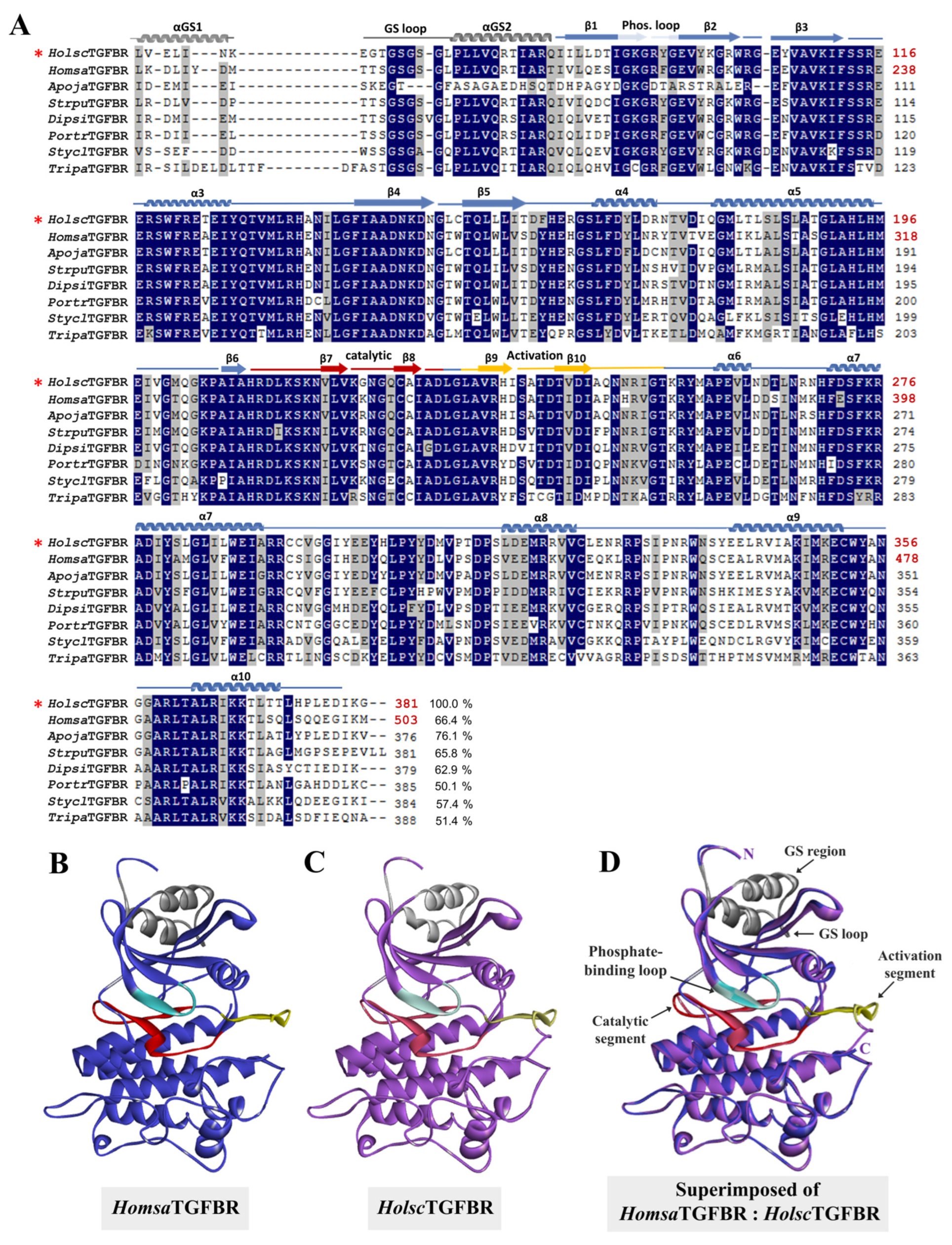
2.3. Bioinformatic Analysis: Amino Acid Sequencing Alignment, Phylogenetic Tree, and Structural Analysis of H. scabra TGF-β Receptor

A phylogenetic analysis of HolscTGFBR compared with the TGF-β receptor I proteins from various species provided valuable insights into its evolutionary relationships and divergence. The analysis demonstrated that HolscTGFBR was closely related to the TGF-β receptors of Apostichopus japonicus (ApojaTGFBR), Strongylocentrotus purpuratus (StrpuTGFBR), Acanthaster planci (AcaplTGFBR), and Patiria miniata (PatmiTGFBR), indicating a conserved functional role of this receptor within the echinoderm phylum. Notably, HolscTGFBR also exhibited significant similarity to the human TGF-β receptor (HomsaTGFBR), suggesting a potential evolutionary link that extends across distant taxa (Figure 4). Sequence alignment revealed a 66.4% identity between HolscTGFBR and HomsaTGFBR. Several highly conserved regions were identified, including the GS loop (residues 185–194 in HomsaTGFBR and 63–72 in HolscTGFBR), the αGS2 region (195–204 in HomsaTGFBR and 73–82 in HolscTGFBR), the phosphate-binding loop (211–217 in HomsaTGFBR and 88–94 in HolscTGFBR), the catalytic segment (331–351 in HomsaTGFBR and 209–229 in HolscTGFBR), and the activation region (354–375 in HomsaTGFBR and 232–253 in HolscTGFBR) (Figure 5A). Furthermore, the three-dimensional structure of HolscTGFBR was predicted using the human Type I TGF-β receptor (PDB ID: 1b6c.3.B) as a structural template. A comparative structural analysis confirmed that key functional domains are conserved and exhibit high structural alignment. These findings support the functional significance of HolscTGFBR in mediating TGF-β receptor activation and inhibition (Figure 5B–D) [25,26].

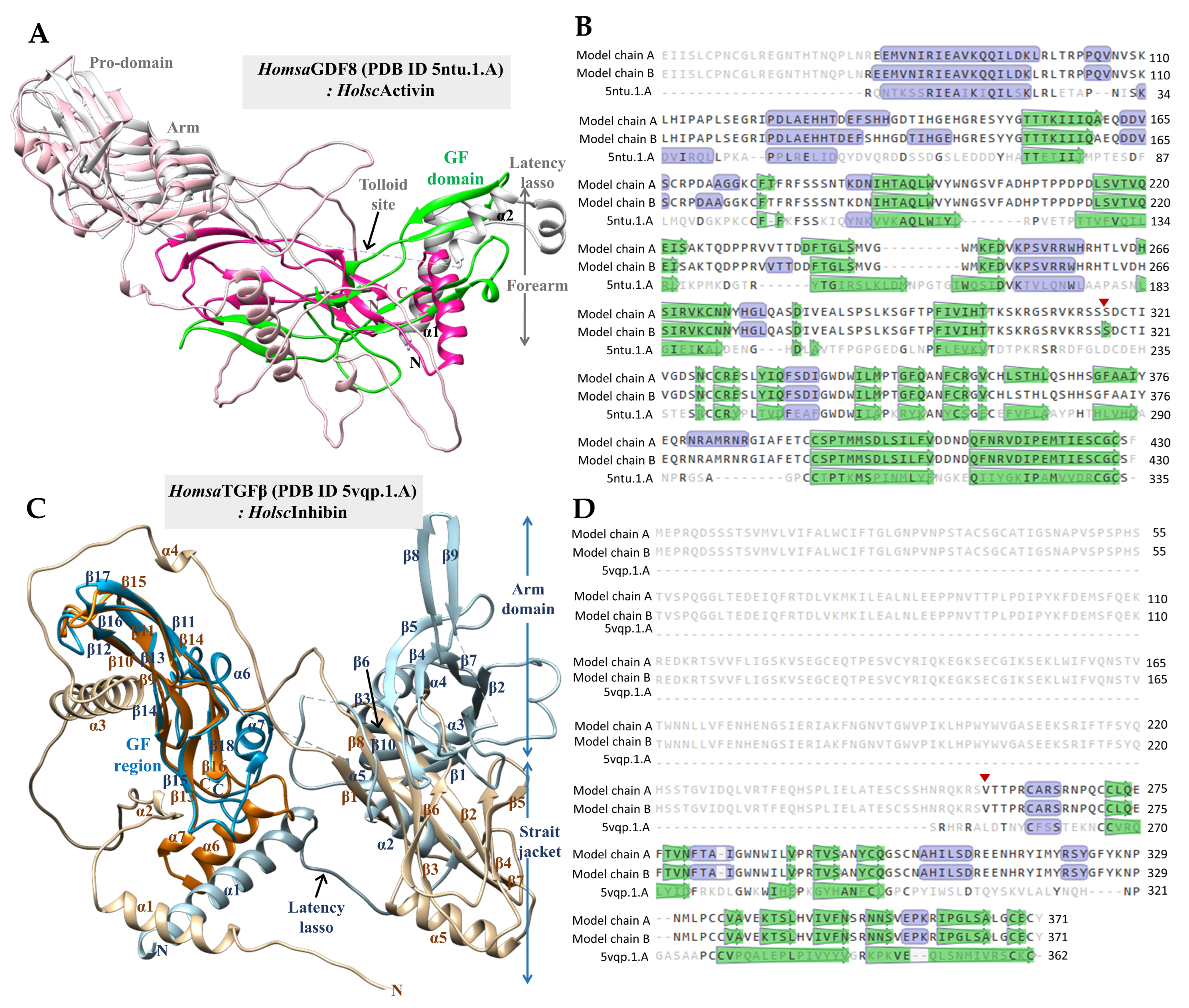
2.4. Three-Dimensional Structural Modeling of H. scabra Activin and Inhibin Proteins

Three-dimensional structural models of H. scabra activin and inhibin (HolscActivin and HolscInhibin) were generated using AlphaFold [27]. The results showed the predicted structures with pLDDT confidence scores of 69.6 (HolscActivin) and 70.1 (HolscInhibin) and the predicted template modeling (pTM) scores of 0.685 and 0.464, respectively. To assess the structural precision and sequence similarity relative to experimentally determined structures, SWISS-MODEL was employed for comparative analyses. The templates with the highest sequence similarity were identified as HomsaGDF8 (human pro-myostatin precursor; 2.6 Å resolution; PDB ID: 5ntu.1.A) for HolscActivin and HomsaTGFβ (human pro-TGF-β1; PDB ID: 5vqp.1.A) for HolscInhibin [28,29].
The structural superposition of the monomeric growth factor domains of HomsaGDF8 and HolscActivin is illustrated in Figure 6A. In this comparison, HomsaGDF8 is depicted with a gray ribbon (pro-domain) and a green ribbon (mature domain), while HolscActivin is illustrated with a pale pink ribbon (pro-domain) and a deep pink ribbon (mature domain). The mature domain of HolscActivin aligns closely with HomsaGDF8, preserving the canonical cystine knot fold, critical for receptor binding and signaling. The pro-domain structures exhibit moderate overlap, including subdomains such as the forearm, latency lasso, and arm regions, suggesting conserved latency control architecture despite evolutionary divergence. Sequence analysis indicates an amino acid identity of 30.43% between HomsaGDF8 and HolscActivin, reflecting moderate conservation, typical of TGF-β family orthologs across metazoans. Additionally, cysteine residues essential for disulfide bond formation are strictly conserved, ensuring the correct stability of the tertiary structure. The red triangle in Figure 6B indicates the N-terminal start site of the HolscActivin mature domain (S316–F430), showing slight positional variation within the structure. Moreover, mature HolscActivin monomer consists of 114 amino acids and forms homodimers with a molecular mass of approximately 25.7 kDa.
Figure 6C presents the structural superposition of HomsaTGFβ (depicted in pale blue for the pro-domain and deep blue for the mature domain) with HolscInhibin (tan for the pro-domain and orange for the mature domain). The mature domain of HolscInhibin aligns well with that of HomsaTGFβ, maintaining the characteristic β-strand and α-helix arrangements of TGF-β family growth factors [30,31,32]. However, notable structural differences are observed in the pro-domain, particularly within the latency lasso and arm regions, indicating species-specific adaptations that may influence latency mechanisms. Sequence alignment reveals an amino acid identity of 28.32% between HomsaTGFβ and HolscInhibin. The mature domain of HolscInhibin spans from V258 to Y371, consisting of 114 amino acids, with a predicted molecular weight of approximately 25.3 kDa for homodimers. Furthermore, the cysteine residues crucial for cystine knot motif formation are conserved, ensuring structural integrity (Figure 6D).

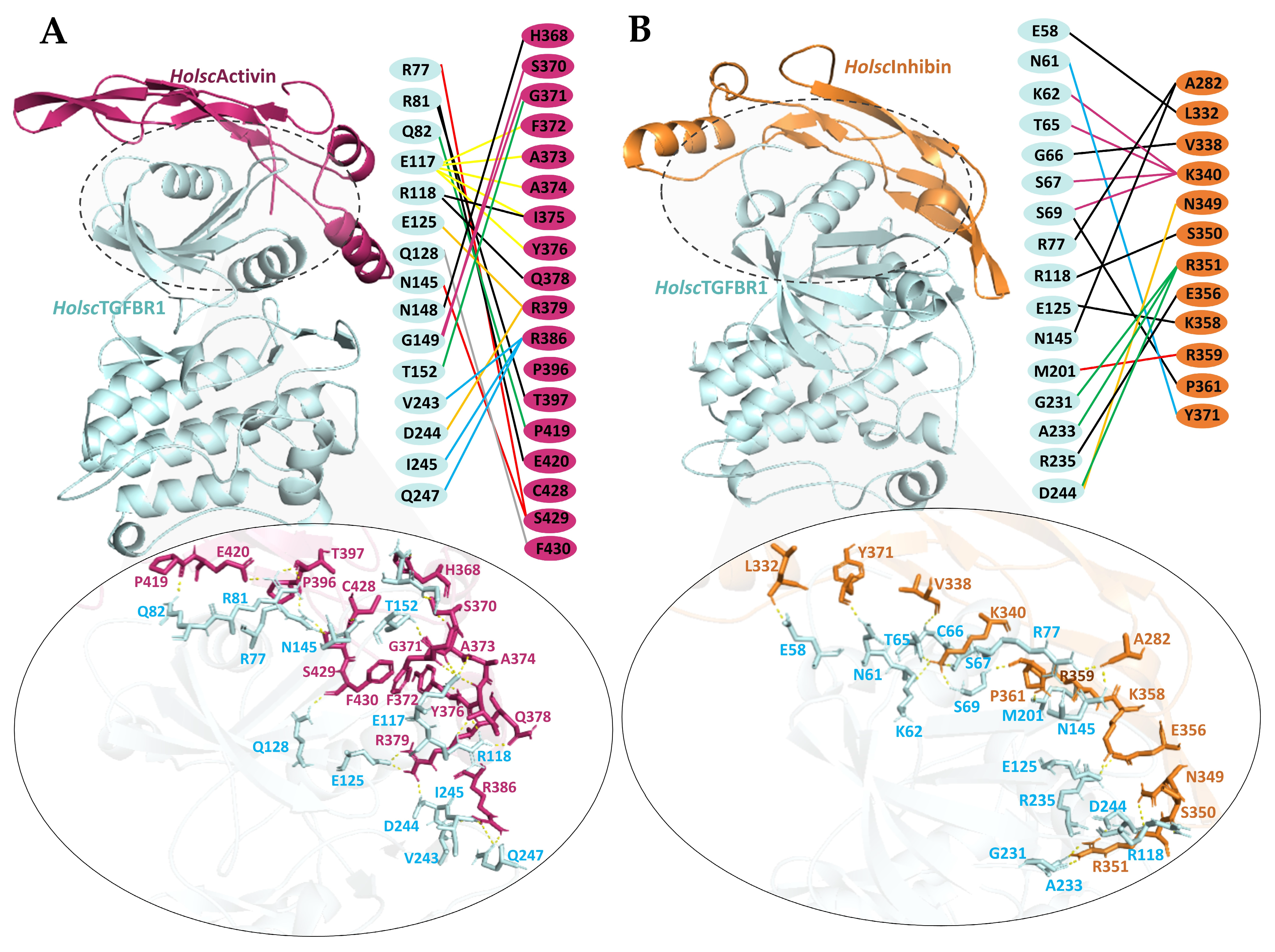
2.5. Molecular Docking of H. scabra TGF-β Proteins and TGF-β Receptors

To investigate the potential role of H. scabra TGF-β ligands (HolscActivin and HolscInhibin) in signaling through the H. scabra type I transmembrane serine/threonine kinase receptor (HolscTGFBR), molecular docking analyses were performed. The complex with the highest coefficient-weighted score, representing the most stable binding interaction, was selected (Table 1). The docking complexes of HolscActivin and HolscInhibin with HolscTGFBR, using human TGFBR structure (PDB ID: 1b6c.3.B) as a template, are shown in Figure 7A,B. HolscActivin (depicted as a deep pink ribbon) interacts with the extracellular domain of HolscTGFBR (light blue ribbon), forming multiple stabilizing interactions. Hydrogen bonding interactions (yellow dotted lines) are observed between key HolscActivin residues, including H368, G371, S370, R379, F330, T397, C428, and Q378, and receptor residues such as R77, R81, E117, R118, E125, N145, D244, I245, and Q247. The mapping of the interaction interface (middle panel) reveals direct contact residues forming both polar and hydrophobic interactions, underscoring their contribution to binding affinity and complex stabilization. Importantly, HolscActivin did not bind to the key functional regions of HolscTGFBR, including the serine/threonine-rich GS loop (residues 63EGTGSGSGLP72), the αC-helix of the kinase domain involved in structural stabilization, the active site residue E123, and the critical ATP-binding activation segment residue R250, which are essential for receptor activation and signal transduction (Figure 7A) [26,29]. In Figure 7B, the HolscInhibin–HolscTGFBR complex is shown, where HolscInhibin (orange ribbon) binds the receptor (light blue ribbon) at an overlapping but slightly shifted site compared to HolscActivin. Hydrogen bonding interactions are identified between HolscInhibin residues L332, V338, K340, N349, K358, S350, E356, Y371, and receptor residues, including E58, K62, T65, S67, G66, S69, R77, N145, E125, R235, and D244. The critical interaction is that K340 and V338 of HolscInhibin bind to T65, G66, S67, and S69, which are in the serine/threonine-rich GS loop (Figure 7B). This interaction may physically block the access of the type II receptor kinase (TβRII) and prevent phosphorylation, thereby inhibiting the receptor activation [25,33]. Similarly, in humans, inhibin functions as a natural antagonist that prevents activin from binding to and activating TGF-β receptors, specifically TGFBR1 [26].

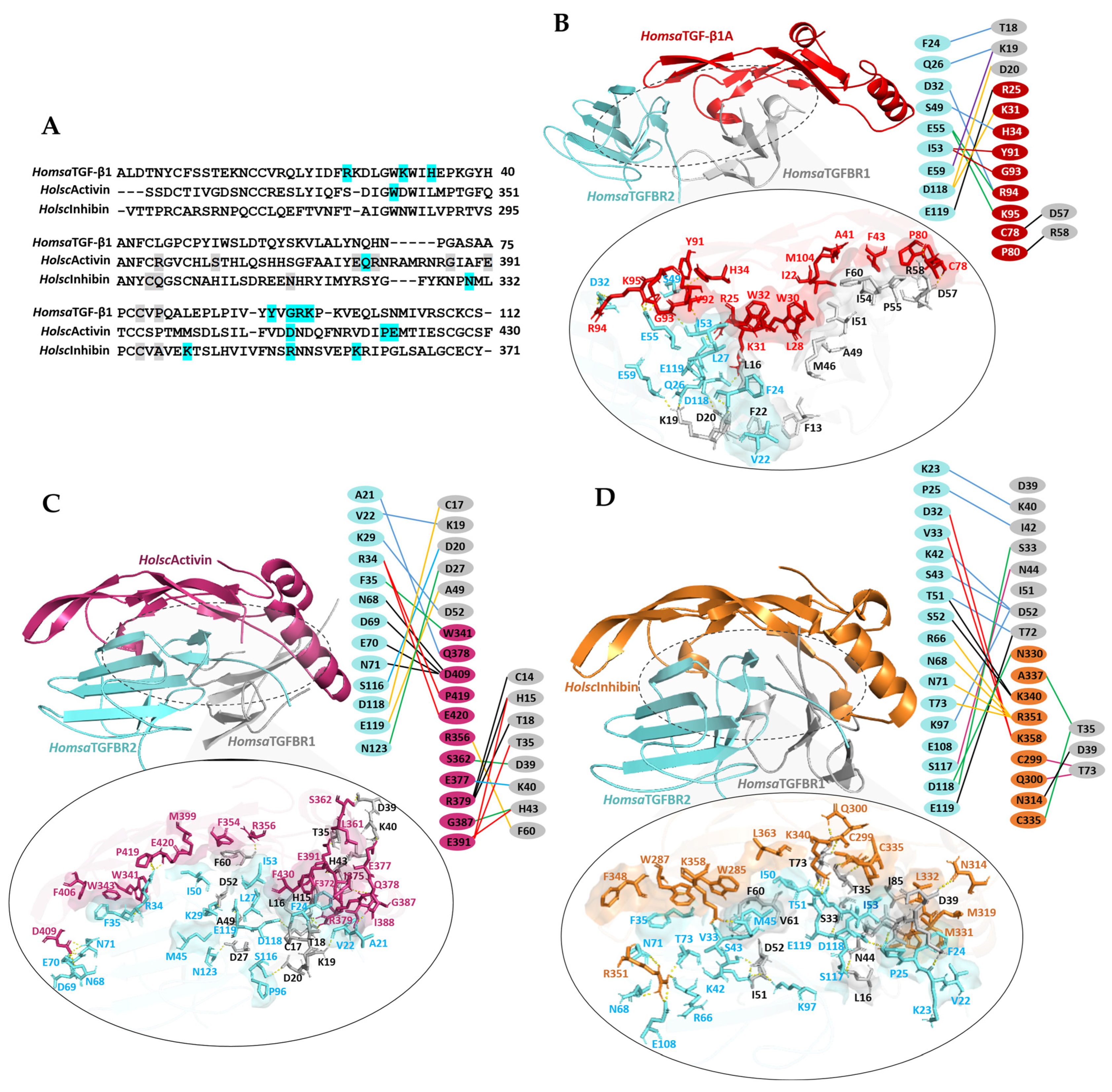
2.6. Ternary Complex Docking Analysis of Human TGF-β1 and H. scabra TGF-β Ligands with Human TGFβ Receptors

To analyze the sequence conservation and receptor-binding residues among human TGF-β1, HolscActivin, and HolscInhibin, multiple sequence alignment was performed (Figure 8A). The results reveal that all three proteins retain conserved cysteine residues essential for cystine knot motif formation, which underpins tertiary structure stability and biological function, consistent with the structural requirements of TGF-β superfamily ligands. Residues highlighted in cyan and gray represent amino acids involved in hydrogen bonding with HomsaTGFBR2 and HomsaTGFBR1, respectively, as identified from structural docking analyses. For HomsaTGF-β1A, critical residues forming hydrogen bonds with TGFBR2 include W30, Y91, R94, and K97, consistent with previously reported receptor-binding interfaces [34,35].
The ternary complex of HomsaTGF-β1 (red), HomsaTGFBR2 (blue), and HomsaTGFBR1 (gray) was used as an experimental template. Docking analysis yielded a binding energy of −1054.2 kcal/mol (Table 1). The complex forms multiple hydrogen bonds, including the residues R25, K31, H34, Y91, G93, R94, and R95 of the TGF-β1A interacting with D32, S49, E55, D118, and E119 of TGFBR2. The residues C78 and P80 of TGF-β1A also form hydrogen bonds with D57 and R58 of TGFBR1. Additionally, interactions involved F24, Q26, E59, and D118 of TGFBR2 binding to T18, K19, and D20 of TGFBR1. A central hydrophobic interface was identified, comprising residues I22, L28, W30, W32, A41, F43, V92, and M104 on TGF-β1, which engage hydrophobic patches on TGFBR2 and TGFBR1 (Figure 8B). This hydrophobic core contributes to stable receptor bridging and activation. The HolscActivin ligand (warm pink ribbon) formed a ternary complex with HomsaTGFBR2 and HomsaTGFBR1, exhibiting a docking energy of −930.5 kcal/mol.
HolscActivin residues W341, Q378, D409, P419, and E420 form hydrogen bonds with F35, A21, N68, D69, E70, and N71 of TGFBR2. Residues R356, S362, E377, R379, G387, and E391 interact with C14, H15, T18, T35, D39, K40, H43, and F60 of TGFBR1. Additionally, interactions include V22, K29, S116, D118, E119, and N123 of TGFBR2 binding to C17, K19, D20, D27, A49, and D52 of TGFBR1. Hydrophobic residues, including F406, W343, M399, F354, F430, F372, I388, L361, and I375, form significant interfacial contacts with TGFBR2 and TGFBR1 (Figure 8C), stabilizing the complex despite weaker binding energy compared to endogenous TGF-β1. The HolscInhibin ligand (orange ribbon) forms a stable ternary complex with a docking energy of −1002.3 kcal/mol, comparable to endogenous TGF-β1. Hydrogen bonding interactions involve the N330, K340, R351, and K358 residues of HolscInhibin forming bonds with D32, V33, T51, S52, R66, N68, N71, T73, and D118 of TGFBR2. Moreover, the ligand residues C229, Q300, N314, C335, and A337 bind with T35, D39, and T73 of TGFBR1. The inter-receptor contacts include K23, P25, K97, S117, D118, and E119 of TGFBR2 binding to K40, I42, S33, N44, D52, and T72 of TGFBR1. Extensive hydrophobic contacts are observed with residues W287, W285, M319, L332, M331, L363, and F348, enhancing complex stability and receptor occupancy (Figure 8D).
Table 1. The highest coefficient weight scores from PIPER energy calculations were obtained for the complexes of human and H. scabra TGF-β ligands with their respective TGFBR receptors, following docking analysis using ClusPro 2.0.
Complex ModelCluster MembersWeight Score a
HolscActivin–HolscTGFBR57−958.0
HolscInhibin–HolscTGFBR70−887.7
HomsaTGF-β1–HomsaTGFBR2107−804.7
HolscActivin–HomsaTGFBR245−748.4
HolscInhibin–HomsaTGFBR236−844.2
HomsaTGF-β1–HomsaTGFBR2–HomsaTGFBR181−1054.2
HolscActivin–HomsaTGFBR2–HomsaTGFBR172−930.5
HolscInhibin–HomsaTGFBR2–HomsaTGFBR195−1002.3
a The PIPER protein interaction energy was calculated from E = w1Erep + w2Eattr + w3Eelec + w4EDARS, where Erep and Eattr represent the repulsive and attractive components of the van der Waals interaction energy, and Eelec indicates an electrostatic energy term. EDARS reveals the pairwise structure-based potential constructed using the decoys as the reference state (DARS) method [36].

3. Discussion

This study is the first to investigate the localization, sequence conservation, structural alignment, and potential functional roles of H. scabra TGF-β proteins (activin and inhibin) across various organs, including the intestine, respiratory tree, ovary, testis, and inner body wall [5,24]. Additionally, we examined the specific gene expression and localization of the H. scabra TGF-β receptor. A comparative sequence alignment was also conducted between H. scabra TGF-β receptors and those of multiple species, particularly Homo sapiens. Furthermore, molecular docking was performed to assess the relevance of H. scabra TGF-β proteins in binding adaptively to the human TGF-β receptor. These findings provided fundamental insights into the potential biomedical applications of H. scabra-derived proteins [2,29,37,38].
For specific localization investigation, the in situ hybridization results indicated that the H. scabra TGF-β genes, including activin and inhibin, were localized across all targeted organs. In the intestine, the mRNA for activin and inhibin were prominently observed in the epithelial layer, aligning with their roles in nutrient absorption and digestion [39]. In the respiratory tree, signals were detected in the lining of the respiratory tubules, suggesting a possible role in gas exchange or osmoregulation [40]. In the testis, intense signals were localized to somatic cells surrounding the germinal epithelium, indicating a regulatory role in spermatogenesis. Similarly, signals were concentrated in the follicular layer surrounding developing oocytes, implying involvement in oocyte maturation [41,42]. Notably, the TGF-β genes were expressed in the inner wall, suggesting their presence in connective tissue and specific cells, which may imply a role in extracellular matrix remodeling, structural integrity, and repair processes [43,44]. These observations are consistent with the known functions of TGF-β family members in cellular communication, matrix control, and tissue homeostasis [45]. Consequently, the findings suggest that activin and inhibin may play a potential role in extracellular matrix remodeling and maintaining structural integrity [46,47,48].
Interestingly, the H. scabra TGF-β receptor (HolscTGFBR) expression exhibited high sequence identity with the human TGF-β receptor type I, which is involved in signal transduction [25]. The tissue-specific expression pattern of HolscTGFBR indicated its functional significance in important physiological processes. Its exclusive expression in the intestinal epithelium, spermatocytes, oocytes, and inner body wall implies potential roles in digestion, reproduction, and tissue remodeling, in line with the roles of TGF-β signaling in invertebrate development and homeostasis [49]. The widespread expression of HolscTGFBR in reproductive and digestive tissues was confirmed by RT-PCR. The absence of expression in neural tissues, such as the radial nerve cord and nerve ring, suggests a tissue-specific regulatory role rather than direct involvement in neural signaling. These findings highlight HolscTGFBR as a key regulator in H. scabra physiology, warranting further investigation into its downstream signaling pathways and functional implications [50].
The phylogenetic and structural analyses of HolscTGFBR underscore its evolutionary conservation and potential functional significance. Phylogenetic comparisons show a close relationship between HolscTGFBR and TGF-β receptor homologs in other echinoderms. This similarity suggests a conserved role in fundamental biological processes, such as development, regeneration, and immune regulation, consistent with the TGF-β signaling functions in marine invertebrates [51]. Remarkably, HolscTGFBR exhibits significant sequence similarity to the human TGF-β type I receptor (HomsaTGFBR), particularly in key functional regions like the GS loop, αGS regions, phosphate-binding loop, catalytic segment, and activation region. This conservation implies that HolscTGFBR likely employs similar phosphorylation-dependent mechanisms for receptor activation as observed in vertebrates. The preserved catalytic segment and phosphate-binding loop further support its potential kinase activity in signal transduction [52].
We investigated the structural conservation and receptor-binding potential of two H. scabra TGF-β superfamily ligands, HolscActivin and HolscInhibin, to assess their functional parallels with vertebrate TGF-β family members. AlphaFold modeling indicated moderate confidence, with both proteins displaying overall structural similarity to canonical TGF-β templates. SWISS-MODEL analyses identified HomsaGDF8 (pro-myostatin precursor) as the closest homolog for HolscActivin and HomsaTGFβ1 for HolscInhibin, consistent with expected divergence across metazoan orthologs. Importantly, both HolscActivin and HolscInhibin retained all cysteine residues necessary for cystine knot motif formation, ensuring correct folding and structural stability [35,53]. Structural superposition showed that mature HolscActivin aligned closely with HomsaGDF8, preserving the canonical cysteine knot fold critical for receptor binding. HolscInhibin similarly maintained the β-strand and α-helix arrangements characteristic of mature HomsaTGFβ. While their mature domains were well conserved, notable species-specific differences were observed in pro-domain regions, particularly in HolscInhibin. These included variations within the forearm, latency lasso, and arm subdomains, which regulate ligand latency and activation mechanisms, suggesting evolutionary adaptations that may influence receptor affinity or signaling dynamics [34,54,55]. These findings indicate that HolscActivin and HolscInhibin share conserved structural features with vertebrate TGF-β family ligands, supporting their potential roles in H. scabra cellular regulation, including growth, differentiation, and immune responses [53,56].
For molecular docking, we investigated the molecular interactions of HolscActivin and HolscInhibin with their putative receptor (HolscTGFBR) to evaluate their potential signaling and inhibitory properties within the TGF-β superfamily framework. HolscActivin docked onto HolscTGFBR, forming multiple hydrogen bonds with the receptor residues, including R77, E117, and R118, and stabilizing hydrophobic contacts while avoiding critical catalytic regions, activation segments, and the GS loop. This indicated that HolscActivin may function similarly to endogenous activin ligands as well as activating TGF-β signaling [57,58]. Conversely, HolscInhibin showed binding to receptor regions overlapping with HolscActivin but critically engaged the serine/threonine-rich GS loop through K340 and V338 residues interacting with T65, G66, S67, and S69. Such interactions could physically obstruct type II receptor recruitment and block receptor activation, consistent with the established inhibitory mechanism of vertebrate inhibins, which prevent activin-induced signaling by sequestering receptor sites [38]. A balance between these ligands controls cell proliferation and differentiation and plays an important role in normal development and homeostasis [59,60].
Furthermore, we concentrated on these TGF-β proteins from the inner wall, as they possess the potential to be developed into functional food and medical applications [25,55]. Interestingly, cross-species docking analysis further provided insights into potential functional conservation between H. scabra and human TGF-β proteins. Ternary complex docking with human receptors (HomsaTGFBR1 and HomsaTGFBR2) further elucidated the competitive binding potential of HolscActivin and HolscInhibin. The crystal structure of the HomsaTGF–β1–HomsaTGFBR2–HomsaTGFBR1 ternary complex (PDB ID: 3KFD) served as a template, enabling the structural modeling to predict their ability to mimic endogenous TGF-β ligand binding and receptor activation [34]. Molecular docking analysis revealed distinct roles of H. scabra TGF-β proteins in receptor activation and inhibition. Ternary complex docking with human TGF-β receptors further supported these interpretations. Endogenous human TGF-β1 exhibited the strongest binding energy (−1054.2 kcal/mol) with extensive hydrogen bonding and hydrophobic interactions bridging TGFBR2 and TGFBR1, consistent with its potent activation capability [37]. Importantly, the complete complex exhibited both hydrogen bonding and hydrophobic interactions at regions and specific residues consistent with those in the experimental crystal structure [34]. This approach can therefore be applied to HolscActivin and HolscInhibin for cross-species docking analyses, providing further insights into the potential function of H. scabra proteins. HolscActivin showed moderate binding (−930.5 kcal/mol), forming multiple interfacial interactions yet lacking the extensive hydrophobic core stabilizing endogenous TGF-β1, suggesting partial receptor engagement. In contrast, HolscInhibin displayed a docking energy (−1002.3 kcal/mol) approaching that of TGF-β1, forming robust inter-receptor contacts and occupying the receptor interface in a manner indicative of competitive inhibition without activation. These results suggested that these ligands can mimic endogenous TGF-β binding modes and potentially modulate receptor-mediated signal transduction [35,37].
In short, we predicted the functional conservation of H. scabra TGF-β proteins, with HolscActivin functioning as a potential agonist and HolscInhibin serving as an antagonist of activin signaling in human TGF-β receptor responses [59,60]. These findings will lead to medical application investigations utilizing H. scabra as a naturally functioning resource. Nonetheless, further studies are required to assess their efficacy, safety, and interactions with human receptors and signaling pathways [61]. Additionally, incorporating these proteins into nanoparticle-based or other targeted delivery systems may enhance their therapeutic potential. This research supports the broader pursuit of nature-sourced approaches for novel functional food as well as biomedical applications in the future [62,63].

4. Materials and Methods

4.1. Ethics Statement

All animals involved in this experiment were approved by the Institutional Animal Care and Use Committee (IACUC) of Thammasat University (Protocol Number 019/2561) according to the established guidelines for scientific research. In this study, we aimed to investigate the distribution of transforming growth factor superfamily (TGF-β), including activin and inhibin, in H. scabra. Wild specimens (weighing 300–500 g, with random sexes) were collected from Krabi Province, Thailand, following Songkoomkrong et al., 2024 [4]. Subsequently, tissue samples of H. scabra, including the intestine, respiratory tree, gonads, and inner wall, were harvested and immediately frozen in liquid nitrogen or fixed in 4% paraformaldehyde. The frozen samples were kept at −80 °C for later RNA extraction, while the fixed samples were processed for histological examination and in situ hybridization [64].

4.2. In Situ Hybridization of the Activin, Inhibin, and TGF-β Receptor Type I in H. scraba

The DIG-labeled sense and anti-sense RNA probes for activin and inhibin were derived from the study by Kornthong et al., 2021 [6,20]. Tissue slides of all organs were routinely prepared using the paraffin method. Following de-paraffinization and rehydration, the slides were immersed in PBS containing DEPC, followed by PBS with 0.3% Tween-20 (PBST), and then incubated with Proteinase K (Roche, Mannheim, Germany) in 1× TNE buffer (500 mM NaCl, 10 mM Tris, 1 mM EDTA, pH 8.0). The slides were washed with 2X Sodium Saline Citrate (SSC) and fixed in 4% paraformaldehyde. For the hybridization procedure, the tissues were incubated in the hybridization solution with RNA probes for HolscActivin, HolscInhibin, and TGF-β receptor type I (HolscTGFBR) overnight at 52 °C. Afterward, the slides were thoroughly washed with 4×, 2×, and 1× SSC solution and blocked for non-specific binding with maleic acid buffer containing 2% sheep serum at room temperature for 3–4 h. The slides were then incubated with a 1:500 dilution of AP-conjugated anti-DIG antibody overnight at 4 °C. After washing, the tissue slides were stained using the NBT/BCIP detection system. Positive expression sites of activin and inhibin were examined and photographed using a Leica compound microscope (Leica, Wetzlar, Germany). The complete hybridization procedure followed the methodology described by Kornthong et al., 2021 [6].

4.3. Bioinformatic Analysis of Activin, Inhibin, and TGF-β Receptor Type I in H. scraba and Other Echinoderms

As a previous report, the transcriptome data set of growth factor transcripts in various organs of H. scabra is available in NCBI (Accession no: MW728942 to MW728950) [6,25]. The translational amino acid sequences of the HolscActivin (accession no: MW728947), HolscInhibin (accession no: MW728945), and HolscTGFBR (accession no: PV173756) were identified using BLASTx (https://blast.ncbi.nlm.nih.gov/Blast.cgi, accessed on 28 April 2024). The molecular mass of mature proteins was calculated using the online software ExPASy Peptide Mass program (http://web.expasy.org/compute_pi/, accessed on 30 April 2024). The analysis of multiple amino acid sequence alignments of HolscTGFBR compared to other species was performed using MEGA11 software, utilizing MUSCLE for alignment. The conserved amino acids were generated with Multiple Align Show (https://www.bioinformatics.org/SMS/multi_align.html, accessed on 3 May 2024). Phylogenetic tree analysis was conducted with MEGA11 software and modified by iTOL v6 software (https://itol.embl.de/upload.cgi, accessed on 3 May 2024).

4.4. Tissue Distribution of the TGF-β Receptor Type I mRNA in H. scabra

The collected tissue samples were extracted for total RNA extraction. The concentration and purity of each RNA sample were determined using a nanodrop spectrophotometer at wavelengths of 260 and 280 nm (ND-3800-OD Nano DOT Microspectrophotometer, Hercuvan Lab Systems, Brixton, UK) [65]. cDNA synthesis was then carried out using the QuantiNova Reverse Transcription Kit (Qiagen, Hilden, Germany), and the resulting cDNA served as a template for PCR with gene-specific primers, as shown in Table 2.

4.5. Structural Modeling Prediction and Sequence Analysis

Full-length sequences of HolscActivin and HolscInhibin were obtained from previously published transcriptome data [6]. The three-dimensional structures of both proteins were utilized using AlphaFold2, accessible on https://colab.research.google.com/github/sokrypton/ColabFold/blob/main/AlphaFold2.ipynb, accessed on 4 July 2025 [27]. The predicted local distance difference test (pLDDT) was used to assess model confidence, while the predicted aligned error (PAE) estimated positional uncertainties for each amino acid. Modeling parameters included 20 recycles and a root mean square deviation (RMSD) tolerance of 0.5 Å. For single-chain predictions, ranking was based on pLDDT scores, whereas complex models were ranked by predicted TM-score. Five models were generated per run, and their respective pTM and pLDDT scores were recorded. Additional structural features were retrieved using SWISS-MODEL (https://swissmodel.expasy.org/, accessed on 5 July 2025). After predicting the 3D structures, HolscActivin and HolscInhibin were analyzed and structurally aligned using the crystal structures of the closest human homologs as templates, including HomsaGDF8 (human pro-myostatin precursor; 2.6 Å resolution, PDB ID: 5ntu.1.A) for HolscActivin and HomsaTGFβ (human pro-TGF-β1, PDB ID: 5vqp.1.A) for HolscInhibin. Additionally, the 3D structure of HolscTGFBR was generated by using the SWISS-MODEL, accessed on 15 May 2024, using HomsaTGFBR (PDB ID: 1b6c.3.B) as a template. Refinement and annotation were performed using Chimera UCSF software (version 1.6.1).

4.6. Molecular Docking

Complex structures of HolscActivin and HolscInhibin with the HolscTGFBR were docked using the cytoplasmic domain of the type I TGF-β receptor (PDB ID: 1b6c) as a complex structural template. For ternary complex docking, HomsaTGFβ was initially docked with HomsaTGFBR2. The complexes with the most favorable (lowest) energy scores were selected, and HomsaTGFBR1 was subsequently recruited to form the complete ternary complex. The crystal structure of the ternary complex (PDB ID: 3KFD) was used as the experimental template. This protocol was similarly applied using HolscActivin and HolscInhibin in place of HomsaTGFβ. All molecular docking complexes were performed on ClusPro 2.0, a widely recognized server for protein–protein docking (https://cluspro.org/help.php, accessed on 6 July 2025). The interaction energy of the protein complexes is based on the PIPER protein interaction energy, using the equation E = w1Erep + w2Eattr + w3Eelec + w4EDARS. The PIPER algorithm has demonstrated strong performance in CAPRI assessments, calculating docking energies on a grid using the Fast Fourier Transform (FFT) and pairwise interaction potentials, which enables the rapid identification of near-native conformations [36]. The resulting structures were then clustered based on pairwise RMSD to optimize their representation as follows [4,66]. The top-ranked models were subsequently visualized using PyMol.

5. Conclusions

The expression of HolscActivin and HolscInhibin was observed across all targeted tissues in the gene expression analysis and in situ hybridization. In the intestine and respiratory tree, these genes were identified in the intestinal and coelomic epithelium, connective tissues, and epithelial cells. Both genes were also present in spermatocytes within the testis and in oocytes of the ovary. Furthermore, their localization was specific to the connective tissue cells and fibers of the inner body wall. HolscTGFBR exhibited a similar expression pattern, except for its absence in the respiratory tree. Phylogenetic and structural analyses revealed that HolscTGFBR is evolutionarily conserved, sharing similarities with human TGF-β receptors. The presence of conserved sequence motifs and structural domains supports the functional significance of HolscTGFBR in TGF-β signaling. Molecular docking studies demonstrated that HolscActivin binds to HolscTGFBR, potentially functioning as an agonist, whereas HolscInhibin likely inhibits receptor activation. The ternary complex interactions suggested a strong role for HolscInhibin and HolscActivin in competitive TGF-β signaling, potentially contributing to tumor suppression and immune response enhancement. These findings illuminate the conserved and distinct mechanisms between H. scabra and human TGF-β signaling. Such insights have significant implications for health-related applications and the development of functional foods in the future.

Supplementary Materials

The following supporting information can be downloaded at https://www.mdpi.com/article/10.3390/ijms26146998/s1.

Author Contributions

Conceptualization, N.K. and S.N.; methodology, S.N., J.C., C.R. and L.S.; software, S.N.; validation and formal analysis, N.K., S.N., S.D. and S.S.; resources, N.K., S.D., S.S. and P.A.; data curation, N.K., S.N., J.S., N.N. and P.S.; writing—original draft preparation, S.N.; writing—review and editing, N.K., J.S. and S.N.; visualization, S.N.; supervision, N.K.; project administration, N.K.; funding acquisition, N.K., S.D. and P.A. All authors have read and agreed to the published version of the manuscript.

Funding

This work was supported by the Thammasat University Research Unit in Biotechnology and its application to aquatic animals. The authors also gratefully acknowledge the financial support provided by the Thailand Science Research and Innovation Fundamental Fund, fiscal year 2024, and the Research Fund of Chulabhorn International College of Medicine, Thammasat University (Grant No. T1/2565).

Institutional Review Board Statement

The study was conducted in accordance with the Declaration of Helsinki and was approved by the Institutional Animal Care and Use Committee (IACUC) of Thammasat University (Protocol Number 019/2561) according to the established guidelines for scientific research.

Informed Consent Statement

Not applicable.

Data Availability Statement

The original contributions presented in this study are included in this article. Further inquiries can be directed to the corresponding author.

Acknowledgments

The authors are especially grateful to the Center of Scientific Equipment for Advanced Research, Office of Advanced Science and Technology, Thammasat University.

Conflicts of Interest

The authors declare no conflicts of interest.

Abbreviations

The following abbreviations are used in this manuscript:
TGF-β Transforming growth factor-β
TGFBR1TGF-β receptor type I
RT-PCRReverse Transcription Polymerase Chain Reaction
H/EHematoxylin and Eosin
bpBase pair
PDBProtein data bank
PBSPhosphate-buffered saline
DEPCDiethyl Pyrocarbonate
PCRPolymerase Chain Reaction
DIG Digoxigenin
APAlkaline Phosphatase

References

  1. Wolkenhauer, S.M.; Uthicke, S.; Burridge, C.; Skewes, T.; Pitcher, R. The Ecological Role of Holothuria scabra (Echinodermata: Holothuroidea) within Subtropical Seagrass Beds. J. Mar. Biol. Assoc. UK 2010, 90, 215–223. [Google Scholar] [CrossRef]
  2. Pangestuti, R.; Arifin, Z. Medicinal and Health Benefit Effects of Functional Sea Cucumbers. J. Tradit. Complement. Med. 2018, 8, 341–351. [Google Scholar] [CrossRef] [PubMed]
  3. Pranweerapaiboon, K.; Noonong, K.; Apisawetakan, S.; Sobhon, P.; Chaithirayanon, K. Methanolic Extract from Sea Cucumber, Holothuria scabra, Induces Apoptosis and Suppresses Metastasis of PC3 Prostate Cancer Cells Modulated by MAPK Signaling Pathway. J. Microbiol. Biotechnol. 2021, 31, 775–783. [Google Scholar] [CrossRef] [PubMed]
  4. Songkoomkrong, S.; Nonkhwao, S.; Duangprom, S.; Saetan, J.; Manochantr, S.; Sobhon, P.; Kornthong, N.; Amonruttanapun, P. Investigating the Potential Effect of Holothuria scabra Extract on Osteogenic Differentiation in Preosteoblast MC3T3-E1 Cells. Sci. Rep. 2024, 14, 26415. [Google Scholar] [CrossRef] [PubMed]
  5. Sun, L.; Yang, H.; Chen, M.; Ma, D.; Lin, C. Correction: RNA-Seq Reveals Dynamic Changes of Gene Expression in Key Stages of Intestine Regeneration in the Sea Cucumber Apostichopus japonicus. PLoS ONE 2013, 8, e69441. [Google Scholar] [CrossRef]
  6. Kornthong, N.; Phanaksri, T.; Saetan, J.; Duangprom, S.; Lekskul, B.; Vivattanasarn, T.; Songkoomkrong, S.; Jattujan, P.; Cummins, S.F.; Sobhon, P.; et al. Identification and Localization of Growth Factor Genes in the Sea Cucumber, Holothuria scabra. Heliyon 2021, 7, e08370. [Google Scholar] [CrossRef] [PubMed]
  7. Derynck, R.; Zhang, Y. Smad-Dependent and Smad-Independent Pathways in TGF-β Family Signalling. Nature 2003, 425, 577–584. [Google Scholar] [CrossRef] [PubMed]
  8. Huminiecki, L.; Goldovsky, L.; Freilich, S.; Moustakas, A.; Ouzounis, C.; Heldin, C.H. Emergence, Development and Diversification of the TGF-β Signalling Pathway within the Animal Kingdom. BMC Evol. Biol. 2009, 9, 28. [Google Scholar] [CrossRef] [PubMed]
  9. Namwanje, M.; Brown, C.W. Activins and Inhibins: Roles in Development, Physiology, and Disease. Cold Spring Harb. Perspect. Biol. 2016, 8, a021881. [Google Scholar] [CrossRef] [PubMed]
  10. Rivier, J.; Spiess, J.; McClintock, R.; Vaughan, J.; Vale, W. Purification and Partial Characterization of Inhibin from Porcine Follicular Fluid. Biochem. Biophys. Res. Commun. 1985, 133, 120–127. [Google Scholar] [CrossRef] [PubMed]
  11. Ying, S.Y. Inhibins, Activins, and Follistatins: Gonadal Proteins Modulating the Secretion of Follicle-Stimulating Hormone. Endocr. Rev. 1988, 9, 267–293. [Google Scholar] [CrossRef] [PubMed]
  12. Du, R.; Wen, L.; Niu, M.; Zhao, L.; Guan, X.; Yang, J.; Zhang, C.; Liu, H. Activin Receptors in Human Cancer: Functions, Mechanisms, and Potential Clinical Applications. Biochem. Pharmacol. 2024, 222, 116061. [Google Scholar] [CrossRef] [PubMed]
  13. Massagué, J.; Blain, S.W.; Lo, R.S. TGFβ Signaling in Growth Control, Cancer, and Heritable Disorders. Cell 2000, 103, 295–309. [Google Scholar] [CrossRef] [PubMed]
  14. Fukamachi, H.; Sasaki, F.; Ohshiro, K.; Sawada, H. Developmental Regulation of Activin βA and βB mRNAs in the Rat Gastrointestinal Tract. Pediatr. Res. 1993, 33, 125–128. [Google Scholar] [CrossRef]
  15. Abdipranoto-Cowley, A.; Park, J.S.; Croucher, D.; Daniel, J.; Henshall, S.; Galbraith, S.; Mervin, K. Activin A Is Essential for Neurogenesis Following Neurodegeneration. Stem Cells 2009, 27, 1330–1339. [Google Scholar] [CrossRef] [PubMed]
  16. Wietecha, M.S.; Pensalfini, M.; Cangkrama, M.; Müller, B.; Jin, J.; Brinckmann, J.; Mazza, E.; Werner, S. Activin-Mediated Alterations of the Fibroblast Transcriptome and Matrisome Control the Biomechanical Properties of Skin Wounds. Nat. Commun. 2020, 11, 2604. [Google Scholar] [CrossRef] [PubMed]
  17. Morianos, I.; Papadopoulou, G.; Semitekolou, M.; Xanthou, G. Activin-A in the Regulation of Immunity in Health and Disease. J. Autoimmun. 2019, 104, 102314. [Google Scholar] [CrossRef] [PubMed]
  18. Heldin, C.H.; Moustakas, A. Signaling Receptors for TGF-β Family Members. Cold Spring Harb. Perspect. Biol. 2016, 8, a022053. [Google Scholar] [CrossRef] [PubMed]
  19. Massagué, J.; Sheppard, D. TGF-β Signaling in Health and Disease. Cell 2023, 186, 4007–4037. [Google Scholar] [CrossRef] [PubMed]
  20. Principe, D.R.; Doll, J.A.; Bauer, J.; Jung, B.; Munshi, H.G.; Bartholin, L.; Pasche, B.; Lee, C.; Grippo, P.J. TGF-β: Duality of Function between Tumor Prevention and Carcinogenesis. J. Natl. Cancer Inst. 2014, 106, djt369. [Google Scholar] [CrossRef] [PubMed]
  21. Zhang, M.; Zhang, Y.Y.; Chen, Y.; Wang, J.; Wang, Q.; Lu, H. TGF-β Signaling and Resistance to Cancer Therapy. Front. Cell Dev. Biol. 2021, 9, 786728. [Google Scholar] [CrossRef] [PubMed]
  22. Wang, J.; Xiang, H.; Lu, Y.; Wu, T. Role and Clinical Significance of TGF-β1 and TGF-βR1 in Malignant Tumors (Review). Int. J. Mol. Med. 2021, 47, 55. [Google Scholar] [CrossRef] [PubMed]
  23. Pinkas, J.; Teicher, B.A. TGF-β in Cancer and as a Therapeutic Target. Biochem. Pharmacol. 2006, 72, 523–529. [Google Scholar] [CrossRef] [PubMed]
  24. Geng, C.; Tian, Y.; Shang, Y.; Wang, L.; Jiang, Y.; Chang, Y. Effect of Acute Salinity Stress on Ion Homeostasis, Na+/K+-ATPase and Histological Structure in Sea Cucumber Apostichopus japonicus. Springerplus 2016, 5, 1977. [Google Scholar] [CrossRef] [PubMed]
  25. Suwansa-Ard, S.; Chaiyamoon, A.; Talarovicova, A.; Tinikul, R.; Tinikul, Y.; Poomtong, T.; Elphick, M.R.; Cummins, S.F.; Sobhon, P. Transcriptomic Discovery and Comparative Analysis of Neuropeptide Precursors in Sea Cucumbers (Holothuroidea). Peptides 2018, 99, 231–240. [Google Scholar] [CrossRef] [PubMed]
  26. Huse, M.; Chen, Y.G.; Massagué, J.; Kuriyan, J. Crystal Structure of the Cytoplasmic Domain of the Type I TGF-β Receptor in Complex with FKBP12. Cell 1999, 96, 425–436. [Google Scholar] [CrossRef] [PubMed]
  27. Jumper, J.; Evans, R.; Pritzel, A.; Green, T.; Figurnov, M.; Ronneberger, O.; Tunyasuvunakool, K.; Bates, R.; Žídek, A.; Potapenko, A.; et al. Highly Accurate Protein Structure Prediction with AlphaFold. Nature 2021, 596, 583–589. [Google Scholar] [CrossRef] [PubMed]
  28. Cotton, T.R.; Fischer, G.; Wang, X.; McCoy, J.C.; Czepnik, M.; Thompson, T.B.; Hyvönen, M. Structure of the Human Myostatin Precursor and Determinants of Growth Factor Latency. EMBO J. 2018, 37, 367–383. [Google Scholar] [CrossRef] [PubMed]
  29. Zhao, B.; Xu, S.; Dong, X.; Lu, C.; Springer, T.A. Prodomain-Growth Factor Swapping in the Structure of Pro-TGF-β1. J. Biol. Chem. 2018, 293, 1579–1589. [Google Scholar] [CrossRef] [PubMed]
  30. Pilus, N.S.M.; Muhamad, A.; Shahidan, M.A.; Yusof, N.Y.M. Potential of Epidermal Growth Factor-Like Peptide from the Sea Cucumber Stichopus horrens to Increase the Growth of Human Cells: In Silico Molecular Docking Approach. Mar. Drugs 2022, 20, 596. [Google Scholar] [CrossRef] [PubMed]
  31. Wang, X.; Fischer, G.; Hyvönen, M. Structure and Activation of Pro-Activin A. Nat. Commun. 2016, 7, 12052. [Google Scholar] [CrossRef] [PubMed]
  32. Constam, D.B. Regulation of TGFβ and Related Signals by Precursor Processing. Semin. Cell Dev. Biol. 2014, 32, 85–97. [Google Scholar] [CrossRef] [PubMed]
  33. Pardali, K.; Moustakas, A. Actions of TGF-β as Tumor Suppressor and Pro-Metastatic Factor in Human Cancer. Biochim. Biophys. Acta 2007, 1775, 21–62. [Google Scholar] [CrossRef] [PubMed]
  34. Radaev, S.; Zou, Z.; Huang, T.; Lafer, E.M.; Hinck, A.P.; Sun, P.D. Ternary Complex of Transforming Growth Factor-β1 Reveals Isoform-Specific Ligand Recognition and Receptor Recruitment in the Superfamily. J. Biol. Chem. 2010, 285, 14806–14814. [Google Scholar] [CrossRef] [PubMed]
  35. Hinck, A.P.; Mueller, T.D.; Springer, T.A. Structural Biology and Evolution of the TGF-β Family. Cold Spring Harb. Perspect. Biol. 2016, 8, a022103. [Google Scholar] [CrossRef] [PubMed]
  36. Kozakov, D.; Hall, D.R.; Xia, B.; Porter, K.A.; Padhorny, D.; Yueh, C.; Beglov, D.; Vajda, S. The ClusPro Web Server for Protein-Protein Docking. Nat. Protoc. 2017, 12, 255–278. [Google Scholar] [CrossRef] [PubMed]
  37. Groppe, J.; Hinck, C.S.; Samavarchi-Tehrani, P.; Zubieta, C.; Schuermann, J.P.; Taylor, A.B.; Schwarz, P.M.; Wrana, J.L.; Hinck, A.P. Cooperative Assembly of TGF-β Superfamily Signaling Complexes Is Mediated by Two Disparate Mechanisms and Distinct Modes of Receptor Binding. Mol. Cell 2008, 29, 157–168. [Google Scholar] [CrossRef] [PubMed]
  38. Kleawyothatis, W.; Jattujan, P.; Chumphoochai, K.; Chalorak, P.; Sobhon, P.; Meemon, K. Holothuria scabra Extracts Confer Neuroprotective Effect in C. elegans Model of Alzheimer’s Disease by Attenuating Amyloid-β Aggregation and Toxicity. J. Tradit. Complement. Med. 2022, 13, 93–104. [Google Scholar] [CrossRef] [PubMed]
  39. Attisano, L.; Wrana, J.L. Smads as Transcriptional Co-Modulators. Curr. Opin. Cell Biol. 2000, 12, 235–243. [Google Scholar] [CrossRef] [PubMed]
  40. Saito, A.; Horie, M.; Nagase, T. TGF-β Signaling in Lung Health and Disease. Int. J. Mol. Sci. 2018, 19, 2460. [Google Scholar] [CrossRef] [PubMed]
  41. Hedger, M.P.; Winnall, W.R. Regulation of Activin and Inhibin in the Adult Testis and the Evidence for Functional Roles in Spermatogenesis and Immunoregulation. Mol. Cell. Endocrinol. 2012, 359, 30–42. [Google Scholar] [CrossRef] [PubMed]
  42. Monsivais, D.; Matzuk, M.M.; Pangas, S.A. The TGF-β Family in the Reproductive Tract. Cold Spring Harb. Perspect. Biol. 2017, 9, a022251. [Google Scholar] [CrossRef] [PubMed]
  43. Wu, X.; Ruan, Y.; Chen, T.; Yu, Z.; Huo, D.; Li, X.; Hu, F.; Jiang, X.; Ren, C. First Echinoderm Alpha-Amylase from a Tropical Sea Cucumber (Holothuria leucospilota): Molecular Cloning, Tissue Distribution, Cellular Localization and Functional Production in a Heterogeneous E. coli System with Codon Optimization. PLoS ONE 2020, 15, e0239044. [Google Scholar] [CrossRef] [PubMed]
  44. Eisapour, M.; Salamat, N.; Salari, M.A.; Bahabadi, M.N.; Salati, A.P. Post-Autotomy Regeneration of Respiratory Tree in Sea Cucumber Holothuria parva. J. Exp. Zool. B Mol. Dev. Evol. 2022, 338, 155–169. [Google Scholar] [CrossRef] [PubMed]
  45. Massagué, J.; Gomis, R.R. The Logic of TGFβ Signaling. FEBS Lett. 2006, 580, 2811–2820. [Google Scholar] [CrossRef] [PubMed]
  46. Makanji, Y.; Harrison, C.A.; Robertson, D.M. Feedback Regulation by Inhibins A and B of the Pituitary Secretion of Follicle-Stimulating Hormone. Vitam. Horm. 2011, 85, 299–321. [Google Scholar] [CrossRef] [PubMed]
  47. Hübner, G.; Alzheimer, C.; Werner, S. Activin: A Novel Player in Tissue Repair Processes. Histol. Histopathol. 1999, 14, 295–304. [Google Scholar] [CrossRef] [PubMed]
  48. Ding, Z.; Jiang, M.; Qian, J.; Gu, D.; Bai, H.; Cai, M.; Yao, D. Role of Transforming Growth Factor-β in Peripheral Nerve Regeneration. Neural Regen. Res. 2024, 19, 380–386. [Google Scholar] [CrossRef] [PubMed]
  49. Medina-Feliciano, J.G.; García-Arrarás, J.E. Regeneration in Echinoderms: Molecular Advancements. Front. Cell Dev. Biol. 2021, 9, 768641. [Google Scholar] [CrossRef] [PubMed]
  50. Morikawa, M.; Derynck, R.; Miyazono, K. TGF-β and the TGF-β Family: Context-Dependent Roles in Cell and Tissue Physiology. Cold Spring Harb. Perspect. Biol. 2016, 8, a021873. [Google Scholar] [CrossRef] [PubMed]
  51. Ben Khadra, Y.; Sugni, M.; Ferrario, C.; Bonasoro, F.; Oliveri, P.; Martinez, P.; Candia Carnevali, M.D. Regeneration in Stellate Echinoderms: Crinoidea, Asteroidea and Ophiuroidea. Results Probl. Cell Differ. 2018, 65, 285–320. [Google Scholar] [CrossRef] [PubMed]
  52. Moustakas, A.; Heldin, C.H. The Regulation of TGFβ Signal Transduction. Development 2009, 136, 3699–3714. [Google Scholar] [CrossRef] [PubMed]
  53. Massagué, J. TGFβ Signalling in Context. Nat. Rev. Mol. Cell Biol. 2012, 13, 616–630. [Google Scholar] [CrossRef] [PubMed]
  54. Shi, Y.; Massagué, J. Mechanisms of TGF-Beta Signaling from Cell Membrane to the Nucleus. Cell 2003, 113, 685–700. [Google Scholar] [CrossRef] [PubMed]
  55. Hinck, A.P. Structural Studies of the TGF-βs and Their Receptors—Insights into Evolution of the TGF-β Superfamily. FEBS Lett. 2012, 586, 1860–1870. [Google Scholar] [CrossRef] [PubMed]
  56. Wang, Y.; Tian, M.; Chang, Y.; Xue, C.; Li, Z. Investigation of Structural Proteins in Sea Cucumber (Apostichopus japonicus) Body Wall. Sci. Rep. 2020, 10, 18744. [Google Scholar] [CrossRef] [PubMed]
  57. Huse, M.; Muir, T.W.; Xu, L.; Chen, Y.G.; Kuriyan, J.; Massagué, J. The TGF Beta Receptor Activation Process: An Inhibitor- to Substrate-Binding Switch. Mol. Cell 2001, 8, 671–682. [Google Scholar] [CrossRef] [PubMed]
  58. Loomans, H.A.; Andl, C.D. Intertwining of Activin A and TGFβ Signaling: Dual Roles in Cancer Progression and Cancer Cell Invasion. Cancers 2014, 7, 70–91. [Google Scholar] [CrossRef] [PubMed]
  59. Kim, B.G.; Malek, E.; Choi, S.H.; Ignatz-Hoover, J.J.; Driscoll, J.J. Novel Therapies Emerging in Oncology to Target the TGF-β Pathway. J. Hematol. Oncol. 2021, 14, 55. [Google Scholar] [CrossRef] [PubMed]
  60. Melisi, D.; Garcia-Carbonero, R.; Macarulla, T.; Pezet, D.; Deplanque, G.; Fuchs, M.; Trojan, J.; Kozloff, M.; Simionato, F.; Cleverly, A.; et al. TGFβ Receptor Inhibitor Galunisertib Is Linked to Inflammation- and Remodeling-Related Proteins in Patients with Pancreatic Cancer. Cancer Chemother. Pharmacol. 2019, 83, 975–991. [Google Scholar] [CrossRef] [PubMed]
  61. Yao, L.; Zhao, B.; Wang, Q.; Jiang, X.; Han, S.; Hu, W.; Li, C. Contribution of the TGFβ Signaling Pathway to Pigmentation in Sea Cucumber (Apostichopus japonicus). Front. Mar. Sci. 2023, 10, 1101725. [Google Scholar] [CrossRef]
  62. Giarratana, A.O.; Prendergast, C.M.; Salvatore, M.M.; Capaccione, K.M. TGF-β Signaling: Critical Nexus of Fibrogenesis and Cancer. J. Transl. Med. 2024, 22, 594. [Google Scholar] [CrossRef] [PubMed]
  63. Vander Ark, A.; Cao, J.; Li, X. TGF-β Receptors: In and Beyond TGF-β Signaling. Cell. Signal. 2018, 52, 112–120. [Google Scholar] [CrossRef] [PubMed]
  64. Duangprom, S.; Ampansri, W.; Suwansa-ard, S.; Chotwiwatthanakun, C.; Sobhon, P.; Kornthong, N. Identification and Expression of Prostaglandin E Synthase (PGES) Gene in the Central Nervous System and Ovary during Ovarian Maturation of the Female Mud Crab, Scylla olivacea. Anim. Reprod. Sci. 2018, 198, 220–232. [Google Scholar] [CrossRef] [PubMed]
  65. Kornthong, N.; Duangprom, S.; Suwansa-Ard, S.; Saetan, J.; Phanaksri, T.; Songkoomkrong, S.; Kheowkae, S.; Pollawat, J.; Sobhon, P. Molecular Characterization of a Vitellogenesis-Inhibiting Hormone (VIH) in the Mud Crab (Scylla olivacea) and Temporal Changes in Abundances of VIH mRNA Transcripts during Ovarian Maturation and Following Neurotransmitter Administration. Anim. Reprod. Sci. 2019, 208, 106122. [Google Scholar] [CrossRef] [PubMed]
  66. Nonkhwao, S.; Rungsa, P.; Buraphaka, H.; Klaynongsruang, S.; Daduang, J.; Kornthong, N.; Daduang, S. Characterization and Localization of Sol g 2.1 Protein from Solenopsis geminata Fire Ant Venom in the Central Nervous System of Injected Crickets (Acheta domestica). Int. J. Mol. Sci. 2023, 24, 14814. [Google Scholar] [CrossRef] [PubMed]
Figure 1. (A,B) In situ hybridization of HolscActivin and HolscInhibin, respectively, in various sea cucumber organs. Positive cell localization is indicated by blue staining. (C) Upper: H/E stain is the section stained with hematoxylin and eosin. Below: Negative control of hybridization was performed with a DIG-labeled sense strand probe. The red box is represented on the right at higher magnification. Submucosa (sub), serosa (ser), muscle (mus), mucosa (muc), lumen (lum), brush borders (bb), oocyte (Oc), spermatocyte (Sp), coelomic epithelium (ce), muscular layer (ml), connective tissue (ct), infusoria (in), lining epithelium (le), mucosal epithelium (me), inner wall (IW), and radial nerve cord (RNC).
Ijms 26 06998 g001
Figure 2. The nucleotide sequences and the deduced amino acid sequences of the HolscTGFBR. The red letter indicates the first amino acid of the mature protein, and (*) represents the stop codon.
Ijms 26 06998 g002
Figure 3. (A) In situ hybridization of HolscTGFBR was specifically localized in various organs of the H. scabra. (B) Negative control of hybridization was performed with a DIG-labeled sense strand probe. (C) Tissue-specific expression of HolscTGFBR using RT-PCR; the 16s rRNA expression was used as a housekeeping gene. RNC: radial nerve cord, NR: nerve ring, IW: inner wall, TT: testis, OV: ovary, RT: respiratory tree, IN: intestine, LM: longitudinal muscle, N: non-template control. (D) The illustration of H. scabra TGF-β proteins and receptor distribution in the inner wall tissue. Submucosa (sub), serosa (ser), muscle (mus), mucosa (muc), lumen (lum), brush borders (bb), oocyte (Oc), and spermatocyte (Sp). The red box shows a magnified view of the interactions between HolscActivin and HolscInhibin with HolscTGFBR.
Ijms 26 06998 g003
Figure 4. Phylogenetic analysis comparison of TGF-β receptor (TGFBR) and various species, including echinoderms, chordates, arthropods, crustaceans, nematodes, tunicates, and hemichordates Holothuria scabra (HolscTGFBR), Apostichopus japonicus (ApojaTGFBR), Acanthaster planci (AcaplTGFBR), Patiria miniata (PatmiTGFBR), Strongylocentrotus purpuratus (StrpuTGFBR), Styela clava (StyclTGFBR), Trichinella sp. (TrispTGFBR), Trichinella papuae (TripaTGFBR), Hirudo medicinalis (HirmeTGFBR), Homo sapiens (HomsaTGFBR), Petromyzon marinus (PetmaTGFBR), Mus musculus (MusmuTGFBR), Arvicola amphibius (ArvamTGFBR), Rana temporaria (RanteTGFBR), Branchiostoma floridae (BraflTGFBR), Saccoglossus kowalevskii (SackoTGFBR), Neodiprion virginiana (NeoviTGFBR), Diprion similis (DipsiTGFBR), Daphnia pulicaria (DappuTGFBR), Procambarus clarkii (ProclTGFBR), and Portunus trituberculatus (PortrTGFBR).
Ijms 26 06998 g004
Figure 5. (A) Amino acid sequence alignment of HolscTGFBR (red asterisk) and TGFBR from various species. The conserved and similar amino acid residues are labeled in dark blue and light blue, respectively. The numbers on the right indicate amino acid numbers. (B) HomsaTGFBR (template, PDB ID: 1b6c.3.B, blue ribbon) and (C) HolscTGFBR (model, purple ribbon). (D) Superimposition of human Type I TGF-β Receptor (HomsaTGFBR) and HolscTGFBR. The GS regions and loop, phosphate-binding, catalytic segment, and activation segments were colored as gray, cyan, red, and yellow, respectively.
Ijms 26 06998 g005
Figure 6. (A) Structural superimposition of monomeric HomsaGDF8 (human pro-myostatin precursor; 2.6 Å resolution; PDB ID: 5ntu.1.A; gray and green ribbons) and monomeric HolscActivin (pale pink and deep pink ribbons). (B) Amino acid sequence alignment of HomsaGDF8 and HolscActivin (model chains A and B). (C) Structural superimposition of HomsaTGFβ (pro-TGF-β1, PDB ID: 5vqp.1.A, pale blue and deep blue ribbons) and HolscInhibin (tan and orange ribbons). (D) Amino acid sequence alignment of HomsaTGFβ and HolscInhibin (model chains A and B). The red triangle indicates the start residues of mature proteins: G230–S335 for HomsaGDF8, S316–F430 for HolscActivin, L253–S363 for HomsaTGFβ, and V258–Y371 for HolscInhibin.
Ijms 26 06998 g006
Figure 7. Molecular docking analysis of mature H. scabra TGF-β proteins and HolscTGFBR complexes using HomsaTGFBR as the receptor template (PDB ID: 1b6c.3.B). (A) HolscActivin–TGFBR (deep pink and light blue ribbons), (B) HolscInhibin–TGFBR (orange and light blue ribbons). The right panel coloring lines represent the TGF-β proteins and TGFBR interaction. Oval insert, yellow dotted lines represent hydrogen bonds.
Ijms 26 06998 g007
Figure 8. (A) Sequence analysis shows conserved residues in HomsaTGF-β1, HolscActivin, and HolscInhibin. Cyan and gray highlights indicate hydrogen bonds with TGFBR2 and TGFBR1, respectively, based on docking results. (B) The ternary docking complex of HomsaTGF-β3 (red), HomsaTGFBR2 (blue), and HomsaTGFBR1 (gray) shows extensive interfacial interactions, obtained from crystal structure (PDB ID: 3KFD). (C,D) The ternary complex of HolscActivin (deep pink) and HolscInhibin (orange) that were docked with HomsaTGFBR2 (blue) and HomsaTGFBR1 (gray). In the right panels of (BD), the colored lines represent the hydrogen bonding interactions between the TGF-β proteins and the receptors. Oval insert, yellow dotted lines and the surface represent hydrogen bonds and hydrophobic interaction, respectively.
Ijms 26 06998 g008
Table 2. Gene-specific primer sequences for molecular cloning and RT-PCR of HolscTGFBR.
GenePrimersPrimer Sequences (5′–3′)Size (bp)Purpose
HolscTGFBRHscTGFR-F6TGGCGACACTTATTGCCAGT101Gene validation
HscTGFR-R6AGGAAGGTACGGAGGCATCT
HolscTGFBRHscTGFR-F1AGATGCCTCCGTACCTTCCT580Gene validation
HscTGFR-R1TGACCGTTCCCTTTGACGAG
HolscTGFBRHscTGFR-F2TCGTCAAAGGGAACGGTCAG652Gene validation
HscTGFR-R2TGGGTCAGCAAAACACCCTT
HolscTGFBRHscTGFR-F4AAGGGTGTTTTGCTGACCCA280Gene validation
HscTGFR-R4AGTGGCGTAAGTCCCAACTG
HolscTGFBRHscTGFRRT-FGGGGAGTATGTCGCAGTCAAGA207Gene expression
HscTGFRRT-RACTGACCGTTCCCTTTGACGAG
16s rRNA16SFGAAAGACGAGAAGACCCTGTCGAG187Gene expression
16SRCTTTTTCCGATTACCAGTTTCTGGTTC
Disclaimer/Publisher’s Note: The statements, opinions and data contained in all publications are solely those of the individual author(s) and contributor(s) and not of MDPI and/or the editor(s). MDPI and/or the editor(s) disclaim responsibility for any injury to people or property resulting from any ideas, methods, instructions or products referred to in the content.

